Embed Size (px)
Citation preview

Pharmacology 501January 10 & 12, 2005David Robertson, M.D.
Page 1
Pharmacology of the Autonomic Nervous System
Lecture OutlineI. IntroductionII. AnatomyIII. Biochemistry
A. NeurotransmittersB. Nonclassical NeurotransmittersC. Synthesis and Metabolism of AcetylcholineD. Synthesis and Metabolism of CatecholaminesE. Summary of Intervention Mechanisms
IV. Norepinephrine, Epinephrine and DopamineA. AdrenoreceptorsB. Alpha1 AgonistsC. Alpha1 AntagonistsD. Alpha2 AgonistsE. Alpha2 AntagonistsF. Beta AgonistsG. Beta AntagonistsH. Dopamine Agonists and AntagonistsI. Indirectly Acting Phenylethylamines
V. AcetylcholineA. Acetylcholine ReceptorsB. Muscarinic AgonistsC. Muscarinic AntagonistsD. Nicotinic AgonistsE. Nicotinic Antagonists (Ganglionic Blockers)F. Cholinesterase InhibitorsG. War Gases
VI. Skeletal Muscle RelaxantsVII. Bibliography

Pharmacology 501January 10 & 12, 2005David Robertson, M.D.
Page 2
Learning Objectives: Autonomic and Neuromuscular Pharmacology1) An understanding of the clinical physiology of the autonomic nervous system
a) Key structures in central cardiovascular controlb) Neurotransmitters involved in major central and peripheral neuronal pathwaysc) Synthesis and metabolism of norepinephrine (NE) and acetylcholine (Ach)
2) An understanding of a and b adrenoreceptors, their subtypes and the clinical spectrum oftheir general and selective stimulation and blockadea) Key uses and side effects of major drugs in each categoryb) Clinical circumstances where these agents may be beneficial
3) An understanding of muscarinic agonists and antagonists, and cholinesterase inhibitors4) An understanding of agents that stimulate or relax skeletal muscle, including the cholinergic
neuromuscular agonists and antagonists as well as the neuromuscular agents acting atnoncholinergic sites.
Pharmacology 501January 10 & 12, 2005David Robertson, M.D.
Page 3
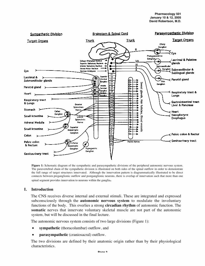
I. Introduction The CNS receives diverse internal and external stimuli. These are integrated and expressedsubconsciously through the autonomic nervous system to modulate the involuntaryfunctions of the body. This overlies a strong circadian rhythm of autonomic function. Thesomatic nerves that innervate voluntary skeletal muscle are not part of the autonomicsystem, but will be discussed in the final lecture. The autonomic nervous system consists of two large divisions (Figure 1):∑ sympathetic (thoracolumbar) outflow, and∑ parasympathetic (craniosacral) outflow. The two divisions are defined by their anatomic origin rather than by their physiologicalcharacteristics.
Figure 1: Schematic diagram of the sympathetic and parasympathetic divisions of the peripheral autonomic nervous system.The paravertebral chain of the sympathetic division is illustrated on both sides of the spinal outflow in order to demonstratethe full range of target structures innervated. Although the innervation pattern is diagrammatically illustrated to be directconnects between preganglionic outflow and postganglionic neurons, there is overlap of innervation such that more than onespinal segment provides innervation to neurons within the ganglia.

Pharmacology 501January 10 & 12, 2005David Robertson, M.D.
Page 4
II. AnatomyA. Central
The circadian rhythm of autonomic function originates in the suprachiasmatic nucleus(SCN) in the hypothalamus, and is entrained by light falling on melanopsin-containingretinal ganglion cell dendrites (not rods or cones) in the eye and transmitted to the SCNby the retinohypothalamic tract. The integration of autonomic outflow to thecardiovascular system lies in the medulla. Stretch-sensitive mechanoreceptors in theblood vessels of the thorax and neck relay information about blood pressure and bloodvolume through the glossopharyngeal (from carotid arteries) and vagus (from aorta)nerves to the nucleus tract solitarii (NTS) in the posterior medulla. Excitatory neurons from the NTS innervate the dorsal motor nucleus of the vagus,where parasympathetic outflow is regulated. Inhibitory neurons, using gamma-aminobutyric acid (GABA) as neurotransmitter, innervate areas in the ventrolateralmedulla from which sympathetic outflow is regulated. The most important such site isthe rostral ventrolateral medulla (RVLM).
Destruction of the NTS or its afferent input in experimental animals (Figure 2) orby tumors, radiation or infarction in patients can lead to the syndrome ofbaroreflex failure. A family in Nashville with a genetic defect leading to tumors inthe carotid body and other paragangliomas has taught us most of what we knowabout baroreflex failure. There is an acute period of dramatic hypertension duringwhich stroke or pulmonary edema may occur. This is followed by a syndrome ofwide swings in blood pressure, from hypotensive to hypertensive levels, withpressure determined by anxiety (pressor), sedation (depressor), noise (pressor),and sunlight (pressor).
Efferent parasympathetic outflow to the cardiovascular system goes through the vagusnerve. Efferent sympathetic outflow from the RVLM travels in the bulbospinal tractto the intermediolateral column of the spinal cord.
Glossopharyngeal neuralgia (glossopharyngeal syncope) is a disorder occurringin patients whose 9th cranial nerve becomes damaged (usually by neck tumor).Paroxysms of severe throat pain associated with hypotension and bradycardiaoccur. Attacks are due to massive spontaneous afferent discharges of the
Figure 2: Contrast between clinical effects of NTS (afferent) destructive lesions on left and RVLM (efferent) destructivelesions on the right. The contast is due to an inhibitory neuron that communicates from the NTS to the RLM.

Pharmacology 501January 10 & 12, 2005David Robertson, M.D.
Page 5
glossopharyngeal nerve, providing excessive input into the NTS, and elicitingparasympathetic activation and sympathetic withdrawal. Although a pacemakermay be helpful in preventing bradycardia, the hypotension is sometimes so severethat surgical section of the glossopharyngeal nerve is required.
The vasomotor neurons of the bulbospinal activate preganglionic cells sympatheticnerves.
III. BiochemistryA. Neurotransmitters
The primary neurochemical mediator of both sympathetic and parasympatheticpreganglionic neurons is acetylcholine (ACh). The primary mediator of sympatheticpostganglionic fibers is usually norepinephrine (NE), but at least some sympatheticpostganglionic fibers to sweat glands are cholinergic (acetylcholine). The mediator ofparasympathetic postganglionic fibers is acetylcholine. Epinephrine is found in theadrenal medulla, the central nervous system and the para-aortic bodies (organs ofZuckerkandl). Dopamine is a neurochemical mediator in the central nervous systemand probably also in some neurons in the superior cervical ganglion and the kidney.Norepinephrine, epinephrine and dopamine are sometimes collectively referred to ascatecholamines. Outside the United States, norepinephrine is often callednoradrenaline, and epinephrine, adrenaline. These endogenous compounds plus drugsthat resemble them functionally and structurally are also called sympathomimeticamines.
Figure 3: Efferent sympathetic outflowLegend: AP, area postrema; NTS, nucleus tractus solitarii; RVLM, rostral ventrolateral medulla (C1 area);intermediolateral column of the spinal cord; glutamate-releasing neuron; GABA, g �-aminobutyric acidreleasing neuron; ACh, acetylcholine-releasing neuron; NA, norepinephrine-releasing neuron
Pharmacology 501January 10 & 12, 2005David Robertson, M.D.
Page 6
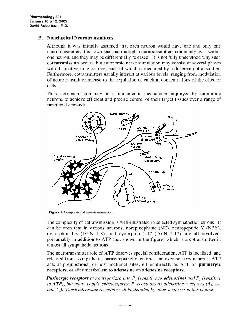
B. Nonclassical Neurotransmitters Although it was initially assumed that each neuron would have one and only oneneurotransmitter, it is now clear that multiple neurotransmitters commonly exist withinone neuron, and they may be differentially released. It is not fully understood why suchcotransmission occurs, but autonomic nerve stimulation may consist of several phaseswith distinctive time courses, each of which is mediated by a different cotransmitter.Furthermore, cotransmitters usually interact at various levels, ranging from modulationof neurotransmitter release to the regulation of calcium concentrations of the effectorcells. Thus, cotransmission may be a fundamental mechanism employed by autonomicneurons to achieve efficient and precise control of their target tissues over a range offunctional demands.
The complexity of cotransmission is well-illustrated in selected sympathetic neurons. Itcan be seen that in various neurons, norepinephrine (NE), neuropeptide Y (NPY),dynorphin 1-8 (DYN 1-8), and dynorphin 1-17 (DYN 1-17), are all involved,presumably in addition to ATP (not shown in the figure) which is a cotransmitter inalmost all sympathetic neurons. The neurotransmitter role of ATP deserves special consideration. ATP is localized, andreleased from, sympathetic, parasympathetic, enteric, and even sensory neurons. ATPacts at prejunctional or postjunctional sites, either directly as ATP on purinergicreceptors, or after metabolism to adenosine on adenosine receptors. Purinergic receptors are categorized into P1 (sensitive to adenosine) and P2 (sensitiveto ATP), but many people subcategorize P1 receptors as adenosine receptors (A1, A2,and A3). These adenosine receptors will be detailed by other lecturers in this course.
Figure 4: Complexity of neurotransmission.
Pharmacology 501January 10 & 12, 2005David Robertson, M.D.
Page 7
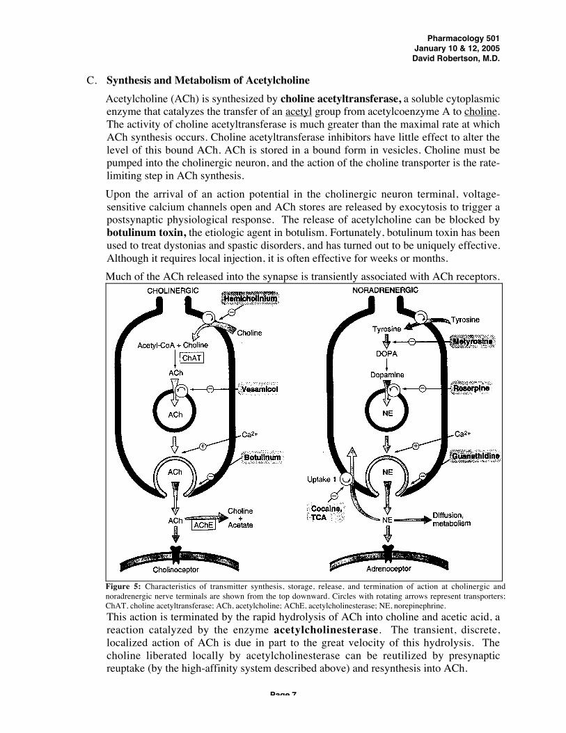
C. Synthesis and Metabolism of Acetylcholine Acetylcholine (ACh) is synthesized by choline acetyltransferase, a soluble cytoplasmicenzyme that catalyzes the transfer of an acetyl group from acetylcoenzyme A to choline.The activity of choline acetyltransferase is much greater than the maximal rate at whichACh synthesis occurs. Choline acetyltransferase inhibitors have little effect to alter thelevel of this bound ACh. ACh is stored in a bound form in vesicles. Choline must bepumped into the cholinergic neuron, and the action of the choline transporter is the rate-limiting step in ACh synthesis. Upon the arrival of an action potential in the cholinergic neuron terminal, voltage-sensitive calcium channels open and ACh stores are released by exocytosis to trigger apostsynaptic physiological response. The release of acetylcholine can be blocked bybotulinum toxin, the etiologic agent in botulism. Fortunately, botulinum toxin has beenused to treat dystonias and spastic disorders, and has turned out to be uniquely effective.Although it requires local injection, it is often effective for weeks or months. Much of the ACh released into the synapse is transiently associated with ACh receptors.
This action is terminated by the rapid hydrolysis of ACh into choline and acetic acid, areaction catalyzed by the enzyme acetylcholinesterase. The transient, discrete,localized action of ACh is due in part to the great velocity of this hydrolysis. Thecholine liberated locally by acetylcholinesterase can be reutilized by presynapticreuptake (by the high-affinity system described above) and resynthesis into ACh.
Figure 5: Characteristics of transmitter synthesis, storage, release, and termination of action at cholinergic andnoradrenergic nerve terminals are shown from the top downward. Circles with rotating arrows represent transporters;ChAT, choline acetyltransferase; ACh, acetylcholine; AChE, acetylcholinesterase; NE, norepinephrine.

Pharmacology 501January 10 & 12, 2005David Robertson, M.D.
Page 8
In addition to acetylcholinesterase (true cholinesterase) which is found near cholinergicneurons and in red blood cells (but not in plasma), there is also a non-specificcholinesterase (pseudocholinesterase or butyrylcholinesterase) which is present inplasma and in some organs but not in the red blood cell or the cholinergic neuron. The genetic abnormalities in pseudocholinesterase can result in a marked deficiency.One in 30,000 people are homozygotes for the most common functionally abnormalvariant, dibucaine-resistant pseudocholinesterase (3.8% of people are thereforeheterozygotes for this gene). Cholinergic nerve activity in such people is normal butsome drugs such as succinylcholine (used during anesthesia) which are normally brokendown by pseudocholinesterase, are very poorly metabolized by this variant enzyme.Such patients may have prolonged muscle paralysis from succinylcholine.
D. Synthesis and Metabolism of Catecholamines Tyrosine in the bloodstream is taken up into nerves and converted into catecholamine.The five main enzymes whose functions are critical to the formation of catecholaminesare discussed below (Figure 6). Tyrosine hydroxylase (tyrosine to dopa) is the rate-limiting step in NE synthesis and is located in thecytoplasm. Catecholamines act as feedback inhibitorsof this enzyme. During increased sympatheticstimulation, dopa production is increased in twoways: a) more enzyme is synthesized, and b) thephysical properties of the enzyme are altered(allosteric activation) so that affinity for tyrosine isincreased and affinity for end products like NE isreduced. A clinically useful inhibitor of this enzymeis metyrosine (a-methyl-p-tyrosine).
Dopa decarboxylase (dopa to dopamine) is found inthe cytoplasm of many nonneural as well as neuraltissues and had been called "aromatic-L-amino aciddecarboxylase" because of its broad substratespecificity. Peripheral (non-neuronal) dopadecarboxylase can be inhibited by carbidopa whenone is trying to prevent formation of peripheraldopamine during dopa therapy of Parkinsonism. This limits dopamine production to thecentral nervous system during dopa therapy, thus limiting peripheral side effects. Dopamine-ß-hydroxylase (dopamine to norepinephrine) is a copper-containing enzymelocated primarily within the membrane of amine storage granules.
Some individuals have been found to have dopamine-ß-hydroxylase deficiency.They present with lifelong orthostatic hypotension, and ptosis of the eyelids. Theirsympathetic neurons contain large quantities of dopamine, but little or nonorepinephrine. They can be treated with the drug dihydroxyphenylserine (DOPS),which is decarboxylated directly into norepinephrine by dopa decarboxylase, thusrestoring the appropriate neurotransmitter.
Figure 6: Metabolic pathway ofcatecholamine synthesis.

Pharmacology 501January 10 & 12, 2005David Robertson, M.D.
Page 9
Phenylethanolamine-N-methyltransferase (norepinephrine to epinephrine) is restrictedto the adrenal medulla, the brain and the organ of Zuckerkandl, with only trace amountsin other locations. It is strongly inhibited by physiological concentrations ofepinephrine providing feedback regulation of enzyme synthesis. Glucocorticoidincreases enzyme activity. Much neuronal NE is located in neuronal vesicles. They store NE and protect it frombreakdown by monoamine oxidase (MAO) in the surrounding cytoplasm. Thesevesicles are subsequently transported to the neuron terminal region for release. Release occurs when acetylcholine liberated from preganglionic neurons inducesdepolarization of postganglionic sympathetic neurons by acting on a nicotinic receptor(see below). The influx of calcium stimulates migration of vesicles to the cellmembrane for excretion, by membrane fusion and exocytosis. Local synaptic concentrations of catecholamines modulate their own release byinteracting with presynaptic a2-receptors to reduce release of additionalnorepinephrine and presynaptic b2-receptors to increase release of norepinephrine(more about this below). The reason for this bidirectional control is not known with certainty, but may function tostabilize synaptic neurotransmitter levels. In addition, other substances mayincrease (angiotensin, and acetylcholine via a nicotinic receptor) and decrease(dopamine, histamine, serotonin, adenosine, PGD2, PGE2 and acetylcholine via amuscarinic receptor) norepinephrine release in selected tissues. Released catecholamines may a) be retaken up into the neuron (norepinephrinetransporter or uptake I), b) be taken up by the extraneuronal tissue (uptake II), or c)be washed into the extracellular fluid and ultimately into the circulation. Terminationof action of released catecholamine varies with organ site. Thus, heavily innervatedtissues like the heart with narrow synaptic clefts tend to rely heavily on thenorepinephrine transporter (90% uptake of released NE), while tissues such as the aortawith wide synaptic clefts and less dense innervation tend to rely on it less. Thecatecholamines may be metabolized by one of two enzymes. Monoamine oxidase occurs in two forms (A and B). Monoamine oxidase is located inthe outer membrane of mitochondria as well as extraneuronally. It convertscatecholamines to their corresponding aldehydes. Inhibitors include pargyline,tranylcypromine, and selegiline (Deprenyl®) that will be covered in part 2 of the coursewith COMT inhibitors below. Catechol-o-methyltransferase (COMT) converts NE into normetanephrine andepinephrine into metanephrine. It is found especially in liver and kidney. Uptake into the neuron terminal by the NE transporter is very efficient. Fully half of anintravenous infusion of NE is taken up and stored in neurons, primarily in heart, spleen,and blood vessels, The structure of the NE transporter and elucidation of its regulationwas achieved by Vanderbilt’s Dr. Randy D. Blakely. The uptake mechanism is anenergy requiring, saturable membrane transport system, that can be blocked tricyclicantidepressants, amphetamine and cocaine (more from Dr. Sanders-Bush, next section).

Pharmacology 501January 10 & 12, 2005David Robertson, M.D.
Page 10
In healthy persons at rest, plasma NE is about 250 pg/ml and plasma A is 25 pg/ml.Normally, plasma norepinephrine level is doubled by standing, but it is several foldelevated by running, and in myocardial infarction (MI), delirium tremens (DT’s), andpheochromocytoma (pheo).
E. Summary of Intervention Mechanisms1. Cholinergic neurotransmission can be modified at several sites, including:
a) Precursor transport blockade hemicholiniumb) Choline acetyltransferase inhibition no clinical examplec) Promote transmitter release choline, black widow spider
venom (latrotoxin)d) Prevent transmitter release botulinum toxine) Storage vesamicol prevents ACh storagef) Cholinesterase inhibition physostigmine, neostigmineg) Receptors agonists and antagonists
2. There are also many sites at which pharmacological alteration of sympathetic
noradrenergic function can take place (Figure 4). They are reviewed below:a) Precursor transport blockade no clinical exampleb) Tyrosine hydroxylase inhibition metyrosine, used to treat
pheochromocytomac) Dopa decarboxylase inhibition carbidopad) Dopamine-ß-hydroxylase inhibition disulfirame) Monoamine oxidase inhibition pargyline, tranylcypromine,
selegilinef) Storage reserpine prevents NE storageg) Release guanethidine, guanadrel cause
initial release of NE leading to depletion of catecholamine;bretylium blocks NE release
h) Receptors a-and b-agonists and antagonistsi) Norepinephrine transporter cocaine, tricyclic antidepressants
(Uptake I) blockadej) Catechol-o-methyltransferase entacapone
inhibitionk) Uptake II glucocorticoids

Pharmacology 501January 10 & 12, 2005David Robertson, M.D.
Page 11
IV. Norepinephrine, Epinephrine, and DopamineA. Adrenoreceptors
It is convenient to distinguish several receptor types in explaining the effects ofcatecholamines. Many more receptor-types can be subtly distinguished by their affinityfor different agonists and antagonists and, in even more cases, by their structures. Forpractical purposes, however, it is probably sufficient to use a classification scheme suchas the following:
1. a1 (three subtypes): a1A, a1B, a1C
2. a2 (three subtypes): a2A, a2B, a2C
3. b2
4. b2
5. b3
6. D1, D2, D3, D4, D5
Most people reserve the term "adrenoreceptor" for a1, a2, b1, b2, and b3-receptors. The Dreceptors are dopamine receptors and are highly relevant to behavior and to Parkinson’sdisease and will be treated only briefly in my lectures.∑ The expressions "adrenoreceptor", "adrenoceptor" and "adrenergic receptor" are
synonymous. The actions of a and ß adrenoreceptors are mediated by diverse intracellularmechanisms. Activation of ß-adrenoreceptors by neurotransmitter, hormone or drugleads to synthesis of cAMP by adenylyl cyclase at the cytoplasmic facet of the plasmamembrane. The hormone-receptor ("liganded receptor") interacts with a stimulatoryguanine nucleotide-binding regulatory protein (Gs), which then activates the adenylylcyclase. A related regulatory protein (Gi) also binds to GTP in the presence of hormonestimulation of the a2 adrenoreceptor (or muscarinic M2 receptor). The interaction of Gileads to inhibition of adenylyl cyclase. The Gi regulatory protein sometimes alsointeracts with ion channels to activate (K+ channels) or inhibit (voltage-gated Ca++
Figure 7: Simplified summary of parasympathetic, sympathetic somaticinnervation.
Pharmacology 501January 10 & 12, 2005David Robertson, M.D.
Page 12
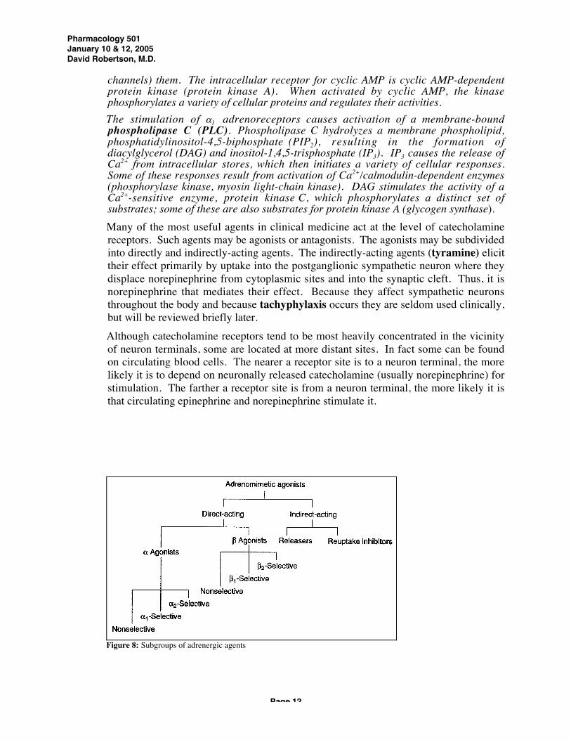
channels) them. The intracellular receptor for cyclic AMP is cyclic AMP-dependentprotein kinase (protein kinase A). When activated by cyclic AMP, the kinasephosphorylates a variety of cellular proteins and regulates their activities. The stimulation of a1 adrenoreceptors causes activation of a membrane-boundphospholipase C (PLC). Phospholipase C hydrolyzes a membrane phospholipid,phosphatidylinositol-4,5-biphosphate (PIP2), resulting in the formation ofdiacylglycerol (DAG) and inositol-1,4,5-trisphosphate (IP3). IP3 causes the release ofCa2+ from intracellular stores, which then initiates a variety of cellular responses.Some of these responses result from activation of Ca2+/calmodulin-dependent enzymes(phosphorylase kinase, myosin light-chain kinase). DAG stimulates the activity of aCa2+-sensitive enzyme, protein kinase C, which phosphorylates a distinct set ofsubstrates; some of these are also substrates for protein kinase A (glycogen synthase). Many of the most useful agents in clinical medicine act at the level of catecholaminereceptors. Such agents may be agonists or antagonists. The agonists may be subdividedinto directly and indirectly-acting agents. The indirectly-acting agents (tyramine) elicittheir effect primarily by uptake into the postganglionic sympathetic neuron where theydisplace norepinephrine from cytoplasmic sites and into the synaptic cleft. Thus, it isnorepinephrine that mediates their effect. Because they affect sympathetic neuronsthroughout the body and because tachyphylaxis occurs they are seldom used clinically,but will be reviewed briefly later. Although catecholamine receptors tend to be most heavily concentrated in the vicinityof neuron terminals, some are located at more distant sites. In fact some can be foundon circulating blood cells. The nearer a receptor site is to a neuron terminal, the morelikely it is to depend on neuronally released catecholamine (usually norepinephrine) forstimulation. The farther a receptor site is from a neuron terminal, the more likely it isthat circulating epinephrine and norepinephrine stimulate it.
Figure 8: Subgroups of adrenergic agents
Pharmacology 501January 10 & 12, 2005David Robertson, M.D.
Page 13
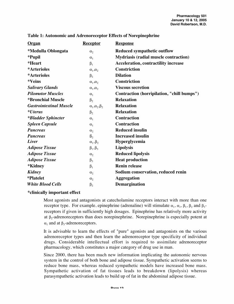
Table 1: Autonomic and Adrenoreceptor Effects of NorepinephrineOrgan Receptor Response*Medulla Oblongata a2 Reduced sympathetic outflow*Pupil a1 Mydriasis (radial muscle contraction)*Heart b1 Acceleration, contractility increase*Arterioles a1,a2 Constriction*Arterioles b2 Dilation*Veins a1,a2 ConstrictionSalivary Glands a1,a2 Viscous secretionPilomotor Muscles a1 Contraction (horripilation, "chill bumps")*Bronchial Muscle b2 RelaxationGastrointestinal Muscle a1,a2,b2 Relaxation*Uterus b2 Relaxation*Bladder Sphincter a1 ContractionSpleen Capsule a1 ContractionPancreas a2 Reduced insulinPancreas ß2 Increased insulinLiver a1, b2 HyperglycemiaAdipose Tissue b1, b3 LipolysisAdipose Tissue a2 Reduced lipolysisAdipose Tissue b3 Heat production*Kidney b1 Renin releaseKidney a2 Sodium conservation, reduced renin *Platelet a2 Aggregation White Blood Cells b2 Demargination
*clinically important effect Most agonists and antagonists at catecholamine receptors interact with more than onereceptor type. For example, epinephrine (adrenaline) will stimulate a1, a2, b1, b2 and b3-receptors if given in sufficiently high dosages. Epinephrine has relatively more activityat b2-adrenoreceptors than does norepinephrine. Norepinephrine is especially potent ata1 and at b3-adrenoreceptors.
It is advisable to learn the effects of "pure" agonists and antagonists on the variousadrenoreceptor types and then learn the adrenoreceptor type specificity of individualdrugs. Considerable intellectual effort is required to assimilate adrenoreceptorpharmacology, which constitutes a major category of drug use in man. Since 2000, there has been much new information implicating the autonomic nervoussystem in the control of both bone and adipose tissue. Sympathetic activation seems toreduce bone mass, whereas reduced sympathetic models have increased bone mass.Sympathetic activation of fat tissues leads to breakdown (lipolysis) whereasparasympathetic activation leads to build up of fat in the abdominal adipose tissue.
Pharmacology 501January 10 & 12, 2005David Robertson, M.D.
Page 14
B. Alpha1-Agonists: The most important effects of a 1-agonists (phenylephrine, methoxamine,norepinephrine) are apparent from inspection of Table 1. The dilator muscle of thepupil is constricted giving mydriasis (dilated pupil). Some of the smooth muscle tissuein the eyelids is constricted leading to a widened palpebral fissure. Most arterioles areconstricted and peripheral vascular resistance is increased, raising blood pressure.Veins (capacitance vessels) are also constricted leading to a central redistribution ofblood into the thorax. Stimulation of pilomotor nerves causes hair to "stand on end"(horripilation or piloerection). The associated stimulation of myoepithelial tissue in thevicinity of the apocrine glands (axilla, crural areas) causes gland emptying although theglands themselves are not stimulated. (The eccrine sweat glands are stimulated bysympathetic postganglionic fibers that are cholinergic and hence do not fit into either aor b classification, but rather respond to acetylcholine and are blocked by atropine.)Bladder sphincters are contracted by a1-stimulation. The spleen capsule is contracted.There is CNS stimulation with agents which cross the blood-brain barrier(norepinephrine, dopamine, and epinephrine do not). Some agonists at a1-adrenoreceptors increase myocardial contractility (but not heart rate) in somecircumstances, but b1 stimulation of contractility is more important clinically.
Phenylephrine (Neosynephrine®) and methoxamine are far more potent instimulating a1-receptors than in stimulating other receptor types. For practical purposes,they can be considered pure a1-agonists. In clinical practice they are used systemicallyto treat hypotensive states. Locally they cause mydriasis. They are useful in treatingnasal congestion. Phenylephrine is occasionally used to restore paroxysmal atrialtachycardia to normal sinus rhythm (via baroreceptor-mediated enhancement of vagaltone).
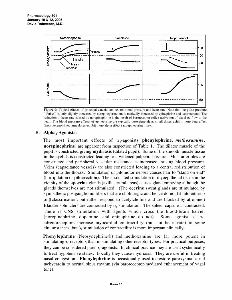
Figure 9: Typical effects of principal catecholamines on blood pressure and heart rate. Note that the pulse pressure(“Pulse”) is only slightly increased by norepinephrine but is markedly increased by epinephrine and isoproterenol. Thereduction in heart rate caused by norepinephrine is the result of baroreceptor reflex activation of vagal outflow to theheart. The blood pressure effects of epinephrine are typically dose-dependent: small doses exhibit more beta effect(isoproterenol-like; large doses exhibit more alpha effect ( norepinephrine-like).

Pharmacology 501January 10 & 12, 2005David Robertson, M.D.
Page 15
Norepinephrine is very close to phenylephrine in its effects and has enjoyed widerclinical use in the treatment of shock. Norepinephrine differs from phenylephrineprimarily in having a greater capacity to stimulate b1-adrenoreceptors as well as a1-adrenoreceptors. Epinephrine is used clinically primarily to support blood pressure,especially during anaphylaxis. Dopamine, the immediate metabolic precursor ofnorepinephrine, has wide use in the drug treatment of shock. At high but not at lowdosages, it stimulates a1-adrenoreceptors. (More in Dr. Blackwell’s lecture).
These agents are sometimes used with local anesthetics; by causing vasoconstriction atthe site of the injection, they delay the absorption of the local anesthetic and prolonganesthesia.
C. Alpha1-Antagonists Blockade of the a1-receptor negates the responses discussed above. In subjects on noother medications, a1-blockers (prazosin, phentolamine, tolazoline, phenoxybenzamine)reduce blood pressure, especially in the upright posture. Some agents (prazosin)selectively block the a1- receptor. Others (phenoxybenzamine, tolazoline,phentolamine) block the a2-receptor as well.
Phentolamine is a competitive, short-acting a-antagonist. It is used to determinewhether a given level of hypertension is catecholamine-mediated. This is sometimeshelpful in diagnosing pheochromocytoma at the bedside. In addition to its a blockingproperties phentolamine antagonizes some effects of serotonin. Its major side effect iscardiac stimulation (arrhythmias and angina pectoris). Abdominal cramping, ulcerexacerbation and diarrhea occur with chronic use. Phenoxybenzamine is a noncompetitive, long-acting a -antagonist. Unlikephentolamine it can be reliably given orally with its clinical effect developing overhours and lasting several days. Like phentolamine, it frequently causes posturalhypotension. Its main clinical use is in the medical management of pheochromocytoma. Prazosin differs from phentolamine, tolazoline and phenoxybenzamine in that itselective blocks a1-receptors without blocking the a2-receptors that mediate feedbackinhibition of norepinephrine synthesis/release. Thus there is less spillover stimulationof a-receptors with prazosin than in the case of the other two agents. Prazosin is used inhypertension and in congestive heart failure. The major problem in its use has been"prazosin syncope," fainting that occasionally occurs on standing 2-4 hours after thefirst oral dose, and a tendency toward reduced efficacy with chronic use. Terazosin anddoxazosin are similar to prazosin and have been used to relieve the symptoms of benignprostatic hypertrophy. (More about the a-adrenergic blockers from Dr. Oates later.)
D. Alpha2-Agonists The most important effects of a2-agonists (clonidine, guanabenz, guanfacine, and a-methylnorepinephrine) are only partially apparent from Table 1. In many tissuespresynaptic a2- stimulation mediates feedback-inhibition of norepinephrinerelease. When there is sufficient norepinephrine in the synaptic cleft to effect aresponse, it would be uneconomical of the neuron to continue to release still moretransmitter.

Pharmacology 501January 10 & 12, 2005David Robertson, M.D.
Page 16
Certain postsynaptic a2 receptors in the vicinity of the NTS and the RVLM areimportant determinants of sympathetic outflow. There is currently great interest inunderstanding these receptors better since they have differences from most other a2
adrenoreceptors. Some of them functionally resemble "imidazoline receptors"; no oneknows for sure the identity of the endogenous agonist for imidazoline receptors in thebrain. Clonidine stimulation of brainstem a2-receptors and binding to imidazolinereceptors significantly reduces sympathetic outflow to the cardiovascular system:hypotension and bradycardia result. This effect accounts for much of the usefulness ofclonidine in treating hypertension. Methyldopa, used as an antihypertensive agent, appears to be effective because itsmetabolite, a-methylnorepinephrine, stimulates these receptors. High doses of a2-agonists may stimulate peripheral postsynaptic vascular a 2-receptors mediatingvasoconstriction and thus actually raise blood pressure. Thus, we sometimes speak ofclonidine as having a "therapeutic window."
Causalgia is a pain syndrome that develops in the joints especially after nerveinjuries. Other names include Sudeck's atrophy, Sudeck's dystrophy, algodystrophy,shoulder-hand syndrome, and reflex sympathetic dystrophy. The major featuresare (1) pain; (2) dystrophy in involved skin, tissue, muscle, and bone; and (3)abnormal sweating and blood flow regulation in the affected area. Sometimesthere is also hypertrichosis and ridging of nails. Weeks or months after myocardialinfarction, this syndrome may develop in the left arm and hand ("shoulder-handsyndrome" or "Dressler's syndrome") and mimic the pain of angina pectoris. After years of skepticism, most investigators now acknowledge the key role of thesympathetic nervous system in mediating causalgia. Destruction of the relevantsympathetic nerves often completely eliminates the pain. There is recentexperimental evidence that blockade of a2-adrenoreceptors may also be helpful.The precise mechanism of autonomic mediation of causalgia remains unknown.
E. Alpha2-Antagonists While phentolamine and phenoxybenzamine block a2-receptors, their major clinicalaction is to block a 1-receptors. The only widely available, relatively specific a2-antagonist is yohimbine. By blocking a2-adrenoreceptors in the medulla, it increasessympathetic outflow. By blocking presynaptic a2-adrenoreceptors in the periphery, itenhances norepinephrine release. Yohimbine has long been reputed to be anaphrodisiac, for which purpose the plant from which it is derived it has been soldthroughout the world. Studies during the last several years seem to confirm that a2-agonists reduce and a2-antagonists increase copulatory behavior in rats.
F. Beta Agonists Isoproterenol stimulates both b1- and b 2-receptors. The heart contracts with greaterforce (increased contractility) and heart rate is increased. Cardiac output rises. There isan increased likelihood of premature heartbeats and other arrhythmias. Many arteriolesare dilated, with consequent reduced resistance. Mean blood pressure is at leasttransiently lowered. The gravid uterus is relaxed. Bronchioles are relaxed (useful in

Pharmacology 501January 10 & 12, 2005David Robertson, M.D.
Page 17
asthmatics) and metabolic effects on the liver and adipose tissue lead to hyperglycemiaand lipolysis. Some CNS stimulation occurs. While it is advantageous to stimulate b2-receptors in the bronchial tree of asthmaticpatients or the uterus of a woman in premature labor, the attendant b 1-cardiacstimulation is an unwanted effect. This has led to efforts to achieve selective b2
stimulation. A variety of putative b2-agonists have been developed, but their selectivityis partial. (More later in the course when asthma therapy is discussed). On the other hand, in certain patients, the cardiac stimulation of b-agonists is desirable(pulmonary edema, coronary bypass post-op) and a relatively selective b1-agonist likedobutamine is indicated. Moderate doses of dobutamine increase myocardialcontractility without significantly altering blood pressure. The relatively small effect ofdobutamine on blood pressure is due to counterbalancing effects of b1 stimulation and b2
stimulation on arteriolar and venous tone. A b3 adrenoreceptor has recently been identified that is sensitive to norepinephrine andnot easily blocked by the usual b-antagonists. It mediates heat production and energyexpenditure in adipose tissue.
G. Beta Antagonists (see Dr. Awad’s lecture on this topic for greater detail) Propranolol is a competitive inhibitor of sympathomimetic amines at both the b1- andb2- receptor. It counteracts the effect of isoprenaline in the vasculature, heart and liver.In persons on no medication, propranolol reduces heart rate, contractility and bloodpressure. Atrioventricular conduction is slowed. There is increased bronchiolar tone;therefore, the drug is avoided in asthmatics and in patients with chronic obstructivepulmonary disease. The reduced myocardial function may worsen heart failure inpatients with borderline cardiac status but low doses of beta-blockers are sometimeshelpful in limiting excessive sympathetic stimulation in severe heart failure. In additionto its b-blocking properties, propranolol possesses "quinidine-like" antiarrhythmicproperties and is a local anesthetic. Propranolol is widely used to reduce heart work in patients with angina pectoris and totreat ventricular arrhythmias. It is also an effective antihypertensive agent, probably byreducing renin production. It produces subjective improvement in thyrotoxicosis andcertain anxiety states and may reduce the incidence of migraine headaches. There isincreasing evidence that long-term treatment of post-myocardial infarction patients withbeta blockers (timolol, metoprolol, propranolol) reduces mortality, perhaps becausethese drugs prevent fatal arrhythmias. Side effects of propranolol include worsenedheart failure, reduced AV conduction, worsened obstructive lung disease, vividnightmares, fatigue, and cold extremities. Metoprolol is a relatively selective blocker of the b1-receptor and may give cardiaceffects with minimal worsening of asthma. Atenolol is also relatively selective for theb1-receptor, does not cross the blood-brain barrier well, and can be given once daily inmanaging hypertension. Timolol eye drops are used in glaucoma patients to reduceintraocular pressure. Pindolol is primarily a beta-antagonist but has some agonistactivity (intrinsic sympathomimetic effect).

Pharmacology 501January 10 & 12, 2005David Robertson, M.D.
Page 18
H. Dopamine Agonists and Antagonists There are distinct dopaminergic receptors in several peripheral tissues, notably in therenal vasculature. Agonists at the receptor include fenoldopam, apomorphine,bromocriptine, and other agents used in treating Parkinson's disease (More later incourse when Parkinson’s Disease therapy discussed). Fenoldopam is a dopaminergic agonist with nine-fold specificity for the D1 receptor. D1receptors mediate vasodilatation in the coronary, cerebrovascular, renal, and mesentericvascular beds, whereas D2 receptors also cause emesis and inhibition of prolactinFenoldopam's ultimate role in therapy remains uncertain, but it may be helpful inhypertension, heart failure, myocardial ischemia, and as a diuretic. Recently, an abnormality in an exon of the D4 receptor has been reported to correlatewith innovation and risk-taking behavior. If confirmed, this may open a new era ofunderstanding human behavior. Other dopaminergic antagonists include metoclopramide, haloperidol, domperidone, anda variety of phenothiazine derivatives that will be discussed during the lectures onpsychotropic drugs and gastrointestinal motility.
I. Indirectly Acting Phenylethylamines Tyramine enters noradrenergic neurons via the norepinephrine transporter anddisplaces NE from the "labile pool" (non-stored NE) and into the synaptic cleft ontopostsynaptic receptors. This drug is present in cheddar cheese, certain wines, marmite,country ham, and broadbeans (fava beans). Although norepinephrine is usually rapidly metabolized by monoamine oxidase,patients on inhibitors of this enzyme (e.g., pargyline) may have profound hypertensionfrom over-indulgence in tyramine-containing foods. Ephedrine is the active ingredient in the herbal product Ephedra (Ma Huang). It is botha directly and indirectly acting agent. The drug has been used to promote weight loss,in therapy of asthma (over-the-counter, OTC) and as a nasal decongestant. Because itincreases skeletal muscle tone (slightly), it has been used as adjunctive therapy inmyasthenia gravis. CNS effects (anxiety, insomnia, mental stimulation) may beprominent, and it has been used to treat narcolepsy. Amphetamine is a congener ofephedrine with even more potent CNS effects. These and similar agents have been usedto induce weight loss (OTC). Ephedra’s widespread availability over the counter causedit to be used to achieve weight loss and to enhance physical performance of athletes. Inhot weather and with the thermic stimulus of exercise, Ephedra’s own hyperthermiceffect has caused illness and death. After several years of hesitation, the FDA initiatedactions to remove this agent from sale in the United States in December 2003.
Horner's syndrome occurs when sympathetic nerves to the eye are interrupted.Three consecutive neurons are involved in conveying sympathetic outflow from themedulla to the eye, and a lesion of any one of them leads to Horner's syndrome.The syndrome includes miosis, ipsilateral anhidrosis, and ipsilateral ptosis. Thediagnosis is made by documenting that an a 1-adrenoreceptor agonist(phenylephrine) will dilate the patient's constricted pupil. Hydroxyamphetamine, atyramine-like agent, will also dilate the pupil if the neuron innervating the iris is

Pharmacology 501January 10 & 12, 2005David Robertson, M.D.
Page 19
intact (that is, if the lesion is more central). If the most peripheral nerve is thedamaged one, hydroxyamphetamine will not work. This differentiation isimportant since the etiologies of more central lesions are very different than thoseof more peripheral lesions.
There are a large number of sympathomimetics in nasal decongestants, nose drops, andinhalers. Phenylpropanolamine achieved great popularity as an over-the-counterweight loss aid ("Dexatrim®"), for which it is probably ineffective. Some of theseagents act directly and others indirectly. Their vasoconstrictive properties areresponsible for increasing tone in boggy mucosal surfaces; this "shrinks" mucosalmembranes. The FDA removed phenylpropranolamine from the market in 2000 due toan increased risk of hemorrhagic stroke. Pseudoephedrine (Sudafed®), which closelyresembles phenylpropanolamine, is increasingly used to replace it, but it could be thatall pressor drugs will prove to have the same increased risk of hemorrhagic stroke ifcarefully studied for this rare complication.

Pharmacology 501January 10 & 12, 2005David Robertson, M.D.
Page 20
V. AcetylcholineA. Acetylcholine Receptors
Acetylcholine (ACh) is the postganglionic neurotransmitter in the parasympatheticnervous system. It is also the preganglionic neurotransmitter for both the sympatheticand parasympathetic nervous system. It is also important at non-autonomic sites. Forexample, ACh is the neurotransmitter by which motor nerves stimulate skeletalmuscle, and also the neurotransmitter at many sites in the brain and spinal cord. As might be expected for an ancient and ubiquitous neurotransmitter, a variety of ACh(cholinergic) receptor types have emerged. The most important classification dependson their responsiveness to the agonist drugs, muscarine and nicotine, and thisdistinction is so crucial that the terms muscarinic receptor (M) and nicotinic receptor(N) are used rather then cholinergic receptor. Muscarinic receptors are located...∑ in tissues innervated by postganglionic parasympathetic neurons∑ in presynaptic noradrenergic and cholinergic nerve terminals∑ in non-innervated sites in vascular endothelium∑ in the central nervous system. Nicotinic receptors are located...∑ in sympathetic and parasympathetic ganglia∑ in the adrenal medulla∑ in the neuromuscular junction of the skeletal muscle∑ in the central nervous system. There are at least 5 subtypes of muscarinic receptors, referred to as M1, M2, M3, M4,and M5. They mediate their effects through G proteins coupled to phospholipase C(M1,3,5), or to potassium channels (M2,4). Because we currently have few truly subtype-specific muscarinic agonists and antagonists of clinical utility, there will not be detaileddiscussion of them this year. There are at least two subtypes of nicotinic receptors, referred to as NM and NN. Thisdistinction is clinically important. The NM nicotinic receptor mediates skeletal musclestimulation, while the NN nicotinic receptor mediates stimulation of the ganglia of theautonomic nervous system, for which reason agonists and antagonists at the latter siteare sometimes called ganglionic agonists and ganglionic blockers.
Nicotinic receptors are ligand-gated ion channels whose activation results in arapid increase in cellular permeability to sodium and calcium. They arepentameric arrays of one to four distinct but homologous subunits, surrounding aninternal channel. The a subunit, which has binding sites for ACh, is present in atleast two copies. Agonist molecules induce a conformational change that opensthe channel. Antagonist molecules may bind to these sites but do not elicit theconformational change.

Pharmacology 501January 10 & 12, 2005David Robertson, M.D.
Page 21
B. Muscarinic Agonists Acetylcholine itself is rarely used clinically because of its rapid hydrolysis followingoral ingestion and rapid metabolism following intravenous administration. Fortunately,a number of congeners with resistance to hydrolysis (methacholine, carbachol, andbethanechol) have become available, and bethanechol has the additional favorableproperty of an overwhelmingly high muscarinic (vs nicotinic) specificity. There are alsoseveral other naturally occurring muscarinic agonists such as muscarine, arecholine,and pilocarpine.
The pharmacological effects of acetylcholine and other muscarinic agonists can beseen by inspection of Table 2, where clinically important effects are designated byasterisks. All these effects are parasympathetically mediated except sweat glandfunction, which is the unique sympathetic cholinergic category, much beloved b yexamination writers; these nerves are sympathetic because of their thoracolumbar originand cholinergic because they release acetylcholine.
Table 2: Muscarinic Autonomic Effects of Acetylcholine (*clinically important) *Iris sphincter muscle Contraction (miosis) *Ciliary muscle Contraction (near vision) *SA node Bradycardia Atrium Reduced contractility *AV node Reduced conduction velocity Arteriole Dilation (via nitric oxide) *Bronchial muscle Contraction *Gastrointestinal motility Increased *Gastrointestinal secretion Increased Gallbladder Contraction *Bladder (detrusor) Contraction *Bladder (trigone, sphincter) Relaxation Penis Erection (but not ejaculation) Sweat glands Secretion Salivary glands Secretion Lacrimal glands Secretion Nasopharyngeal glands Secretion
Figure 10: Subgroups of cholinomimetic drugs.

Pharmacology 501January 10 & 12, 2005David Robertson, M.D.
Page 22
Bethanechol (Urecholine®) is used (rarely) to treat gastroparesis, because it stimulatesgastrointestinal motility and secretion. It is also useful in patients with autonomicfailure, in whom modest improvement in gastric emptying and constipation may occur,but at a cost of some cramping abdominal discomfort. If gastric absorption is impaired,subcutaneous administration is sometimes employed. Especially with intravenousadministration, hypotension and bradycardia may occur. Bethanechol is also widelyused to treat urinary retention if physical obstruction (e.g., prostate enlargement) isnot the cause. This agent also occasionally is used to stimulate salivary gland secretionin patients with xerostomia, which entails the dry mouth, nasal passages, and throatoccurring in Sjögren’s syndrome, and in some cases of traumatic or radiation injury. Inrare cases, high doses of bethanechol have seemed to cause myocardial ischemia inpatients with a predisposition to coronary artery spasm, so chest pain in a patient onbethanechol should be taken seriously. Pilocarpine is more commonly used than bethanechol to induce salivation, and also forvarious purposes in ophthalmology. It is especially widely used to treat open-angleglaucoma, for which a topical (ocular) preparation is available. Intraocular pressure islowered within a few minutes following ocular instillation of pilocarpine. It causescontraction of the iris sphincter, which results in miosis (small pupils) and contractionof the ciliary muscle, which results in near (as opposed to distant) focus of vision.Pilocarpine possesses the expected side effect profile, including increased sweating,asthma worsening, nausea, hypotension, bradycardia (slow heart rate), and occasionallyhiccups. Methacholine is often used to provoke bronchoconstriction during diagnostic testing ofpulmonary function. Elicitation of significant bronchoconstriction with inhaledmethacholine challenge sometimes leads to the diagnosis of reactive airways disease(asthma) in patients with little baseline abnormality in pulmonary function.
C. Muscarinic Antagonists
The classical muscarinic antagonists are derived from plants. The deadly nightshade(Atropa belladonna), a relative of the tomato and potato, contains atropine. Jimsonweed (Datura stramonium) is even more widespread in Tennessee than the deadlynightshade. The tiny dark seeds from the Jimson weed pod are sometimes ingested fortheir hallucinogenic effect, a central side effect of atropine-like substances. Henbane(Hyoscyamus niger) contains primarily scopolamine and hyoscine.
Some plants of these families grow well in poor, rocky soil whereas tomatoes growpoorly in these locations. The grafting of tomato plant stalks onto the root systemsof these plants yields an unusually productive "tomato" that bears well even in dry
Figure 11: Subtypes of anticholinergic agents.

Pharmacology 501January 10 & 12, 2005David Robertson, M.D.
Page 23
weather. Abundant large red tomatoes result. Unfortunately, if the grafting is notdone properly, belladonna alkaloids may enter the tomato fruit. Tachycardia andhallucinations (as well as more life-threatening problems) may ensue when suchtomatoes are eaten.
Side effects of muscarinic antagonists include constipation, xerostomia (dry mouth),hypohidrosis (decreased sweating), mydriasis (dilated pupils), urinary retention,precipitation of glaucoma, decreased lacrimation, tachycardia, and decreasedrespiratory secretions. Clinically, atropine is used for raising heart rate during situations where vagal activityis pronounced (for example, vasovagal syncope). It is also used for dilating the pupils.Its most widespread current use is in preanesthetic preparation of patients; in thissituation, atropine reduces respiratory tract secretions and thus facilitates intubation. Itprobably also has some efficacy as a bronchodilator. Inhaled ipratropium is marketedfor maintenance therapy in chronic obstructive pulmonary disease (COPD). It has along half-life. Pirenzepine shows selectivity for the M1 muscarinic receptor. Because of theimportance of this receptor in mediating gastric acid release, M1 antagonists such aspirenzepine help patients with ulcer disease or gastric acid hypersecretion. However,antihistamines and proton pump inhibitors are more useful and more widely used forcontrol of gastric acidity.
D. Nicotinic Agonists Nicotine is the most commonly encountered nicotinic agonist. It is found in the leavesof the tobacco plant (Nicotiana tabacum) in concentrations of 0.3 to 0.7%. It isresponsible for the addicting properties of tobacco. Nicotine's actions are complex. Atlow dosages it stimulates ganglionic nicotinic receptors thus enhancing bothsympathetic and parasympathetic neurotransmission. These are the effects nicotine hasbeen classically considered to have. In practice there is stimulation of nicotinicreceptors in many other sites especially as nicotine dosages increase. Nicotinepossesses some antagonist effect at nicotinic receptors at high dosages. Smoking one cigarette or intravenous administration of 1 mg nicotine will usually raiseblood pressure about 10 mm Hg and increase heart rate 15 beats per minute. Peripheralvascular resistance increases, as do cardiac output and heart work. Vasopressin andepinephrine are released. Higher doses stimulate the heart eliciting the Bezold-Jarischreflex (bradycardia, hypotension, nausea), and may eventually result in weakness,tremors, and convulsions. A dosage of 60 mg is lethal; there are two lethal doses in onecigar (if it were absorbed rather than smoked). Chronic smoking has effects unrelated to nicotine (or related to nicotine in a still poorlyunderstood way). HDL cholesterol is reduced, LDL cholesterol is increased, andplasma fibrinogen is elevated. There is excessive free radical production. All thesechanges could promote atherosclerosis. Smokers have increased metabolic rate thatkeeps them relatively lean; on discontinuation of smoking the reduction in metabolismusually causes a weight gain. On average the babies of smoking mothers weigh 0.5pound less than those of non-smoking mothers. Smokers have an increased risk ofcancer (129,000 extra deaths per year), coronary heart disease (170,000 extra deaths per

Pharmacology 501January 10 & 12, 2005David Robertson, M.D.
Page 24
year), and chronic obstructive pulmonary disease (62,000 extra deaths per year). Thepersistence of widespread tobacco use into the 21st century is an incongruity that futurehistorians will probably find tragic and inexplicable. Addiction to nicotine makes it very difficult for regular cigarette users to stop smoking.Since the overwhelming majority of the bad effects of smoking are due to factors otherthan nicotine itself, nicotine products such as patches (for transdermal nicotineadministration), chewing gum, and nasal sprays have been developed to try toadminister nicotine without the involvement of tobacco use. In general, this approachhas worked well. Only time will tell to what extent the patches and nasal spray willthemselves cause addiction, but experience so far is encouraging. The principal sideeffects noted have included alterations in taste or smell and increased heart rate.
E. Nicotinic Antagonists (Ganglionic Blockers) The actions of drugs on autonomic ganglia are complex. The primary receptors atganglia are cholinergic receptors of the nicotinic (NN) type. The effects observedclinically with ganglionic blocking agents are due to blockade of these receptors.Nearly all effects are predictable from the knowledge that ganglionic blockers reducetransmission in all autonomic ganglia, both sympathetic and parasympathetic. In somesites, sympathetic activation seems to predominate over parasympathetic, while in othersites, the opposite is true. Ganglionic blockade thus "uncovers" the predominantsystem. This class of drugs is now rarely used.
Table 3: Mediators and Effects of Ganglionic Blockade on Organ Systems Tissue Predominant System Ganglionic Blockade Effect Arterioles Sympathetic Vasodilation Veins Sympathetic Vasodilation Heart Parasympathetic Tachycardia Iris Parasympathetic Mydriasis Ciliary muscle Parasympathetic Cycloplegia Gastrointestinal tract Parasympathetic Hypomotility Urinary bladder Parasympathetic Urinary retention Salivary glands Parasympathetic Xerostomia Sweat glands Sympathetic cholinergic AnhidrosisF. Cholinesterase Inhibitors
The muscarinic and nicotinic agonists mimic acetylcholine effect by stimulating therelevant receptors themselves. Another way of accomplishing the same thing is toreduce the destruction of ACh following its release. This is achieved by cholinesteraseinhibitors, which are also called the anticholinesterases. They mimic the effect ofcombined muscarinic and nicotinic agonists. Cholinergic neurotransmission isespecially important in insects, and it was discovered many years ago thatanticholinesterases could be effective insecticides, by “overwhelming the cholinergiccircuits” (see War Gases below) By inhibiting acetylcholinesterase and pseudocholinesterase, these drugs allow ACh tobuild up at its receptors. Thus they result in enhancement of both muscarinic andnicotinic agonist effect.

Pharmacology 501January 10 & 12, 2005David Robertson, M.D.
Page 25
"Reversible" cholinesterase inhibitors are generally short-acting. They includephysostigmine that enters the CNS and neostigmine and edrophonium that do not: Physostigmine enters the CNS and can cause restlessness, apprehension, andhypertension in addition to the effects more typical of muscarinic and nicotinic agonists. Neostigmine is a quaternary amine (tends to be charged) and enters the CNS poorly;its effects are therefore almost exclusively those of muscarinic and nicotinicstimulation. It is used to stimulate motor activity of the small intestine and colon, as incertain types of nonobstructive paralytic ileus. It is useful in treating atony of thedetrusor muscle of the urinary bladder, in myasthenia gravis, and sometimes inglaucoma. Like other cholinesterase inhibitors, neostigmine requires an intactpostganglionic innervation for full development of its actions. Edrophonium (Tensilon®) is a quaternary amine widely used as a clinical test formyasthenia gravis. If this disorder is present, edrophonium will markedly increasestrength. It often causes some cramping, but this only lasts a few minutes.Ambenonium and pyridostigmine are sometimes also used to treat myasthenia. Many phosphorothionates, including parathion and malathion undergo enzymaticoxidation that can greatly enhance anticholinesterase activity. The reaction involves thesubstitution of oxygen for sulphur. Thus, parathion is oxidized to the more potent andmore water-soluble paraoxon. Differences in the hydrolytic and oxidative metabolismin different organisms accounts for the remarkable selectivity of malathion. Inmammals, the hydrolytic process in the presence of carboxyesterase leads toinactivation. This normally occurs quite rapidly, whereas oxidation leading toactivation is slow. In insects, the opposite is usually the case, and those agents are verypotent insecticides. Some patients encounter muscarinic side effects due to the inhibition of peripheralcholinesterase by physostigmine. The most common of these side effects are nausea,pallor, sweating and bradycardia. Concomitant use of anticholinergic drugs which arequaternary amines (e.g., glycopyrrolate or methscopolamine) and which therefore donot cross the blood-brain barrier are recommended to prevent the peripheral side effectsof physostigmine. Several centrally acting drugs produce an acute toxic psychosis characterized byconfusion and the peripheral signs of cholinergic blockade. These drugs include severalplant toxins, antidepressants, H1 receptor antagonists with central effects, and severalantiparkinsonian drugs and antipsychotic drugs. Overdoses of many other drugs canalso lead to this anticholinergic syndrome. Cholinesterase inhibitors that cross the blood-brain barrier are suitable to reverse thecentral anticholinergic syndrome. Physostigmine is the drug of choice. Althoughphysostigmine effectively wakes up such patients briefly, it is not certain that its useresults in a long-term better prognosis. Some patients with Alzheimer's disease havememory improvement transiently after anti-cholinergic drugs. Tacrine (Cognex®), wasfound to elicit hepatotoxicity, but definitely benefited Alzheimer's disease, if only to alimited extent. Two newer agents donepezil (Aricept®) and rivastigmine (Exelon®)have little hepatotoxicity and have replaced tacrine.

Pharmacology 501January 10 & 12, 2005David Robertson, M.D.
Page 26
On the accompanying tables, the effects of intoxication and the therapeutic approach totreatment are outlined. The drug pralidoxime deserves special comment. This drugcounteracts cholinesterase inhibitor intoxication by reactivating the cholinesteraseenzyme. Pralidoxime combines with the anionic site on the enzyme by electrostaticattraction to the quaternary N atom, which orients the nucleophilic oxime group toreact with the electrophilic P atom; the oxime-phosphonate is split off, leaving theregenerated enzyme.
G. War Gases Long-acting or "irreversible" cholinesterase inhibitors (organophosphates) areespecially used as insecticides. Cholinesterase inhibitors enhance cholinergictransmission at all cholinergic sites, both nicotinic and muscarinic. This makes themuseful as poisons. Sarin which is a war nerve gas is a binary agent composed of twocomponents that are not toxic until mixed. Nerve gases such as the cholinesteraseinhibitor, sarin, have been the chemical weapons of choice for over 50 years. Sarin wasfirst produced in 1938. It is a colorless, odorless gas with a lethal dose of just 0.5 mg foran adult human. Sarin is an easily dispersed agent that acts extremely quickly whenabsorbed through the skin or inhaled. Although recently superseded by more deadly andmore persistent compounds such as VX, sarin remains a highly dangerous and effectivepart of mankind's destructive arsenal. Death usually occurs by asphyxiation. The finalstage of sarin synthesis usually takes place while the missile or other delivery vessel is inflight because it is safer to store the component reagents than the more dangerous sarinitself.
Table 4 :Clinical Manifestations of Cholinesterase Inhibitor Intoxication Muscarinic∑ Miosis∑ Blurred vision (spasm of accommodation)∑ Lacrimation∑ Sweating∑ Excessive respiratory secretions∑ Dyspnea (bronchoconstriction)∑ Bradycardia∑ Hypotension∑ Salivation∑ Nausea∑ Cramping (gastrointestinal spasm)∑ Diarrhea∑ Urgency (urinary incontinence) Nicotinic∑ Fasciculations (early)∑ Weakness (late)∑ Adrenomedullary (sympathetic) discharge (early and transient) Central Nervous System∑ Anxiety∑ Insomnia∑ Nightmares∑ Confusion∑ Hypertension (rare)∑ Tremors

Pharmacology 501January 10 & 12, 2005David Robertson, M.D.
Page 27
∑ Convulsions∑ Respiratory depression∑ Circulatory collapse Table 5: Therapy of Cholinesterase Inhibitor Intoxication Mild Poisoning∑ Atropine sulfate, 1-2 mg intravenously, as necessary∑ Termination of exposure∑ Pralidoxime, 1 g infused slowly, or 1-3 orally (if no GI symptoms)∑ Supportive care Severe Poisoning∑ Artificial respiration∑ Atropine sulfate, 2-4 mg intravenously at 5 minute intervals until abatement of symptoms
occurs or signs of atropinization (tachycardia, dilated pupil, drug skin) appear∑ Pralidoxime, 1 g infused slowly∑ Termination of exposure∑ If convulsion, diazepam 5-10 mg intravenously∑ Supportive care∑ Hospitalization for 2-3 daysVI. Skeletal Muscle Relaxants
Skeletal muscle relaxants fall into two broad categories. The neuromuscularblocking drugs are used to produce muscle paralysis and act at the neuromuscularendplate. The spasmolytic drugs have much milder actions and act at sites other thanthe muscle endplate. The pharmacology of the neuromuscular blocking drugs is historically very complex,and several lectures in this course were once devoted to it. This no longer seems to benecessary in order to gain the knowledge required to use these agents appropriately.Much of the complexity of these drugs relates to the varying characteristics of theblockade they induced (depolarizing versus nondepolarizing), which seems simplernow that we understand it better. Since skeletal muscle contraction is elicited by nicotinic (Nm) cholinergic mechanisms,it has similarities to nicotinic neurotransmission at the autonomic ganglia.Interestingly, two different kinds of functional blockade may occur at theneuromuscular endplate. One type mechanistically resembles muscarinic blockade, a-adrenoreceptor blockade and b -blockade described above, and is called“nondepolarizing blockade.” A second type is very different. The depolarizing typeof blockade is elicited by an agonist effect whereby there is stimulation of the nicotinicendplate receptor to depolarize the neuromuscular endplate. This initial depolarizationis accompanied by transient twitching of the skeletal muscle. However, with continuedagonist effect, the skeletal muscle tone cannot be maintained, and, therefore, thiscontinuous depolarization results in a functional muscle paralysis. Thus, the effects ofa depolarizing neuromuscular blocking agent move from a continuous depolarization(phase I) to a gradual repolarization with resistance to depolarization (phase II) Nondepolarizing neuromuscular blocking drugs. Tubocurarine is a prototype forthis class of drugs. It has a comparatively long (60 minutes) half-life, but this can beincreased in patients with impaired renal function. Blockade by agents such astubocurarine, pancuronium, and doxacurium can be reversed by increasing the

Pharmacology 501January 10 & 12, 2005David Robertson, M.D.
Page 28
amount of acetylcholine in the synaptic cleft, for example, by the administration of acholinesterase inhibitor. Depolarizing neuromuscular blocking drugs. Succinylcholine is a prototype for thisclass of drug. It has a shorter half-life (5-10 minutes) and must be given by continuousinfusion if prolonged paralysis is required. In practice, succinylcholine is often used toinitiate paralysis and paralysis is then continued with a non-depolarizing agent. Animportant aspect of succinylcholine is its hydrolysis by pseudocholinesterase. Inpatients with pseudocholinesterase deficiency, succinylcholine half-life is greatlyprolonged, and such patients may regain control of their skeletal muscles slowly after asurgical procedure. This is the most serious complication of pseudocholinesterasedeficiency. While reversal of blockade is relatively easily accomplished with non-depolarizing blockers, the paralysis by depolarizing blockers during phase I isfacilitated by cholinesterase inhibitors while the block achieved during phase II isreversed by cholinesterase inhibitors. Side Effects. It is obvious that patients with myasthenia gravis would be dangerouslysensitive to the effects of neuromuscular blockers, as are patients with certain forms ofcarcinomatous neuropathy. There is a typical pattern of relaxation of muscles after theadministration of an agent such as tubocurarine: extraocular muscles are affected first,then the muscles of the hands and feet, head and neck, abdomen and limbs, and finallythe muscles of ventilation. For this reason, in deep paralysis, patients must bemaintained on a respirator. With the administration of neuromuscular blockers, thereis often histamine release and this can reduce blood pressure, increase respiratorysecretions, and sometimes produce a degree of bronchospasm. Some agents can alsostimulate or block sympathetic and parasympathetic effects on various tissues. Inpractice, some neuromuscular blockers have resulted in very high blood pressures andheart rates in occasional individuals and very low blood pressures and heart rates inothers, primarily because of their disparate effects on autonomic ganglia and muscarinicreceptors. Drug Interactions. A number of drugs may potentiate the effects of neuromuscularblockers. These include a number of antibiotics (aminoglycosides gentamicin,kanamycin, and streptomycin) and inhaled anesthetics such as isoflurane. Spasmolytic Drugs. Neuromuscular blocking agents produce general relaxation of allskeletal muscles. They are not useful for specific muscle relaxation. Furthermore, theyhave to be administered parenterally. Therefore, there is a great need for specificmuscle relaxants, which can be used in spastic states associated with trauma,inflammation or psychogenic disorder. They are of two types, (a) those that act directlyon muscle and (b) those that act indirectly by depressing nerves.
∑ Dantrolene is the only muscle relaxant which reduces muscle tension through adirect effect at a site proximal to the contractile mechanism. It does not affectneuromuscular transmission. Dantrolene reduces the release of activator calciumfrom the sarcoplasmic reticulum. Its effect is dose-dependent and is of longduration. Dantrolene is widely used to treat the muscle contractures associated withmalignant hyperthermia. It may be effective in relieving spasticity due tocerebrovascular damage, spinal cord lesions, multiple sclerosis or cerebral palsy. Itis not useful in the treatment of fibrositis, bursitis, arthritis or acute muscle spasm of

Pharmacology 501January 10 & 12, 2005David Robertson, M.D.
Page 29
local origin. Dantrolene is potentially hepatotoxic, especially in women over 35 yearof age who have taken the drug for 60 days or longer. The lowest effective doseshould be prescribed and thereby should be discontinued if clear benefits are notobserved.
∑ Baclofen is a generally effective muscle relaxant that acts as a partial GABA agonist,probably in the spinal cord.
Finally, there are a number of antianxiety agents that also have a significant ability toreduce nerve stimulation of the muscles (diazepam, chlordiazepoxide, carisoprodol,meprobamate). (More later in course) Glycine, like GABA, is an important CNS inhibitory amino acid neurotransmitter. Itseffects are antagonized by strychnine, which may cause hypersensitivity to stimuli andeventually convulsions. Strychnine was used to stimulate respiration before ventilatorsbecame widely available
Several dozen small churches in Appalachia and the Appalachian diaspora havedeveloped a tradition of drinking strychnine (and handling rattlesnakes) duringcertain religious services. A number of deaths have resulted from such overdose,including some not far from Nashville. Several states have passed laws to preventthis practice, but these laws have been challenged by legal scholars concernedabout civil and religious liberty. On the other hand, some congregants seem tohave ingested strychnine during the services against their better judgment, inresponse to a perceived social pressure.
VII. Bibliography1. Hardman JG, Limbird LE, editors. Goodman & Gilman’s Pharmacological Basis of
Therapeutics, tenth edition (New York: McGraw Hill), 2001.∑ The definitive pharmacology text. The autonomic sections (chapters 6,7,8,9 and 10) are
outstanding. Some minor errors in section on catecholamine metabolism.2. Katzung BG. Basic and Clinical Pharmacology, 9th edition (Norwalk: Lange), 2003.
∑ Unpretentious book with good illustrations and good clinical focus. Some of the figuresare incorporated into these lecture notes.
3. Robertson D, et al. editors. Primer on the Autonomic Nervous System, second edition (NewYork: Academic Press) 2004, pp 1-386.∑ Introductory text of clinical autonomic science.
4. Muszkat M, Sofowora GG, Wood AJJ, Stein CM. Alpha-2 adrenergic receptor-inducedvascular constriction in blacks and whites. Hypertension 2004; 43: 31-35.
5. Shannon JR, et al. Orthostatic intolerance and orthostatic tachycardia associated withnorepinephrine-transporter deficiency. N Eng J Med 2000; 342:541-549
6. Boden G and Hoeldtke RD. Nerves, fat, and insulin resistance. N Eng J Med 2003; 249:1966-1967.
7. Greer CM, Pinkston JO, Baxter JH Jr, Brannon ES. Nor-epinephrine -(3,4-dihydroxyphenyl)-ß-hydroxyethylamine as a possible mediator in the sympathetic division of the autonomicnervous system. J Pharmacol Exp Ther 1938; 62:189- 227.
Pharmacology 501January 10 & 12, 2005David Robertson, M.D.
Page 30
∑ The classic demonstration (carried out 65 years ago by two VU M2 students) thatnorepinephrine rather than epinephrine was the sympathetic neurotransmitter.
8. Resnik H Jr, Mason MF. Effect of the injection of certain nitrogen-containing compoundsinto the cisterna magna on the blood pressure of dogs. Am J Med Sci 1936; 520-525.∑ First demonstration that glutamate and aspartate at the medulla oblongata could affect
neural cardiovascular regulation carried out in Tinsley Harrison's laboratory atVanderbilt.
The Far Side1. Faulkner JM. Nicotine poisoning by absorption through the skin. J Am Med Assoc 1933;
100:1664-1665.∑ Soon after sitting in a chair where a drop of nicotine (used as insecticide) had spilled, this
florist experienced a rapid sequence of autonomic reactions terminating in coma, andprobably a myocardial infarction. After a slow recovery and a complicatedhospitalization, he was at length discharged home … and was reissued the trousers hecame in with … he was readmitted 20 minutes later…
2. Carden KW, Pelton RW. The Persecuted Prophets (New York: A.S. Barnes)∑ A somewhat sympathetic protrayal of the remarkable Holiness sect founded in 1908 in
Appalachia which includes rattlesnake-handling and strychnine-ingestion in theirservices. On pp. 81-101 is the riveting and medically correct (but tragic) account of thestrychnine deaths of Jimmy Ray Williams and Buford Pack.
3. Khan JA. Atropine poisoning in Hawthorne's The Scarlet Letter. N Engl J Med 1984;311:414-416.∑ Did Dr. Chillingworth poison Rev. Dimmesdale with atropine?
4. Hargens AR, Millard RW, Pettersson K, Johansen K. Gravitational haemodynamics andoedema prevention in the giraffe. Nature 1984; 329:59-60.∑ Explains why giraffes don't pass out when they stand up.
5. Ertl AC, et al. Sympathetic nerve activity and plasma noradrenaline kinetics in space. J.Physiology 2002; 538:321-329.∑ Explains why astronauts develop orthostatic intolerance on return from space travel.
6. Virtual Naval Hospital Nerve Gas Site:http://www.vnh.org/MedAspChemBioWar/chapters/chapter_5.htm