Embed Size (px)
Citation preview
File: nss-sa_exam_info_2018_final.doc Release: 2/23/18 Page 1 of 26
PHASE I & II EXAMINATION
INFORMATION FOR SA-NSS-
CANDIDATES
Reviewed and revised February 22nd, 2018. This version is current for 2018 Examination.
Candidates: The Examination Security Form is now a separate file, available from the Examination
section of the Information for Registered Trainees web page. The form is to be signed and returned
(via mail, fax or email) to the AVDC Executive Secretary by October 31st, 2017 for the Phase I
examination in January 2018, and by March 1st, 2018 for the Phase II examination in June 2018.
Available in This Document
Page 1: Equine and Non-Species Specific Examinations
Page 1: Disabilities and Other Health Issues
Page 2: Examination Eligibility, Format, Dates and Location
Page 2: Examination Fees
Page 3: Phase I – Multiple Choice Question Examination
Page 3: Phase I Examination Content Table
Page 13: Phase II- Practical Examination
Page 17: Possible Practical Examination Procedure List
Page 19: Reasons for Failure of Practical Examination Procedures
Page 22: Suggested Reading List
Page 24: Passing Score, Examination Results, Repeat Examinations
Page 25: Examination Security and Candidate Misconduct
Page 26: Appeal of Adverse Decision
Equine and Non-Species-Specific Examinations
This Examination Information document includes details for the Non-Species-Specific AVDC examination
in 2018. It is expected the Equine Phase I and Phase II examinations will be administered on the same
dates as the non-Equine examinations in 2018. Further details of the Equine examination are available in a
separate Equine Examination document.
Disabilities and Other Health Issues
Within the constraints of an examination environment requiring maintenance of anonymity of the
candidates and use by the candidates of equipment during the practical examination, AVDC will endeavor
to accommodate disabilities or other health concerns that are made known to the AVDC prior to the
examination. Any health-related information you elect to submit will be held in confidence. A separate
Disability Accommodation Request document and form is available in the Examination section of the
Information for Registered Trainees web page.
File: nss-sa_exam_info_2018_final.doc Release: 2/23/18 Page 2 of 26
Examination Eligibility and Format, Dates and Location of the Examination
Veterinarians become eligible to take the AVDC certification examination as a result of successful
completion of an AVDC-approved training program and approval of a credentials application.
The examination consists of two Phases, administered separately:
Phase I is a multiple-choice exam and will be administered at PSI regional examination centers on January
18-19, 2018.
Phase II is the Practical examination, requiring candidates to perform procedures on cadavers, which will
be given at the Oquendo Education Center, Las Vegas, NV on June 5-7, 2018.
For veterinarians who became candidates in 2014 or later, entry to Phase II will be limited to candidates
who have passed the Phase I examination. Any individual who fails the Phase I examination three times is
no longer a candidate for the AVDC examination (except as noted under ‘Repeat Examinations’ on pages
13 and 14 of this document).
Individuals who became candidates in 2013 or earlier, and who have previously taken and failed one or
more parts of the examination and have eligibility for an additional attempt, will be allowed to take any of
the part of the examination that they have not yet passed.
Tentative dates for future examinations:
Phase I, Written online examination: second or third Thursday and Friday in January each year.
Phase II Practical examination dates are: June 4-6, 2019.
Examination Fees
The Examination fee is separate from the Credentials Application Fee.
Phase I Examination (Multiple choice examination): $1,500, whether being taken for the first or a
subsequent time.
The signed Phase I Examination Security Form is to be submitted by and the examination fee paid by new
candidates and re-examination candidates by October 31st of the year preceding the examination. This
form is available in the Examination section of the Information for Registered Trainees web page. The
AVDC Phase I examination fee does NOT include the PSI examination center fee. You will be asked to
pay this fee by credit card when you call to make your PSI examination center reservation.
Phase II Examination (Practical). Only candidates who have passed the Phase I examination are eligible
for entry to the Phase II, Practical Examination (with the exception of individuals who became candidates
in 2013 or earlier). The Phase II examination fee for 2017 will be $3,000.
The signed Phase II Examination Security Form is to be submitted by and the examination fee paid by
new candidates and re-examination candidates by March 1st. This form is available in the Examination
section of the Information for Registered Trainees.
Deferral and Refund: Candidates who have paid an examination fee and who subsequently inform AVDC that they are electing
to defer taking the examination no less than 30 days prior to the examination date may request a refund of
the paid examination fee or leave the funds in place as a credit for a subsequent examination attempt. No
refund will be available if the candidate does not inform AVDC 30 or more days prior to the examination,
except for documented personal or family emergency reasons.
File: nss-sa_exam_info_2018_final.doc Release: 2/23/18 Page 3 of 26
Phase I - Multiple Choice Question Examination
Phase I of the examination will consist of two sessions; the scores from the two sessions will be combined
as a single Phase I score in determining Pass or Fail.
Phase I will be given January 18-19, 2018, and will be administered via computer at PSI regional
examination centers in the USA. Eligible candidates will be given information on selecting and registering
for a particular examination center well ahead of the examination dates. Candidates will be allowed 4
hours to complete each session and will be permitted to return to previous questions during the
examination period. Candidates may bring two pieces of blank paper and two #2 pencils into the
examination; the papers are to be turned in to the proctor for destruction at the end of the examination.
Each session of the Phase I examination will include approximately 100 four-part multiple-choice
questions, which may be accompanied by images (radiographs, clinical photos/specimens, dental
instruments and materials etc.) The Phase I examination is designed to assess knowledge of the scientific
literature in topics relevant to veterinary dentistry, plus oral diagnosis and treatment planning abilities,
familiarity with anatomy, materials, supplies and equipment, as well as therapeutic judgment in topics
relevant to veterinary dentistry, as described in the Examination Content Table below. Large animal and
exotic animal questions in the Phase I exam will not exceed 10%.
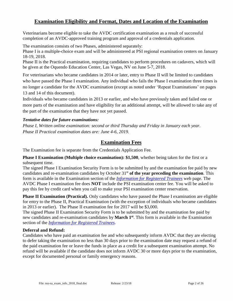
AVDC Phase I Examination Content Table
Summary of Proportion of Content:
Periodontology 19%
Endodontics 16%
Oral Surgery 16%
Operative Dentistry 9%
Orthodontics 5%
Oral Medicine 10%
Anesthesia & Analgesia 13%
Diagnostic Imaging 13%
Periodontology
Understand anatomy, physiology, pathophysiology, and pathology as it relates to periodontology
1. Healing of periodontal tissues
2. Periodontal anatomy
3. Physiology and pathophysiology of periodontal disease
File: nss-sa_exam_info_2018_final.doc Release: 2/23/18 Page 4 of 26
4. Principles of implantology and osseointegration
Assess periodontal health or disease and develop a comprehensive treatment plan
1. Classification systems for dental record keeping (e.g., furcation exposure, gingivitis)
2. Instrumentation for periodontal evaluation
3. Clinical signs and manifestations of periodontal disease
4. Indications, contraindications, materials and techniques for performing professional dental cleanings and
periodontal treatment
5. Indications, contraindications, materials, and techniques for treatment of combined periodontic/endodontic
lesions
6. Indications, contraindications, materials, and techniques for periodontal splinting, guided tissue regeneration,
bone augmentation, and periodontal membranes
7. Techniques, principles and materials to implement home care programs
8. Presence of severe cases of periodontal disease requiring staged treatment, including recognition of systemic
or immunopathic effects
9. Assessment of pretreatment systemic, general and local immunologic health of the animal as it relates to
treatment options
Utilize appropriate periodontal instruments, materials, and techniques and assess outcome/complications for the
treatment plan, and develop follow-up plan
1. Materials and techniques to treat periodontal pockets and exposed root surfaces
2. Care and use of hand instrumentation including curettes and scalers
3. Materials and patterns used to suture a periodontal flap
4. Materials and techniques to perform gingivectomy/gingivoplasty
5. Polishing equipment and materials
6. Care, use, and mechanism of action of power equipment
7. Visualization equipment such as light source, magnification, and mirrors
8. Combined periodontic/endodontic monitoring post-treatment
9. Dietary products, treats, and toys that may promote oral health by retarding plaque and calculus
10. Home care products – indications, use, and contraindications
11. Mechanisms of action of home care products
12. Postoperative care, long-term prognosis, and future assessment
13. Strategies for periodontal disease prevention, maintenance and improvement
File: nss-sa_exam_info_2018_final.doc Release: 2/23/18 Page 5 of 26
14. Evaluation of home care product efficacy and safety
Endodontics
Understand anatomy, physiology, pathophysiology, and pathology as it relates to endodontics
1. Gross and microscopic endodontic and periapical anatomy
2. Physiology and pathophysiology of the pulp-dentin complex and periapical tissues
Assess endodontic health or disease and develop a comprehensive treatment plan
1. Clinical signs of, and methods to assess, endodontic disease, including tooth fractures, tooth resorption,
pulpitis, and developmental defects
2. Tooth-fracture classifications and nomenclature
3. Indications, contraindications, materials, and techniques for vital pulp therapy, plus or minus coronal
reduction
4. Indications, contraindications, materials, and techniques for standard (orthograde) endodontic therapy
5. Indications, contraindications, materials, and techniques for surgical (retrograde) endodontic therapy
6. Indications, contraindications, materials, and techniques for hemisection and root resection
7. Indications, contraindications, materials, and techniques of apexification procedures
8. Physical properties of endodontic materials
Utilize appropriate endodontic instruments, materials, and techniques and assess outcome/complications for the
treatment plan, and develop follow-up plan
1. Appropriate access for standard (orthograde) and surgical (retrograde) endodontic therapy
2. Materials, equipment and methods for root canal debridement and irrigation
3. Materials and methods for obturation
4. Materials and methods for restoration following endodontic therapy
5. Root canal instrumentation, materials, and methods
6. Causes, prevention and treatment of iatrogenic procedural complications of endodontic therapy
7. Radiographic assessment of treatment outcome
Oral Surgery
Understand anatomy, physiology, pathophysiology, and pathology as it relates to oral surgery

File: nss-sa_exam_info_2018_final.doc Release: 2/23/18 Page 6 of 26
1. Anatomy, physiology, pathophysiology, and pathology of orofacial structures
2. Surgical considerations of effects of radiation therapy, chemotherapy, and immunosuppressive medications
on the surgical site
3. Maxillofacial fracture types and biomechanical effects of oral structures
4. Incidence, prevalence, and biological behavior of oral tumors and non-neoplastic diseases that can mimic
neoplasia
5. Pathophysiology and pathology of acquired and congenital hard and soft palate defects
6. Physiology of hard and soft tissue healing
Assess health or disease as it relates to oral surgery and develop a comprehensive treatment plan
1. Assessment of hard and soft palate defects (including oronasal and oro-antral communications)
2. Assessment of head trauma patient prior to surgery
3. Indications, contraindications, and techniques for exodontia of teeth and root remnants
4. Indications and contraindications of incisional vs. excisional biopsy techniques
5. Indications, contraindications, and techniques for partial/total mandibulectomy and maxillectomy
6. Indications, contraindications, and techniques for repair of acquired and congenital hard and soft palate
defects
7. Indications, contraindications, and techniques for maxillofacial fracture repair
8. Techniques, materials, indications and contraindications for repair of traumatic injuries
9. Indications, contraindications, and techniques for salivary gland and lymph node surgery
Utilize appropriate oral surgical instruments, materials, and techniques and assess outcome/complications for the
treatment plan, and develop follow-up plan
1. Closed and open extraction techniques
2. Extraction site management (e.g., protect soft tissue, flap and suture techniques, implant materials)
3. Instrumentation for exodontia
4. Management of teeth in fracture lines
5. Management of temporomandibular joint disease and associated conditions
6. Management of tooth displacement injuries
7. Materials, instrumentation, and techniques for oral and maxillofacial surgery
8. Noninvasive and invasive techniques for treatment of maxillofacial trauma
9. Nonsurgical and surgical methods for treatment of hard and soft palate defects
File: nss-sa_exam_info_2018_final.doc Release: 2/23/18 Page 7 of 26
10. Nonsurgical and surgical treatment of osteomyelitis
11. Nutritional management of the oral surgery patient
12. Complications of extraction procedures and their management
13. Complications of hard and soft palate repair procedures and their management
14. Complications of maxillofacial trauma repair and their management
15. Complications of oral biopsies and their management
16. Complications of partial/total mandibulectomy and maxillectomy and their management
17. Postoperative and follow-up management of the oral surgery patient
Operative Dentistry
Understand anatomy, physiology, pathophysiology, and pathology of tooth structure
1. Normal anatomy and histology of tooth structure, including occlusal contacts
2. Tooth structure, pathophysiology, and pathology and classification of defects
Assess structural integrity of teeth and develop a comprehensive treatment plan
1. Presence of direct or indirect pulp exposure
2. Effects of alteration of normal anatomy or structural integrity
3. Periodontal considerations for restorations
4. Indications and contraindications for placement of dental prostheses
5. Indications, contraindications, types, uses, and wear characteristics for restorative and prosthodontic materials
6. Periodontal considerations for restorations
7. Physical properties of restorative materials
8. Principles of micro- and macro-mechanical retention of dental restorative materials
Utilize appropriate operative dentistry instruments, materials, and techniques and assess outcome/complications
for the treatment plan, and develop follow-up plan
1. Cavity preparation
2. Instrumentation for operative dentistry
3. Materials and techniques for crown buildup procedures
4. Placement and finish of restoration material
5. Techniques and materials for obtaining impressions and model fabrication
File: nss-sa_exam_info_2018_final.doc Release: 2/23/18 Page 8 of 26
6. Techniques for appropriate crown reduction methods
7. Techniques, materials, indications, and contraindications for marginal finish and prosthesis cementation
8. Complications of operative dentistry and their management
9. Postoperative and follow-up management of restorative dentistry patient
Orthodontics
Understand anatomy, physiology, pathophysiology, and pathology of occlusal patterns
1. Occlusal characteristics and skull types
2. Developmental anatomy and embryology
3. Genetic basis for orthodontic problems
4. Normal and abnormal adult anatomy, occlusion, and function
Assess occlusion pattern and develop a comprehensive treatment plan
1. Current nomenclature and classification to accurately describe occlusion/malocclusion
2. Consequences of malocclusion
3. Appearance of secondary trauma associated with malocclusion
4. Age of animal relative to performance of procedure and effects of subsequent growth
5. Effects of orthodontic appliance on development of occlusion
6. Legal and ethical considerations for orthodontic treatment and genetic counseling
7. Probability of short- and long-term success of the orthodontic treatment
8. Techniques for orthodontic movement of various malocclusions
9. Indications and principles of interceptive orthodontics
10. Physical properties of orthodontic materials
11. Time required to complete orthodontic treatment
12. Animal behavior and treatment compliance
13. Indications and contraindications for appliance use
Utilize appropriate orthodontic instruments, materials, and techniques and assess outcome/complications for the
treatment plan, and develop follow-up plan
1. Appliance design, installation, maintenance and removal
2. Equipment and materials for orthodontic treatment

File: nss-sa_exam_info_2018_final.doc Release: 2/23/18 Page 9 of 26
3. Retention devices
4. Indications, contraindications, advantages, and disadvantages of direct and indirect appliance fabrication
5. Complications of orthodontic treatment
Oral Medicine
Understand anatomy, physiology, pathophysiology, and pathology of diseases of the craniofacial region and oral
cavity
1. Normal anatomy and physiology of the craniofacial region and oral cavity
2. Species and breed differences with respect to the incidence and prevalence of diseases of the oral cavity
3. Prevalence and biological behavior of local and systemic diseases affecting the oral cavity and craniofacial
region including developmental, degenerative, allergic, metabolic, inflammatory, infectious, immune mediated,
nutritional, traumatic, toxic, and neoplastic, both benign and malignant
4. Systemic impact of oral disease
5. Regional impact of oral disease (e.g., sinusitis, fistula formation)
6. Radiation therapy, chemotherapy, and immunosuppressive medication effects
Assess craniofacial region and oral cavity health or disease and develop a comprehensive treatment plan
1. Clinical presentations of primary (e.g., stomatitis, masticatory myositis) and secondary diseases (e.g.,
hyperparathyroidism, petechia)
2. Limitations of diagnostic tests
3. Prioritization of pathology and treatment in context of overall patient health and well-being
4. Adjunctive therapy for specific tumor types
5. Indications and contraindications for medical and surgical therapies
6. Treatment options for client education
7. Appropriate diagnostic modalities for primary disease, secondary diseases, and staging of neoplasia
8. Therapeutic effects and side effects of medical and surgical therapies
9. Indications for antimicrobial drug use
10. Nutritional requirements
Utilize appropriate oral medicine instruments, materials, and techniques and assess outcome/complications for the
treatment plan, and develop follow-up plan

File: nss-sa_exam_info_2018_final.doc Release: 2/23/18 Page 10 of 26
1. Diagnostic equipment and techniques for tissue sampling (e.g., cytology, biopsy, culture)
2. Treatment modalities (e.g., pharmaceuticals, immunological agents, chemotherapeutic agents, radiation, laser
therapy, physical therapy)
3. Treatment options of systemic disease
4. Nutritional management and feeding techniques
5. Cytological preparations, special stains, and microscopic evaluation
6. Follow-up for medical and surgical therapies for primary and secondary disease
7. Management of complications and side effects of medical and surgical therapies
8. Appropriate modification of long-term medical therapies, based on patient response and potential for adverse
patient reactions to the selected medical therapies
9. Prognosis and response to treatment
Anesthesia and Analgesia
Understand anatomy, physiology, and pharmacology of anesthesia and analgesia
1. Species and breed differences (e.g., brachycephalic, sighthound, lagomorph) for delivery of anesthesia,
sedation, and regional analgesia
2. Injectable, inhalant, multimodal anesthetics, anesthesia, analgesia, sedation, constant rate infusions and their
related metabolism
3. Systemic physiology related to anesthesia with particular emphasis on cardiopulmonary, renal, and hepatic
physiology
4. Regional anatomy (e.g., maxillofacial innervation, vascularization, and important structures) for regional
analgesia
5. Pharmacology of local/regional anesthetics, analgesia, and related metabolism
6. Anesthetic drug interactions with concurrent medications (e.g., seizure, behavioral, homeopathic,
cardiovascular, renal)
7. Contraindications of medications used for medical problems and anesthesia/analgesia
8. Anesthetic considerations for pediatric and geriatric patients
Assess health and disease for appropriate pre-anesthetic evaluation of the dentistry and oral surgery patient and
develop a comprehensive anesthesia and analgesia treatment plan
1. Patient history, signalment, and physical examination
2. Assessment of preoperative laboratory testing (e.g., CBC, chemistry panel, urinalysis, endocrine testing,
radiography, ancillary imaging)
3. Animal demeanor in relation to anesthetic protocol
File: nss-sa_exam_info_2018_final.doc Release: 2/23/18 Page 11 of 26
4. American Society of Anesthesiologists (ASA) physical status classification
5. Concomitant disease (e.g., cardiac, renal, endocrine, trauma) conditions that can impact the safety of
anesthesia delivery, protocol design/drug selection, and anesthesia monitoring
6. General anesthesia and analgesia protocols, indications, and contraindications
7. Individual anesthetic and analgesic plans for patients with concomitant disease (e.g., cardiac, renal, endocrine,
trauma)
8. Regional/local analgesia techniques, indications, and contraindications
9. Multimodal pain control, acute pain, chronic pain, and cancer pain
10. Indications for extraoral endotracheal tube placement, example: pharyngotomy
11. Maintaining homeostasis (e.g., thermoregulation, hemodynamics)
Utilize appropriate anesthesia and analgesia instruments, materials, and techniques and assess
outcome/complications for the treatment plan, and develop follow-up plan
1. Anesthesia machines and circuit types
2. Anesthesia physiological monitoring equipment (e.g., capnography, pulse oximetry, electrocardiogram, blood
pressure, temperature) and data interpretation
3. Anesthesia thermoregulation devices (e.g., circulating water blankets, force air convection warming devices,
carbon fiber blankets, IV fluid warmers)
4. Regional and local anesthetic administration
5. Anesthesia delivery mechanisms (e.g., intravenous, inhalant, CRI)
6. Pharyngostomy and tracheostomy tube placement
7. Anesthetic reversal agents (e.g., atipamezole, flumazenil, naloxone)
8. Re-warming devices
9. Emergency procedures (e.g., tracheostomy, CPR) and equipment
10. Crystalloid, colloid, and blood product support
11. Emergency drug indications and routes of delivery
12. Recognition and management of common arrhythmias
13. Management of hypotensive crises
Diagnostic Imaging
Understand anatomy, physiology, pathophysiology, and pathology as related to diagnostic imaging
File: nss-sa_exam_info_2018_final.doc Release: 2/23/18 Page 12 of 26
1. Anatomy and physiology of the dental and periodontal tissues
2. Anatomy and physiology of the skull and soft tissues of the head and neck
3. Normal radiographic development of the teeth and the jaws
4. Developmental and acquired abnormalities of the teeth, jaws, and soft tissues of the head
5. Fundamentals of diagnostic imaging (e.g., radiographs, CT, MRI, ultrasound)
Assess patient health or disease utilizing diagnostic imaging and develop a comprehensive plan to obtain
diagnostic images
1. Radiographic interpretation of endodontic and periodontal anatomy and pathology
2. Extraoral imaging (e.g., radiographs, CT, MRI) interpretation of maxillofacial health and disease
3. Radiographic interpretation of anatomical/developmental normal structures and anomalies
4. Radiographic diagnosis of bone lesions
5. Radiographic/imaging signs of benign and malignant lesions, including determination of radiographic
margins of neoplastic disease
6. Radiographic interpretation of dental and oral/maxillofacial trauma
7. Radiographic pathology of developmental or congenital anomalies
8. Indications and contraindications of various diagnostic imaging modalities
9. Patient and operator protection and radiation safety guidelines
10. Patient immobilization for obtaining diagnostic images
Utilize appropriate diagnostic imaging instruments, materials, and techniques to obtain and interpret diagnostic
images, assess outcome/complications, and develop follow-up plan based on diagnostic images obtained
1. Operation of X-ray generators and advanced imaging modalities
2. Patient preparation and positioning
3. Radiographic imaging including conventional film and digital radiography
4. Parallel, bisecting angle, and occlusal techniques
5. Developing, processing, and positioning
6. Imaging artifacts
7. Labial and lingual mounting techniques
8. Identification and archival storage of images
9. Interpretation of intraoral and extraoral radiographs
10. Interpretation of magnetic resonance imaging studies
File: nss-sa_exam_info_2018_final.doc Release: 2/23/18 Page 13 of 26
11. Interpretation of computed tomography studies
12. Radiographic procedures to determine presence and location of retained roots
13. Radiographic/imaging interpretation of previous treatments
Phase II (Practical Examination)
Phase II of the examination will be given on June 6-7, 2018 at the Oquendo Center in Las Vegas, NV with
a mandatory orientation session in the evening of June 5, 2018. This examination is designed to assess the
clinical technical skills of the candidate. The examination will be given in four core sessions (two sessions
starting Wednesday morning and two sessions Thursday morning). The candidates will perform two
procedures within each of the four core disciplines: periodontics; endodontics; oral surgery; and restorative
dentistry/prosthodontics/orthodontics. The core schedule and time limit for each session is listed below
and is for the 2018 cycle only. The format of the examination will be explained further at the exam
security meeting and at the beginning of the examination. Plan your work sequence at the start of the
session and continue to be aware of the remaining time during the testing session.
2018 Examination Location and Schedule
Tuesday June 5th, 2018
Location: Oquendo Center, 2425 Oquendo Rd., Las Vegas, NV 89120
5pm - 6pm Candidate Orientation Meeting/Candidate Security Registration**
6pm - 8pm Candidate practical station set-up
** Attendance at this meeting is mandatory in order to complete the AVDC security and anonymity
procedures.
Access to the laboratory to start set-up for the practical examination will not be permitted until after the
orientation and security meetings have finished.
Two hours (120 minutes) will be made available for workstation set-up (regardless of when the orientation
and security meetings end). All candidates will be allowed into the laboratory at the same time, given the
opportunity to find their work station, and then allowed to set up, and to practice using the radiographic
equipment; cadaver specimens will be provided.
Wednesday June 6th, 2018
Location: Oquendo Center, 2425 Oquendo Rd., Las Vegas, NV 89120
7:30 am: Candidates may enter the testing room
8:00 am: Session 1 (Endodontics) begins
11:00 am: End of session 1
11:00 am- 12:00 pm- Break for cleanup of station and setup for next session and short lunch break.
It is advised that candidates bring their lunch to eat at Oquendo during this
break or travel only a short distance to a local restaurant. Session 2 begins

File: nss-sa_exam_info_2018_final.doc Release: 2/23/18 Page 14 of 26
promptly at 12:00 pm.
12:00 pm: Session 2 (Restoratives/Orthodontics/Prosthodontics) begins
2:30 pm: End of session 2
2:30-3:00 pm: Cleanup of station, candidates exit testing room promptly at 3:00 pm
Thursday June 7th, 2018
Location: Oquendo Center, 2425 Oquendo Rd., Las Vegas, NV 89120
7:30 am: Candidates may enter the testing room
8:00 am: Session 3 (Periodontics) begins
11:00 am: End of Session 3
11:00-12:00 pm: Break for cleanup of station and setup for next session, short lunch break. It
is advised that candidates bring their lunch to eat at Oquendo during this
break or travel only a short distance to a local restaurant. Session 2 begins
promptly at 11:30 am.
12:00 pm: Session 4 (Oral Surgery) begins
2:30 pm: End of Session 4
2:30-3:30 pm: Cleanup and breakdown of workstations, all candidates.
ALL CANDIDATES MUST BE PRESENT FOR THE EXAMINATION SECURITY MEETING
AND NO CANDIDATES WILL BE ALLOWED TO PACK UP THEIR STATIONS UNTIL THE
END OF THE LAST SESSION. Candidates will only be permitted in the testing room during their
required core sessions and designated set-up/clean-up times.
Candidates should not schedule flights home earlier than 6 pm on Thursday June 7th.
While every effort is made to ensure consistency between specimens to ensure fairness, as in clinical
practice not all specimens are exactly the same. Each candidate should work with their specimen(s) to the
best of their ability.
Candidates are to work independently, and no candidate is allowed to receive help on any phase of the
practical examination. Sharing of equipment or materials among candidates during the examination
sessions is not permitted, as this has been found to be disruptive to the examination process. With the
exception of minimal conversation with a proctor directed toward the use of AVDC-provided equipment,
candidates are not to engage in conversation during the examination. Pets, family members, friends, staff,
and personal belongings not related to the examination will not be allowed in the examination area.
Electronic music players and earphones are not allowed (see Examination Security and Candidate
Misconduct). You may use earplugs if you wish to reduce ambient noise.
Work Station:
File: nss-sa_exam_info_2018_final.doc Release: 2/23/18 Page 15 of 26
Dental high-speed units and hand-pieces:
Candidates will be allowed to use the scaler hand pieces provided on the unit. One high-speed hand
piece will also be provided. No other hand-pieces or scalers will be provided – you are required to bring a
low speed handpiece, back up high- and low-speed hand-pieces, couplers, tips and burs you wish to use
in the examination.
This year candidates will be randomly assigned either a Midmark VetPro 1000 dental unit with LED
piezo scaler or an iM3 GS Deluxe LED unit with piezo scaler.
Midmark Equipment Specifications
The Midmark VetPro 1000 has conventional 4-hole and 5-hole (fiberoptic) connections, and a piezo
electric scaler hand-piece. Candidates wishing to use the scaler must provide their own piezo scaling
tips. Piezo tips must be Acteon Satelec brand tips only. Other brands may fit into the hand-piece but may
not work normally in the Midmark units. Acteon Satelec tips can be purchased through dealers or directly
from Midmark.
http://ahstore.midmark.com/index.php?route=product/search&filter_name=tip
AVDC cannot guarantee that hand-pieces with fiberoptic capability will function properly with all
candidate-provided fiberoptic lights at the hand-piece head.
The Midmark Vet Pro 1000 has water bottles that are used to provide water to the built-in hand-piece lines.
Distilled water will be provided for use in the bottles in the Vet Pro dental units and that water is available
via the high-speed hand-piece line and the air-water syringe line. Ports on the back of the dental unit can
be used for attaching additional equipment, provided the candidate brings an appropriate coupler for the
water to flow through from the back of the VetPro 1000 to the additional equipment.
iM3 Equipment Specifications
The GS Deluxe LED is equipped with an air-driven, LED light on the high-speed hand-piece. If you
provide your own hand-piece that has a fiberoptic light, the light will NOT work on the GS Deluxe LED.
5-hole handpieces will NOT work on iM3 units.
The iM3 GS Deluxe LED dental unit has a conventional 4-hole connection, LED, 360 degree swivel
handpiece, and a P6 piezo electric scaler. Candidates wishing to use the scaler must provide their own
piezo scaling tips. Piezo tips must be Acteon Satelec brand tips only. Other brands may fit into the hand-
piece but may not work normally in the iM3 units. Acteon Satelec tips can be purchased through dealers or
directly from iM3.
http://www.im3vet.com
The GS Deluxe LED has water bottles that are used to provide water to the built-in hand-piece lines.
Distilled water will be provided for use in the bottles in the GS Deluxe LED dental units and that water is
available via the high-speed hand-piece line and the air-water syringe line. Ports on the back of the dental
unit can be used for attaching additional equipment, provided the candidate brings an appropriate coupler
for the water to flow through from the back of the GS Deluxe LED to the additional equipment.
File: nss-sa_exam_info_2018_final.doc Release: 2/23/18 Page 16 of 26
Instruments and materials such as digital radiographic film, hand-pieces, restorative material, curing
lights, impression materials etc. are not supplied by the AVDC. The Examination Committee discourages
the use of thermoplasticized gutta percha for endodontic procedures due to the temperature of the materials
provided. The use of surgical adhesives for closure is not allowed because this prevents evaluation of
surgical technique. Due to safety regulations, use of two-part, liquid-powder methyl methacrylate products
and chloroform are not allowed; candidates must find an alternate material for procedures that might call
for the use of such products. Candidates will not be required to use amalgam for restorative procedures.
Candidates will not be required to pour stone models. To summarize, all materials necessary to complete
the practical examination sessions, and which were not mentioned in this document as being supplied by
the AVDC, are the responsibility of the candidate. No reading materials associated with dental equipment
or supplies may be brought into the examination room, except product information sheets that were
originally packaged with the equipment or material.
Set-up: In addition to the set-up time in the evening prior to the first session, the candidates will be
allowed into the examination room 30 minutes prior to the scheduled start time of the first session of each
day, to set up their equipment. To avoid disruptions to others, all candidates are required to set-up and
breakdown equipment at the same time, regardless of cores they are taking.
Radiographs: A digital radiographic system will be used by all candidates. Digital radiography will be
used to create and expose images using the CR7 system, available from IM3. Candidates will be required
to purchase their own phosphor plates for the examination. It is recommended that candidates bring one or
two size 4 phosphor plates and two size 2 phosphor plates. Plates can be purchased through IM3 directly
(800-664-6348); size 2 plates (X7121) are $65.25 each ($261.00 for a pack of four); size 4 plates (X7140)
are $176.25 each (prices subject to change) - be sure to mention that you are purchasing the plates for
the AVDC examination to obtain the discounted rate. AVDC will provide barrier sleeves for use with
the phosphor plates when radiographing your specimen; therefore, you are not required to purchase barrier
sleeves; however, AVDC strongly recommends using a barrier sleeve when you are using phosphor plates
in clinical practice or when practicing for the examination, to prevent the plate from becoming scratched or
dirty. The sleeves are available from IM3 in packs, at a cost of $55.80 for the size 4 plate and $106.20 for
the size 2 plate.
USB flash drives will be provided with each specimen for storage of images. The radiographic images will
be viewed on Microsoft Surface Pro 3 tablets (or something comparable) provided by AVDC. This is a
touch-screen Windows-based device and will allow image files to be accessed from the thumb drive folder
in the same manner they would be viewed on a PC. The model being used has the following specifications:
Microsoft Surface Pro 3 iCore 3; 4GB RAM; 64GB Solid state drive; 12 inch screen; Windows 8 pro
operating system.
Submission of items for grading:
Examination materials must be handed in on time. The final five minutes of the examination will be
recorded. The entire examination period may be video recorded. Radiograph generators will be turned off
File: nss-sa_exam_info_2018_final.doc Release: 2/23/18 Page 17 of 26
with 5 minutes remaining; unprocessed films placed at the development station immediately following the
generator being powered down will be processed; if a processing backlog occurs, any undeveloped films
will be processed and considered “turned in” upon the call of time despite not being viewed by the
candidate. All specimens and related materials for grading (such as resected specimens, impressions or
USB flash drives containing radiographs) must be placed in plastic boxes with the top closed, and the box
must be placed on the floor before or at the time that the end of the examination is announced.
Time remaining in the session will be announced periodically by the proctor.
Candidates will be informed by announced countdown of the last 15 seconds prior to the end of the
examination.
If a specimen is not in the plastic box on the floor at the announced end of the examination, proctors will
physically collect the specimens and place them in the plastic box. A red tag will be attached to that
specimen box (which will be removed before the specimen box is seen by the graders). These red tag
specimens will be penalized 35% of the actual scores given by the graders for procedures performed on
those specimens. Physical resistance by the candidate to collection of the specimen by the proctor will
cause the proctor to back away with the result that the specimen will not being graded at all. Additional
materials such as resected tissues, impressions or extracted teeth not in the plastic box when collected by
the proctor will still be submitted for grading but will receive a 35% penalty for the portion of the grading
that the item pertains to.
Any items being submitted for evaluation (such as impression trays) must be completely devoid of any
identifying mark other than the specimen numbers that will be assigned at the time of the examination.
Candidates must submit only what is specifically requested on the examination instructions. Any other
material submitted will not be evaluated and may compromise the anonymity of the candidate.
Safety issues: Taking dangerous chemicals (e.g. chloroform, bleach) on airplanes is illegal. Candidates
currently residing outside the USA should be aware that the voltage in the United States is 110v. Given the
travel security arrangements now in place, review carefully what you need to bring with you. Contact your
airline and/or the US Transport Security Administration if you have any questions or concerns.
Possible Practical Examination (Phase II) Procedure List
While this list is representative of the types of procedures that will be included in the examination,
the AVDC and the Examination Committee reserve the right to include other procedures. Any
procedures not on the list will not require equipment or supplies beyond those necessary for
performing the procedures on the list.
The goal of the practical examination is to evaluate clinical skills, judgment, and treatment
planning.
The choice of technique and materials to be used for each procedure is part of treatment planning,
and it is up to the examinee to select an appropriate technique and to execute the procedure.
Radiographs will be required for some procedures.
File: nss-sa_exam_info_2018_final.doc Release: 2/23/18 Page 18 of 26
Use this list to determine what equipment, instruments and supplies may be needed, so that you are
fully prepared.
Periodontics
1. Perform routine periodontal treatment (“prophylaxis”) on an assigned area.
2. Open curettage of single or multiple teeth.
3. Flap procedures of assigned type and location.
4. Use of a guided tissue regeneration technique for management of a periodontal defect. You will
NOT be required to provide and place an actual guided tissue regeneration membrane.
5. Type II Crown Lengthening procedure.
Endodontics
1. Pulpectomy (standard root canal treatment) or partial coronal pulpectomy (vital pulp therapy),
specific tooth as directed.
2. Apicoectomy (surgical root canal treatment), specific tooth as directed.
3. Pulp capping, specific tooth as directed.
4. Treatment of endodontic complications (e.g. perforated root).
Restorative
1. Crown preparation for a metal or porcelain crown in response to a fractured crown with most of the
crown intact; appropriate impressions and bite registrations.
2. Functional direct crown build-up on a tooth fractured off 2 mm coronal to the gingival margin; this
may involve placement of post and/or pins.
3. Restoration of a specific class and type of defect with an appropriate restorative for such, or with a
restorative such as glass ionomer or composite as specified in the examination. Use of amalgam
will NOT be required.
4. Crown wall restoration, including subgingival finish.
5. Crown lengthening procedure as part of placing a restoration.
Oral Surgery
1. Surgical and/or non-surgical extraction of specified tooth or teeth.
2. Repair of an oronasal fistula on a specified area.
3. Intra-osseous or interdental wiring of specified teeth or area.
4. Palatal surgery.
5. Dental arch resection for the treatment of a neoplasm.
6. Noninvasive fracture repair techniques (intraoral splints).
Orthodontics
1. Take an impression of a specified area with an appropriate material.
2. Cementation of brackets and buttons.

File: nss-sa_exam_info_2018_final.doc Release: 2/23/18 Page 19 of 26
3. Application of a bracket, button, appliance, wire, elastics, or power chain as required or requested,
appropriate for movement of a specified tooth or teeth.
4. Diagnosis of a malocclusion, recommendation of a treatment plan and preparation of laboratory
instructions.
Reasons for Failure of Practical Examination Procedures
Here is a comprehensive list of reasons contributing to grading a procedure as a failure, from recent AVDC
examinations.
General:
Some requested items were not submitted. Results in 35% penalty for all grading criteria related to
that item.
Requested radiographs do not show the required structure(s).
Stated specifications have not been met (e.g. mm of crown length to be created).
Patient care concerns, i.e, anything that would cause clinical problems in a patient if not attended to. Below
is a list of examples. This list is not all inclusive. Neglect of patient care results in a 5% penalty on all
grading for that specimen.
Gauze or gross debris left in the mouth.
Lip sutured to skin left in place.
Mouth gag left in specimen’s mouth.
Major procedural complications, I.e. anything that would cause the procedure to fail clinically. Below is a
list of examples. This list is not all inclusive. Major complications result in the grade for the
procedure being multiplied by 0.70 as a penalty.
Incorrect tooth/area treated
Wrong procedure performed
Untreated pulp exposure
Lack of biologic width preservation
Life-threatening/catastrophic complications, I.e. anything that would result in mortality or significant
morbidity. This list is not all inclusive. Life-threatening complications result in automatic failure of
the entire examination.
Brain perforation/herniation
Orbital penetration
Unnecessary removal of tissues such as the lips or tongue not related to the requested procedure
Soft Tissue:
Inappropriate location or length of incision.
Irregular edges of incised tissues.
Major vessel appears to be severed but not ligated.
Un-necessary exposure of bone.
Inappropriate size of suture material.
Gaps between sutures, sutures are too loose or too tight or are crowded, or suture knots are not secure.
File: nss-sa_exam_info_2018_final.doc Release: 2/23/18 Page 20 of 26
Tension at suture line.
Debris present.
Adjacent soft tissue has been damaged.
Dental structures:
Inadequate or excessive removal of enamel or dentin, or unsupported enamel is present.
Exposed dental surfaces have not been smoothed.
Root is gouged or rough.
Tooth gouged during preparation of adjacent tissues.
Gingiva and Periodontal Bone:
In addition to items in ‘Soft Tissue’, above:
Calculus remaining on treated teeth.
Biologic width is inadequate, gingiva is damaged or poorly adapted.
Bone is rough or inappropriately shaped.
Root is exposed.
Perforation near or into the nasal cavity.
Flap is poorly designed, and is insufficient to cover the defect without tension, or the width:length ratio is
inadequate.
Flap is loose or is perforated or is poorly adapted to bone.
Tooth damage created during preparation of bone.
Crown lengthening: In addition to above, insufficient additional length achieved; incorrect technique (I vs.
II).
Impressions and Bite Registrations:
Impression tray is not included, is flexible or is too large.
Impression material is not fully mixed; light body/wash not appropriately distributed.
Not all relevant teeth are included in the impression.
Cuspal show-through as a result of insufficient height of impression material.
Bubbles or other defects such as drag lines are included in the impression.
Bite registration is inappropriately designed and fabricated – the bite is not correctly registered.
Oral Surgery:
Poor or absent blood vessel management.
Mass removal:
Inadequate or excessive extent of tissue resection.
Inappropriate design and length of flap incisions, causing tension or e.g. lip tuck, or tooth contacts
flap.
Extractions:
Excessive bone removal and inadequate alveoloplasty.
Excessive undermining of flaps and damage to adjacent tissue.
Inadequate preparation or over-preparation of flap recipient site.
File: nss-sa_exam_info_2018_final.doc Release: 2/23/18 Page 21 of 26
Bone surfaces rough and irregular, debris in alveolus beneath suture line or on exposed bone.
Retained root tip; root tip in mandibular canal.
Exposure of mandibular canal.
Alveolus of canine tooth is fractured and mobile.
Fracture repair:
Poor wiring technique, splint is excessive or design is poor - prevents occlusal closure or causes
excessive soft-tissue coverage.
Splint has rough edges or debris found; weak bonding of splint to teeth – easily displaced; occlusal
interference.
Endodontics
Access is misdirected or is too shallow or over-prepared or there is damage of adjacent enamel.
Canal is over-instrumented or is inappropriately instrumented.
Failure to clean the coronal portion if a separate access is made.
Obturation is incomplete or of variable density or has obvious voids.
Tooth split by excess obturation pressure.
Debris in access site, or sealer is present on walls of access site.
Excessive apical extrusion.
Apicoectomy: Inappropriate location or length of incision; inadequate bone preparation or excessive or
rough edges to cavity in bone; root surface at apicoectomy not smooth; perforation into nasal cavity; voids
in apical fill; overfill and flash; restoration inadequate or not performed; site closure incomplete or sutures
are tight.
Operative Dentistry:
Preparation for restoration: insufficient or of excessive depth, or enamel is undercut, or extends to the bone
edge or margins and surfaces are not smooth, or bone management is poor.
Root trauma.
Buccal bulge is excessive or preparation is over-filled.
Crown preparation margin is irregular in width, in height to gingiva or is undercut, and surface of
preparation is gouged or rough.
Restorative material is not fully cured.
Soft tissue damage.
Orthodontics
Inappropriate choice of or location of attachment device for anchor and/or target teeth.
Active force device not appropriately loaded.
Appliance will cause occlusal interference or soft tissue damage.
Appliance not securely attached.
Appliance design will not cause required tooth movement.
Appliance not finished.
Gingival or tooth damage created.
File: nss-sa_exam_info_2018_final.doc Release: 2/23/18 Page 22 of 26
Suggested Reading List for Candidates and Trainees
The following list is provided as suggested reading material. It is not all inclusive of every potential
reference and publication, because the body of scientific literature is fluid and always changing.
Much of veterinary dental knowledge has been derived from human dentistry. This is
reflected in the suggested reading list and will also be reflected in the examination itself.
Books
Anatomy:
1. Evans HE. Miller’s Anatomy of the Dog. 4th ed. Philadelphia: WB Saunders, 2012.
2. Schroeder HE. Oral Structural Biology. New York: Thieme Medical Publishers, 1991.
Anesthesia:
1. Tranquilli WJ and et al eds. Lumb & Jones Veterinary Anesthesia and Analgesia. 4th ed. Baltimore:
Williams & Wilkins, 2007.
Dental Materials:
1. Anusavice KJ. Philips’ Science of Dental Materials. 12th ed. Philadephia: WB Saunders, 2013.
2. Powers JM. Craig’s Restorative Dental Materials. 12th ed. St. Louis: Mosby Elsevier, 2006.
Embryology:
1. Nanci A. Ten Cate’s Oral Histology: Development, Structure, and Function. 8th ed. St. Louis:
Mosby, 2012.
2. Avery, James K. Essentials of Oral Histology and Embryology: A Clinical Approach. 3rd ed.
Mosby Elsevier. 2002.
Endodontics:
1. Hargreaves KM and Cohen MA. Cohen’s Pathways of the Pulp. 10th ed. St. Louis: Mosby, 2010.
Equine:
1. Easley J, Dixon PM, and Schumacher J. Equine Dentistry. 3rd ed. Philadelphia: Saunders, 2010.
2. Easley J. Advances in Equine Dentistry. Veterinary Clinics of North America: Equine Practice
29(2). Philadelphia: Saunders-Elsevier, 2013
Exotics:
1. Capello V, Gracis M, and Lennox, A. Rabbit and Rodent Dentistry. Philadelphia: WB Saunders,
2005.
Orthodontics:
1. Proffit WR and Fields HW. Contemporary Orthodontics. 5th ed. St. Louis: Mosby-Year Book,
2013.
Pathology:
1. Regezi JA, Sciubba JJ, Jordan RCK. Oral Pathology: Clinical Pathologic Correlations. 5th ed.
Philadelphia: Saunders, 2007.
Periodontology:
File: nss-sa_exam_info_2018_final.doc Release: 2/23/18 Page 23 of 26
1. Newman MG, Takei H, Klokkevold PR, Carranza FA. Carranza’s Clinical Periodontology. 12th ed.
Philadelphia: Saunders, 2014.
2. Wolf HF, Rateitschak KH, Rateitschak EM, Hassell TM. Color Atlas of Dental Medicine –
Periodontology. 3rd ed. New York: Thieme Medical Publishers, 2005.
Radiology:
1. DuPont GA and DeBowes LJ. Atlas of Dental Radiography in Dogs and Cats. St. Louis: Saunders
Elsevier, 2009.
2. White SC and Pharoah MJ. Oral Radiology: Principles and Interpretation. 6th ed. St. Louis: Mosby,
2008.
Restorative Dentistry / Prosthodontics:
1. Roberson TM, Heyman HO, and Swift EJ. Sturdevant’s Art and Science of Operative Dentistry. 6th
ed. St. Louis: Mosby, 2013.
Surgery:
1. Fossum TW. Small Animal Surgery. 3rd ed. St. Louis: Mosby, 2007.
2. Hupp JR, Ellis III E, Tucker MR. Contemporary Oral and Maxillofacial Surgery. 6th ed. St. Louis:
Mosby, 2013.
3. Slatter D. Textbook of Small Animal Surgery. 3rd ed. Philadelphia: WB Saunders, 2003.
4. Verstraete FJM and Lommer MJ. Oral and Maxillofacial Surgery in Dogs and Cats. Philadelphia:
Saunders, 2012.
Small Animal Dentistry:
1. Holmstrom SE, Frost P, Eisner ER. Veterinary Dental Techniques. 3rd ed. Philadelphia: WB
Saunders, 2004.
2. Holmstrom SE. Veterinary Dentistry. Veterinary Clinics of North America: Small Animal Practice.
35(4), p. 763-1072. Philadelphia: Saunders-Elsevier, 2005.
3. Holmstrom SE. Clinical Veterinary Dentistry. Veterinary Clinics of North America: Small Animal
Practice. 43(3) p. 447-689. Philadelphia: Saunders-Elsevier, 2013.
4. Tutt C, Deeprose J, Crossley DA, eds. BSAVA Manual of Small Animal Dentistry. 3rd ed. Quedgeley:
British Small Animal Veterinary Association, 2007.
6. Verstraete FJM. Self-Assessment Color Review of Veterinary Dentistry. London: Manson
Publishing and Ames: Iowa State University Press, 1999.
7. Wiggs RB and Lobprise HB. Veterinary Dentistry Principles & Practice. Philadelphia:
Lippincott-Raven, 1997.
Journals and Periodicals:
1. Journal of Veterinary Dentistry: Volume 16(1) 1999 to present.
2. Dixon PM (2008) Equid Dentistry. The Veterinary Journal, 178(3) 307-424.
3. Journal of Endodontics* (articles as referenced in JVD Abstracts section)
4. Journal of Periodontology* (articles as referenced in JVD Abstracts section)
5. Journal of Clinical Periodontology* (articles as referenced in JVD Abstracts section)
6. Journal American Dental Association* (articles as referenced in JVD Abstracts section)
7. American Journal of Dentistry*(articles as referenced in JVD Abstracts section)
File: nss-sa_exam_info_2018_final.doc Release: 2/23/18 Page 24 of 26
8. Frontiersin.org, particularly the Veterinary Dentistry and Oromaxillofacial Surgery section (online
journal: http://journal.frontiersin.org/journal/veterinary-science/section/veterinary-dentistry-and-
oromaxillofacial-surgery#archive)
Other suggested journals that contain some valuable oral-dental articles, and that are AVDC-approved for
meeting the AVDC Credentials Publication Requirement:
1. American Journal of Veterinary Research
2. Journal of the American Animal Hospital Association
3. Journal of the American Veterinary Medical Association
4. Journal of Small Animal Practice
5. Journal of Feline Medicine and Surgery
6. Journal of Veterinary Internal Medicine
7. Veterinary Comparative Orthopaedics and Traumatology
8. Veterinary Pathology
9. Veterinary Radiology & Ultrasound
10. Veterinary & Comparative Oncology
11. Veterinary Surgery
Passing Score, Examination Results, Repeat Examinations
Phase I - Multiple Choice Question Examination
A pre-set Pass cut score is NOT used. All questions are ‘criterion-referenced’ using the modified Angoff
procedure to determine the degree of difficulty of that question for a minimally qualified entry-level
veterinary dental specialist. The mean of the Angoff scores of the questions included in the examination is
the pass cut-score. Typically, the mean Angoff score is in the region of or slightly below 70%.
Phase II - Practical Examination
The scoring standards for the practical portion of the examination are scored based on likely clinical
success of the procedure and based on generally accepted dental techniques as found in current textbooks
and journals and practiced by Diplomates of the AVDC. For each procedure, a grading system of 0-100
based on predetermined criteria is used. The passing grade for a single procedure is 70%. All four core
disciplines must be passed to pass the Phase II examination. The core disciplines are: periodontics,
endodontics, oral surgery and operative/restorative/orthodontics. The scores of the graders are averaged for
each specimen. Fractional scores stand as is, and are not rounded. The final grade of the practical
examination is the average score of the procedures assigned. The Examination Committee reserves the
right to recommend to the AVDC Board of Directors to fail a candidate in the practical examination,
irrespective of the score obtained, if an error was performed by the candidate that would, in a clinical
situation, result in serious harm to the patient.
For successful passing of the Phase II examination, a candidate must have passed each core discipline with
>70% for the tested procedures in that core. The examination fee for Phase II will be the same for all
candidates regardless of which cores, or how many cores, are taken.
File: nss-sa_exam_info_2018_final.doc Release: 2/23/18 Page 25 of 26
Candidates returning to take any remaining cores of the Phase II examination will be required to be present
for the entire examination process. All candidates must attend the Examination Security meeting held the
evening before the first examination day. Candidates will not be allowed to remove their equipment or
pack up until after the final core session is completed. The specific day and start time of each core session
will be provided to all candidates at the Examination Security meeting.
Disclosure of Examination Results
Candidates will receive written notification of whether or not they passed Phase I of the examination
within 45 days of the date of the examination. Results for all candidates are sent on the same day.
Candidates who are not successful in passing Phase I will be provided with an explanation of the
deficiencies that prevented their passing the examination.
For Phase I of the examination, information will be made available on whether the candidate passed or
failed each major discipline category of the examination.
For Phase II of the examination the specific procedures that the candidate failed will be listed for the
candidate. In addition, this document includes a comprehensive list of reasons for failing particular
procedures in several recent years.
Actual scores will not be released to candidates.
Repeat Examinations
The AVDC certifying examination has two parts: Phase I (written questions) and Phase II (practical
examination). Candidates must pass Phase I of the certifying examination to be eligible to sit for Phase II.
Beginning with the first examination after approval of the credentials application, candidates shall have a
limit of three attempts in consecutive years for each phase of the examination, with the exception of one
deferral year, and subject to the requirement that candidates must have passed the Phase I examination in
order to be eligible for the Phase II examination. Candidates who fail to pass either phase I or II of the
certifying examination in three consecutive years, including a possible deferral year, may opt to resubmit a
new credentials package fulfilling the current credentials. Exceptions to this limit on number of
examination attempts may be made by the Board of Directors following petition from the candidate for one
additional attempt per phase of the examination; the petition must include the candidate’s proposed
examination preparation action plan, which is subject to review and approval by the Board. The Board
shall have the right to consider extenuating circumstances.
Candidates wishing to retake Phase I or Phase II of the examination are to complete, sign and submit the
Re-Examination Form, which is available for down-loading from the AVDC web site, in the Examination
documents section of the Information for Registered Trainees page.
Examination Security and Candidate Misconduct
Any questions before the examination regarding the examination are to be directed via e-mail to the
Executive Secretary of the AVDC ([email protected]) or, if the Executive Secretary is unavailable, to
the Chair of the Examination Committee. Questions will be answered in writing and copies will be sent to
all candidates. It is strictly forbidden to have direct or indirect contact with other members of the
Examination Committee regarding the process, format or content of the examination, from the date that an
File: nss-sa_exam_info_2018_final.doc Release: 2/23/18 Page 26 of 26
applicant is notified that s/he is a candidate for the examination until the examination has been completed.
Any breach of these rules can be considered reason for action by the Board of Directors to deny a
candidate admission to the examination.
The NSS-SA Examination Committee leaders for the 2018 Examination consist of: Chris Smithson
(Chair), Kendall Taney (Past-Chair), and Michael Jennings (Chair-Designate)
The Equine Examination Committee leaders for the 2018 Examination consist of: Jack Easley (Chair), and
Hubert Simhofer (Past Chair).
Tom Kiger, PhD from HumRRO test consulting services will be assisting with examination security
procedures, calibration, and overall test analysis.
Examination security is a primary concern for AVDC. Do not bring personal materials (e.g. notes,
books, tape recorders, photographic devices, calculators, computers, cellular phones) to the examination
room. References are not to be consulted during the examination process. The examination material is not
to be divulged to others. See the specific language in the Examination Security Form.
Candidates: Complete and sign the Examination Security Form and return it to the AVDC
Executive Secretary by October 31st of the year before the examination for the Phase I examination
and by March 1st for the Phase II examination. The Examination Security Form is available in the
Examination section of the Information for Registered Trainees web page
AVDC POLICY ON APPEAL OF ADVERSE DECISIONS
The AVDC policy on appeal of adverse decisions is available on the AVDC web site by link from the
Information for Registered Trainees page.