Embed Size (px)
Citation preview
PHASE RELATIONSHIP BETWEEN SKIN TEMPERATURE AND SLEEP-WAKE RHYTHMS IN WOMEN WITH VASCULAR DYSREGULATIONAND CONTROLS UNDER REAL-LIFE CONDITIONS
Britta Gompper,1 Vivien Bromundt,1 Selim Orgül,2 Josef Flammer,2 andKurt Kräuchi,11Thermophysiological Chronobiology, Centre for Chronobiology,Psychiatric Hospital of the University of Basel, Switzerland2University Eye Clinic, Basel, Switzerland
The aim of the study was to investigate whether women with primary vascular dysre-gulation (VD; main symptoms of thermal discomfort with cold extremities) and diffi-culties initiating sleep (DIS) exhibit a disturbed phase of entrainment (Ψ) undereveryday life conditions. The authors predicted a phase delay of the distal-proximalskin temperature gradient and salivary melatonin rhythms with respect to the sleep-wake cycle in women with VD and DIS (WVD) compared to controls (CON), similarto that found in their previous constant-routine laboratory data. A total of 41 younghealthy women, 20 with WVD and 21 matched CON without VD and normal sleeponset latency (SOL), were investigated under ambulatory conditions (following theirhabitual bedtimes) during 7 days of continuous recording of skin temperatures, sleep-wake cycles monitored by actimetry and sleep-wake diaries, and single evening salivacollections for determining the circadian marker of dim light melatonin onset(DLMO). Compared to CON, WVD showed increased distal vasoconstriction atmidday and in the evening, as indicated by lower distal (DIST; hands and feet) andfoot-calf skin temperatures, and distal-proximal skin temperature gradients ( p< .05).WVD manifested distal vasoconstriction before lights-off that also lasted longer afterlights-off than in CON. In parallel, WVD exhibited a longer SOL ( p< .05). To defineinternal phase-relationships, cross-correlation analyses were performed using diurnalrhythms of wrist activity and foot skin temperature. WVD showed a phase delay infoot skin temperature (CON versus WVD: 3.57± 17.28 min versus 38.50±16.65 min; p< .05) but not in wrist activity. This finding was validated by additionalwithin-subject cross-correlation analyses using the diurnal wrist activity pattern asreference. DLMO and habitual sleep times did not differ between CON and WVD.The authors conclude that WVD exhibit a phase delay of distal vasodilatation withrespect to their habitual sleep-wake cycle and other circadian phase markers, such asDLMO. A full factorial design will have to show whether the finding is specific to
1778
Address correspondence to Kurt Kräuchi, Thermophysiological Chronobiology, Centre forChronobiology, Psychiatric Hospital of the University of Basel, Wilhelm Klein Strasse 27, 4012 Basel,Switzerland. Tel: ++ 41 61 325 5508; Fax: ++ 41 61 325 5556; Email: [email protected]
Submitted May 3, 2010, Returned for revision June 4, 2010, Accepted August 31, 2010
Chronobiology International, 27(9–10): 1778–1796, (2010)Copyright © 2010 Informa Healthcare USA, Inc.ISSN 0742-0528 print/1525-6073 onlineDOI: 10.3109/07420528.2010.520786
primary vascular dysregualtion, to DIS, or to their interaction. (Author correspon-dence: [email protected])
Keywords Ambulatory measurements; Circadian rhythm thermoregulation;Melatonin; Skin temperatures; Difficulties initiating sleep; Vascular dysregulation
INTRODUCTION
Under real-life conditions, the endogenous circadian pacemaker inthe suprachiasmatic nuclei (SCN) is precisely entrained to the 24-h solarday. Each overt rhythm is characterized by a certain “phase of entrain-ment” (Ψ), either with respect to other circadian rhythms (= internal Ψ)or with respect to the zeitgeber, e.g., light-dark cycle (= external Ψ).Both internal and external Ψ are characteristics of the organism’sentrained state (Roenneberg et al., 2003a). The two most extensively usedmarker rhythms in human circadian studies are core body temperature(CBT) and salivary or plasma melatonin. Important for a stable CBT isthat body heat production and heat loss be in balance (Aschoff, 1971;Werner, 2009). The distal skin regions (mainly hands and feet) play acrucial role in the regulation of the endogenous circadian CBT rhythm(Aschoff & Heise, 1972).
The endogenous circadian time course of distal skin temperatures(hands and feet) exhibits an inverse and higher amplitude rhythm thanCBT when measured during the demasking conditions of a constant-routine protocol (CR) (Kräuchi & Wirz-Justice, 1994; Kräuchi et al.,2006). In contrast, temperatures of proximal skin regions, e.g., thigh,infraclavicular region, stomach, forehead, passively follow CBT withsimilar amplitude (Aschoff & Heise, 1972; Kräuchi & Wirz-Justice, 1994;Kräuchi et al., 2006). This opposite circadian rhythm in distal and proxi-mal skin temperature rhythms reflects the differences in thermophysiolo-gical regulatory mechanisms.
Heat loss via distal skin regions is essentially promoted by opening ofarteriovenous anastomoses (AVAs), exclusively found in these glabrousskin regions. AVAs are shunts between arterioles and venules, and whenthey are open blood streams rapidly from the core to the distal skinregions, leading to efficient body heat loss and subsequent decrease inCBT (Hales, 1985). The distal-proximal skin temperature gradient (DPG)provides a selective measure of distal skin blood flow, and, hence, of effi-cient body heat loss via the extremities (House & Tipton, 2002;Rubinstein & Sessler, 1990; Severens et al., 2010). It has previously beenshown that the increase in distal skin temperature and decline in CBT inthe evening is associated with melatonin secretion onset, subjective sleepi-ness, and ease of falling asleep (Campbell & Broughton, 1994; Kräuchiet al., 2007; Lack et al., 2008).
Phase Relationship Under Real-Life Conditions 1779
Part of the general population, mostly women before menopause,exhibit a primary vasospastic dysregulation (VD) (Flammer et al., 2001).These individuals appear to maintain a large thermophysiological shell(Aschoff, 1971) with constricted vessels in distal skin regions to protecttheir core from cooling. Women with VD have a diathesis of respondingwith spasm to stimuli like cold temperature or emotional stress, particu-larly concerning the hands and feet. Additionally, they often suffer fromdifficulties initiating sleep (DIS). It is a prevalent symptom of high clinicalrelevance. In a recent epidemiological survey, we found that nearly 30%of women between the age of 20 and 40 yrs suffer from VD, but only 7%of men. In both sexes, the relative risk for DIS was doubled in subjectswith VD (Kräuchi et al., 2008). In addition, we have shown that, underconstant-routine laboratory conditions, women with both VD and DIS(WVD) exhibit a phase delay of the thermoregulatory system with respectto their sleep-wake rhythm (changed internal Ψ). As a consequence, theirdistal skin temperature is still low before lights-off, suggesting that theyare not ready thermophysiologically to go to sleep, and this goes togetherwith their observed prolonged sleep onset latency (Vollenweider et al.,2008). However, there is no information how WVD are entrained in theirnormal everyday life. WVD can be considered a “naturalistic model”—these are healthy subjects, not sleep insomniacs—to study the relationshipbetween sleep induction and thermoregulation. In this study, we specifi-cally tested the hypothesis whether WVD exhibit a phase delay of distalskin temperatures with respect to their sleep-wake cycle under normallife circumstances, i.e., whether they have lower distal skin temperaturesin the evening before lights-off than controls (CON).
METHODS
Participants
Subjects were recruited by poster and Internet advertisements at theUniversities of Basel (Switzerland), Freiburg i. Breisgau, Tübingen, andMainz (Germany). A total of 20 healthy WVD (women with both VD andDIS) and 21 healthy CON (women without VD and DIS) were selectedby means of screening questionnaires.
Two questions referring to the leading symptoms of VD were usedfor its definition:
1. During the past month, how intensively did you suffer from coldhands?
2. During the past month, how intensively did you suffer from cold feet?
B. Gompper et al.1780
The answers were assessed on a Likert-scale: “not at all”= 1, “alittle”= 2, “quite”= 3, “extraordinary”= 4. The criterion for VD was ful-filled if the subject answered both questions with either “quite” or “extra-ordinary” (>2). CON had to answer both questions with “not at all” or “alittle” (<3). These criteria have been independently validated by fingerskin temperature measurements after a cold stress (Kräuchi et al., 2008).
Three questions referring to the leading symptoms of DIS were usedfor its definition:
1. During the past month, how often was your sleep onset latency longerthan 30 minutes? Answer categories: “never”= 1, “seldom”= 2, “1–2times per week”= 3, “≥ 3 times per week”= 4.
2. During the past month, was it a problem for you to fall asleep? Answercategories: “not at all”= 1, “a little”= 2, “quite”= 3, “extraordinary”= 4.
3. How long did you take to fall asleep? Answer categories: <5 min= 1,5–14 min= 2, 15–24 min= 3, >25 min= 4.
DIS was rated when the sum score of all three questions was >6 andquestions 1 and 2 were answered with >2. No DIS was rated when thesum score was <6 and all three answer categories were <3.
Furthermore, subjects were asked in screening questionnaires abouttheir health (“How is your current health condition?”), and three ques-tions addressed stress factors (“How often did you have stress in the pastfour weeks?”; “Do you get cold hands due to stress?”; and “Do you getwarm hands due to stress?”). Answer categories are described in thelegend to Table 1.
Excluded were subjects with acute and chronic mental and physicaldiseases, nickel allergy, body mass index <17.5, migraine, smoking, medi-cation or drug consumption, shiftwork within 3 months or transmeridiantravel within 1 month before the study, excessive caffeine (≤2 cups/day)or alcohol consumption (≤1 glass/day). Other exclusion criteria werereports of disturbances in the sleep-wake cycle: delayed sleep phase syn-drome (DSPS), advanced sleep phase syndrome, or sleep duration <6 or>9 h.
In women without hormone supplementation, data sampling tookplace during the luteal phase, defined as the interval between day 14 andthe end of the menstrual cycle. Eleven CON and 10 WVD were takingcontraceptives. Based on the fact that no significant effects of contracep-tives on the output variables (skin temperatures, sleep habits, or wristactivity) were found, the factor “contraceptives” was not taken intoaccount (data not shown).
The study was conducted between the end of winter and beginningof summer (number of participants: N= 8 [February], 12 [March], 11[April], 10 [May], 1 [June 2006]; the distribution of WVD and CON did
Phase Relationship Under Real-Life Conditions 1781
not differ in a statistically significantly manner). Each subject signed aninformed consent form approved by the local Ethical Committee(Ethikkommission beider Basel) for research on human subjects. Thestudy conformed to the Declaration of Helsinki and met the ethical stan-dards of the journal (Portaluppi et al., 2008). All subjects completed thestudy without any complaints.
Skin Temperature Recordings
Skin temperatures were recorded during a 1-wk ambulatory protocolunder habitual life conditions. Wireless temperature sensors (DS 1922LThermochron iButtons®; diameter × height: 17 × 6 mm, accuracy0.0625°C; Maxim, Dallas, USA) were used to record skin temperaturescontinuously in 2.5-min intervals (Smith et al., 2010). The iButtons®
were fixed to the skin with thin, air-permeable adhesive surgical tape(Fixomull®; Beiersdorf, Hamburg, Germany) on the left and right side ofthe body: foot (on the inner calcaneus bone), wrist (palm side of the wriston the os lunatum), calf, thigh, and infraclavicular region, and one temp-erature sensor on the sternum (Van Marken Lichtenbelt et al., 2006).The temperature sensors can be worn under normal life conditions,including when taking a shower and participating in sports. Temperature
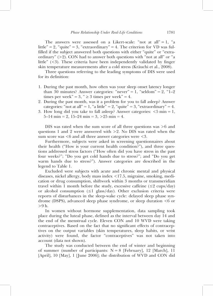
TABLE 1 Biological and sleep parameters (subjective characteristics)
Variable CON WVD P-value
Age (years) 25.00 ± 1.00 25.80 ± 1.00 .56BMI (kg/m2) 21.60 ± .50 20.40 ± .40 .07Health (Likert scale) 1.43 ± .11 1.95 ± .15 .008∗
Stress (Likert scale) 2.52 ± .19 1.80 ± 1.56 .005∗
Cold hands due to stress (Likert scale) 3.38 ± .19 2.32 ± .20 .004∗
Warm hands due to stress (Likert scale) 3.57 ± .15 3.17 ± .19 .09Time in bed before lights off (min) 14.60 ± 1.40 27.00 ± 2.30 .03∗
Lights off (h) 23.90 ± .30 24.00 ± .20 .63Sleep latency (min) 7.80 ± 1.10 29.20 ± 4.30 <.001∗
Sleep midpoint (h) 3.90 ± .20 4.20 ± .20 .30Sleep duration (h) 7.70 ± .20 7.40 ± .20 .26Lights on (h) 7.70 ± .20 7.80 ± .30 .65Time in bed after lights on (min) 24.60 ± 2.70 39.10 ± 7.20 .06Waking during night (number) .20 ± .04 0.40 ± .07 .005∗
Getting up during night (number) .06 ± .02 0.08 ± .02 .5Sleep quality (Likert scale) 5.80 ± .30 5.00 ± .20 .05Restorative sleep (Likert scale) 5.60 ± .30 5.00 ± .20 .11Time outdoors (h) 33.57 ± 5.11 21.11 ± 4.39 .68
Note. Values are means ± SEM over 1 week. p values are performed by Mann-Whitney U test andone-way ANOVA. Age, body mass index (BMI), health (1 = very good, 2 = good, 3 = moderate, 4 =bad, 5 = very bad), and stress factors (1 = frequent, 2 = sometimes, 3 = infrequent, 4 = never) weredetermined by screening questionnaires. Other variables were collected by the use of sleep-wake diaries.Sleep quality and restorative sleep were also measured by Likert scales (1 = very poor; 8 = very good).
B. Gompper et al.1782
data with their time stamps were then transferred to the statistic program(see below). “Proximal skin temperature” is defined as averaged skintemperatures of thigh, infraclavicular regions, and sternum. The meanskin temperatures of the hands and feet constitute the “distal skin temp-erature.” All other skin measurements are calculated as the mean of theleft and right side of the body.
Actimetric Recordings
Actimetry was used to monitor motor activity using Actiwatch®-Ldevices (Cambridge Neurotechnology, UK). Wrist activity of the non-dominant arm was recorded and stored in 1-min intervals, and dailysleep-wake logs were filled in by the subjects upon waking. Activity datawere edited so that gaps due to actigraph removal, e.g., when showering,were filled with activity counts from the 24-h-day average. If >3 h/day or1 h/night of data were missing, then the data of the whole day wereexcluded from further analysis. Sleep parameters were calculated for 18CON and 20 WVD by the Sleep and Activity Analysis Software 7.23V(Cambridge Neurotechnology). The Non-Parametric Circadian RhythmAnalysis (NPCRA; Van Someren et al., 1999), calculated the relativeamplitude of the 10-h of highest activity compared with the 5 h of leastactivity as a comprehensive representation of the amplitude of the sleep-wake cycles (CON N= 18; WVD N= 19).
Saliva Sampling for Melatonin Determination
For the determination of the dim light melatonin onset (DLMO),nine saliva samples were collected on one evening under dim light con-ditions (subjects had to wear sunglasses) using Salivettes® (Sarstedt,Nümbrecht, Germany). Samples were taken at 30-min intervals starting4 h before bedtime, and subjects were instructed to stay at home andavoid high activity this evening. Melatonin was assayed by a validatedradioimmunoassay (Bühlmann Laboratories, Allschwil, Switzerland(Weber et al., 1997). DLMO was determined by linear interpolation ofthe evening melatonin rise across a 3 pg/mL threshold (Pandi-Perumalet al., 2007). Three subjects collected melatonin samples after the ambu-latory week, but under similar life conditions. The data of two subjectswere excluded from statistical analyses due to lack of compliance.
Sleep-Wake Diaries
Subjects rated their sleep episode directly in their sleep-wake diaries,immediately after awakening. The items were bedtime, lights-off, esti-mated sleep onset latency, sleep disturbances (waking bouts, get-up times
Phase Relationship Under Real-Life Conditions 1783
during night), wake-up time (lights-on), and get-up time in the morning.Additionally, they rated their sleep quality, restorative sleep, andsleepiness before lights-off. The wake diary was divided into four timespans of the day: morning (06:00–12:00 h), afternoon (12:00–18:00 h),evening (18:00 h–sleep time), and shortly before lights-off. Estimationswere made on 100 mm bipolar visual analogue scales (VAS): alert–sleepy,relaxed–tense, concentrated–unfocused, satiated–hungry, and goodmood–bad mood (e.g., 0 mm= extremely alert, 100 mm= extremelysleepy). Additionally, self-ratings on bipolar VAS for thermal comfortwere estimated for different skin regions: feet, body, and hands (0 mm=feeling extremely cold, 100 mm= feeling extremely hot). Subjects notedtiming of activity, time spent outdoors, and time of stressful psychologicalsituations.
Statistical Analysis
The statistical packages StatView 5.0 (Abacus Concepts, Berkley, CA)and Statistica 7 for Windows (StatSoft, Tulsa, OK) were used. Statisticalanalyses were based on weekly averaged daily profiles per subject.In order to reduce short-term fluctuations and the number of time seg-ments, continuously recorded data were averaged in 30-min blocks. Thetime courses were statistically analyzed by two-way, repeated-measure-ments analysis of variance (ANOVA). Huynh-Feldt (H-F) statistics wereused to adjust the covariance matrix for violations of sphericity. H-F’sp values were based on corrected degrees of freedom, but the originaldegrees of freedom are reported. When the F ratio proved significant,Curran-Everret, multiple-range, post hoc tests were applied to locate sig-nificant differences between means (Curran-Everett, 2000). Results arereported in detail when the significance level was p< .05. Simple-groupdifferences between WVD and CON were calculated by factorial one-way ANOVA. Intraindividual phase relationships between feet skintemperatures and wrist activity were determined by cross-correlationanalyses, using time series over 7 days of 5-min binned data (seeKräuchi & Wirz-Justice, 1994; Kräuchi et al., 2006). Additionally, inter-individual phase relationships between feet skin temperatures and wristactivity were analyzed for WVD and CON. Mean CON values (N= 21)were taken as the reference curve for each subject. Time lags ofmaximum and minimum r-values were extracted from smoothed (by 65-min moving averaged) individual curves between time lags of ±960 min.Mean cross-correlation curves were calculated after Fisher z-transform-ation of r values, which were retransformed for Figure 3. The relationbetween the two variables was statistically tested by least-squares corre-lation analysis.
B. Gompper et al.1784
RESULTS
Subjects’ Characteristics
Table 1 summarizes the biological and sleep behavioral characteristicsof the study subjects. Age and body mass index were similar betweengroups. All 41 subjects were in good health, though WVD were slightlyworse than CON. WVD exhibited a higher level of stress and higheroccurrence of cold hands due to stress. With respect to sleep character-istics, WVD reported significantly more awakenings during the night,longer time in bed before lights-off, and longer sleep onset latency.Subjectively estimated and objectively measured sleep latencies (by wrist-actimetry) were significantly correlated ( p< .05).
Physiological Data Analyses
In a first step, the daily time course of skin temperatures and wristactivity of WVD and CON were analyzed according to clock time (solartime), neglecting possible differences in intra- and interindividual sleep-wake patterns, which was taken into account in a second step of analysiswith data adjustment to the individual sleep-wake cycle (percentilizeddata for sleep and wake phases, separately). This kind of analysis has thedisadvantage of expansion or contraction of the time axis, but gives a firstimpression of phase of entrainment with respect to the sleep-wake cycle.Thirdly, the phase relationships between the variables were calculated bycross-correlation analyses.
Daily Time Course of Clock Time (Adjusted Data)
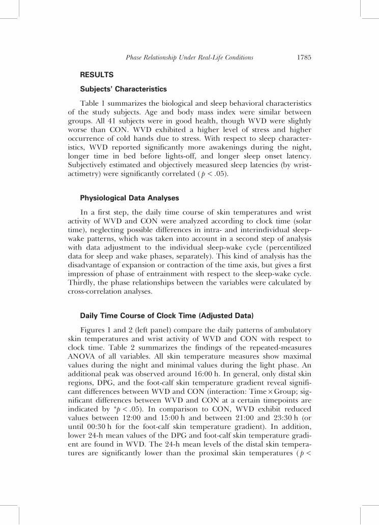
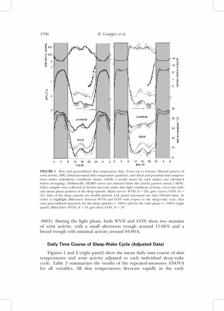
Figures 1 and 2 (left panel) compare the daily patterns of ambulatoryskin temperatures and wrist activity of WVD and CON with respect toclock time. Table 2 summarizes the findings of the repeated-measuresANOVA of all variables. All skin temperature measures show maximalvalues during the night and minimal values during the light phase. Anadditional peak was observed around 16:00 h. In general, only distal skinregions, DPG, and the foot-calf skin temperature gradient reveal signifi-cant differences between WVD and CON (interaction: Time ×Group; sig-nificant differences between WVD and CON at a certain timepoints areindicated by ∗p< .05). In comparison to CON, WVD exhibit reducedvalues between 12:00 and 15:00 h and between 21:00 and 23:30 h (oruntil 00:30 h for the foot-calf skin temperature gradient). In addition,lower 24-h mean values of the DPG and foot-calf skin temperature gradi-ent are found in WVD. The 24-h mean levels of the distal skin tempera-tures are significantly lower than the proximal skin temperatures ( p<
Phase Relationship Under Real-Life Conditions 1785
.0001). During the light phase, both WVD and CON show two maximaof wrist activity, with a small afternoon trough around 15:00 h and abroad trough with minimal activity around 04:00 h.
Daily Time Course of Sleep-Wake Cycle (Adjusted Data)
Figures 1 and 2 (right panel) show the mean daily time course of skintemperatures and wrist activity adjusted to each individual sleep-wakecycle. Table 3 summarizes the results of the repeated-measures ANOVAfor all variables. All skin temperatures decrease rapidly in the early
FIGURE 1 Raw and percentilized skin temperature data. From top to bottom: Diurnal pattern ofwrist activity, DPG (distal-proximal skin temperature gradient), and distal and proximal skin tempera-tures under ambulatory conditions (mean ±SEM; a weekly mean for each subject was calculatedbefore averaging). Additionally, DLMO curves are inserted below the activity pattern (mean± SEM).Saliva samples were collected at 30-min intervals under dim light conditions at home. Grey bars indi-cate mean phase position of the sleep episode. Black curves: WVD (N= 20); grey curves: CON (N=21). Data of the sleep episode are double plotted. Left panel represents raw data (30-min bins). Inorder to highlight differences between WVD and CON with respect to the sleep-wake cycle, datawere percentilized separately for the sleep episode (= 100%) and for the wake phase (= 100%) (rightpanel). Black lines: WVD, N= 19; grey lines: CON, N= 19.
B. Gompper et al.1786
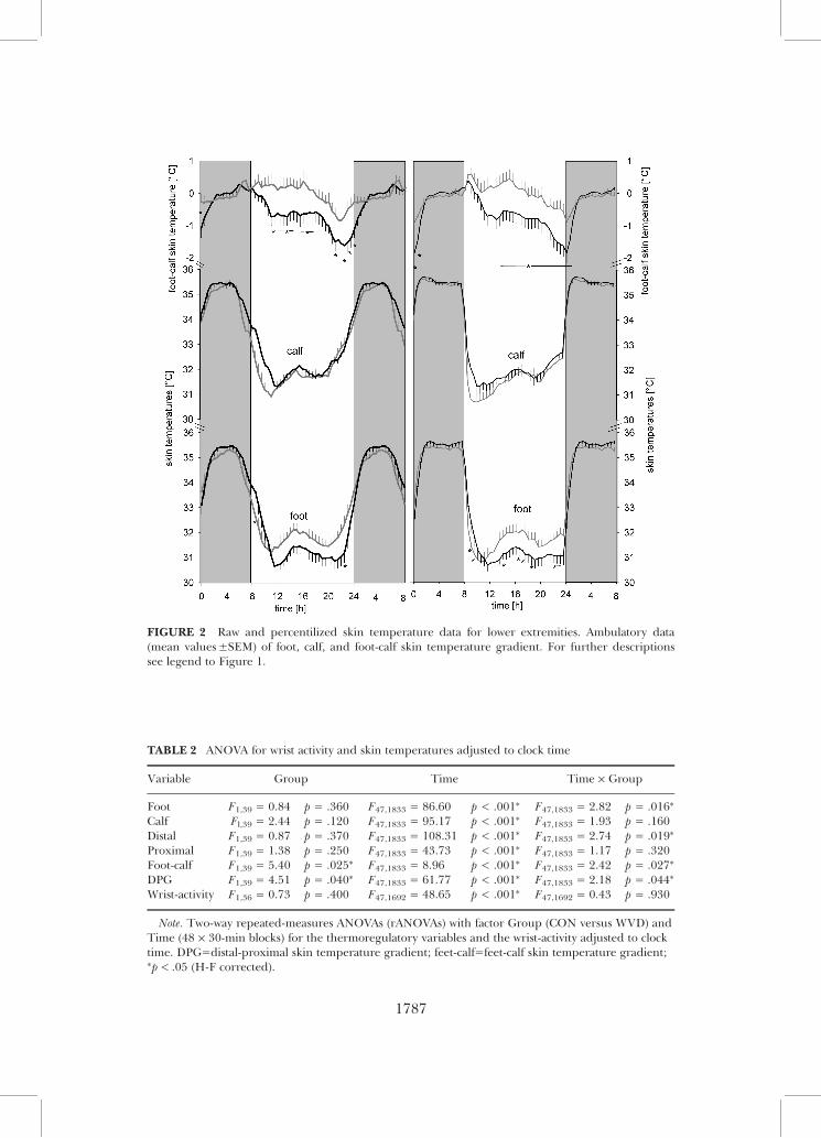
FIGURE 2 Raw and percentilized skin temperature data for lower extremities. Ambulatory data(mean values±SEM) of foot, calf, and foot-calf skin temperature gradient. For further descriptionssee legend to Figure 1.
TABLE 2 ANOVA for wrist activity and skin temperatures adjusted to clock time
Variable Group Time Time × Group
Foot F1,39 = 0.84 p = .360 F47,1833 = 86.60 p < .001∗ F47,1833 = 2.82 p = .016∗
Calf Fl,39 = 2.44 p = .120 F47,1833 = 95.17 p < .001∗ F47,1833 = 1.93 p = .160Distal F1,39 = 0.87 p = .370 F47,1833 = 108.31 p < .001∗ F47,1833 = 2.74 p = .019∗
Proximal F1,39 = 1.38 p = .250 F47,1833 = 43.73 p < .001∗ F47,1833 = 1.17 p = .320Foot-calf F1,39 = 5.40 p = .025∗ F47,1833 = 8.96 p < .001∗ F47,1833 = 2.42 p = .027∗
DPG F1,39 = 4.51 p = .040∗ F47,1833 = 61.77 p < .001∗ F47,1833 = 2.18 p = .044∗
Wrist-activity F1,36 = 0.73 p = .400 F47,1692 = 48.65 p < .001∗ F47,1692 = 0.43 p = .930
Note. Two-way repeated-measures ANOVAs (rANOVAs) with factor Group (CON versus WVD) andTime (48 × 30-min blocks) for the thermoregulatory variables and the wrist-activity adjusted to clocktime. DPG=distal-proximal skin temperature gradient; feet-calf=feet-calf skin temperature gradient;∗p< .05 (H-F corrected).
1787
morning after lights-on and, conversely, rise rapidly after lights-off.Around midday, WVD and CON differ significantly in distal, foot, foot-calf, and DPG skin temperatures (significant interaction: Time ×Group;significant differences between WVD and CON at certain time points areindicated by ∗p< .05. Proximal skin temperatures did not differ signifi-cantly between WVD and CON (middle panel). All skin temperaturemeasures show maximal and minimal values during the night and lightphase, respectively.
Actimetry revealed normally entrained sleep-wake rhythms withminimum movement during the sleep episode when skin temperaturesexhibit maximal values. Two peaks of daytime activity occurred, one atmidday and another in the evening. The time course of wrist activity wasnot significantly different in WVD compared to CON, and, in addition,the amplitude of daytime to nighttime activity in the percentilized datadid not reveal any difference between the two groups (314.3 ± 160.4versus 329.6 ± 160.4; F(1, 36) = 0.09, p = .773). The relative amplitude(RA) calculated from the 10 h of highest and 5 h of least activity, as a cir-cadian parameter, tended to be higher in WVD compared to CON (RA;0.88 ± 0.08 versus 0.92 ± 0.04; F(1, 36) = 3.3, p = .078).
Phase Relationship Analyses
The intraindividual phase relationships between foot skin tempera-tures and wrist activity were quantified by cross-correlation analysis(Figure 3, upper panel). In comparison to CON, WVD show an asym-metric cross-correlation curve, with distinct differences between lags −8and −1 h. Individual extracted times of minimum r values in CON reveala significant phase advance of foot skin temperatures with respect to wristactivity ( p< .05; phase of wrist activity was set as 0). Compared withCON, the phase position of WVD (wrist-foot correlation) is significantly
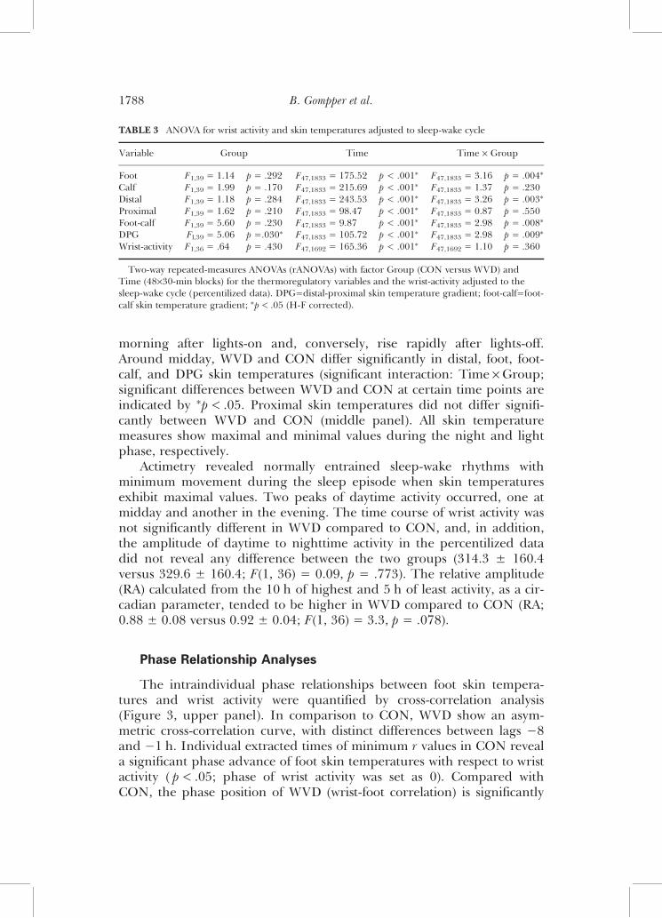
TABLE 3 ANOVA for wrist activity and skin temperatures adjusted to sleep-wake cycle
Variable Group Time Time × Group
Foot F1,39 = 1.14 p = .292 F47,1833 = 175.52 p < .001∗ F47,1833 = 3.16 p = .004∗
Calf F1,39 = 1.99 p = .170 F47,1833 = 215.69 p < .001∗ F47,1833 = 1.37 p = .230Distal F1,39 = 1.18 p = .284 F47,1833 = 243.53 p < .001∗ F47,1833 = 3.26 p = .003∗
Proximal F1,39 = 1.62 p = .210 F47,1833 = 98.47 p < .001∗ F47,1833 = 0.87 p = .550Foot-calf F1,39 = 5.60 p = .230 F47,1833 = 9.87 p < .001∗ F47,1833 = 2.98 p = .008∗
DPG Fl,39 = 5.06 p =.030∗ F47,1833 = 105.72 p < .001∗ F47,1833 = 2.98 p = .009∗
Wrist-activity F1,36 = .64 p = .430 F47,1692 = 165.36 p < .001∗ F47,1692 = 1.10 p = .360
Two-way repeated-measures ANOVAs (rANOVAs) with factor Group (CON versus WVD) andTime (48×30-min blocks) for the thermoregulatory variables and the wrist-activity adjusted to thesleep-wake cycle (percentilized data). DPG=distal-proximal skin temperature gradient; foot-calf=foot-calf skin temperature gradient; ∗p< .05 (H-F corrected).
B. Gompper et al.1788
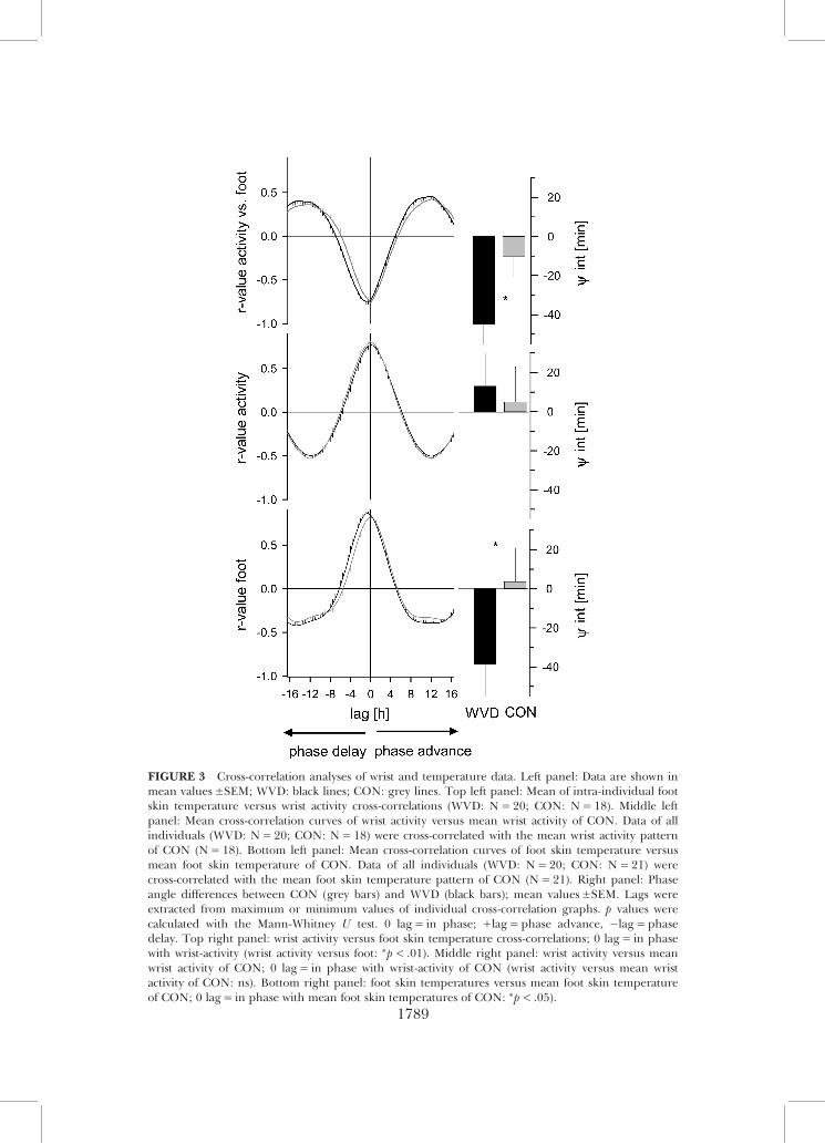
FIGURE 3 Cross-correlation analyses of wrist and temperature data. Left panel: Data are shown inmean values±SEM; WVD: black lines; CON: grey lines. Top left panel: Mean of intra-individual footskin temperature versus wrist activity cross-correlations (WVD: N= 20; CON: N= 18). Middle leftpanel: Mean cross-correlation curves of wrist activity versus mean wrist activity of CON. Data of allindividuals (WVD: N= 20; CON: N= 18) were cross-correlated with the mean wrist activity patternof CON (N= 18). Bottom left panel: Mean cross-correlation curves of foot skin temperature versusmean foot skin temperature of CON. Data of all individuals (WVD: N= 20; CON: N= 21) werecross-correlated with the mean foot skin temperature pattern of CON (N= 21). Right panel: Phaseangle differences between CON (grey bars) and WVD (black bars); mean values±SEM. Lags wereextracted from maximum or minimum values of individual cross-correlation graphs. p values werecalculated with the Mann-Whitney U test. 0 lag= in phase; +lag= phase advance, −lag= phasedelay. Top right panel: wrist activity versus foot skin temperature cross-correlations; 0 lag= in phasewith wrist-activity (wrist activity versus foot: ∗p< .01). Middle right panel: wrist activity versus meanwrist activity of CON; 0 lag= in phase with wrist-activity of CON (wrist activity versus mean wristactivity of CON: ns). Bottom right panel: foot skin temperatures versus mean foot skin temperatureof CON; 0 lag= in phase with mean foot skin temperatures of CON: ∗p< .05).
1789
phase delayed by 35 min ( p< .01). The cross-correlation curves of wristactivity are similar in WVD and CON (Figure 3, middle panel), and theextracted phases do not differ statistically. The cross-correlation curve offoot skin temperatures of WVD and CON reveal clear differences(Figure 3, lower panel). Compared with CON, the time course of footskin temperatures in WVD is significantly phase-delayed by 42 min ( p<.05). Taken together, the daily time course of foot skin temperatures inWVD is significantly phase delayed by 35–42 min with respect to wristactivity and CON.
Subjective Measures
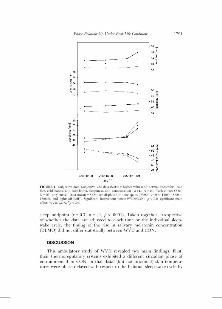
Sleepiness and concentration measured by VAS exhibit a significantinteraction (Time ×Group, p< .05; Figure 4), showing that WVD are lesstired and more concentrated than CON before lights-off. Morning ratingsshow an inverse pattern, leading to lower daily fluctuations in WVD.
WVD showed higher ratings of cold sensations than CON for all skinregions (Figure 4; main effect Group, p<. 02). Additionally, WVD exhib-ited a stronger sensation of cold feet than CON before sleep (Time ×Group, p< .02).
WVD did not differ from CON in subjective ratings of mood, tension,and feelings of hunger. All subjects were more hungry, more relaxed,and in better mood in the evening than morning.
Salivary Melatonin Concentrations in the Evening and DLMO
The time course of salivary melatonin concentrations is presented inFigure 1 for clock time (left panel, 19:00–23:00 h) and lights-off (sleep-wake cycle)—adjusted data (right panel). Statistical comparisons were per-formed by two-way, repeated-measures ANOVA. Neither clock time norsleep-wake–adjusted time courses reveal significant differences betweenWVD and CON. Clock-time–adjusted data for: Group, WVD versusCON, F(1, 37)= 2.00, p = .16, n.s.; Time, 9 timepoints, F(8, 296)=55.58, p< .0001; Time ×Group, F(8, 296)= 2.83, p = .067; p< .05 (H-Fcorrected). Lights-off adjusted data for: Group, WVD versus CON, F(1,37)= 0.01, p = .92, n.s.; Time, 9 timepoints, F(8, 296)= 66.47, p< .0001;Time ×Group, F(8, 296)= 0.34, p= .95, n.s.
Statistical analysis of the DLMO times reveals no differences betweenthe groups. Clock-time adjusted data: WVD versus CON, 21.54± 0.26versus 21.47± 0.28 h, F(1, 37)= 0.04, p= .852, n.s.; lights-off adjusteddata: WVD versus CON, 2.14± 0.21 versus 1.83± 0.23 h before lights-off, F(1, 37)= 0.99, p= .326, n.s.
DLMO correlated significantly with the phase of activity (r= .66, n=38, p< .0001), feet skin temperature (r= .66, n= 41, p< .0001), and
B. Gompper et al.1790
sleep midpoint (r= 0.7, n= 41, p< .0001). Taken together, irrespectiveof whether the data are adjusted to clock time or the individual sleep-wake cycle, the timing of the rise in salivary melatonin concentration(DLMO) did not differ statistically between WVD and CON.
DISCUSSION
This ambulatory study of WVD revealed two main findings. First,their thermoregulatory systems exhibited a different circadian phase ofentrainment than CON, in that distal (but not proximal) skin tempera-tures were phase delayed with respect to the habitual sleep-wake cycle by
FIGURE 4 Subjective data. Subjective VAS data (worse= higher values) of thermal discomfort (coldfeet, cold hands, and cold body), sleepiness, and concentration (WVD: N= 20, black curve; CON:N= 21, grey curve). Data (mean± SEM) are displayed in time spans (06:00–12:00 h; 12:00–18:00 h;18:00 h, and lights-off [loff]). Significant interaction: time ×WVD/CON, ∗p< .05; significant maineffect: WVD/CON, #p< .05.
Phase Relationship Under Real-Life Conditions 1791
about 40 min. Second, WVD, who complain of thermal discomfort withcold extremities, do, in fact, exhibit objectively measured cooler skintemperatures than CON, selectively at the extremities, and this occursthroughout the day, but not during the sleep episode. The two subjectgroups were selected with respect to VD and DIS. Therefore, furtherstudies including all four combinations of VD and DIS are required toelucidate whether it is VD and/or DIS that is essential for the phasedelay of distal skin temperature in WVD.
The discussion of the findings is structured into differences in phaseof entrainment and vascular regulation. In the last part, we try to eluci-date how the thermoregulatory system interacts with the circadian system.
Differences in Phase of Entrainment
The starting point of the present investigation was our recent con-stant-routine (CR) laboratory study showing parallel phase delays ofabout 1 h in all measured circadian variables (CBT, skin temperatures,DPG, salivary melatonin, and subjective ratings of sleepiness) in WVDwith respect to CON (Kräuchi, 2007). Similar to the present study, sleeptimes (time of lights-off and lights-on) did not differ between the twogroups. However, in contrast to the CR study, under ambulatory con-ditions only the diurnal pattern of DPG was phase delayed, whereasDLMO did not differ.
How can this discrepancy between CR and ambulatory conditions beexplained? The differences between these two protocols lay not only indifferences in body position, physical exercise, food intake patterns, andincreasing sleepiness (masking effects), but most importantly also in thelighting conditions. It is well known that overt circadian rhythms, includ-ing that of melatonin, are generated by the circadian pacemaker systemlocalized in the SCN, which is efficiently entrained by light (for reviewsee Dijk & von Schantz, 2005). Subjects in a CR experience a <8-luxenvironment that allows the measurement of unmasked circadian phaseand amplitude. In the real world, they are exposed to much brighterlight intensities at variable times of the day, depending on their work andfree-time habits. Thus, the masking and entraining effects of light may beresponsible for the similar phases of WVD and CON in DLMO, wristactivity, and proximal skin temperature under ambulatory conditions.Only the phase delay in distal skin temperature and the diurnal vasocon-striction remained also during ambulatory conditions. Thus, we can linkthe delayed sleep onset latency to the delayed onset of vasodilatation inthe evening and not to a phase shift of the circadian clock.
Many studies have shown that the distal heat loss regulatory systemdoes not act unidirectionally on sleep initiation. All kinds of relaxation be-havior prior to sleep, whether muscular or psychological in nature (e.g.,
B. Gompper et al.1792
lying down, sleep initiation rituals), trigger a pronounced increase indistal vasodilatation. All temperatures increase rapidly after lights-off,whether under ambulatory or CR conditions (Kräuchi et al., 1999; Lack& Gradisar, 2002). Such a feedback regulatory system can be used thera-peutically for sleep onset disturbances, e.g., by applying bed socks, auto-suggestion of warmth, autogenic training, and yoga (Kräuchi et al., 1997,1999).
In the present study, we also found that WVD reported lower sleepquality and more awakenings during sleep than CON, which could haveinfluenced alertness the following morning. Indeed, WVD showed a ten-dency to stay longer in bed and to be sleepier in the morning, bothindicative of a more pronounced sleep inertia process (Kräuchi et al.,2006). This finding fits together with the longer duration of the distalvasoconstriction process in the morning in WVD; the disappearance ofsleep inertia has been shown to correlate with the rate of distal vasocon-striction (Kräuchi et al., 2004). WVD rated not only more sleepiness inthe morning but also less sleepiness in the evening (Figure 4), whichmight indicate a phase delay of the circadian sleep propensity rhythm aspreviously found in a CR study (Vollenweider et al., 2008). Thisinterpretation is in accord with previous findings showing a close relation-ship between the circadian rhythm of sleepiness and distal vasodilatation(Kräuchi et al., 2006; Lack et al., 2008).
Differences in Vascular Regulation
Our study revealed interesting findings with respect to circadianthermoregulation. The daily subjective ratings of cold sensation through-out the day confirmed the global rating of the screening instrument.WVD had higher ratings of cold sensation, most pronounced in distalskin regions, especially their feet, before lights-off. This finding providesinformation about the status of the thermoregulatory systems of WVDand CON. All subjects usually try to reach a state of thermal comfort,which requires a thermoregulatory system in equilibrium. Therefore, rec-ognition of thermal discomfort is claimed to be an indication of a devi-ation in the subjects’ thermoregulatory “set point” (Cabanac, 1969). WVDnot only feel that they have cooler distal skin regions while awake, butthey, indeed, do have cooler measured distal skin temperatures thanCON. Interestingly, both groups had similar proximal skin temperatures,leading, therefore, to a clear distinction between the two groups in DPGduring the wake phase. The skin temperatures of the calf and foot, andtheir difference, may provide an additional insight into thermoregulatoryheat loss mechanisms via the lower legs. The calf skin temperatureexhibits its lowest values in the morning, increasing thereafter, withthe exception of a small trough in the afternoon, to high values before
Phase Relationship Under Real-Life Conditions 1793
lights-off. Even though the spatial distance between the temperatureprobes of the calf and foot was only about 25 cm, the temperature differ-ence between them during the wake phase was quite large (average:0.73°C in WVD). This indicates that effective thermoregulatory mechan-isms have to be activated to induce such a large gradient in skin tempera-tures within this short distance, e.g., closing of AVAs and countercurrentback stream of blood via inner veins. Within this context, it is noteworthythat the parallel bimodal pattern of proximal and distal skin temperaturesoccurring during the daytime, with maximum values around 16:00 h, areprobably not of thermoregulatory origin and suggest a behavioral one(e.g., food intake). Such a bimodal pattern of ambulatory distal skintemperature has been previously documented (Sarabia et al., 2008).
Remarkable is that WVD exhibit a slightly higher life stress level thanCON, and a higher propensity to react with cold hands in stressful situ-ations. Further support for a higher stress sensitivity in WVD comes fromother analyses where we have shown that WVD in comparison to CONexhibit reduced parasympathetic activity and increased sympathovagalbalance, as derived from heart rate variation (HRV) spectral analyses(Anders et al., 2010), and a higher anger-turned-inwards level (Von Arbet al., 2009), which in turn could be responsible for the increased sym-pathovagal balance and distal vasoconstriction.
Analysis of percentilized data (Figures 1 and 2, right panel) illustrateshow vasodilatation and vasoconstriction are tightly linked to the sleep-wake cycle after lights-off and lights-on. In both WVD and CON, distalskin temperatures reached their minimum and maximum levels later inthe morning and night, respectively, than proximal skin temperatures,confirming the findings under controlled CR conditions (Kräuchi et al.,2006). As a consequence, distal skin regions exhibited a 2–3°C lowerdiurnal temperature level than proximal skin regions. The thermoregula-tory difference between WVD and CON lies rather in the different temp-erature levels, i.e., lower DPG levels during the diurnal phase in WVD,than in the kinetics of the distal thermoregulatory heat loss system.
In conclusion, WVD live with a larger thermoregulatory “shell”during the wake phase resulting in increased body heat retention, a stateoccurring when the thermoregulatory “set levels” are not reached.Increased distal vasoconstriction may be caused by their documentedelevated life stress level. Having vasoconstricted distal skin regions beforelights-off are known to prolong sleep onset latency. The demonstratedphase delayed distal skin temperature rhythm with respect to the sleep-wake cycle in WVD could also be caused by lower diurnal distal skintemperatures. It should be noted that we studied a healthy populationwhose delayed sleep onset could be linked to the above thermoregulatorydisturbance, while maintaining similar circadian phase as controls. Sleep-onset disturbances in the clinic, however, have a variety of origins; for
B. Gompper et al.1794
example, some could be related to thermoregulation and stress andothers to delayed internal clock time. What we have shown is, in thismodel population, that the degree of vascular dysregulation of a subjectmay represent a crucial thermophysiological property that is necessaryfor initiating sleep.
ACKNOWLEDGMENTS
This work was supported by the Swiss National Science FoundationGrant SNF 3100A0-102182/1 (to K. Kräuchi), and the Schwickert-Stiftung. The authors are grateful to Anna Wirz-Justice for her helpfulcomments on the manuscript. The authors are grateful to Anna Wirz-Justice for her helpful comments on the manuscript and Sarah Chellapafor her linguistic corrections. Furthermore, the authors acknowledgeClaudia Renz, Marie France Dattler, and Giovanni Balestrieri for theirtechnical aid.
Declaration of Interest: The authors report no conflicts of interest.The authors alone are responsible for the content and writing of thepaper.
REFERENCES
Anders D, Vollenweider S, Cann J, Hofstetter M, Flammer J, Orgül S, Kräuchi K. (2010). Heart ratevariability in women during 40 hour prolonged wakefulness. Chronobiol. Int. 27:1609–1628.
Aschoff J. (1971). Temperaturregulation. In Gauer OH, Kramer K, Jung R (eds). Energiehaushalt undTemperaturregulation. Physiologie des Menschen. München: Urban und Schwarzenberg, 43–112.
Aschoff J, Heise A. (1972). Thermal conductance in man: its dependence on time of day and ofambient temperature. In Itoh S, Ogata K, Yoshimura H (eds) Advances in climatic physiology.Tokyo: Igako Shoin, 334–348.
Cabanac M. (1969). Plaisir ou deplaisir de la sensation thermique et homeothermie. Physiol. Behav.4:359–364.
Campbell SS, Broughton RJ. (1994). Rapid decline in body temperature before sleep: fluffing thephysiological pillow? Chronobiol. Int. 11:126–131.
Curran-Everett D. (2000). Multiple comparisons: philosophies and illustrations. Am. J. Physiol. Regul.Integr. Comp. Physiol .279:R1–R8.
Dijk D, von Schantz M. (2005). Timing and consolidation of human sleep, wakefulness, and perform-ance by a symphony of oscillators. J. Biol. Rhythms 20:279–290.
Flammer J, Pache M, Resink T. (2001). Vasospasm, its role in the pathogenesis of diseases with par-ticular reference to the eye. Prog. Retin. Eye Res. 20:319–349.
Hales JRS. (1985). Skin arteriovenous anastomoses, their control and role in thermoregulation.In Johansen K, Burggren WW (eds). Cardiovascular shunts. Alfred Benzon Symposium,Copenhagen: Munksgaard, 433–451.
House JR, Tipton MJ. (2002). Using skin temperature gradients or skin heat flux measurements todetermine thresholds of vasoconstriction and vasodilatation. Eur. J. Appl. Physiol. 88:141–145.
Kräuchi K. (2007). The thermophysiological cascade leading to sleep initiation in relation to phase ofentrainment. Sleep Med. Rev. 11:439–451.
Kräuchi K, Wirz-Justice A. (1994). Circadian rhythm of heat production, heart rate, and skin andcore temperature under unmasking conditions in men. Am. J. Physiol. 267:R819–R829.
Kräuchi K, Cajochen C,Wirz-Justice A. (1997). A relationship between heat loss and sleepiness: effectsof postural change and melatonin administration. J. Appl. Physiol. 83:134–139.
Phase Relationship Under Real-Life Conditions 1795
Kräuchi K, Cajochen C, Werth E, Wirz-Justice A. (1999). Warm feet promote the rapid onset ofsleep. Nature 401:36–37.
Kräuchi K, Cajochen C, Werth E, Wirz-Justice A. (2000). Functional link between distal vasodilationand sleep-onset latency? Am. J. Physiol. Regul. Integr. Comp. Physiol. 278:R741–R748.
Kräuchi K, Cajochen C, Wirz-Justice A. (2004). Waking up properly: is there a role of thermoregula-tion in sleep inertia? J. Sleep Res. 13:121–127.
Kräuchi K, Knoblauch V, Wirz-Justice A, Cajochen C. (2006). Challenging the sleep homeostat doesnot influence the thermoregulatory system in men: evidence from a nap vs. sleep-deprivationstudy. Am. J. Physiol. Regul. Integr. Comp. Physiol. 290:R1052–R1061.
Kräuchi K, Fontana Gasio P, Vollenweider S, Von Arb M, Dubler B, Orgül S, Flammer J, Stutz E.(2008). Cold extremities and difficulties initiating sleep: evidence of co-morbidity from a randomsample of a Swiss urban population. J. Sleep Res. 17:420–426.
Lack L, Gradisar M. (2002). Acute finger temperature changes preceding sleep onsets over a 45-hperiod. J. Sleep Res. 11:275–282.
Lack L, Gradisar M, Van Someren EJW, Wright HR, Lushington K. (2008). The relationshipbetween insomnia and body temperatures. Sleep Med. Rev. 12:307–317.
Pandi-Perumal SR, Smits M, Spence W, Srinivasan V, Cardinali DP, Lowe AD, Kayumov L. (2007).Dim light melatonin onset (DLMO): a tool for the analysis of circadian phase in human sleepand chronobiological disorders. Prog. Neuropsychopharmacol. Biol. Psychiatry 31:1–11.
Portaluppi F, Touitou Y, Smolensky M. (2008). Ethical and methodological standards for laboratoryand medical biological rhythm research. Chronobiol. Int. 25:999–1016.
Roenneberg T, Daan S, Merrow M. (2003a). The art of entrainment. J. Biol. Rhythms 18:183–194.Roenneberg T, Wirz-Justice A, Merrow M. (2003b). Life between clocks: daily temporal patterns of
human chronotypes. J. Biol. Rhythms 18:80–90.Rubinstein E, Sessler D. (1990). Skin-surface temperature gradients correlate with fingertip blood
flow in humans. Anesthesiology 73:541–545.Sarabia J, Rol M, Mendiola P, Madrid J. (2008). Circadian rhythm of wrist temperature in normal-
living subjects A candidate of new index of the circadian system. Physiol. Behav .95:570–580.Severens NM, van Marken Lichtenbelt WD, Frijns AJ, Kingma BR, de Mol B A, van Steenhoven A A.
(2010). Measurement of model coefficients of skin sympathetic vasoconstriction. Physiol. Meas.31:77–93.
Smith AD, Crabtree DR, Bilzon JL, Walsh NP. (2010). The validity of wireless iButtons and thermis-tors for human skin temperature measurement. Physiol. Meas. 31:95–114.
Van Marken Lichtenbelt WD, Daanen HA, Wouters L, Fronczek R, Raymann RJ, Severens NM,Van Someren EJ. (2006). Evaluation of wireless determination of skin temperature usingiButtons. Physiol. Behav. 88:489–497.
Van Someren EJ, Swaab DF, Colenda CC, Cohen W, McCall WV, Rosenquist PB. (1999). Bright lighttherapy: improved sensitivity to its effects on rest-activity rhythms in Alzheimer patients by appli-cation of nonparametric methods. Chronobiol. Int. 16:505–518.
Vollenweider S, Wirz-Justice A, Flammer J, Orgül S, Kräuchi K. (2008). Chronobiological characteriz-ation of women with primary vasospastic syndrome: body heat loss capacity in relation to sleepinitiation and phase of entrainment. Am. J. Physiol. Regul. Integr. Comp. Physiol. 294:R630–R638.
Von Arb M, Gompper B, Meyer AH, Stutz EZ, Orgül S, Flammer J, Kräuchi K. (2009). Relationshipbetween gender role, anger expression, thermal discomfort and sleep onset latency in women.Biopsychosoc. Med. 3:1–7.
Weber J, Schwander J, Unger I, Meier D. (1997). A direct ultrasensitive RIA for the determination ofmelatonin in human saliva: comparison with serum levels. Sleep Res. 26:A112–A113.
Werner J. (2010). System properties, feedback control and effector coordination of human tempera-ture regulation. Eur. J. Appl. Physiol. 109:13–25.
Wever R. (1979). The circadian system of man—results of experiments under temporal isolation. New York:Springer Verlag.
B. Gompper et al.1796