Embed Size (px)
Citation preview

Phenotypic and Molecular Identification of SporothrixIsolates from an Epidemic Area of Sporotrichosis in Brazil
Manoel Marques Evangelista Oliveira • Rodrigo Almeida-Paes •
Mauro Medeiros Muniz • Maria Clara Gutierrez-Galhardo •
Rosely Maria Zancope-Oliveira
Received: 26 January 2011 / Accepted: 30 May 2011 / Published online: 24 June 2011
� Springer Science+Business Media B.V. 2011
Abstract Sporotrichosis has significantly increased
in Brazil in the last decade, particularly in the state of
Rio de Janeiro, with the occurrence of an epidemic
related to zoonotic transmission from cats to humans.
Recently, four new phylogenetic species were incor-
porated into the Sporothrix species complex based on
the phenotypic and molecular characteristics, and a
new species name (Sporothrix brasiliensis) was
proposed for some of the Sporothrix isolates from
this epidemic. This study describes the characteriza-
tion of 246 isolates obtained from patients attending
the Laboratory of Infectious Dermatology, IPEC-
FIOCRUZ, between 1998 and 2008, together with
one environmental sample. Two hundred and six of
the isolates (83.4%) were characterized as S. brasil-
iensis, 15 (6.0%) as S. schenckii, and one (0.5%) as
S. mexicana. Twenty-five isolates (10.1%) could not
be identified according to their phenotype and were
classified as Sporothrix spp. The calmodulin gene
was sequenced to confirm the identity of these
isolates. The molecular analysis demonstrated that
24 of the isolates were S. brasiliensis, with the
remainder being a S. globosa isolate. The isolate
characterized phenotypically as S. mexicana was
clustered on the S. schenckii clade. The correlation
between molecular data and phenotypic characteris-
tics described in this study is fundamental to the
identification of the Sporothrix complex.
Keywords Sporothrix species complex �Sporotrichosis � Rio de Janeiro � Calmodulin gene �Identification � Taxonomy
Introduction
Sporotrichosis is a subcutaneous mycosis with a
global distribution, with areas of high endemicity in
Latin America [1–3]. This infection is caused by the
dimorphic fungus previously described as the single
species Sporothrix schenckii, which is associated in
the environment with plants and soil [4]. In humans,
sporotrichosis is a disease that usually occurs in the
form of isolated cases or small outbreaks, involving
people exposed to plants or soil rich in organic matter
such as farmers, florists, gardeners and miners, and
occasionally laboratory technicians [5, 6].
Pathogenic fungi of the genus Sporothrix are dimor-
phic; in the environment or when cultured in the
M. M. E. Oliveira � R. Almeida-Paes �M. M. Muniz � R. M. Zancope-Oliveira (&)
Laboratorio de Micologia, Instituto de Pesquisa Clınica
Evandro Chagas, Fundacao Oswaldo Cruz, Rio de Janeiro,
RJ 21045-900, Brazil
e-mail: [email protected]
M. C. Gutierrez-Galhardo
Laboratorio de Pesquisa Clınica em Dermatologia
Infecciosa, Instituto de Pesquisa Clınica Evandro Chagas,
Fundacao Oswaldo Cruz, Rio de Janeiro, RJ, Brazil
123
Mycopathologia (2011) 172:257–267
DOI 10.1007/s11046-011-9437-3

laboratory at 25–30�C, in the filamentous form they
present as hyaline, septate, branched hyphae with
single-celled conidia of two types—hyaline and brown
(dematiaceous). The hyaline conidia are small, ovoid
and usually occur in the apical portion of conidiophores.
The dematiaceous conidia are large and ovoid, with a
thick cell wall and are present along the entire length of
the hyphae [7]. Macroscopically, filamentous S. sche-
nckii colonies are initially white and gradually become
brown to dark black, as conidia are produced by the
fungus [8]. In contrast, in the parasitic phase or when
cultured in appropriate culture media at 35–37�C, S.
schenckii appears as cigar-shaped oval yeast cells that
may have one or more buds [9, 10]. Macroscopically
colonies of S. schenckii grown at 378C have a yellowish-
beige color with a creamy aspect [11, 12].
Rio de Janeiro State in Brazil is an epidemic area for
sporotrichosis; thus, in a study conducted from 1998 to
2004, Schubach and co-workers [13, 14] identified 759
cases of human sporotrichosis, 1,503 of feline sporotri-
chosis, and sixty-four cases of canine S. schenckii
infection. 83.4% of the human infections were associ-
ated with prior contact with cat sporotrichosis cases
[14]. This differs from other worldwide sporotrichosis
outbreaks that have been associated with infection via a
plant source containing the fungus, rather than by
domestic cats infected with S. schenckii [14, 15]. In the
Rio de Janeiro outbreak, the cutaneous-lymphatic form
of the disease was most common, followed by localized
and disseminated cutaneous forms. Uncommon mani-
festations of the disease involving nasal and conjunc-
tival mucosa [13, 14] as well hypersensitivity
manifestations, such as erythema nodosum and ery-
thema multiform [16, 17], were also noted.
Marimon et al. [18] have recently suggested on the
basis of a combination of phenotypic and genetic
features that S. schenckii should not be considered a
single taxon, which causes sporotrichosis, and have
identified three new species, S. brasiliensis, S.
globosa, and S. mexicana. S. globosa has been
defined as having a worldwide distribution [19, 20],
whereas S. mexicana is apparently restricted to
Mexico, and S. brasiliensis to Brazil. Recent work
by the same group has also suggested that S.
schenckii var. luriei should be promoted to a new
species, S. luriei [21]. Additionally, other phyloge-
netic analysis based on rDNA and b-tubulin regions
from S. albicans, S. pallida, and S. nivea revealed a
significant similarity, and there is a proposition that
all the three species were called as S. pallida, the first
of three species first described [22]. An identification
key for the Sporothrix complex has now been
proposed [18] that includes analysis of conidial
morphology, auxonogram analysis using raffinose
and sucrose, and genotyping via polymerase chain
reaction (PCR) amplification of the calmodulin gene.
Based on this last analysis, Romeo and collaborators
(2011) studying the molecular phylogeny and epide-
miology of S. schenckii species complex isolated in
Italy demonstrated that twenty-six environmental
strains co-clustered with S. albicans, and two clinical
isolates grouped with S. schenckii stricto sensu [24].
The objective of this study was characterizing, at
the species level, Sporothrix strains isolated during
the course of the Rio de Janeiro sporotrichosis
epidemic, and maintained in our fungal collection,
using the newer methods of taxonomic analysis
developed by Marimon et al. [18], and evaluate the
accuracy of the classic methodologies applied in the
Sporothrix identification.
Materials and Methods
Strains
This study was approved by the Research Ethics
Committee of IPEC/Fiocruz. Two hundred and forty-
seven isolates from the Fungal Culture Collection of
the Laboratorio de Micologia/IPEC/Fiocruz, two
control strains (IOC 1113, IOC 1824) isolated on
1926 and 1984, respectively, prior to the Rio de
Janeiro State sporotrichosis epidemic, and S. brasil-
iensis CBS 120339 (former IPEC16490) type strain
characterized by Marimon et al. [19]were included in
this study—each had been characterized previously
as S. schenckii by traditional morphological identifi-
cation methods [22]. Two hundred and forty-six of
the isolates were from human sporotrichosis patients
identified between 1998 and 2008 and the remainder
was isolated from the environment. All strains were
previously identified as S. schenckii.
Fungal Re-isolation and Phenotypic
Characterization
Filamentous fungal colonies grown on Sabouraud
Dextrose Agar were visually examined using
258 Mycopathologia (2011) 172:257–267
123

Lactophenol Cotton Blue (Fluka Analyted, France),
and slide cultures were prepared as previously
described for Sporothrix identification [22]. Dimor-
phism was demonstrated by conversion to the yeast-
like form on Brain Heart Infusion (BHI) agar medium
(DifcoTM Becton, Dickinson and Company/Sparks,
MD 21152 USA) for 7 days at 37�C. In order to study
and confirm macroscopic features and sporulation,
isolates were sub cultured on potato dextrose agar
plates (PDA—DifcoTM Becton, Dickinson and Com-
pany/Sparks, MD 21152 USA) and Corn Meal Agar
slants (BBLTM Becton, Dickinson and Company/
Sparks, MD 21152 USA) and incubated at 30�C in
the dark. After 10–12 days, the microscopic features
were determined. Colony diameter on PDA was
determined after 21 days according to the previously
described protocol [18, 20]. To check growth at 37�C,
the strains were grown on PDA and incubated at 37�C
for 3 weeks. Carbohydrate assimilation tests were
performed using freshly prepared yeast nitrogen base
(YNB) medium (DifcoTM Becton, Dickinson and
Company/Sparks, MD 21152 USA) and tested for
sucrose and raffinose using the technique described
previously protocol [18, 20]. Cultures on YNB sup-
plemented with glucose were used as positive control
for growth and YNB without carbohydrates was used
as a negative control. Experiments were performed at
least three times on different days and, in case of
discordant results, repeated two additional times.
Molecular Identification
Genomic DNA was extracted and purified from
Sporothrix spp mycelial phase by phenol/chloro-
form/isoamyl alcohol method as previously described
[25]. For the partial sequencing of the nuclear
calmodulin (CAL) gene, we used the specific condi-
tions described by Oliveira et al. [20]. Briefly, the
PCR mix consisted of MgCl2 2 mM (Invitrogen,
USA), dNTP mix 200 lM (Invitrogen, USA), 2.5U
Taq Gold DNA polymerase (Applied Biosystems,
USA), 30 pmol of each primer CL1 (50-GA(GA)T
(AT)CAAGGAGGCCTTCTC-30), and CL2A (50-TT
TTTGCATCATGAGTTGGAC-30) [20, 23]. In the
PCR reaction, the annealing temperature was 60�C.
Automated sequencing was done using the Sequenc-
ing Platform at Fundacao Oswaldo Cruz—PDTIS/
FIOCRUZ, Brazil [26]. Sequences from both DNA
strands were generated and edited with the
Sequencher ver. 4.6 software package (Genes Codes
Corporation, USA), followed by alignment by means
of the Mega version 4.0.2 software. The sequences of
our strains were compared by BLAST (Basic Local
Alignment Search Tool- NIH) with sequences avail-
able from NCBI GenBank (Sporothrix AM 398382.1/
AM 398393.1/AM 117444.1/AM 116899.1/AM
116908.1). All phylogenetic analyses were performed
based on Tamura et al. [27], using MEGA vers. 4.0.2
(http://www.megasoftware.net/). The phylogenetic
relationships among isolates were evaluated from tree
topologies: (1) the Neighbor-joining (NJ) algorithm
[28] was generated using the genetic distances model
of Kimura 2-parameter [29] with pair-wise deletion
(gaps/missing data) and uniform rates among sites;
(2) the Maximum parsimony (MP) method [29] was
employed to infer trees using the close-neighbor-
interchange algorithm [30]. A Bootstrap test [31]
with 1,000 replicates was conducted for both NJ and
MP analyses.
Statistical Analyses
All data were subjected to statistical analysis with the
use of GraphPad PRISM vers. 4.00, San Diego, CA (
http://www.graphpad.com/quickcalcs/) and Statistical
Package for Social Science (SPSS�) vers. 11.0,
Chicago, IL, EUA. P values (\0.005) were calculated
by analysis of variance, chi-square test, or McNe-
mar’s correlation analysis with 95% of confidence
interval (CI) and 5% significance level.
Nucleotide sequence accession numbers. All
sequences from isolates included in genotypic analysis
were deposited in the GenBank database under acces-
sion numbers GU456632, HQ426928 to HQ426962.
Results
Phenotypic characterization. All fungal isolates
included in the study exhibited similar macroscopic
morphology in their filamentous form. After 21 days
of incubation, at 30�C on PDA, white, brown, or
black colonies with velvety or glabrous texture were
observed (data not shown). Pigmented colonies were
not seen in only three Sporothtrix isolates (IPEC
16919, IPEC 27022, and IPEC 27087). Microscopic
examination of these colonies revealed thin, hyaline,
and septate hyphae. All 247 isolates when cultured on
Mycopathologia (2011) 172:257–267 259
123

Corn Meal Agar for 12 days at 30�C developed
interim or terminal conidia in sympodial conidio-
phores along the hyphae. These conidia were hyaline
or pigmented, usually globose to subglobose or pear
shaped. In addition, sessile conidia were also
observed, which were dark, thick-walled, and con-
nected individually throughout the hyphae via tiny
denticles. Globose, elongated or triangular (Fig. 1),
and dematiaceous conidia were predominantly
observed in our isolates (Table 1). The IPEC
17692, IPEC 18782B, and IPEC 28790 strains
(1.2%) produced just hyaline, globose to subglobose,
or pear shaped conidia, and three other isolates, IPEC
25406, IPEC 28329, and IPEC 28487 (1.2%) dem-
onstrated elongated and pigmented conidia. Triangu-
lar, pigmented conidia characteristic of S. schenckii
were seen on five strains (Table 1).
The mean diameter of the colonies together with
statistical measures of dispersion was also evaluated
in our study (Table 1). Overall, the best fungal growth
was at 30�C, with minimum colony diameter ranging
from 9 to 14 mm, and maximum from 42 to 51 mm at
this temperature. Colony growth at 37�C was lower
than that observed at 30�C, with 2–3 mm diameter
minima and maxima of 14–20 mm. The mean colony
diameter at 30�C was higher in all strains studied
when compared with that at 37�C (Table 1).
After 10 days, glucose was assimilated by all
isolates tested including the controls. Fifteen isolates
(6%) also assimilated sucrose and raffinose, and 22
isolates (8.9%) only assimilated sucrose and glucose.
The remaining 84.7% (n = 210) assimilated only
glucose. None of the isolates assimilated only raffi-
nose and none was able to grow in the control without
a carbohydrate source. The biochemical profile of
Sporothrix isolates that assimilated sucrose only was
assessed and repeated five times using five samplings
on five consecutive days of each strain in order to
evaluate the occurrence of variable sugar assimilation.
In all evaluations, Sporothrix isolates presented the
same biochemical profile, just assimilating sucrose.
Table 1 shows the phenotypic characteristics
observed in each species. Of the total, 25 (10.1%)
strains could not be identified according to the proposed
identification key previously reported [18]. Twenty-two
thermotolerant isolates (8.9%) with dematiaceous
conidia, diameter of the colonies not exceeding
50 mm, on PDA at 30�C after 21 days of incubation
presented biochemical profile of assimilation for species
S. globosa or S. albicans. In the previous species
definition, [18] S. globosa is described as being not
thermotolerant, and S. albicans typically produces
colonies[50 mm, and does not produce dematiaceous
conidia (Table 1). In the other group of strains, 1.2% of
our isolates (IPEC 17692, IPEC 28790, and IPEC
18782B) did not produce dematiaceous conidia, grew
colonies less than 50 mm, showed growth at 37�C, and
did not assimilate raffinose and sucrose (Table 1). This
carbohydrate assimilation profile is characteristic of S.
brasiliensis, whereas the conidial structure is typical of
S. albicans.
The conidia type produced by different species of
the genus Sporothrix included in this study is also
shown in Table 1. Two hundred and two isolates of S.
brasiliensis (98%) showed dematiaceous and oval
conidia and two isolates (1.0%) triangular conidia.
Most isolates (60%) characterized as S. schenckii
produced oval conidia without the presence of
dematiaceous and triangular conidia, which accord-
ing to studies by Marimon et al. [18] could be a
peculiar feature of the strains characterized as
belonging to the species S. schenckii. Another isolate,
also characterized as S. schenckii, produced oval,
elongated, and pigmented conidial morphology. The
correlation among the isolates characterized as S.
schenckii with the presence of triangular conidia was
performed using the statistics test McNemar’s and
was not statistically significant (P = 0.0961). Just
one isolate (IPEC 27722) was characterized as S.
mexicana based on the key proposed previously [18],
since dematiaceous and oval conidia were observed.
The correlation among colony growth on PDA at
30�C and 37�C for 21 days on our isolates is shown
in Fig. 2. The isolate characterized as S. mexicana
had the largest diameter at 30�C and lowest at 37�C.
The growth of S. schenckii isolates was higher at
37�C when compared to S. brasiliensis, although
statistical differences (P = 0.57) were not observed.
S. brasiliensis isolates and the Sporothrix spp. were
not identified at the species level and demonstrated
similar colony growth at 30�C (P = 0.79) and 37�C
(P = 0.58). The comparison of growth at 30�C of S.
schenckii and S. brasiliensis isolates showed statis-
tically significant differences (P = 0.012).
The phenotypic tests of type strain CBS120339
(IPEC 16490) characterized as S. brasiliensis by
Marimon et al. [18] were re-assessed. The colonies on
PDA attained a diameter of 13 mm at 30�C and of
260 Mycopathologia (2011) 172:257–267
123

35 mm 37�C after 21 days of incubation and were
unable to assimilate sucrose and raffinose. These
phenotypic data are characteristic of S. brasiliensis.
However, it was found microscopically that this
strain produced triangular, dematiaceous conidia, in
addition to the globose to subglobose, elongated,
dematiaceous conidia (Fig. 1). All control strains
from Brazil (IOC1113 and IOC1824) produced
colonies on PDA with diameters of 45 and 38 mm
at 30�C, and 10 and 9 mm at 37�C, with triangular
dematiaceous conidia in all strains. All assimilated
sucrose and raffinose. These phenotypic characteris-
tics are consistent with the species S. schenckii.
Molecular Analysis. Molecular assessment was
used to identify the 25 (10.1%) isolates that could not
be identified via phenotypic characterization and to
confirm the taxonomy of one strain phenotypically
characterized as S. mexicana, together with the
control strains (n = 3) and isolates with the pheno-
typic profiles of S. brasiliensis or S. schenckii
(n = 6). The primers CL1 and CL2A were used to
amplify a fragment of 785 base pairs (bp) of the CAL
gene, which was then sequenced. The BLASTn
program (Basic Local Alignment Search Tool—
NIH) was used to compare all the partial Sporothrix
sequences of the CAL locus with datasets of Sporo-
thrix sequences in the NCBI public databank, which
showed high similarity with previously deposited
sequences, ranging from 97 to 100%. We selected
sequences that could provide 100% of query coverage
and maxima similarity. The phylogenetic tree of the
CAL locus analyzed by Neighbor Joining—NJ
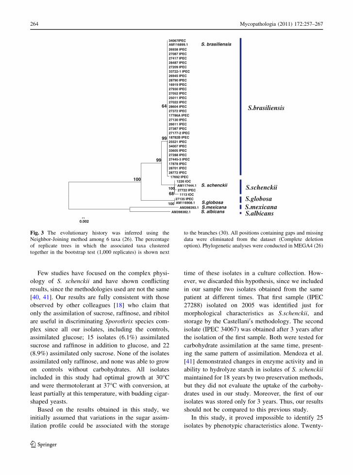
revealed (Fig. 3) three distinct and major clades
represented by S. brasiliensis, S. schenckii, and S.
globosa. Similar results were obtained when the MP
method was employed to analyze the CAL locus of
our isolates (data not shown). The significance of the
tree was assessed using bootstrap confidence [32]. In
our assessment, in 1,000 replicates, the confidence
level of occurrence of nodes was 96–99% (Fig. 3).
Bootstrap values lower than 70% are not shown.
Twenty-four of our isolates with inconclusive
Fig. 1 Morphology of conidia Sporothrix spp. a Interim or
terminal conidia in sympodial conidiophores type placed along
the hypha, b hyaline and pigmented conidia, c sessile and dark
conidia connected individually in throughout the hyphae, and
d triangular conidia. Bars 10 lm. Magnification 940
Mycopathologia (2011) 172:257–267 261
123

phenotypic profile were grouped on cluster S. brasil-
iensis and shared 100% similarity with a previously
published S. brasiliensis AM116899.1. The isolate
IPEC27722 phenotypically characterized as S. mex-
icana was grouped in cluster S. schenckii
(AM117444.1) with 99% of similarity. The taxonomy
of S. globosa was confirmed in the isolate IPEC
27135 by this methodology showing 99% similarity
with a previously published S. globosa
(AM116908.1). The control strains and our isolates
with the phenotypic profile of S. brasiliensis or S.
schenckii randomly selected to confirm their identi-
fication remained grouped into the cluster S. brasil-
iensis and S. schenckii, respectively (Fig. 3). Overall,
there was 11.7% disagreement between the species
classification using phenotypic and genotypic
methodologies.
Discussion
During the last decade, sporotrichosis has signifi-
cantly increased in Brazil, especially in the State of
Rio de Janeiro, with the occurrence of an epidemic
related to zoonotic transmission from cats to humans
causing small outbreaks amongst cat’s owners and
professionals taking care of sick cats [14]. This form
of zoonotic transmission of disease has led to changes
in the usual clinical manifestations of sporotrichosis,
with examples of primary involvement of the nasal
mucosa [33], erythema nodosum [16], erythema
multiform [17], and primary conjunctivitis [13]. In
addition, there have been a large number of cases of
disseminated cutaneous sporotrichosis, which is typ-
ically a less common clinical form of the disease
[34].
Previous studies using methodologies based on the
analysis of nucleic acids, such as RAPD and partial
sequencing of some genes [35–37], showed that
Sporothrix isolates retrieved during the outbreak in
Rio de Janeiro State could be grouped in clades
distinct to other isolates from different geographical
regions in Brazil and from other countries. A high
degree of genetic similarity was observed among
isolates from the epidemic. However, it was possible
to discriminate between 5 and 10 genotypic profiles,
indicating the presence of more than one population
of S. schenckii [37]. These findings suggested a
common source of infection between humans andTa
ble
1P
hen
oty
pic
char
acte
rist
ics
of
Sp
oro
thri
xsp
ecie
sco
mp
lex
Sp
ecie
s(%
)C
on
idia
Co
lon
yd
iam
eter
Ass
imil
atio
nte
st
Dem
atia
ceo
us
Hy
alin
eR
ang
eA
ver
age
±S
D*
Glo
bo
se
N(%
)
Tri
ang
ula
r
N(%
)
Elo
ng
ated
N(%
)
Hy
alin
e
N(%
)
30�C
37
�C3
0�C
37
�CG
luco
seS
ucr
ose
Raf
fin
ose
Sp
oro
thri
xb
rasi
lien
sis
(83
.4)
20
2(9
8)
2(1
)2
(1)
09
–4
52
–1
82
6.9
±7
.76
.3±
2.5
Po
siti
ve
Neg
ativ
eN
egat
ive
Sp
oro
thri
xsp
p(1
0.1
)§2
2(8
8)
00
3(1
2)
14
–4
23
–1
42
7.3
±7
.56
.4±
3.7
Po
siti
ve
Po
siti
ve
Neg
ativ
e
Sp
oro
thri
xsc
hen
ckii
(6.0
)0
9(6
0)
5(3
3)
1(7
)0
12
–4
92
–2
03
3.2
±1
0.2
7.0
±3
.4P
osi
tiv
eP
osi
tiv
eP
osi
tiv
e
Sp
oro
thri
xm
exic
an
a(0
.5)
01
(10
0)
00
05
13
51
.0±
0.0
3.0
±0
.0P
osi
tiv
eP
osi
tiv
eP
osi
tiv
e
*S
tan
dar
dd
evia
tio
n§
Sam
pli
ng
no
tid
enti
fied
tosp
ecie
sle
vel
by
ph
eno
typ
icte
sts
262 Mycopathologia (2011) 172:257–267
123

cats, and that animal’s act as vectors for transmission
of the fungus.
In order to confirm the identity of species within
Sporothrix complex isolated during the epidemic in
Rio de Janeiro, 247 isolates (representing 11.8% of
the isolates maintained in our laboratory) were re-
analyzed. The predominant species found was S.
brasiliensis (83.4%), which confirms previous data
that strains of S. brasiliensis are associated with the
Rio de Janeiro sporotrichosis epidemic [18]. How-
ever, S. schenckii and S. globosa were also isolated
from patients’ resident at these epidemic areas, which
indicate that other species of Sporothrix are also
contributing to the persistence of this epidemic
disease in Rio de Janeiro.
The saprophytic form of S. schenckii is usually
characterized by the types of conidia [38]. It should be
emphasized that on the description of new species the
authors [18] observed terminal or intercalary conidial
cluster on sympodial conidiophores arranged along
the hyphae, hyaline or lightly pigmented, usually
globose to subglobose or pear shaped. Besides these
sessile conidia, dark, thick-walled, connected indi-
vidually throughout the hyphae of tiny denticles were
also detected, as well as dematiaceous conidia with
triangular morphology seen only in S. schenckii.
Based on the morphology of conidia found in our
isolates, three of them (IPEC25406, IPEC28329, and
IPEC28487) showed elongated, globose, and
dematiaceous conidia, typical of S. schenckii, corrob-
orating data presented earlier in which the presence of
these conidia were associated with the morphology of
S. schenckii [18]. However, only isolate IPEC28329
was characterized as S. schenckii via the other
phenotypic methods, whilst the isolates IPEC25406
and IPEC28487 were characterized as S. brasiliensis
by other phenotypic tests. The same observation was
made when the previously characterized S. brasilien-
sis-type strain CBS120339 (IPEC16490) was re-
analyzed in this study. The colonies of this isolate
when grown on PDA attained a diameter of 35 mm at
30�C and 13 mm at 37�C, and the fungus was unable
to assimilate sucrose and raffinose. These phenotypic
aspects are characteristic of S. brasiliensis. However,
microscopic examination determined that this strain
produced dematiaceous triangular conidia, structures
not previously described on this strain, which have
been only associated previously with strains of S.
schenckii. Similar data were also described by Mari-
mon et al. [18] who showed that the type strain S.
schenckii (CBS 359.36), which traditionally produces
pigmented conidia, formed only sympodial and hya-
line conidia. These data suggest that the identification
of Sporothrix species using only the morphology of
conidia as a parameter of classification should be used
with restrictions, and that the application of other
phenotypic tests is mandatory, since these isolates
may fail their ability to produce some morphological
structures or may modify them due to some external
factor or even after several culture passages. This fact
was recently demonstrated in Italy where a fungal
isolate from an immunocompetent patient with lym-
phocutaneous sporotrichosis was characterized as S.
schenckii by conventional filamentous morphology
and conversion to yeast form in BHI at 37�C [39].
This isolate was subcultured several times to be used
as a standard strain in environmental studies. After
these subcultures, the yeast form remained unchanged
even when grown at room temperature [12]. In
addition, Marimon et al. [18] related that morphology
of conidia would be a parameter to be considered in
the characterization of S. schenckii, since the isolates
included in their studies tended to have triangular
conidiation. Our data contradict this assertion, since
statistical analysis by the McNemar’s method showed
no correlation between the strains characterized
phenotipically as S. schenckii and morphology of
conidia.
Fig. 2 Comparison of colony growth (mean diameters of
colonies) in PDA among species of the genus Sporothrix in two
different temperatures of incubation
Mycopathologia (2011) 172:257–267 263
123
Few studies have focused on the complex physi-
ology of S. schenckii and have shown conflicting
results, since the methodologies used are not the same
[40, 41]. Our results are fully consistent with those
observed by other colleagues [18] who claim that
only the assimilation of sucrose, raffinose, and ribitol
are useful in discriminating Sporothrix species com-
plex since all our isolates, including the controls,
assimilated glucose; 15 isolates (6.1%) assimilated
sucrose and raffinose in addition to glucose, and 22
(8.9%) assimilated only sucrose. None of the isolates
assimilated only raffinose, and none was able to grow
on controls without carbohydrates. All isolates
included in this study had optimal growth at 30�C
and were thermotolerant at 37�C with conversion, at
least partially at this temperature, with budding cigar-
shaped yeasts.
Based on the results obtained in this study, we
initially assumed that variations in the sugar assim-
ilation profile could be associated with the storage
time of these isolates in a culture collection. How-
ever, we discarded this hypothesis, since we included
in our sample two isolates obtained from the same
patient at different times. That first sample (IPEC
27288) isolated on 2005 was identified just for
morphological characteristics as S.schenckii, and
storage by the Castellani0s methodology. The second
isolate (IPEC 34067) was obtained after 3 years after
the isolation of the first sample. Both were tested for
carbohydrate assimilation at the same time, present-
ing the same pattern of assimilation. Mendoza et al.
[41] demonstrated changes in enzyme activity and in
ability to hydrolyze starch in isolates of S. schenckii
maintained for 18 years by two preservation methods,
but they did not evaluate the uptake of the carbohy-
drates used in our study. Moreover, the first of our
isolates was stored only for 3 years. Thus, our results
should not be compared to this previous study.
In this study, it proved impossible to identify 25
isolates by phenotypic characteristics alone. Twenty-
34067IPECAM116899.1 S. brasiliensis26938 IPEC27087 IPEC27417 IPEC28487 IPEC27209 IPEC33722-1 IPEC26945 IPEC28790 IPEC16919 IPEC27930 IPEC27052 IPEC25011 IPEC27022 IPEC28604 IPEC27372 IPEC17786A IPEC27130 IPEC26611 IPEC27387 IPEC27177-2 IPEC18782B IPEC25521 IPEC34007 IPEC33605 IPEC27288 IPEC27445-3 IPEC17878 IPEC28701 IPEC28772 IPEC17692 IPEC
1226 IOCAM117444.1 S. schenckii27722 IPEC1113 IOC
27135 IPECAM116908.1 S.globosa
AM398393.1 S.mexicanaAM398382.1 S. albicans
68100
100
100
99
99
64
0.002
S.brasiliensis
S.schenckii
S.globosaS.mexicanaS.albicans
Fig. 3 The evolutionary history was inferred using the
Neighbor-Joining method among 6 taxa (26). The percentage
of replicate trees in which the associated taxa clustered
together in the bootstrap test (1,000 replicates) is shown next
to the branches (30). All positions containing gaps and missing
data were eliminated from the dataset (Complete deletion
option). Phylogenetic analyses were conducted in MEGA4 (26)
264 Mycopathologia (2011) 172:257–267
123

two of these isolates showed a biochemical pattern of
carbohydrate assimilation typical to that of S. globosa
or S. albicans by the Marimon0s key. However, the
average colony diameter at 30�C excluded S. albicans
in these cases; in addition, the latter produces
dematiaceous conidia. They also could not be clas-
sified as S. globosa since these isolates were
thermotolerant. The identification was concluded
after sequencing of the CAL locus, which confirmed
that 21 of the isolates were actually S. brasiliensis
and one was S. globosa.
Identification of isolates IPEC17692,
IPEC18782B, and IPEC28790, also proved inconclu-
sive, based on observation of hyaline conidia, which
are characteristic features of the species S. albicans or
S. lurie [18, 21], together with an auxonogram profile
typical of S. brasiliensis. Recently our group dem-
onstrated that one of these isolates (IPEC18782B),
whilst being unable to produce dematiaceous conidia,
was able to produce low levels of melanin at 30�C,
although in insufficient amounts to either darken
colonies or to appear in fungal cell walls [42]. Thus,
this isolate probably lost its ability to produce visible
amount of melanin during its growth. However, it
cannot yet be ruled out that there is further as yet
unappreciated variability within isolates classified as
S. brasiliensis, and studies are underway in our
laboratory to clarify this hypothesis. Another three
strains classified as S. brasiliensis (IPEC16919,
IPEC27022, and IPEC27087) have not demonstrated
macroscopically pigmented colonies, but did develop
oval conidia. These results also suggest phenotypic
variability within this species, and that the production
of melanin should be verified by both macroscopic
and microscopic examination of colonies.
Some isolates examined in this study deserve
special attention. Isolate IPEC27722, characterized
phenotypically as S. mexicana, showed colony
diameters exceeding 50 mm (51 mm) at 30�C and
3 mm at 37�C, coincident with those proposed for the
classification of this species. Physiological character-
istics also inferred that this isolate was S. mexicana.
Thus far this species has only been isolated from
environmental sources - rosewood soil and carnation
leaves—and is considered non-pathogenic to man
[18]. Therefore, genotypic analysis was performed to
confirm its taxonomy given that IPEC27722 was
isolated from the forearm injury of an immunocom-
promised patient, who had a previous report of an
uncommon form of histoplasmosis [43]. The sporo-
trichosis regressed spontaneously, supporting previ-
ous data that S. mexicana acts more as opportunist
fungi. Molecular analysis of this isolate clustered it
on the S. schenckii clade. For this reason, we suggest
that molecular characterization should be applied to
isolates of the Sporothrix complex that grow colonies
around 50 mm in diameter at 30�C.
Molecular analysis of 25 isolates that showed
inconclusive results on phenotypic studies enabled
the formation of a clade composed by 24 isolates with
100% similarity with the species S. brasiliensis
demonstrating again the importance of analysis at
the level of genotype to identify species. Moreover,
one of our isolates (IPEC27135) was clustered with
the species S. globosa (AM116908.1) isolated in
Spain (Fig. 3). Accordingly, strain IPEC27135 was
classified as S. globosa and represents the first
isolation of this pathogen in Brazil [20]. In addition,
our study has shown that the strain IPEC28329,
classified phenotypically as S. schenckii, actually
demonstrates similarity to the S. brasiliensis
(AM116899.1) type strain. Therefore, our data is in
disagreement with those previously published [18]
that make the assumption that the differentiation of
species within the complex Sporothrix can be easily
accomplished without the involvement of molecular
methods. In contrast, we believe that the correlation
between molecular data and phenotypic characteris-
tics is crucial in identifying these species and suggest
that other genetic markers should also be evaluated in
taxonomic studies of the Sporothrix species complex.
This study represents the most extensive charac-
terization of strains yet undertaken of the Sporothrix
complex, based on phenotypic and genetic analysis.
We have characterized phenotypically 247 isolates
from the epidemic of sporotrichosis in Rio de Janeiro
state, Brazil. And have confirmed S. brasiliensis as
the most prevalent species (83.4% of isolates). We
have also identified an important discrepancy with
the previously reported data concerning the micros-
copy of conidia produced by these newly described
Sporothrix species. More studies are necessary to
fully understand the conidial profile of each Sporo-
thrix species.
Acknowledgments Financial support for this work was
provided by FAPERJ (Grant Proc. E-26/111.619/2008).
R.M.Z.O. is in part supported by CNPq 350338/2000-0. We
Mycopathologia (2011) 172:257–267 265
123

thank Andrew J. Hamilton for help in preparing this
manuscript. Automated sequencing was done using the
Genomic Platform-DNA Sequencing Platform at Fundacao
Oswaldo Cruz—PDTIS/FIOCRUZ (RPT01A), Brazil.
References
1. Conti-Diaz IA. Epidemiology of sporotrichosis in Latin
America. Mycopathologia. 1989;108:113–6.
2. Kovarik CL, Neyra E, Bustamante B. Evaluation of cats as
the source of endemic sporotrichosis in Peru. Med. Mycol.
2008;46:53–6.
3. Pappas PG, Tellez I, Deep AE, Nolasco D, Holgado W,
Bustamanate B. Sporotrichosis in Peru: description of an
area of hyperendemicity. Clin Infect Dis. 2000;30:65–70.
4. Carrada-Bravo T. New observations on the epidemiology
and pathogenesis of sporothrichosis. Ann Trop Med
Parasitol. 1975;69:267–73.
5. Cooper CR, Dixon DM, Salkin IF. Laboratory-acquired
sporotrichosis. J Med Vet Mycol. 1992;30:169–71.
6. Hajjeh R, McDonnel S, Reef S, Licitra C, Hankins M, Toth
B, Padhye A, Kaufman L, Passarell L, Cooper CR, Hut-
wagner L, Hopkins R, McNeil M. Outbreak of sporotri-
chosis among three nursery workers. J Infect Dis. 1997;
176:499–504.
7. St-Germain G, Summerbell R. Identifying filamentous
fungi. In: A clinical laboratory handbook. California: Star
Publishing Company; 1996.
8. Sigler L, Harris JL, Dixon DM, Flis AL, Salkin IF, Kemna
M, Duncan RA. Microbiology and potential virulence of
Sporothrix cyanescens, a fungus rarely isolated from blood
and skin. J Clin Microbiol. 1990;28:1009–15.
9. Chandler FW, Kaplan W, Ajello L. A colour atlas and
textbook of histopathology of mycotic diseases. Wolfe
Medical Publications Ltd.; 1980, pp 112–115.
10. Ramos-e-Silva M, Vasconcelos C, Carneiro S, Cestari T.
Sporotrichosis. Clin Dermatol. 2007;25:181–7.
11. Morris-Jones R. Sporotrichosis. Clin. Exp. Dermatology.
2002;27:427–31.
12. Criseo G, Zungriand D, Romeo O. Stable yeast-like form
of Sporothrix schenckii: lack of dimorphic stage. J Clin
Microbiol. 2008;46:3870–1.
13. Schubach A, de Lima Barros MB, Schubach TM, Fran-
cesconi-do-Valle AC, Gutierrez-Galhardo MC, Sued M, de
Matos Salgueiro M, Fialho-Monteiro PC, Reis RS, Mar-
zochi KB, Wanke B, Conceicao-Silva F. Primary con-
junctival sporotrichosis: two cases from a zoonotic
epidemic in Rio de Janeiro, Brazil. Cornea. 2005;24:491–3.
14. Schubach A, Barros MB, Wanke B. Epidemic sporotri-
chosis. Curr. Opin. Infect. Dis. 2008;21:129–33.
15. Freitas DF, Valle AC, Almeida-Paes R, Bastos FI, Gal-
hardo MC. Zoonotic Sporotrichosis in Rio de Janeiro,
Brazil: a protracted epidemic yet to be curbed. Clin Infect
Dis. 2010;50:453.
16. Gutierrez-Galhardo MC, de Oliveira Schubach A, de Lima
Barros MB, Moita Blanco TC, Cuzzi-Maya T, Pacheco
Schubach TM, dos Santos Lazera M, do Valle AC. Ery-
thema nodosum associated with sporotrichosis. Int J Der-
matol. 2002;41:114–6.
17. Gutierrez-Galhardo MC, Barros MB, Schubach AO, Cuzzi
T, Schubach TM, Lazera MS, Valle AC. Erythema multi-
forme associated with sporotrichosis. J. Eur. Acad. Der-
matol. Venereol. 2005;19:507–9.
18. Marimon R, Cano J, Gene J, Sutton DA, Kawasaki M,
Guarro J. Sporothrix brasiliensis, S. globosa, and S. mex-icana, three new Sporothrix species of clinical interest.
J Clin Microbiol. 2007;45(10):3198–206.
19. Madrid H, Cano J, Gene J, Bonifaz A, Toriello C, Guarro J.
Sporothrix globosa, a pathogenic fungus with widespread
geographical distribution. Rev. Iberoam. Micol. 2009;26(3):
218–22.
20. Oliveira MME, Almeida-Paes R, Muniz MM, Barros
MBL, Gutierrez-Galhardo MC, Zancope-Oliveira RM.
Sporotrichosis caused by Sporothrix globosa in Rio de
Janeiro, Brazil: case report. Mycopathologia. 2010;169:
359–63.
21. Marimon R, Gene J, Cano J, Guarro J. Sporothrix luriei:rare fungus from clinical origin. Med. Mycol. 2008;
46(6):621–5.
22. de Meyer EM, de Beer ZW, Summerbell RC, Moharram
AM, de Hoog GS, Vismer HF, Wingfield MJ. Taxonomy
and phylogeny of new wood- and soil-inhabiting Sporo-thrix species in the Ophiostoma stenoceras-Sporothrixschenckii complex. Mycologia. 2008;100(4):647–61.
23. Romeo O, Scordino F, Criseo, G. New insight into
molecular phylogeny and epidemiology of Sporothrixschenckii species complex based on calmodulin-encoding
gene analysis of Italian isolates. Mycopathologia 2011;
Published online: 02 April 2011.
24. Dixon DM, Salkin IF, Duncan RA, Hurd NJ, Haines JH,
Kemna ME, Coles FB. Isolation and characterization of
Sporothrix schenckii from clinical and environmental
sources associated with the largest US epidemic of spo-
rotrichosis. J Clin Microbiol. 1991;29:1106–13.
25. Woods JP, Kersulyte D, Goldman WE, Berg DE. Fast
DNA isolation from Histoplasma capsulatum: methodol-
ogy for arbitrary primer polymerase chain reaction-based
epidemiological and clinical studies. J Clin Microbiol.
1993;31(2):463–4.
26. Otto TD, Vasconcellos EA, Gomes LHF, Moreira AS,
Degrave WM, Mendonca-Lima L, Alves-Ferreira M.
ChromaPipe: a pipeline for analysis, quality control and
management for a DNA sequencing facility. Genet Mol
Res. 2008;7(3):861–71.
27. Tamura K, Dudley J, Nei M, Kumar S. MEGA4: Molecular
Evolutionary Genetics Analysis (MEGA) software version
4.0. Mol Biol Evol. 2007;24(8):1596–9.
28. Saitou N, Nei M. The neighbor-joining method: A new
method for reconstructing phylogenetic trees. Molec. Biol.
Evol. 1987;4:406–25.
29. Kimura M. A simple method for estimating evolutionary
rates of bases substitutions through comparative studies of
nucleotide sequences. J Mol Evol. 1980;16(2):111–20.
30. Eck RV, Dayhoff MO. Atlas of protein sequence and
structure. National Biomedical Research Foundation.
Maryland: Silver Springs; 1966.
31. Nei M, Kumar S. Molecular Evolution and Phylogenetics.
Oxford University, NY: Oxford University Press; 2000.
32. Felsenstein J. Confidence limits on phylogenies: An
approach using the bootstrap. Evolution. 1985;39:783–91.
266 Mycopathologia (2011) 172:257–267
123

33. Schubach TMP, Schubach AO, Cuzzi-Maya T, Okamoto
T, Reis RS, Monteiro PCF, Gutierrez-Galhardo MC,
Wanke B. Pathology of sporotrichosis in 10 cats of Rio de
Janeiro. Vet Rec. 2003;152:172–5.
34. Barros MBL, Schubach AO, Gutierrez-Galhardo MC,
Schubach TMP, Reis RS, Conceicao MJ, Valle ACF.
Sporotrichosis with widespread cutaneous lesions: report
of 24 cases related to transmission by domestic cats in Rio
de Janeiro, Brazil. Int J Dermatol. 2003;42:677–81.
35. Marimon R, Gene J, Cano J, Sutton DA, Trilles L, Dos
Santos Lazera M, Guarro J. Molecular phylogeny of Spo-rothrix schenckii. J Clin Microbiol. 2006;44(9):3251–6.
36. Gutierrez-Galhardo MC, Zancope-Oliveira RM, Valle AC,
Almeida-Paes R, Silva-Tavares PM, Monzon A, Mellado
E, Rodriguez-Tudela JL, Cuenca-Estrella M. Molecular
epidemiology and antifungal susceptibility patterns of
Sporothrix schenckii isolates from a cat-transmitted epi-
demic of sporotrichosis in Rio de Janeiro, Brazil. Med.
Mycol. 2008;46:141–51.
37. Reis RS, Almeida-Paes R, Muniz MM, Tavares PM,
Monteiro PC, Schubach TM, Gutierrez-Galhardo MC,
Zancope-Oliveira RM. Molecular characterisation of Spo-rothrix schenckii isolates from humans and cats involved in
the sporotrichosis epidemic in Rio de Janeiro. Brazil.Mem.
Inst. Oswaldo. Cruz. 2009;104:769–74.
38. de Hoog GS, de Vries GA. Two new species of Sporothrixand their relation to Blastobotrys nivea. Antonie Van.
Leeuwenhoek. 1973;39:515–20.
39. Criseo G, Malara G, Romeo O, Puglisi Guerra A. Lym-
phocutaneous sporotrichosis in an immunocompetent
patient: a case report from extreme southern Italy. Myco-
pathologia. 2008;166:159–62.
40. Gosh A, Maity PK, Hemashettar BM, Sharmaand VK,
Chakrabarti A. Physilogical characters of Sporothrixschenckii isolates. Mycoses. 2002;45:449–54.
41. Mendoza M, Alvarado P, Dias de Torres E, Lucena L, de
Albornoz MC. Physiological comportment and in vivo
sensitivity of Sporothrix schenckii isolates maintained for
18 years by two preservation methods. Rev. Iberoam.
Micol. 2005;22:151–6.
42. Almeida-Paes R, Frases S, Monteiro PC, Gutierrez Gal-
hardo MC, Zancope-Oliveira RM, Nosanchuk JD. Growth
conditions influence melanization of the pathogenic fungus
Sporothrix schenckii. Microb. Infect. 2009;11:554–62.
43. Valle AC, Moreira LC, Almeida-Paes R, Moreira JS,
Pizzini CV, Muniz MM, Zancope-Oliveira RM. Chronic
disseminated histoplasmosis with lesions restricted to the
mouth: case report. Rev Inst Med Trop Sao Paulo. 2006;
48:113–6.
Mycopathologia (2011) 172:257–267 267
123