Embed Size (px)
Citation preview
PHL - 224 Biochemistry II
1
PHL 224 Biochemistry II
Lab. Manual
Academic Year
1438/1439 - 2017/2018
Kingdom of Saudi Arabia
Ministry of Higher Education
Prince Sattam Bin Abdulaziz University
College of Pharmacy
Department of Pharmacology
PHL - 224 Biochemistry II
2
Contents
Remarks Name of the Experiment DATE S.NO.
1
2
3
4
5
6
7
8
9
10
11
12
13
14
PHL - 224 Biochemistry II
3
Introduction to the Laboratory:
This course is intended to introduce you to some of the most widely used experimental procedures in Biochemistry. Prior
to each lab period, you will need to spend some time reading the Laboratory Manual. This reading will provide background
information and an outline of the procedures to be performed. If you do not do this, you will find yourself wasting large
amounts of class time, and annoying both your lab partners and your Instructor.
SAFETY:
Laboratories contain hazards of various kinds. Everyone is required to wear closed-toe shoes, long pants, goggles with side
shields, and a lab coat while performing laboratory work. Students should not work in the laboratory if the instructor is not
present.
COMMON LABORATORY TOOLS & EQUIPMENT USED IN BIOCHEMISTRY:
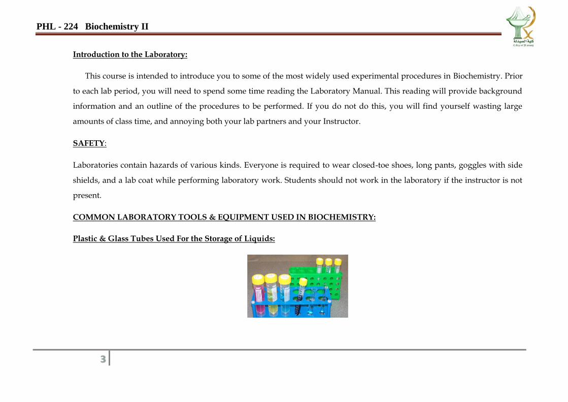
Plastic & Glass Tubes Used For the Storage of Liquids:
PHL - 224 Biochemistry II
4
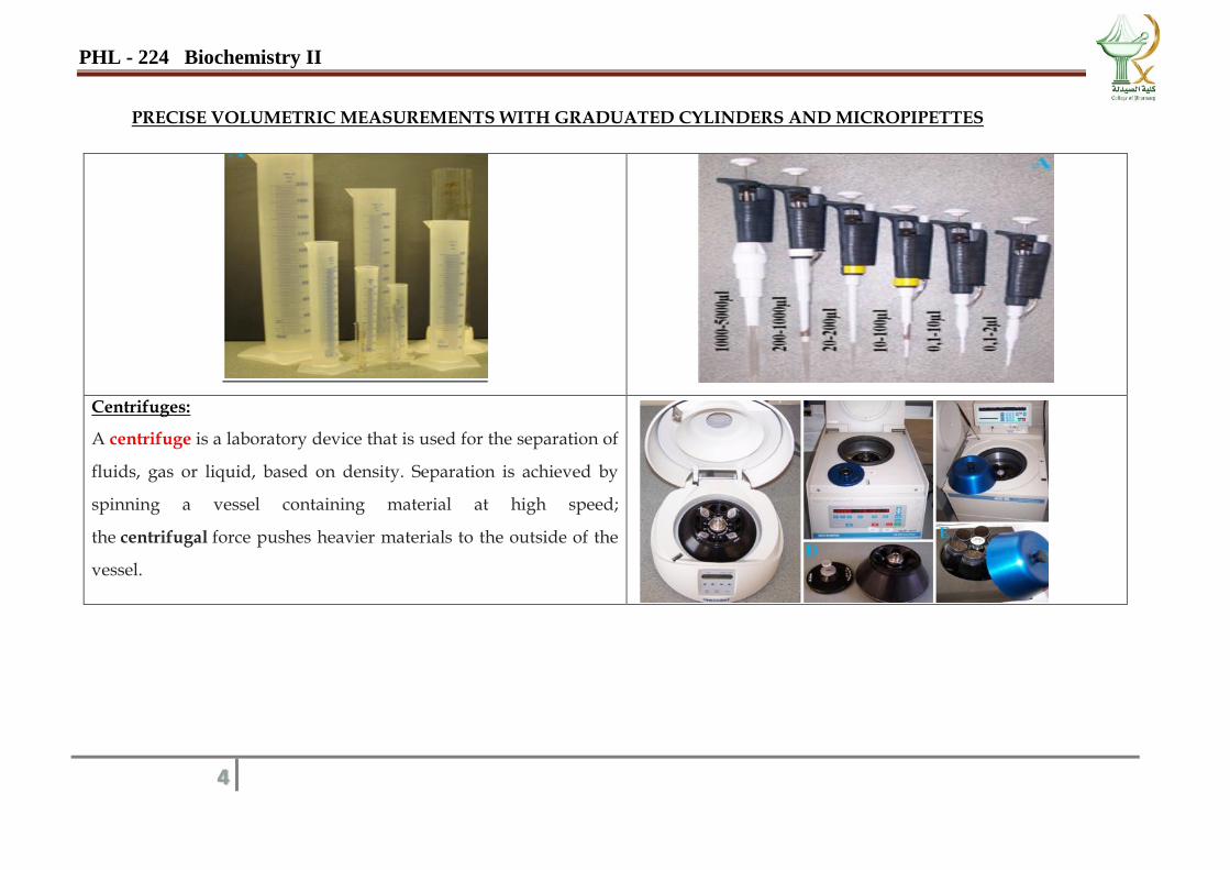
PRECISE VOLUMETRIC MEASUREMENTS WITH GRADUATED CYLINDERS AND MICROPIPETTES
Centrifuges:
A centrifuge is a laboratory device that is used for the separation of
fluids, gas or liquid, based on density. Separation is achieved by
spinning a vessel containing material at high speed;
the centrifugal force pushes heavier materials to the outside of the
vessel.
PHL - 224 Biochemistry II
5
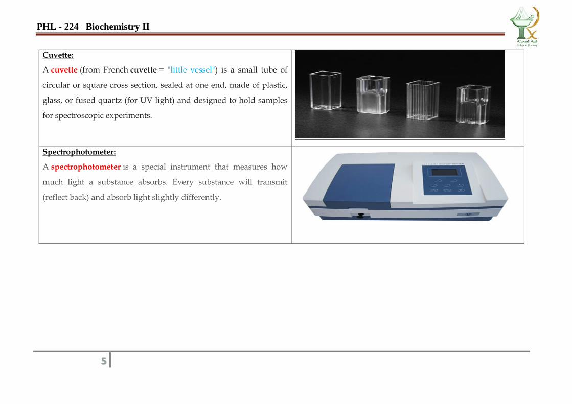
Cuvette:
A cuvette (from French cuvette = "little vessel") is a small tube of
circular or square cross section, sealed at one end, made of plastic,
glass, or fused quartz (for UV light) and designed to hold samples
for spectroscopic experiments.
Spectrophotometer:
A spectrophotometer is a special instrument that measures how
much light a substance absorbs. Every substance will transmit
(reflect back) and absorb light slightly differently.
PHL - 224 Biochemistry II
6
PREPARATION OF BUFFER’s
PREPARATION OF BUFFER AND MEASURING pH AIM:
To study the nature of the buffer and prepare a buffer at the required pH
INTRODUCTION AND PRINCIPLE:
Buffers are defined as the solutions that resist changes in the pH of systems. Regulation of the pH of body fluids and tissues
consistent with life and normal functions are obtained by the presence of buffer
The pH meter measures an electrical potential developed by a pair of electrode pins in a solution. For the measurement of pH, an
electrode system sensitive to change in H+ ion concentration of a solution is used.
PROCEDURE:
An acidic buffer is prepared by adding a weak acid to salt of that acid. A basic buffer is prepared by adding a weak base to the
salt of base.
The required pH of two buffers is given. The obtained pH can be adjusted by adding HCl for a more acidic or NaOH for a more
basic solution.
PHL - 224 Biochemistry II
7
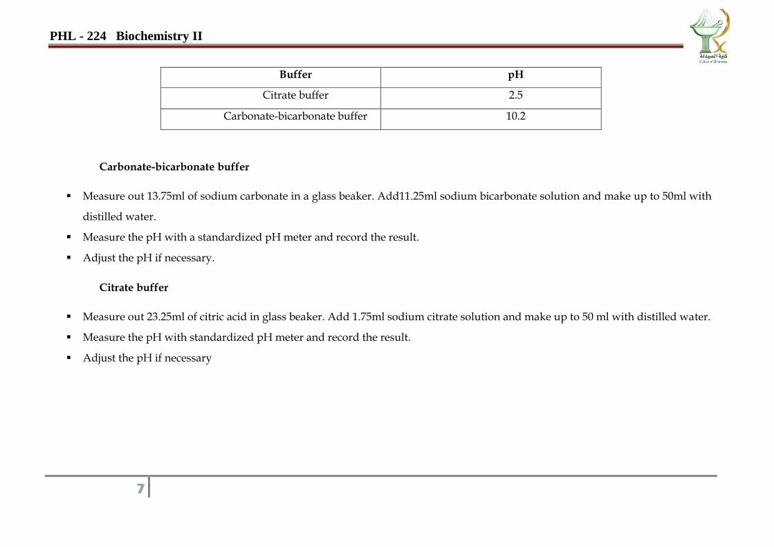
Buffer pH
Citrate buffer 2.5
Carbonate-bicarbonate buffer 10.2
Carbonate-bicarbonate buffer
Measure out 13.75ml of sodium carbonate in a glass beaker. Add11.25ml sodium bicarbonate solution and make up to 50ml with
distilled water.
Measure the pH with a standardized pH meter and record the result.
Adjust the pH if necessary.
Citrate buffer
Measure out 23.25ml of citric acid in glass beaker. Add 1.75ml sodium citrate solution and make up to 50 ml with distilled water.
Measure the pH with standardized pH meter and record the result.
Adjust the pH if necessary
PHL - 224 Biochemistry II
8
RESULTS AND DISCUSSION:
Buffer pH obtained HCl added?
(Yes/No)
NaOH added?
(Yes/No)
REPORT:
_______________________________________________________________________________________________________________
_______________________________________________________________________________

PHL - 224 Biochemistry II
9
Qualitative analysis
PHL - 224 Biochemistry II
10
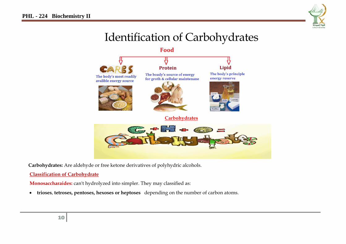
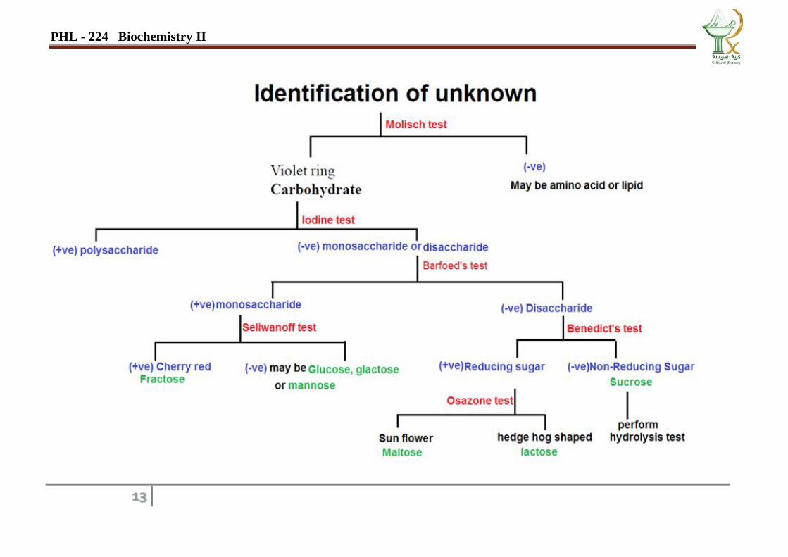
Identification of Carbohydrates
Carbohydrates
Carbohydrates: Are aldehyde or free ketone derivatives of polyhydric alcohols.
Classification of Carbohydrate
Monosaccharaides: can't hydrolyzed into simpler. They may classified as:
trioses, tetroses, pentoses, hexoses or heptoses depending on the number of carbon atoms.
PHL - 224 Biochemistry II
11
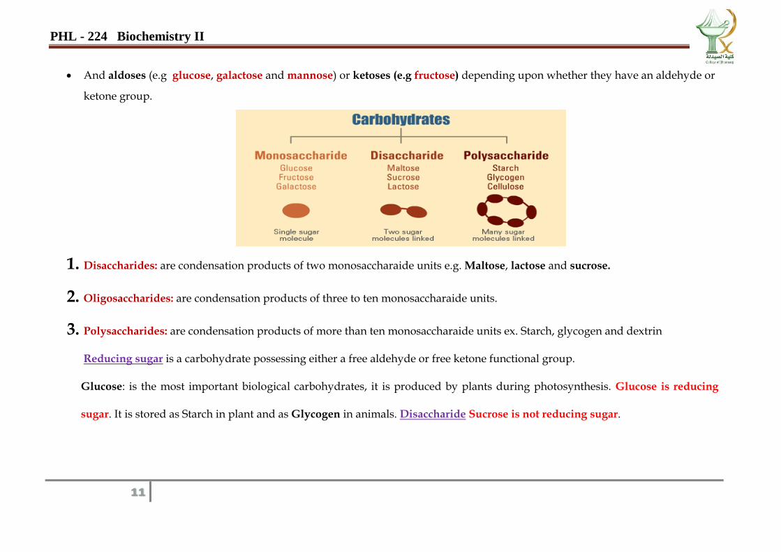
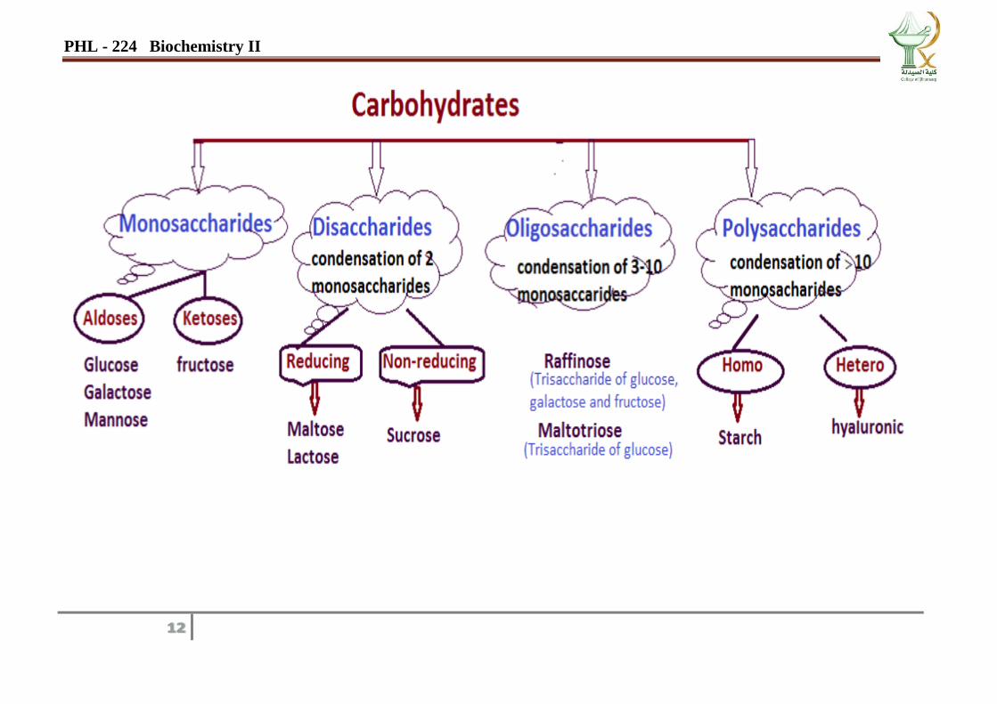
And aldoses (e.g glucose, galactose and mannose) or ketoses (e.g fructose) depending upon whether they have an aldehyde or
ketone group.
1. Disaccharides: are condensation products of two monosaccharaide units e.g. Maltose, lactose and sucrose.
2. Oligosaccharides: are condensation products of three to ten monosaccharaide units.
3. Polysaccharides: are condensation products of more than ten monosaccharaide units ex. Starch, glycogen and dextrin
Reducing sugar is a carbohydrate possessing either a free aldehyde or free ketone functional group.
Glucose: is the most important biological carbohydrates, it is produced by plants during photosynthesis. Glucose is reducing
sugar. It is stored as Starch in plant and as Glycogen in animals. Disaccharide Sucrose is not reducing sugar.
PHL - 224 Biochemistry II
12
PHL - 224 Biochemistry II
13
PHL - 224 Biochemistry II
14
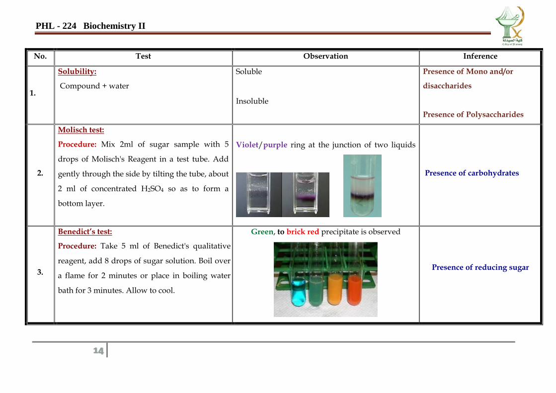
No. Test Observation Inference
1.
Solubility:
Compound + water
Soluble
Insoluble
Presence of Mono and/or
disaccharides
Presence of Polysaccharides
2.
Molisch test:
Procedure: Mix 2ml of sugar sample with 5
drops of Molisch's Reagent in a test tube. Add
gently through the side by tilting the tube, about
2 ml of concentrated H2SO4 so as to form a
bottom layer.
Violet/purple ring at the junction of two liquids
Presence of carbohydrates
3.
Benedict’s test:
Procedure: Take 5 ml of Benedict's qualitative
reagent, add 8 drops of sugar solution. Boil over
a flame for 2 minutes or place in boiling water
bath for 3 minutes. Allow to cool.
Green, to brick red precipitate is observed
Presence of reducing sugar
PHL - 224 Biochemistry II
15
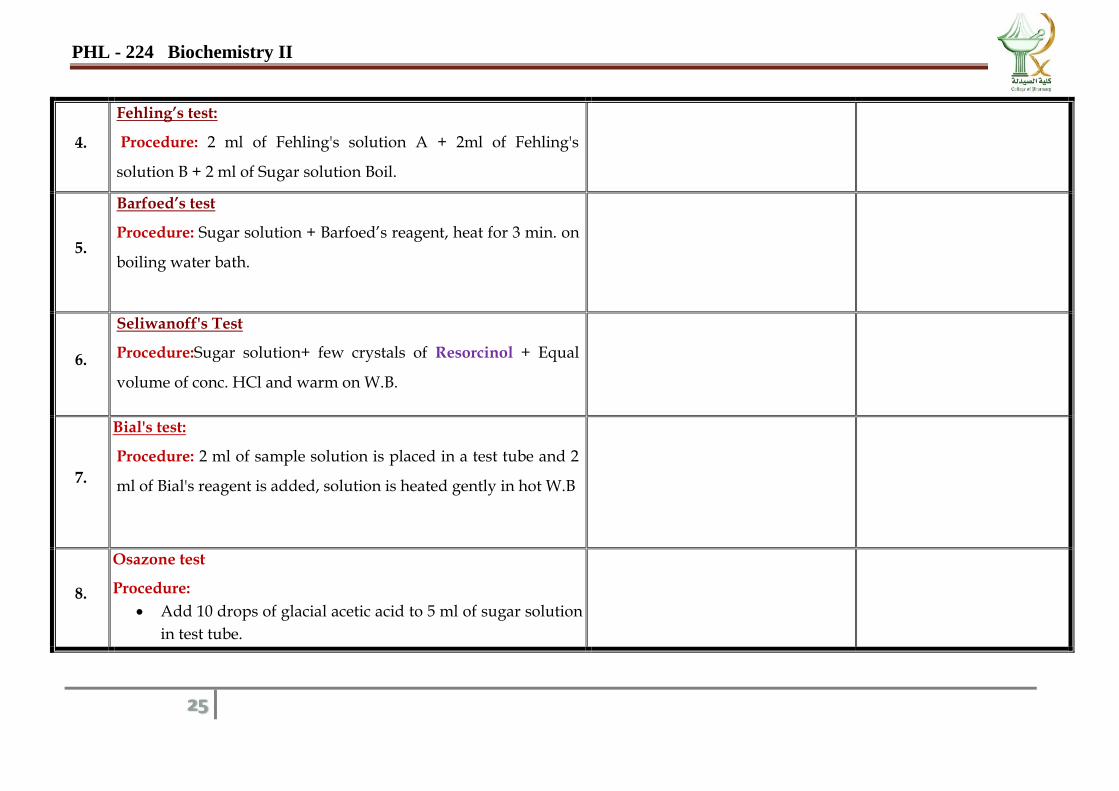
4. 44
4
4
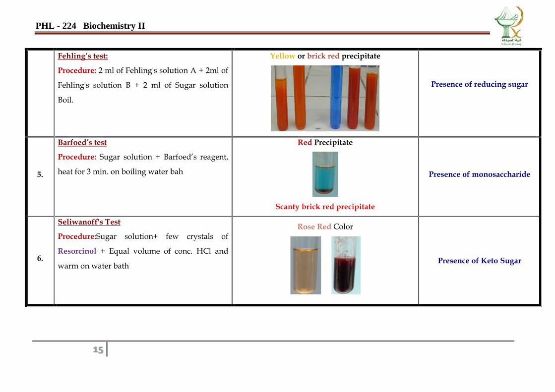
Fehling’s test:
Procedure: 2 ml of Fehling's solution A + 2ml of
Fehling's solution B + 2 ml of Sugar solution
Boil.
Yellow or brick red precipitate
Presence of reducing sugar
5.
Barfoed’s test
Procedure: Sugar solution + Barfoed’s reagent,
heat for 3 min. on boiling water bah
Red Precipitate
Scanty brick red precipitate
Presence of monosaccharide
6.
Seliwanoff's Test
Procedure:Sugar solution+ few crystals of
Resorcinol + Equal volume of conc. HCl and
warm on water bath
Rose Red Color
Presence of Keto Sugar
PHL - 224 Biochemistry II
16
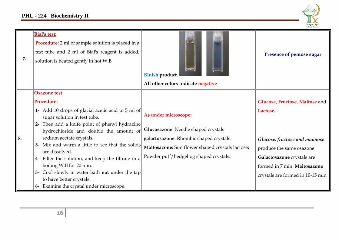
7-
Bial's test:
Procedure: 2 ml of sample solution is placed in a
test tube and 2 ml of Bial's reagent is added,
solution is heated gently in hot W.B
Bluish product.
All other colors indicate negative
Presence of pentose sugar
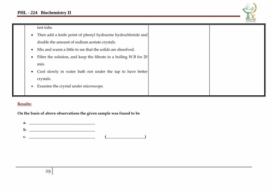
8.
Osazone test
Procedure:
1- Add 10 drops of glacial acetic acid to 5 ml of
sugar solution in test tube.
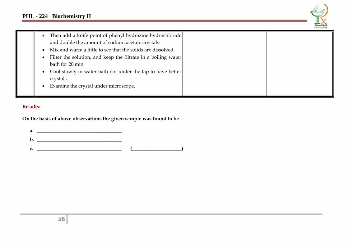
2- Then add a knife point of phenyl hydrazine
hydrochloride and double the amount of
sodium acetate crystals.
3- Mix and warm a little to see that the solids
are dissolved.
4- Filter the solution, and keep the filtrate in a
boiling W.B for 20 min.
5- Cool slowly in water bath not under the tap
to have better crystals.
6- Examine the crystal under microscope.
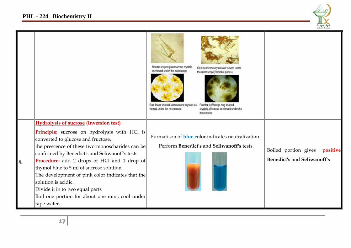
As under microscope:
Glucosazone: Needle shaped crystals
galactosazone: Rhombic shaped crystals.
Maltosazone: Sun flower shaped crystals lactose:
Powder puff/hedgehog shaped crystals.
Glucose, Fructose, Maltose and
Lactose.
Glucose, fructose and mannose
produce the same osazone
Galactosazone crystals are
formed in 7 min. Maltosazone
crystals are formed in 10-15 min
PHL - 224 Biochemistry II
17
9.
Hydrolysis of sucrose (Inversion test)
Principle: sucrose on hydrolysis with HCl is
converted to glucose and fructose.
the prescence of these two monoscharides can be
confirmed by Benedict's and Seliwanoff's tests.
Procedure: add 2 drops of HCl and 1 drop of
thymol blue to 5 ml of sucrose solution.
The development of pink color indicates that the
solution is acidic.
Divide it in to two equal parts
Boil one portion for about one min., cool under
tape water.
Formatison of blue color indicates neutralization .
Perform Benedict's and Seliwanoff's tests.
Boiled portion gives positive
Benedict's and Seliwanoff's
PHL - 224 Biochemistry II
18
Neutralize both portions by adding 2% sodium
carbonate drop by drop.
10.
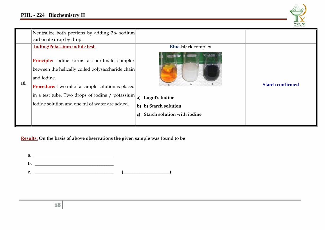
Iodine/Potassium iodide test:
Principle: iodine forms a coordinate complex
between the helically coiled polysaccharide chain
and iodine.
Procedure: Two ml of a sample solution is placed
in a test tube. Two drops of iodine / potassium
iodide solution and one ml of water are added.
Blue-black complex
a) Lugol's Iodine
b) b) Starch solution
c) Starch solution with iodine
Starch confirmed
Results: On the basis of above observations the given sample was found to be
a. __________________________________
b. __________________________________
c. __________________________________ (____________________)
PHL - 224 Biochemistry II
19
PHL - 224 Biochemistry II
20
Monosaccharides
Fructose
Galactose
Glucose
Fructose Date……………….
No. Test Observation Inference
1.
Solubility:
Compound + water
2.
Molisch test:
Procedure: Mix 2ml of sugar sample with 5 drops
of Molisch's Reagent in a test tube. Add gently
through the side by tilting the tube, about 2 ml of
concentrated H2SO4 so as to form a bottom layer.
PHL - 224 Biochemistry II
21
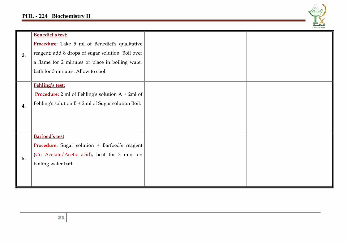
3.
Benedict’s test:
Procedure: Take 5 ml of Benedict's qualitative
reagent; add 8 drops of sugar solution. Boil over
a flame for 2 minutes or place in boiling water
bath for 3 minutes. Allow to cool.
4.
Fehling’s test:
Procedure: 2 ml of Fehling's solution A + 2ml of
Fehling's solution B + 2 ml of Sugar solution Boil.
5.
Barfoed’s test
Procedure: Sugar solution + Barfoed’s reagent
(Cu Acetate/Acetic acid), heat for 3 min. on
boiling water bath
PHL - 224 Biochemistry II
22
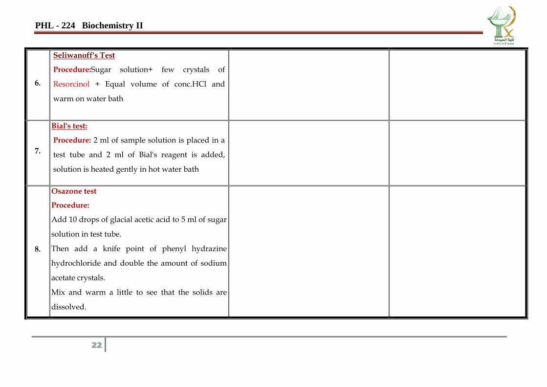
6.
Seliwanoff's Test
Procedure:Sugar solution+ few crystals of
Resorcinol + Equal volume of conc.HCl and
warm on water bath
7.
Bial's test:
Procedure: 2 ml of sample solution is placed in a
test tube and 2 ml of Bial's reagent is added,
solution is heated gently in hot water bath
8.
Osazone test
Procedure:
Add 10 drops of glacial acetic acid to 5 ml of sugar
solution in test tube.
Then add a knife point of phenyl hydrazine
hydrochloride and double the amount of sodium
acetate crystals.
Mix and warm a little to see that the solids are
dissolved.
PHL - 224 Biochemistry II
23
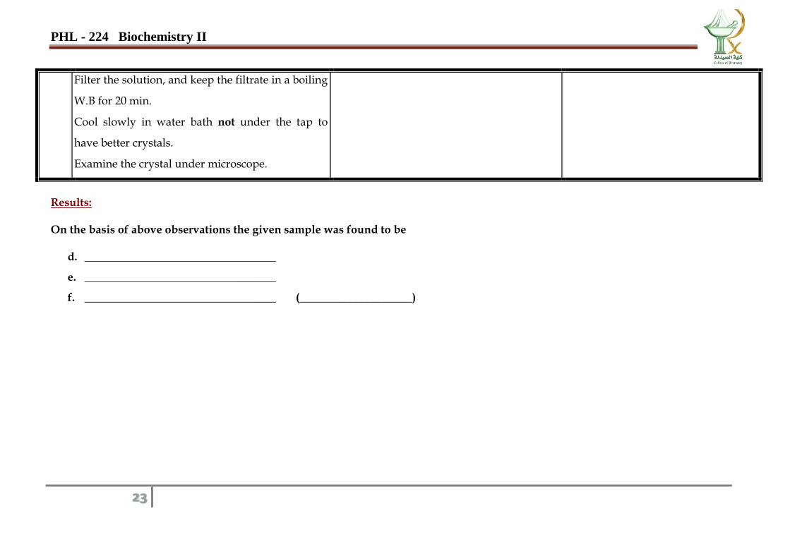
Filter the solution, and keep the filtrate in a boiling
W.B for 20 min.
Cool slowly in water bath not under the tap to
have better crystals.
Examine the crystal under microscope.
Results: On the basis of above observations the given sample was found to be
d. __________________________________
e. __________________________________
f. __________________________________ (____________________)
PHL - 224 Biochemistry II
24
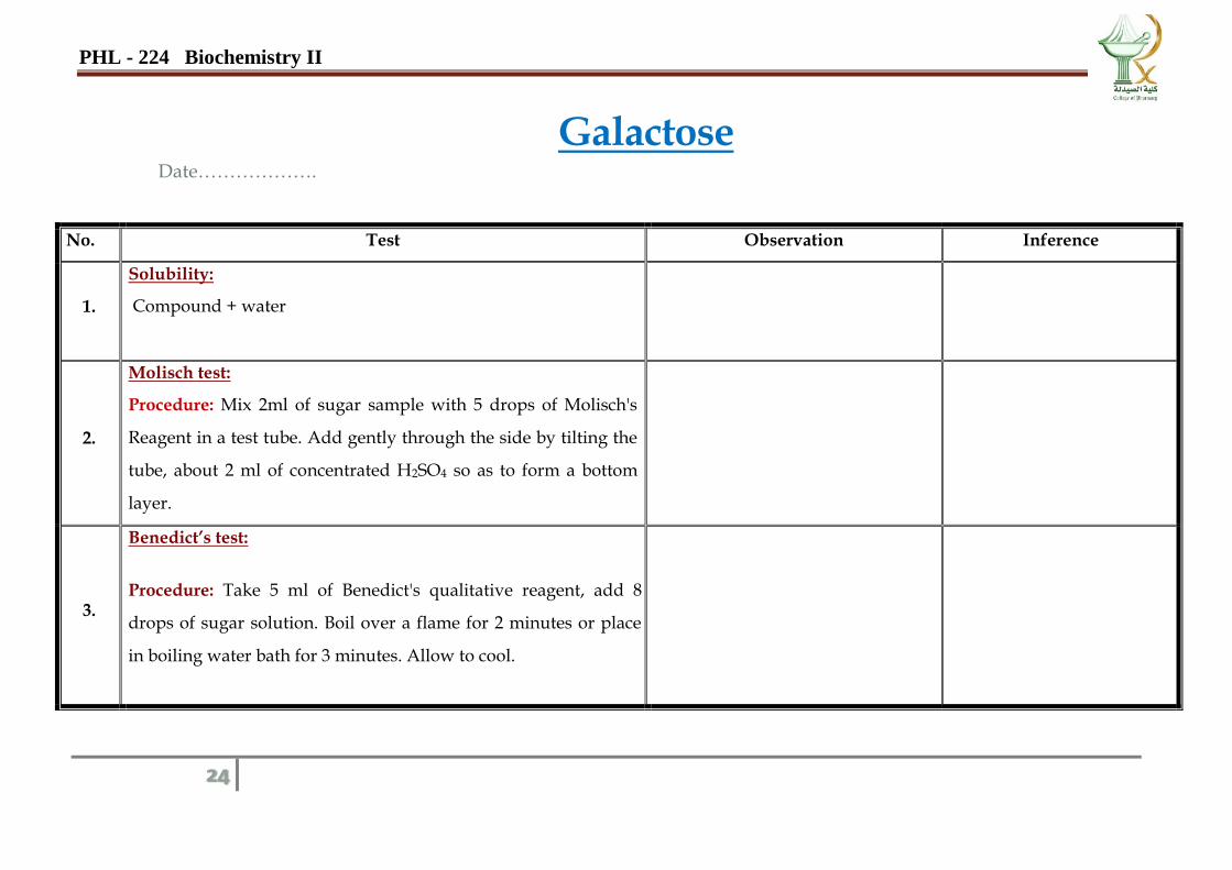
Galactose Date……………….
No. Test Observation Inference
1.
Solubility:
Compound + water
2.
Molisch test:
Procedure: Mix 2ml of sugar sample with 5 drops of Molisch's
Reagent in a test tube. Add gently through the side by tilting the
tube, about 2 ml of concentrated H2SO4 so as to form a bottom
layer.
3.
Benedict’s test:
Procedure: Take 5 ml of Benedict's qualitative reagent, add 8
drops of sugar solution. Boil over a flame for 2 minutes or place
in boiling water bath for 3 minutes. Allow to cool.
PHL - 224 Biochemistry II
25
4.
Fehling’s test:
Procedure: 2 ml of Fehling's solution A + 2ml of Fehling's
solution B + 2 ml of Sugar solution Boil.
5.
Barfoed’s test
Procedure: Sugar solution + Barfoed’s reagent, heat for 3 min. on
boiling water bath.
6.
Seliwanoff's Test
Procedure:Sugar solution+ few crystals of Resorcinol + Equal
volume of conc. HCl and warm on W.B.
7.
Bial's test:
Procedure: 2 ml of sample solution is placed in a test tube and 2
ml of Bial's reagent is added, solution is heated gently in hot W.B
8.
Osazone test
Procedure:
Add 10 drops of glacial acetic acid to 5 ml of sugar solution
in test tube.
PHL - 224 Biochemistry II
26
Then add a knife point of phenyl hydrazine hydrochloride
and double the amount of sodium acetate crystals.
Mix and warm a little to see that the solids are dissolved.
Filter the solution, and keep the filtrate in a boiling water
bath for 20 min.
Cool slowly in water bath not under the tap to have better
crystals.
Examine the crystal under microscope.
Results: On the basis of above observations the given sample was found to be
a. __________________________________
b. __________________________________
c. __________________________________ (____________________)
PHL - 224 Biochemistry II
27
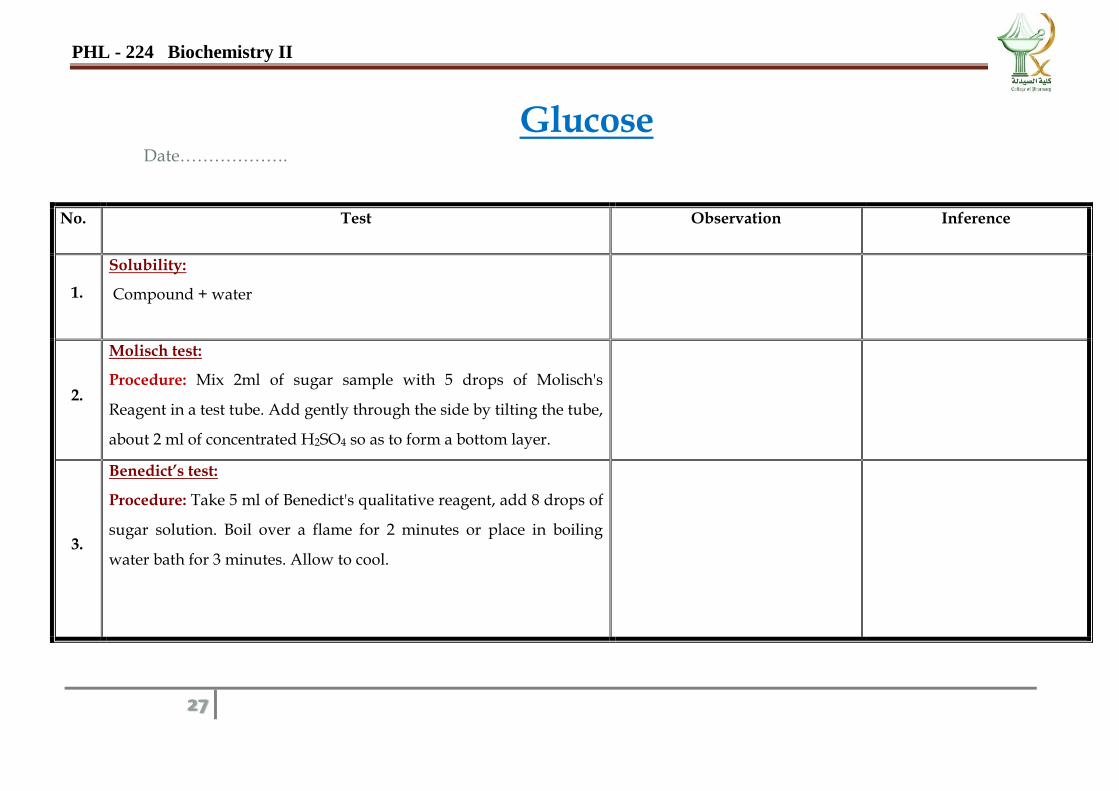
Glucose Date……………….
No. Test Observation Inference
1.
Solubility:
Compound + water
2.
Molisch test:
Procedure: Mix 2ml of sugar sample with 5 drops of Molisch's
Reagent in a test tube. Add gently through the side by tilting the tube,
about 2 ml of concentrated H2SO4 so as to form a bottom layer.
3.
Benedict’s test:
Procedure: Take 5 ml of Benedict's qualitative reagent, add 8 drops of
sugar solution. Boil over a flame for 2 minutes or place in boiling
water bath for 3 minutes. Allow to cool.
PHL - 224 Biochemistry II
28
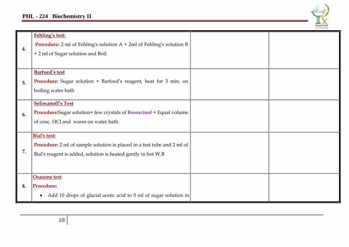
4.
Fehling’s test:
Procedure: 2 ml of Fehling's solution A + 2ml of Fehling's solution B
+ 2 ml of Sugar solution and Boil.
5.
Barfoed’s test
Procedure: Sugar solution + Barfoed’s reagent, heat for 3 min. on
boiling water bath
6.
Seliwanoff's Test
Procedure:Sugar solution+ few crystals of Resorcinol + Equal volume
of conc. HCl and warm on water bath.
7.
Bial's test:
Procedure: 2 ml of sample solution is placed in a test tube and 2 ml of
Bial's reagent is added, solution is heated gently in hot W.B
8.
Osazone test
Procedure:
Add 10 drops of glacial acetic acid to 5 ml of sugar solution in
PHL - 224 Biochemistry II
29
test tube.
Then add a knife point of phenyl hydrazine hydrochloride and
double the amount of sodium acetate crystals.
Mix and warm a little to see that the solids are dissolved.
Filter the solution, and keep the filtrate in a boiling W.B for 20
min.
Cool slowly in water bath not under the tap to have better
crystals.
Examine the crystal under microscope.
Results: On the basis of above observations the given sample was found to be
a. __________________________________
b. __________________________________
c. __________________________________ (____________________)
PHL - 224 Biochemistry II
30
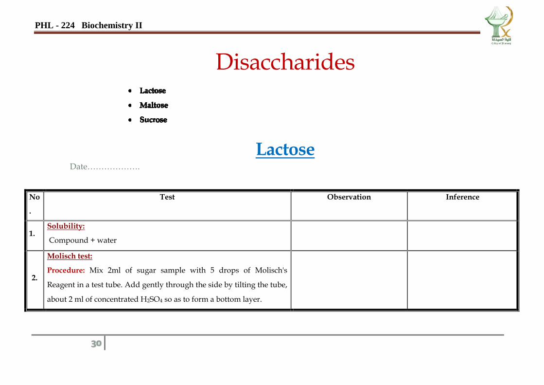
Disaccharides
Lactose Date……………….
No
.
Test Observation Inference
1. Solubility:
Compound + water
2.
Molisch test:
Procedure: Mix 2ml of sugar sample with 5 drops of Molisch's
Reagent in a test tube. Add gently through the side by tilting the tube,
about 2 ml of concentrated H2SO4 so as to form a bottom layer.
PHL - 224 Biochemistry II
31
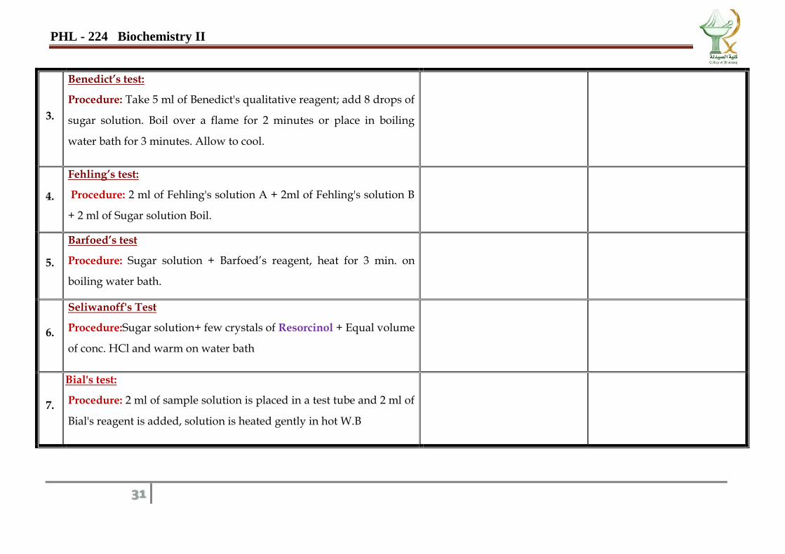
3.
Benedict’s test:
Procedure: Take 5 ml of Benedict's qualitative reagent; add 8 drops of
sugar solution. Boil over a flame for 2 minutes or place in boiling
water bath for 3 minutes. Allow to cool.
4.
Fehling’s test:
Procedure: 2 ml of Fehling's solution A + 2ml of Fehling's solution B
+ 2 ml of Sugar solution Boil.
5.
Barfoed’s test
Procedure: Sugar solution + Barfoed’s reagent, heat for 3 min. on
boiling water bath.
6.
Seliwanoff's Test
Procedure:Sugar solution+ few crystals of Resorcinol + Equal volume
of conc. HCl and warm on water bath
7.
Bial's test:
Procedure: 2 ml of sample solution is placed in a test tube and 2 ml of
Bial's reagent is added, solution is heated gently in hot W.B
PHL - 224 Biochemistry II
32
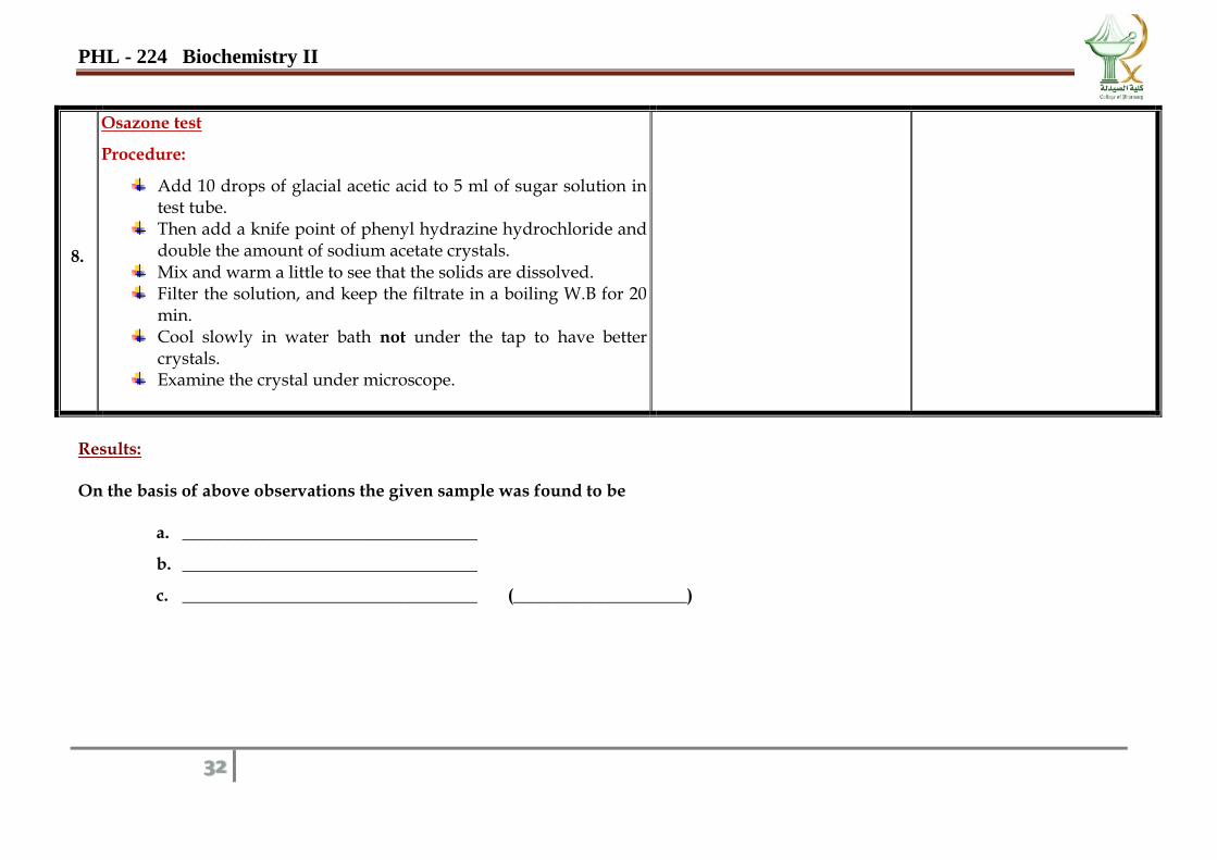
8.
Osazone test
Procedure:
Add 10 drops of glacial acetic acid to 5 ml of sugar solution in test tube.
Then add a knife point of phenyl hydrazine hydrochloride and double the amount of sodium acetate crystals.
Mix and warm a little to see that the solids are dissolved. Filter the solution, and keep the filtrate in a boiling W.B for 20
min. Cool slowly in water bath not under the tap to have better
crystals. Examine the crystal under microscope.
Results: On the basis of above observations the given sample was found to be
a. __________________________________
b. __________________________________
c. __________________________________ (____________________)
PHL - 224 Biochemistry II
33
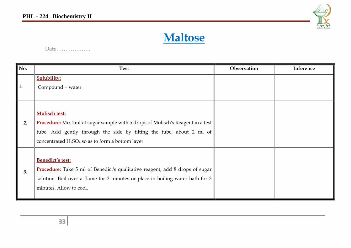
Maltose Date……………….
No. Test Observation Inference
1.
Solubility:
Compound + water
2.
Molisch test:
Procedure: Mix 2ml of sugar sample with 5 drops of Molisch's Reagent in a test
tube. Add gently through the side by tilting the tube, about 2 ml of
concentrated H2SO4 so as to form a bottom layer.
3.
Benedict’s test:
Procedure: Take 5 ml of Benedict's qualitative reagent, add 8 drops of sugar
solution. Boil over a flame for 2 minutes or place in boiling water bath for 3
minutes. Allow to cool.
PHL - 224 Biochemistry II
34
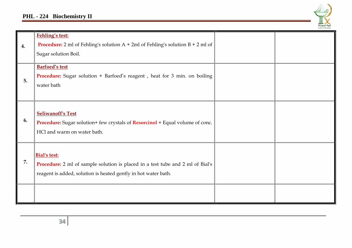
4.
Fehling’s test:
Procedure: 2 ml of Fehling's solution A + 2ml of Fehling's solution B + 2 ml of
Sugar solution Boil.
5.
Barfoed’s test
Procedure: Sugar solution + Barfoed’s reagent , heat for 3 min. on boiling
water bath
6.
Seliwanoff's Test
Procedure: Sugar solution+ few crystals of Resorcinol + Equal volume of conc.
HCl and warm on water bath.
7.
Bial's test:
Procedure: 2 ml of sample solution is placed in a test tube and 2 ml of Bial's
reagent is added, solution is heated gently in hot water bath.
PHL - 224 Biochemistry II
35
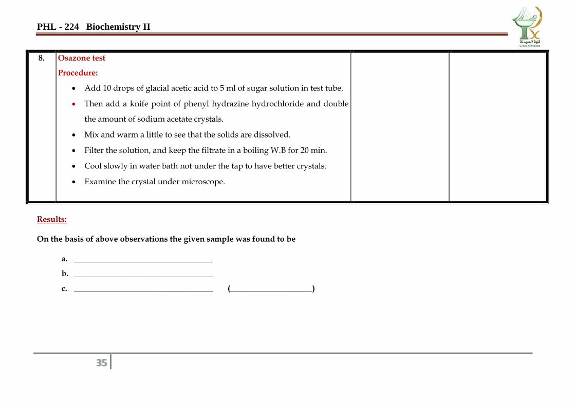
8. Osazone test
Procedure:
Add 10 drops of glacial acetic acid to 5 ml of sugar solution in test tube.
Then add a knife point of phenyl hydrazine hydrochloride and double
the amount of sodium acetate crystals.
Mix and warm a little to see that the solids are dissolved.
Filter the solution, and keep the filtrate in a boiling W.B for 20 min.
Cool slowly in water bath not under the tap to have better crystals.
Examine the crystal under microscope.
Results: On the basis of above observations the given sample was found to be
a. __________________________________
b. __________________________________
c. __________________________________ (____________________)
PHL - 224 Biochemistry II
36
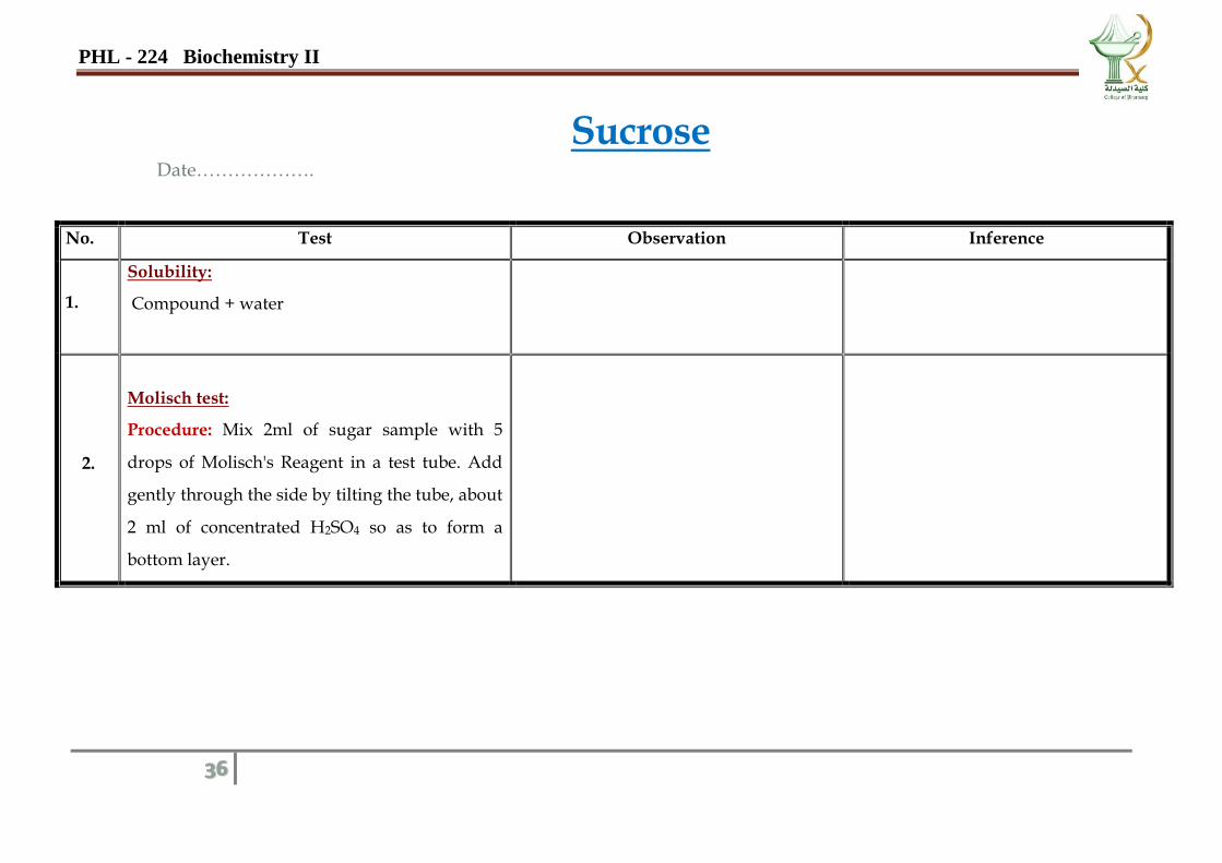
Sucrose Date……………….
No. Test Observation Inference
1.
Solubility:
Compound + water
2.
Molisch test:
Procedure: Mix 2ml of sugar sample with 5
drops of Molisch's Reagent in a test tube. Add
gently through the side by tilting the tube, about
2 ml of concentrated H2SO4 so as to form a
bottom layer.
PHL - 224 Biochemistry II
37
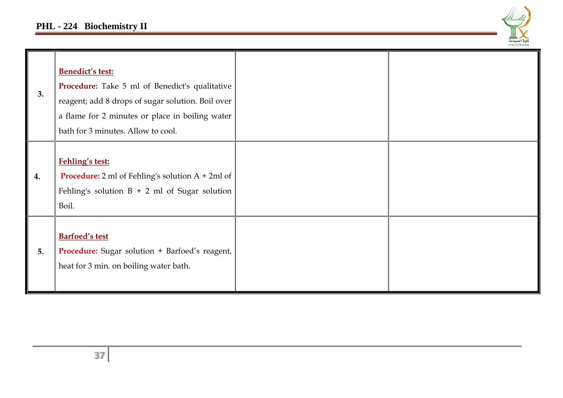
3.
Benedict’s test:
Procedure: Take 5 ml of Benedict's qualitative
reagent; add 8 drops of sugar solution. Boil over
a flame for 2 minutes or place in boiling water
bath for 3 minutes. Allow to cool.
4.
Fehling’s test:
Procedure: 2 ml of Fehling's solution A + 2ml of
Fehling's solution B + 2 ml of Sugar solution
Boil.
5.
Barfoed’s test
Procedure: Sugar solution + Barfoed’s reagent,
heat for 3 min. on boiling water bath.
PHL - 224 Biochemistry II
38
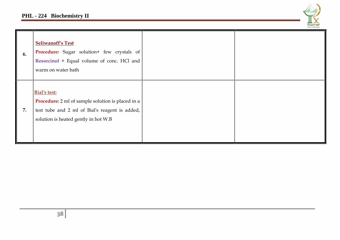
6.
Seliwanoff's Test
Procedure: Sugar solution+ few crystals of
Resorcinol + Equal volume of conc. HCl and
warm on water bath
7.
Bial's test:
Procedure: 2 ml of sample solution is placed in a
test tube and 2 ml of Bial's reagent is added,
solution is heated gently in hot W.B
PHL - 224 Biochemistry II
39
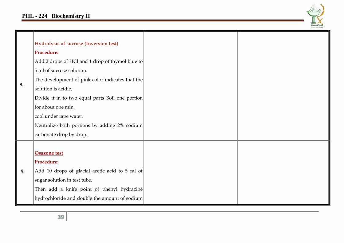
8.
Hydrolysis of sucrose (Inversion test)
Procedure:
Add 2 drops of HCl and 1 drop of thymol blue to
5 ml of sucrose solution.
The development of pink color indicates that the
solution is acidic.
Divide it in to two equal parts Boil one portion
for about one min.
cool under tape water.
Neutralize both portions by adding 2% sodium
carbonate drop by drop.
9.
Osazone test
Procedure:
Add 10 drops of glacial acetic acid to 5 ml of
sugar solution in test tube.
Then add a knife point of phenyl hydrazine
hydrochloride and double the amount of sodium
PHL - 224 Biochemistry II
40
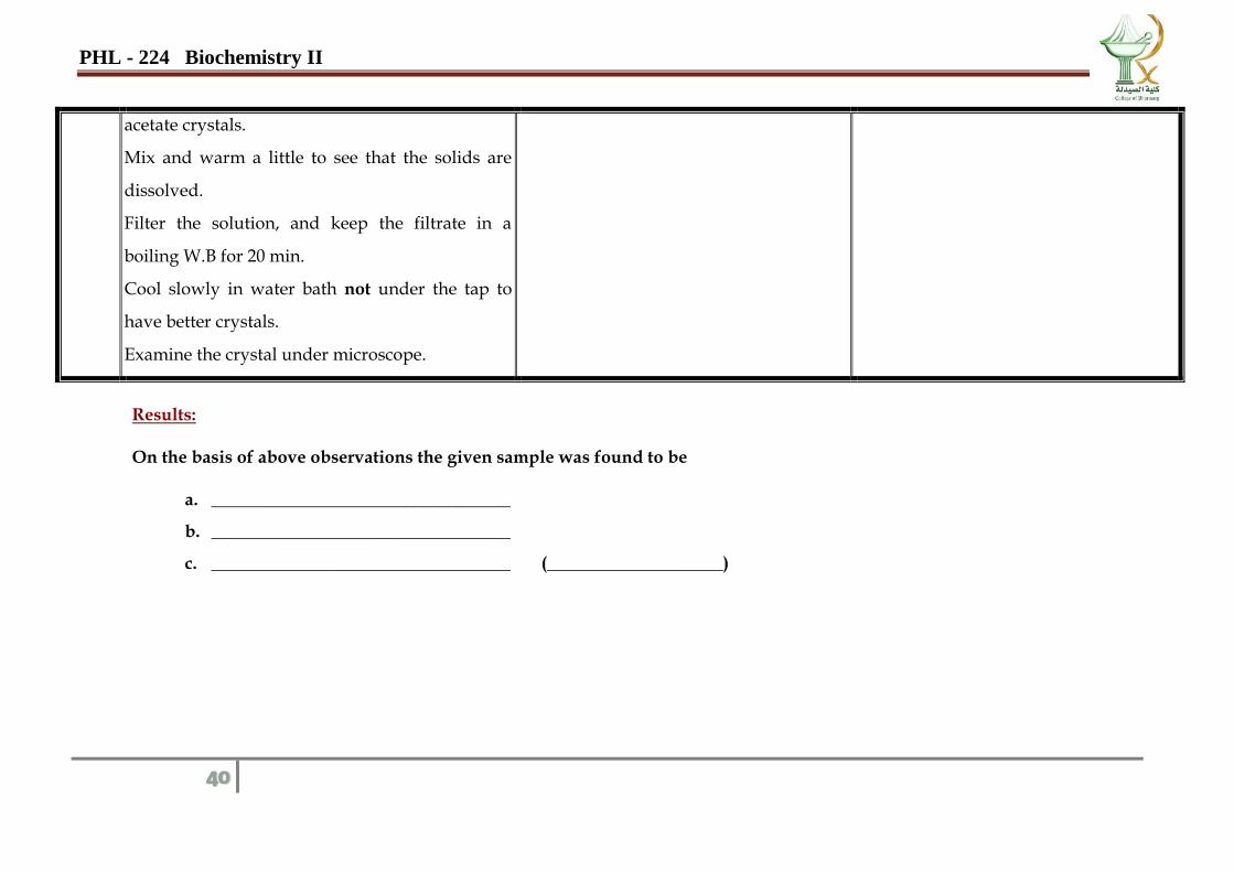
acetate crystals.
Mix and warm a little to see that the solids are
dissolved.
Filter the solution, and keep the filtrate in a
boiling W.B for 20 min.
Cool slowly in water bath not under the tap to
have better crystals.
Examine the crystal under microscope.
Results: On the basis of above observations the given sample was found to be
a. __________________________________
b. __________________________________
c. __________________________________ (____________________)
PHL - 224 Biochemistry II
41
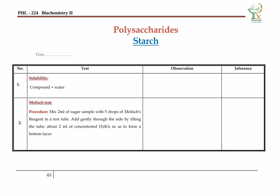
Polysaccharides Starch
Date……………….
No. Test Observation Inference
1.
Solubility:
Compound + water
2.
Molisch test:
Procedure: Mix 2ml of sugar sample with 5 drops of Molisch's
Reagent in a test tube. Add gently through the side by tilting
the tube, about 2 ml of concentrated H2SO4 so as to form a
bottom layer.
PHL - 224 Biochemistry II
42
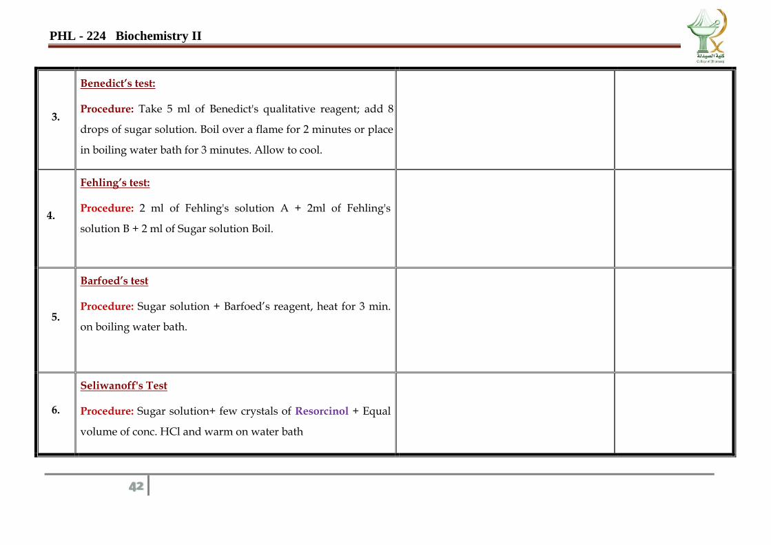
3.
Benedict’s test:
Procedure: Take 5 ml of Benedict's qualitative reagent; add 8
drops of sugar solution. Boil over a flame for 2 minutes or place
in boiling water bath for 3 minutes. Allow to cool.
4.
Fehling’s test:
Procedure: 2 ml of Fehling's solution A + 2ml of Fehling's
solution B + 2 ml of Sugar solution Boil.
5.
Barfoed’s test
Procedure: Sugar solution + Barfoed’s reagent, heat for 3 min.
on boiling water bath.
6.
Seliwanoff's Test
Procedure: Sugar solution+ few crystals of Resorcinol + Equal
volume of conc. HCl and warm on water bath
PHL - 224 Biochemistry II
43
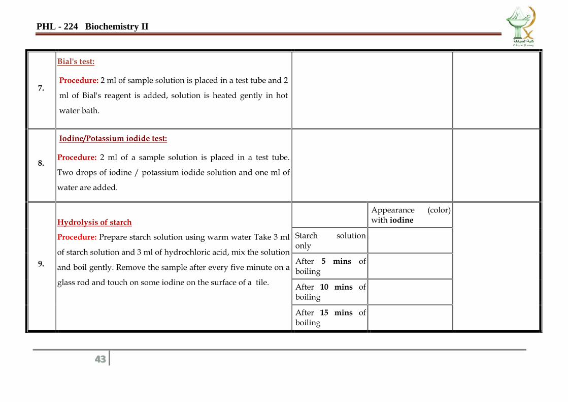
7.
Bial's test:
Procedure: 2 ml of sample solution is placed in a test tube and 2
ml of Bial's reagent is added, solution is heated gently in hot
water bath.
8.
Iodine/Potassium iodide test:
Procedure: 2 ml of a sample solution is placed in a test tube.
Two drops of iodine / potassium iodide solution and one ml of
water are added.
9.
Hydrolysis of starch
Procedure: Prepare starch solution using warm water Take 3 ml
of starch solution and 3 ml of hydrochloric acid, mix the solution
and boil gently. Remove the sample after every five minute on a
glass rod and touch on some iodine on the surface of a tile.
Appearance (color) with iodine
Starch solution only
After 5 mins of boiling
After 10 mins of boiling
After 15 mins of boiling
PHL - 224 Biochemistry II
44
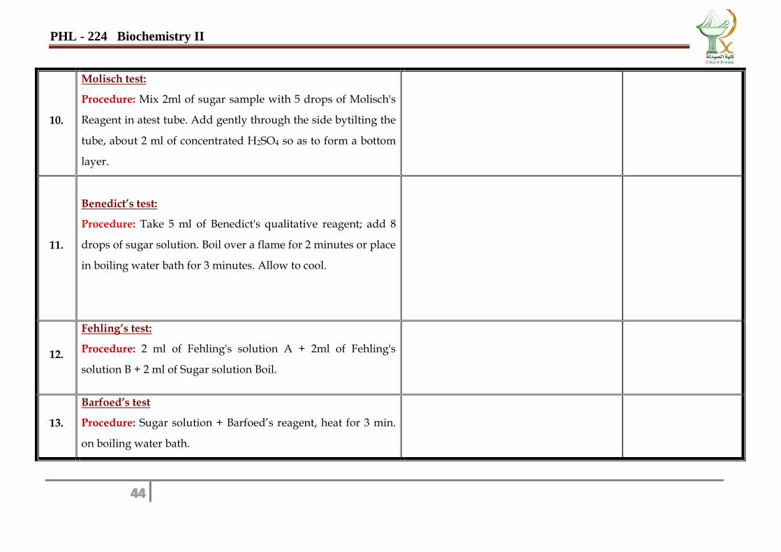
10.
Molisch test:
Procedure: Mix 2ml of sugar sample with 5 drops of Molisch's
Reagent in atest tube. Add gently through the side bytilting the
tube, about 2 ml of concentrated H2SO4 so as to form a bottom
layer.
11.
Benedict’s test:
Procedure: Take 5 ml of Benedict's qualitative reagent; add 8
drops of sugar solution. Boil over a flame for 2 minutes or place
in boiling water bath for 3 minutes. Allow to cool.
12.
Fehling’s test:
Procedure: 2 ml of Fehling's solution A + 2ml of Fehling's
solution B + 2 ml of Sugar solution Boil.
13.
Barfoed’s test
Procedure: Sugar solution + Barfoed’s reagent, heat for 3 min.
on boiling water bath.
PHL - 224 Biochemistry II
45
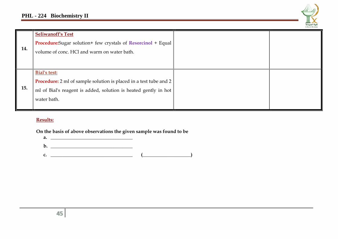
14.
Seliwanoff's Test
Procedure:Sugar solution+ few crystals of Resorcinol + Equal
volume of conc. HCl and warm on water bath.
15.
Bial's test:
Procedure: 2 ml of sample solution is placed in a test tube and 2
ml of Bial's reagent is added, solution is heated gently in hot
water bath.
Results: On the basis of above observations the given sample was found to be
a. __________________________________
b. __________________________________
c. __________________________________ (____________________)
PHL - 224 Biochemistry II
46
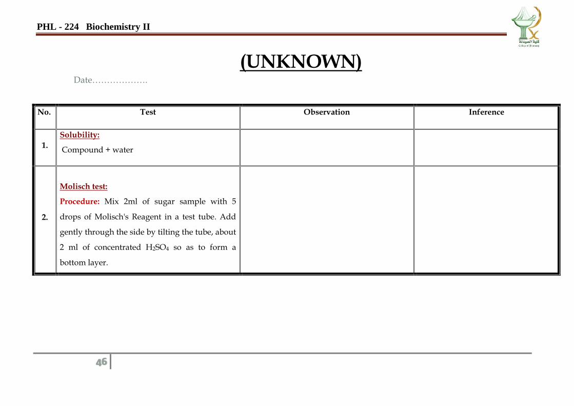
(UNKNOWN) Date……………….
No. Test Observation Inference
1.
Solubility:
Compound + water
2.
Molisch test:
Procedure: Mix 2ml of sugar sample with 5
drops of Molisch's Reagent in a test tube. Add
gently through the side by tilting the tube, about
2 ml of concentrated H2SO4 so as to form a
bottom layer.
PHL - 224 Biochemistry II
47
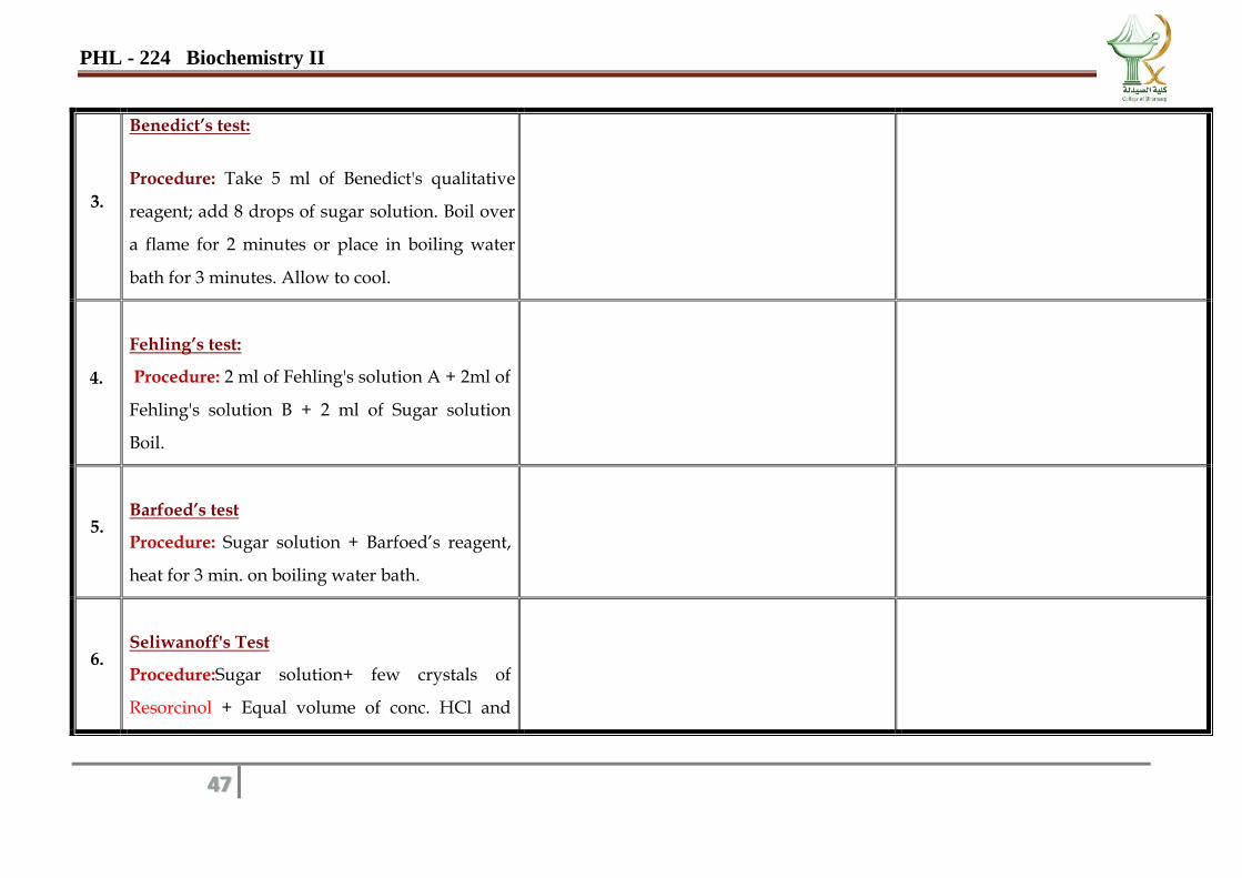
3.
Benedict’s test:
Procedure: Take 5 ml of Benedict's qualitative
reagent; add 8 drops of sugar solution. Boil over
a flame for 2 minutes or place in boiling water
bath for 3 minutes. Allow to cool.
4.
Fehling’s test:
Procedure: 2 ml of Fehling's solution A + 2ml of
Fehling's solution B + 2 ml of Sugar solution
Boil.
5.
Barfoed’s test
Procedure: Sugar solution + Barfoed’s reagent,
heat for 3 min. on boiling water bath.
6.
Seliwanoff's Test
Procedure:Sugar solution+ few crystals of
Resorcinol + Equal volume of conc. HCl and
PHL - 224 Biochemistry II
48
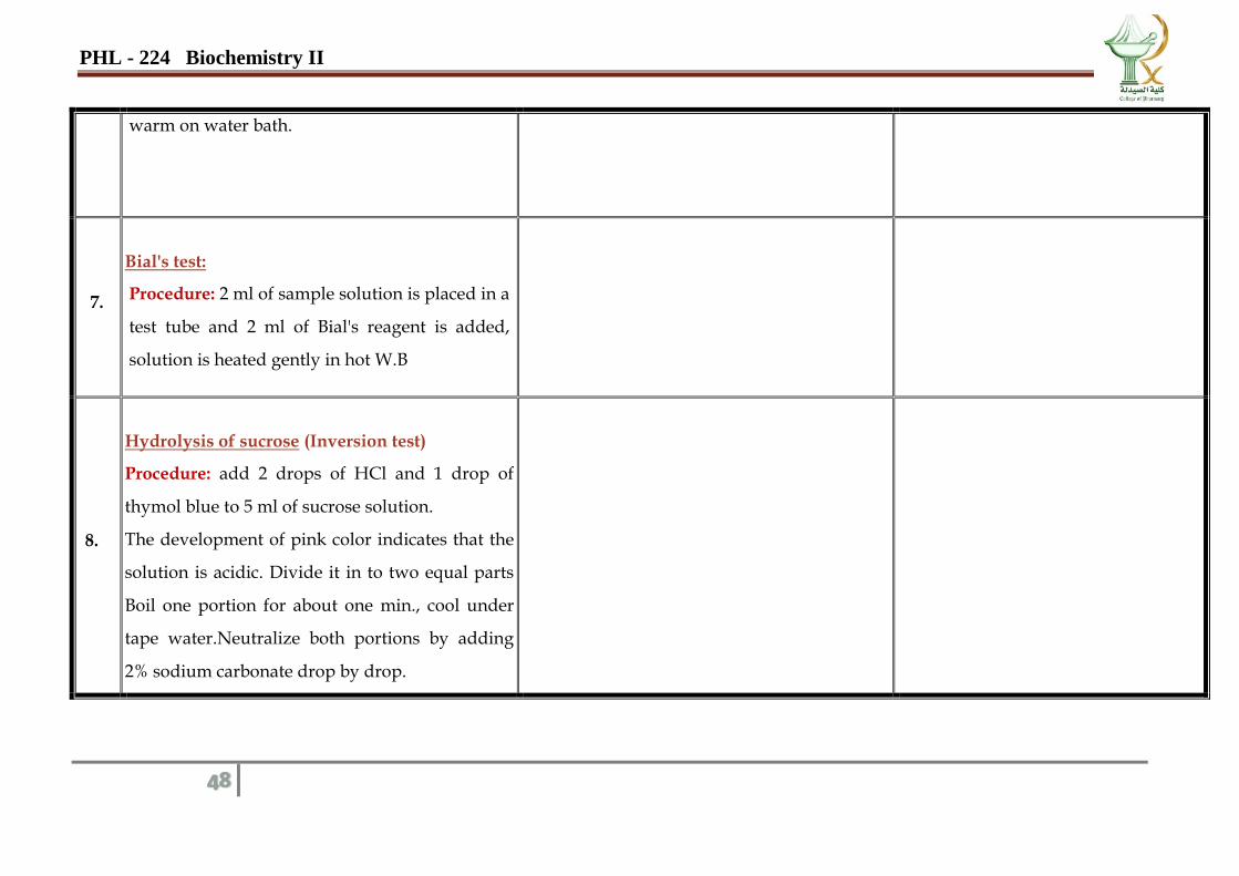
warm on water bath.
7.
Bial's test:
Procedure: 2 ml of sample solution is placed in a
test tube and 2 ml of Bial's reagent is added,
solution is heated gently in hot W.B
8.
Hydrolysis of sucrose (Inversion test)
Procedure: add 2 drops of HCl and 1 drop of
thymol blue to 5 ml of sucrose solution.
The development of pink color indicates that the
solution is acidic. Divide it in to two equal parts
Boil one portion for about one min., cool under
tape water.Neutralize both portions by adding
2% sodium carbonate drop by drop.
PHL - 224 Biochemistry II
49
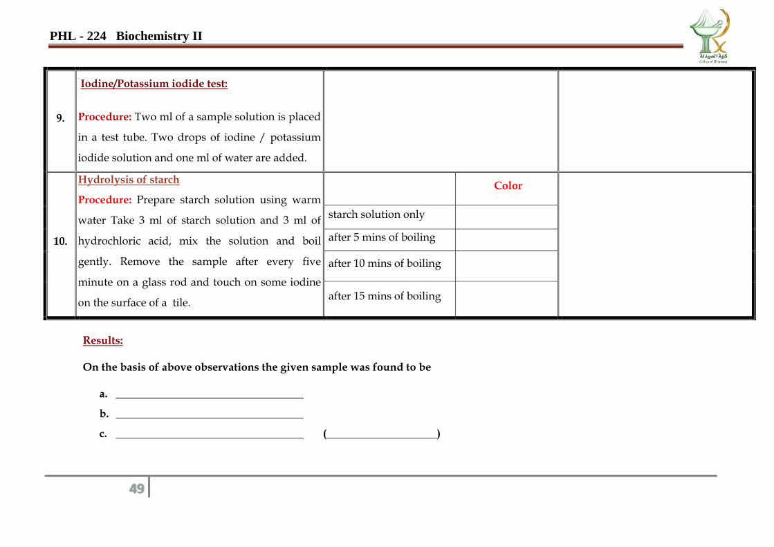
9.
Iodine/Potassium iodide test:
Procedure: Two ml of a sample solution is placed
in a test tube. Two drops of iodine / potassium
iodide solution and one ml of water are added.
10.
Hydrolysis of starch
Procedure: Prepare starch solution using warm
water Take 3 ml of starch solution and 3 ml of
hydrochloric acid, mix the solution and boil
gently. Remove the sample after every five
minute on a glass rod and touch on some iodine
on the surface of a tile.
Color
starch solution only
after 5 mins of boiling
after 10 mins of boiling
after 15 mins of boiling
Results: On the basis of above observations the given sample was found to be
a. __________________________________
b. __________________________________
c. __________________________________ (____________________)
PHL - 224 Biochemistry II
50
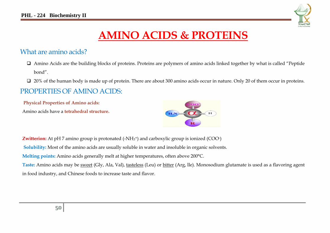
AMINO ACIDS & PROTEINS
What are amino acids?
Amino Acids are the building blocks of proteins. Proteins are polymers of amino acids linked together by what is called “Peptide
bond”.
20% of the human body is made up of protein. There are about 300 amino acids occur in nature. Only 20 of them occur in proteins.
PROPERTIES OF AMINO ACIDS:
Physical Properties of Amino acids:
Amino acids have a tetrahedral structure.
Zwitterion: At pH 7 amino group is protonated (-NH3+) and carboxylic group is ionized (COO-)
Solubility: Most of the amino acids are usually soluble in water and insoluble in organic solvents.
Melting points: Amino acids generally melt at higher temperatures, often above 200°C.
Taste: Amino acids may be sweet (Gly, Ala, Val), tasteless (Leu) or bitter (Arg, lle). Monosodium glutamate is used as a flavoring agent
in food industry, and Chinese foods to increase taste and flavor.
PHL - 224 Biochemistry II
51
Importance of amino acids:
Amino acids have an influence on the function of organs, glands, tendons and arteries. They are furthermore essential for healing
wounds and repairing tissue, especially in the muscles, bones, skin and hair as well as for the removal of all kinds of waste deposits
produced in connection with the metabolism.
Essential Amino Acids: (cannot be synthesized by an organism and must be obtained in the diet) Histidine, Isoleucine, Leucine, Lysine,
Methionine, Phenylalanine, Threonine, Tryptophan, and Valine.
Non-essential Amino Acids: Alanine, Asparagine, Aspartic Acid, Glutamic Acid.
Conditional Amino Acids: Arginine (essential in children, not in adults), Cysteine, Glutamine, Glycine, Proline, Serine, and Tyrosine.
PHL - 224 Biochemistry II
52
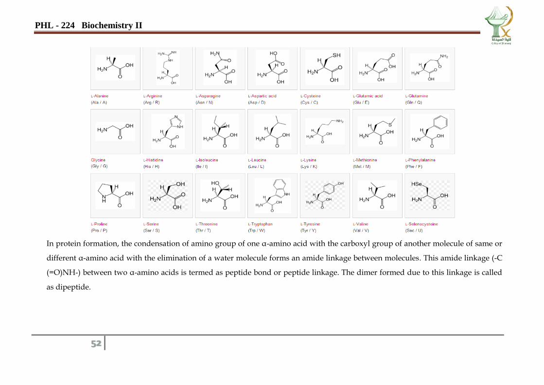
In protein formation, the condensation of amino group of one α-amino acid with the carboxyl group of another molecule of same or
different α-amino acid with the elimination of a water molecule forms an amide linkage between molecules. This amide linkage (-C
(=O)NH-) between two α-amino acids is termed as peptide bond or peptide linkage. The dimer formed due to this linkage is called
as dipeptide.
PHL - 224 Biochemistry II
53
1- Solubility Tests:
Principle:
The solubility of amino acids and proteins is largely dependent on the solution pH. The structural changes in an amino acid or
protein that take place at different pH values alter the relative solubility of the molecule. In acidic solutions, both amino and
carboxylic groups are protonated. In basic solutions, both groups are deprotonated.
Amino acids are essentially soluble in water. Their solubility in water dilute alkali and dilute acid vary from one compound to the
other depending on the structure of their side chains. Apply this test to glycine, tyrosine, glutamic acid and cysteine.
Procedure:
1- Note the solubility of amino acids in water and alcohol by placing a small amount in a test tube, adding a few mL of solvent and
warming if necessary.
2- Determine the amino acid solution is acidic or basic by using a litmus paper while testing the solubility in water.
3- Repeat the solubility test using dilute HCl and dilute NaOH.
PHL - 224 Biochemistry II
54
Experimental Procedure:
No. Test Observation Inference &Interpretation
I. General Test for amino acid and protein (Amine groups in proteins, peptones and amino acids)
1.
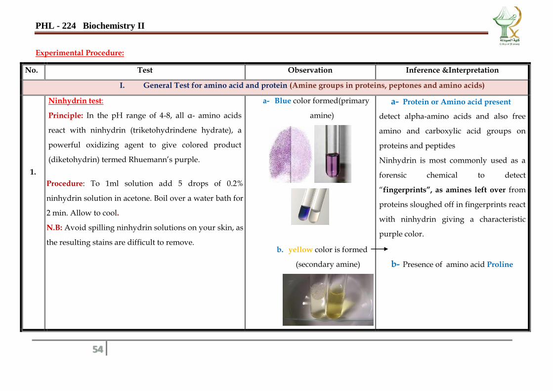
Ninhydrin test:
Principle: In the pH range of 4-8, all α- amino acids
react with ninhydrin (triketohydrindene hydrate), a
powerful oxidizing agent to give colored product
(diketohydrin) termed Rhuemann’s purple.
Procedure: To 1ml solution add 5 drops of 0.2%
ninhydrin solution in acetone. Boil over a water bath for
2 min. Allow to cool.
N.B: Avoid spilling ninhydrin solutions on your skin, as
the resulting stains are difficult to remove.
a- Blue color formed(primary
amine)
b. yellow color is formed
(secondary amine)
a- Protein or Amino acid present
detect alpha-amino acids and also free
amino and carboxylic acid groups on
proteins and peptides
Ninhydrin is most commonly used as a
forensic chemical to detect
“fingerprints”, as amines left over from
proteins sloughed off in fingerprints react
with ninhydrin giving a characteristic
purple color.
b- Presence of amino acid Proline
PHL - 224 Biochemistry II
55
II. Tests for protein
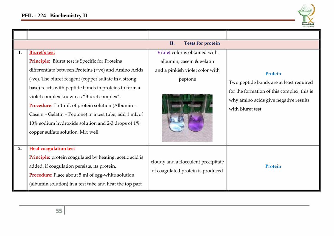
1. Biuret’s test
Principle: Biuret test is Specific for Proteins
differentiate between Proteins (+ve) and Amino Acids
(-ve). The biuret reagent (copper sulfate in a strong
base) reacts with peptide bonds in proteins to form a
violet complex known as “Biuret complex”.
Procedure: To 1 mL of protein solution (Albumin –
Casein – Gelatin – Peptone) in a test tube, add 1 mL of
10% sodium hydroxide solution and 2-3 drops of 1%
copper sulfate solution. Mix well
Violet color is obtained with
albumin, casein & gelatin
and a pinkish violet color with
peptone
Protein
Two peptide bonds are at least required
for the formation of this complex, this is
why amino acids give negative results
with Biuret test.
2. Heat coagulation test
Principle: protein coagulated by heating, acetic acid is
added, if coagulation persists, its protein.
Procedure: Place about 5 ml of egg-white solution
(albumin solution) in a test tube and heat the top part
cloudy and a flocculent precipitate
of coagulated protein is produced Protein
PHL - 224 Biochemistry II
56
of the solution only.
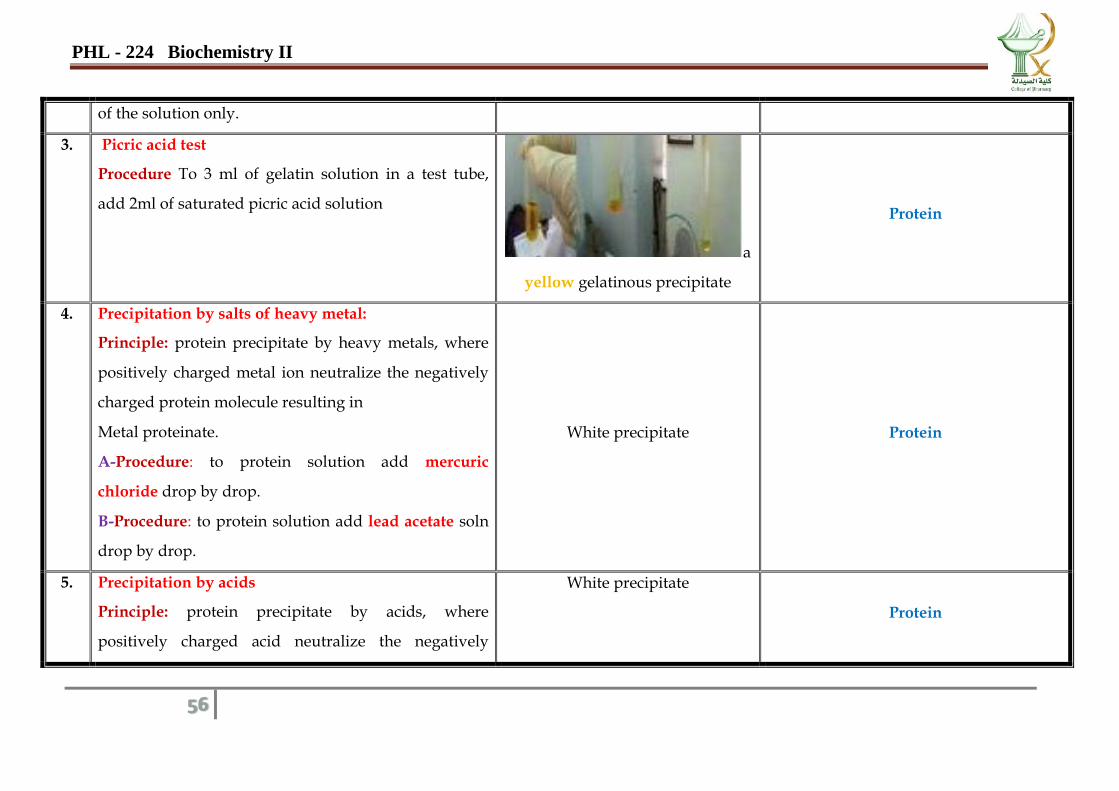
3. Picric acid test
Procedure To 3 ml of gelatin solution in a test tube,
add 2ml of saturated picric acid solution
a
yellow gelatinous precipitate
Protein
4. Precipitation by salts of heavy metal:
Principle: protein precipitate by heavy metals, where
positively charged metal ion neutralize the negatively
charged protein molecule resulting in
Metal proteinate.
A-Procedure: to protein solution add mercuric
chloride drop by drop.
B-Procedure: to protein solution add lead acetate soln
drop by drop.
White precipitate Protein
5. Precipitation by acids
Principle: protein precipitate by acids, where
positively charged acid neutralize the negatively
White precipitate
Protein
PHL - 224 Biochemistry II
57
charged protein
A - Procedure: to protein solution add sulphasalicylic
acid drop by drop.
B - Procedure: to protein solution add tannic acid
drop by drop.
A light brownprecipitate
6. Esbach's test
Principle: protein precipitate by Esbchs reagent, where
positively charged reagent neutralize the negatively
charged protein
Procedure: to protein solution add 5 ml of
esbachsreagent drop by drop.
yellow precipitate
Protein
III. Test for α -amino-acid glycine
1. p-nitrobenzoyl chloride & pyridine test:
Principle: Glycine in quantities as small as 0.5 g
detected as an orange-red color by reaction with p-
nitrobenzoylchloride & pyridine. The procedure can
be used both as a qualitative spot test and as a sensitive
Orange-red to maroon color
develops immediately, varying in
shade with the concentration of
glycine.
Glycine
N.B.As little as 0.5 g. of glycine develops
a perceptible orange-red color. All other
amino acids fail to react in concentrations
below 0.5 mg with larger quantities, a
PHL - 224 Biochemistry II
58
quantitative method, the color is due to aziactone
formation.
Procedure: A few crystals of powder sample, on a filter
paper strip or a glass slide, and approximately 1 mg. of
solidp-nitrobenzoyl chloride is placed on top. One to
three drops of pyridine are then added to wet the
mixture.
The color is soluble in polar solvents such as
chloroform, dichioroethylene, tetrachloroethane,
ethylacetate, and also in excess pyridine.
pale bluish color, in contrast to colorless
blanks, develops with some amino acids.
Acetylglycine, glycyiglycine,hippuric
acid, and salicyluric acid do not react in
the cold, but on warming, a yellow color
is produced with milligram quantities.
IV. Test for amino-acids contain: activated benzene rings ( Tyrosine and Tryptophan)
1.
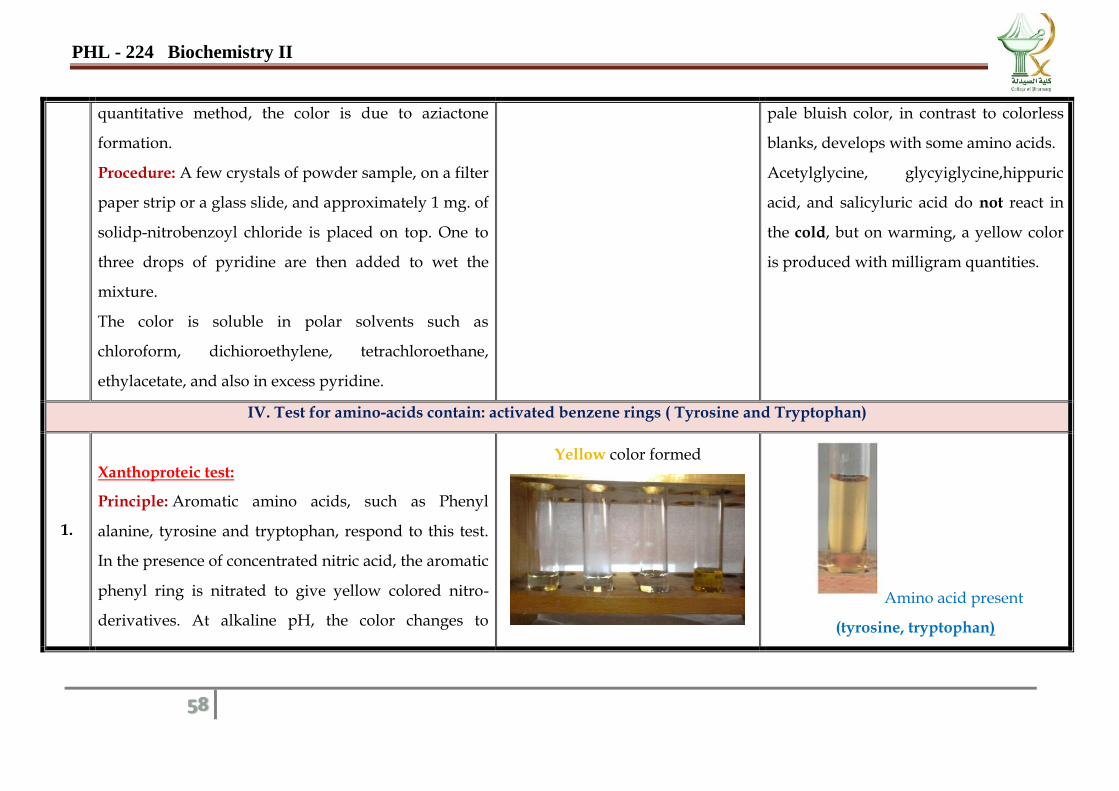
Xanthoproteic test:
Principle: Aromatic amino acids, such as Phenyl
alanine, tyrosine and tryptophan, respond to this test.
In the presence of concentrated nitric acid, the aromatic
phenyl ring is nitrated to give yellow colored nitro-
derivatives. At alkaline pH, the color changes to
Yellow color formed
Amino acid present
(tyrosine, tryptophan)
PHL - 224 Biochemistry II
59
orange due to the ionization of the phenolic group.
Procedure: To 2ml of solution in a boiling test tube,
add an equal volume of conc. HNO3. Heat over a flame
for 2 min and observe the color. Now COOL
THOROUGHLY under the tap and CAUTIOSLY run
in sufficient 40% NaOH to make the solution strongly
alkaline.
V. Test for amino-acids contain: Indol group
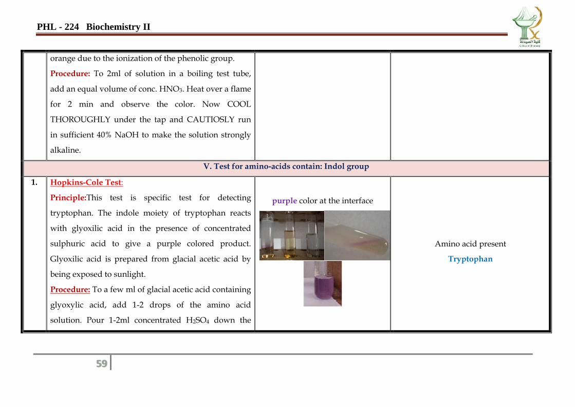
1. Hopkins-Cole Test:
Principle:This test is specific test for detecting
tryptophan. The indole moiety of tryptophan reacts
with glyoxilic acid in the presence of concentrated
sulphuric acid to give a purple colored product.
Glyoxilic acid is prepared from glacial acetic acid by
being exposed to sunlight.
Procedure: To a few ml of glacial acetic acid containing
glyoxylic acid, add 1-2 drops of the amino acid
solution. Pour 1-2ml concentrated H2SO4 down the
purple color at the interface
Amino acid present
Tryptophan
PHL - 224 Biochemistry II
60
side of the sloping test tube to form a layer underneath
the acetic acid.
2. Ehrlich's test:To 0.5ml of the amino acid solution, add
2ml Ehrlich reagent
A colored complex formed
Amino acid present
Tryptophan
VI. Test for : phenolic amino acid
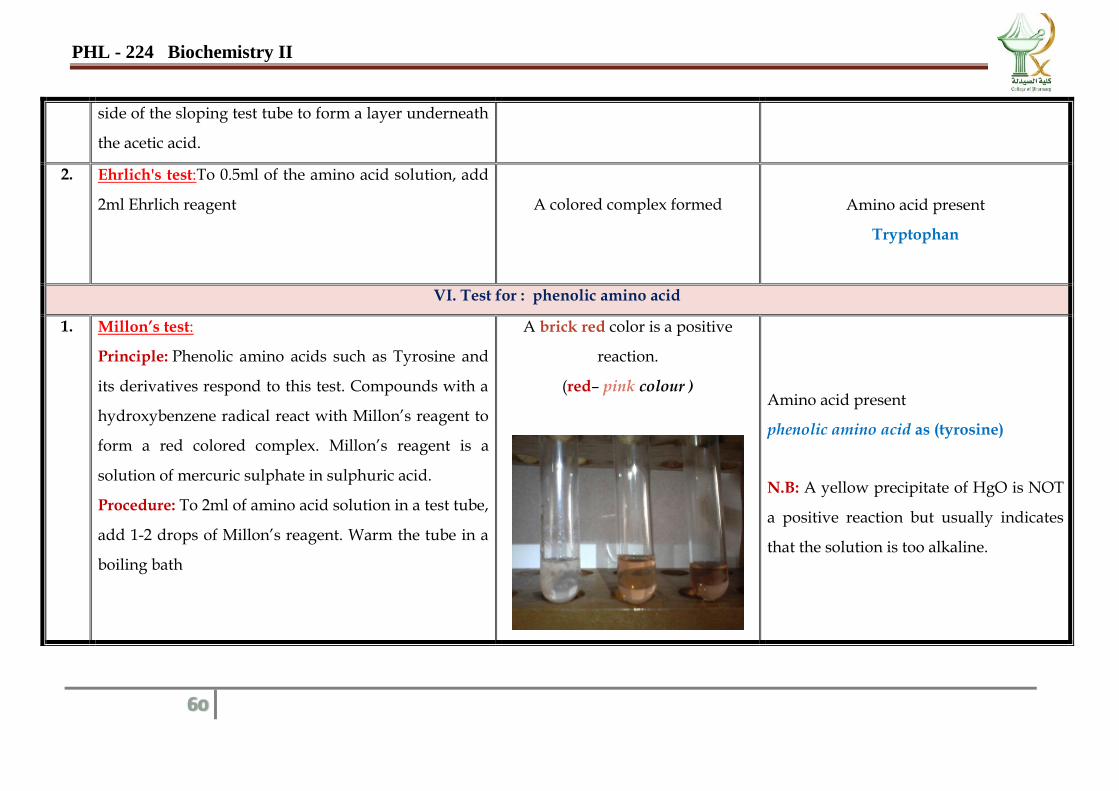
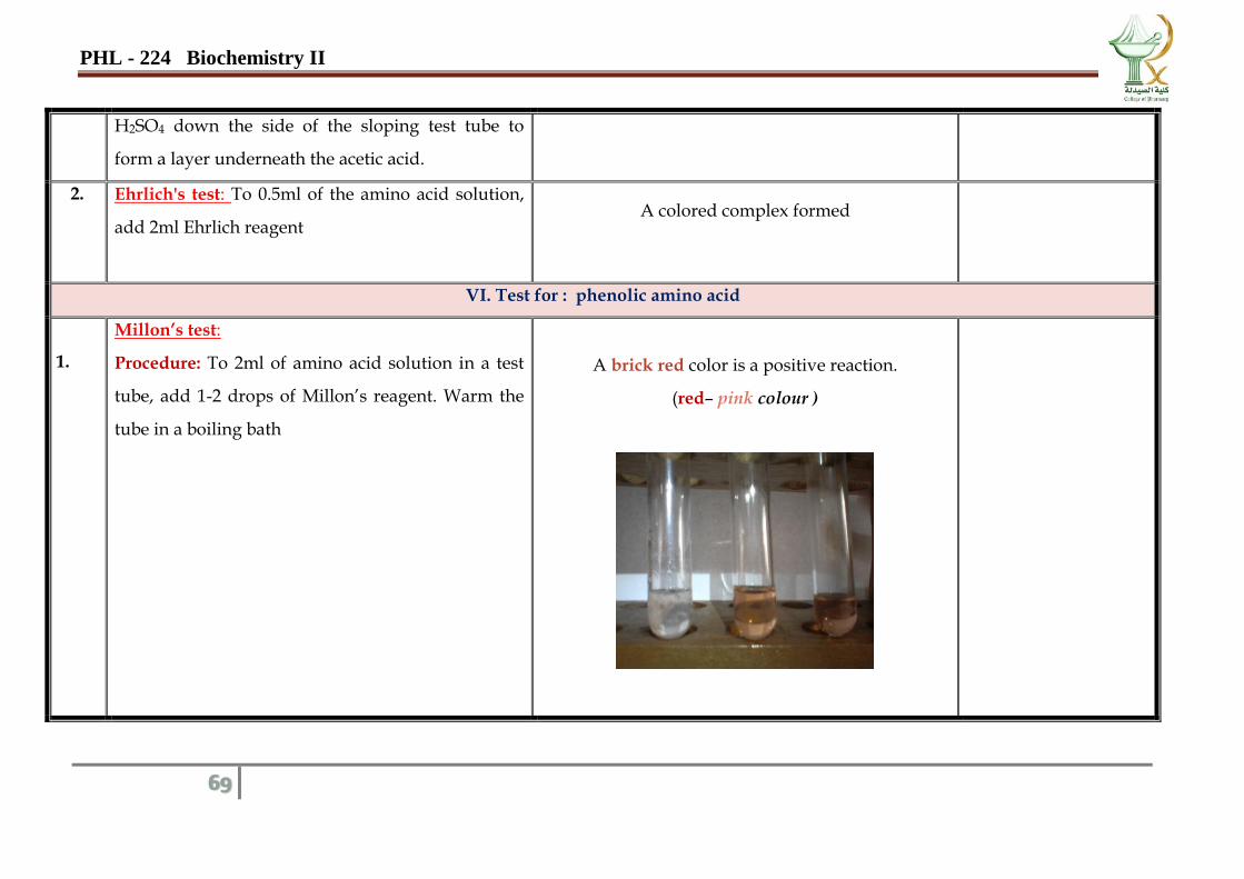
1. Millon’s test:
Principle: Phenolic amino acids such as Tyrosine and
its derivatives respond to this test. Compounds with a
hydroxybenzene radical react with Millon’s reagent to
form a red colored complex. Millon’s reagent is a
solution of mercuric sulphate in sulphuric acid.
Procedure: To 2ml of amino acid solution in a test tube,
add 1-2 drops of Millon’s reagent. Warm the tube in a
boiling bath
A brick red color is a positive
reaction.
(red– pink colour )
Amino acid present
phenolic amino acid as (tyrosine)
N.B: A yellow precipitate of HgO is NOT
a positive reaction but usually indicates
that the solution is too alkaline.
PHL - 224 Biochemistry II
61
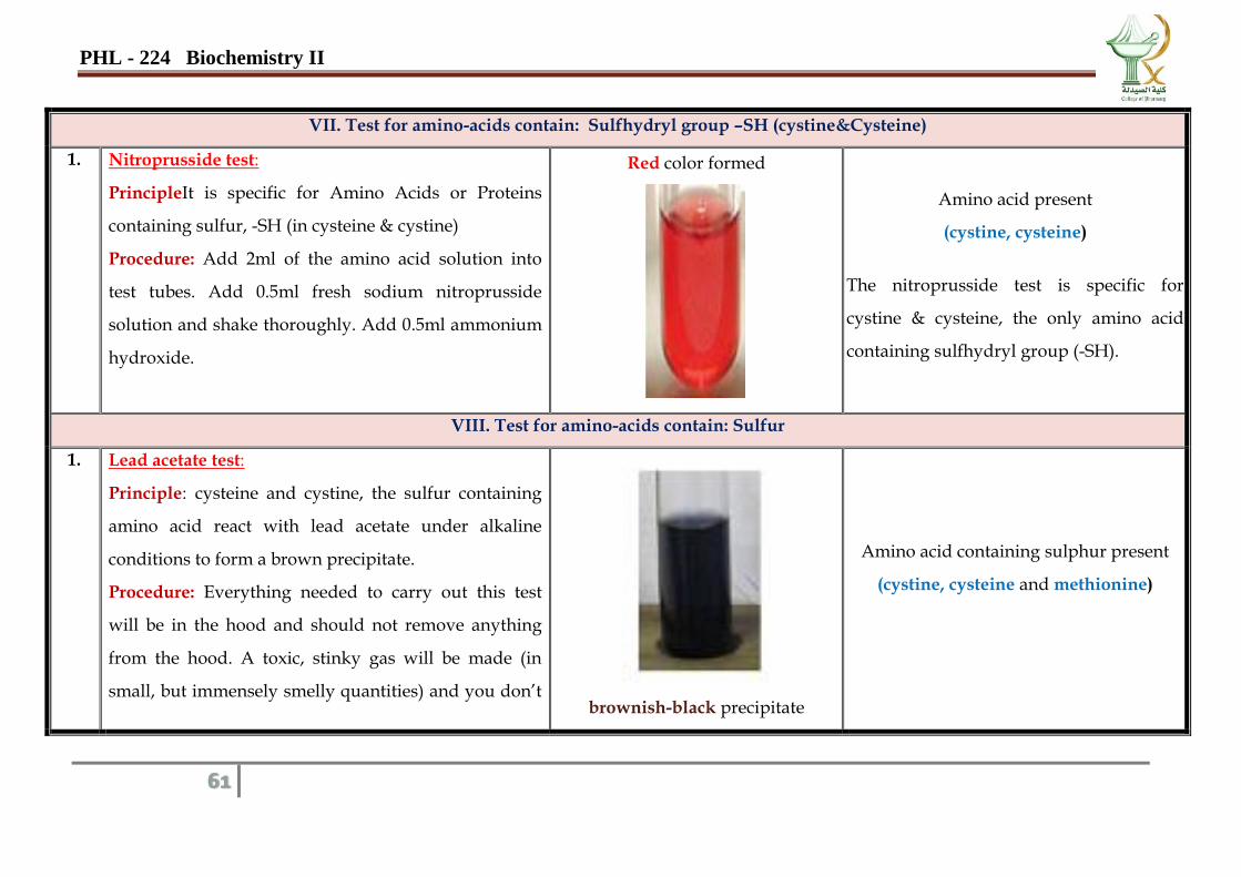
VII. Test for amino-acids contain: Sulfhydryl group –SH (cystine&Cysteine)
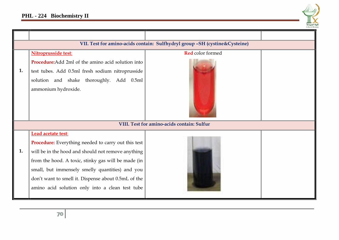
1. Nitroprusside test:
PrincipleIt is specific for Amino Acids or Proteins
containing sulfur, -SH (in cysteine & cystine)
Procedure: Add 2ml of the amino acid solution into
test tubes. Add 0.5ml fresh sodium nitroprusside
solution and shake thoroughly. Add 0.5ml ammonium
hydroxide.
Red color formed
Amino acid present
(cystine, cysteine)
The nitroprusside test is specific for
cystine & cysteine, the only amino acid
containing sulfhydryl group (-SH).
VIII. Test for amino-acids contain: Sulfur
1. Lead acetate test:
Principle: cysteine and cystine, the sulfur containing
amino acid react with lead acetate under alkaline
conditions to form a brown precipitate.
Procedure: Everything needed to carry out this test
will be in the hood and should not remove anything
from the hood. A toxic, stinky gas will be made (in
small, but immensely smelly quantities) and you don’t
brownish-black precipitate
Amino acid containing sulphur present
(cystine, cysteine and methionine)
PHL - 224 Biochemistry II
62
want to smell it. Dispense about 0.5mL of the amino
acid solution only into a clean test tube (found in the
hood, where you will leave it when you are done).
Add 0.5mL of 20% NaOH and insert the test tube in a
boiling water bath for 1 min. Add 2 drops of lead (II)
acetate solution.
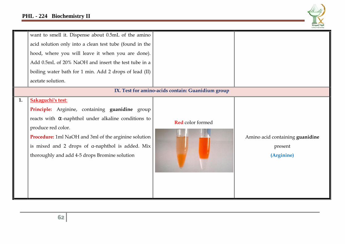
IX. Test for amino-acids contain: Guanidium group
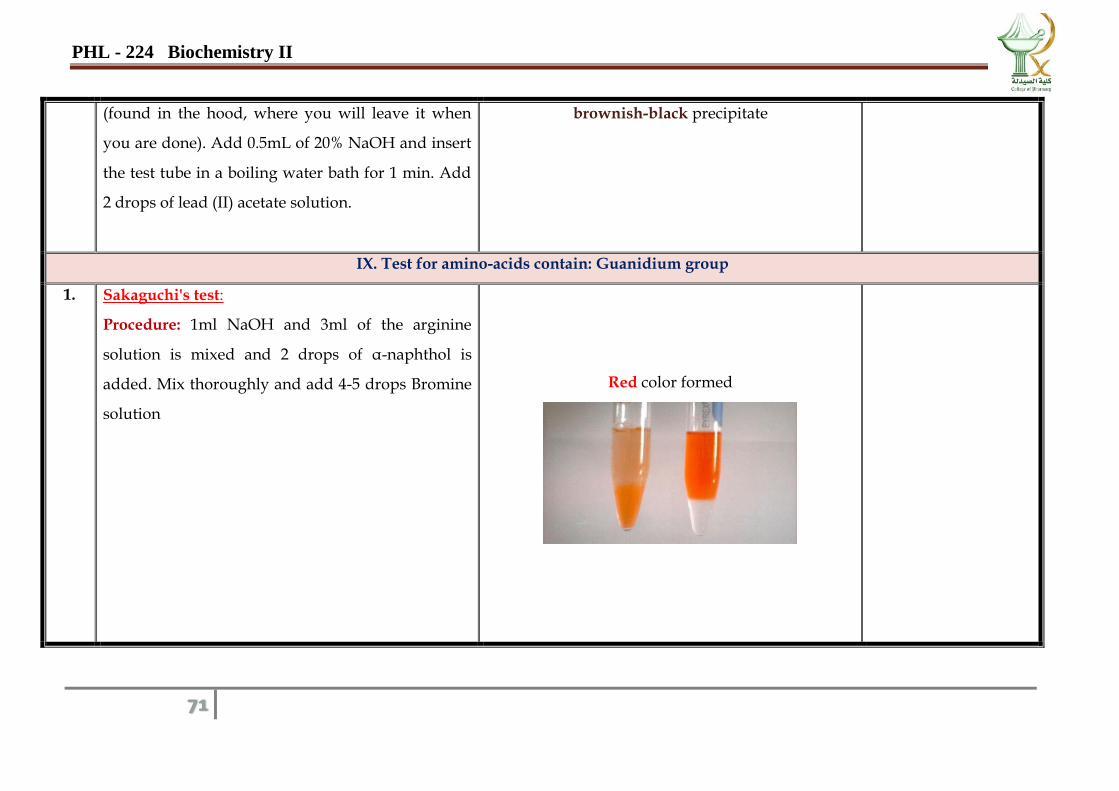
1. Sakaguchi's test:
Principle: Arginine, containing guanidine group
reacts with α -naphthol under alkaline conditions to
produce red color.
Procedure: 1ml NaOH and 3ml of the arginine solution
is mixed and 2 drops of α-naphthol is added. Mix
thoroughly and add 4-5 drops Bromine solution
Red color formed
Amino acid containing guanidine
present
(Arginine)
PHL - 224 Biochemistry II
63
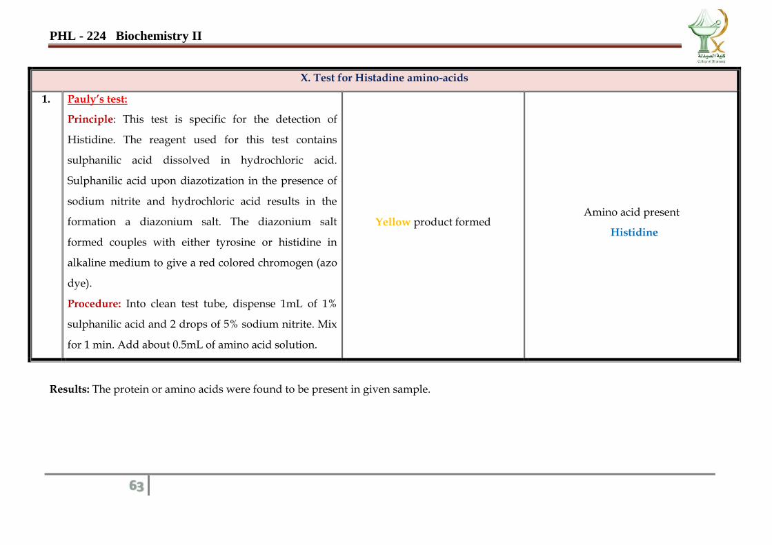
X. Test for Histadine amino-acids
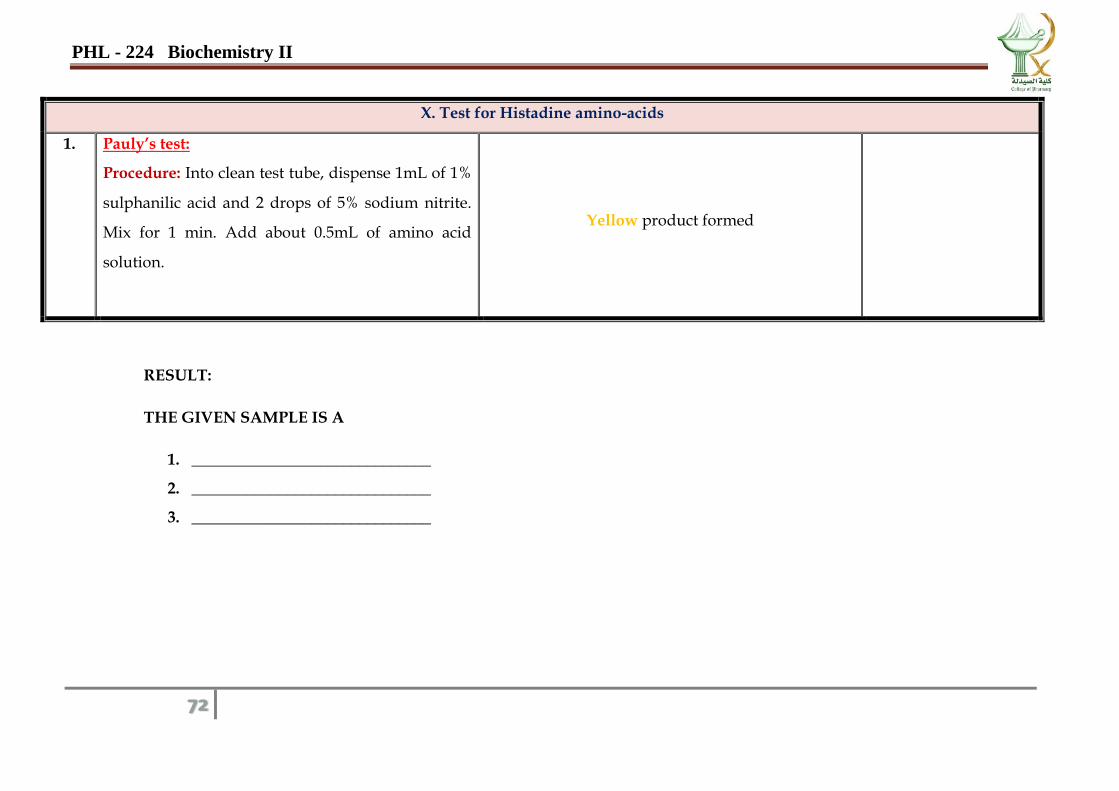
1. Pauly’s test:
Principle: This test is specific for the detection of
Histidine. The reagent used for this test contains
sulphanilic acid dissolved in hydrochloric acid.
Sulphanilic acid upon diazotization in the presence of
sodium nitrite and hydrochloric acid results in the
formation a diazonium salt. The diazonium salt
formed couples with either tyrosine or histidine in
alkaline medium to give a red colored chromogen (azo
dye).
Procedure: Into clean test tube, dispense 1mL of 1%
sulphanilic acid and 2 drops of 5% sodium nitrite. Mix
for 1 min. Add about 0.5mL of amino acid solution.
Yellow product formed Amino acid present
Histidine
Results: The protein or amino acids were found to be present in given sample.
PHL - 224 Biochemistry II
64
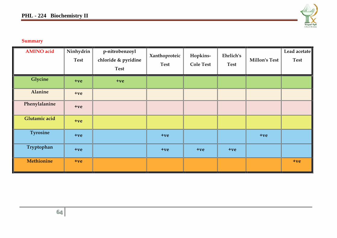
Summary
AMINO acid Ninhydrin
Test
p-nitrobenzoyl
chloride & pyridine
Test
Xanthoproteic
Test
Hopkins-
Cole Test
Ehrlich's
Test Millon's Test
Lead acetate
Test
Glycine +ve +ve
Alanine +ve
Phenylalanine +ve
Glutamic acid +ve
Tyrosine +ve +ve +ve
Tryptophan +ve +ve +ve +ve
Methionine +ve
+ve
PHL - 224 Biochemistry II
65
Date……………….
No. Test Observation
Inference
&Interpretation
1. General Test for amino acid and protein (Amine groups in proteins, peptones and amino acids)
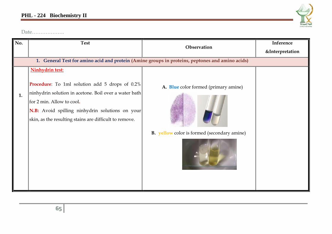
1.
Ninhydrin test:
Procedure: To 1ml solution add 5 drops of 0.2%
ninhydrin solution in acetone. Boil over a water bath
for 2 min. Allow to cool.
N.B: Avoid spilling ninhydrin solutions on your
skin, as the resulting stains are difficult to remove.
A. Blue color formed (primary amine)
B. yellow color is formed (secondary amine)
PHL - 224 Biochemistry II
66
III. Tests for protein
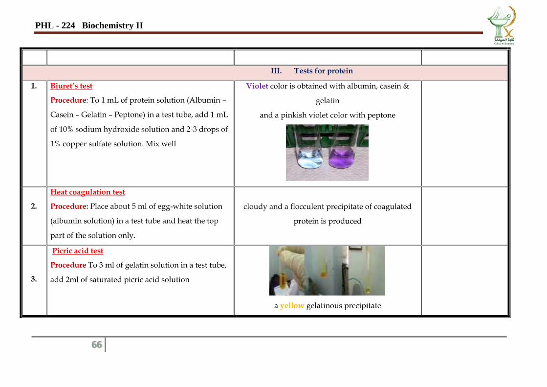
1. Biuret’s test
Procedure: To 1 mL of protein solution (Albumin –
Casein – Gelatin – Peptone) in a test tube, add 1 mL
of 10% sodium hydroxide solution and 2-3 drops of
1% copper sulfate solution. Mix well
Violet color is obtained with albumin, casein &
gelatin
and a pinkish violet color with peptone
2.
Heat coagulation test
Procedure: Place about 5 ml of egg-white solution
(albumin solution) in a test tube and heat the top
part of the solution only.
cloudy and a flocculent precipitate of coagulated
protein is produced
3.
Picric acid test
Procedure To 3 ml of gelatin solution in a test tube,
add 2ml of saturated picric acid solution
a yellow gelatinous precipitate
PHL - 224 Biochemistry II
67
4.
Precipitation by salts of heavy metal:
A-Procedure: to protein solution add mercuric
chloride drop by drop.
B-Procedure: to protein solution add lead acetate
solution drop by drop.
White precipitate
5.
Precipitation by acids
A - Procedure: to protein solution add
sulphasalicylic acid drop by drop.
B - Procedure: to protein solution add tannic acid
drop by drop.
White precipitate
A light brown precipitate
6.
Esbach's test
Procedure: to protein solution add 5 ml of esbachs
reagent drop by drop.
yellow precipitate
III. Test for α -amino-acid glycine
p-nitrobenzoyl chloride& pyridine test:
Procedure: A few crystals of powder sample, on a
filter paper strip or a glass slide, and approximately
1 mg. of solid p-nitrobenzoyl chloride is placed on
Orange-red to maroon color develops immediately,
varying in shade with the concentration of glycine.
PHL - 224 Biochemistry II
68
1.
top. One to three drops of pyridine are then added
to wet the mixture.
The color is soluble in polar solvents such as
chloroform, dichioroethylene, tetrachloroethane,
ethylacetate, and also in excess pyridine.
IV. Test for amino-acids contain: activated benzene rings ( Tyrosine and Tryptophan)
1.
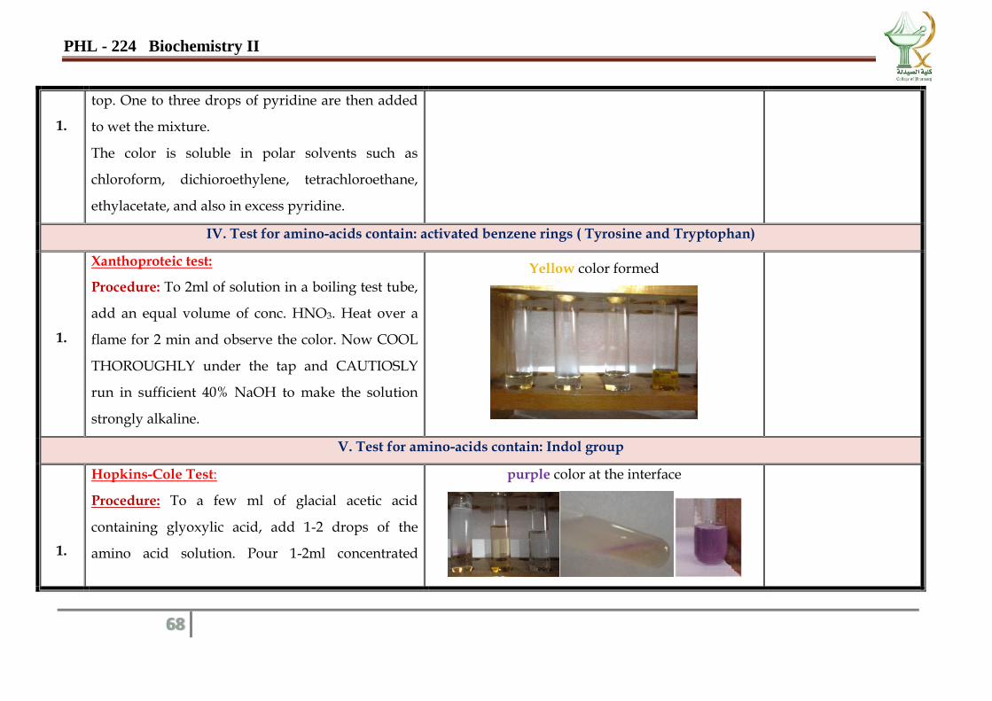
Xanthoproteic test:
Procedure: To 2ml of solution in a boiling test tube,
add an equal volume of conc. HNO3. Heat over a
flame for 2 min and observe the color. Now COOL
THOROUGHLY under the tap and CAUTIOSLY
run in sufficient 40% NaOH to make the solution
strongly alkaline.
Yellow color formed
V. Test for amino-acids contain: Indol group
1.
Hopkins-Cole Test:
Procedure: To a few ml of glacial acetic acid
containing glyoxylic acid, add 1-2 drops of the
amino acid solution. Pour 1-2ml concentrated
purple color at the interface
PHL - 224 Biochemistry II
69
H2SO4 down the side of the sloping test tube to
form a layer underneath the acetic acid.
2. Ehrlich's test: To 0.5ml of the amino acid solution,
add 2ml Ehrlich reagent
A colored complex formed
VI. Test for : phenolic amino acid
1.
Millon’s test:
Procedure: To 2ml of amino acid solution in a test
tube, add 1-2 drops of Millon’s reagent. Warm the
tube in a boiling bath
A brick red color is a positive reaction.
(red– pink colour )
PHL - 224 Biochemistry II
70
VII. Test for amino-acids contain: Sulfhydryl group –SH (cystine&Cysteine)
1.
Nitroprusside test:
Procedure:Add 2ml of the amino acid solution into
test tubes. Add 0.5ml fresh sodium nitroprusside
solution and shake thoroughly. Add 0.5ml
ammonium hydroxide.
Red color formed
VIII. Test for amino-acids contain: Sulfur
1.
Lead acetate test:
Procedure: Everything needed to carry out this test
will be in the hood and should not remove anything
from the hood. A toxic, stinky gas will be made (in
small, but immensely smelly quantities) and you
don’t want to smell it. Dispense about 0.5mL of the
amino acid solution only into a clean test tube
PHL - 224 Biochemistry II
71
(found in the hood, where you will leave it when
you are done). Add 0.5mL of 20% NaOH and insert
the test tube in a boiling water bath for 1 min. Add
2 drops of lead (II) acetate solution.
brownish-black precipitate
IX. Test for amino-acids contain: Guanidium group
1. Sakaguchi's test:
Procedure: 1ml NaOH and 3ml of the arginine
solution is mixed and 2 drops of α-naphthol is
added. Mix thoroughly and add 4-5 drops Bromine
solution
Red color formed
PHL - 224 Biochemistry II
72
X. Test for Histadine amino-acids
1. Pauly’s test:
Procedure: Into clean test tube, dispense 1mL of 1%
sulphanilic acid and 2 drops of 5% sodium nitrite.
Mix for 1 min. Add about 0.5mL of amino acid
solution.
Yellow product formed
RESULT:
THE GIVEN SAMPLE IS A
1. ______________________________
2. ______________________________
3. ______________________________
PHL - 224 Biochemistry II
73
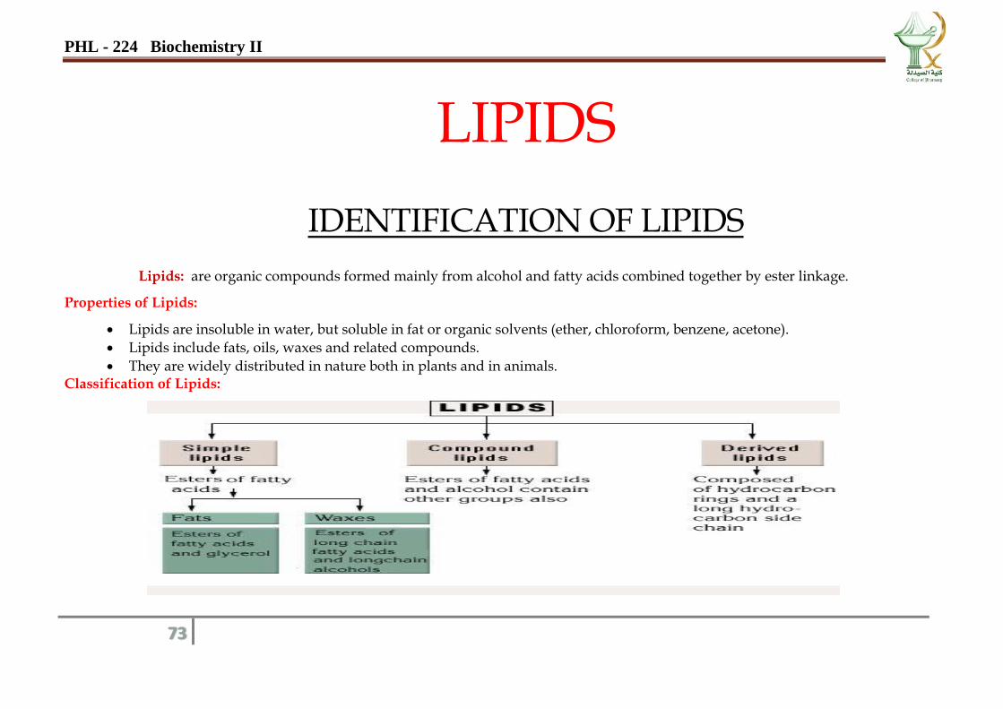
LIPIDS
IDENTIFICATION OF LIPIDS
Lipids: are organic compounds formed mainly from alcohol and fatty acids combined together by ester linkage.
Properties of Lipids:
Lipids are insoluble in water, but soluble in fat or organic solvents (ether, chloroform, benzene, acetone).
Lipids include fats, oils, waxes and related compounds.
They are widely distributed in nature both in plants and in animals. Classification of Lipids:
PHL - 224 Biochemistry II
74
Why Analysis and Identification of fats and oils (Fat Constants) is needed?
• Fat constants or numbers are tests used for:
1. Checking the purity of fat for detection of adulteration. 3. To quantitatively estimate certain properties of fat. 2. To identify the biological value and natural characteristics of fat. 4. Detection of fat rancidity and presence of toxic fatty acids.
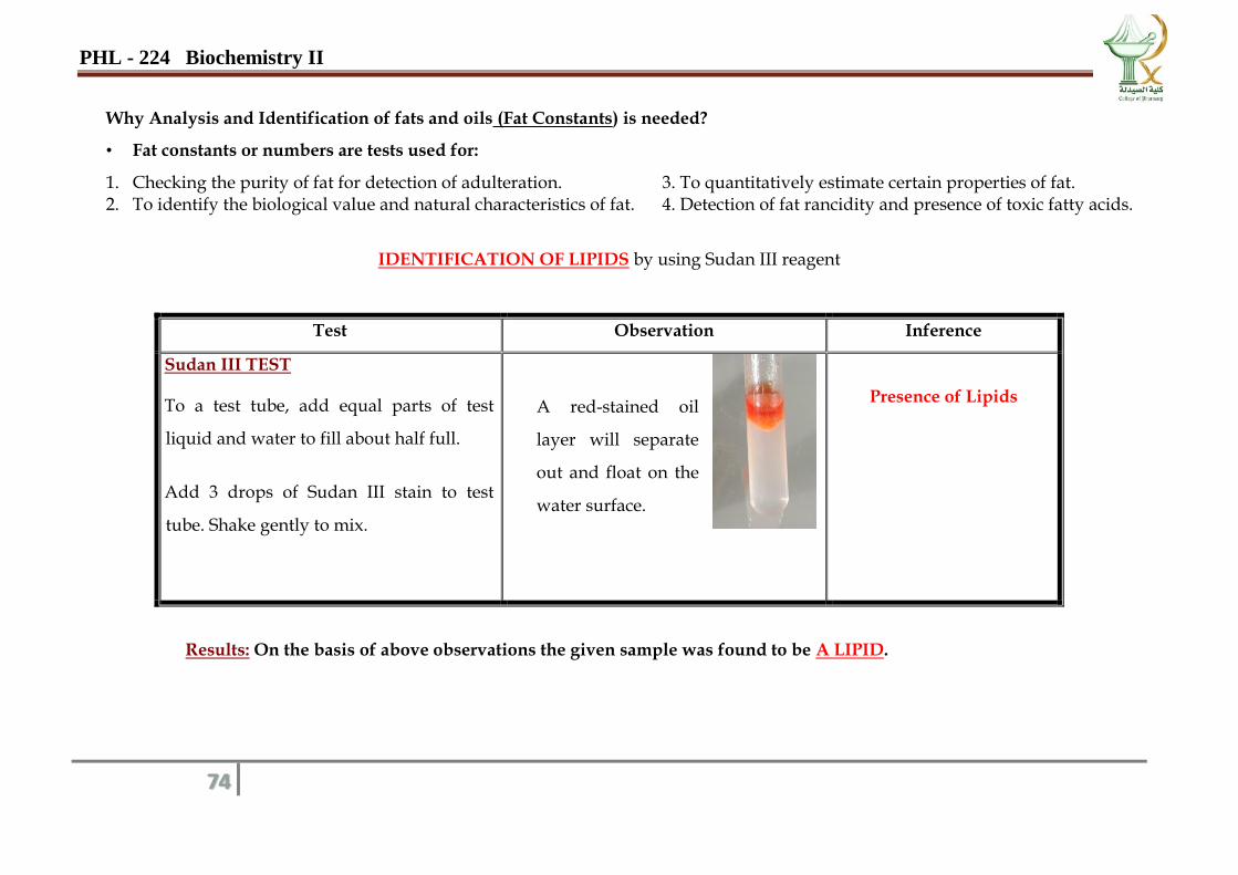
IDENTIFICATION OF LIPIDS by using Sudan III reagent
Test Observation Inference
Sudan III TEST
To a test tube, add equal parts of test
liquid and water to fill about half full.
Add 3 drops of Sudan III stain to test
tube. Shake gently to mix.
A red-stained oil
layer will separate
out and float on the
water surface.
Presence of Lipids
Results: On the basis of above observations the given sample was found to be A LIPID.
PHL - 224 Biochemistry II
75
REPORT
Unknown
Date……………….
Test Observation Inference
Sudan III TEST
To a test tube, add equal parts of test
liquid and water to fill about half full.
Add 3 drops of Sudan III stain to test
tube. Shake gently to mix.
Results: The given sample was found to be ______________________.
PHL - 224 Biochemistry II
76
QUANTITATIVE ANALYSIS
ENZYMES
Definition: Enzymes are highly specific biologic catalysts that greatly speed up the rate of a chemical reaction occurring in living
cells. Enzymes are found in low concentration in body fluids.
The enzyme activity is expressed in the international unit (I.U).
Classification of Enzymes:
Oxidoreductases:
There is a hydrogen donor and a hydrogen acceptor.
a. Aerobic oxidases: use O2 as H-acceptor forming H2O e.g. Tyrosinase.
b. Aerobic dehydrogenases: use O2 as H-acceptor forming H2O2 e.g. Glucose oxidase.
c. Anaerobic dehydrogenases: use co-enzyme as H-acceptor e.g. LDH
Transferases:
Transfer a group from one organic compound to another (CH3, NH2, and Phosphate etc.) e.g. Aminotransferases,
Kinases, Transketolases.
Hydrolases: hydrolyse the substrate
Lyases: Remove groups without hydrolysis leaving a double bond e.g. Decarboxylases.
Isomerases: convert one pair of isomers into another e.g. Racemases.
Ligases = Synthetases: linking two molecules together coupled with the breakdown of phosphate bond.
PHL - 224 Biochemistry II
77
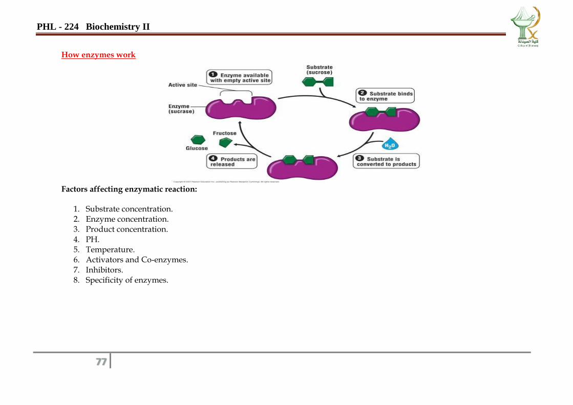
How enzymes work
Factors affecting enzymatic reaction:
1. Substrate concentration. 2. Enzyme concentration. 3. Product concentration. 4. PH. 5. Temperature. 6. Activators and Co-enzymes. 7. Inhibitors. 8. Specificity of enzymes.
PHL - 224 Biochemistry II
78
Date……………….
1. Estimation of Serum Lactate Dehydrogenase Activity (LDH)
Type: anaerobic dehydrogenase enzyme.
Occurrence: Heart > liver > skeletal muscle> erythrocytes > pancreas
Introduction
A lactate dehydrogenase (LDH) test measures the amount of LDH in the blood.
Lactate dehydrogenase is an enzyme that the body uses during the process of turning sugar into energy for your cells to use.
LDH is found in many of the body's tissues and organs, including the muscles, liver, heart, pancreas, kidneys, brain and
blood cells. The LDH test is mainly used to help identify the location and severity of tissue damage in the body. It's also
sometimes used to monitor how far certain conditions have progressed including:
kidney disease
liver disease
some types of cancer
LDH catalyzes the oxidation of lactate to pyruvate in the presence of NAD, which is subsequently reduced to NADH. The
rate of NADH formation measured at 340 nm is directly proportional to serum LDH-L activity
PHL - 224 Biochemistry II
79
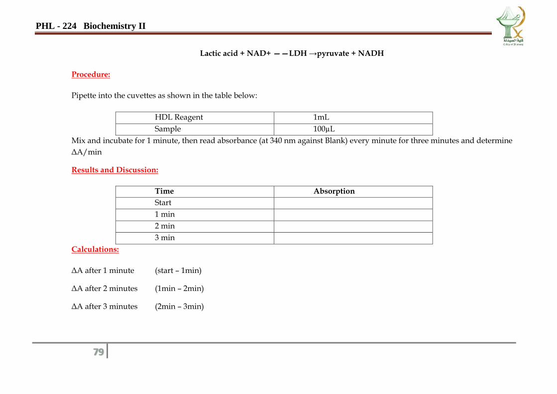
Lactic acid + NAD+ ——LDH →pyruvate + NADH
Procedure:
Pipette into the cuvettes as shown in the table below:
HDL Reagent 1mL
Sample 100µL
Mix and incubate for 1 minute, then read absorbance (at 340 nm against Blank) every minute for three minutes and determine
ΔA/min
Results and Discussion:
Time Absorption
Start
1 min
2 min
3 min
Calculations:
ΔA after 1 minute (start – 1min)
ΔA after 2 minutes (1min – 2min)
ΔA after 3 minutes (2min – 3min)
PHL - 224 Biochemistry II
80
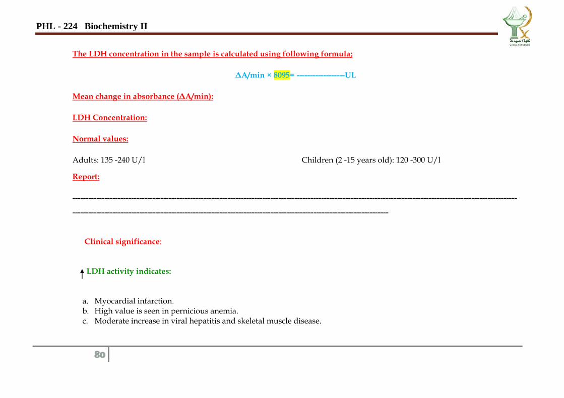
The LDH concentration in the sample is calculated using following formula;
ΔA/min × 8095= ------------------UL
Mean change in absorbance (ΔA/min):
LDH Concentration:
Normal values:
Adults: 135 -240 U/l Children (2 -15 years old): 120 -300 U/l
Report:
----------------------------------------------------------------------------------------------------------------------------------------------------------------------
----------------------------------------------------------------------------------------------------------------------
Clinical significance:
LDH activity indicates:
a. Myocardial infarction. b. High value is seen in pernicious anemia. c. Moderate increase in viral hepatitis and skeletal muscle disease.
PHL - 224 Biochemistry II
81
Date……………….
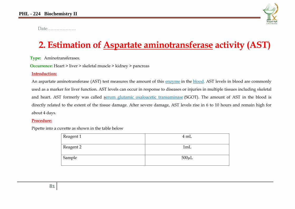
2. Estimation of Aspartate aminotransferase activity (AST) Type: Aminotransferases.
Occurrence: Heart > liver > skeletal muscle > kidney > pancreas
Introduction:
An aspartate aminotransferase (AST) test measures the amount of this enzyme in the blood. AST levels in blood are commonly
used as a marker for liver function. AST levels can occur in response to diseases or injuries in multiple tissues including skeletal
and heart. AST formerly was called serum glutamic oxaloacetic transaminase (SGOT). The amount of AST in the blood is
directly related to the extent of the tissue damage. After severe damage, AST levels rise in 6 to 10 hours and remain high for
about 4 days.
Procedure:
Pipette into a cuvette as shown in the table below
Reagent 1 4 mL
Reagent 2 1mL
Sample 500µL
PHL - 224 Biochemistry II
82
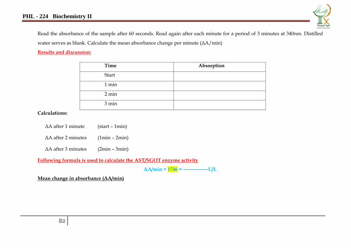
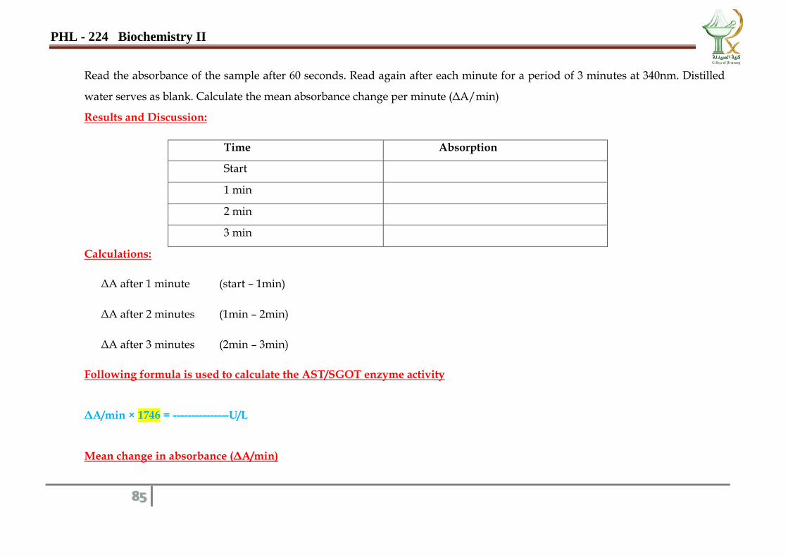
Read the absorbance of the sample after 60 seconds. Read again after each minute for a period of 3 minutes at 340nm. Distilled
water serves as blank. Calculate the mean absorbance change per minute (ΔA/min)
Results and discussion:
Time Absorption
Start
1 min
2 min
3 min
Calculations:
ΔA after 1 minute (start – 1min)
ΔA after 2 minutes (1min – 2min)
ΔA after 3 minutes (2min – 3min)
Following formula is used to calculate the AST/SGOT enzyme activity
ΔA/min × 1746 = ---------------U/L
Mean change in absorbance (ΔA/min)
PHL - 224 Biochemistry II
83
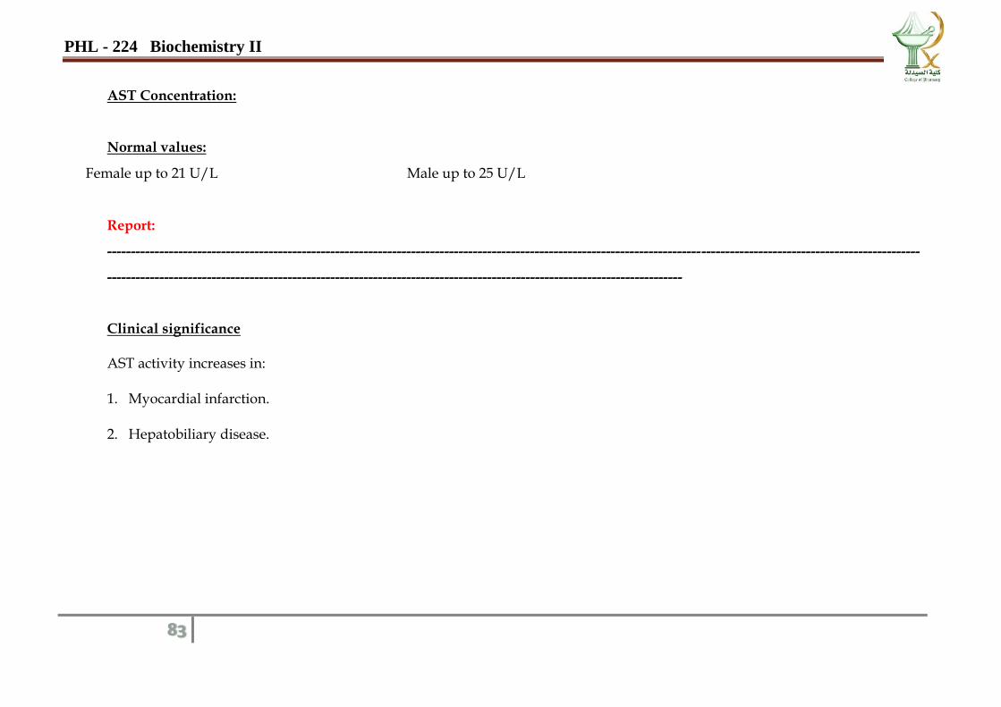
AST Concentration:
Normal values:
Female up to 21 U/L Male up to 25 U/L
Report:
---------------------------------------------------------------------------------------------------------------------------------------------------------------------------
-------------------------------------------------------------------------------------------------------------------------
Clinical significance
AST activity increases in:
1. Myocardial infarction.
2. Hepatobiliary disease.
PHL - 224 Biochemistry II
84
Date……………….
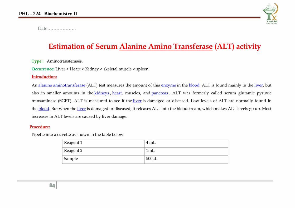
Estimation of Serum Alanine Amino Transferase (ALT) activity
Type : Aminotransferases.
Occurrence: Liver > Heart > Kidney > skeletal muscle > spleen
Introduction:
An alanine aminotransferase (ALT) test measures the amount of this enzyme in the blood. ALT is found mainly in the liver, but
also in smaller amounts in the kidneys , heart, muscles, and pancreas . ALT was formerly called serum glutamic pyruvic
transaminase (SGPT). ALT is measured to see if the liver is damaged or diseased. Low levels of ALT are normally found in
the blood. But when the liver is damaged or diseased, it releases ALT into the bloodstream, which makes ALT levels go up. Most
increases in ALT levels are caused by liver damage.
Procedure:
Pipette into a cuvette as shown in the table below
Reagent 1 4 mL
Reagent 2 1mL
Sample 500µL
PHL - 224 Biochemistry II
85
Read the absorbance of the sample after 60 seconds. Read again after each minute for a period of 3 minutes at 340nm. Distilled
water serves as blank. Calculate the mean absorbance change per minute (ΔA/min)
Results and Discussion:
Time Absorption
Start
1 min
2 min
3 min
Calculations:
ΔA after 1 minute (start – 1min)
ΔA after 2 minutes (1min – 2min)
ΔA after 3 minutes (2min – 3min)
Following formula is used to calculate the AST/SGOT enzyme activity
ΔA/min × 1746 = ---------------U/L
Mean change in absorbance (ΔA/min)
PHL - 224 Biochemistry II
86
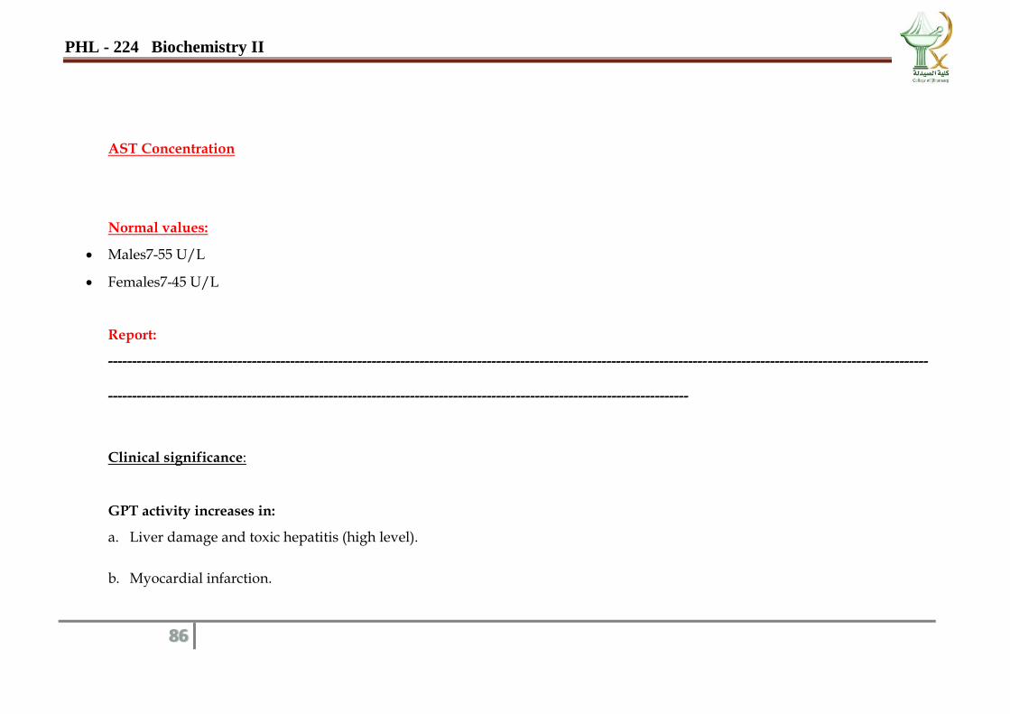
AST Concentration
Normal values:
Males7-55 U/L
Females7-45 U/L
Report:
---------------------------------------------------------------------------------------------------------------------------------------------------------------------------
-------------------------------------------------------------------------------------------------------------------------
Clinical significance:
GPT activity increases in:
a. Liver damage and toxic hepatitis (high level).
b. Myocardial infarction.
PHL - 224 Biochemistry II
87
Date……………….
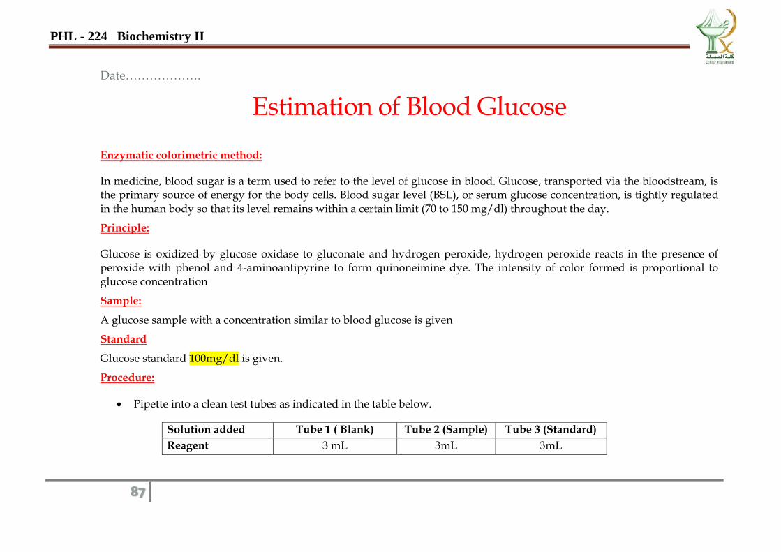
Estimation of Blood Glucose
Enzymatic colorimetric method:
In medicine, blood sugar is a term used to refer to the level of glucose in blood. Glucose, transported via the bloodstream, is the primary source of energy for the body cells. Blood sugar level (BSL), or serum glucose concentration, is tightly regulated in the human body so that its level remains within a certain limit (70 to 150 mg/dl) throughout the day.
Principle:
Glucose is oxidized by glucose oxidase to gluconate and hydrogen peroxide, hydrogen peroxide reacts in the presence of peroxide with phenol and 4-aminoantipyrine to form quinoneimine dye. The intensity of color formed is proportional to glucose concentration
Sample:
A glucose sample with a concentration similar to blood glucose is given
Standard
Glucose standard 100mg/dl is given.
Procedure:
Pipette into a clean test tubes as indicated in the table below.
Solution added Tube 1 ( Blank) Tube 2 (Sample) Tube 3 (Standard)
Reagent 3 mL 3mL 3mL
PHL - 224 Biochemistry II
88
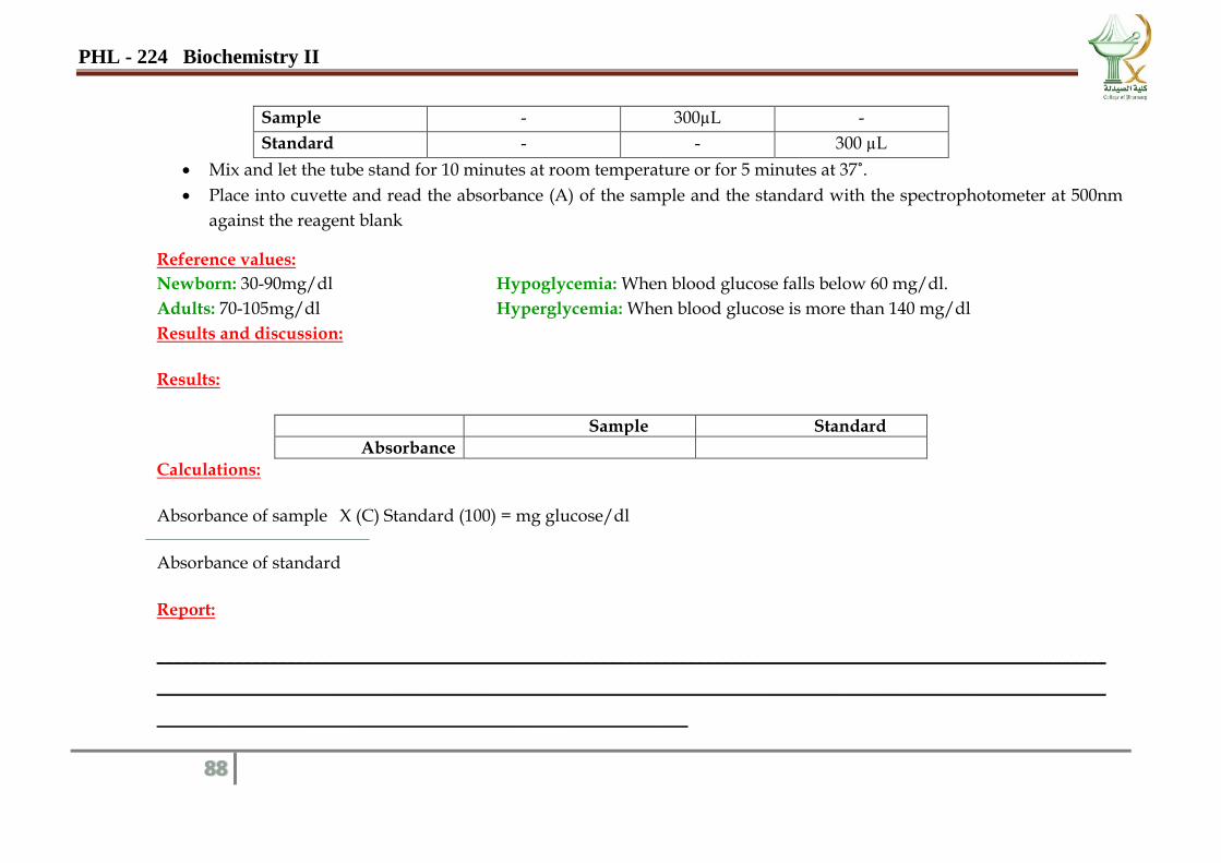
Sample - 300µL -
Standard - - 300 µL
Mix and let the tube stand for 10 minutes at room temperature or for 5 minutes at 37˚.
Place into cuvette and read the absorbance (A) of the sample and the standard with the spectrophotometer at 500nm
against the reagent blank
Reference values:
Newborn: 30-90mg/dl Hypoglycemia: When blood glucose falls below 60 mg/dl.
Adults: 70-105mg/dl Hyperglycemia: When blood glucose is more than 140 mg/dl
Results and discussion:
Results:
Sample Standard
Absorbance
Calculations:
Absorbance of sample X (C) Standard (100) = mg glucose/dl
Absorbance of standard
Report:
_____________________________________________________________________________________________________________
_____________________________________________________________________________________________________________
_____________________________________________________________
PHL - 224 Biochemistry II
89
PROTEIN AND NUCLEIC ACIDS
PURIFICATION AND EXTRACTION

PHL - 224 Biochemistry II
90