Embed Size (px)
Citation preview
Effect of cholesterol in suspension on the incorporation of phosphate into phospholipid by macrophages in vitro
A. J. DAY, N. H. FIDGE, and G. N. WILKINSON
Department of Human Physiology and Pharmacology, University of Adelaide, and Division of Mathematical Statistics, C.S.I.R.O., Adelaide, South Australia
ABSTRACT, Macrophages obtained from the peritoneal cavity of rabbits were incubated with pho~phate-~~P in order to investigate the synthesis of phospholipid by these cells. After 6 hr of incubation 0.25% of the phosphate added to the medium had been incorporated into phospholipid by the macrophages, mainly into lecithin and sphingomyelin, but partly also into phosphatidyl ethanolamine and inositol phosphatide.
The addition of cholesterol to the macrophage suspensions increased the rate of incorporation by 20% with 1 mg of cholesterol added, and 440/, with 2.5 mg added. The increase was similar for all the phospholipid fractions, and was not ac- companied by any increase in oxygen uptake by the cells. The addition of carbon particles (as a specific check for phagocytic effects) had only a small effect on the rate of incorporation.
The data provide support for the concept that cholesterol stimulates phospholipid synthesis by similar cells in the ar- terial wall during atherogenesis.
KEY WORDS pho~phate-~~P . incorporation . phospho- lipid . biosynthesis . macrophages . rabbit . cholesterol . carbon . suspension . oxygen uptake . phagocytosis . randomized block . atheroma
IT IS WELL KNOWN that in both experimental and human atheroma the phospholipid content of the ar- terial wall increases with that of cholesterol. Zilversmit and coworkers (1, 2) have demonstrated that this phos- pholipid accumulation occurs as a result of synthesis in the arterial wall rather than by deposition from the blood. The phospholipid that accumulates in the experi- mental atheromatous lesion, however, does so intracel- lularly in the foam cell (3); this suggests that during development of experimental atheroma, the phospho- lipid synthesis that is occurring in the arterial wall takes place in the foam cell. Recently Dunnigan (4) has
demonstrated that in human atheroma also, phosphc- lipid accumulates intracellularly in foam cells. In view of these facts, some further investigation of lipid synthe- sis by these cells or their precursors is indicated.
Investigation of the in situ synthesis of lipid by foam cells is impracticable by other than histochemical means, and a more thorough investigation of their lipid metabolism requires a preparation free from other cells. Although it was previously thought that foam cells arise from tissue macrophages, there is evidence that some of these cells may arise from smooth muscle cells (5). However, a large proportion of them is derived from blood mono- cytes which infiltrate the lesion, become macrophages, and engulf lipid (6). Following the injection of liquid paraffin, monocytes are mobilized into the peritoneal cavity; these cells can be collected and their lipid metabo- lism studied. While it cannot be asserted with certainty that the metabolism of such cells is the same as that of arterial wall macrophages or of foam cells, it is likely to be similar.
In previous papers (7, 8) it has been shown, with monocytes from the peritoneal cavity of rabbits, that 14C-labeled fatty acid and 14C-labeled acetate both can be incorporated into phospholipid by macrophages in vitro. We have now studied the incorporation of 32P- labeled phosphate into macrophages in vitro, in order, firstly, to establish that synthesis of phospholipid by macrophages occurs, secondly, to identify the phospho- lipid synthesized, and thirdly, to determine the effect of cholesterol uptake on this process.
MATERIALS AND METHODS 32P-labeled carrier-free orthophosphate in dilute HC1 was obtained from the Radiochemical Centre, Amer-
132 JOURNAL OF LIPID RESEARCH VOLUME 7, 1966
by guest, on March 17, 2019
ww
w.jlr.org
Dow
nloaded from

sham, U.K. This was diluted with 0.9y0 sodium chloride solution to give a stock solution for use.
Phospholipid standards for chromatography were ob- tained as follows. Synthetic dipalmitoyl lecithin, phos- phatidyl ethanolamine, phosphatidyl serine, and inositol phosphatide were obtained from the Nutritional Bio- chemicals Corporation (Cleveland, Ohio). Sphingomyelin was prepared from beef lung as described by Thann- hauser, Benotti, and Boncoddo (9). This preparation gave positive tests for choline (described later) and sphin- gosine (10) and the RF value was identical with that of a sample of sphingomyelin supplied by the Common- wealth Serum Laboratories (Melbourne, Australia). Lysolecithin was prepared from synthetic dipalmitoyl lecithin by the method of Renkonen (11) and had the same R , value as a sample of lysolecithin supplied by the Commonwealth Serum Laboratories (Melbourne, Aus- tralia). I t also gave a positive test for choline.
Carbon suspension [carbon C11/1413a (Gunther Wagner, Hanover, Germany)] 32 mg/ml was diluted with water before use. The mean particle diameter was 0.05 p.
A cholesterol suspension was prepared by slowly adding a warm solution of 500 mg of cholesterol in acetone to 200 ml of water. The acetone was removed by evaporation, and the suspension concentrated to 10 mg of cholesterol per milliliter by boiling. This suspension was stable, did not separate on standing even over a period of many months, and contained particles with a mean diameter of 1 p.
Rabbit macrophages were obtained from the peri- toneal cavity of rabbits as previously described (7).
Experimental Procedure Two series of replicate experiments were carried out, the first to demonstrate the incorporation by macrophages in vitro of 32P-labeled phospholipid and to identify the labeled phospholipid by fractionation, and the second to study the effect, on the rate of incorporation of labeled phosphate into phospholipid, of either added choles- terol or added carbon particles, the latter as a control for phagocytic effects.
In each experiment siliconized Warburg flasks con- taining an aliquot (5-6 X lo7 cells, approx 35 mg wet wt) of macrophages from a single freshly prepared stock, 60 I.LC of carrier-free 32P-labeled orthophosphate, and, in the second series of experiments, additions of either cholesterol or carbon suspension or an equivalent vol- ume of water, were made up to a final volume of 4 ml with Hanks' solution-serum 2 : 1 (v/v) and incubated for various periods of time at 37'.
The first series comprised seven replicate experiments in which flasks were incubated in duplicate for 1, 2, 4, 6, and (in five out of seven experiments) 12 hr. The second
series comprised two sets, (u) and ( b ) , of four and three replicate experiments respectively, in which 12 flasks (in three groups of four with either added cholesterol, carbon, or distilled water as described above) were in- cubated for 1, 2, 4, and 6 hr respectively. The amounts of added cholesterol were either ( u ) 2.5 mg or ( b ) 1.0 mg respectively; and of carbon either (u) 0.5 mg or ( b ) 1.0 mg. [In the set (u) of experiments the amounts of added cholesterol or carbon suspension contained comparable numbers of particles; in (6) they were equal by weight.] Additional flasks containing dead cells (killed by heating at 70 for 10 min) were investigated in some experiments.
Oxygen uptakes were recorded throughout incubation in all experiments, with manometer readings at 20-min intervals. After the appropriate incubation time, the cells were separated by centrifugation (1500 rpm for 5 min), the medium being discarded. The cells were washed with 0.9% sodium chloride and centrifuged again, and the lipid was extracted with chloroform- methanol 2 : 1 (v/v) by the method of Folch, Lees, and Sloane Stanley (12). A portion of the lipid extract WBS
taken for determination of lipid phosphorus as described by Brown (13), and a second portion for liquid scintilla- tion counting, appropriate correction for 32P decay being made.
Duplicate samples from the remainder of the extract were chromatographed on silicic acid-impregnated paper, using the solvent system diisobutylketone-acetic acid-water 40 : 25 : 5 (v/v/v) of Marinetti (1 4). After development, the papers were cut into strips 1.5 inches wide and scanned with a Nuclear-Chicago 4 ?r Actigraph paper chromatograph scanner. The area of the peaks obtained by triangulation was then used to measure the 32P-labeled phosphate incorporated into individual phos- pholipids at the various time intervals. Radioautographs were prepared for further inspection of the separation achieved, and the papers were then stained with Rhod- amine 6G or other spot test reagents as set out below. In some experiments the spots were cut out and eluted with 1.7 N HC1 in methanol and the lipid phosphorus was determined in the eluates. However, since there was too little lipid phosphorus in any spot but that containing lecithin, it was possible to determine only the distribu- tion of radioactivity and not the specific activity of the individual phospholipids.
Identification of Phospholipids The phospholipid components separated on silicic acid- impregnated paper were identified by comparing their mobilities with those of reference phospholipids, by the use of specific spot tests, and by co-chromatography (1 5). The R , values of the unknown spots were compared with those of the reference compounds and the papers were then stained with either 0.25y0 ninhydrin in ace-
DAY, FIDGE, AND WILKINSON Phospholipid Synthesis by Macrophages 133
by guest, on March 17, 2019
ww
w.jlr.org
Dow
nloaded from
tone-lutidine 9 : l (v/v) for detection of the amino- phospholipids, with phosphomolybdate-stannous chlo- ride reagent for detection of choline-containing phospho- lipids, or with Rhodamine 6G, which fluoresced with all phospholipids (1 4). For co-chromatography, the spot located by radioautography was cut out, combined with the corresponding spot from other chromatograms, and eluted for three successive 24 hr periods with chloro- form-methanol-water 75 : 25 : 5 (1 5). The combined eluates were centrifuged to remove particles derived from the paper, the supernatant solutions were evaporated to dryness under N2, and the residues co-chromatographed with the appropriate reference compound. The solvent system diisobutylketone-acetic acid-water was used, either in the proportions 40:25:5 or 40:30:7 (see Re- sults). Radioautographs were then prepared, the radio- active spots being marked and the papers stained accord- ing to the type of reference phospholipid. If the radio- active spot coincided accurately with the stained spot, the unknown labeled material was considered to be identical with the reference compound.
Statistical Methods Specific activities (cps/pg lipid P) and assays of total phospholipid (P, pg) were transformed to logarithms (base 10) for statistical analysis to obtain additivity of systematic effects and homogeneity of experimental variability. Transformation was, however, unnecessary for the percentage counts in the distribution of counts between phospholipid fractions, these values being already expressed on a relative basis.
Both series of experiments have the standard “ran- domized blocks” structure with replicate experiments constituting the blocks, and in the second series the 12 treatments have the 2-way factorial structure of three initial treatments (addition of cholesterol, carbon, or water) x four periods of incubation. This structure and the assumption of additivity of treatment and replicate effects determines the primary analysis of variance, in which error variation is measured by the treatment X replicate interaction mean square. However, there was a considerable degree of nonadditivity in the experimental data; a different preparation of macrophages was used in each experiment, and there was in consequence ap- preciable variation between experiments in the observed kinetic effects. I t was necessary, therefore, to partition the analysis of variance further and eliminate the varia- tion attributable to nonadditive effects from the estimate of error variation.
In the tabulation of results means of logarithms have been reconverted to natural units, and comparisons of logarithmic values expressed as relative differences, with appropriate standard errors. In Table 2 (ii) the entries have a constant relative error (coefficient of variation)
so that, for comparisons of entries, percentage differences should be calculated. (The coefficients of variation given with the table are the appropriate standard errors for testing percentage differences.)
In the graphical presentation of results (Figs. 1-3) the logarithmic scales used in the analysis have been retained so that the plotted points are all of the same accuracy visually. However, the scales have been calibrated in natural units for ease of reference. The time scale is also logarithmic, so that a hypothetically constant rate of up- take should be represented by a straight line with a slope o f l .
RESULTS
Viability of Macrophages during Incubation During the first 4 hr of incubation, oxygen uptake pro- ceeded at a constant rate in all experiments of approxi- mately 5 pmoles of oxygen per 108 cells per hr, a value similar to that reported previously (8). In some experi- ments a slight drop in rate of uptake occurred in the 4-6 hr interval, but in most experiments there was no drop in rate until after 6 hr. The mean values for total phospholipid extracted from the macrophages (Table 1) also remain constant during the first 4 hr but show some reduction at 6 and 12 hr. These results indicate that there was no appreciable loss in viability during the first 4-6 hr of incubation.
Incorporation by Macrophages of 32P-Labeled Phosphate into Phospholipid No significant incorporation of 32P-labeled phosphate into phospholipid was detected in test flasks containing dead cells only; oxygen uptake was also negligible.
The results obtained for viable cells are summarized in Table 2. The data presented in Table 2 are for the controls in both series of experiments; the effects of added cholesterol in series I1 on the rate of incorporation are shown separately in Table 3. The results are illus- trated graphically in Fig. 1. Synthesis of phospholipid by the macrophages is demonstrated by the increasing
TABLE 1 AMOUNT OF LIPID PHOSPHORUS EXTRACTED FROM
MACROPHAGES AFTER VARIOUS PERIODS OF INCUBATION
Total Lipid P* Incubation Time
hr 1 2 4 6 12
PE 41.6 39.1 39.6 35.5 32.3
(SEM 1.20)
* Means at each time interval for the two series of experiments together with the combined standard error of the mean.
134 JOURNAL OF LIPID RESEARCH VOLUME 7,1966
by guest, on March 17, 2019
ww
w.jlr.org
Dow
nloaded from
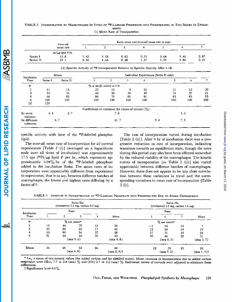
TABLE 2 INCORPORATION BY MACROPHAGES IN VITRO OF s 2 P - L ~ ~ ~ ~ ~ ~ PHOSPHATE INTO PHOSPHOLIPID, IN Two SERIES OF EXPERI- MENTS
(i) Mean Rate of Incorporation
Ratio mean rate/over-all mean rate in a p t . Over-all
mean rate 1 2 ' 3 4 5 6 7
@/fig lipid P/hr Series I 15.8 1.42 1.18 0.83 0.91 0.64 1.46 0.87 Series I1 19.1 0.36 1.64 0.48 1.51 1.39 i .a6 0.91
(ii) Specific Activity of 82P-Incorporated Relative to Specific Activity After 6 Hr
Means Individual Experiments (Series I1 only) Incubation
Time Series I Series I1 1 2 3 4 5 6 7
hr % of spccijc activity at 6 hr 1 11 15 23 12 9 21 11 12 20 2 33 36 42 20 30 45 36 27 61 4 66 83 86 70 81 81 83 85 93 6 100 100 100 100 100 100 100 100 100
12 129
Coefficients of variation for ratios of entries (%): In same 6.1 2.7 7 .8 5 . 3
In different 6.7 11.7 9.4 7.9 column :
columns :
specific activity with time of the 32P-labeled phospho- lipid.
The over-all mean rate of incorporation for all control experiments [Table 2 (i)] (averaged on a logarithmic scale over all times of incubation) was approximately 17.5 cps (32P)/pg lipid P per hr, which represents ap- proximately 0.04yO/hr of the 32P-labeled phosphate added to the incubation flasks. The mean rates of in- corporation were appreciably different from experiment to experiment, that is to say, between different batches of macrophages, the lowest and highest rates differing by a factor of 5 .
The rate of incorporation varied during incubation [Table 2 (ii)]. After 4 hr of incubation there was a pro- gressive reduction in rate of incorporation, indicating transition towards an equilibrium state, though the rates during this period may also have been affected somewhat by the reduced viability of the macrophages. The kinetic curves of incorporation [see Table 2 (ii)] also varied appreciably between different batches of macrophages. However, there does not appear to be any close correla- tion between these variations in trend and the corre- sponding variations in mean rate of incorporation [Table 2 (ill.
TABLE 3 INCREASE IN INCORPORATION OF a 2 P - L ~ ~ ~ ~ ~ PHOSPHATE INTO PHOSPHOLIPID DUE TO ADDED CHOLESTEROL
Series IIa Series IIb (cholesterol 2.5 mg, carbon 0.5 mg) (cholesterol 1.0 mg, carbon 1.0 mg)
~~ ~
Expt. Expt. Incubation
Time 1 2 3 4 Mean 5 6 7 Mean
hr % over control* 1 13 44 79 12 34 2 23 80 60 17 42 4 63 80 56 37 58 6 51 60 26 39 43
(SEM 9.6) (SEM 4.8)
% over control* -1 10 13 7 13 30 14 19 17 31 19 22 20 46 29 31
(SEM 6.5) (SEM 3.7)
Mean 36 65 54 26 44 (SEM 4.8) (SEM 2.4) t
12 29 19 20 (SEM 3.2) (SEM 1.9) t
* I.e., a mean of two control values (for added carbon and for distilled water). Mean increases in incorporation due to added carbon suspension were (IIa), 7.7 f 2.8 (SEM) % and (IIb) 2.7 f 2.2 (SEM) %. Individual means of controls were adjusted to eliminate these differences.
t Significance level 0.1 %.
DAY, FIDGE, AND WILKINSON Phospholipid Synthesis by Macrophages 135
by guest, on March 17, 2019
ww
w.jlr.org
Dow
nloaded from

SPECIFIC AC ctr l u c Ire
150-
100-
8 0-
60- 5 0 -
40-
30-
20-
15-
IO-
TlVlTY LIPID P
X---x Carbon 0-0 Control
t I I I I l l
I 2 4 6 8 10 12 TIME (hr)
FIG. 1. Specific activity of phospholipid (cps/pg lipid phosphorus) following incubation of macrophages with 32P-Iabeled phosphate (logarithmic scales: see text and Appendix). Mean of seven experi- ments.
RELATIVE SPECIFIC ACTIVITY
'I' M Cholesterol X Carbon
100
8 0
30
20
I5
I I I 1
I 2 4 6 TIME (hr )
FIG. 2. The effect of 1 mg each of cholesterol and of carbon SUS- pension on the incorporation of phosphate-=P into phospholipid by macrophages in vitro. Logarithmic scales (see text). Mean relative specific activity for three experiments (percentage of that present in the control at 6 hr) against time.
Efect of Added Carbon Particles A very small increase (average 5.2y0) in rate of incorpo- ration was detected with added carbon particles. This effect was slight in comparison with that induced by added cholesterol (see below).
Efect of Added Cholesterol The increases attributable to added cholesterol in the rate of incorporation of phosphate-82P into phospholipid are given in Table 3, expressed as percentages of the corresponding control values, and are illustrated in Figs. 2 and 3. The over-all mean increases in rate for the series of experiments I Ia and IIb were 44 and 20Y0 respec- tively, the increases in both series being highly significant (P < O.lyo). These values are closely commensurate with the amounts of added cholesterol in each case, 2.5 and 1 .O mg respectively. The addition of cholesterol, how- ever, produced no detectable increase in oxygen uptake.
There were appreciable differences in kinetic behavior according to the amount of cholesterol added. With the lower level of added cholesterol the difference in the amount of incorporated anP increased progressively from 7% at 1 hr to 31% of control at 6 hr, whereas with the higher level of added cholesterol the difference reached a peak of 58% at 4 hr and decreased thereafter. These results are kinetically consistent in that it would be ex- pected that, with a higher rate of incorporation ini- tially, transition towards equilibrium would occur some-
r I I 1 I 2 4 6
TIME (hr)
FIG. 3. The effect of 2.5 mg of cholesterol and 0.5 mg of carbon suspension on the incorporation of phosphate-a'P into phospholipid by macrophages in vitro. Logarithmic scales. Mean relative specific activity for four experiments (percentage of that present in the con- trol at 6 hr) against time.
136 JOURNAL OF LIPID RESEARCH VOLUME 7,1966
by guest, on March 17, 2019
ww
w.jlr.org
Dow
nloaded from
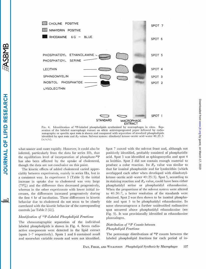
a CHOLINE POSITIVE
NINHYDRIN POSITIVE
m RHODAMINE 6 G - BLUE
.I SPOT 7
PHOSPHATIDYL ETHANOLAMINE - PHOSPHATIDYL SERINE - LEClTH IN
SPOT 6
SPOT 5
(SsisD SPOT 4
SPHINGOMY ELlN
INOSITOL PHOSPHATIDE I LYSOLECITHIN
/
SPOT 3
SPOT 2
SPOT I - - STANDARD MACROPHAGE
EXTRACT FIG. 4. Sep- aration of the labeled macrophage extract on silicic acid-impregnated paper followed by radio- autography or specific spot tests is shown and compared with separation of standard phospholipids identified by spot tests and RF values. Solvent system: diisobutyl ketone-acetic acid-water 40 : 25 : 5
Identification of ZP-labeled phospholipids synthesized by macrophages in vitro.
(v/v/v).
what sooner and more rapidly. However, it could also be inferred, particularly from the data for series IIb, that the equilibrium level of incorporation of ph~spha te -~~P has also been affected by the uptake of cholesterol, though the data are not conclusive on this point.
The kinetic effects of added cholesterol varied appre- ciably between experiments, mainly in series IIa, but in a consistent way. In experiment 3 (Table 3) the initial increase in uptake due to cholesterol was very large (79y0) and the difference then decreased progressively, whereas in the other experiments with lower initial in- creases, the differences increased progressively during the first 4 hr of incubation. These differences in kinetic behavior due to cholesterol do not seem to be closely correlated with the kinetic behavior of the corresponding controls [see Table 2 (ii) 1. Identi5cation of 32P-Labeled Phospholipid Fractions The chromatographic separation of the individual labeled phospholipids is shown in Fig. 4. Seven radio- active components were detected in the lipid extract (spots 1-7 respectively). Spots 1 and 6 contained small and somewhat variable counts and were not identified.
Spot 7 moved with the solvent front and, although not positively identified, probably consisted of phosphatidic acid. Spot 3 was identified as sphingomyelin and spot 4 as lecithin. Spot 2 did not contain enough material to produce a color reaction. Its RF value was similar to that for inositol phosphatide and for lysolecithin (which overlapped each other when developed with diisobutyl- ketone-acetic acid-water 40 : 25 : 5). Spot 5, according to its staining reaction and R F value, could have been either phosphatidyl serine or phosphatidyl ethanolamine. When the proportions of the solvent system were altered to 40:30:7, a better resolution of the standards were obtained. Spot 2 was then shown to be inositol phospha- tide and spot 5 to be phosphatidyl ethanolamine. In some chromatograms a further unidentified radioactive spot occurred above phosphatidyl ethanolamine (see Fig. 5). It was provisionally identified as ethanolamine plasmalogen.
Distribution of 32P Counts between Phospholipid Fractions The percentage distributions of 32P counts between the labeled phospholipid fractions for each period of in-
DAY, FIDGE, AND WILKINSON Phospholipid.Synthesis by Macrophages 137
by guest, on March 17, 2019
ww
w.jlr.org
Dow
nloaded from
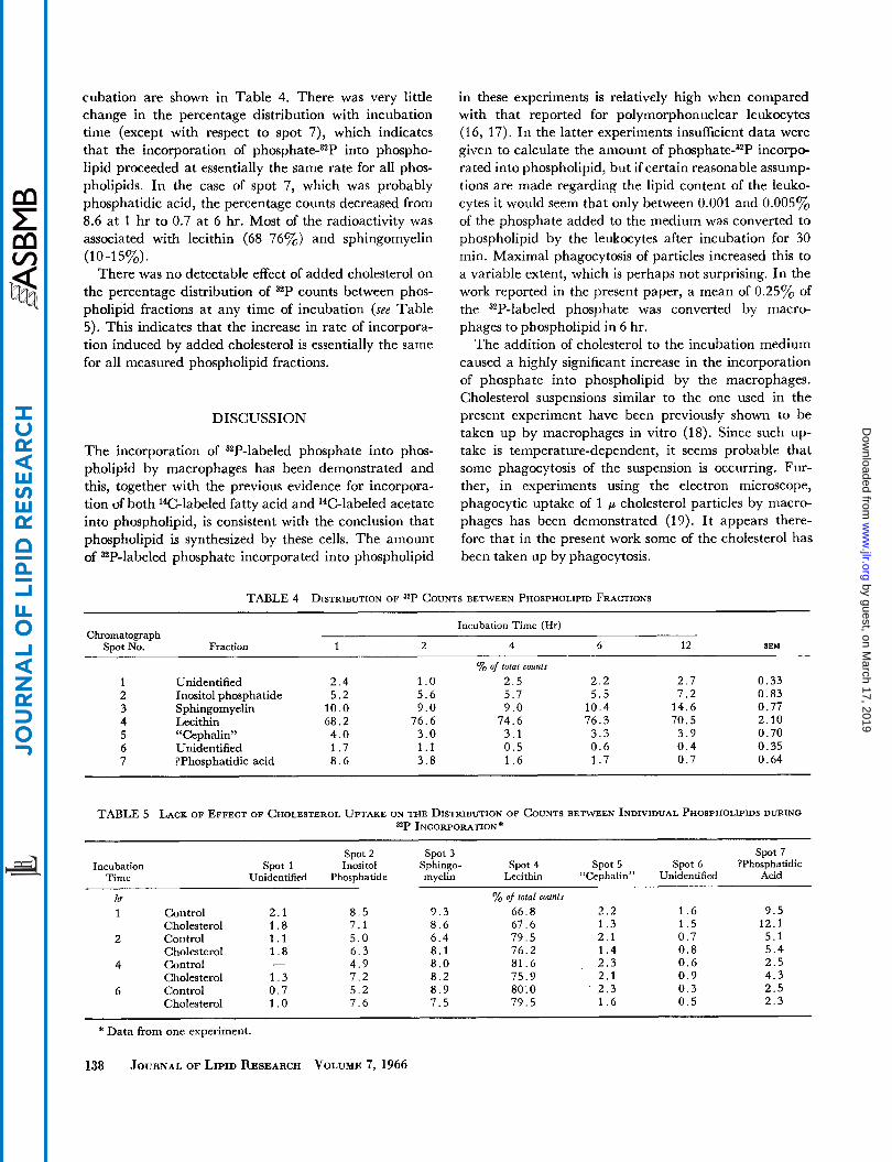
cubation are shown in Table 4. There was very little change in the percentage distribution with incubation time (except with respect to spot 7), which indicates that the incorporation of pho~phate-~~P into phospho- lipid proceeded at essentially the same rate for all phos- pholipids. In the case of spot 7, which was probably phosphatidic acid, the percentage counts decreased from 8.6 at 1 hr to 0.7 at 6 hr. Most of the radioactivity was associated with lecithin (68-76%) and sphingomyelin
There was no detectable effect of added cholesterol on the percentage distribution of 32P counts between phos- pholipid fractions at any time of incubation (see Table 5). This indicates that the increase in rate of incorpora- tion induced by added cholesterol is essentially the same for all measured phospholipid fractions.
(1 0-1 5%).
DISCUSSION
The incorporation of 32P-labeled phosphate into phos- pholipid by macrophages has been demonstrated and this, together with the previous evidence for incorpora- tion of both 14C-labeled fatty acid and 14C-labeled acetate into phospholipid, is consistent with the conclusion that phospholipid is synthesized by these cells. The amount of 32P-labeled phosphate incorporated into phospholipid
in these experiments is relatively high when compared with that reported for polymorphonuclear leukocytes (16, 17). In the latter experiments insufficient data were given to calculate the amount of pho~phate-~~P incorpo- rated into phospholipid, but if certain reasonable assump- tions are made regarding the lipid content of the leuko- cytes it would seem that only between 0.001 and 0.005% of the phosphate added to the medium was converted to phospholipid by the leukocytes after incubation for 30 min. Maximal phagocytosis of particles increased this to a variable extent, which is perhaps not surprising. In the work reported in the present paper, a mean of 0.25% of the 32P-labeled phosphate was converted by macro- phages to phospholipid in 6 hr.
The addition of cholesterol to the incubation medium caused a highly significant increase in the incorporation of phosphate into phospholipid by the macrophages. Cholesterol suspensions similar to the one used in the present experiment have been previously shown to be taken up by macrophages in vitro (18). Since such up- take is temperature-dependent, it seems probable that some phagocytosis of the suspension is occurring. Fur- ther, in experiments using the electron microscope, phagocytic uptake of 1 p cholesterol particles by macro- phages has been demonstrated (19). It appears there- fore that in the present work some of the cholesterol has been taken up by phagocytosis.
TABLE 4 DISTRIBUTION OF 32P COUNTS BETWEEN PHOSPHOLIPID FRACTIONS
Chromatograph Spot No.
Incubation Time (Hr)
SEM Fraction
Unidentified Inositol phosphatide Sphingomyelin Lecithin “Cephalin” Unidentified ?Phosphatidic acid
1 2 4 6 12
Yo of total counts 2 . 4 1 .o 2 . 5 2 . 2 2 . 7 5 . 2 5 . 6 5 . 7 5 . 5 7 . 2
1 0 . 0 9 . 0 9 . 0 1 0 . 4 1 4 . 6 6 8 . 2 7 6 . 6 7 4 . 6 7 6 . 3 7 0 . 5 4 . 0 3 . 0 3 . 1 3 . 3 3 . 9 1 . 7 1 . 1 0 . 5 0 . 6 0 . 4 8 . 6 3 . 8 1 . 6 1 . 7 0 . 7
0 . 3 3 0 . 8 3 0 . 7 7 2 . 1 0 0 . 7 0 0 . 3 5 0 . 6 4
TABLE 5 LACK OF EFFECT OF CHOLESTEROL UPTAKE ON THE DISTRIBUTION OF COUNTS BETWEN INDIVIDUAL PHOSPHOLIPIDS DURING **P INCORPORATION*
spot 2 Spot 3 spot 7 Incubation spot 1 Inositol Sphingo- spot 4 spot 5 Spot 6 ?Phosphatidic
Time Unidentified Phosphatide myelin Lecithin “Cephalin” Unidentified Acid
hr % of total counts 1 Control 2 . 1 8 . 5 9 . 3 6 6 . 8 2 . 2 1 . 6 9 . 5
Cholesterol 1 . 8 7 . 1 8 . 6 6 7 . 6 1 . 3 1 . 5 1 2 . 1
Cholesterol 1 . 8 6 . 3 8 . 1 7 6 . 2 1 . 4 0 . 8 5 . 4
Cholesterol 1 . 3 7 . 2 8 . 2 7 5 . 9 2 . 1 0 . 9 4 . 3
2 Control 1 . 1 5 . 0 6 . 4 7 9 . 5 2 . 1 0 . 7 5 . 1
4 Control - 4 . 9 8 . 0 8 1 . 6 2 . 3 0 . 6 2 . 5
6 Control 0 . 7 5 . 2 8 . 9 80: 0 2 . 3 0 . 3 2 . 5 Cholesterol 1 .o 7 . 6 7 . 5 7 9 . 5 1 . 6 0 . 5 2 . 3
* Data from one experiment.
138 JOURNAL’ OF LIPID RESEARCH VOLUME 7, 1966
by guest, on March 17, 2019
ww
w.jlr.org
Dow
nloaded from
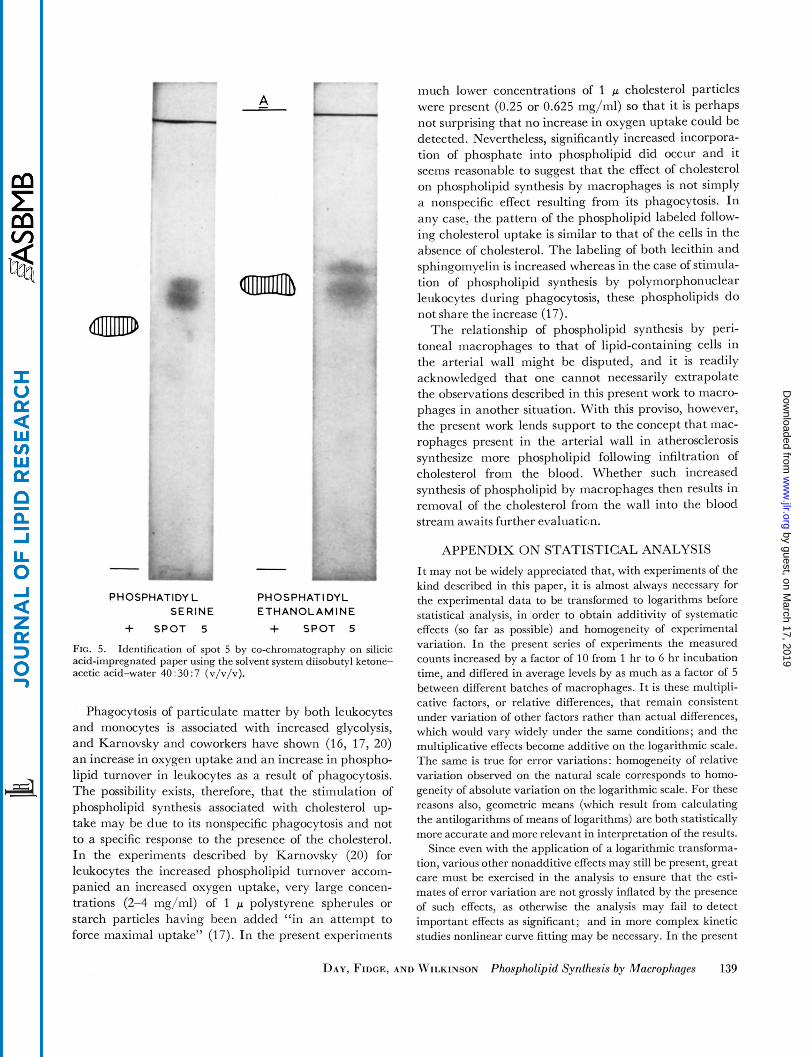
- - PHOSPHATIDYL PH 0 S PH AT I DY L
SERINE ETHANOLAMINE
+ SPOT 5 + SPOT S
FIG. 5. Identification of spot 5 by co-chromatography on silicic acid-impregnated paper using the solvent system diisobutyl ketone- acetic acid-water 40 : 30 : 7 (v/v/v).
Phagocytosis of particulate matter by both leukocytes and monocytes is associated with increased glycolysis, and Karnovsky and coworkers have shown (16, 17, 20) an increase in oxygen uptake and an increase in phospho- lipid turnover in leukocytes as a result of phagocytosis. The possibility exists, therefore, that the stimulation of phospholipid synthesis associated with cholesterol up- take may be due to its nonspecific phagocytosis and not to a specific response to the presence of the cholesterol. In the experiments described by Karnovsky (20) for leukocytes the increased phospholipid turnover accom- panied an increased oxygen uptake, very large concen- trations (2-4 mg/ml) of 1 p polystyrene spherules or starch particles having been added “in an attempt to force maximal uptake” (17). In the present experiments
much lower concentrations of 1 p cholesterol particles were present (0.25 or 0.625 mg/ml) so that it is perhaps not surprising that no increase in oxygen uptake could be detected. Nevertheless, significantly increased incorpora- tion of phosphate into phospholipid did occur and it seems reasonable to suggest that the effect of cholesterol on phospholipid synthesis by macrophages is not simply a nonspecific effect resulting from its phagocytosis. In any case, the pattern of the phospholipid labeled follow- ing cholesterol uptake is similar to that of the cells in the absence of cholesterol. The labeling of both lecithin and sphingomyelin is increased whereas in the case of stimula- tion of phospholipid synthesis by polymorphonuclear leukocytes during phagocytosis, these phospholipids do not share the increase (17).
The relationship of phospholipid synthesis by peri- toneal macrophages to that of lipid-containing cells in the arterial wall might be disputed, and it is readily acknowledged that one cannot necessarily extrapolate the observations described in this present work to macro- phages in another situation. With this proviso, however, the present work lends support to the concept that mac- rophages present in the arterial wall in atherosclerosis synthesize more phospholipid following infiltration of cholesterol from the blood. Whether such increased synthesis of phospholipid by macrophages then results in removal of the cholesterol from the wall into the blood stream awaits further evaluation.
APPENDIX ON STATISTICAL ANALYSIS
It may not be widely appreciated that, with experiments of the kind described in this paper, it is almost always necessary for the experimental data to be transformed to logarithms before statistical analysis, in order to obtain additivity of systematic effects (so far as possible) and homogeneity of experimental variation. In the present series of experiments the measured counts increased by a factor of 10 from 1 hr to G hr incubation time, and differed in average levels by as much as a factor of 5 between different batches of macrophages. It is these multipli- cative factors, or relative differences, that remain consistent under variation of other factors rather than actual differences, which would vary widely under the same conditions; and the multiplicative effects become additive on the logarithmic scale. The same is true for error variations: homogeneity of relative variation observed on the natural scale corresponds to homo- geneity of absolute variation on the logarithmic scale. For these reasons also, geometric means (which result from calculating the antilogarithms of means of logarithms) are both statistically more accurate and more relevant in interpretation of the results.
Since even with the application of a logarithmic transforma- tion, various other nonadditive effects may still be present, great care must be exercised in the analysis to ensure that the esti- mates of error variation are not grossly inflated by the presence of such effects, as otherwise the analysis may fail to detect important effects as significant; and in more complex kinetic studies nonlinear curve fitting may be necessary. In the present
DAY, FIDGE, AND WILKINSON Phospholipid Synthesis by Macrophages 139
by guest, on March 17, 2019
ww
w.jlr.org
Dow
nloaded from

series of experiments all comparisons of initial treatments and of incubation times were effected with aliquots taken from a common stock of macrophages in each experiment, so that the only relevant error variation is that arising from the experi- mental handling of the incubation flasks and subsequent assay errors. In the second series of experiments there were no dupli- cates for estimating error variation directly, and if standard randomized block analyses had been applied, the error mean squares obtained would have been approximately six times larger than those finally obtained after the elimination of non- additive effects. Most of the nonadditive effects arose from the very appreciable differences in kinetic behavior of different batches of macrophages.
This work was carried out under a grant-in-aid from the Na- tional Heart Foundation of Australia. We are also indebted to Misses M. Kleeman and M. Wynbergen for technical assist- ance, and to Dr. J. Casley-Smith of the Department of Zoology for measuring particle sizes of the carbon and cholesterol sus- pensions by electron microscopy. Manuscript received 23 August 7965; accepted 30 September 7965.
REFERENCES
1. Zilversmit, D. B., M. L. Shore, and R. F. Ackerman. Circulation 9: 581, 1954.
2. Zilversmit, D. B., and E. L. McCandless. J. Lipid Res. 1:
3. Day, A. J. J. Atherosclerosis Res. 2: 350, 1962. 4. Dunnigan, M. G. J. Atherosclerosis Res. 4: 144, 1964. 5. Buck, R. C. Brit. J . Exptl. Puthol. 43: 236, 1962. 6. Still, W. J. S. Exptl. Mol. Pathol. 2: 491, 1963. 7. Day, A. J., and N. H. Fidge. J. Lipid Res. 3: 333, 1962. 8. Day, A. J., and N. H. Fidge. J. Lipid Res. 5: 163, 1964. 9. Thannhauser, S. J., J. Benotti, and N. F. Boncoddo. J.
118, 1959.
Biol. Chem. 166: 669, 677, 1946. 10. Saito, K. J. Biochem. (Tokyo) 47: 573, 1960. 11. Renkonen, 0. J. Lipid Res. 3: 181, 1962. 12. Folch, J., M. Lees, and G. H. Sloane Stanley. J. Bid.
13. Brown, W. D. Australian J . Exptl. Biol. 32: 677, 1954. 14. Marinetti, G. V. J. Lipid Res. 3: 1, 1962. 15. Hokin, L. E., and M. R. Hokin. J. Biol. Chem. 233: 805,
16. Sbarra, A. J., and M. L. Karnovsky. J . Bid. Chem. 235:
17. Karnovsky, M. L., and D. F. H. Wallach. J. Biol. Chem.
18. Day, A. J. Quart. J . Exptl. Physiol. 46: 383, 1961. 19. Casley-Smith, J. R., and A. J. Day. Quart. J . Exptl. Physiol.,
20. Karnovsky, M. L. Phvsiol. Rev. 42: 143, 1962.
Chem. 226: 497, 1957.
1958.
2224, 1960.
236: 1895, 1961.
in press (1966).
140 JOURNAL OF LIPID RESEARCH VOLUME 7, 1966
by guest, on March 17, 2019
ww
w.jlr.org
Dow
nloaded from


![Lipid Biosynthesis in Eukaryotic Cells - pub.epsilon.slu.sepub.epsilon.slu.se/1173/1/Avhandling_nr_078[1].2006_Tryckfil.pdf · Lipid Biosynthesis in Eukaryotic Cells Studies on Enzyme](https://img.pdfslide.net/doc/110x75/5cbc44c388c9937f418c630e/lipid-biosynthesis-in-eukaryotic-cells-pub-12006tryckfilpdf-lipid-biosynthesis.jpg)