Embed Size (px)
Citation preview

0163-769X/88/0901-0038$02.00/0Endocrine ReviewsCopyright © 1988 by The Endocrine Society
Vol. 9, No. 1Printed in U.S.A.
Phosphorylation of Cell Surface Receptors: A Mechanismfor Regulating Signal Transduction PathwaysDAVID R. SIBLEY, JEFFREY L. BENOVIC, MARC G. CARON, ANDROBERT J. LEFKOWITZ
Howard Hughes Medical Institute, Departments of Medicine, Biochemistry and Physiology, Duke UniversityMedical Center, Durham, North Carolina 27710
Introduction
SPECIFIC receptors, located in the plasma mem-branes of cells, are crucially involved in mediating
the acute and chronic effects of a wide variety of hor-mones, drugs, growth factors, antigens, and other ligands.Some membrane receptors are involved in transportingvarious important molecules into the cell by a processknown as receptor-mediated endocytosis. An importantinsight which has emerged from recent research is thatboth the function and subcellular distribution of manysuch receptors are dynamically regulated.
Phosphorylation/dephosphorylation represents per-haps the most intensively studied reversible covalentmodification of enzymes which is known to have regu-latory significance (1, 2). Considerably more is knownabout the kinases than the phosphatases. Only recentlyhas it begun to be appreciated that the function ofnoncatalytic proteins, such as receptors, may also beregulated in this way. We review here the importantplasma membrane receptor systems thus far studiedwhose function and/or cellular trafficking has been dem-onstrated to be regulated by phosphorylation. Availableinformation suggests a number of principles which maybe generally applicable as well as several testable hy-potheses.
/?-Adrenergic Receptor-Coupled AdenylateCyclase
The jS-adrenergic receptor-coupled adenylate cyclasesystem may be considered to be prototypical of a widevariety of receptors which are coupled to their effectorsystems via guanine nucleotide regulatory (G) proteins.G proteins represent a family of closely related proteinswhich are heterotrimeric consisting of GTP binding andhydrolyzing a-subunits as well as /3- and 7-subunits (3-
Address requests for reprints and all correspondence to: Dr. DavidR. Sibley, Experimental Therapeutics Branch, Building 10, Room 5C-108, National Institutes of Health, Bethesda, Maryland 20892.
5). Receptors may either stimulate adenylate cyclase viathe stimulatory guanine nucleotide regulatory protein(G8) or inhibit it via the inhibitory guanine nucleotideregulatory protein (Gi). It is now readily apparent thatsome forms of regulation of adenylate cyclase may beachieved through phosphorylation of its protein compo-nents. One of the most extensively investigated forms ofthis type of regulation is that of desensitization. Desen-sitization is defined as a process through which prolongedexposure of target cells to hormone, drug, or neurotrans-mitter agonists results in a diminished cellular respon-siveness to further agonist stimulation.
Desensitization of 0-adrenergic receptor-coupledadenylate cyclase is generally divided into two majorcategories (6-8). One form is referred to as agonist spe-cific or homologous and is distinguished by the fact thatonly stimulation by 0-adrenergic agonists is attenuated.Conversely, agonist-nonspecific or heterologous desen-sitization is characterized by diminished responsivenessto additional receptor agonists and to nonreceptor acti-vators such as guanine nucleotides and fluoride ion.Investigations of homologous desensitization have dem-onstrated sequestration or down-regulation of the recep-tors away from the cell surface in addition to an uncou-pling of receptor-adenylate cyclase interaction (6-8). Ho-mologous desensitization does not appear to be mediatedby cAMP as this form of refractoriness can be demon-strated in the absence of cAMP generation (6-8). Het-erologous desensitization, by contrast, does not involvereceptor sequestration or down-regulation but instead isprimarily associated with functional uncoupling of thereceptors from adenylate cyclase. Moreover, heterologousdesensitization appears to be mediated, at least partially,by cAMP (6-8). Since heterologous desensitization isassociated with decrements in fluoride ion and guaninenucleotide-stimulated activities (effectors which bypassthe receptors) this form of desensitization must, at aminimum, involve alterations in either the regulatoryproteins and/or the catalytic moiety of adenylate cyclase.
38

February, 1988 RECEPTOR PHOSPHORYLATION 39
Recent studies have, in fact, demonstrated that thestimulatory and perhaps the inhibitory guanine nucleo-tide regulatory proteins (Gs and Gj, respectively) arefunctionally modified in heterologous desensitization.Kassis and Fishman (9) demonstrated that Gs extractedfrom heterologously desensitized fibroblasts and recon-stituted into S49 eye" cell membranes (S49 cyc~ cellslack G8 but possess the other components of adenylatecyclase) was functionally impaired relative to controls.Similarly, heterologous desensitization induced by pros-taglandin Ex in liver (10) and by human CG in ovaries(11) results in an impaired functionality of Gs as deter-mined with S49 eye" membrane reconstitution. In con-trast, Rich et al. (12) have reported that glucagon-in-duced heterologous desensitization in MDCK cells wasnot associated with G8 alterations but instead involvedincreases in the apparent levels of the inhibitory guanineregulatory protein, Gi. This suggests that alterations inthe G8/Gi stoichiometry may be another mechanism bywhich heterologous desensitization is achieved.
Additional evidence for a functional alteration in G8
in heterologous desensitization has come from studieswith avian erythrocytes. Briggs et al. (13) found thatwhen Gs was extracted from catecholamine-desensitizedturkey erythrocytes and carefully quantitated by labelingwith [32P]NAD+ and cholera toxin, desensitization re-sulted in a significant reduction in the ability of Gs toreconstitute adenylate cyclase activity in S49 cyc~ mem-branes. Although heterologous desensitization can ap-parently result in impaired G8 functionality, this doesnot exclude other potential lesions in the adenylate cy-clase system. In fact, the observation that catecholamine-stimulated adenylate cyclase activity is desensitized byabout 50% whereas the fluoride ion and guanine nucleo-tide activities are reduced by only 10-20% after agonist-induced desensitization is indicative of other processeswhich occur as well (6,13).
Indeed, using [32P]orthophosphate incorporation, ithas been shown that during the heterologous desensiti-zation in both avian and amphibian erythrocytes, the /3-adrenergic receptor undergoes phosphorylation (14-16).Sibley et al. (15) have investigated this phosphorylationprocess in detail using turkey erythrocytes. In these cells,the /3-adrenergic receptor is stoichiometrically phospho-rylated under basal conditions containing 0.7-1.0 molphosphate/mol receptor with this stoichiometry increas-ing to 2-3 mol/mol upon maximal desensitization (15).This phosphorylation occurs exclusively on serine resi-dues in the receptor (16, 17). We have also shown thatthe phosphate/receptor stoichiometry is tightly corre-lated with the degree of desensitization (15). For in-stance, the time courses for receptor phosphorylationand adenylate cyclase desensitization are identical as arethe rates of resensitization and the return of the phos-
phate/receptor stoichiometry to control levels (15).Moreover, incubation of the cells with membrane-perme-able cAMP analogs causes submaximal phosphorylationof the /3-adrenergic receptor which is correlated with thepartial desensitization of adenylate cyclase which theseanalogs evoke (15). These data thus indicate that inerythrocytes, heterologous desensitization is tightly cor-related with phosphorylation of the 0-adrenergic recep-tor.
Since cAMP analogs can reproduce the catecholamine-induced receptor phosphorylation and desensitization inavian (15) and amphibian erythrocytes (16), then pre-sumably the cAMP-dependent protein kinase is involvedin this desensitization. In fact, Benovic et al. (18) haverecently demonstrated a cAMP-mediated phosphoryla-tion of mammalian lung 0-adrenergic receptor that issimilar to the process exhibited by erythrocytes. Usingpure receptor and pure c AMP-dependent protein kinase,it is shown that isoproterenol enhanced the rate of re-ceptor phosphorylation (on serine residues) by about 2-fold. Reconstitution of the phosphorylated receptor withthe G8 protein demonstrated diminished agonist-pro-moted receptor-mediated stimulation of the GTPase ac-tivity compared with controls. Thus, the cAMP-depend-ent protein kinase-promoted phosphorylation is func-tionally significant. Since any hormone or drug thatraises intracellular cAMP levels will presumably lead tosuch /3-adrenergic receptor phosphorylation, this type ofregulation is of the heterologous type.
A fascinating question concerning heterologous desen-sitization is why incubation of avian erythrocytes withcAMP analogs produces only partial effects (15). Onehypothesis, suggested by Benovic et al. (18), is that inorder to obtain maximal phosphorylation of the /3-adre-nergic receptor, agonist occupancy must occur. Anotherpossibility is that the receptor phosphorylation is notcompletely mediated by cAMP and that other proteinkinase systems may phosphorylate the /3-adrenergic re-ceptor as well. In this regard, we (19) and others (20)have shown that phorbol esters, compounds which po-tently activate protein kinase C, are capable of stimulat-ing /3-adrenergic receptor phosphorylation concomitantlywith adenylate cyclase desensitization. Interestingly, induck erythrocytes (19) the phorbol ester-induced recep-tor phosphorylation is nonadditive with that producedby isoproterenol, suggesting a common mechanism orpathway of action.
Recently, protein kinase C has been shown to directlyphosphorylate the purified /3-adrenergic receptor in vitroalbeit to a lower stoichiometry than that of the cAMP-dependent protein kinase (21). The phosphorylation byprotein kinase C occurs on serine residues and is notenhanced by agonist occupancy of the receptor. Impor-tantly, the sites on the receptor that are phosphorylated

40 SIBLEY ET AL. Vol. 9, No. 1
by protein kinase C and the cAMP-dependent proteinkinase appear to be identical as determined by peptidemapping techniques (21).
The relationship of the protein kinase C and cAMP-dependent protein kinase phosphorylation sites has beenfurther elucidated by recent molecular cloning and se-quence analysis of the j82-adrenergic receptor (22). Figure1 shows the j82-adrenergic receptor as it is proposed to beorganized in the plasma membrane. As can be seen inFig. 1, there are two sites on the receptor consisting ofthe general amino acid sequence Arg-Arg-X-Ser, whichrepresents a consensus recognition site for the cAMP-dependent protein kinase (23). Both of these sequencesalso contain a basic amino acid (lysine) either at or closeto the carboxy-terminal side of the relevant serine resi-due, which renders these excellent recognition sites forprotein kinase C as well (24).
It thus appears as if protein kinase C and the cAMP-dependent protein kinase both phosphorylate the 0-ad-renergic receptor on identical sites. The phosphorylationby the cAMP-dependent protein kinase is enhanced byreceptor agonist occupancy and represents a classicalnegative feedback regulatory loop. Phosphorylation ofthe /3-adrenergic receptor by protein kinase C may rep-resent a cross talk pathway whereby receptor systemswhich stimulate phosphatidylinositol turnover and acti-vate protein kinase C can negatively modulate adenylatecyclase-coupled receptors. In support of this hypothesisis the observation that muscarinic receptor agonists havebeen shown to promote /3-adrenergic receptor desensiti-zation in the heart where muscarinic receptors are cou-pled to phosphatidylinositol hydrolysis (25).
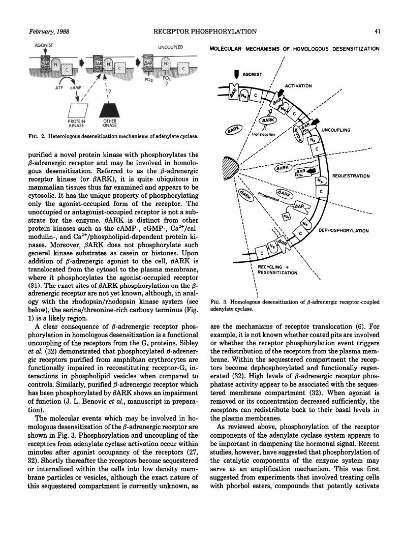
In summary, the molecular mechanisms which arethought to be involved in heterologous desensitizationare shown in Fig. 2. In this form of desensitization,receptor function is regulated by phosphorylation in theabsence of receptor sequestration or down-regulation.
This covalent modification serves to functionally uncou-ple the receptors, that is, to impair their interactionswith the guanine nucleotide regulatory proteins. Severalprotein kinases seem to be capable of promotingphosphorylation of the receptors including the cAMP-dependent kinase and protein kinase C. In addition tothe receptor modification, heterologous desensitizationseems to be associated with functional modifications(phosphorylation?) at the level of the nucleotide regula-tory proteins (G8 and GO.
The mechanisms by which agonists promote homolo-gous desensitization of the /3-adrenergic receptor alsoappear to involve receptor phosphorylation. Evidence forthis was first obtained using amphibian erythrocyteswhere /?-adrenergic agonists were shown to promote ho-mologous desensitization of adenylate cyclase and phos-phorylation of /3-adrenergic receptor (26). The agonist-induced receptor phosphorylation is pharmacologicallyspecific and stoichiometric, occurring to about 2 molphosphate/mol receptor. Prostaglandin Ei does not pro-mote /3-adrenergic receptor phosphorylation, althoughthis hormone elevates cAMP levels in these cells. Thissuggests that the observed receptor phosphorylation isnot mediated by the cAMP-dependent protein kinase.Additional evidence that protein kinase A is not involvedin homologous-induced receptor phosphorylation hascome from studies using S49 lymphoma cells (27). It wasobserved that in the S49 mutant cell lines cyc~ and kin",which are deficient in the G8 protein and c AMP-depend-ent protein kinase, respectively, agonists promoted 0-adrenergic receptor phosphorylation to the same degreeas in wild type cells. These results indicate that 1)receptor-G8 coupling is not necessary for receptor phos-phorylation to occur—agonist occupancy is sufficient;and 2) the receptor phosphorylation is not mediated bythe cAMP-dependent protein kinase.
Benovic et al. (28-30) have recently identified and
Adrenergic Receptor
FIG. 1. Structure of the /8-adrenergic re-ceptor as it is proposed to be organizedin the plasma membrane. The sites ofphosphorylation by the cAMP-depend-ent protein kinase (and probably proteinkinase C) are indicated. The solid circlesrepresent the serine- and threonine-richregion in the carboxy terminus whichmay serve as sites of /3ARK phosphor-ylation (see text).
February, 1988 RECEPTOR PHOSPHORYLATION 41
UNCOUPLED
PROTEIN OTHERKINASE KINASE
FIG. 2. Heterologous desensitization mechanisms of adenylate cyclase.
purified a novel protein kinase with phosphorylates thei8-adrenergic receptor and may be involved in homolo-gous desensitization. Referred to as the j8-adrenergicreceptor kinase (or /3ARK), it is quite ubiquitous inmammalian tissues thus far examined and appears to becytosolic. It has the unique property of phosphorylatingonly the agonist-occupied form of the receptor. Theunoccupied or antagonist-occupied receptor is not a sub-strate for the enzyme. 0ARK is distinct from otherprotein kinases such as the cAMP-, cGMP-, Ca2+/cal-modulin-, and Ca2+/phospholipid-dependent protein ki-nases. Moreover, jSARK does not phosphorylate suchgeneral kinase substrates as casein or histones. Uponaddition of 0-adrenergic agonist to the cell, 0ARK istranslocated from the cytosol to the plasma membrane,where it phosphorylates the agonist-occupied receptor(31). The exact sites of 0ARK phosphorylation on the /8-adrenergic receptor are not yet known, although, in anal-ogy with the rhodopsin/rhodopsin kinase system (seebelow), the serine/threonine-rich carboxy terminus (Fig.1) is a likely region.
A clear consequence of j8-adrenergic receptor phos-phorylation in homologous desensitization is a functionaluncoupling of the receptors from the G8 proteins. Sibleyet al. (32) demonstrated that phosphorylated jS-adrener-gic receptors purified from amphibian erythrocytes arefunctionally impaired in reconstituting receptor-Gs in-teractions in phospholipid vesicles when compared tocontrols. Similarly, purified /3-adrenergic receptor whichhas been phosphorylated by /? ARK shows an impairmentof function (J. L. Benovic et al., manuscript in prepara-tion).
The molecular events which may be involved in ho-mologous desensitization of the 0-adrenergic receptor areshown in Fig. 3. Phosphorylation and uncoupling of thereceptors from adenylate cyclase activation occur withinminutes after agonist occupancy of the receptors (27,32). Shortly thereafter the receptors become sequesteredor internalized within the cells into low density mem-brane particles or vesicles, although the exact nature ofthis sequestered compartment is currently unknown, as
MOLECULAR MECHANISMS OF HOMOLOGOUS DESENSITIZATION
ACTIVATION
UNCOUPLING
SEQUESTRATION
DEPHOSPHORYLATION
\ RECYCLING +\ RESENSITIZATION
FIG. 3. Homologous desensitization of j8-adrenergic receptor-coupledadenylate cyclase.
are the mechanisms of receptor translocation (6). Forexample, it is not known whether coated pits are involvedor whether the receptor phosphorylation event triggersthe redistribution of the receptors from the plasma mem-brane. Within the sequestered compartment the recep-tors become dephosphorylated and functionally regen-erated (32). High levels of /S-adrenergic receptor phos-phatase activity appear to be associated with the seques-tered membrane compartment (32). When agonist isremoved or its concentration decreased sufficiently, thereceptors can redistribute back to their basal levels inthe plasma membranes.
As reviewed above, phosphorylation of the receptorcomponents of the adenylate cyclase system appears tobe important in dampening the hormonal signal. Recentstudies, however, have suggested that phosphorylation ofthe catalytic components of the enzyme system mayserve as an amplification mechanism. This was firstsuggested from experiments that involved treating cellswith phorbol esters, compounds that potently activate

42 SIBLEY ET AL. Vol. 9, No. 1
protein kinase C (33), and examining their effects onadenylate cyclase activity. It was observed that phorbolester treatment resulted in enhanced basal adenylatecyclase activity as well as that stimulated by a variety ofhormonal and nonhormonal effectors (34-39). This am-plification effect has also been demonstrated by thedirect addition of activated protein kinase C to plasmamembranes (40).
Using amphibian erythrocytes, we have demonstratedthat incubation of these cells with phorbol esters resultsin a dramatic 100-300% amplification of /3-adrenergicagonist-, prostaglandin-, guanine nucleotide-, as well asMn2+-, and forskolin-stimulated enzyme activities (34).The observation that the forskolin- and Mn2+-stimulatedactivities are increased is especially interesting sincethese compounds can directly stimulate the catalytic unitof adenylate cyclase. This suggests that protein kinase Cmay phosphorylate the catalytic unit resulting in ampli-fied activity. In fact, using erythrocytes, we have dem-onstrated that activation of protein kinase C with phor-bol esters results in stoichiometric phosphorylation ofthe catalytic unit of adenylate cyclase (41). Under basalconditions there is no observable phosphate incorporatedinto the catalytic unit purified from [32P]-labeled cells,however, from phorbol ester-treated cells, the catalyticunit is phosphorylated to about 3 mol phosphate/molenzyme. We have also shown that purified protein kinaseC can directly phosphorylate the adenylate cyclase cat-alytic unit purified from bovine brain (41). This proteinkinase C-catalyzed phosphorylation of the adenylate cy-clase catalytic unit and amplification of enzyme activitymay provide a physiological mechanism by which recep-tor systems that promote phosphatidylinositol turnoverand protein kinase C activation can modulate receptorsystems coupled to adenylate cyclase.
Other G Protein-Coupled Receptors
It has become increasingly apparent that diverse bio-logical phenomena are characterized by adaptive proc-esses analogous to the desensitization observed in 0-adrenergic receptor-coupled adenylate cyclase systems.Not surprisingly, many of the other G protein-coupledreceptors display desensitization phenomena qualita-tively similar to those described above. Thus far, littleinformation is available concerning the molecular mech-anisms involved. It will be of interest to see whetherthese are identical to those uncovered for the /3-adrener-gic receptors.
A number of receptors, including ai-adrenergic andmuscarinic cholinergic, which are coupled via G proteinsto phosphatidylinositol turnover and protein kinase Cactivation (33, 42) demonstrate regulatory phenomenasimilar to adenylate cyclase-coupled receptors. Most no-
tably, the ax-adrenergic receptor-stimulated phosphati-dylinositol turnover response in cultured smooth musclecells has been shown to be desensitized by phorbol estertreatment (43, 44). Moreover, this desensitization is as-sociated with phosphorylation of the ai-adrenergic recep-tor (44). More recently, it has been shown that ax-adrenergic agonists also promote ai-adrenergic receptordesensitization, sequestration, and phosphorylation in atemporally correlated fashion (45). Unlike the situationfor the adenylate cyclase-coupled /8-adrenergic receptors,there is as yet no evidence for specific receptor kinasesinvolved in these phosphorylation reactions. In fact, itappears that protein kinase C itself directly phosphoryl-ates the ai-adrenergic receptor and that the rate but notthe extent of this reaction is enhanced by agonist occu-pancy (21). This is analogous to the cAMP-dependentprotein kinase-mediated phosphorylation of the 0-adre-nergic receptor (18). A classical feedback loop thus pre-sumably operates whereby diacylglycerol, generated byagonist stimulation of phosphatidylinositol turnover, ac-tivates protein kinase C, which phosphorylates and de-sensitizes the ai-adrenergic receptor. The cAMP-de-pendent protein kinase will also directly phosphorylatethe ai-adrenergic receptor in vitro, although agonists donot promote this reaction (21).
The situation for the muscarinic cholinergic receptorseems quite analogous to that of the «i-adrenergic recep-tor. Desensitization of muscarinic receptor-stimulatedphosphatidylinositol hydrolysis and receptor down-reg-ulation has been observed in response to cholinergicagonist or phorbol ester stimulation (46-50). Further-more, Kwatra and Hosey (51) have recently demon-strated muscarinic receptor phosphorylation in cardiactissue in response to agonist activation. The proteinkinase(s) involved in this reaction has not yet beenidentified.
An additional G protein-coupled receptor systemwhich demonstrates well characterized adaptive phe-nomena is the rhodopsin system in retinal rod outersegments. As reviewed in detail elsewhere (22, 52), thissystem is structurally and functionally analogous tohormone-activated adenylate cyclase. The analogouscomponents are a photon of light instead of hormone,rhodopsin instead of receptor, a GTP-binding and hy-drolyzing protein termed transducin instead of G8, anda cyclic GMP (cGMP) phosphodiesterase, which con-trols retinal concentrations of cGMP, instead of thecatalytic unit of adenylate cyclase. Rhodopsin undergoesa phosphorylation reaction that is catalyzed by a specifickinase termed rhodopsin kinase (53-57). Phosphoryla-tion by rhodopsin kinase requires the light (agonist)activated form of rhodopsin and occurs on multiple serineand threonine residues within the carboxy terminus ofrhodopsin (58). Phosphorylation of rhodopsin results in

February, 1988 RECEPTOR PHOSPHORYLATION 43
an impaired ability of rhodopsin to interact with trans-ducin. This system is thus desensitized by an agonist-promoted phosphorylation reaction that uncouples thereceptor from its GTP-binding regulatory protein. Al-though phosphorylation of rhodopsin will reduce its in-teraction with transducin, an additional protein is re-quired for complete uncoupling of this system. Thisprotein, referred to as 48K protein, S antigen, or arrestin,only binds to the phosphorylated form of rhodopsin (59,60). This observation raises the interesting hypothesisthat there may be an analogous 48K protein in theadenylate cyclase system whose action is necessary toexpress the full functional effects of receptor phosphor-ylation.
The picture that emerges from these findings is of avariety of pathways by which G protein-coupled recep-tors are regulated. There are classical feedback regula-tory loops, such as the protein kinase A phosphorylationof the /3-adrenergic receptor or protein kinase C phos-phorylation of the ai-adrenergic receptor. The rates ofthese reactions are enhanced by agonist occupancy of thereceptors. Cross-talk pathways also exist by which, forexample, protein kinase C can phosphorylate the aden-ylate cyclase-coupled /?-adrenergic receptor or proteinkinase A the ai-adrenergic receptor. These reactions arenot promoted by agonist occupancy of the substratereceptor. Although their physiological regulatory signif-icance is less clear, these reactions could play an impor-tant amplification role in tissues where a physiologicalor biochemical response of the target cell is reciprocallycontrolled by hormones using two distinct signal trans-duction pathways. Finally, there are specific receptorkinases, such as rhodopsin kinase and /8ARK, whichphosphorylate and desensitize specific receptors in acompletely agonist-dependent fashion. Such kinases leadonly to agonist-specific or homologous desensitizationsince agonist-promoted conformational changes in thereceptor are required to transform them into substratesfor the regulatory enzyme.
Insulin Receptors
Insulin induces a wide variety of biological effects intarget cells leading to alterations in cellular metabolismand growth. These actions are mediated by specific highaffinity cell surface receptors. Insulin receptors are in-tegral membrane glycoproteins comprised of two a-sub-units (Mr a 130,000) and two 0-subunits (Mr s 90,000)linked by disulfide bonds (reviewed in Refs. 61 and 62).Insulin appears to interact with the a-subunit of thereceptor which results in enhanced tyrosine kinase activ-ity which is intrinsic to the |8-subunit. Both the a- and0-subunits are known to be derived from a single glyco-sylated precursor protein Mr = 190,000. Recently, com-
plementary DNAs (cDNAs) encoding the precursor havebeen cloned which predict a polypeptide consisting ofeither 1,370 (63) or 1,382 (64) amino acids. The a-subunitcomprises the N-terminal portion of the precursor, fol-lowed by an enzyme cleavage site and then the /3-subunitcontaining a single transmembrane spanning sequence.The /3-subunit also contains a protein kinase domainwhich exhibits homology with the epidermal growth fac-tor (EGF) receptor and the src family of tyrosine kinases.A schematic representation of the insulin receptor isshown in Fig. 4.
Although the physiological effects of insulin receptorstimulation are well known, the initial biochemical sig-nals have been difficult to elucidate. One of the earliestmeasurable responses to insulin stimulation, however, isthat of receptor phosphorylation. Kasuga et al. (65, 66)initially demonstrated that insulin stimulates phosphor-ylation of the 0-subunit of the receptor in 32P-labeledlymphocytes and hepatoma cells. Similar results havebeen observed using isolated adipocytes (67) and rat
NH 2 N H 2
EXTRACELLULARLIGAND BINDING
DOMAIN
N H 2
TRANSMEMBRANEDOMAIN
CYTOPLASMICTYROSINE KINASE
DOMAIN
a
N H 2
COOHCOOH
T
-TYR 960 (972)
-TYR 1150 (1162)-TYR 1151 (1163)
•TYR 1316 (1328)
COOH COOH
FIG. 4. Schematic representation of the insulin receptor. Regions ofhigh cysteine concentration are shown as hatched boxes, transmem-brane domains as solid boxes, and tyrosine kinase domains as openboxes.

44 SIBLEY ET AL. Vol. 9, No. 1
hepatocytes (68). In most cells, under basal conditionsthe 0-subunit contains predominantly phosphoserineand to a lesser extent phosphothreonine. Upon additionof insulin to the cells, increased levels of phosphoserineand phosphothreonine are observed in addition to phos-phorylation of tyrosine residues in the j3-subunit (67, 68-75). Interestingly, Pang et al. (71) have shown that theinsulin-stimulated tyrosine phosphorylation temporallyprecedes the increased serine phosphorylation of the /3-subunit. Peptide mapping of the phosphorylated /3-sub-units from intact cells indicates the presence of multiplesites of serine, and to a lesser extent threonine, phos-phorylation whereas the tyrosine phosphorylation occurson two to three major distinct sites (72-74). Recently,Stadtmauer and Rosen (76) have presented evidencesuggesting that tyrosine l.lSO1 is one of the phosphoryl-ated residues in insulin-stimulated cells (Fig. 4). In intactcells, the a-subunit does not appear to be phosphorylated.
In contrast to intact cells, when insulin-stimulatedreceptor phosphorylation is examined under cell-freeconditions, using [7-32P]ATP, phosphorylation occursexclusively on tyrosine residues. A large number of lab-oratories have characterized this reaction using solubi-lized insulin receptors in various stages of purity from avariety of tissues (see Refs. 61, 62, and 77 for review andreferences). It has been well documented that the cell-free phosphorylation is a result of autophosphorylationof the /3-subunit and that the insulin receptor itself is atyrosine kinase. Autophosphorylation of the /3-subunitappears to be intramolecular as dilution of insulin recep-tors does not decrease the rate of 0-subunit phosphoryl-ation (78, 79). The /J-subunit possesses an ATP bindingsite (80-82), and the cloned sequence predicts a proteinkinase domain with homology to other tyrosine kinases(63, 64). Tyrosine phosphorylation of the a-subunit hasalso been observed under cell-free conditions (83, 84);however, as this subunit appears to be extracellular (Fig.4), this reaction may not be physiologically significant.
Reports on the stoichiometry of insulin receptor phos-phorylation indicate that in vitro the /3-subunit is phos-phorylated up to 2 mol phosphate/mol receptor (78, 79).Peptide mapping of the in vitro phosphorylated 0-subunitindicates that there are two to three distinct sites oftyrosine phosphorylation (72, 74, 78,79,85). In the studyof White et al. (72), two of the in vitro phosphorylationsites corresponded to the two tyrosine phosphorylationsites which were observed in intact cells.
Evidence is now available which indicates that the
1 The two reported amino acid sequences for the insulin receptorprecursor differ in that the sequence of Ebina et al. (64) contains aninsertion of 12 amino acids in the carboxy terminus of the a-subunitwhen compared to that of Ullrich et al. (63). In this review, we willrefer to the sequence of Ullrich et al. (63) when designating amino acidresidues.
activity of the insulin receptor tyrosine kinase is en-hanced by tyrosine phosphorylation of its |8-subunit.Rosen et al. (86) initially showed that autophosphoryla-tion of the receptor results in increased tyrosine kinaseactivity as measured with exogenous substrates. More-over, after autophosphorylation, the activated tyrosinekinase is rendered insulin independent. Dephosphoryla-tion of the receptor with alkaline phosphatase restoresthe insulin dependency of the tyrosine kinase. Otherlaboratories have shown that autophosphorylation of thereceptor both in vitro (85, 87) and in intact cells (74, 88)leads to enhanced tyrosine kinase activity of the /3-subunit. In the studies of Yu and Czech (85) and Kwoket al. (87) the enhanced activity correlated with thephosphorylation of a single tryptic peptide site. •
Using antibodies directed to peptides derived from theknown sequence of the ^-subunit, Herrera and Rosen(89) have provided evidence that tyrosines 1150 and 1316(Fig. 4) are autophosphorylated in vitro. It was furthershown that autophosphorylation of Tyr-1150, which isalso phosphorylated in intact cells (76), is best correlatedwith kinase activation. Interestingly, antibodies directedagainst the sequence containing Tyr-960 block auto-phosphorylation and thus exogenous kinase activity (90),yet this residue does not appear to be autophos-phorylated in vitro (76). Using microsequencing tech-niques, Tornqvist et al. (91) have also demonstrated invitro autophosphorylation of tyrosine residues 1150 and1316 as well as tyrosines 1146,1151, and 1322. Recently,Ellis et al. (92) have demonstrated that point mutationsin tyrosines 1150 and 1151 will reduce insulin-stimulatedautophosphorylation and tyrosine kinase activity in par-allel with a decrease in insulin-stimulated 2-deoxyglucoseuptake when expression is performed in CHO cells.Taken together, these results argue strongly for a role ofautophosphorylation of tyrosines 1150 and 1151 in acti-vation of tyrosine kinase activity, although the molecularmechanism by which this activation occurs is not yetunderstood.
The observation that in intact cells insulin receptorsundergo phosphorylation on serine and threonine resi-dues in addition to tyrosine residues suggests that otherprotein kinases are involved in regulating insulin recep-tor function. A role for the cAMP-dependent proteinkinase has been suggested by Pessin et al. (93) whoshowed that incubation of adipocytes with /?-adrenergicagonists, which raise intracellular cAMP levels, inhibithigh affinity binding of insulin to its receptor. Insulin-stimulated glucose transport is also inhibited, and theseeffects are mimicked by incubation with dibutyryl-cAMP. Haring et al. (94) have reported similar findingsand further showed that /3-adrenergic agonist treatmentof cells reduces the tyrosine kinase activity of the recep-tor by increasing its Michaelis-Menten constant (Km)

February, 1988 RECEPTOR PHOSPHORYLATION 45
for ATP. Similarly, Stadtmauer and Rosen (75) demon-strated that increasing the cAMP of IM-9 cells dimin-ishes insulin receptor tyrosine kinase activity and thatthis is correlated with increased serine and threoninephosphorylation on the /3-subunit. Although these datasuggest that c AMP leads to attenuation of insulin actionby altering the phosphorylation state of the insulin re-ceptor, it is not clear whether the cAMP-dependentprotein kinase directly phosphorylates the receptor. In-spection of the amino acid sequence of the 0-subunitindicates the absence of classic consensus recognitionsites for this enzyme (63, 64) suggesting that multiplekinases may be involved in this modification.
Protein kinase C also appears to be involved in regu-lating insulin receptor function. Treatment of cells withphorbol esters, which activate protein kinase C, resultsin increased phosphorylation of serine and threonineresidues in the 0-subunit (69, 73, 95, 96). In some cells,but not others, this modification results in a reductionin affinity of insulin for binding to the receptor. Taka-yama et al. (69) and Haring et al. (96) have shown thatphorbol ester-induced receptor phosphorylation addi-tionally results in a reduction of tyrosine kinase activityof the /3-subunit. Recently, Bollag et al. (97) demon-strated that protein kinase C can directly phosphorylatethe /3-subunit of the insulin receptor in vitro to stoichi-ometric levels. This phosphorylation is accompanied bya 65% reduction in the tyrosine kinase activity of thereceptor.
As noted earlier, treatment of intact cells with insulinresults in enhanced receptor phosphorylation on serineand threonine residues. Peptide mapping experimentsindicate that these residues appear, for the most part, tobe distinct from those phosphorylated in response toprotein kinase C activation (69, 73). This may suggestthat insulin treatment leads to the activation of otherserine/threonine kinases or that insulin occupancy of thereceptor renders it a better substrate for those kinases.Recent results have indicated that long term insulintreatment of cells can lead to reductions in receptortyrosine kinase activity (98, 99) which may be linked tothe observed insulin-induced increase in serine/threo-nine phosphorylation.
Insulin receptors thus appear to be under both positiveand negative control through phosphorylation of their 0-subunits. One of the earliest events in insulin activationis autophosphorylation of the #-subunit leading to en-hanced tyrosine kinase activity toward exogenous sub-strates. Tyr-1150 and 1151 appear to be intimately in-volved in this phosphorylation although other tyrosineresidues cannot be ruled out as yet. In contrast, phos-phorylation of the /3-subunit on serine and perhaps thre-onine residues results in decreased autophosphorylationand tyrosine kinase activity of the receptor. The serine
and threonine residues involved in this modification havenot yet been identified. A number of serine/threonineprotein kinases may be involved in this phosphorylationincluding the cAMP-dependent protein kinase and pro-tein kinase C. Whether or not insulin occupancy resultsin receptor phosphorylation by additional kinases re-mains to be determined.
Insulin-Like Growth Factor (IGF) Receptors Iand II
IGF-1 or somatomedin C is a polypeptide growth factorwith an amino acid sequence which is highly homologousto insulin. IGF-I initiates its mitogenic response in targetcells by binding to specific receptors on the plasmamembrane. Like the insulin receptor, the IGF-I receptoris a disulfide-linked heterotetramer that can be resolvedinto Mr s= 130,000 a-subunits which bind IGF-I and Mr
= 98,000 /3-subunits which exhibit tyrosine kinase activ-ity (100). In analogy with the insulin receptor, IGF-Ireceptor a- and /3-subunits are encoded with a single Mx
= 180,000 glycosylated receptor precursor for whichcDNAs have been recently cloned and sequenced (101).
As with the receptor for insulin, the IGF-I receptor isa tyrosine protein kinase capable of autophosphoryla-tion. Incubation of 32P-labeled IM-9 and Hep-G2 cellswith IGF-I promotes phosphorylation on tyrosine resi-dues of the IGF-I receptor /3-subunit (73). In addition, inboth partially and highly purified preparations of IGF-1receptors, IGF-I stimulates phosphorylation of tyrosineresidues on the /3-subunit of the receptor (95, 102-107).Evidence has been presented that this is an intramole-cular reaction (105, 107). As with insulin receptors, au-tophosphorylation of the IGF-I receptor is believed toenhance its tyrosine kinase activity. IGF-I receptor-me-diated phosphorylation of exogenous tyrosine-containingsubstrates is enhanced by prior autophosphorylation, andthis enhancement is reversed by phosphatase treatmentof the receptor (105,107).
In intact cells, the /?-subunits of the IGF-I receptor areconstitutively phosphorylated on serine and threonineresidues (73). Incubation of cells with biologically activephorbol esters increase this serine and threonine phos-phorylation by about 4-fold (73, 95). In IM-9 cells, IGF-I also promotes serine and threonine phosphorylation onthe /3-subunit, although apparently on different sitesthan those observed with phorbol esters (73). The func-tional significance of these serine and threonine phos-phorylations is at present unclear; however, in some cellsphorbol esters have been shown to reduce IGF-I bindingactivity (108).
Like IGF-I, IGF-II is a peptide hormone which isstructurally homologous with insulin. The receptor forIGF-II is distinct from the IGF-1 receptor and is com-

46 SIBLEY ET AL. Vol. 9, No. 1
posed of a single polypeptide with Mr = 250,000 (109).The IGF-II receptor appears to be functionally differentfrom insulin and IGF-I receptors in that binding of IGF-II to its receptor does not directly trigger acute metabolicor growth-permissive effects. Moreover, the IGF-II re-ceptor does not appear to possess tyrosine kinase activity(110). Rather it has been suggested that internalizationand recycling of the IGF-II receptor may be closely linkedto IGF-II action (111). Interestingly, Oka et al. (112,113)have observed that insulin treatment of adipocytes leadsto increased numbers of IGF-II receptors in the plasmamembrane. In addition, there is a concomitant decreasein IGF-II receptors located in a low density microsomalmembrane compartment.
Corvera and Czech (114) have investigated the molec-ular basis of this phenomenon and demonstrated a rolefor IGF-II receptor phosphorylation. They found thatunder basal conditions about 80-90% of the total cellularIGF-II receptors reside in a microsomal fraction with10-20% being located in the plasma membrane. More-over, the receptors in the plasma membrane are stoichi-ometrically phosphorylated possessing 2- to 3-fold morephosphate than those receptors in the microsome frac-tion. Insulin treatment produces a specific and dramaticdecrease in the phosphorylation state of the plasmamembrane receptors which is correlated with the ap-pearance of receptors on the cell surface. These datasuggest a model in which the IGF-II receptor is persist-ently phosphorylated on the cell surface leading to inter-nalization into a microsomal compartment where de-phosphorylation can occur. Insulin perturbs this processby inhibiting the IGF-II receptor kinase or activating aphosphatase or both which leads to decreased internali-zation and increased IGF-II receptor expression on thecell surface. Interestingly, this model is analogous to thatdescribing the role of phosphorylation in /3-adrenergicreceptor sequestration and recycling (see above).
The nature of the protein kinase(s) involved in IGF-IIreceptor phosphorylation is at present not known. Corv-era et al. (110), however, have demonstrated that theIGF-II receptor can be phosphorylated by a tyrosinekinase in adipocyte membranes although, as mentionedabove, this is not an autophosphorylation event.
The EGF Receptor
EGF is a polypeptide mitogen which is capable ofstimulating the proliferation of a variety of epidermaland epithelial cells (115). The receptor for EGF is a170,000 dalton glycosylated transmembrane proteinwhich exhibits intrinsic tyrosine kinase activity (115,116). Activation of the receptor with EGF leads to en-hanced intracellular protein phosphorylation on tyrosineresidues (see Refs. 77,117, and 118 for reviews). Recent
molecular cloning of the EGF receptor indicates that itconsists of 1,186 amino acids, contains a single mem-brane-spanning domain, and exhibits an extracellularamino terminus and a cytosolic carboxy terminus (119-121). The tyrosine kinase domain of the receptor is inthe cytoplasmic region and demonstrates high homologyto the transforming protein v-erb B of avian erythro-blastosis virus (122). A schematic representation of theEGF receptor is shown in Fig. 5. In intact cells, the EGFreceptor is constitutively phosphorylated on serine, thre-onine, and tyrosine residues (123-125). Evidence hasaccumulated suggesting that these phosphorylation sitesmay be intimately linked with regulating receptor func-tion.
Stimulation of the EGF receptor with EGF rapidlyleads to receptor autophosphorylation on tyrosine resi-dues (reviewed in Refs. 77 and 115-118). Downward etal. (126) have shown that tyrosine residues 1,068, 1,148,and 1,173 in the carboxy terminus are involved in thisautophosphorylation with residue 1,173 being the mostheavily phosphorylated (Fig. 5). This reaction was shownby Weber et al. (127) to be intramolecular in nature. Gilland colleagues (128, 129) have suggested that this auto-
N H 2
EXTRACELLULARLIGAND BINDING
DOMAIN
ITRANSMEMBRANE
DOMAINL-THR 654
CYTOPLASMICTYROSINE KINASE
DOMAIN
-TYR 1068
-TYR 1148-TYR 1173
COOH
FIG. 5. Schematic representation of the EGF receptor. Regions of highcysteine concentration are shown as hatched boxes, the transmembranedomain as a solid box, and the tyrosine kinase domain as an open box.

February, 1988 RECEPTOR PHOSPHORYLATION 47
phosphorylation enhances the tyrosine kinase activity ofthe receptor. When phosphorylation of exogenous pep-tide substrates is assayed as a function of receptor au-tophosphorylation, tyrosine kinase activity is enhancedby up to 3-fold at a phosphate/receptor stoichiometry of1-2 mol/mol. High concentrations of peptide substratescan inhibit phosphorylation of both the receptor and thepeptides, but this inhibition can be relieved by initiallyautophosphorylating the receptor. These results suggestthen that autophosphorylation of the carboxy terminusmay remove an inhibitory steric constraint, thus allowingaccess of exogenous substrates to the active site andincreased tyrosine kinase activity.
In contrast to the above studies, an early report byCassel et al. (130) indicated that EGF-induced autophos-phorylation of the EGF receptor in A431 cell membranesdoes not affect its tyrosine kinase activity. In addition,the rate of EGF-induced exogenous peptide phosphoryl-ation does not show a lag during the time period in whichreceptor phosphorylation was" submaximal. ^Similarly,Downward et al. (131) have provided evidence that vari-ation in the extent of receptor autophosphorylation from0.1-2.8 mol phosphate/mol receptor does not influencereceptor kinase activity either in purified or membrane-bound receptor preparations. Moreover, Gullick et al.(132) have shown that antibodies raised against syntheticpeptides containingjthe EGF receptor tyrosine autophos-phorylation sites can inhibit autophosphorylation of thenative receptor without affecting EGF-stimulated tyro-sine kinase activity toward exogenous substrates. In ad-dition, Yarden and Schlessinger (133) have presentedevidence favoring an intermolecular rather than an in-tramolecular mechanism of EGF receptor activation. Itis thus not yet clear whether receptor autophosphoryla-tion on tyrosine residues results in enhanced tyrosinekinase activity of the EGF receptor. It is expected thatexperiments involving site-directed mutagenesis of therelevant tyrosine residues in the EGF receptor will re-solve this important issue. :
In addition to phosphorylation on tyrosine residues,treatment of intact cells with EGF results in1 enhancedphosphorylation of serine and threonine residues in thereceptor (123-125, 134). Since EGF has been shown topromote phosphatidylinositol turnover (135, 136) gen-erating diacylglycerol which activates protein kinase C(137), it was proposed that this kinase phosphorylatesthe receptor. Indeed, a number of studies have demon-strated that treatment of cells with phorbol esters, whichdirectly activate protein kinase C, results in attenuationof EGF binding to its receptor (138-144). Moreover,phorbol esters induce phosphorylation of serine and thre-onine residues in the EGF receptor in intact 32P-labeledcells (124, 125, 134, 145, 146). Although treatment ofcells with phorbol esters enhances phosphorylation of
both serine and threonine residues, peptide mappingexperiments indicate the appearance of unique peptidescontaining only phosphothreonine (124, 134, 145, 146).Hunter et al. (145) and Davis and Czech (147) haveidentified threonine 654 in the EGF receptor as theunique site of phosphorylation in response to phorbolesters in intact cells. This residue is located on thecytoplasmic side of the receptor nine residues away fromthe plasma membrane (Fig. 5). It is thus in an ideallocation to affect interaction between the extracellularand intracellular domains of the receptor. Protein kinaseC added directly to A431 cell membranes will also phos-phorylate the EGF receptor on the same sites as thosepromoted by phorbol esters (134).
Phosphorylation of EGF receptors by protein kinaseC appears to inhibit receptor tyrosine kinase activity.Cells exposed to phorbol esters exhibit decreased phos-photyrosine levels in EGF receptors and other proteins(124, 134, 148). These studies also demonstrated thatEGF-induced autophosphorylation of the receptor ontyrosine residues is reduced after phorbol esters treat-ment. Recently, Downward et al. (131) have also shownthat phosphorylation of the EGF receptor by proteinkinase C results in decreased tyrosine kinase activitytoward exogenous substrates.
As discussed previously, phosphorylation of the EGFreceptor by protein kinase C (via phorbol ester treat-ment) results in functional alterations in EGF binding.In some cells this is apparent as a reduction in the highaffinity component of EGF-receptor binding, whereas inothers the result is a decrease in the number of EGFreceptors at the cell surface. Davis and Czech (149) haveshown that reduction of the apparent affinity of the EGFreceptor by protein kinase C correlates with phosphor-ylation of threonine 654 but not with phosphorylation ofother residues on the receptor. Lin et al. (150) havefurther shown that mutating threonine 654 to an alanineresidue blocks phorbol ester-induced EGF receptor in-ternalization in a cellular expression system. These datasuggest that phosphorylation of threonine 654 by proteinkinase C regulates the affinity of the receptor for EGF,the activity of the tyrosine kinase domain, as well as theintracellular location of the receptor.
It should be noted that the mechanisms by which EGFinduces internalization of its receptor may be distinctfrom those mediated by the actions of phorbol esters.First, phorbol esters appear to promote internalizationof EGF receptors in the absence of receptor degradation(151, 152), whereas EGF promotes both internalizationand degradation (153). Moreover, in the study of Lin etal. (150) the alanine 654 mutant receptors were stillinternalized and degraded in response to EGF. Therethus appears to be two independent mechanisms bywhich EGF receptors are internalized. One of these in-

48 SIBLEY ET AL. Vol. 9, No. 1
volves phosphorylation on threonine 654 of the receptorand can be promoted by both phorbol esters and EGF(154). The other, which is promoted only by EGF, alsoleads to receptor degradation and may or may not involvephosphorylation. In this regard, it is interesting thatmonoclonal antibodies to the EGF receptor can promotereceptor internalization in the apparent absence of phos-phorylation (155).
Although EGF stimulation leads to rapid autophos-phorylation on tyrosine residues which may result inenhanced kinase activity. EGF treatment also results inthe phosphorylation of threonine 654 (154) leading toeventual receptor internalization and tyrosine kinaseinhibition (156). In addition to this negative feedbackloop induced by EGF, the protein kinase C-induced re-ceptor phosphorylation may also represent a mechanismof heterologous regulation by other hormone ligandswhich activate protein kinase C. Platelet-derived growthfactor (PDGF), fibroblast growth factor, bombesin, andvasopressin, all of which stimulate phosphatidylinositolturnover, decrease EGF binding to its receptor (157-161). Davis and Czech (162) have further shown thatPDGF will induce phosphorylation on threonine 654 ofthe EGF receptor. Moreover, synthetic derivatives ofdiacyglycerol, the endogenous activator of protein kinaseC, will promote EGF receptor phosphorylation (163-165).
The EGF receptor is also potentially regulated throughphosphorylation by the cAMP-dependent protein kinase.This kinase has been shown to phosphorylate membrane-bound and purified EGF receptor on serine and threonineresidues (166, 167). Evidence that this phosphorylationmay be physiologically significant has come from studiesof Pessin et al. (93). They observed that treatment ofadipocytes with both /3-adrenergic agonists, which in-crease intracellular cAMP levels, and with dibutyryl-cAMP leads to a decreased affinity of the receptor forEGF. Phosphorylation of the EGF receptor by thecAMP-dependent protein kinase may thus lead to inhi-bition of EGF action.
The PDGF Receptor
PDGF is one of the major mitogens found in serumwhich stimulates DNA and protein synthesis as well asamino acid uptake and phosphatidylinositol turnover ina number of cells (168). Interestingly, PDGF is structur-ally homologous with the product of the v-sis oncogene(169). The PDGF receptor has been identified by affinitycross-linking experiments as a Mr = 180,000 single poly-peptide glycoprotein (170, 171). Like the receptors forinsulin, EGF and IGF-I, the PDGF receptor possesestyrosine kinase activity. Initial studies using fibroblastmembranes demonstrated that PDGF promotes the in-
corporation of phosphate into tyrosine residues of a Mr
= 180,000 protein (172-174). Pike et al. (175) and Heldinet al. (170) subsequently provided evidence suggestingthat the phosphoprotein observed in fibroblast mem-branes is the PDGF receptor. It was also observed thatthe Mr = 180,000 protein contains phosphoserine andphosphothreonine which are enhanced upon PDGFtreatment (175). The PDGF receptor has been recentlypurified using a novel antiphosphotyrosine antibody(176-178). These studies indicate that the Mr s= 180,000substrate of the PDGF-stimulated tyrosine kinase is, infact, the PDGF receptor and like the other tyrosinekinase receptors is capable of autophosphorylation. Veryrecently, the primary structure of the PDGF receptorhas been obtained through molecular cloning techniques(179). The single polypeptide contains an extracellularligand binding domain, a single membrane spanningregion, and an intracellular tyrosine kinase domainwhich is homologous with other tyrosine kinases (179).
Since the functional consequences of PDGF receptorautophosphorylation are not yet known, it will be inter-esting to investigate whether this covalent modificationis linked to tyrosine kinase activation as suggested forthe other tyrosine kinase receptors. Similarly, it will beimportant to establish a role for the phosphorylation ofthe PDGF receptor on serine and threonine residues(175) and to identify the kinase(s) involved. Since PDGFpromotes phosphatidylinositol turnover in several celltypes, it is tempting to speculate that protein kinase Cmay somehow be involved in these phosphorylationevents. However, a recent report by Sturani et al. (180)has suggested that phorbol esters do not affect PDGFreceptor function, at least in fibroblasts.
The Nicotinic Acetylcholine Receptor
The nicotinic acetylcholine receptor is an integralmembrane protein that functions as a neurotransmitter-dependent ion channel at the vertebrate neuromuscularjunction (181). The nicotinic receptor has been mostextensively studied in the Torpedo electric organ andexists as an oligomer of four polypeptide chains withmasses of 40 (a), 50 (0), 60 (7), and 65 (5) kilodaltons(kD) with a stoichiometry of a2fiy8 (182). Phosphoryla-tion of the nicotinic acetylcholine receptor has beenshown to occur both in intact cells and in vitro with asmany as nine phosphoserines being identified, distrib-uted 1, 1, 2, and 5 among the a, /?, 7, and 8 subunits,respectively (183). Postsynaptic membranes rich in thenicotinic receptor appear to contain both endogenousprotein kinase (184-186) and protein phosphatase activ-ities (187).
The endogenous protein kinases include protein kinaseC which phosphorylates the a- and 5-subunits of the

February, 1988 RECEPTOR PHOSPHORYLATION 49
receptor on serine residues (188). In addition, an endog-enous tyrosine protein kinase which phosphorylates the/?-, 7-, and 5-subunits of the receptor has been reported(188). This kinase phosphorylates a single tryosine resi-due on each of these subunits with stoichiometries ofapproximately 0.5 mol phosphate/mol subunit attainedin postsynaptic membranes. It has also been reportedthat purified pp608rc of Rous sarcoma virus as well asA431 cell membranes, which are rich in EGF receptor,both specifically phosphorylate purified nicotinic acetyl-choline receptor on the #-, 7-, and 5-subunits (188).
The cAMP-dependent protein kinase (protein kinaseA) also phosphorylates the 7- and 5-subunits of thenicotinic receptor on serine residues (189, 190). Thisphosphorylation can be completely blocked by the spe-cific inhibitor protein of the cAMP-dependent proteinkinase. Huganir and Greengard (189) have demonstratedthat the 7- and 5-subunits of the purified receptor arephosphorylated by the catalytic subunit of protein kinaseA to stoichiometries of 1.0 and 0.89 mol phosphate/molreceptor, respectively. Based on amino acid sequencesand subunit and substrate specificities, Huganir et al.(188) proposed the phosphorylation sites for proteinkinase A as serine 354 (7-subunit) and serine 361 (5-subunit). Moreover, antibodies raised against syntheticpeptides encompassing the proposed phosphorylationsites inhibit phosphorylation of the receptor by proteinkinase A (191).
Several recent studies have provided evidence for afunctional alteration of the acetylcholine receptor whenphosphorylated by protein kinase A (192-194). Twogroups have demonstrated that treatment of rat skeletalmuscle with forskolin, a potent activator of adenylatecyclase, leads to desensitization of nicotinic receptorfunction (192-194). Moreover, Huganir et al. (193) haveprovided direct evidence in a reconstituted system thatphosphorylation of the receptor by protein kinase Aincreases the rate of agonist-induced desensitization ofthe receptor by 7- to 8-fold. These results provide thefirst direct evidence that phosphorylation regulates thefunction of an ion channel receptor protein.
Intracellular Transport Receptors
Eukaryotic cells internalize many macromolecules andnutrients by receptor-mediated endocytosis. The intra-cellular pathways of this process have been investigatedin a variety of receptor systems (reviewed in Refs. 195and 196). Upon binding of ligand, the receptors move toand cluster in specialized regions of the plasma mem-brane termed coated pits. These structures contain theprotein clathrin which may be involved in trapping ofthe receptor-ligand complex. Subsequently, the coatedpits invaginate to form coated vesicles which rapidly give
rise to uncoated vesicles referred to as endosomes orreceptosomes. The receptosome is a low pH compartmentwhich promotes ligand-receptor dissociation. At thispoint the pathways of endocytotic receptors may diverge,with some classes of receptors being recycled back to thecell surface while others are degraded in lysosomes. Thetransreticular Golgi system may play a role in this sortingprocess (153). One of the major questions concerningreceptor-mediated endocytosis is what signals are in-volved in triggering receptor internalization, sorting, andreceptor recycling.
Recent attention has focused on phosphorylation ofthe receptor proteins as a mechanism of regulating theirintracellular distribution. One well characterized recy-cling receptor, the transferrin receptor, has indeed beenshown to undergo phosphorylation/dephosphorylationreactions. The transferrin receptor is an integral mem-brane glycoprotein composed of two identical subunitsof Mr = 90,000 which are linked by a disulfide bond toform a dimer (197, 198). This receptor mediates theendocytosis of transferrin, resulting in cellular uptake ofFe2+. Recently, it has been reported that endocytosis ofthe transferrin receptor can be promoted by phorbolesters (199, 200). Phorbol ester treatment results in aredistribution in approximately 50% of the transferrinreceptors from the plasma membrane to an endosomecompartment with no loss in total receptor number. InHL60 cells this event correlates with an increase in thephosphorylation state of the transferrin receptor (200,201). Although phorbol esters and transferrin induceinternalization of transferrin receptors in a nonadditivefashion, transferrin itself does not promote receptorphosphorylation. This may suggest that although phos-phorylation results in receptor internalization, it is notthe primary signal for transferrin-induced receptor en-docytosis. Recently, Davis et al. (202) have identifiedserine 24 as the site on the transferrin receptor phospho-rylated by protein kinase C.
In contrast to the effects of phorbol esters, recent workhas demonstrated that insulin and various growth factorscan induce the redistribution of transferrin receptorsfrom an intracellular endosome compartment to theplasma membrane (203, 204). In view of the observationthat insulin promotes a similar redistribution of IGF-IIreceptors by reducing their phosphorylation state (seeabove), it is tempting to speculate that insulin andgrowth factors induce dephosphorylation of transferrinreceptors leading to their enhanced cell surface expres-sion.
Other recycling receptors may be regulated by phos-phorylation events. The asialoglycoprotein receptor isphosphorylated on serine residues (205, 206) althoughthe functional significance of this modification is notknown. Phorbol esters have been shown to reduce the

50 SIBLEY ET AL. Vol. 9, No. 1
binding activity of low density lipoprotein (LDL) recep-tors (207); however receptor phosphorylation was notexamined. In contrast, Kishimoto et al. (208) have dem-onstrated phosphorylation of the LDL receptor on serine833 in the cytoplasmic domain by a casein kinase II-likeenzyme purified from adrenal cortex. The physiologicalsignificance of this modification is unknown as mutatingthis serine to an alanine has no effect on the rate of LDLreceptor internalization (209).
Receptors Mediating Immune Function
T Lymphocyte Receptors
The T cell antigen receptor is found on T lymphocyteswhere it mediates the activation of these cells by specificantigens. Clonotypic antibodies and cDNA cloning bysubtractive screening methods have shown that the maincore of the receptor is formed of an a (40-50 kD)- and fi(43 kD)-subunit heterodimer (reviewed in Ref. 210).However, immunoprecipitation and cross-linking exper-iments have suggested that the functioning receptor mayinvolve several additional protein components. A com-plex of proteins termed the T3 complex has been char-acterized both in the human and murine systems (211-214). This complex is comprised of two glycoproteins of20-22 kD (gp21) and 26 kD (gp26), and two nonglyco-sylated peptides of 25-26 kD (p25) and 16 kD (pl6). Anadditional nonglycosylated subunit of 21 kD (p21) hasrecently been identified in the murine system (215).Activation of the T cell antigen receptor complex byclonotypic antibodies or specific antigens leads to theincreased hydrolysis of polyphosphoinositides, suggest-ing that activation of protein kinase C may at least inpart mediate T cell activation (216, 217). Although abiochemical role for each of the putative components ofthe receptor complex has not yet been established, sev-eral studies have suggested that many of these polypep-tides are phosphorylated in response to T cell activationby specific antigens or mitogens. In human T lympho-cytes, Cantrell et al. (218) have reported phosphorylationof two peptides of the T3 complex (Mr = 26,000 and to alesser extent Mr = 21,000) upon activation of cells withphorbol esters. Samuelson et al. (215) have shown inmurine T cell hybridomas that phorbol esters lead tophosphorylation of p25 and gp21 on serine residues.Antigen activation of these cells leads to the phosphor-ylation of the same gp21 subunit and a previously uni-dentified 21 kD polypeptide on tyrosine residues. It hasbeen speculated that phosphorylation of the various Tcell receptor complex subunits might be involved in theprocess of down-regulation or desensitization of the Tcell receptor, a process induced by phorbol esters (218).Recently, the suggestion has been made (219) that thephosphorylation pattern of one of these subunits (p21,
tyrosine phosphorylation) may be abnormally constitu-tive in a mouse model of lymphoproliferative disease.Thus, these data suggest that phosphorylation of the Tcell antigen receptor complex may modulate several ofits functions.
Another T lymphocyte-specific receptor is that whichbinds T cell growth factor (TCGF) or interleukin-2 (IL-2), a Mr = 14,800 glycopeptide hormone necessary forantigen- or mitogen-mediated activation of T cell prolif-eration (220). The receptor for IL-2 is a glycoprotein ofMr = 55,000 with a nonglycosylated amino acid core ofMr = 28,428 (220). The mechanisms of signal transduc-tion for this hormone effector system are still not com-pletely understood. However, IL-2 has been shown toinduce a redistribution of protein kinase C activity (221),a property which is shared by hormones that use thephosphatidylinositol/Ca++ pathway as a transducingmechanism.
Several studies have documented that the IL-2 recep-tor is a phosphoprotein. Leonard et al. (222) showed thathuman IL-2 receptors isolated from mitogen-stimulatedperipheral lymphocytes and leukemic HUT 102B2 T cellsare constitutively phosphorylated. Shackelford andTrowbridge (223) have shown that treatment of activatedT cells or leukemic cells with phorbol esters induces thephosphorylation of the IL-2 receptor which is associatedwith increased expression of IL-2 receptors on the sur-face of these cells. More recently, Gaulton and Eardley(224) have shown that exposure of mitogen-activatedsplenic T cells to physiological concentrations of IL-2causes rapid phosphorylation of several membrane pro-teins amongst which is the IL-2 receptor protein itself.By site-directed mutagenesis, Gallis et al. (225) haveshown that cytoplasmic serine 247 is likely to be the siteof phosphorylation of the IL-2 receptor. From these datait has been suggested that phosphorylation of the IL-2receptor may be involved in the expression and mainte-nance of the receptor at the cell surface.
Immunogbbulin (Ig) receptors
Mast cells and basophils express, on their surface,specific receptors for IgE. Binding of IgE to these recep-tors initiates the noncytotoxic release and synthesis of avariety of chemical mediators of inflammation such ashistamine and leukotrienes from these cells (226). TheIgE receptor is heterotetramer composed of one a (45kD)-, one fi (33 kD)-, and two 7-subunits (9 kD) (226).The a-subunit is thought to be extracellular and servesas the ligand binding site for IgE whereas the j8-subunitappears to be transmembrane. The two 7-subunits inter-act with the jS-subunit within the cytoplasmic domain(226).
The biochemical signalling mechanisms by which IgE
February, 1988 RECEPTOR PHOSPHORYLATION 51
initiates its effects are still poorly understood; nonethe-less, phosphorylation of the IgE receptor has been ex-amined as a possible control mechanism for IgE-me-diated effects. Hempstead et al. (227, 228) have reportedthat in normal rat mast cells, the a- and /3-subunits ofthe receptor are constitutively phosphorylated. Antigenactivation of these cells results in increased phosphor-ylation of the a-subunit whereas the 0-subunit phos-phorylation is unaltered. The increase in a-subunit phos-phorylation occurs within the time scale of cell activationleading to the hypothesis that this covalent modificationis a primary event in receptor action. In contrast, Metz-ger and colleagues (229, 230), using basophilic leukemiacells, observed phosphorylation of the /3- and 7-subunitsof the IgE receptor but no phosphorylation of the a-subunit. In these cells, antigen activation results in in-creased phosphorylation of the /3-subunit whereas the 7-subunit phosphorylation is decreased. In light of thesecontroversial findings, it is clear that more work will benecessary to define a functional role for IgE receptorphosphorylation.
Polymeric IgA, the major immunoglobulin in externalsecretions, is synthesized in plasma cells of various mu-cosal tissues as well as exocrine glands (231). Receptorsfor IgA are synthesized by a variety of epithelial cells,including hepatocytes, and mediate the secretion of poly-IgA via receptor-mediated transport across the epithelia(232). In hepatocytes, the mature form of the receptor(120 kD) arises from two intermediate precursors, a 105kD initial precursor which becomes glycosylated to a 116kD secondary form. The final maturation of the 116 kDprecursor to the mature 120 kD form is associated withphosphorylation of the intracellular domain of the recep-tor on serine residues (233). Whereas this active form ofthe IgA receptor appears to be phosphorylated, the bio-logical significance of this phosphorylation is still notunderstood. One possibility is that it is involved in thesorting patterns and intracellular processing of the re-ceptor protein (233).
Conclusions
A rapidly expanding body of literature now indicatesthat the functionality and/or cellular distribution of cellsurface receptors for hormones, drugs, neurotransmit-ters, and growth factors can be regulated by phosphor-ylation. In many instances where phosphorylation di-rectly alters receptor function, this appears to be in anegative direction. Thus, receptor phosphorylation mayresult in decreased agonist ligand binding or a diminishedability of the receptor to interact with its biochemicaleffector. In some cases, the enzymatic activity of a recep-tor molecule may be inhibited. In contrast, autophos-phorylation of the tyrosine kinase receptors may result
in increased enzymatic activity and enhanced receptorfunction. In those receptor systems which exhibit inter-nalization and recycling phenomena, phosphorylationappears to result in enhanced receptor internalization.The biological role for this internalization event may bequite varied and system specific. In some cases, theconsequence of receptor internalization may be dephos-phorylation and resensitization, whereas in others theresult is enhanced endocytosis of a specific ligand. Futurework in this rapidly growing field will undoubtedly focuson identifying a role for phosphorylation in other recep-tor systems, characterizing the protein kinases and phos-phatases involved, and elucidating the molecular mech-anisms by which phosphorylation produces its functionaleffects.
AcknowledgmentWe wish to thank Mary M. Holben for her expert secretarial help
in the preparation of this manuscript.
References1. Krebs EG, Beavo JA 1979 Phosphorylation-dephosphorylation of
enzymes. Annu Rev Biochem 48:9232. Flockhart DA, Corbin JD 1982 Regulatory mechanisms in the
control of protein kinases. CRC Crit Rev Biochem 12:1333. Gilman, AG 1984 G proteins and dual control of adenylate cyclase.
Cell 36:5774. Stryer, L, Bourne HR 1986 G-Proteins: a Family of signal trans-
ducers. Annu Rev Cell Biol 2:3915. Spiegel, AM 1987 Signal transduction by guanine nucleotide
binding proteins. Mol Cell Endocrinol 49:16. Sibley, DR, Lefkowitz RJ 1985 Molecular mechanisms of receptor
desensitization using the /3-adrenergic receptor-coupled adenylatecyclase system as a model. Nature 317:124
7. Hertel C, Perkins JP 1984 Receptor-specific mechanisms of de-sensitization of /3-adrenergic receptor function. Mol Cell Endocri-nol 37:245
8. Harden TK 1983 Agonist-induced desensitization of the /3-adre-nergic receptor-linked adenylate cyclase. Pharmacol Rev 36:5
9. Kassis S, Fishman PH 1982 Different mechanisms of desensiti-zation of adenylate cyclase by isoproterenol and prostaglandin Eiin human fibroblasts: role of regulatory components in desensiti-zation. J Biol Chem 257:5312
10. Garrity MJ, Andreasen TJ, Storm DR, Robertson RP 1983 Pros-taglandin E-induced heterologous desensitization of hepatic ad-enylate cyclase: consequences on the guanyl nucleotide regulatorycomplex. J Biol Chem 258:8692
11. Kirchik HJ, Iyengar R, Birnbaumer L 1983 Human chorionicgonadotropin-induced heterologous desensitization of adenylatecyclase from highly lutenized rat ovaries: attenuation of regulatoryN8 component activity. Endocrinology 113:1638
12. Rich KA, Codina J, Floyd G, Sekura R, Hildebrandt JD, IyengarR 1984 Glucagon-induced heterologous desensitization of theMDCK cell adenylate cyclase. J Biol Chem 259:7893
13. Briggs MM, Stadel JM, Iyengar R, Lefkowitz RJ 1983 Functionalmodification of the guanine nucleotides regulatory protein afterdesensitization of turkey erythrocytes by catecholamines. ArchBiochem Biophys 224:142
14. Stadel JM, Nambi P, Shorr RGL, Sawyer DF, Caron MG, Lef-kowitz RJ 1983 Catecholamine-induced desensitization of turkeyerythrocyte adenylate cyclase is associated with phosphorylationof the /8-adrenergic receptor. Proc Natl Acad Sci USA 80:3173
15. Sibley DR, Peters JR, Nambi P, Caron MG, Lefkowitz RJ 1984Desensitization of turkey erythrocyte adenylate cyclase: /3-Adre-

52 SIBLEY ET AL. Vol. 9, No. 1
nergic receptor phosphorylation is correlated with attenuation of 34.adenylate cyclase activity. J Biol Chem 259:9742
16. Sibley DR, Daniel KD, Strader CD, Lefkowitz RJ 1987 Phos-phorylation of the /8-adrenergic receptor in intact cells: relation- 35.ship to heterologous and homologous mechanisms of adenylatecyclase desensitization. Arch Biochem Biophys 258:24 36.
17. Stadel JM, Rebar R, Shorr RGL, Nambi P, Crooke ST 1986Biochemical characterization of phosphorylated /3-adrenergic re-ceptors from catecholamine-desensitized turkey erythrocytes. 37.Biochemistry 25:3719
18. Benovic JL, Pike LJ, Cerione RA, Staniszewski C, Yoshimasa T,Codina J, Caron MG, Lefkowitz RJ 1985 Phosphorylation of the 38.mammalian /8-adrenergic receptor by cyclic AMP-dependent pro-tein kinase. J Biol Chem 260:7094
19. Sibley DR, Nambi P, Peters JR, Lefkowitz RJ 1984 Phorboldiesters promote /3-adrenergic receptor phosphorylation and ad- 39.enylate cyclase desensitization in duck erythrocytes. BiochemBiophys Res Commun 121:973
20. Kelleher DJ, Pessin JE, Ruoho AE, Johnson GL 1984 Phorbol 40.ester induces desensitization of adenylate cyclase and phosphor-ylation of the /S-adrenergic receptor in turkey erythrocytes. ProcNatl Acad Sci USA 81:4316 41.
21. Bouvier M, Leeb-Lundberg LMF, Benovic JL, Caron MG, Lef-kowitz RJ 1986 Regulation of adrenergic receptor function byphosphorylation. II. Effects of agonist occupancy on phosphoryl-ation of «i- and 02-adrenergic receptors by protein kinase C andthe cyclic AMP-dependent protein kinase. J Biol Chem 262:3106 42.
22. Dixon RAF, Kobilka BK, Strader DJ, Benovic JL, Dohlman HG,Frielle T, Bolanowski MA, Bennett CD, Rands E, Diehl RE, 43.Mumford RA, Slater EE, Sigal IS, Caron MG, Lefkowitz RJ,Strader CD 1986 Cloning of the gene and cDNA for mammalian/3-adrenergic receptor and homology with rhodopsin. Nature321:75 44.
23. Glass DB, Krebs EG 1980 Protein phosphorylation catalyzed bycyclic AMP-dependent and cyclic GMP-dependent protein ki-nases. Annu Rev Pharmacol Toxicol 20:363
24. Kishimoto A, Nishiyama K, Nakanishi H, Vratsuji NH, Takay-ama Y, Nishizuka Y 1985 Studies on the phosphorylation of 45.myelin basic protein by protein kinase C and adenosine 3':5'-monophosphate dependent protein kinase. J Biol Chem 260:12492
25. Limas CJ, Limas C 1985 Carbachol induces desensitization ofcardiac /3-adrenergic receptors through muscarinic Mi receptors.Biochem Biophys Res Commun 128:699
26. Sibley DR, Strasser RH, Caron MG, Lefkowitz RJ 1985 Homol- 46.ogous desensitization of adenylate cyclase is associated with phos-phorylation of the /3-adrenergic receptor. J Biol Chem 260:3883
27. Strasser RH, Sibley DR, and Lefkowitz RJ 1986 A novel cate-cholamine-activated adenosine cyclic 3',5'monophosphate inde-pendent pathway for /3-adrenergic receptor phosphorylation in 47.wild-type and mutant S49 lymphoma cells: mechanism of homol-ogous desensitization of adenylate cyclase. Biochemistry 25:1371
28. Benovic JL, Strasser RH, Caron MG, Lefkowitz RJ 1986 /3-Adrenergic receptor kinase: identification of a novel protein ki- 48.nase that phosphorylates the agonist-occupied form of the recep-tor. Proc Natl Acad Sci USA 83:2797
29. Benovic JL, Mayor Jr F, Somers RL, Caron MG, Lefkowitz RJ 49.1986 Light-dependent phosphorylation of rhodopsin by 0-adre-nergic receptor kinase. Nature 322:869
30. Benovic JL, Mayor F, Staniszewski C, Lefkowitz RJ, Caron MG 50.1987 Purification and characterization of the 0-adrenergic recep-tor kinase. J Biol Chem 262:9026
31. Strasser RH, Benovic JL, Caron MG, Lefkowitz RJ 1986 /?-agonist- and prostaglandin Ei-induced translocation of the /8- 51.adrenergic receptor kinase: evidence that the kinase may act onmultiple adenylate cyclase-coupled receptors. Proc Natl Acad SciUSA 83:6362 52.
32. Sibley DR, Strasser RH, Benovic JL, Daniel K, Lefkowitz RJ1986. Phosphorylation/dephosphorylation of the /3-adrenergic re- 53.ceptor regulates its functional coupling to adenylate cyclase andsubcellular distribution. Proc Natl Acad Sci USA 83:9408 54.
33. Nishizuka Y 1984 The role of protein kinase C in cell surfacesignal transduction and tumour promotion. Nature 308:693
Sibley DR, Jeffs RA, Daniel K, Nambi P, Lefkowitz RJ 1986Phorbol diester treatment promotes enhanced adenylate cyclaseactivity in frog erythrocytes. Arch Biochem Biophys 244:373Bell JD, Brunton LL 1986 Enhancement of adenylate cyclaseactivity in S49 cells by phorbol esters. J Biol Chem 260:2625Cronin MJ, Canonico PO 1985 Tumor promoters enhance basaland growth hormone releasing factor stimulated cAMP levels inanterior pituitary cells. Biochem Biophys Res Commun 129:404Sulakhe PV, Johnson DD, Phan NT, Wilcons R 1985 Phorbolester inhibits myoblast fusion and activates j8-adrenergic receptor-coupled adenylate cyclase. FEBS Lett 186:281Hollingsworth EB, Ukena D, Daly JW 1986 The protein kinaseC activator phorbol-12-myristate-13-acetate enhances cyclicAMP accumulation in pheochromocytoma cells. FEBS Lett196:131Johnson JA, Goka TJ, Clark RB 1986 Phorbol ester-inducedaugmentation and inhibition of epinephrine-stimulated adenylatecyclase in S49 lymphoma cells. J Cyclic Nucleotide Res 151:199Naghshineh S, Noguchi N, Huang KP, Londos C 1986 Activationof adipocyte adenylate cyclase by protein kinase C. J Biol Chem261:14534Yoshimasa T, Sibley DR, Bouvier M, Lefkowitz RJ, Caron MG1987 Cross-talk between second messenger generating systems:phorbol esters induce phosphorylation of the catalytic unit ofadenylate cyclase activity and enhancement of its activity. Nature327:67Berridge MJ, Irvine RF 1984 Inositol triphosphate, a novel secondmessenger in cellular signal transduction. Nature 312:315Cotecchia S, Leeb-Lundberg LMF, Hagen P-O, Lefkowitz RJ,Caron MG 1985 Phorbol ester effects on ai-adrenoceptor bindingand phosphatidylinositol metabolism in cultured vascular smoothmuscle cells. Life Sci 37:2389Leeb-Lundberg LMF, Cotecchia S, Lomasney J, DeBernardis JF,Lefkowitz RJ, Caron MG 1985 Phorbol esters promote ai-adre-nergic receptor phosphorylation and receptor uncoupling frominositol phospholipid metabolism. Proc Natl Acad Sci USA82:5651Leeb-Lundberg LMF, Cottechia S, DeBlasi A, Caron MG, Lef-kowitz RJ 1987 Regulation of adrenergic receptor function byphosphorylation. I. Agonist-promoted desensitization and phos-phorylation of ai-adrenergic receptors coupled to inositol phos-pholipid metabolism in DDTi MF-2 smooth muscle cells. J BiolChem 262:3098Vicentini LM, Virgilo, FD, Ambrosini A, Pozzan T, Meldolesi J1985 Tumor promoter 12-myristate, 13-acetate inhibits phosphi-nositide hydrolysis and cytosolic Ca2+ rise induced by the activa-tion of muscarinic receptors in PC 12 cells. Biochem Biophys ResCommun 127:310Masters SB, Quinn MT, Brown JH 1985 Agonist-induced desen-sitization of muscarinic receptor-mediated calcium efflux withoutconcomitant desensitization of phosphoinositide hydrolysis. MolPharmacol 27:325Orellana SA, Solski PA, Brown JH 1985 Phorbol ester inhibitsphosphoinostide hydrolysis and calcium mobilization in culturedastrocytoma cells. J Biol Chem 260:5236Harden TK, Petch LA, Traynelis SF, Waldo GL 1985 Agonist-induced alteration in the membrane form of muscarinic cholin-ergic receptors. J Biol Chem 260:13060Liles WC, Hunter DD, Meier KE, Nathanson NM1986 Activationof protein kinase C induces rapid internalization and subsequentdegradation of muscarinic acetylcholine receptors in neuro-blastoma cells. J Biol Chem 261:5307Kwatra MM, Hosey MM 1986 Phosphorylation of the cardiacmuscarinic receptor in intact chick heart and its regulation by amuscarinic agonist. J Biol Chem 261:12429Stryer L 1986 Cyclic GMP cascade of vision. Annu Rev Neurosci9:87Kuhn H, Dreyer WJ 1972 Light-dependent phosphorylation ofrhodopsin by ATP. FEBS Lett 20:1Kuhn H, Cook JH, Dreyer WJ 1973 Phosphorylation of rhodopsinin bovine photoreceptor membranes: a dark reaction after illu-mination. Biochemistry 12:2495

February, 1988 RECEPTOR PHOSPHORYLATION 53
55. Kuhn H 1978 Light-regulated binding of rhodopsin kinase and 78.other proteins to cattle photoreceptor membranes. Biochemistry17:4389
56. Kuhn H, Hargrave PA 1981 Light-induced binding of guanosine 79.triphosphatase to bovine photoreceptor membranes: effect of lim-ited proteolysis of the membranes. Biochemistry 20:2410
57. Bownds D, Dawes J, Miller J, Stahlman M 1972 Phosphorylationof frog photoreceptor membranes induced by light. Nature 237:125 80.
58. Hargrave PA, Fong SL, McDowell JH, Mas MT, Curtis DR,Wang JK, Juszcak E, Smith DP 1980 The partial primary struc- 81.ture of bovine rhodopsin and its topography in the retinal rod celldisc membrane. Neurochem Intern 1:231
59. Kuhn H, Hall SW, Wilden U 1984 Light-induced binding of 48kDa protein to photoreceptor membranes is highly enhanced by 82.phosphorylation of rhodopsin. FEBS Lett 176:473
60. Wilden U, Hall SW, Kuhn H 1986 Phosphodiesterase activation 83.by photoexcited rhodopsin is quenched when rhodopsin is phos-phorylated and binds the intrinsic 48-kDa protein of rod outersegments. Proc Natl Acad Sci USA 83:1174 84.
61. Czech MP 1985 The nature and regulation of the insulin receptor:structure and function. Annu Rev Physiol 47:357
62. Kahn CR 1985 The molecular mechanism of insulin action. Annu 85.Rev Med 36:429
63. Ullrich A, Bell JR, Chen EY, Herrera R, Petruzzelli LM, Dull TJ,Gray A, Coussens L, Liao Y-C, Tsubokawa M, Mason A, Seeburg 86.PH, Grunfeld C, Rosen OM, Ramachandran J 1985 Humaninsulin receptor and its relationship to the tyrosine kinase familyof oncogenes. Nature 313:756 87.
64. Ebina Y, Ellis L, Jarnigan, Edery M, Graf L, Clauser E, Ou, J-h,Masiarz F, Kan YW, Goldfine ID, Roth RA, Rutter WJ 1985 Thehuman insulin receptor cDNA: the structural basis for hormone-activated transmembrane signalling. Cell 40:747 88.
65. Kasuga M, Karlsson FA, Kahn CR 1981 Insulin stimulates thephosphorylation of the 95,000 dalton subunit of its own receptor.Science 215:185 89.
66. Kasuga M, Zick Y, Blith, Karlsson FA, Haring HU, Kahn CR1982 Insulin stimulation of phosphorylation of the /3 subunit of 90.the insulin receptor. J Biol Chem 257:9891
67. Haring H-U, Kasuga M, Kahn CR 1982 Insulin receptor phos-phorylation in intact adipocytes and in a cell-free system.Biochem Biophys Res Commun 108:1538 91.
68. Gazzano H, Kowalski A, Fehlmann M, Von Obberghen E 1983Two different protein kinase activities are associated with theinsulin receptor. Biochem J 216:575 92.
69. Takayama S, White MF, Lauris V, Kahn CR 1984 Phorbol estersmodulate insulin receptor phosphorylation and insulin action incultured hepatoma cells. Proc Natl Acad Sci USA 81:7797
70. Haring H-U, Kasuga M, White MF, Crettaz M, Kahn CR 1984 93.Phosphorylation and dephosphorylation of the insulin receptor:evidence against an intrinsic phosphatase activity. Biochemistry23:3298 94.
71. Pang DT, Sharma BR, Shafer JA 1985 Predominance of tyrosinephosphorylation of insulin receptors during the initial responseof intact cells to insulin. J Biol Chem 260:7131
72. White MF, Takayama S, Kahn CR 1985 Differences in the sites 95.of phosphorylation of the insulin receptor in vivo and in vitro. JBiol Chem 260:9470
73. Jacobs S, Cuatrecasas P 1986 Phosphorylation of receptors for 96.insulin and insulin-like growth factor I. J Biol Chem 261:934
74. Yu K-T, Czech MP 1986 Tyrosine phosphorylation of insulinreceptor /3 subunit activates the receptor tyrosine kinase in intactH-35 hepatoma cells. J Biol Chem 261:4715 97.
75. Stadtmauer L, Rosen OM 1986 Increasing the cAMP content ofIM-9 cells alters the phosphorylation state and protein kinaseactivity of the insulin receptor. J Biol Chem 261:3402
76. Stadtmauer L, Rosen OM 1986 Phosphorylation of synthetic 98.insulin receptor peptides by the insulin receptor kinase and evi-dence that the preferred sequence containing Tyr-1150 is phos-phorylated in vitro. J Biol Chem 261:10000 99.
77. Pike LJ, Krebs EG 1986 Protein tyrosine kinase activity ofhormone and growth factor receptors. In: Conn PM (ed) TheReceptors. Academic Press, New York, vol 3:93-134 100.
White MF, Haring H-U, Kasuga M, Kahn CR 1984 Kineticproperties and sites of autophosphorylation of the partially puri-fied insulin receptor from hepatoma cells. J Biol Chem 259:255Petruzzelli L, Herrera R, Rosen OM 1984 Insulin receptor is aninsulin-dependent tyrosine protein kinase: copurification of in-sulin-binding activity and protein kinase activity to homogeneityfrom human placenta. Proc Natl Acad Sci USA 81:3327Roth RA, Cassell DJ 1983 Insulin receptor: evidence that it is aprotein kinase. Science 219:299Van Obberghen E, Rossi B, Kowalski A, Gazzano H, Ponzio G1983 Receptor-mediated phosphorylation of the hepatic insulinreceptor: evidence that the Mr 95,000 receptor subunit is its ownkinase. Proc Natl Acad Sci USA 88:945Shia MA, Pilch PF 1983 The 0 subunit of the insulin receptor isan insulin-activated protein kinase. Biochemistry 22:717Kasuga M, Zick Y, Blithe DL, Crettaz M, Kahn, Kahn CR 1982Insulin stimulates tyrosine phosphorylation of the insulin recep-tor in a cell-free system. Nature 298:667Zick Y, Kasuga M, Kahn CR, Roth J 1983 Characterization ofinsulin-mediated phosphorylation of the insulin receptor in a cell-free system. J Biol Chem 258:75Yu K-T, Czech MP 1984 Tyrosine phosphorylation of the insulinreceptor /S subunit activates the receptor-associated tyrosine ki-nase activity. J Biol Chem 259:5277Rosen OM, Herrera R, Olowe Y, Petruzzelli LM, Cobb MH 1983Phosphorylation activates the insulin receptor tyrosine proteinkinase. Proc Natl Acad Sci USA 80:3237Kwok YC, Nemenoff RA, Powers AC, Avruch J 1986 Kineticproperties of the insulin receptor tyrosine protein kinase: activa-tion through an insulin-stimulated tyrosine-specific, intramo-lecular autophosphorylation. Arch Biochem Biophys 244:102Klein HH, Freidenberg GR, Kladde M, and Olefsky JM 1986Insulin activation of insulin receptor tyrosine kinase in intact ratadipocytes. J Biol Chem 261:4691Herrera R, Rosen OM 1986 Autophosphorylation of the insulinreceptor in vitro. J Biol Chem 261:11980Herrera R, Petruzzelli L, Thomas N, Bramson HN, Kaiser ET,Rosen OM 1985 An antipeptide antibody that specifically inhibitsinsulin receptor autophosphorylation and protein kinase activity.Proc Natl Acad Sci USA 82:7899Tornqvist HE, Pierce MW, Frackelton R, Nemenoff RA, AvruchJ 1987 Identification of insulin receptor tyrosine resdues auto-phosphorylated in vitro. J Biol Chem 262:10212Ellis L, Clauser E, Morgan DO, Edery M, Roth RA, Rutter WA1986 Replacement of insulin receptor tyrosine residues 1162 and1163 compromises insulin-stimulated kinase activity and uptakeof 2-deoxyglucose. Cell 45:721Pessin JE, Gitomer W, Oka Y, Oppenheimer CL, Czech MP 1983/3-Adrenergic regulation of insulin and epidermal growth factorreceptors in rat adipocytes. J Biol Chem 258:7386Haring H, Kirsch D, Obermaier B, Ermel B, Machicao F 1986Decreased tyrosine kinase activity of insulin receptor isolatedfrom rat adipocytes rendered insulin-resistant by catecholaminetreatment in vitro. Biochem J 234:59Jacobs S, Sahyoun NE, Saltiel AR, Cuatrecasas 1983 Phorbolesters stimulate the phosphorylation of receptors for insulin andsomatomedin C. Proc Natl Acad Sci USA 80:6211Haring H, Kirsch D, Obermaier B, Ermel B, Machicao F 1986Tumor-promoting phorbol esters increase the Km of the ATP-binding site of the insulin receptor kinase from rat adipocytes. JBiol Chem 261:3869Bollag GE, Roth RA, Beaudoin J, Mochly-Rosen D, Koshland JrDE 1986 Protein kinase C directly phosphorylates the insulinreceptor ire vitro and reduces its protein-tyrosine kinase activity.Proc Natl Acad Sci USA 83:5822Arsenis G, Livingston JN 1986 Alterations in the tyrosine kinaseactivity of the insulin receptor produced by in vitro hyperinsuli-nemia. J Biol Chem 261:147Blake AD, Hayes NS, Slater EE, Strader CD 1987 Insulin receptordesensitization correlates with attenuation of tyrosine kinaseactivity but not of receptor endocytosis. Biochem J 245:357Morgan DO, Jarnigan K, Roth RA 1986 Purification and char-

54 SIBLEY ET AL. Vol. 9, No. 1
acterization of the receptor for insulin-like growth factor I. Bio-chemistry 25:5560
101. Ullrich A, Gray A, Tam AW, Yang-Feng T, Tsubokawa M, CollinsC, Henzel W, Le Bon T, Kathuria S, Chen E, Jacobs, Francke U,Ramachandran J, Fujita-Yamaguchi Y 1986 Insulin-like growthfactor I receptor primary structure: comparison with insulin re-ceptor suggests structural determinants that define functionalspecificity. EMBO J 5:2503
102. Jacobs S, Kull Jr F, Earp HS, Svoboda ME, Van Wyk JJ,Cuatrecasas P 1983 Somatomedin-C stimulates the phosphoryla-tion of the /3-subunit of its own receptor. J Biol Chem 258:9581
103. Rubin JB, Shia MA, Pilch PF 1983 Stimulation of tyrosine-specific phosphorylation in vitro by insulin-like growth factor I.Nature 305:438
104. Zick Y, Sasaki N, Rees-Jones RW, Grunberger G, Nissley SP,Rechler MM 1984 Insulin-like growth factor-I (IGF-I) stimulatestyrosine kinase activity in purified receptors from a rat liver cellline. Biochem Biophys Res Commun 119:6
105. Sasaki N, Rees-Jones RW, Zick Y, Nissley SP, Rechler MM 1985Characterization of insulin-like growth factor I-stimulated tyro-sine kinase activity associated with the /8-subunit of type I insulin-like growth factor receptors of rat liver cells. J Biol Chem 260:9793
106. Catanese VM, Grigorescu F, King GL, Kahn CR 1986 The humanerythrocyte insulin-like growth factor I receptor: characterizationand demonstration of ligand-stimulated autophosphorylation. JClin Endocrinol Metab 62:692
107. Yu K-T, Peters MA, Czech MP 1986 Similar control mechanismsregulate the insulin and type I insulin-like growth factor receptorkinases. J Biol Chem 261:11341
108. Fouis M, Thomopoulos P, Postel-Vinay M, Testa U, Guyda HJ,Posner BI 1984 The effect of phorbol esters on the receptors forinsulin-like peptides (ILAs). Mol Physiol 5:123
109. Oppenheimer CL, Czech MP 1983 Purification of the type IIinsulin-like growth factor receptor from rat placenta. J Biol Chem258:8539
110. Corvera S, Whitehead RE, Mottola C, Czech MP 1986 Theinsulin-like growth factor II receptor is phosphorylated by atyrosine kinase in adipocyte plasma membranes. J Biol Chem261:7675
111. Oka Y, Czech MP 1986 The type II insulin-like growth factorreceptor is internalized and recycles in the absence of ligand. JBiol Chem 261:9090
112. Oka Y, Mottola C, Oppenheimer CL, Czech MP 1984 Insulinactivates the appearance of insulin-like growth factor II receptorson the adipocyte cell surface. Proc Natl Acad Sci USA 81:4028
113. Oka Y, Rozek LM, Czech MP 1985 Direct demonstration of rapidinsulin-like growth factor II receptor internalization and recyclingin rat adipocytes. J Biol Chem 260:9435
114. Corvera S, Czech MP 1985 Mechanism of insulin action onmembrane protein recycling: a selective decrease in the phos-phorylation state of insulin-like growth factor II receptors in thecell surface membrane. Proc Natl Acad Sci USA 82:7314
115. Carpenter G 1983 The biochemistry and physiology of the recep-tor-kinase for epidermal growth factor. Mol Cell Endocrinol 31:1
116. Cohen S, Fava RA, Sawyer ST 1982 Purification and character-ization of epidermal growth factor receptor/protein kinase fromnormal mouse liver. Proc Natl Acad Sci USA 79:6237
117. Sefton BM, Hunter T 1984 Tyrosine protein kinases. Adv CyclicNucleotide Protein Phosphorylation Res 18:195
118. Hunter T, Cooper JA 1985 Protein-tyrosine kinases. Annu RevBiochem 54:897
119. Ullrich A, Coussens L, Hayflick JS, Dull TJ, Gray A, Tam AW,Lee J, Yarden LY, Liebemann TA, Schlessinger J, Downward J,Mayes ELV, Whittle N, Waterfield MD, Seeburg PH 1984 Humanepidermal growth factor receptor cDNA sequence and aberrantexpression of the amplified gene in A431 epidermoid carcinomacells. Nature 309:418
120. Xu Y-h, Ishii S, Clark AJL, Sullivan M, Wilson RK, Ma DP, RoeBA, Merlino GT, Pastan I 1984 Human epidermal growth factorreceptor cDNA is homologous to a variety of RNAs overproducedin A431 carcinoma cells. Nature 309:806
121. Lin CR, Chen WS, Kruiger W, Stolarsky LS, Weber W, Evans
RM, Verma IM, Gill GN, Rosenfeld MG 1984 Expression ofcloning of human EGF receptor complementary DNA: gene am-plification and three related messenger RNA products in A431cells. Science 25:843
122. Downward J, Yarden Y, Mayes E, Scrace G, Totty N, StockwellP, Ullrich A, Schlessinger J, Waterfield MD 1984 Close similarityof epidermal growth factor receptor and v-erb-B oncogene proteinsequences. Nature 307:521
123. Hunter T, Cooper JA 1981 Epidermal growth factor induces rapidtyrosine phosphorylation of proteins in A431 human tumor cells.Cell 24:741
124. Iwashita S, Fox CF 1984 Epidermal growth factor and potentphorbol tumor promoters induce epidermal growth factor receptorphosphorylation in a similar but distinctively different manner inhuman epidermoid carcinoma A431 cells. J Biol Chem 259:2559
125. Davis RJ, Czech MP 1984 Tumor-promoting phorbol diestersmediate phosphorylation of the epidermal growth factor receptor.J Biol Chem 259:8545
126. Downward J, Parker P, Waterfield MD 1984 Autophosphorylationsites on the epidermal growth factor receptor. Nature 311:483
127. Weber W, Bertics PJ, Gill GN 1984 Immunoaffinity purificationof the epidermal growth factor. J Biol Chem 259:14631
128. Bertics PJ, Weber W, Cochet C, Gill GN 1985 Regulation of theepidermal growth factor receptor by phosphorylation. J Biol Chem29:195
129. Bertics PJ, Gill GN 1985 Self-phosphorylation enhances theprotein tyrosine kinase activity of the epidermal growth factorreceptor. J Biol Chem 260:14642
130. Cassel D, Pike LJ, Grant GA, Krebs EG, Glaser L1983 Interactionof epidermal growth factor-dependent protein kinase with endog-enous membrane proteins and soluble peptide substrates. J BiolChem 258:2945
131. Downward J, Waterfield MD, Parker PJ 1985 Autophosphoryla-tion and protein kinase C phosphorylation of the epidermalgrowth factor receptor. J Biol Chem 260:14538
132. Gullick WJ, Downward J, Waterfield MD 1985 Antibodies to theautophosphorylation sites of the epidermal growth factor receptorprotein-tyrosine kinase as probes of structure and function.EMBO J 4:2869
133. Yarden Y, Schlessinger J 1987 Self-phosphorylation of epidermalgrowth factor receptor: evidence for a model of intramolecularallosteric activation. Biochemistry 26:1434
134. Cochet C, Gill GN, Meisenhelder J, Cooper JA, Hunter T 1984C-Kinase phosphorylates the epidermal growth factor receptorand reduces its epidermal growth factor stimulated tyrosine pro-tein kinase activity. J Biol Chem 259:2553
135. Sawyer ST, Cohen S 1981 Enhancement of calcium uptake andphosphatidylinositol turnover by epidermal growth factor in A-431 cells. Biochemistry 20:6280
136. Pike LJ, Eakes AT 1987 Epidermal growth factor stimulates theproduction of phosphatidylinositol monophosphate and thebreakdown of polyphosphoinositides in A431 cells. J Biol Chem262:1644
137. Bell RM 1986 Protein kinase C activation by diacylglycerol secondmessengers. Cell 45:631
138. Lee L-S, Weinstein IB 1978 Tumor-promoting phorbol estersinhibit binding of epidermal growth factor to cellular receptors.Science 202:313
139. Lee L-S, Weinstein IB 1979 Mechanism of tumor promoter inhi-bition of cellular binding of epidermal growth factor. Proc NatlAcad Sci USA 76:5168
140. Brown KD, Dicker P, Rozengurt E 1979 Inhibition of epidermalgrowth factor binding to surface receptors by tumor promotors.Biochem Biophys Res Commun 86:1037
141. Shoyab M, DeLarco JE, Todaro GJ 1979 Biologically activephorbol esters specifically alter affinity of epidermal growth factormembrane receptors. Nature 279:387
142. Magun BE, Matrisian LM, Bowden GT 1980 Epidermal growthfactor. J Biol Chem 255:6373
143. Salomon DS 1981 Inhibition of epidermal growth factor bindingto mouse embryonal carcinoma cells by phorbol esters mediatedby specific phorbol ester receptors. J Biol Chem 256:7958

February, 1988 RECEPTOR PHOSPHORYLATION 55
144. King AC, Cuatrecasas P 1982 Resolution of high and low affinityepidermal growth factor receptors. J Biol Chem 257:3053
145. Hunter T, Ling N, Cooper JA 1984 Protein kinase C phosphor-ylation of the EGF receptor at a threonine residue close to thecytoplasmic face of the plasma membrane. Nature 311:480
146. Chinkers M, Garbers DL 1984 Phorbol ester-induced threoninephosphorylation of the human epidermal growth factor receptoroccurs within the EGF binding domain. Biochem Biophys ResCommun 123:618
147. Davis RJ, Czech MP 1985 Tumor-promoting phorbol diesterscause the phosphorylation of epidermal growth factor receptorsin normal fibroblasts at threonine-654. Proc Natl Acad Sci USA82:1974
148. Friedman BA, Frackelton Jr AR, Ross AH, Connors JM, FujikiH, Sugimura T, Rosner MR 1984 Tumor promoters block tyro-sine-specific phosphorylation of the epidermal growth factor re-ceptor. Proc Natl Acad Sci USA 81:3034
149. Davis RJ, Czech MP 1986 Inhibition of the apparent affinity ofthe epidermal growth factor receptor caused by phorbol diesterscorrelates with phosphorylation of threonine-654 but not othersites on the receptor. Biochem J 233:435
150. Lin CR, Chen WS, Lazar CS, Carpenter CD, Gill GN, Evans RM,Rosenfeld MG 1986 Protein kinase C phosphorylation at Thr 654of the unoccupied EGF receptor and EGF binding regulate func-tional receptor loss by independent mechanisms. Cell 44:839
151. Beguinot L, Hanover JA, Ito S, Richert ND, Willingham MC,Pastan I 1985 Phorbol esters induce transient internalizationwithout degradation of unoccupied epidermal growth factor recep-tors. Proc Natl Acad Sci USA 82:2774
152. Fearn JC, King AC 1985 EGF receptor affinity is regulated byintracellular calcium and protein kinase C. Cell 40:991
153. Pastan I, Hanover J, Willingham M 1985 The cellular entry ofEGF and transferrin: a problem in intracellular sorting. Curr TopCell Regul 26:17
154. King CS, Cooper JA 1986 Effects of protein kinase C activationafter epidermal growth factor binding on epidermal growth factorreceptor phosphorylation. J Biol Chem 261:10073
155. Sunada H, Magun BE, Mendelsohn J, MacLeod CL 1986 Mono-clonal antibody against epidermal growth factor receptor is inter-nalized without stimulating receptor phosphorylation. Proc NatlAcad Sci USA 83:3825
156. Chinkers M, Garbers DL 1986 Suppression of protein tyrosinekinase activity of the epidermal growth factor receptor by epider-mal growth factor. J Biol Chem 261:8295
157. Rozengurt E, Brown KD, Pettican P 1980 Vasopressin inhibitionof epidermal growth factor binding to cultured mouse cells. J BiolChem 256:716
158. Rozengurt E, Collins M, Brown KD, Pettican P 1982 Inhibitionof epidermal growth factor binding to mouse cultured cells byfibroblast-derived growth factor. J Biol Chem 257:3680
159. Wharton W, Leof E, Pledger WJ, O'Keefe EJ 1982 Modulationof the epidermal growth factor receptor by platelet-derived growthfactor and choleragen: effects on mitogenesis. Proc Natl Acad SciUSA 79:5567
160. Collins MKL, Sinnett-Smith JW, Rozengurt E 1983 Platelet-derived growth factor treatment decreases the affinity of theepidermal growth factor receptors of Swiss 3T3 cells. J Biol Chem258:11689
161. Brown KD, Blay J, Irvine RF, Heslop JP, Berridge MJ 1984Reduction of epidermal growth factor receptor affinity by heter-ologous ligands: evidence for a mechanism involving the break-down of phosphoinositides and the activation of protein kinaseC. Biochem Biophys Res Commun 123:377
162. Davis RJ, Czech MP 1985 Platelet-derived growth factor mimicsphorbol diester action on epidermal growth factor receptor phos-phorylation at threonine-654. Proc Natl Acad Sci USA 82:4080
163. McCaffrey PH, Friedman BA, Rosner MR 1984 Diacylglycerolmodulates binding and phosphorylation of the epidermal growthfactor receptor. J Biol Chem 259:12502
164. Davis RJ, Ganong BR, Bell RM, Czech MP 1985 sn-l,2-Dioctan-oylglycerol. J Biol Chem 260:1562
165. Davis RJ, Ganong BR, Bell RM, Czech MP 1985 Structural
requirements for diacylglycerols to mimic tumor-promoting phor-bol diester action on the epidermal growth factor receptor. J BiolChem 260:5315
166. Rackoff WR, Rubin RA, Earp HS 1984 Phosphorylation of thehepatic EGF receptor with cAMP-dependent protein kinase. MolCell Endocrinol 34:113
167. Ghosh-Dastidar P, Fox CF 1984 cAMP-dependent protein kinasestimulates epidermal growth factor-dependent phosphorylation ofepidermal growth factor receptors. J Biol Chem 259:3864
168. Ross R, Vogel A 1978 The platelet derived growth factor. Cell14:203
169. Robbins KC, Antoniades HN, Devare SG, Hunkapillar MW,Aaronson SA 1983 Structural and immunological similaritiesbetween simian sarcoma virus gene product(s) and human plate-let-derived growth factor. Nature 305:605
170. Heldin C-H, Ek B, Ronnstrand L 1983 Characterization of thereceptor for platelet-derived growth factor on human fibroblasts.J Biol Chem 258:10054
171. Williams LT, Tremble PM, Lain MF, Sunday ME 1984 Platelet-derived growth factor receptors form a high affinity state inmembrane preparations. J Biol Chem 259:5287-5294
172. Ek B, Heldin C-H 1982 Characterization of a tyrosine-specifickinase activity in human fibroblast membranes stimulated byplatelet-derived growth factor. J Biol Chem 257:10486
173. Ek B, Westermark B, Wasteson, Heldin C-H 1982 Stimulation oftyrosine-specific phosphorylation by platelet-derived growth fac-tor. Nature 295:419
174. Nishimura J, Huang JS, Deuel TF 1982 Platelet-derived growthfactor stimulates tyrosine-specific protein kinase activity in SwissMouse 3T3 membranes. Proc Natl Acad Sci USA 79:4303
175. Pike LJ, Bowen-Pope DF, Ross R, Krebs EG 1983 Characteriza-tion of platelet-derived growth factor-stimulated phosphorylationin cell membranes. J Biol Chem 258:9383
176. Frackelton Jr AR, Tremble PM, Williams LT 1984 Evidence forthe platelet-derived growth factor-stimulated tyrosine phosphor-ylation of the platelet-derived growth facator receptor in vivo. JBiol Chem 259:7909
177. Daniel TO, Tremble PM, Frackelton Jr AR, Williams LT 1985Purification of the platelet-derived growth factor receptor by usingan anti-phosphotyrosine antibody. Proc Natl Acad Sci USA82:2684
178. Bishayee S, Ross AH, Womer R, Scher CD 1986 Purified humanplatelet-derived growth factor receptor has ligand-stimulated ty-rosine kinase activity. Proc Natl Acad Sci USA 83:6756
179. Yarden Y, Escobedo JA, Kuang W-J, Yang-Feng TL, Daniel TO,Tremble PM, Chen EY, Ando ME, Harkins RN, Francke U, FriedVA, Ullrich A, Williams LT 1986. Structure of the receptor forplatelet-derived growth factor helps define a family of closelyrelated growth factor receptors. Nature 323:226
180. Sturani E, Vicentini LM, Zippel R, Toschi L, Pandiella-AlonsoA, Comoglio PM, Meldolesi J 1986 PDGF-induced receptor phos-phorylation and phosphoinositide hydrolysis are unaffected byprotein kinase C activation in Mouse Swiss 3T3 and human skinfibroblasts. Biochem Biophys Res Commun 137:343
181. Changeux J-P 1981 The acetylcholine receptor: an "allosteric"membrane protein. Harvey Lect 75:85
182. Reynolds JA, Karlin A 1978 Molecular weight in detergent solu-tion of acetylcholine receptor from Torpedo Californica. Biochem-istry 17:2035
183. Vandlen RL, Wu WC-S, Eisenach JC, Raftery MA 1979 Studiesof the composition of purified Torpedo Californica acetylcholinereceptor and of its subunits. Biochemistry 18:1845
184. Gordon AS, Davis CG, Milfay D, Diamond 11977 Phosphorylationof acetylcholine receptor by endogenous membrane protein kinasein receptor-enriched membranes of Torpedo Californica. Nature267:539
185. Teichberg VI, Sobel A, Changeux J-P 1977 In vitro phosphoryla-tion of the acetylcholine receptor. Nature 267:540
186. Saitoh T, Changeux J-P 1981 Change in state of phosphorylationof acetylcholine receptor during maturation of the electromotorsynapse in Torpedo Mamorata electric organ. Proc Natl Acad SciUSA 78:4430

56 SIBLEY ET AL. Vol. 9, No. 1
187. Gordon AS, Milfay D, Davis CG, Diamond I 1979 Protein phos-phatase activity in acetylcholine receptor-enriched membranes.Biochem Biophys Res Commun 87:876
188. Huganir RL, Miles K, Greengard P 1984 Phosphorylation of thenicotinic acetylcholine receptor by an endogenous tyrosine-spe-cific protein kinase. Proc Natl Acad Sci USA 81:6968
189. Huganir RL, Greengard P 1983 cAMP-dependent protein kinasephosphorylates the nicotinic acetylcholine receptor. Proc NatlAcad Sci USA 80:1130
190. Zavoico GA, Comerci C, Subers E, Egan JJ, Huang C-K, FeinsteinMB, Smilowitz H 1984 cAMP, not Ca2+/calmodulin, regulates thephosphorylation of acetylcholine receptor in Torpedo Californicaelectroplax. Biochim Biophys Acta 770:225
191. Souroujon MC, Neumann D, Pizzighella S, Fridkin M, Fuchs S1986 Mapping of the cAMP-dependent phosphorylation sites onthe acetylcholine receptor. EMBO J 5:543
192. Albuquerque EX, Deshpande SS, Aracava Y, Alkondon M, DalyJW 1986 A possible involvement of cycle AMP in the expressionof desensitization of the nicotinic acetylcholine receptor. FEBSLett 199:113
193. Huganir RL, Delcour AH, Greengard P, Hess GP 1986 Phosphor-ylation of the nicotinic acetylcholine receptor regulates its rate ofdesensitization. Nature 321:774
194. Middleton P, Jaramillo F, Schuetze SM 1986 Forskolin increasesthe rate of acetylcholine receptor desensitization at rat soleusendplates. Proc Natl Acad Sci USA 83:4967
195. Stahl P, Schwartz AL 1986 Receptor-mediated endocytosis. J ClinInvest 77:657
196. Goldstein JL, Brown MS, Anderson RGW, Russel, DW, Schnei-der WJ 1985 Receptor-mediated endocytosis: concepts emergingfrom the LDL receptor system. Annu Rev Cell Biol 1:1
197. Schneider C, Owen MJ, Banville D, Williams JG 1984 Primarystructure of human transferrin receptor deduced from the mRNAsequence. Nature 311:675
198. McClelland A, Kuhn LC, Ruddle FH 1984 The human transferrinreceptor gene: genomic organization, and complete primary struc-ture of the receptor deduced from a cDNA sequence. Cell 39:267
199. Klausner RD, Harford J, van Renswoude J 1984 Rapid internal-ization of the transferrin receptor in K562 cells is triggered byligand binding or treatment with a phorbol ester. Proc Natl AcadSci USA 81:3005
200. May WS, Jacobs S, Cuatrecasas P 1984 Association of phorbolester-induced hyperphosphorylation and reversible regulation oftransferrin membrane receptors in HL60 cells. Proc Natl AcadSci USA 81:2016
201. May WS, Sahyoun N, Jacobs S, Wolf M, Cuatrecasas P 1985Mechanism of phorbol diester-induced regulation of surface trans-ferrin receptor involves the action of activated protein kinase Cand an intact cytoskeleton. J Biol Chem 260:9419
202. Davis RJ, Johnson GL, Kelleher DJ, Anderson JK, Mole JE,Czech MP 1986 Identification of serine 24 as the unique site onthe transferrin receptor phosphorylated by protein kinase C. JBiol Chem 261:9034
203. Davis RJ, Czech MJ 1986 Regulation of transferrin receptorexpression at the cell surface by insulin-like growth factors,epidermal growth factor and platelet-derived growth factor.EMBO J 5:653
204. Davis RJ, Corvera S, Czech MP 1986 Insulin stimulates cellulariron uptake and causes the redistribution of intracellular trans-ferrin receptors to the plasma membrane. J Biol Chem 261:8708
205. Schwartz AL 1984 Phosphorylation of the human asialoglycopro-tein receptor. Biochem J 223:481
206. Takahashi T, Nakada H, Okumura T, Sawamura T, Tashiro Y1985 Phosphorylation of the rat hepatocyte asialoglycoproteinreceptor. Biochem Biophys Res Commun 126:1054
207. Rouis M, Goldstein S, Thromopoulos P, Berthelier M, Hervy C,and Testa U 1984 Phorbol esters inhibit the binding of low-density lipoproteins (LDL) to U-937 monocytelike cells. J CellPhysiol 121:540
208. Kishimoto A, Brown MS, Slaughter CA, Goldstein JL 1987 Phos-phorylation of serine 833 in cytoplasmic domain of low densitylipoprotein receptor by a high molecular weight enzyme resem-bling casein kinase II. J Biol Chem 262:1344
209. Davis CG, Van Driel IR, Russel DW, Brown MS, Goldstein JL1987 The low density lipoprotein receptor. J Biol Chem 262:4075
210. Collins MKL, Owen MJ 1985 The T cell antigen receptor.Biochem J 230:281
211. Borst J, Prendiville MA, Terhorst C 1983 The T3 complex onhuman thymus-derived lymphocytes contains two different sub-units of 20 kDa. Eur J Immunol 13:576
212. Kanellopoulos JM, Wigglesworth NM, Owen MJ, Crumpton MJ1983 Biosynthesis and molecular nature of the T3 antigen of Tlymphocytes. EMBO J 3:1807
213. Samelson LE, Harford JB, Klausner RD 1985 Identification ofthe components of the murine T cell antigen receptor complex.Cell 43:223
214. Samelson LE, Harford, J, Schwarz RD, Klausner RD 1985 A 20-kDa protein associated with the murine T-cell antigen receptor isphosphorylated in response to activation by antigen or con-canavalin A. Proc Natl Acad Sci USA 82:1969
215. Samuelson LE, Patel MD, Weissman AM, Harford JB, KlausnerRD 1986 Antigen activation of murine T cells induces tyrosinephosphorylation of a polypeptide associated with the T cell anti-gen receptor. Cell 46:1083
216. Imboden JB, Stobo JD 1985 Transmembrane signaling by the Tcell antigen receptor. J Exp Med 161:446
217. Patel M, Samelson LE, Klausner RD, Multiple kinases and signaltransduction: phosphorylation of the T cell antigen receptor com-plex. Nature, in press
218. Cantrell DA, Davies AS, Crumpton MJ 1985 Activators of proteinkinase C down-regulate and phosphorylate the T3/T-cell antigenreceptor complex of human T lymphocytes. Proc Natl Acad SciUSA 82:8158
219. Samelson LE, Davidson WF, Morse III HC, Klausner RD, Ab-normal constituitive tyrosine phosphorylation of the T cell anti-gen receptor in murine lymphoproliferative disorders. Nature, inpress
220. Greene WC, Leonard WJ 1986 The human interleukin-2 receptor.Annu Rev Immunol 4:69
221. Farrar WL, Anderson WB 1985 Interleukin-2 stimulates associ-ation of protein kinase C with plasma membrane. Nature 315:233
222. Leonard WJ, Depper JM, Kronke M, Robb RJ, Waldman TA,Greene WC 1985 The human receptor for T-cell growth factor:evidence for variable post-translational processing, phosphoryla-tion, sulfation and the ability of precursor forms of the receptorto bind TCGF. J Biol Chem 260:1872
223. Shackelford DA, Trowbridge IS 1984 Induction of expression andphosphorylation of the human interleukin 2 receptor by a phorboldiester. J Biol Chem 259:11706
224. Gaulton GN, Eardley DD 1986 Interleukin 2-dependent phos-phorylation of interleukin 2 receptors and other T cell membraneproteins. J Immunol 136:2470
225. Gallis B, Lewis A, Wignall J, Alpert A, Mochizuki DY, CosmanD, Hopp T, and Urdal D 1986 Phosphorylation of the humaninterleukin-2 receptor and a synthetic peptide identical to its C-terminal, cytoplasmic domain. J Biol Chem 261:5075
226. Metzger H, Alcaraz G, Hohman R, Kinet J-P, Pribluda V, QuartoR 1986 The receptor with high affinity for immunoglobulin E.Annu Rev Immunol 4:419
227. Hempstead BL, Kulczycki Jr A, Parker CW1981 Phosphorylationof the IgE receptor from ionophore A23187 stimulated intact ratmast cells. Biochem Biophys Res Commun 98:815
228. Hempstead BL, Parker CW, Kulczycki Jr A 1983 Selective phos-phorylation of the IgE receptor in antigen-stimulated rat mastcells. Proc Natl Acad Sci USA 80:3050
229. Fewtrell C, Goetz A, Metzger H 1982 Phosphorylation of thereceptor for immunoglobulin E. Biochemistry 21:2004
230. Perez-Montfort G, Fewtrell C, Metzger H 1983 Changes in thereceptor for Immunoglobulin E coincident with receptor-mediatedstimulation of basophilic leukemia cells. Biochemistry 22:5733
231. Hanson LA, Ahlstedt A, Andersson B, Carlsson B, Cole MF, CruzJR, Dahlgren V, Ericsson TH, Jalil F 1983 Mucosal immunity.Ann NY Acad Sci 409:1
232. Courtoy PJ, Limet JN, Quintart J, Schneider Y-J, Vaerman J-P,Bauduiin P 1983 Transfer of IgA into rat bile: ultrastructuraldemonstration. Ann NY Acad Sci 409:799
233. Larkin JM, Sztul ES, Palade GE 1986 Phosphorylation of the rathepatic polymeric IgA receptor. Proc Natl Acad Sci USA 83:4759


![Phosphorylation of mitophagy and pexophagy receptors ...labs.biology.ucsd.edu/subramani/documents/Farre_Suresh4.pdf · is known to interact with Atg30 [4]. By co-immunoprecipitation](https://img.pdfslide.net/doc/110x75/5fc23e702fd1747c672d7811/phosphorylation-of-mitophagy-and-pexophagy-receptors-labs-is-known-to-interact.jpg)