Embed Size (px)
Citation preview
University of Northern Iowa University of Northern Iowa
UNI ScholarWorks UNI ScholarWorks
Dissertations and Theses @ UNI Student Work
2000
Photodecomposition of crystal violet dye in water solution and Photodecomposition of crystal violet dye in water solution and
suspensions of metal oxides suspensions of metal oxides
Tatiana Yurievna Zakharian University of Northern Iowa
Let us know how access to this document benefits you
Copyright ©2000 Tatiana Zakharian
Follow this and additional works at: https://scholarworks.uni.edu/etd
Part of the Organic Chemistry Commons
Recommended Citation Recommended Citation Zakharian, Tatiana Yurievna, "Photodecomposition of crystal violet dye in water solution and suspensions of metal oxides" (2000). Dissertations and Theses @ UNI. 649. https://scholarworks.uni.edu/etd/649
This Open Access Thesis is brought to you for free and open access by the Student Work at UNI ScholarWorks. It has been accepted for inclusion in Dissertations and Theses @ UNI by an authorized administrator of UNI ScholarWorks. For more information, please contact [email protected].
Copyright by Tatiana Yurievna Zakharian
2000 All Rights Reserved

PHOTODECOMPOSITION OF CRYSTAL VIOLET DYE IN WATER SOLUTION
AND SUSPENSIONS OF METALS OXIDES
An Abstract of a Thesis
Submitted
in Partial Fulfullment
of the Requirements for the Degree
Master of Arts
Tatiana Yurievna Zakharian
University of Northern Iowa
July 2000
ABSTRACT
Photodecomposition and photodecolorization of crystal violet dye were studied in
water solution, hydrogen peroxide solution and suspensions of metal oxides.
Spectroscopic evidence for the formation of methylated pararosanilines and demethylated
derivatives of Michler's ketone as the reaction intermediates was obtained for the
photodecomposition of crystal violet in water solution. The mechanism of formation of
the intermediates was discussed in terms of charges of individual atoms calculated using
Spartan software.
Photooxidation of crystal violet was shown to occur faster in hydrogen peroxide
solution due to the higher oxidation potential of hydrogen peroxide and hydroxyl
radicals. A mechanism similar to photooxidation in water solution was proposed since the
formation of the same intermediates was observed.
The semiconducting oxides Ti02 and ZnO were shown to be more efficient in
crystal violet photodecomposition than any of the insulating oxides studied: Si 0 2, Ah03,
and MgO. Adsorption and wavelength dependent studies of titanium dioxide suspensions
are consistent with a pathway in which a dye is oxidized by the surface-bound hydroxyl
radicals formed by injection of an electron from adsorbed hydroxyl groups to
photogenerated holes. Photogenerated electrons are subsequently scavenged by molecular
oxygen to form 0 2- radical anions and ultimately hydroxyl radicals that can also oxidize
the dye. Intermediates/products of photodecomposition were found to be adsorbed on the
titanium dioxide surface, which could lower the catalytic efficiency of titanium dioxide
as the reaction proceeds. Zinc oxide was found to be the most efficient oxide for crystal

violet photodecomposition, but its low photochemical stability makes it unsuitable for
industrial catalysis. Photochemically inert oxides were shown to influence the rate of
crystal violet photodecomposition. Acceleration of photodecomposition in the presence
of aluminum oxide can be explained by stabilization of the excited state of the dye by the
aluminum oxide surface, and inhibition of photodecomposition in the presence of silicon
dioxide can be explained by the formation of dye clusters on the silicon dioxide sUJface.
Fast decolorization in magnesium oxide suspensions is due to the reaction of crystal
violet with hydroxide ion to form a carbinol base. Titanium dioxide was concluded to be
the best prospective catalyst for crystal violet photodecomposition of those studied, since
it is highly efficient, relatively cheap and chemkally stable.
PHOTODECOMPOSITION OF CRYSTAL VIOLET DYE IN WATER SOLUTION
AND SUSPENSIONS OF METALS OXIDES
A Thesis
Submitted
in Partial Fulfullment
of the Requirements for the Degree
Master of Arts
Tatiana Yurievna Zakharian
University of Northern Iowa
July 2000
This Study by: Tatiana Yurievna Zakharian
Entitled: Photodecomposition of Crystal Violet Dye in Water Solution and
Suspensions of Metal Oxides
has been approved as meeting the thesis requirement for the
Degree of Master of Arts
~ {~ 2fJ1Jo Date Dr. Sh hanna R. Coon, Chair, Thesis Committee
& fr)/00 Date . Laura M. Hoistad, Thesis Committee Member
C, /1~/00 Date Dr. Paul E. Rider, Thesis Committee Member
Itw 1s:Urro Date
• John W. Somervill, Dean, Graduate College
II

111
ACKNOWLEDGEMENTS
I thank Dr. Shoshanna Coon for being a great research advisor always ready to
discuss new ideas and suggest interesting experiments, encouraging to work harder and to
think deeper. She did a great job in helping me to submit my thesis, which made
completion of my work possible.
I appreciate my committee members, Dr. Laura Hoistad, Dr. Paul Rider and Dr.
Vyacheslav Pak for fast and careful reading of this manuscript: Their suggestions and
ideas definitely made it more complete.
I thank Dr. Duane Bartak and Dr. Shilov who started the graduate exchange
program between the University of Northern Iowa and the Hertzen State University and
the UNI Graduate College and College of Natural Sciences for financial support.

IV
TABLE OF CONTENTS
PAGE
LIST OFT ABLES VI
LIST OF FIGURES . . . . . . . . . . . . . . . . . . . . . . . . . . . . . . . . . . . . . . . . . . . . . . . . . . . . . . . . . . . . . . . . . . . . . . . VII
CHAPTER 1. INTRODUCTION . . . . . . . . . . . . . . . . . . . . . . . . . . . . . . . . . . . . . . . . . . . . . . . . . . . . . . . 1
CHAPTER 2. EXPERIMENTAL SECTION........................................... 10
Materials . . . . . . . . . . . . . . . . . . . . . . . . . . . . . . . . . . . . . . . . . . . . . . . . . . . .. . . . . . . . . . . . . . . . . . . . . . 10
Light Source . . . . . . . . . . . . . . . . . . . . . . . . . . . . . . . . . . . . . . . . . . . . . .. . . . . . . . . . . . . . . . . . . . . ... 10
Procedures and Analysis . . . . . . . . . . . . . . . . . . . . . . . . . . . . . . . . . . . . . . . . . . . . . . . . . . . . . . . 12
CHAPTER 3. RESULTS AND DISCUSSION . . . . . . . . . . . . . . . . . . . . . . . .. . . ....... ...... 14
Photodecomposition of Crystal Violet in Water Solution . . . . . . . . . . . . . . . .. . 14
Photodecomposition of Crystal Violet in Hydrogen Peroxide . . . . . . . . . . . . 23
Photodecomposition of Crystal Violet in Water Suspensions of Metal Oxides ... ............ . ................ .. ... . .. ................. ... ................. 26
Photodecomposition of Crystal Violet in Titanium Dioxide Suspensions . . . . . . . . . . . . . . . . . . . . . . . . . . . . . . . . . . . . . . . . . . . . . . . . . . . . . . . . . . . . . 26
Mechanism of photodegradation . . . . . . . . . . . . . . . . . . . . . . . . . . . . 27
Intermediates and products . . .. . .. . .. . . . . .. . . . . . . . . . . . . . . . . . . . 31
Photodecomposition of Crystal Violet in Zinc Oxide 33 Suspensions ..... ... ........... ......................................... .
Photodecomposition of Crystal Violet in Silicon Dioxide Suspensions . . . . . . . . . . . . . . . . . . . . . . . . . . . . . . . . . . . . . . . . . . . . . . . . . . . . . . . . . . . . . 34
Photodecomposition of Crystal Violet in Aluminum Oxide Suspensions . . . . . . . . . . . . . . . . . . . . . . . . . . . . . . . . . . . . . . . . . . . . . . . . . . . . . . . . . . . . . 35
Photodecomposition of Crystal Violet in Magnesium Oxide Suspensions . . . . . . . . . . . . . . . . . . . . . . . . . . . . . . . . . . . . . . . . . . . . . . . . . . . .. . . . . . . . . 36
Comparison of Different Methods of Crystal Violet Photodecomposition . . . . . . . . . . . . . . . . . . . . . . . . . . . . . . . . . . . . . . . . . . .. . . . . .. . . . 38
CHAPTER 4. CONCLUSIONS . . . . . . . . . . . . . . . . . . . . . . . . . . . . . . . . . . . . . . . . . . . . . . . . . . . . . . . . . . 40
REFERENCES . . . . . . . . . . . . . . . . . . . . . . . . . . . . . . . . . . . . . . . . . . . . . . . . . . . . . . . . . . . . . . . . . . . . . . . . . . . . .. 44
V

TABLE
1
2
LIST OF TABLES
Charges of individual atoms in a crystal violet molecule calculated using Spartan .......... ....... ................ ......................................................... .
Charges of individual atoms in a pararosaniline molecule calculated using Spartan .......... ......... ....... ......... ......... ............................ ........... ....... .
VI
PAGE
20
20
FIGURE
1
2
3
4
5
6
7
8
9
10
11
12
13
LIST OF FIGURES
Structure of three triphenylmethane dyes: Basic Green 4 (Malachite Green), Basic Red (Pararosaniline) and Basic Violet 3 (Crystal Violet) ........................................... .... ...... ... ....... ... ..... ... .. ..... .
Photosensitization of a dye-semiconductor system and photoexcitation of a semiconductor surface .. ................ ... .. ... ..... .... .. .
Reduction of crystal violet.. ............................................... ...... ... .... . ..
The spectrum of sunlight measured by a spectroradiometer with 2 nm resolution ............. ..... .......................................... ........... .......... .
The spectrum of sunlight through the window of McColl um Science Hall, measured with a spectroradiometer with 2 nm resolution ................................................................ .. .......... ..... .... .... .. .
Photodecomposition of crystal violet in water solution under full spectrum illumination .... ... ... ... ...... ..... ....... ........ ............. .. ................. .
Photodecomposition of crystal violet in water solution under full spectrum illumination (fragment) .. ... ... ......... ............ ............ .... ........ .
The decrease in the integrated area of the dye peak in the visible region as a function of time for the dye solutions under visible and full spectrum illumination (corrected by photon flux) ... ... ... ..... ... .. .. ..
Photodecomposition of crystal violet in air-saturated and oxygenpurged water solutions after one day of full spectrum illumination ..
The mechanism of demethylation process ............ ........................... ..
Structure of Michler's ketone ......................... ................................ ... .
Photodecomposition of Michler's ketone under full spectrum illumination ...... .... .. ... ... .............. ... .. ............ .. ... .. ... ......... ... ....... ... .. .... .
Photodecomposition of pararosaniline under full spectrum illumination ...... ....... ....................................... .... ........... .... .... ....... ..... .
VII
PAGE
2
6
8
11
11
15
15
16
16
17
18
18
19

14
15
16
17
18
19
20
21
22
23
24
25
26
Mechanism of crystal violet photooxidation proposed by Kuramoto and Kitao ........................................................................................... .
The mechanism of crystal violet photodecomposition in the water solution ......... .............. ....... ..... ...... ............. .... .... ...... ............... ..... ..... .
Photodecomposition of crystal violet in hydrogen peroxide solution under full spectrum illumination ............ ......... ...... .. ............ .
Decomposition of crystal violet in hydrogen peroxide solution in the dark and in the light (an hour reaction time) ............. .............. .
The decrease in absorbance at 590 nm for the solutions with different concentrations of hydrogen peroxide after 20 minutes of full spectrum illumination .... ........................ .. ..... .............. ..... ... .... .... .
Photodecomposition of pararosaniline in hydrogen peroxide solution ............................................................................................ .. .
Photodecomposition of crystal violet in the absence and presence of titanium dioxide after one hour of illumination ............................ .
The decrease in the integrated area of the dye peak in the visible region as a function of time for crystal violet water solution and titanium dioxide suspension under visible and full spectrum illumination (corrected by photon flux) ............................. ............... .
Absorbance of crystal violet solution as a function of stirring time with titanium dioxide powder (in the dark) ...................................... .
Absorbance spectra of titanium dioxide surface after three hours of illumination and of Michler's ketone adsorbed on titanium dioxide surface ............................................................................................... .
The mechanism of crystal violet photodecomposition in titanium dioxide suspensions .......................................................................... .
Photodecomposition of crystal violet in titanium dioxide and zinc oxide suspensions after three days of visible illumination ............... .
Photodecomposition of crystal violet in silicon dioxide suspension after three days of full spectrum illumination ... .... .... ................. ....... .
VIII
21
22
23
24
25
26
27
27
28
32
32
34
35

IX
27 Photodecomposition of crystal violet in aluminum oxide 36 suspension after three days of full spectrum illumination ................ .
28 Decolorization of crystal violet solution half an hour after addition 37 of MgO under full spectrum illumination ......................................... .
29 The reaction of crystal violet with base ..... ....................................... . 37

CHAPTER I
INTRODUCTION
Dyes are intensely colored chemical compounds which, being applied to a
substrate, impart color to their substrate. Their ability to produce color arises from the
absorption of light in the visible range (400-700 nm), as a result of the interaction of the
radiation energy with the electron system of the molecule. The absorption spectrum
depends on the electronic structure of the dye.
The efficiency of dyes in absorbing specific wavelengths and reflecting others is
evaluated by the extinction coefficient, which is a measure of the fraction of light lost to
scattering and absorption per unit distance in the dye. For dyes with higher extinction
coefficients, lesser amounts are required to give the same depth of shade, which makes
them more economical in industry.
Dyes can be classified on the basis of the functional groups present. Azo,
carbonyl and triphenylmethane dyes are the most widely used classes of dyes.
1
Azo dyes contain azo groups (-N=N-) linked to two radicals, with at least one, but
more usually both, being aromatic. Azo dyes have high extinction coefficients, cover all
shades from yellow to navy and are relatively inexpensive, which makes them widely
used for dyeing of apparel and household textiles. The disadvantage of azo dyes is that
with bathochromic (red) shades, the ability to preferentially absorb light at only specific
wavelengths is reduced, with the resulting dulling of the shade. Further, the light fastness
of azo dyes is usually not very high: many of them undergo reductive and oxidative

photochemical reactions on exposure to strong light with the azo chromophore being
destroyed which makes the molecule either colorless or at best pale yellow.
Carbonyl dyes contain at least one carbonyl group (-C=O). Carbonyl dyes in
general are compact, which makes them excellent in obtaining high-quality dyeings in
pale shades on difficult to dye fabrics, and have good to excellent lightfastness. The
principal disadvantage of most carbonyl dyes is that they are tinctorially weak and based
on complex and expensive chemicals and chemical processes.
2
Triphenylmethane dyes contain three phenyl rings connected to an sp2-hybridized
carbon. The formulas of some common triphenylmethane dyes are shown in Figure l.
Cl
Figure 1. Structure of three triphenylmethane dyes (clockwise from upper left) : Basic Green 4 (Malachite Green), Basic Red (Pararosaniline) and Basic Violet 3 (Crystal Violet) .

3
Triphenylmethane dyes are of brilliant hue, exhibit high tinctorial strength and
can be applied to a large range of substrates. However, they are seriously deficient in
fastness properties, especially fastness to light and washing. Consequently, they are used
in outlets where brightness and cost effectiveness rather than permanence are paramount,
for example, the coloration of paper, waxes, plastics, drugs and ceramics. 1 Crystal violet,
N,N,N',N',N",N"-hexamethylpararosaniline, is widely used in pulp and paper
manufacture for coloration of paper and in laboratories for staining human and veterinary
tissue cells .2
Because of the high extinction coefficients of dyes, their presence in waste-
waters even at low concentrations may significantly lower transmission of sunlight- an
important component of life for most organisms. In addition, many triphenylmethane
dyes exhibit toxic effects toward microbial populations and can be toxic and mutagenic to
animals.3 Some dyes, including crystal violet, are known to be enzymatically degraded in
the human digestive system to produce carcinogenic substances.4 These environmental
effects state the necessity of decolorization for nontoxic and noncarcinogenic dyes to
form colorless and harmless dye derivatives and decomposition for the others to degrade
them into nonhazardous compounds, preferably CO2 and water.
Significant losses occur during manufacture and processing of dyes. Recent
estimates indicate that 12% of the synthetic textile dyes used yearly are lost to waste
streams.5 Because of their hazardous environmental effect, contamination of drinking
water supplies is of concern, and an efficient and cheap way of treating dye-contaminated
waters should be found.
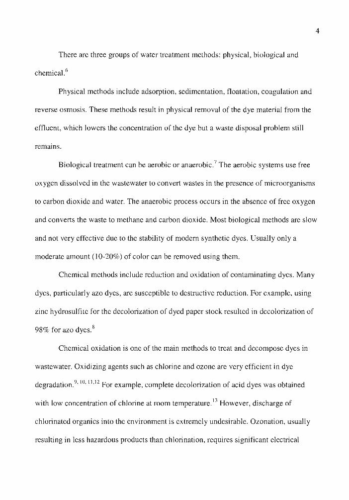
There are three groups of water treatment methods: physical, biological and
chemical.6
Physical methods include adsorption, sedimentation, floatation, coagulation and
reverse osmosis. These methods result in physical removal of the dye material from the
effluent, which lowers the concentration of the dye but a waste disposal problem still
remains.
4
Biological treatment can be aerobic or anaerobic. 7 The aerobic systems use free
oxygen dissolved in the wastewater to convert wastes in the presence of microorganisms
to carbon dioxide and water. The anaerobic process occurs in the absence of free oxygen
and converts the waste to methane and carbon dioxide. Most biological methods are slow
and not very effective due to the stability of modem synthetic dyes . Usually only a
moderate amount (10-20%) of color can be removed using them.
Chemical methods include reduction and oxidation of contaminating dyes. Many
dyes, particularly azo dyes, are susceptible to destructive reduction. For example, using
zinc hydrosulfite for the decolorization of dyed paper stock resulted in decolorization of
98% for azo dyes. 8
Chemical oxidation is one of the main methods to treat and decompose dyes in
wastewater. Oxidizing agents such as chlorine and ozone are very efficient in dye
degradation.9•
10•
11•12 For example, complete decolorization of acid dyes was obtained
with low concentration of chlorine at room temperature. 13 However, discharge of
chlorinated organics into the environment is extremely undesirable. Ozonation , usually
resulting in less hazardous products than chlorination, requires significant electrical
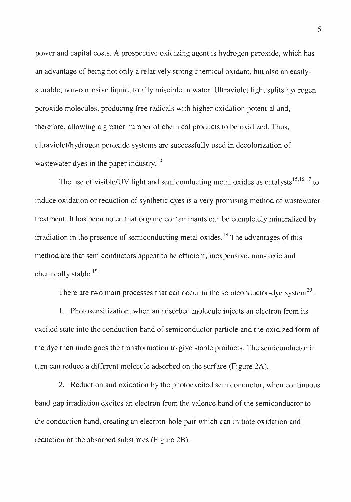
power and capital costs. A prospective oxidizing agent is hydrogen peroxide, which has
an advantage of being not only a relatively strong chemical oxidant, but also an easily
storable, non-corrosive liquid, totally miscible in water. Ultraviolet light splits hydrogen
peroxide molecules, producing free radicals with higher oxidation potential and,
therefore, allowing a greater number of chemical products to be oxidized. Thus,
ultraviolet/hydrogen peroxide systems are successfully used in decolorization of
d . h . d 14 wastewater yes m t e paper m ustry.
5
The use of visible/UV light and semiconducting metal oxides as catalysts 15•16
•17 to
induce oxidation or reduction of synthetic dyes is a very promising method of wastewater
treatment. It has been noted that organic contaminants can be completely mineralized by
irradiation in the presence of semiconducting metal oxides. 18 The advantages of this
method are that semiconductors appear to be efficient, inexpensive, non-toxic and
chemically stable. 19
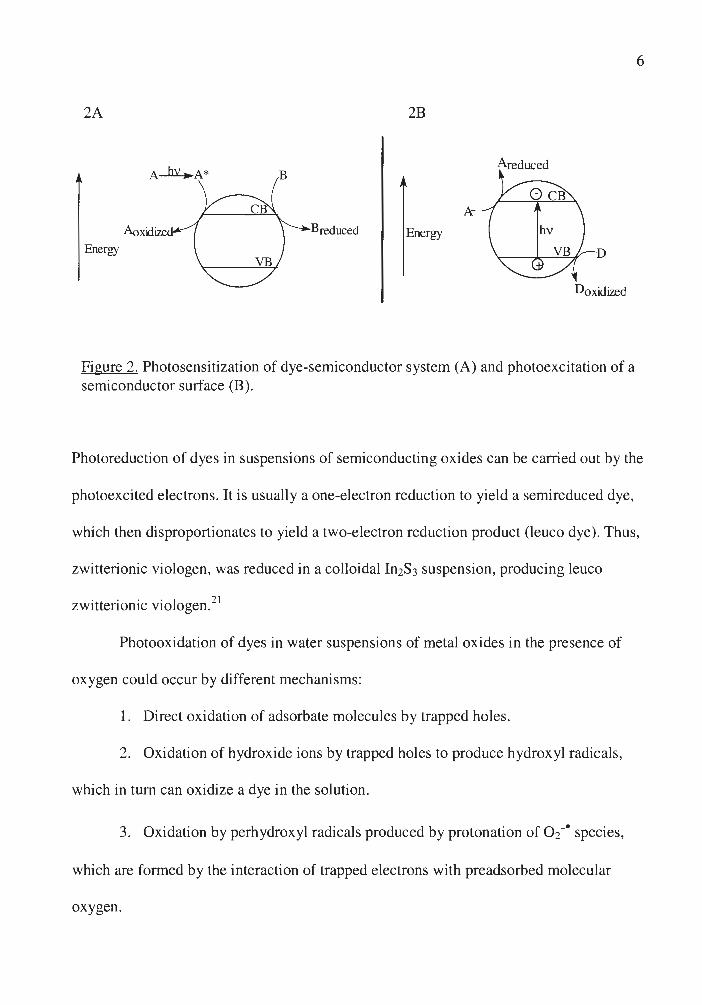
There are two main processes that can occur in the semiconductor-dye system20:
1. Photosensitization, when an adsorbed molecule injects an electron from its
excited state into the conduction band of semiconductor particle and the oxidized form of
the dye then undergoes the transformation to give stable products. The semiconductor in
turn can reduce a different molecule adsorbed on the surface (Figure 2A).
2. Reduction and oxidation by the photoexcited semiconductor, when continuous
band-gap irradiation excites an electron from the valence band of the semiconductor to
the conduction band, creating an electron-hole pair which can initiate oxidation and
reduction of the absorbed substrates (Figure 2B).
2A 2B
Areduced
Breduced Energy Energy D
Doxidiz.ed
Figure 2. Photosensitization of dye-semiconductor system (A) and photoexcitation of a semiconductor surface (B).
6
Photoreduction of dyes in suspensions of semiconducting oxides can be carried out by the
photoexcited electrons. It is usually a one-electron reduction to yield a semireduced dye,
which then disproportionates to yield a two-electron reduction product (leuco dye). Thus,
zwitterionic viologen, was reduced in a colloidal In2S3 suspension, producing leuco
zwitterionic viologen. 21
Photooxidation of dyes in water suspensions of metal oxides in the presence of
oxygen could occur by different mechanisms:
1. Direct oxidation of adsorbate molecules by trapped holes.
2. Oxidation of hydroxide ions by trapped holes to produce hydroxyl radicals,
which in tum can oxidize a dye in the solution.
3. Oxidation by perhydroxyl radicals produced by protonation of 0 2-• species,
which are formed by the interaction of trapped electrons with preadsorbed molecular
oxygen.
7
These oxidation routes are hard to differentiate. The chemical identification of
hydroxylated oxidation intermediates and the ESR detection of hydroxyl radical appear to
support the hydroxyl radical mechanism22, but a recent diffuse reflectance flash
photolysis experiment in nonaqueous solutions presented evidence in favor of direct
oxidation by trapped holes. 23 A competitive mechanism, where an organic molecule and
hydroxide adsorbed on the surface compete for the trapped holes, has also been
proposed. 24
In this investigation, crystal violet was selected to be a model compound to study
photodecomposition and photodecolorization of dyes on metal oxide surfaces.
Photodecolarization and photodecomposition of crystal violet was previously
studied both in the solutions of different solvents and adsorbed on different surfaces.25
Crystal violet can be photooxidized or photoreduced depending on experimental
conditions. Photoreduction may involve either an electron or hydrogen abstraction
process. Thus, it is favored under anaerobic conditions and in the presence of compounds
that are oxidized more readily than the dye, for example, protein substrates.
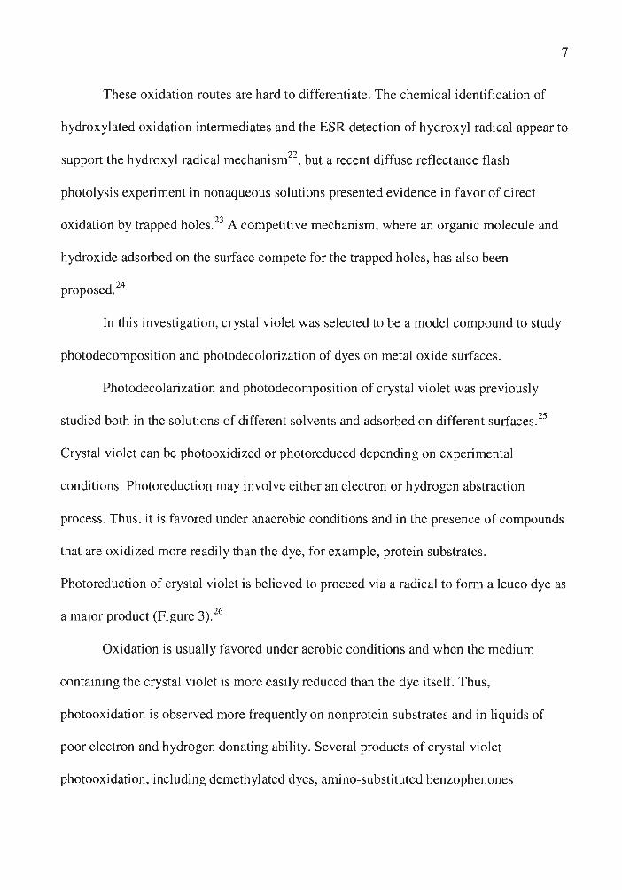
Photoreduction of crystal violet is believed to proceed via a radical to form a leuco dye as
a major product (Figure 3). 26
Oxidation is usually favored under aerobic conditions and when the medium
containing the crystal violet is more easily reduced than the dye itself. Thus,
photooxidation is observed more frequently on nonprotein substrates and in liquids of
poor electron and hydrogen donating ability. Several products of crystal violet
photooxidation, including demethylated dyes, amino-substituted benzophenones
CV -----1•
Crystal Violet Radical Leuco Crystal Violet
Figure 3. Reduction of crystal violet
and leuco dyes, have been identified. 27 The proposed mechanisms for the formation of
these products will be discussed in this thesis.
8
Although photodecomposition of crystal violet has been intensively studied, little
work has been done in terms of photodegradation and photodecolorization of crystal
violet in the suspensions of semiconducting metal oxides. Effects of crystal violet
concentration, amount of semiconductor, light intensity and pH have been investigated,28
but the products of the reaction were not identified and the mechanism of
photodecomposition was unclear.
The focus of this research was photoreactions of crystal violet dye in hydrogen
peroxide solution and in water suspensions of metal oxides under illumination. The aim
of the research was to determine the mechanism of photodecolorization and
photodecomposition of crystal violet and to evaluate the efficiency of photooxidation. In
order to do that, five oxides were studied and compared: zinc oxide and titanium dioxide,
which are semiconductors, and magnesium oxide, aluminum oxide and silicon dioxide,
which are insulators. Photodecomposition of other triphenylmethane dyes was also
studied.
9

CHAPTER II
EXPERIMENT AL SECTION
Materials
10
Crystal violet (N,N,N',N',N",N"-hexamethylpararosaniline (chloride), CI 42555,
Sigma Chemical Co), Basic fuchsin (pararosaniline hydrochloride, CI 42500, Fisher
Scientific Company) and Michler' s ketone ( 4,4' -bis( dimethylamino )benzophenone,
Acros) were of reagent grade and were used without further purification.
The following oxide powers were used: Degussa Fumed TiO2 P-25 grade (80% anatase,
20% rutile, 21 nm average particle size), rutile TiO2 (Rutile Titanium Dioxide, 94%, 0.3-
1.0 µm particle size, Aremco Products, Inc.), ZnO (Zinc Oxide, 99.9%, 1-5 µm particle
size, Aremco Products, Inc.), MgO (Magnesium Oxide, 99.9%, 1-5 µm particle size,
Aremco Products, Inc.), SiO2 (Cab-O-Sil, Amorphous fumed silica, 99.8%, 0.2-0.3 µm
particle size, Cabot Co), A!iO3 (Aluminum Oxide, Baker Chenucal Co). The powders
were subjected to heating at 400°C for at least an hour before each experiment to remove
CO2 adsorbed on the surface.
Light Source
All the experiments were performed in the sunlight passing through the window
of the third floor of McColl um Science Hall at the University of Northern Iowa. The
spectra of the sunlight and sunlight through the window, recorded using a
spectroradiometer (Licor Model LI-1800), are shown in Figure 4 and 5. The plastic
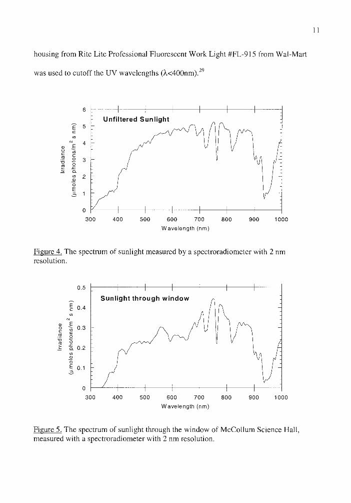
housing from Rite Lite Professional Fluorescent Work Light #FL-915 from Wal-Mart
was used to cutoff the UV wavelengths (A<400nm). 29
6
Unfiltered Sunlight E 5 C
en N 4
Q) E u ---C en
C (I]
.8 3 "O
~ 0 .c C. en ~
2
0 E 2, 1
0
300 400 500 600 700 800 900 1000
Wavelength (nm)
Figure 4. The spectrum of sunlight measured by a spectroradiometer with 2 nm resolution.
0 .5
E c 0 .4
Q) _§ u en 0.3 g § "O -~ 0 - -g_ 0.2
en ~ 0
~ 0 .1
0
300
Sunlight through window
400 500 600 700 800 900 1000
Wavelength (nm)
Figure 5. The spectrum of sunlight through the window of McColl um Science Hall , measured with a spectroradiometer with 2 nm resolution.
11
12
Procedures and Analysis
Unless otherwise noted, aqueous oxide suspensions were prepared by adding
0.23 g of an oxide powder to 25ml of 5xl0-5 M dye solution. Prior to irradiation, the
suspensions were magnetically stirred in the dark for 30 minutes to ensure establishment
of adsorption/desorption equilibrium of the dye on the oxide surface. At given irradiation
time intervals, samples were collected and centrifuged to remove the catalyst. The
supematents were analyzed by UV-Vis spectroscopy with a Shimadzu UV-2101PC or
UV-2401PC double beam spectrophotometer to monitor the loss of the dye and the
possible formation of intermediate species.
All the comparisons, such as different catalysts, dyes and etc., were done in
simultaneous trials to reassure that the differences in the reaction rates are due to the
activities of reactants, not different sunlight intensities on different days.
Thin layer chromatography on silica plates was used to confirm some of the
UV/vis data. The eluting solution was a 90:9: 1 mixture of 1-propanol to water to acetic
acid.
TiO2 pellets used for diffuse reflectance spectroscopy experiments were produced
by centrifugation and dried in the dark for several days prior to the measurements.
Diffuse reflectance spectroscopy measurements were made using a Shimadzu UV2101PC
double beam spectrophotometer with the ISR-260 intergrating sphere accessory. Spectra
were referenced to Degussa P-25 TiO2.
Molecular modeling was carried out using Spartan IBM Version 5.0.3 Open GL,
running on the IBM RS6000 computers in the Chemistry Department at the University of
13
Northern Iowa. The geometry of the crystal violet and pararosaniline cations was
optimized using the AMI semi-empirical method, with the options set to "optcycle=500
maxcycle=500 hess=unit". Hartree-Fock ab initio calculations using the STO-3O basis
set were then done on the optimized structure to estimate the charges on the individual
atoms.

CHAPTER III
RESULTS AND DISCUSSION
Crystal violet undergoes photodecolorization and photodecomposition in water
solution and in slurry with solid substrates. The rates of the reactions depend on the
chemical properties of the substrates and on the wavelengths of illumination.
Photodecomposition of Crystal Violet in Water Solutions
14
Crystal violet is relatively stable in water solution in the dark, but it decomposes
when exposed to light, which indicates that excitation of the dye is necessary for the
reaction to occur. When illumination of the crystal violet solution was carried out in the
sunlight and monitored by UV-visible spectroscopy, both visible and UV bands
underwent a hypsochromic shift and decreased in intensity (Figure 7). New species with
absorbance at about 360nm were formed and then decomposed with further illumination
(Figure 6 and 7) .
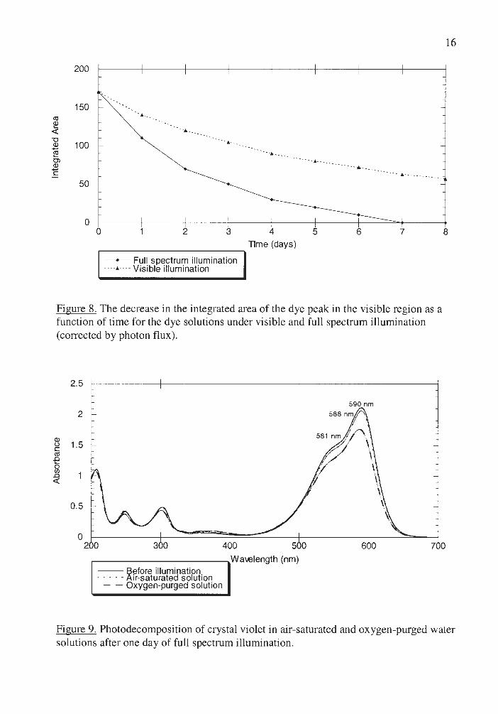
Figure 8 displays the wavelength dependence of the crystal violet
photodecomposition in water solution. Crystal violet decolorized with visible
illumination (A< 400 nm) slower than with full spectrum illumination.
Photodecomposition rate was observed to increase with increasing concentration
of dissolved oxygen (Figure 9) indicating that photooxidation is a major pathway of
photodecomposition.

Q) (.) C ro .0 0 Cl) .0 <(
2.5
2
1.5
0.5
0 200 300 400 500
Wm,elength (nm)
-- Before illumination ---------After 4 days of illumination
- - After 6 days of ill umination -- After 7 days of illumination
590 nm
600
Figure 6. Photodecomposition of crystal violet in water solution under full spectrum illumination.
0.14 360nm -
-___; - -0.12 _,.. -- - 351 nm
--- ----. 374nm ·.
' 0.1 ' " 0.08 ' ' '-.
0.06 '-. ......
'-.
0.04 ......
---0.02
15
700
340 360 380 400 420 Wa\€length (nm)
-- Before illumination - - - - - After 4 days of illumination
- - After 6 days of illumination
Figure 7. Photodecomposition of crystal violet in water solution under full spectrum illumination.
200
150 C1) (I)
~ . ·•-"O
100 (I)
~
-. ,. __
Ol (I)
c ··- -•-- ---- ------.... _
··--· ----------50
0 0 2 3 4 5 6 7
Time (days) ..--------------.. --+-- Full spectrum illumination ----•---- Visible illumination
Figure 8. The decrease in the integrated area of the dye peak in the visible region as a function of time for the dye solutions under visible and full spectrum illumjnation (corrected by photon flux).
2.5
2
(I)
1.5 (.) C C1) .0 0 (J)
.0 <(
0.5
0
590 nm
581 nm //,, ' '7./ \ r_,/ • // \
,1/ \ fl \
\
16
8
2 0 7 0
~----------~Wa\€length (nm) -- Before illumination · · - - - Air-saturated solution
- - Oxygen-purged solution
Figure 9. Photodecomposition of crystal violet in air-saturated and oxygen-purged water solutions after one day of full spectrum illumination.

17
The blue shift in the absorption spectra with illumination can be attributed to
crystal violet demethylation to form methyl violet and other methylated parorosanilines,
identified using TLC. Pararosaniline (see Figure 1), the final product of demethylation,
was detected to be in the solution that had been irradiated for four days. The mechanism
of demethylation has been proposed (Figure 10). 30
Figure 10. The mechanism of demethylation process.
In this mechanism, demethylation occurs via N-oxide precursors, which are too unstable
to be detected. The final products of methyl group oxidation are carbon dioxide and
water.
The overall decrease of absorbance in the visible region indicates that
decomposition to colorless products is occurring concurrently with demethylation,
thereby lowering the total concentration of methylated pararosanilines. The increase in
absorbance at about 360 nm and then its decrease can be explained by the formation of
intermediates in the decomposition to colorless products. The absorption spectra and a
positive ketone test suggest that these intermediates are demethylated derivatives of
18
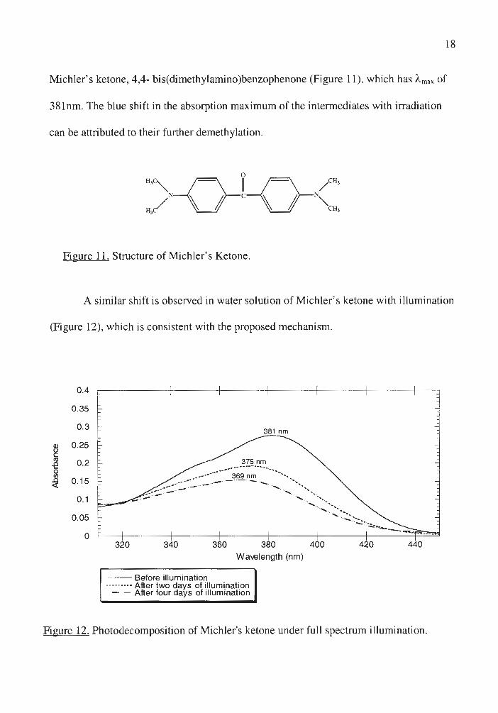
Michler's ketone, 4,4- bis(dimethylamino)benzophenone (Figure 11), which has Amax of
381nm. The blue shift in the absorption maximum of the intermediates with irradiation
can be attributed to their further demethylation.
Figure 11. Structure of Michler's Ketone.
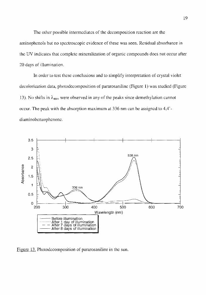
A similar shift is observed in water solution of Michler's ketone with illumination
(Figure 12), which is consistent with the proposed mechanism.
0.4
0.35
0.3
8 0.25 C Cl) 0.2 -e 0 (/)
~ 0.15
0.1
0.05
0
381 nm
375 nm ___ ............. ----... -· . --- -....... ...
-- 369 nm - ---- ··· - ---...._ __ ....... ........ · ....... ··., .
320 340 360 380 Wavelength (nm)
400
--Before illumination --------- After two days of illumination
- - After four days of illumination
420 440
Figure 12. Photodecomposition of Michler's ketone under full spectrum illumination .
19
The other possible intermediates of the decomposition reaction are the
aminophenols but no spectroscopic evidence of these was seen. Residual absorbance in
the UV indicates that complete mineralization of organic compounds does not occur after
20 days of illumination .
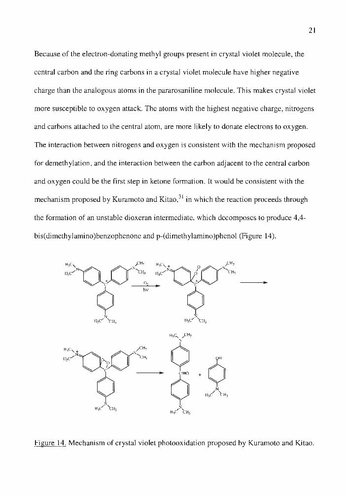
In order to test these conclusions and to simplify interpretation of crystal violet
decolorization data, photodecomposition of pararosaniline (Figure 1) was studied (Figure
13). No shifts in "-max were observed in any of the peaks since demethylation cannot
occur. The peak with the absorption maximum at 336 nm can be assigned to 4,4' -
diaminobenzophenone.
3.5
3
538 nm 2.5
Q) 2 (.)
C ro -e 1.5 0 (/)
..a <t:
0.5
0 200 300 400 500 600 700
Wa\€length (nm) .---------------... -- Before illumination --------- After 1 day of illumination
- - After 7 days of illumination --After 8 days of illumination
Figure 13. Photodecomposition of pararosaniline in the sun.

20
Complete decolorization was observed after 7 and 8 days of illumination for
crystal violet and pararosaniline respectively. This result can be rationalized in terms of
point charges of individual atoms (Table 1 and Table 2) calculated using Spartan.
Table 1. Charges of individual atoms in a crystal violet molecule (see Figure 1) calculated using Spartan
Atoms Atom Char es Natural Mulliken
Central C 0.23 0.12 Attaching C -0.12 -0.05 Ortho C 0.04 -0.02 MetaC -0.12 -0.1 ParaC 0.24 0.17 Ortho H 0 .05 0.08 MetaH 0.05 0.08 N -0.25 -0.27 N-C 0 .01 -0.05 H2C-H 0.06 0 .08 HC-H2 0 .05 0.07
Table 2. Charges of individual atoms in a pararosaniline molecule (see Figure 1) calculated using Spartan.
Atoms Atom Char es Natural Mulliken
Central C 0 .24 0.13 Attaching C -0.12 -0.05 Ortho C 0 .04 -0.02 MetaC -0.12 -0.09 Para C 0.27 0.19 Ortho H 0.06 0.09 MetaH 0.05 0.07 N -0.41 -0.42 N-H 0.23 0 .23
21
Because of the electron-donating methyl groups present in crystal violet molecule, the
central carbon and the ring carbons in a crystal violet molecule have higher negative
charge than the analogous atoms in the pararosaniline molecule. This makes crystal violet
more susceptible to oxygen attack. The atoms with the highest negative charge, nitrogens
and carbons attached to the central atom, are more likely to donate electrons to oxygen .
The interaction between nitrogens and oxygen is consistent with the mechanism proposed
for demethylation, and the interaction between the carbon adjacent to the central carbon
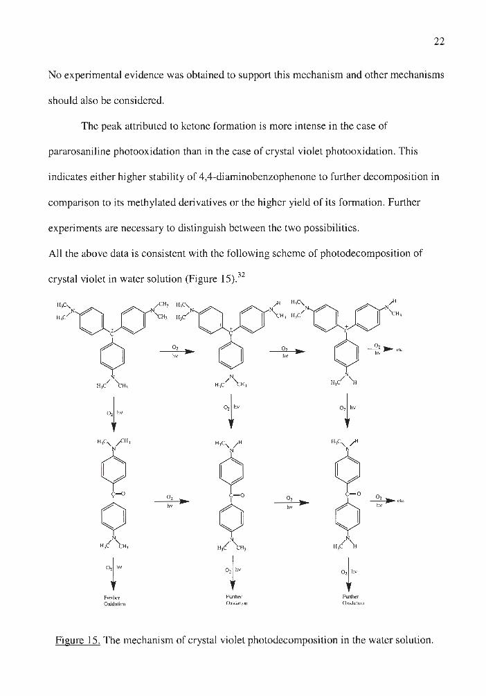
and oxygen could be the first step in ketone formation. It would be consistent with the
mechani sm proposed by Kuramoto and Kitao,31 in which the reaction proceeds through
the formation of an unstable dioxeran intermediate, which decomposes to produce 4,4-
bis(dimethylamjno )benzophenone and p-(dimethylamino)phenol (Figure 14).
Figure 14. Mechani sm of crystal violet photooxidation proposed by Kuramoto and Ki tao.
22
No experimental evidence was obtained to support this mechanism and other mechanisms
should also be considered.
The peak attributed to ketone formation is more intense in the case of
pararosaniline photooxidation than in the case of crystal violet photooxidation. This
indicates either higher stability of 4,4-diaminobenzophenone to further decomposition in
comparison to its methylated derivatives or the higher yield of its formation. Further
experiments are necessary to distinguish between the two possibilities.
All the above data is consistent with the following scheme of photodecomposition of
crystal violet in water solution (Figure 15).32
+ t + 11,c, /Cll ' 11,c, / 11 11,c, ~
¢ ¢ ¢ 0
o, C= O o,
0 ~ etc. • ¢ • hv hv hv
N N N
11/ t 11, 11,( t 11 , 11/ \i
t ,,,. 1''"
Further Further Funher Ox..idation Oxidation Oxidaticu
Figure 15. The mechanism of crystal violet photodecomposition in the water solution .
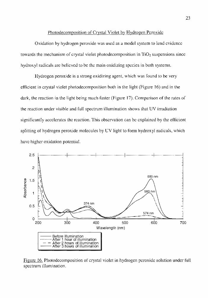
Photodecomposition of Crystal Violet by Hydrogen Peroxide
Oxidation by hydrogen peroxide was used as a model system to lend evidence
towards the mechanism of crystal violet photodecomposition in Ti02 suspensions since
hydroxyl radicals are believed to be the main oxidizing species in both systems.
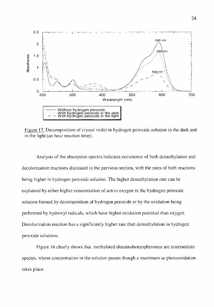
Hydrogen peroxide is a strong oxidizing agent, which was found to be very
efficient in crystal violet photodecomposition both in the light (Figure 16) and in the
dark, the reaction in the light being much faster (Figure 17). Comparison of the rates of
the reaction under visible and full spectrum illumination shows that UV irradiation
significantly accelerates the reaction. This observation can be explained by the efficient
splitting of hydrogen peroxide molecules by UV light to form hydroxyl radicals, which
have higher oxidation potential.
2.5
2
(I) 1.5 (.) C (1j .0 .... 0 U) .0 ~
0.5
23
0 200 300 400 500 600 700
Wa1.elength (nm)
-- Before illumination ········- After 1 hour of illumination
- - After 2 hours of illumination --After 3 hours of illumination
Figure 16. Photodecomposition of crystal violet in hydrogen peroxide solution under full spectrum illumination.
2.5
2
j 1.5
j 0.5
0
.,,-,·' /
,· · /
590 nm
58~nm '-. -- ,/ \ '.
24
200 300 400 500 600 700 Wa\€length (nm)
--W ithout hydrogen peroxide ········- With hydrogen peroxide in the dark
- - With hydrogen peroxide in the light
Figure 17. Decomposition of crystal violet in hydrogen peroxide solution in the dark and in the light (an hour reaction time) .
Analysis of the absorption spectra indicates occurrence of both demethylation and
decolorization reactions discussed in the previous section, with the rates of both reactions
being higher in hydrogen peroxide solution. The higher demethylation rate can be
explained by either higher concentration of active oxygen in the hydrogen peroxide
solution formed by decomposition of hydrogen peroxide or by the oxidation being
performed by hydroxyl radicals, which have higher oxidation potential than oxygen.
Decolorization reaction has a significantly higher rate than demethylation in hydrogen
peroxide solutions.
Figure 16 clearly shows that methylated diaminobenzophenones are intermediate
species , whose concentration in the solution passes though a maximum as photooxidation
takes place.

25
Figure 18 shows the effect of the increase of hydrogen peroxide concentration on
the absorbance of crystal violet solution at 590 nm.
2.5
2
~ 1.5 C ca -e &1 .0 <(
0.5
0
Concentration (mol/L)
Figure 18. The decrease in absorbance at 590 nm for the solutions with different concentrations of hydrogen peroxide after 20 minutes of full spectrum illumination .
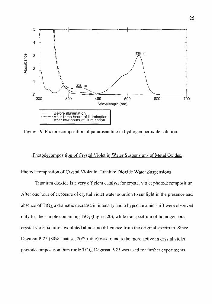
The spectrum of pararosaniline photooxidation in hydrogen peroxide solution is
analogous to that in water solution, with rate of decolorization being much faster for the
former even at relatively low hydrogen peroxide concentrations (Figure 19).
Based on these data it was concluded that the mechanism of the photoreaction of
crystal violet with hydrogen peroxide is similar to the mechanism of photodecomposition
of crystal violet in water solutions.
(l) (.) C co -e 5l .0 ~
5
4
3
2
0 200 300 400 500
Wa\A91ength (nm)
-- Before illumination ········- After three hours of illumination
- - After four hours of illumination
538 nm
600
Figure 19. Photodecomposition of pararosaniline in hydrogen peroxide solution.
Photodecomposition of Crystal Violet in Water Suspensions of Metal Oxides.
Photodecompostion of Crystal Violet in Titanium Dioxide Water Suspensions
26
700
Titanium dioxide is a very efficient catalyst for crystal violet photodecomposition.
After one hour of exposure of crystal violet water solution to sunlight in the presence and
absence of Ti 0 2, a dramatic decrease in intensity and a hypsochromic shift were observed
only for the sample containing TiO2 (Figure 20), while the spectrum of homogeneous
crystal violet solution exhibited almost no difference from the original spectrum. Since
Degussa P-25 (80% anatase, 20% rutile) was found to be more active in crystal violet
photodecomposition than rutile Ti 0 2, Degussa P-25 was used for further experiments.

27
2 .5 --- Without titanium dioxide -- ------- With titanium dioxide 590 nm
2
0 .5
0 200 300 400 500 600 700
Wavelength (nm)
Figure 20. Photodecomposition of crystal violet in the absence and presence of titanium dioxide after an hour of illumination.
Mechanism of photodegradation. In order to distinguish between the two main
processes that can occur in the semiconductor-dye system, photosensitization and
oxidation/reduction by the photoexcited semiconductor, the rates of crystal violet
photodecomposition under visible and full spectrum illumination were compared
(Figure 21).
200
'-.. '-..
150 '-..
i 11. "-
-g 100 1!! ~ c
50
0
"-
50
· ··· -- • ············ • ······-····· • ····· ...... _ . ...... . . . .... ..... . ... . .
'·---
1 0
--- ---
1 0
Time (min)
--• - --2 0 2 0
Figure 21. The decrease in the integrated area of the dye peak in the visible region as a function of time for crystal violet water solution and titanium dioxide suspension under visible and full spectrum illumjnation (corrected by photon flux).

28
Photosensitization does occur, since the presence of titanium dioxide accelerates
decolorization of crystal violet under visible illumination, but the much higher rate of the
reaction for the full spectrum illumination indicates that oxidation/reduction by the
photoexcited semiconductor is the main mechanism of crystal violet photodecomposition .
Photoreduction by the photoexcited semiconductor surface would require efficient
preadsorption of the dye to the TiO2 lattice for the direct electron transfer to occur. 33
When TiO2 was added to crystal violet solution and the mixture was kept in the dark, a
small increase in crystal violet concentration was observed for the solution exposed to
TiO2 powder for a short period of time (Figure 22).
E C 0 (]) LO
'lil ~ C Ct1 -e j
1.7
1.65
1.6
1.55
1.5
1.45
1.4
1.35
1.3 0 5 10 15 20 25 30
Time (min)
Figure 22. Absorbance of crystal violet solution as a function of stirring time with titanium dioxide powder (in the dark).

The initial increase in absorbance indicates that water is adsorbed on the Ti02
lattice better than crystal violet. The subsequent decrease in the dye concentration
indicates its secondary adsorption by the surface hydroxyl groups either formed by
dissociation of water molecules on the Ti0 2 surface or adsorbed from water solution .
29
Since water is adsorbed preferentially to crystal violet initially, and taking the low
concentration of crystal violet into account, I conclude that crystal violet occupies only a
small fraction of the surface-solution interface, with solvent occupying most of it.
Moreover, no spectroscopic evidence for any reduction products was seen, either in the
supematent of the centrifuged CV /H20/Ti02 solution or on the surface of Ti 0 2. These
data rule out photoreduction as a possible mechanism of crystal violet
photodecomposition in Ti02 suspensions under illumination.
In the case of oxidation by the photoexcited semjconductor, one or more of
several species could perform the oxidation :
1. trapped holes
2. hydroxyl radicals formed by the interaction of hydroxide ions with the
photoexcited semiconductor
3. perhydroxyl radicals formed by the protonation of 0 2- • .
Direct oxidation is expected to be less important than oxidation by hydroxyl
radicals or by perhydroxyl radicals since it also requires strong adsorption of the dye at
the semiconductor lattice, which was shown not to be the case with crystal violet.
Additional evidence for the importance of oxidation by hydroxyl radicals comes
from the inhibition of crystal violet photooxidation by isopropanol, which is known as a

30
good QH. quencher,34 and from the higher activity of anatase TiO2 in photooxidation of
crystal violet in comparison to rutile TiO2. It has been shown that anatase TiO2 abstracts
water better and forms hydroxyl radicals more readily than rutile TiO2.35
Since the solution after preadsorption in the dark decolorizes two times faster than
the solution without preadsorption, secondary adsorbtion on the hydroxyl groups at the
titanium dioxide surface has a great influence on the rate of the reaction. Besides,
hydroxyl radicals are extremely reactive and are not likely to migrate far from the surface
even at low concentrations of oxidizable reactants. Therefore, the oxidation of crystal
violet in the solution by free hydroxyl radicals was concluded to be negligible.
The best explanation for the observations is photogeneration of surface-bound
hydroxyl radicals, which oxidize molecules at the surface layer without diffusing into the
bulk solution.
In order to keep the photoxidation process going, it is necessary to avoid
accumulation of electrons in the conduction band of semiconductor particles, which
would increase the electron-hole recombination rate and lower the quantum yield of the
reaction. Because the conduction band of TiO2 is nearly isoenergetic with the reduction
potential of oxygen, adsorbed oxygen serves as a trap for photogenerated conduction
electrons.36 Since oxygen reacts slowly with the electrons of most semiconductors,
including Ti 0 2, the rate of electron transfer from the particles to oxygen is usually both
yield and rate-controlling in the photocatalytic process. 37 0 2-• and o/ -generated species
can either directly oxidize the dye or protonate to form perhydroxyl and hydroxyl
radicals:
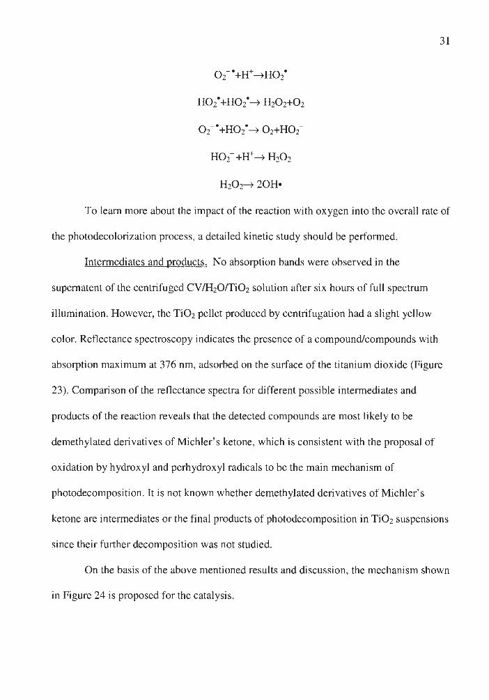
0 2- •+Ir• HO/
Ho2·+Ho2·• H202+02
02- •+Ho2•• 02+H02-
H02- +H+• H202
H202• 20H•
31
To learn more about the impact of the reaction with oxygen into the overall rate of
the photodecolorization process, a detailed kinetic study should be performed.
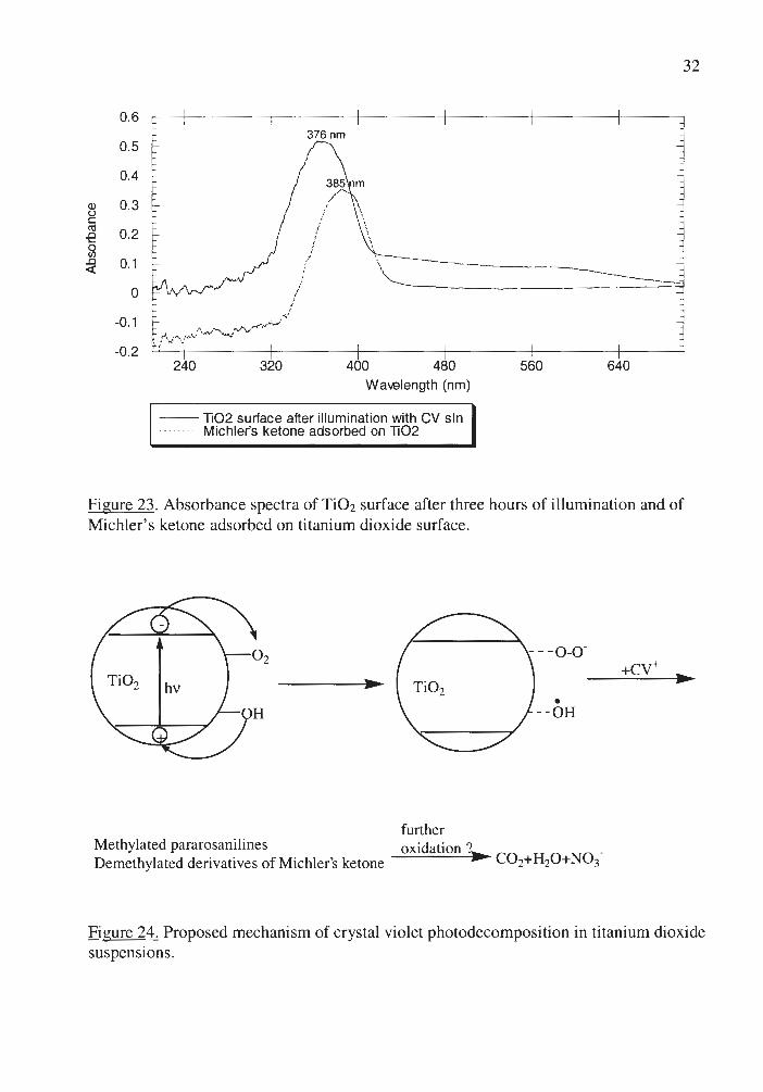
Intermediates and products. No absorption bands were observed in the
supernatent of the centrifuged CV/H20/Ti02 solution after six hours of full spectrum
illumination. However, the Ti02 pellet produced by centrifugation had a slight yellow
color. Reflectance spectroscopy indicates the presence of a compound/compounds with
absorption maximum at 376 nm, adsorbed on the surface of the titanium dioxide (Figure
23). Comparison of the reflectance spectra for different possible intermediates and
products of the reaction reveals that the detected compounds are most likely to be
demethylated derivatives of Michler's ketone, which is consistent with the proposal of
oxidation by hydroxyl and perhydroxyl radicals to be the main mechanism of
photodecomposition. It is not known whether demethylated derivatives of Michler's
ketone are intermediates or the final products of photodecomposition in Ti02 suspensions
since their further decomposition was not studied.
On the basis of the above mentioned results and discussion, the mechanism shown
in Figure 24 is proposed for the catalysis.
0.6
0.5
0.4
Q) ()
0.3 C (1j
0.2 -e 0 Cl)
..Cl 0.1 ~
0
-0.1
-0.2 240 320
376 nm
i
i i
400 480 Wa-.elength (nm)
-- li02 surface after illumination with CV sin ········- Michler's ketone adsorbed on li02
32
560 640
Figure 23. Absorbance spectra of TiO2 surface after three hours of ill umination and of Michler's ketone adsorbed on ti tanium dioxide surface.
• Ti0 2 •
-- OH
furt her Methylated pararosanili nes Demethylated derivatives of Michler's ketone
oxidation ?• C02+H20+N03-
Figure 24. Proposed mechanism of crystal violet photodecomposition in titanium dioxide suspensions.
33
Photocatalysis starts by the excitation of the semiconductor by UV irradiation to
produce electron-hole pairs on the surface of a semiconductor particle. An electron can be
transferred from a surface hydroxyl group to a photogenerated hole on the Ti02 particle
to form surface OH•, which after its formation is still associated with the surface.
Hydroxyl radicals can then oxidize crystal violet molecules to produce methylated
pararosanilines and demethylated derivatives of Michler's ketone. Preadsorbed oxygen
can be reduced by the conduction band electrons to form 0 2-• radicals, which can
protonate to form perhydroxyl and hydroxyl radicals that most likely also oxidize crystal
violet. Further studies should be carried out to determine the final products of crystal
violet photodecomposition on Ti02 surface.
Photodecomposition of Crystal Violet in Zinc Oxide Suspensions
Zinc oxide is another semiconducting oxide, which was shown to be even more
efficient in photodecolorization of crystal violet than titanium dioxide. Zinc oxide was
found to have higher initial adsorption of crystal violet and more efficient
photosensitization than Ti02 (Figure 25). The peak attributed to ketone formation was
observed to be higher in ZnO suspensions than in Ti02 suspensions. This result can be
explained by either the higher rate of formation of demethylated benzophenones in ZnO
suspensions or by lower adsorption of these intermediates on the ZnO surface.
Demethylation was shown to be less efficient for ZnO than for Ti 0 2.

34
1.6
1.2 Q) 0 C ro ..a 0.8 .... 0 (/)
..a <(
0.4
0 200 300 400 500 600 700
Wa-elength (nm)
-- No catalyst --------- With titanium dioxide
- - With zinc oxide
Figure 25. Photodecomposition of crystal violet in titanium dioxide and zinc oxide suspensions after three days of visible illumination. Faster decrease in the intensity of the main adsorption maxima in case of zinc oxide indicates the process of photosensitization being more efficient (the starting amount of adsorbed crystal violet is the same for ZnO and Ti02).
Photodecomposition of Crystal Violet in Silicon Dioxide Suspensions
Silicon dioxide is an insulator with a band gap of at least 4.9 eV and therefore is a
photochemically inert surface. The fast decrease in crystal violet concentration after the
addition of silicon dioxide to a crystal violet solution and the bright purple color of the
centrifuged Si02 powder indicate high adsorption of crystal violet on the surface of Si 0 2.
This is caused by the intense interaction of the cationic dye and negatively charged
silanol groups. Strong localized surface interactions restrict the mobility of the adsorbed
molecule, slowing down the diffusion of molecular oxygen to the dye layers stabilized by
n-n stacking and, consequently, decrease the rate of demethylation (Figure 26).

2.5
2
(I) 1.5 (.) C (U
-e 0 (J)
.Cl <x:
0.5
'- - - -0 200 300
--Before illumination --------- Without silicon dioxide
- - With silicon dioxide
400 500 Wa-.elength (nm)
590 nm
585 nm
600
35
700
Figure 26. Photodecomposition of crystal violet in silicon dioxide suspension after three days of full spectrum illumination. Smaller hypsochromic shift indicates that Si02 slows down demethylation reaction.
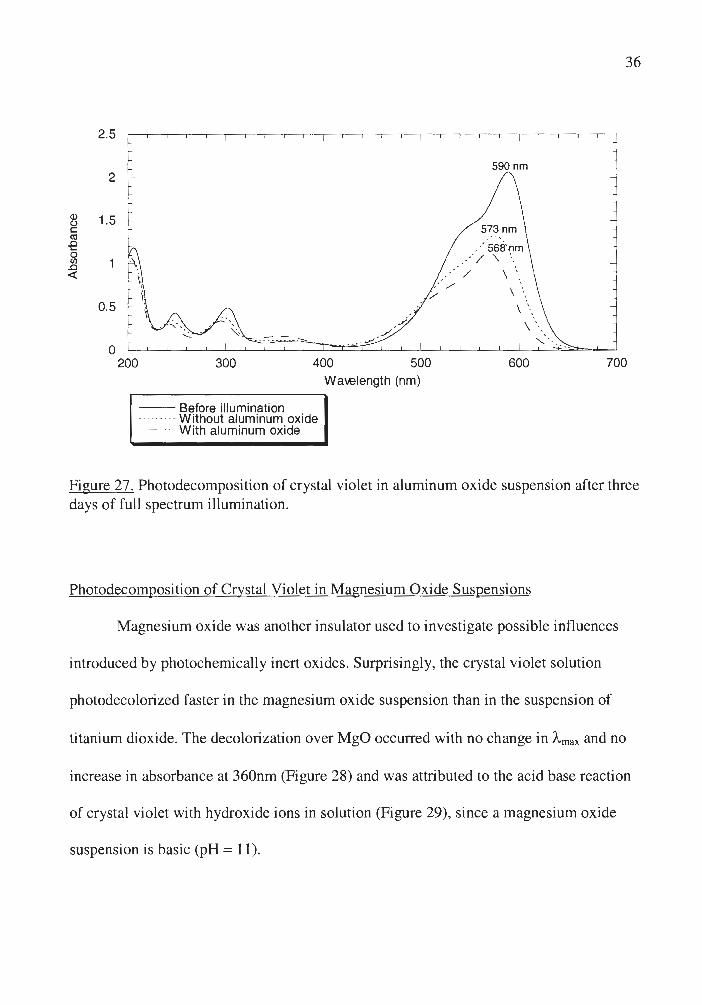
Photodecomposition of Crystal Violet in Aluminum Oxide Suspensions
Aluminum oxide, an insulator, was found to accelerate the reaction of crystal
violet with oxygen (Figure 27) since a larger hypsochromic shift is observed in the
presence of A]i03 than in homogeneous crystal violet solution after the same time of
illumination. Aluminum oxide has been shown to stabilize the excited states of aromatic
compounds,38 which could enhance the efficiency of the reaction of crystal violet with
oxygen since it is dependent on the lifetime of the excited state of the dye .
2.5
2
Q) 1.5 (..) C Cll .0 0 <Jl .0 <(
0.5
0
.·· / .· ./
/
590 nm
36
, -- . 200 300 400 500 600 700
Wa-.elength (nm)
-- Before illumination --- ------ Without aluminum oxide
- - With aluminum oxide
Figure 27 . Photodecomposition of crystal violet in aluminum oxide suspension after three days of full spectrum illumination.
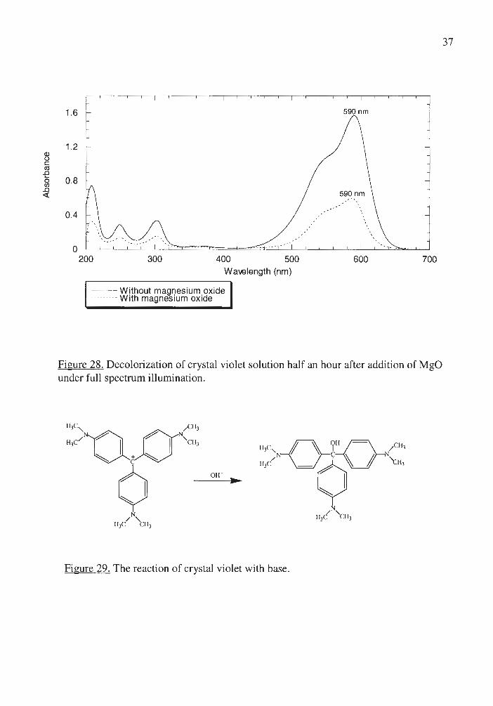
Photodecomposition of Crystal Violet in Magnesium Oxide Suspensions
Magnesium oxide was another insulator used to investigate possible influences
introduced by photochemically inert oxides. Surprisingly, the crystal violet solution
photodecolorized faster in the magnesium oxide suspension than in the suspension of
titanium dioxide. The decolorization over MgO occurred with no change in Amax and no
increase in absorbance at 360nm (Figure 28) and was attributed to the acid base reaction
of crystal violet with hydroxide ions in solution (Figure 29), since a magnesium oxide
suspension is basic (pH= 11).
Q) (.) C cu .0 .... 0 (/)
.0 <(
1.6 590 nm
1.2
0.8
0.4
.-
0 200 300 400 500 600 700
Wa-.elength (nm)
--Without magnesium oxide --------- W ith magnesium ox ide
Figure 28. Decolorization of crystal violet solution half an hour after addition of MgO under fu ll spectrum illumination.
ow •
Figure 29. The reaction of crystal violet with base.
37

The formation of the colorless carbinol was supported by reverse reaction with
acid to form crystal violet cation. The rate of the reaction in the dark was found to be
lower than with illumination.
38
Pararosaniline was found to react faster with base than crystal violet does. It can
be explained by the fact that pararosaniline, lacking electron-donating groups at the
nitrogens, is more electron deficient than crystal violet, and therefore is more active in the
reaction with the nucleophile.
According to the results of Spartan point charge calculations for individual atoms
(Table 1), the central carbon in a pararosaniline molecule has higher positive charge than
the analogous atom in a crystal violet molecule, which is in a good agreement with the
proposed mechanism.
Comparison of Different Methods of Crystal Violet Photodecomposition
Hydrogen peroxide is efficient in crystal violet photodecomposition but it is
consumed during the reaction and it absorbs only short UV wavelengths, utilizing only a
very small part of solar radiation.
Anatase titanium dioxide is a cheap, chemically and biologically inert catalyst,
and it can absorb short visible wavelengths (all wavelengths below 400 nm; from 2 to 5%
of sunlight). 39 On the other hand, being a heterogeneous catalyst, titanium dioxide might
adsorb the products of the reaction on its surface, which could result in lowering or even
loss of its catalytic activity. The rate of photodecomposition of the intermediates, as well
as the possible adsorption of the reaction products, should be taken into account when

39
discussing the possibility of using titanium dioxide for the treatment of dye-contaminated
waste-waters on the industrial scale.
Zinc oxide was found to be more efficient than titanium dioxide in crystal violet
photodecomposition, but it has been found that a significant quantity of Zn2+ can be
dissolved into solution during the photochemical reactions. It is known that ZnO
electrode undergoes anodic photocorrosion according to the equation40
ZnO+2h+• Zn2++O*
Therefore, ZnO powder is not suitable to be a photocatalyst for wastewater treatment
unless a means of removing Zn2+ is also used.
None of the insulating oxides under investigation (SiO2, A'2O3, MgO) are
prospective catalysts for crystal violet photodecomposition because of the very low
reaction rates.

CHAPTER IV
CONCLUSIONS
40
Dyes are among the most widely synthesized chemicals. Since some dyes,
including crystal violet, are hazardous and significant losses occur during their
manufacture and processing, efficient and easy ways of dye decomposition should be
investigated. Chemical oxidation and photooxidation in the presence of semiconducting
oxides have been shown to be one of the most prospective methods of treating dye
contaminated waters.
The focus of my research was to determine the mechanism of crystal violet
photodecomposition in the presence of hydrogen peroxide and metal oxides, to study the
influence of different factors on the rate of the reaction and to compare the efficiency of
the photochemical processes.
Photodecomposition of crystal violet in water solutions was first studied in the
absence of catalysts. It was shown that full spectrum illumination is more efficient in
crystal violet decomposition than visible illumination and the increase in oxygen
concentration accelerates the reaction. Two concurrent reactions, demethylation and
ketone formation were found to occur to form demethylated dyes and demethylated
derivatives of Michler's ketone. Point charges of individual atoms, calculated using
Spartan, were used to identify the most probable mechanism of their formation .
Photoreaction of crystal violet with hydrogen peroxide was suggested to have the
same mechanism as the reaction of crystal violet with oxygen. Its higher rate is due to the
higher oxidation potential of hydrogen peroxide and hydroxyl radicals .

The results on the rates of photodecomposition were obtained and compared for
the suspensions of several oxides: titanium dioxide and zinc oxide, which are
semiconductors and magnesium oxide, aluminum oxide and silicon dioxide, which are
insulators.
41
Semiconducting oxides were shown to be the most efficient in crystal violet
photodecomposition in comparison to other oxides studied. Wavelength dependence and
diffuse reflectance spectroscopy studies of the intermediates adsorbed on the surface
allowed proposing oxidation by the photoexcited semiconductor as the main mechanism
of crystal violet photodecomposition in titanium dioxide suspensions. It was concluded
that surface-bound hydroxyl radicals perform the oxidation of crystal violet molecules
preadsorbed by the surface hydroxyl groups. It was shown that oxygen is required for
crystal violet photodecomposition since it serves as an electron acceptor, preventing
accumulation of electrons on the semiconductor particles.
Studies of crystal violet photodecomposition in the presence of aluminum oxide
and silicon dioxide indicate that a photochemically inert surface may influence the rate of
the reaction . Aluminum oxide accelerates the reaction by stabilizing of the excited state
of the dye and silicon dioxide inhibits crystal violet photodecomposition by forming dye
clusters at the surface.
The reaction of crystal violet with hydroxide ion occurs in magnesium oxide
suspensions (pH= 11) to form a carbinol base.
The efficiency of using different reagents and catalysts for treatment of crystal
violet contaminated wastewaters was evaluated. Only hydrogen peroxide and

42
semiconducting oxides have a potential of being used in industry for this purpose,
although they also have certain disadvantages: Hydrogen peroxide is consumed during
the reaction; TiO2 might adsorb products of the reaction on the surface; and ZnO is
photochemically unstable. TiO2 was concluded to be the most prospective industrial
photocatalyst because of its cheapness, reasonable effectiveness, chemical and biological
stability.
In future research on the photodegradation of crystal violet in water solutions and
suspensions of metal oxides, I would suggest the following improvements and directions:
1. HPLC and Total Carbon Analysis should be performed to identify the
intermediates and final products of photodecomposition.
2. Experiments with singlet and triplet state quenchers may provide the
information about the role of the singlet and triplet states of crystal violet in its reaction
with oxygen.
3. ESR studies should be performed in order to eliminate the possibility of the
direct hole mechanism in semiconducting oxide suspensions and to detect the formation
of hydroxyl radicals.
4 . A detailed kinetic study is required to support the proposed mechanism of
crystal violet photodecomposition in the suspensions of semiconducting oxides. The use
of laser pulse photolysis to investigate the rapid recombination and trapping events
occurring in and on the semiconductor photocatalyst may allow the measurements of the
kinetic parameters.

Several changes can be made in the systems in order to make crystal violet
photodecomposition more efficient:
43
1. A common feature of photocatalytic reactions occurring on metal oxides
suspended in aqueous solution is dependence of the reaction rate on solution pH. Surface
charge is expected to be positive at pH lower than isoelectric point and negative at pH
higher than isoelectric point. The charge of a crystal violet molecule can also change
significantly, which will definitely change the adsorptivity of the dye. Changes in
adsorbtivity, in the bandgap positions of the semiconductor and redox potential of the dye
will determine the photocatalytic activity at certain pH.
2. A simple and elegant approach to suppress the back electron transfer is to
produce a long-distance charge separated state, with electrons and holes far from each
other through the use of coupled, i.e. the combination of two or more, semiconductors
with appropriate energy levels. In these coupled systems, for instance, ZnO/CdS, the
energy levels of the semiconductors are such that the electrons, photo generated or
injected in CdS , are quickly transferred to the lower lying conduction band of ZnO. As a
result, the electrons and holes are physically separated, thus reducing the possibility of
back electron transfer and suppressing the wasteful charge recombination.
3. Metal deposition over the semiconductor catalyst is another method of
enhancing of photoreaction efficiency. It improves the charge transfer rates to 0 2 and
consequently lowers the rate of wasteful recombination .

REFERENCES
1 Pratt, L.S. The Chemistry And Physics Of Organic Pigment; John Wiley: New York, 1947; p 143.
44
2 Conn, H.J. Biological Stains, A Handbook On The Nature And Uses Of The Dyes Employed in the Biological Laboratory, 4th ed.; Biotech. Publications: New York, 1940; p 150.
3 Clemmensen, S.; Jensen, J.C.; Jensen, N.J; Meyer, O.; Olsen, P.; Wurtzen, G. Arch. Toxicol. 1984, 56, 43.
4 Drake, J.J.-P. Toxicology 1976, 5, 3.
5 Clarke, E.A; Anliker, R. Environmental Chemistry, Anthropogenic Compounds Springier-Verlag: New York, 1980, Vol.3, Part A, p 181.
6 Park, J.; Shore, J. J Soc. Dyers Colors 1984, JOO, 383.
7 Jones, E.L.; Alspaugh, T.C.; Stokes, H.B.; Water, J. Poll. Cont. Fed. 1962, 34 (5), 495.
8 Barton, R.W. TAPP! 1962, 45(2), 178A.
9 Tong, S.; Young, B. Water Poll. Cont. Fed. 1974, 73, 584.
10 Mills, J .R. Water Resources Research Institute Bulletin l'/2 46, Water Conservation Technical Text-State of the Art; Auburn University: Alabama, 1982; p 17.
11 Green, J.M.; Sokol , C. Am. Dyest. Rep. 1985, 74(4), 50.
12 Gould, J.P .; Groff, K.A. Ozone Sci. Eng. 1987, 9(2), 153.
13 Namboodri, C.G.; Perkins, W.S.; Walsh, W.K. Am. Dye. Rep. 1994, 83, 17.
14 Prat, C.; Vicente, M . Wat. Res. 1988, 22, 663.
15 Trillas, M .; Pujol, M.; Domenech, X. J Chem. Tech. Biotechnol., 1992, 55, 85.
16 Haarstrick, A; Kut, O.M.; Heinzle, E. Environ. Sci. Technol. 1996, 30, 817.
17 Kamat, P.V. Chem. Rev. 1993, 93, 267.
18 Hisanaga, T.; Harada, K.; Tanaka, K. J Photochem. Photobiol. A: Chem. 1990, 54, 113.
19 Fox, M.A. CHEMTECH 1992, 22, 680-685 .
20 Linsebigler, A.L.; Lu, G.; Yates, J.T.Jr. Chem.Rev. 1995, 95, 735.
21 Mulvaney, P. ; Swayambunathan, V.; Grieser, F.; Meisel, D. J Phys. Chem. 1988, 92, 6732.
22 Turchi, C.S.; Ollis, D.F. J Cata!., 1989, 119, 483 ; 1990, 122, 178.
23 Draper, R.B; Fox, M.A. Langmuir 1990, 6, 1396.
45
24 Minero, C.; Aliberti, C.; Pelizzetti, E.; Terzian, R. ; Serpone, N. Langmuir 1991 , 7, 928.
25 Duxbury, D.F. Chem. Rev. 1993, 93, 381.
26 Leaver, I.H. Photochem. Photobiol. 1972, 16, 189.
27 Desai, C.M.; Vaidya, B.K.J Indian. Chem. Soc. 1954, 31, 261.
28 Rao, P. ; Patel, G.; Sharma, S.L.; Ameta, S.C. Tax. Env. Chem. 1997, 60, 155.
29 Bumpus, J. Private Communication, UNI, 1999
30 Porter, J.J. Text. Res. J, 1973, 45, 735.
31 Kuramoto N.; Kitao T. Dyes Pigments 1982, 3, 49.
32 Banget, R. ; Aichele, W.; Schollmeyer, E.; Wemann, B. ; Hedinger, H. Melliand Textilber, 1977, 58, 399.
33 Gerischer, H. J Phys. Chem. 1984, 88, 6096.
34 Richard, C. J Photochem. Photobiol. A: Chem. 1993, 72, 179.
35 Ding, Z.; Lu, G.Q.; Greenfield, P.F. J Phys.Chem. B 2000, 104, 4815.
36 Fox, M.A.; Dulay, M.T. Chem. Rev. 1993, 93, 341 .
37 Nelsen, S.F.; Teasely, M.F.; Kapp, P.L. J Am. Chem. Soc. 1986, 108, 5503.
38 Kerry, J.T., Chem. Rev. 1993, 93, 301.
39 Kormann, C.; Bahnemann, D.W. ; Hoffmann, M.R. JPhys.Chem. 1988, 92, 5196.
40 Spathis, P.; Paulios, I. Corr. Science 1995, 37, 673.