Embed Size (px)
Citation preview

Photodynamic Therapy Mediated by Nontoxic Core−ShellNanoparticles Synergizes with Immune Checkpoint Blockade ToElicit Antitumor Immunity and Antimetastatic Effect on BreastCancerXiaopin Duan,† Christina Chan,† Nining Guo,†,‡ Wenbo Han,† Ralph R. Weichselbaum,‡
and Wenbin Lin*,†
†Department of Chemistry, The University of Chicago, 929 East 57th Street, Chicago, Illinois 60637, United States‡Department of Radiation and Cellular Oncology and The Ludwig Center for Metastasis Research, The University of Chicago, 5758South Maryland Avenue, Chicago, Illinois 60637, United States
*S Supporting Information
ABSTRACT: An effective, nontoxic, tumor-specific immunotherapy is the ultimate goal inthe battle against cancer, especially the metastatic disease. Checkpoint blockade-basedimmunotherapies have been shown to be extraordinarily effective but benefit only theminority of patients whose tumors have been pre-infiltrated by T cells. Here, we show thatZn-pyrophosphate (ZnP) nanoparticles loaded with the photosensitizer pyrolipid (ZnP@pyro) can kill tumor cells upon irradiation with light directly by inducing apoptosis and/ornecrosis and indirectly by disrupting tumor vasculature and increasing tumorimmunogenicity. Furthermore, immunogenic ZnP@pyro photodynamic therapy (PDT)treatment sensitizes tumors to checkpoint inhibition mediated by a PD-L1 antibody, notonly eradicating the primary 4T1 breast tumor but also significantly preventing metastasisto the lung. The abscopal effects on both 4T1 and TUBO bilateral syngeneic mouse modelsfurther demonstrate that ZnP@pyro PDT treatment combined with anti-PD-L1 results in the eradication of light-irradiatedprimary tumors and the complete inhibition of untreated distant tumors by generating a systemic tumor-specific cytotoxic T cellresponse. These findings indicate that nanoparticle-mediated PDT can potentiate the systemic efficacy of checkpoint blockadeimmunotherapies by activating the innate and adaptive immune systems in tumor microenvironment.
■ INTRODUCTION
Breast cancer is the most common cancer for females in theUnited States and the second most common cause of cancer-related death in women.1 In particular, metastatic triple-negative breast cancer (mTNBC) is associated with a poorprognosis and has no effective targeted therapy available,making this breast cancer subtype almost fatal.2 The relativeineffectiveness of surgical interventions, radiation, and cytotoxicchemotherapies has driven interest in immunotherapy as aprimary treatment modality.3 Tumor immunotherapy operateson the premise that cancer cells can be eliminated by hostcytotoxic CD8+ T cells,4 although these cells themselves can besubjected to various suppressive mechanisms includinginhibition by regulatory T (Treg) cells,5 myeloid derivedsuppressor cells,6 and induced expression of programmeddeath-1 (PD-1) and other inhibitory checkpoint receptors,7 alllimiting the antitumor functions of cytotoxic lymphocytes.Targeting T cell inhibitory checkpoint signaling pathways
overexpressed in tumors with antibodies has provided apromising strategy for tumor-specific immunotherapy.8 Theunusually high density of transmembrane protein PD-L1expressed on tumors presents the PD-1/PD-L1 pathway as avaluable target:9 two PD-1 targeted antibodies, nivolumab and
pembrolizumab, and one PD-L1 targeted antibody, atezolizu-mab, have already been approved by the Food and DrugAdministration for the treatments of advanced melanoma, non-small cell lung cancer, and bladder cancer, respectively.10
However, only a small minority of cancer patients respond tocheckpoint inhibition due to its reliance on high expression ofPD-L1 on tumors and/or pre-existing tumor-infiltrating CD8+
T cells expressing PD-1.7a,11 This evidence indicates thatstrategies that can induce immunogenic tumor microenviron-ments to enhance T cell infiltration might sensitize tumors tocheckpoint therapy and improve response rates.4d,12
Photodynamic therapy (PDT) is a clinically used, minimallyinvasive therapeutic procedure that has also been shown toinduce antitumor immunity.13 In PDT, a photosensitizer (PS)accumulated in tumors is activated with a specific wavelength oflight in the presence of oxygen to generate reactive oxygenspecies (ROS), predominantly the singlet oxygen (1O2), whichkills tumor cells directly by inducing necrosis and/or apoptosisand indirectly by disrupting tumor vasculature and producingtumor-specific immunity.14 The precise mechanisms involved
Received: September 15, 2016Published: December 2, 2016
Article
pubs.acs.org/JACS
© 2016 American Chemical Society 16686 DOI: 10.1021/jacs.6b09538J. Am. Chem. Soc. 2016, 138, 16686−16695
in PDT-mediated induction of antitumor immunity are not yetfully understood. Potential contributing factors includealterations in the tumor microenvironment via stimulation ofproinflammatory cytokines and direct effects of PDT on thetumor that increase immunogenicity.15 We hypothesize thathighly effective PDT can sensitize tumors to checkpointblockade therapy by inducing acute inflammation andincreasing tumor immunogenicity to broaden the use ofcheckpoint blockade immunotherapies in metastatic cancers.Selective accumulation of PSs in tumors is critical for
effective PDT by minimizing collateral damage to surroundinghealthy tissues. However, typically PSs are hydrophobic andaggregate in aqueous media, which deleteriously affects theirphotophysical (decreased 1O2 formation), chemical (decreasedsolubility) and biological (insufficient tumor localization)properties, thereby diminishing the PDT efficacy.16 Nano-particles can increase the solubility of hydrophobic therapeuticor PDT agents and offer proper size and surface properties toprolong blood circulation, allowing for their selectiveaccumulation in tumors via the enhanced permeability andretention (EPR) effect.17 Tumor accumulation may be furtherimproved by modifying the particle surface with cancertargeting ligands.18 Indeed, a number of nanoparticles havebeen explored as promising delivery vehicles for molecule- ormaterial-based PDT alone or combined with chemotherapeuticagents to cancers in order to enhance the phototreatmentefficiency, and in some cases, encouraging preclinical andclinical data are emerging.19
Here we report the design of nontoxic core−shell nano-particles (ZnP@pyro) with a coordination polymer of Zn andpyrophosphate (ZnP) in the core and the photosensitizerpyrolipid (a lipid conjugate of pyropheophorbide-a) in the shellfor highly effective PDT. ZnP@pyro is optimally biocompatibleas both Zn and pyrophosphate are endogenously found inblood plasma and pyrolipid is nontoxic without lightactivation.20 The particles showed minimal uptake by themononuclear phagocyte system (MPS), prolonged bloodcirculation, and preferential accumulation in the tumor aftersystemic injection, due to the EPR effect. The dual selectivity oftumor-targeted nanomedicine and the spatially controlled lightirradiation minimizes damage to normal tissues to reducesystemic toxicity associated with classical PDT. This novelnanomedicine harnessed the power of PDT for direct cellkilling and stimulation of systemic immune response for cancertreatment. We demonstrated that ZnP@pyro PDT treatmentcould sensitize tumors to checkpoint blockade therapy (Figure1): the combination of ZnP@pyro PDT treatment with PD-L1checkpoint blockade therapy not only eradicated the primarytumors, but also significantly prevented lung metastases in a4T1 mTNBC murine model. In addition, the combinationtherapy produced an efficient abscopal effect on two bilateralsyngeneic mouse models, 4T1 and TUBO, leading to thecomplete inhibition of the non-irradiated pre-existing distanttumors. These findings indicate that the proportion of cancersresponding to checkpoint therapy can be substantially increasedby combining checkpoint blockade with immunogenic conven-tional therapies such as PDT.
■ RESULTS AND DISCUSSIONPreparation and Characterization of ZnP@pyro. ZnP
nanoparticles were first synthesized by the polymerizationbetween Zn2+ ions and pyrophosphate in the presence of 1,2-dioleoyl-sn-glycero-3-phosphate sodium salt (DOPA, Figure
S1). The coordination polymerization between Zn2+ ions andpyrophosphate linkers was confirmed by extended X-rayabsorption fine structure (EXAFS) studies (Figure S1 andTable S1). ZnP particles are capped with a DOPA monolayervia the interactions between phosphate groups of DOPA andfree Zn coordination sites on ZnP which are reinforced byhydrophobic−hydrophobic interactions between DOPA mole-cules. The DOPA coating not only controls the particle size butalso makes the nanoparticles dispersible in organic solvents,facilitating pyrolipid loading into the shell. ZnP exhibited anumber-average diameter of 25.1 ± 0.7 nm and a polydispersityindex (PDI) of 0.13 ± 0.01, as determined by dynamic lightscattering (DLS, Figure S2). The transmission electronmicroscopy (TEM) image showed that ZnP was generallyspherical in shape with good monodispersity (Figure S3).ZnP was further coated with a mixture of lipids containing
1,2-dioleyl-sn-glycero-3-phosphocholine (DOPC), cholesterol,pyrolipid, and 1,2-diastearoyl-sn-glycero-3-phosphoethanol-amine-N-[amino(polyethylene glycol)2000] (DSPE-PEG2k)in a 2:1:1:1 molar ratio to afford the core−shell nanoparticleZnP@pyro. The self-assembled asymmetric lipid bilayercontained pyrolipid as a PS for PDT, DOPC as a lipidcomponent to form a lipid bilayer, cholesterol as a lipidexcipient to order, condense, and stabilize the lipid bilayerstructure, and DSPE-PEG2k to endow “stealth” and longcirculation properties (Figure 2A). Zn@pyro was observed byTEM to be well-dispersed, uniformly spherical nanoparticles(Figure 2B). DLS measurements gave a number-averagediameter, PDI, and zeta potential of 45.4 ± 2.8 nm, 0.13 ±0.01, and −1.5 ± 0.3 mV, respectively (Figure 2C). ZnP@pyroalso exhibited favorable structural stability in a physiologicalenvironment, as evidenced by consistent size and PDI whenincubated in phosphate buffer solution (PBS) containing 5 mg/mL bovine serum albumin (BSA) for up to 24 h (Figure S4).We hypothesize that the small size, near neutral surface charge,and high stability of ZnP@pyro should endow the particle withlow MPS uptake, prolonged blood circulation, and improved
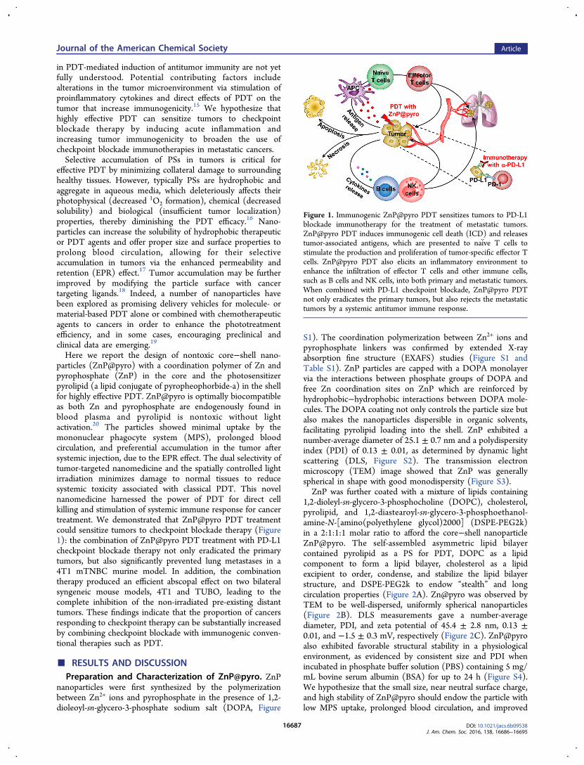
Figure 1. Immunogenic ZnP@pyro PDT sensitizes tumors to PD-L1blockade immunotherapy for the treatment of metastatic tumors.ZnP@pyro PDT induces immunogenic cell death (ICD) and releasestumor-associated antigens, which are presented to naive T cells tostimulate the production and proliferation of tumor-specific effector Tcells. ZnP@pyro PDT also elicits an inflammatory environment toenhance the infiltration of effector T cells and other immune cells,such as B cells and NK cells, into both primary and metastatic tumors.When combined with PD-L1 checkpoint blockade, ZnP@pyro PDTnot only eradicates the primary tumors, but also rejects the metastatictumors by a systemic antitumor immune response.
Journal of the American Chemical Society Article
DOI: 10.1021/jacs.6b09538J. Am. Chem. Soc. 2016, 138, 16686−16695
16687

tumor uptake, making ZnP@pyro ideal for in vivo therapeuticapplications.21
Pyrolipid was incorporated into lipid bilayer at the very highloading of 10.6 ± 0.5 wt%, as determined by UV−vis at 669nm. Due to the high loading, > 97% of the pyrolipidfluorescence was self-quenched when the lipid layer was intact.After the addition of Triton X-100 to disrupt the lipid bilayer,pyrolipid was freed from the ordered structure of ZnP@pyroand regained its fluorescence (Figure S5). The 1O2 generationefficiency of ZnP@pyro was determined in the presence ofsinglet oxygen sensor green (SOSG) regent. When the lipidbilayer was intact, ZnP@pyro generated very little singletoxygen, likely due to the quenching of pyrolipid excited statesbefore it can transfer energy to triplet oxygen. After addition ofTriton X-100 to ZnP@pyro, 1O2 generation upon lightirradiation was restored to a similar efficiency to that of freepyrolipid at the same concentration (Figure S6).ZnP@pyro Shows Long Circulation and High Tumor
Accumulation. The pharmacokinetics (PK) and biodistribu-tion studies on orthotopic 4T1 tumor-bearing BALB/c miceshowed that ZnP@pyro exhibited a prolonged blood circulationhalf-life of 14.5 ± 2.2 h after intravenous (i.v.) injection (Figure2D). ZnP@pyro also showed low distribution in the liver (<5ID%/g), spleen (<8 ID%/g), and kidneys (<6 ID%/g),suggesting that ZnP@pyro can avoid MPS uptake. Slowblood clearance and low MPS uptake led to high tumoraccumulation, with the highest tumor uptake measured to be15.6 ± 2.5 ID%/g at 24 h post i.v. administration (Figure 2E).The confocal laser scanning microscope (CLSM) imagingconfirmed the high distribution of ZnP@pyro in the tumor at
24 h after i.v. injection (Figure S7). By contrast, free pyrolipidshowed a low tumor accumulation of 3.2 ± 1.7 ID%/g and veryhigh accumulation in liver, heart, and spleen at 24 h post i.v.administration (Figure S8).
ZnP@pyro PDT Induces Cell Apoptosis and/orNecrosis in Vitro and in Vivo. ZnP@pyro was rapidlyinternalized by 4T1 tumor cells, with most uptake occurringwithin 1 h followed by stable amounts measured over 24 h(Figure S9). High cellular uptake and negligible efflux (<2%)(Figure S10) ensured the high cellular accumulation of ZnP@pyro. Confocal images showed that the fluorescence of ZnP@pyro was quite dim in the first 2 h incubation, but becamemuch brighter after 2 h incubation (Figure S11). The initialdim signal indicates fluorescence quenching and suggests thatZnP@pyro maintains its structural integrity in cells for the first2 h, followed by the lipid layer dissociation and pyrolipidrelease. After release, pyrolipid can absorb light to generatecytotoxic ROS, killing tumor cells by inducing apoptosis and/ornecrosis.ZnP@pyro induced no cytotoxicity in cells without
irradiation (IC50 > 5 μM), but exhibited very high cytotoxicityafter irradiation at a light dose of 54 J/cm2, given at 60 mW/cm2 for 15 min, as shown by a significant decrease in the IC50value (0.42 ± 0.02 μM) (Figure S12 and Table S2), confirmingthat ZnP@pyro is nontoxic without light activation, and thelocal application of light can specifically control the cytotoxiceffect. Flow cytometry assay showed that ZnP@pyro at aconcentration of 0.2 μM failed to induce apoptosis and/ornecrosis without irradiation, but evoked high levels of apoptosisand/or necrosis consistent with free pyrolipid under irradiation
Figure 2. Preparation and characterization of ZnP@pyro. (A) Scheme showing the Zn-pyrophosphate core and the asymmetric lipid bilayer shell ofZnP@pyro. (B) TEM image showing the spherical and nearly monodispersed morphology of ZnP@pyro (scale bar = 200 nm). (C) Number-averagediameter of ZnP@pyro in PBS, measured by DLS. (D) Blood concentration of pyrolipid over time after i.v. injection of ZnP@pyro at a pyrolipiddose of 6 mg/kg. (E) Biodistribution and tumor uptake of ZnP@pyro in 4T1 tumor-bearing mice. Data are expressed as means ± s.d. (n = 3).
Journal of the American Chemical Society Article
DOI: 10.1021/jacs.6b09538J. Am. Chem. Soc. 2016, 138, 16686−16695
16688
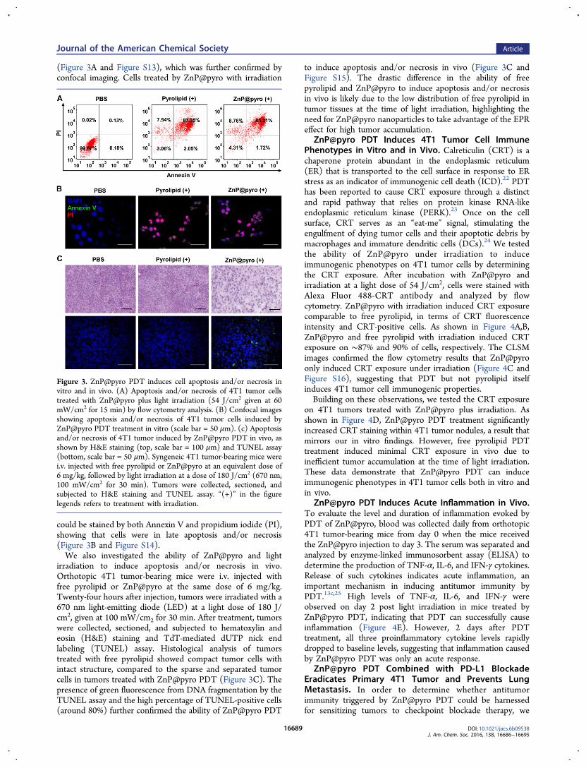
(Figure 3A and Figure S13), which was further confirmed byconfocal imaging. Cells treated by ZnP@pyro with irradiation
could be stained by both Annexin V and propidium iodide (PI),showing that cells were in late apoptosis and/or necrosis(Figure 3B and Figure S14).We also investigated the ability of ZnP@pyro and light
irradiation to induce apoptosis and/or necrosis in vivo.Orthotopic 4T1 tumor-bearing mice were i.v. injected withfree pyrolipid or ZnP@pyro at the same dose of 6 mg/kg.Twenty-four hours after injection, tumors were irradiated with a670 nm light-emitting diode (LED) at a light dose of 180 J/cm2, given at 100 mW/cm2 for 30 min. After treatment, tumorswere collected, sectioned, and subjected to hematoxylin andeosin (H&E) staining and TdT-mediated dUTP nick endlabeling (TUNEL) assay. Histological analysis of tumorstreated with free pyrolipid showed compact tumor cells withintact structure, compared to the sparse and separated tumorcells in tumors treated with ZnP@pyro PDT (Figure 3C). Thepresence of green fluorescence from DNA fragmentation by theTUNEL assay and the high percentage of TUNEL-positive cells(around 80%) further confirmed the ability of ZnP@pyro PDT
to induce apoptosis and/or necrosis in vivo (Figure 3C andFigure S15). The drastic difference in the ability of freepyrolipid and ZnP@pyro to induce apoptosis and/or necrosisin vivo is likely due to the low distribution of free pyrolipid intumor tissues at the time of light irradiation, highlighting theneed for ZnP@pyro nanoparticles to take advantage of the EPReffect for high tumor accumulation.
ZnP@pyro PDT Induces 4T1 Tumor Cell ImmunePhenotypes in Vitro and in Vivo. Calreticulin (CRT) is achaperone protein abundant in the endoplasmic reticulum(ER) that is transported to the cell surface in response to ERstress as an indicator of immunogenic cell death (ICD).22 PDThas been reported to cause CRT exposure through a distinctand rapid pathway that relies on protein kinase RNA-likeendoplasmic reticulum kinase (PERK).23 Once on the cellsurface, CRT serves as an “eat-me” signal, stimulating theengulfment of dying tumor cells and their apoptotic debris bymacrophages and immature dendritic cells (DCs).24 We testedthe ability of ZnP@pyro under irradiation to induceimmunogenic phenotypes on 4T1 tumor cells by determiningthe CRT exposure. After incubation with ZnP@pyro andirradiation at a light dose of 54 J/cm2, cells were stained withAlexa Fluor 488-CRT antibody and analyzed by flowcytometry. ZnP@pyro with irradiation induced CRT exposurecomparable to free pyrolipid, in terms of CRT fluorescenceintensity and CRT-positive cells. As shown in Figure 4A,B,ZnP@pyro and free pyrolipid with irradiation induced CRTexposure on ∼87% and 90% of cells, respectively. The CLSMimages confirmed the flow cytometry results that ZnP@pyroonly induced CRT exposure under irradiation (Figure 4C andFigure S16), suggesting that PDT but not pyrolipid itselfinduces 4T1 tumor cell immunogenic properties.Building on these observations, we tested the CRT exposure
on 4T1 tumors treated with ZnP@pyro plus irradiation. Asshown in Figure 4D, ZnP@pyro PDT treatment significantlyincreased CRT staining within 4T1 tumor nodules, a result thatmirrors our in vitro findings. However, free pyrolipid PDTtreatment induced minimal CRT exposure in vivo due toinefficient tumor accumulation at the time of light irradiation.These data demonstrate that ZnP@pyro PDT can induceimmunogenic phenotypes in 4T1 tumor cells both in vitro andin vivo.
ZnP@pyro PDT Induces Acute Inflammation in Vivo.To evaluate the level and duration of inflammation evoked byPDT of ZnP@pyro, blood was collected daily from orthotopic4T1 tumor-bearing mice from day 0 when the mice receivedthe ZnP@pyro injection to day 3. The serum was separated andanalyzed by enzyme-linked immunosorbent assay (ELISA) todetermine the production of TNF-α, IL-6, and IFN-γ cytokines.Release of such cytokines indicates acute inflammation, animportant mechanism in inducing antitumor immunity byPDT.13c,25 High levels of TNF-α, IL-6, and IFN-γ wereobserved on day 2 post light irradiation in mice treated byZnP@pyro PDT, indicating that PDT can successfully causeinflammation (Figure 4E). However, 2 days after PDTtreatment, all three proinflammatory cytokine levels rapidlydropped to baseline levels, suggesting that inflammation causedby ZnP@pyro PDT was only an acute response.
ZnP@pyro PDT Combined with PD-L1 BlockadeEradicates Primary 4T1 Tumor and Prevents LungMetastasis. In order to determine whether antitumorimmunity triggered by ZnP@pyro PDT could be harnessedfor sensitizing tumors to checkpoint blockade therapy, we
Figure 3. ZnP@pyro PDT induces cell apoptosis and/or necrosis invitro and in vivo. (A) Apoptosis and/or necrosis of 4T1 tumor cellstreated with ZnP@pyro plus light irradiation (54 J/cm2 given at 60mW/cm2 for 15 min) by flow cytometry analysis. (B) Confocal imagesshowing apoptosis and/or necrosis of 4T1 tumor cells induced byZnP@pyro PDT treatment in vitro (scale bar = 50 μm). (c) Apoptosisand/or necrosis of 4T1 tumor induced by ZnP@pyro PDT in vivo, asshown by H&E staining (top, scale bar = 100 μm) and TUNEL assay(bottom, scale bar = 50 μm). Syngeneic 4T1 tumor-bearing mice werei.v. injected with free pyrolipid or ZnP@pyro at an equivalent dose of6 mg/kg, followed by light irradiation at a dose of 180 J/cm2 (670 nm,100 mW/cm2 for 30 min). Tumors were collected, sectioned, andsubjected to H&E staining and TUNEL assay. “(+)” in the figurelegends refers to treatment with irradiation.
Journal of the American Chemical Society Article
DOI: 10.1021/jacs.6b09538J. Am. Chem. Soc. 2016, 138, 16686−16695
16689

investigated the antitumor activity and antimetastatic effect ofZnP@pyro PDT combined with anti-PD-L1 (α-PD-L1, Clone:10F.9G2, Catalog No. BE0101, BioXCell) on 4T1 tumors.Orthotopic 4T1 tumors in the mammary fat pads of miceproduce spontaneous metastases to the lung, making it asuitable experimental animal model for stage IV human breastcancer.26 4T1 tumor-bearing mice were i.v. injected with ZnP@pyro at a pyrolipid dose of 6 mg/kg every 2 days for a total ofthree treatments. Twenty-four hours post injection, tumorswere irradiated with a 670 nm LED at an irradiance of 100mW/cm2 for 30 min. After irradiation, mice were intra-peritoneally (i.p.) injected with anti-PD-L1 antibody at a doseof 75 μg/mouse. As indicated in Figure 5A−C, anti-PD-L1itself failed to delay 4T1 tumor progression. In contrast, ZnP@pyro PDT treatment significantly inhibited 4T1 tumor growthwith a 68% reduction in tumor volume and a 75% reduction intumor weight compared to the PBS control group. Notably,ZnP@pyro PDT combined with anti-PD-L1 treatmentcompletely eradicated the primary 4T1 tumor, indicating thatthe combination treatment was markedly better than eitherZnP@pyro PDT or anti-PD-L1 alone. In addition, no weightloss was observed in ZnP@pyro PDT plus anti-PD-L1 treatedgroup, indicating the absence of severe systemic toxicity (FigureS17).
At the end of the study, 23 days after tumor inoculation, micewere sacrificed and assessed for the extent of metastasis to thelungs by gross examination of tissue for tumor nodules.Compared to the PBS control, ZnP@pyro PDT or anti-PD-L1alone showed little effect on preventing lung metastasis, whilethe combination treatment significantly reduced tumornodules: only one or two tumor nodules were found on thelungs of those receiving combination treatment, compared to31 ± 6 tumor nodules observed in the PBS control group(Figure 5D,E). Lungs were further sectioned and stained withH&E to quantify the proportion of the metastasis area to thewhole lung. As shown in Figure 5F,G, about 37%, 30%, and26% of lungs were occupied by tumors in PBS-, ZnP@pyroPDT-, and anti-PD-L1-treated groups, respectively. Combina-tion treatment significantly decreased the percentage ofmetastasis in the lung to only 0.4%, indicating that thecombination treatment was much more effective in preventinglung metastasis than either ZnP@pyro PDT or anti-PD-L1alone. Lungs were also digested, and the cells were cultured inthe presence of 60 μM 6-thioguanine for 10 days. After fixationwith menthol, colonies formed by clonogenic metastatic cancercells were stained with 0.1% crystal violet. Because 4T1 tumorcells are resistant to 6-thioguanine, only metastasized tumorcells can proliferate and form colony.27 As shown in Figure 5H,the combination treatment significantly reduced the coloniesnumber to only 6 ± 3, compared to PBS-, ZnP@pyro PDT-,and anti-PD-L1-treated groups, which all formed numerouscolonies. The quantitative results showed that the absorbanceof the combination treatment group was only 4.5 ± 1.5% of thePBS control group (Figure 5I), which indicates that there weremuch less clonogenic metastatic cancer cells in the lungstreated by ZnP@pyro PDT plus anti-PD-L1 than that treatedby PBS.Our findings were consistent with previous reports that 4T1
tumor showed no response to anti-PD-L1, possibly due to theirlow expression of PD-L1.28 However, literature reports indicatethat checkpoint blockade immunotherapy can be enhanced bycombining with other immunogenic therapies. For example,ibrutinib was able to convert a weak antitumor T-cell immuneresponse induced by anti-PD-L1 antibody into a powerful one,although it did not affect the PD-L1 expression level intumors.28 In another example, the combination of oxaliplatinwith cyclophosphamide was shown to induce tumor cellimmune phenotypes, trigger adaptive and innate immunity, andsensitize tumors to checkpoint blockade therapy.29 Wedemonstrated that PDT treatment can also increase tumorimmunogenicity and induce acute inflammation, therebyproducing tumor-specific immunity. The tumor-specificimmunity evoked by PDT enhanced the effect of immunecheckpoint therapy, resulting in the eradication of primarytumor and the prevention of lung metastasis.
ZnP@pyro PDT Combined with PD-L1 Blockade NotOnly Prevents Metastasis but Also Completely InhibitsLarger, Pre-existing Metastatic Tumors. A bilateralsubcutaneous 4T1 model was used to determine whether theinduced antitumor immune response by ZnP@pyro PDT plusanti-PD-L1 antibody could be effective against larger, pre-existing metastatic tumors. ZnP@pyro was systemically injectedbut only the right (primary) tumors were irradiated. Asindicated in Figure 6A−C, anti-PD-L1 alone exhibited verylittle effect on the inhibition of either the primary or the distanttumors. ZnP@pyro with irradiation effectively controlledprimary tumor growth but did not significantly inhibit the
Figure 4. ZnP@pyro PDT induces tumor cell immune phenotypesand acute inflammation. (A,B) Quantification of CRT exposure on thesurface of 4T1 cells after treatment with free pyrolipid or ZnP@pyroplus light irradiation (54 J/cm2) by flow cytometry analysis. (C,D)Confocal images showing the CRT exposure on 4T1 tumor cells invitro (C) and in vivo (D) after treatment with free pyrolipid or ZnP@pyro plus light irradiation (scale bar = 50 μm). (E) Pro-inflammatorycytokine levels in the sera of mice treated with PDT of ZnP@pyrofrom day 0 to day 3. Arrows represent the time of nanoparticleadministration (black) and irradiation (red). “(+)” in the figurelegends refers to treatment with irradiation. Data are expressed asmeans ± s.d. (n = 3).
Journal of the American Chemical Society Article
DOI: 10.1021/jacs.6b09538J. Am. Chem. Soc. 2016, 138, 16686−16695
16690

distant tumors, compared to the anti-PD-L1 group. However,the combination of ZnP@pyro with irradiation and PD-L1blockade induced complete eradication of the irradiatedprimary tumors (synergistic effect) and effective control ofthe nonirradiated distant tumors (abscopal effect), eliciting a92% reduction in tumor size compared to the PBS controlgroup. These results indicate that tumors can be sensitized toPD-L1 blockade immunotherapy by ZnP@pyro PDT-mediatedtumor-specific immune responses, and the combination ofZnP@pyro PDT and PD-L1 blockade has the potential tobecome a potent immunotherapy strategy in the managementof patients with metastatic cancer.ZnP@pyro PDT Sensitizes Other Tumors to Immune
Checkpoint Therapy. Finally, we tested whether theantitumor immunity of ZnP@pyro PDT could also sensitize
other tumors to immune checkpoint therapy. We exploredanother syngeneic murine breast cancer model, TUBO, andsimilarly found that TUBO-bearing mice also failed to respondto anti-PD-L1 antibody. However, when combined with ZnP@pyro PDT, anti-PD-L1-mediated checkpoint blockade therapycompletely eradicated primary tumors and significantlyinhibited the growth of distant tumors (Figure 6D−F). Theseresults demonstrate that our findings in the 4T1 mouse modelcould be extended to other tumor types. To further validateearlier findings that our nanoparticle formulation of pyrolipid isnecessary to observe the in vivo effects, we also determined theefficacy of free pyrolipid PDT plus anti-PD-L1, which showedno significant difference from anti-PD-L1 alone (Figure 6D−F).This result was consistent with the earlier results that freepyrolipid PDT induced low apoptosis/necrosis, low CRT
Figure 5. ZnP@pyro PDT combined with PD-L1 blockade eradicates primary 4T1 tumors and prevents lung metastasis. PBS or ZnP@pyro was i.v.injected into an orthotopic 4T1 mouse model at a pyrolipid dose of 6 mg/kg, then tumors were irradiated (670 nm, 100 mW/cm2) for 30 min at 24h after each injection. (A) Tumor growth curves. Arrows represent the time of nanoparticle administration (black) and irradiation (red). (B) Tumorweights at the end point. (C) Photographs of excised tumors at the end point. From top to bottom: PBS, α-PD-L1, ZnP@pyro (+), and ZnP@pyro(+) + α-PD-L1. Rectangle indicates tumors disappeared in the ZnP@pyro (+) + α-PD-L1 group. (D) Representative pictures showing the grossappearance of tumor nodules in the lungs. (E) The numbers of tumor nodules present in the lungs. (F) Representative lung sections stained withH&E. (G) Percentage of lung in metastasis. (H) Representative pictures showing the colonies formed after culturing in the presence of 6-thioguninefor 10 days. (I) Normalized absorbance of crystal violet in different treatment groups. “(+)” in the figure legends refers to treatment with irradiation.*P < 0.05, **P < 0.01, ***P < 0.001.
Journal of the American Chemical Society Article
DOI: 10.1021/jacs.6b09538J. Am. Chem. Soc. 2016, 138, 16686−16695
16691

exposure, and low cytokines level in vivo, compared to ZnP@pyro PDT, due to its inefficient accumulation in tumor tissues.ZnP@pyro PDT Combined with PD-L1 Blockade
Activates Systematic Antitumor Immune Response.The inhibition of distant tumors in 4T1- and TUBO-bearingmice treated with ZnP@pyro PDT plus anti-PD-L1 implied aneffective induction of systemic antitumor immune response. Wefirst validated this hypothesis in TUBO model with an IFN-γELISPOT assay to determine the presence or absence oftumor-specific cytotoxic T cells. Splenocytes were harvestedfrom TUBO-bearing mice at day 12 after the first treatment andstimulated with antigen-presenting 3T3/NKB cells, whichexpress TUBO-derived antigen neu, and the IFN-γ spot-forming cells (SFC) were determined.30 We found that thenumbers of IFN-γ SFC significantly increased in both PD-L1blockade (IFN-γ SFC/104 cells = 2.20 ± 1.54) and ZnP@pyroPDT plus anti-PD-L1 groups (IFN-γ SFC/104 cells = 2.66 ±1.95), compared to the PBS control group (IFN-γ SFC/104
cells = 0.29 ± 0.31) (Figure 7A).After initial indication of systemic immune response, we
further profiled infiltrating leukocytes in the distant tumors.The percentage of CD45+ leukocytes in the total tumor cellssignificantly increased by about 45% in the ZnP@pyro PDTplus anti-PD-L1 treatment group (25.4 ± 6.01%), compared tothe PBS control group (17.5 ± 1.45%) (Figure 7B).Specifically, the percentages of NK cells (17.2 ± 3.89%),CD8+ T cells (1.45 ± 0.65%) and CD4+ T cells (0.97 ± 0.16%)all significantly increased in the anti-PD-L1 treated groupcompared to the PBS control group (NK cells, 9.28 ± 3.07%;CD8+ T cells, 0.63 ± 0.20%; CD4+ T cells, 0.33 ± 0.18%)
(Figure 7C−E), while the percentage of B cells significantlyincreased in ZnP@pyro PDT treated group (6.65 ± 3.64%)compared to the PBS control group (3.01 ± 2.06%) (Figure7F). These results suggest that PD-L1 checkpoint blockadeplays an important role in promoting the dramatically increasedNK cell infiltration and accumulation in the distant tumor sitesand activating tumor-specific T cells responses to control thedistant tumors, while ZnP@pyro PDT evokes B cellsinfiltration to the distant tumors, which can potentially induceantitumor humoral immune responses. PDT and checkpointblockade therapy each initiate unique forms of immuneresponse, which were both found in ZnP@pyro PDT plusanti-PD-L1 treatment (NK cells, 17.6 ± 6.17%; CD8+ T cells,1.72 ± 0.66%; CD4+ T cells, 1.04 ± 0.32%; B cells, 8.28 ±3.25%). The increase in CD8+ T cell infiltration and activitymay have been influenced by the PD-L1 blockade, as PD-L1has been reported to negatively regulate T cells.31 There wasalso a slight, though not statistically significant, decrease in thepercentage of regulatory T cells in tumors treated with ZnP@pyro PDT combined with PD-L1 blockade (Figure S18), whichmay have contributed to the increased CD8+ T cells activity. Acombination of these immune responses were likely requiredfor the eradication of the primary tumor and inhibition ofdistant tumor, supporting the increased efficacy of ZnP@pyroPDT combined with immune checkpoint blockade therapy.The antitumor immune response elicited by ZnP@pyro PDT
in combination with anti-PD-L1 was further confirmed byimmunofluorescence assay. We found that ZnP@pyro withirradiation plus anti-PD-L1 treatment instigated CD3+ T cellinfiltration within the distant tumor tissues, whereas no tumor-
Figure 6. ZnP@pyro PDT sensitizes tumors to immune checkpoint therapy. Bilateral syngeneic tumor models of 4T1 and TUBO were developed bysubcutaneously injecting cancer cells into both the right and left flank regions of each animal. The right tumors were designated as primary tumorsfor light irradiation, and the left tumors were designated as distant tumors and not subjected to light irradiation. ZnP@pyro was i.v. injected intomice, followed by light irradiation at a dose of 180 J/cm2 (670 nm, 100 mW/cm2) at 24 h after nanoparticle injection and i.p. injection of anti-PD-L1at a dose of 75 μg/mouse. The treatment was carried out every other day for a total of three treatments. Primary and distant tumor growth curves in4T1 (A,B) and TUBO (D,E) models. The arrows represent the time of nanoparticle administration (black) and irradiation (red). (C,F) Weight of4T1 (C) and TUBO (F) tumors at the end point of the experiment. “(+)” in the figure legends refers to treatment with irradiation. *P < 0.05, ** P <0.01, *** P < 0.001.
Journal of the American Chemical Society Article
DOI: 10.1021/jacs.6b09538J. Am. Chem. Soc. 2016, 138, 16686−16695
16692
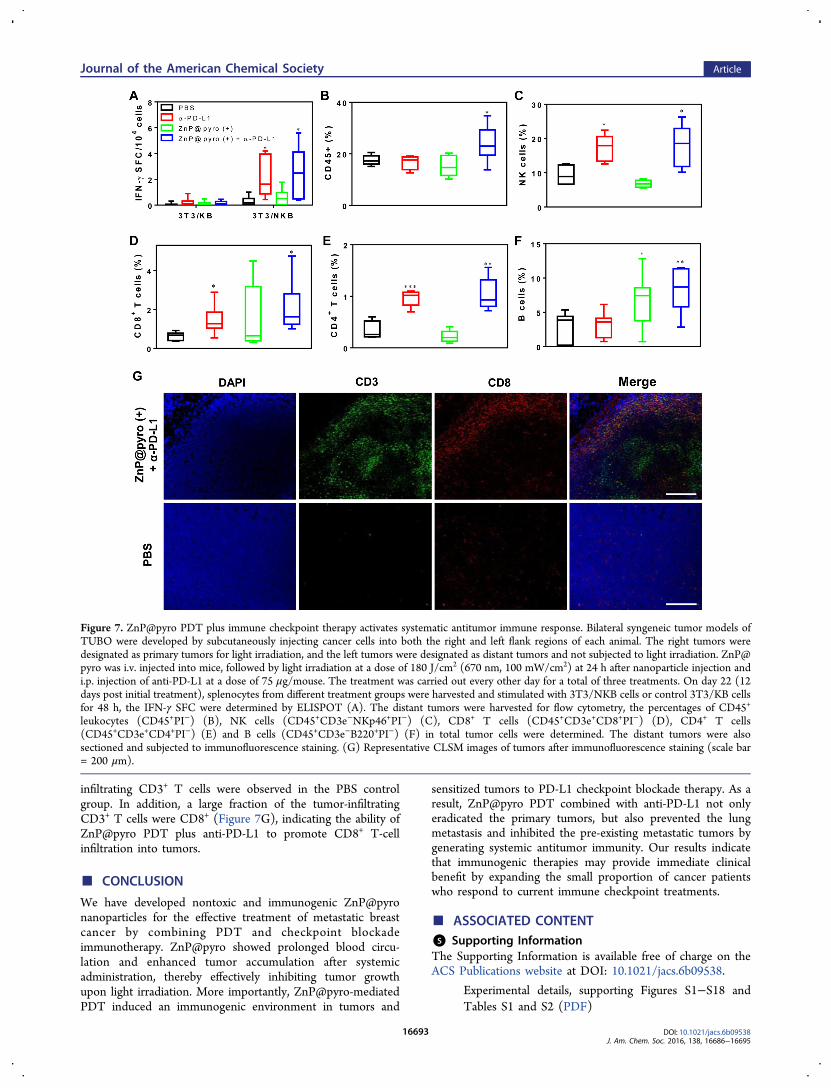
infiltrating CD3+ T cells were observed in the PBS controlgroup. In addition, a large fraction of the tumor-infiltratingCD3+ T cells were CD8+ (Figure 7G), indicating the ability ofZnP@pyro PDT plus anti-PD-L1 to promote CD8+ T-cellinfiltration into tumors.
■ CONCLUSION
We have developed nontoxic and immunogenic ZnP@pyronanoparticles for the effective treatment of metastatic breastcancer by combining PDT and checkpoint blockadeimmunotherapy. ZnP@pyro showed prolonged blood circu-lation and enhanced tumor accumulation after systemicadministration, thereby effectively inhibiting tumor growthupon light irradiation. More importantly, ZnP@pyro-mediatedPDT induced an immunogenic environment in tumors and
sensitized tumors to PD-L1 checkpoint blockade therapy. As aresult, ZnP@pyro PDT combined with anti-PD-L1 not onlyeradicated the primary tumors, but also prevented the lungmetastasis and inhibited the pre-existing metastatic tumors bygenerating systemic antitumor immunity. Our results indicatethat immunogenic therapies may provide immediate clinicalbenefit by expanding the small proportion of cancer patientswho respond to current immune checkpoint treatments.
■ ASSOCIATED CONTENT*S Supporting InformationThe Supporting Information is available free of charge on theACS Publications website at DOI: 10.1021/jacs.6b09538.
Experimental details, supporting Figures S1−S18 andTables S1 and S2 (PDF)
Figure 7. ZnP@pyro PDT plus immune checkpoint therapy activates systematic antitumor immune response. Bilateral syngeneic tumor models ofTUBO were developed by subcutaneously injecting cancer cells into both the right and left flank regions of each animal. The right tumors weredesignated as primary tumors for light irradiation, and the left tumors were designated as distant tumors and not subjected to light irradiation. ZnP@pyro was i.v. injected into mice, followed by light irradiation at a dose of 180 J/cm2 (670 nm, 100 mW/cm2) at 24 h after nanoparticle injection andi.p. injection of anti-PD-L1 at a dose of 75 μg/mouse. The treatment was carried out every other day for a total of three treatments. On day 22 (12days post initial treatment), splenocytes from different treatment groups were harvested and stimulated with 3T3/NKB cells or control 3T3/KB cellsfor 48 h, the IFN-γ SFC were determined by ELISPOT (A). The distant tumors were harvested for flow cytometry, the percentages of CD45+
leukocytes (CD45+PI−) (B), NK cells (CD45+CD3e−NKp46+PI−) (C), CD8+ T cells (CD45+CD3e+CD8+PI−) (D), CD4+ T cells(CD45+CD3e+CD4+PI−) (E) and B cells (CD45+CD3e−B220+PI−) (F) in total tumor cells were determined. The distant tumors were alsosectioned and subjected to immunofluorescence staining. (G) Representative CLSM images of tumors after immunofluorescence staining (scale bar= 200 μm).
Journal of the American Chemical Society Article
DOI: 10.1021/jacs.6b09538J. Am. Chem. Soc. 2016, 138, 16686−16695
16693

■ AUTHOR INFORMATION
Corresponding Author*[email protected]
ORCIDWenbin Lin: 0000-0001-7035-7759NotesThe authors declare no competing financial interest.
■ ACKNOWLEDGMENTS
We thank the National Cancer Institute (U01-CA198989), theUniversity of Chicago Medicine Comprehensive Cancer Center(NIH CCSG: P30 CA014599), the CBI Training Grant (NIH5T32GM008720-15), the Cancer Research Foundation, andthe Ludwig Institute for Metastasis Research for fundingsupport. We thank Mr. Zekai Lin for help with EXAFSexperiments.
■ REFERENCES(1) DeSantis, C.; Ma, J.; Bryan, L.; Jemal, A. Ca-Cancer J. Clin. 2014,64, 52.(2) (a) Schmadeka, R.; Harmon, B. E.; Singh, M. Am. J. Clin. Pathol.2014, 141, 462. (b) Bayraktar, S.; Gluck, S. Breast Cancer Res. Treat.2013, 138, 21.(3) (a) Vanneman, M.; Dranoff, G. Nat. Rev. Cancer 2012, 12, 237.(b) Couzin-Frankel, J. Science 2013, 342, 1432.(4) (a) Restifo, N. P.; Dudley, M. E.; Rosenberg, S. A. Nat. Rev.Immunol. 2012, 12, 269. (b) Rooney, M. S.; Shukla, S. A.; Wu, C. J.;Getz, G.; Hacohen, N. Cell 2015, 160, 48. (c) Schumacher, T. N.;Schreiber, R. D. Science 2015, 348, 69. (d) Gajewski, T. F.; Schreiber,H.; Fu, Y.-X. Nat. Immunol. 2013, 14, 1014.(5) Nishikawa, H.; Sakaguchi, S. Curr. Opin. Immunol. 2014, 27, 1.(6) Talmadge, J. E.; Gabrilovich, D. I. Nat. Rev. Cancer 2013, 13, 739.(7) (a) Sharma, P.; Allison, J. P. Science 2015, 348, 56. (b) Topalian,S. L.; Drake, C. G.; Pardoll, D. M. Cancer Cell 2015, 27, 450.(8) (a) Pardoll, D. M. Nat. Rev. Cancer 2012, 12, 252. (b) Gubin, M.M.; Zhang, X.; Schuster, H.; Caron, E.; Ward, J. P.; Noguchi, T.;Ivanova, Y.; Hundal, J.; Arthur, C. D.; Krebber, W.-J.; et al. Nature2014, 515, 577. (c) Topalian, S. L.; Taube, J. M.; Anders, R. A.;Pardoll, D. M. Nat. Rev. Cancer 2016, 16, 275.(9) (a) Meng, X.; Huang, Z.; Teng, F.; Xing, L.; Yu, J. Cancer Treat.Rev. 2015, 41, 868. (b) Iwai, Y.; Ishida, M.; Tanaka, Y.; Okazaki, T.;Honjo, T.; Minato, N. Proc. Natl. Acad. Sci. U. S. A. 2002, 99, 12293.(c) Blank, C.; Gajewski, T. F.; Mackensen, A. Cancer Immunol.Immunother. 2005, 54, 307. (d) Topalian, S. L.; Drake, C. G.; Pardoll,D. M. Curr. Opin. Immunol. 2012, 24, 207.(10) (a) Webster, R. M. Nat. Rev. Drug Discovery 2014, 13, 883.(b) Leventakos, K.; Mansfield, A. S. BioDrugs 2016, 30, 397.(11) (a) Tumeh, P. C.; Harview, C. L.; Yearley, J. H.; Shintaku, I. P.;Taylor, E. J.; Robert, L.; Chmielowski, B.; Spasic, M.; Henry, G.;Ciobanu, V.; et al. Nature 2014, 515, 568. (b) Rizvi, N. A.; Hellmann,M. D.; Snyder, A.; Kvistborg, P.; Makarov, V.; Havel, J. J.; Lee, W.;Yuan, J.; Wong, P.; Ho, T. S.; et al. Science 2015, 348, 124.(12) (a) Gajewski, T. F.; Woo, S.-R.; Zha, Y.; Spaapen, R.; Zheng, Y.;Corrales, L.; Spranger, S. Curr. Opin. Immunol. 2013, 25, 268.(b) Joyce, J. A.; Fearon, D. T. Science 2015, 348, 74. (c) Shahabi, V.;Postow, M. A.; Tuck, D.; Wolchok, J. D. Am. J. Clin. Oncol. 2015, 38,90.(13) (a) Shams, M.; Owczarczak, B.; Manderscheid-Kern, P.;Bellnier, D. A.; Gollnick, S. O. Cancer Immunol. Immunother. 2015,64, 287. (b) Kwitniewski, M.; Juzeniene, A.; Glosnicka, R.; Moan, J.Photochem. Photobiol. Sci. 2008, 7, 1011. (c) Castano, A. P.; Mroz, P.;Hamblin, M. R. Nat. Rev. Cancer 2006, 6, 535. (d) He, C.; Duan, X.;Guo, N.; Chan, C.; Poon, C.; Weichselbaum, R. R.; Lin, W. Nat.Commun. 2016, 7, 12499. (e) Lu, K.; He, C.; Guo, N.; Chan, C.; Ni,K.; Weichselbaum, R. R.; Lin, W. J. Am. Chem. Soc. 2016, 138, 12502.
(14) (a) Garg, A. D.; Agostinis, P. Photochem. Photobiol. Sci. 2014, 13,474. (b) Dolmans, D. E.; Fukumura, D.; Jain, R. K. Nat. Rev. Cancer2003, 3, 380. (c) Garg, A. D.; Nowis, D.; Golab, J.; Agostinis, P.Apoptosis 2010, 15, 1050. (d) Gollnick, S. O.; Brackett, C. M. Immunol.Res. 2010, 46, 216. (e) Mroz, P.; Hashmi, J. T.; Huang, Y.-Y.; Lange,N.; Hamblin, M. R. Expert Rev. Clin. Immunol. 2011, 7, 75. (f) Mijan,M. C.; Longo, J. P. F.; de Melo, L. N. D.; Simioni, A. R.; Tedesco, A.C.; Azevedo, R. B. J. Nanomed. Nanotechnol. 2014, 5, 218.(15) (a) Sanabria, L. M.; Rodríguez, M. E.; Cogno, I. S.; Vittar, N. B.R.; Pansa, M. F.; Lamberti, M. J.; Rivarola, V. A. Biochim. Biophys. Acta,Rev. Cancer 2013, 1835, 36. (b) Gollnick, S.; Evans, S.; Baumann, H.;Owczarczak, B.; Maier, P.; Vaughan, L.; Wang, W.; Unger, E.;Henderson, B. Br. J. Cancer 2003, 88, 1772. (c) Galluzzi, L.; Kepp, O.;Kroemer, G. EMBO J. 2012, 31, 1055.(16) (a) Celli, J. P.; Spring, B. Q.; Rizvi, I.; Evans, C. L.; Samkoe, K.S.; Verma, S.; Pogue, B. W.; Hasan, T. Chem. Rev. 2010, 110, 2795.(b) Chatterjee, D. K.; Fong, L. S.; Zhang, Y. Adv. Drug Delivery Rev.2008, 60, 1627. (c) Paszko, E.; Ehrhardt, C.; Senge, M. O.; Kelleher,D. P.; Reynolds, J. V. Photodiagn. Photodyn. Ther. 2011, 8, 14.(17) (a) Wang, A. Z.; Langer, R.; Farokhzad, O. C. Annu. Rev. Med.2012, 63, 185. (b) Maeda, H.; Nakamura, H.; Fang, J. Adv. DrugDelivery Rev. 2013, 65, 71. (c) Stylianopoulos, T. Ther. Delivery 2013,4, 421. (d) Allison, R.; Mota, H.; Bagnato, V. S.; Sibata, C. Photodiagn.Photodyn. Ther. 2008, 5, 19.(18) (a) Friedman, A.; Claypool, S.; Liu, R. Curr. Pharm. Des. 2013,19, 6315. (b) Florence, A. T. J. Controlled Release 2012, 164, 115.(c) Subramani, K.; Hosseinkhani, H.; Khraisat, A.; Hosseinkhani, M.;Pathak, Y. Curr. Nanosci. 2009, 5, 135. (d) Stuchinskaya, T.; Moreno,M.; Cook, M. J.; Edwards, D. R.; Russell, D. A. Photochem. Photobiol.Sci. 2011, 10, 822.(19) (a) Lovell, J. F.; Jin, C. S.; Huynh, E.; Jin, H.; Kim, C.;Rubinstein, J. L.; Chan, W. C.; Cao, W.; Wang, L. V.; Zheng, G. Nat.Mater. 2011, 10, 324. (b) Jin, C. S.; Cui, L.; Wang, F.; Chen, J.; Zheng,G. Adv. Healthcare Mater. 2014, 3, 1240. (c) Samia, A. C.; Chen, X.;Burda, C. J. Am. Chem. Soc. 2003, 125, 15736. (d) Cheng, Y.; Doane,T. L.; Chuang, C. H.; Ziady, A.; Burda, C. Small 2014, 10, 1799.(e) Chen, J.; Wang, D.; Xi, J.; Au, L.; Siekkinen, A.; Warsen, A.; Li, Z.-Y.; Zhang, H.; Xia, Y.; Li, X. Nano Lett. 2007, 7, 1318. (f) Lal, S.; Clare,S. E.; Halas, N. J. Acc. Chem. Res. 2008, 41, 1842. (g) Idris, N. M.;Gnanasammandhan, M. K.; Zhang, J.; Ho, P. C.; Mahendran, R.;Zhang, Y. Nat. Med. 2012, 18, 1580. (h) Tian, J.; Ding, L.; Xu, H.-J.;Shen, Z.; Ju, H.; Jia, L.; Bao, L.; Yu, J.-S. J. Am. Chem. Soc. 2013, 135,18850. (i) Luo, D.; Carter, K. A.; Miranda, D.; Lovell, J. F. Adv. Sci.2016, DOI: 10.1002/advs.201600106.(20) (a) Sian, L.; Mingyan, X.; Miller, L. V.; Tong, L.; Krebs, N. F.;Hambidge, K. M. Am. J. Clin. Nutr. 1996, 63, 348. (b) Lomashvili, K.A.; Narisawa, S.; Millan, J. L.; O’Neill, W. C. Kidney Int. 2014, 85,1351. (c) Zhang, M.; Zhang, Z.; Blessington, D.; Li, H.; Busch, T. M.;Madrak, V.; Miles, J.; Chance, B.; Glickson, J. D.; Zheng, G.Bioconjugate Chem. 2003, 14, 709.(21) (a) Duan, X.; Li, Y. Small 2013, 9, 1521. (b) Hirsjarvi, S.;Dufort, S.; Gravier, J.; Texier, I.; Yan, Q.; Bibette, J.; Sancey, L.;Josserand, V.; Passirani, C.; Benoit, J.-P.; Coll, J.-L. Nanomedicine2013, 9, 375. (c) Kulkarni, S. A.; Feng, S.-S. Pharm. Res. 2013, 30,2512.(22) (a) Obeid, M.; Tesniere, A.; Ghiringhelli, F.; Fimia, G. M.;Apetoh, L.; Perfettini, J.-L.; Castedo, M.; Mignot, G.; Panaretakis, T.;Casares, N.; et al. Nat. Med. 2007, 13, 54. (b) Kroemer, G.; Galluzzi,L.; Kepp, O.; Zitvogel, L. Annu. Rev. Immunol. 2013, 31, 51.(23) Garg, A. D.; Krysko, D. V.; Verfaillie, T.; Kaczmarek, A.;Ferreira, G. B.; Marysael, T.; Rubio, N.; Firczuk, M.; Mathieu, C.;Roebroek, A. J.; et al. EMBO J. 2012, 31, 1062.(24) (a) Ma, Y.; Adjemian, S.; Mattarollo, S. R.; Yamazaki, T.;Aymeric, L.; Yang, H.; Catani, J. P. P.; Hannani, D.; Duret, H.; Steegh,K.; et al. Immunity 2013, 38, 729. (b) Zitvogel, L.; Galluzzi, L.; Smyth,M. J.; Kroemer, G. Immunity 2013, 39, 74.(25) (a) Yom, S. S.; Busch, T. M.; Friedberg, J. S.; Wileyto, E. P.;Smith, D.; Glatstein, E.; Hahn, S. M. Photochem. Photobiol. 2003, 78,
Journal of the American Chemical Society Article
DOI: 10.1021/jacs.6b09538J. Am. Chem. Soc. 2016, 138, 16686−16695
16694
75. (b) Ziołkowski, P.; Symonowicz, K.; Milach, J.; Zawirska, B.;Szkudlarek, T. Neoplasma 1996, 44, 192.(26) (a) Pulaski, B. A.; Ostrand-Rosenberg, S. Curr. Protoc. Immunol.2001, 39, 20.2.1. (b) Tao, K.; Fang, M.; Alroy, J.; Sahagian, G. G. BMCCancer 2008, 8, 228.(27) Shan, D.; Chen, L.; Njardarson, J. T.; Gaul, C.; Ma, X.;Danishefsky, S. J.; Huang, X.-Y. Proc. Natl. Acad. Sci. U. S. A. 2005,102, 3772.(28) Sagiv-Barfi, I.; Kohrt, H. E. K.; Czerwinski, D. K.; Ng, P. P.;Chang, B. Y.; Levy, R. Proc. Natl. Acad. Sci. U. S. A. 2015, 112, E966.(29) Pfirschke, C.; Engblom, C.; Rickelt, S.; Cortez-Retamozo, V.;Garris, C.; Pucci, F.; Yamazaki, T.; Poirier-Colame, V.; Newton, A.;Redouane, Y.; Lin, Y.-J.; Wojtkiewicz, G.; Iwamoto, Y.; Mino-Kenudson, M.; Huynh, T. G.; Hynes, R. O.; Freeman, G. J.;Kroemer, G.; Zitvogel, L.; Weissleder, R.; Pittet, M. J. Immunity2016, 44, 343.(30) Park, S.; Jiang, Z.; Mortenson, E. D.; Deng, L.; Radkevich-Brown, O.; Yang, X.; Sattar, H.; Wang, Y.; Brown, N. K.; Greene, M.;Liu, Y.; Tang, J.; Wang, S.; Fu, Y.-X. Cancer Cell 2010, 18, 160.(31) (a) Blank, C.; Gajewski, T. F.; Mackensen, A. Cancer Immunol.Immunother. 2005, 54, 307. (b) Latchman, Y. E.; Liang, S. C.; Wu, Y.;Chernova, T.; Sobel, R. A.; Klemm, M.; Kuchroo, V. K.; Freeman, G.J.; Sharpe, A. H. Proc. Natl. Acad. Sci. U. S. A. 2004, 101, 10691.
Journal of the American Chemical Society Article
DOI: 10.1021/jacs.6b09538J. Am. Chem. Soc. 2016, 138, 16686−16695
16695