Embed Size (px)
Citation preview

THE WILD AND WACKY WORLD OF NEURO‐OPHTHALMOLOGY
M. Tariq Bhatti, MDDepartments of Ophthalmology and Medicine (Division of Neurology)
Duke University Eye Center and Duke University Medical Center
Consultant, Grant and Honorarium Support
EMD Serono PfizerBiogen IdecBayer HealthcareNovartis Pharmaceuticals
NO CONFLICTS OF INTEREST
$10
1. Eye Movementsa. Wall-Eyed
b. Stuck to the right
2. Pupil
a. Funny taste in my mouth
b. The teacher and the pupil
3. Aneurysma. Re-coiled
b. That’s a wrap
4. Optic nerve
a. Knee deep in the nerve
b. Really big cells
OUTLINE
The good physician treats the disease; the great physician treats the patient who has the disease.
Sir William Osler, 1849-1919.
Photography and Neuro‐Ophthalmology
•PhotographyFundusExternalEye Movements
•Pupillography•Optical Coherence Tomography•Ultrasonography•Intravenous Fluorescein Angiography•Magnetic Resonance Imaging
MRA/MRV•Computed Tomography
CTA/CTV•Cerebral Angiography•Pathology
Wall‐Eyed
Diplopia initially only in lateral gaze→ progressed to primary gaze
Family doctor: diagnosed sinusitis treated with erythromycin
Ophthalmologist: referred to Duke for evaluation
42 yo CM with 6 day history of binocular and horizontal diplopia
• POH/PSH: congenital color blindness• PMH: remote history of head trauma• Medications: nose spray as needed for allergies• FMH: stroke• SH & ROS: unremarkable
Wall‐Eyed
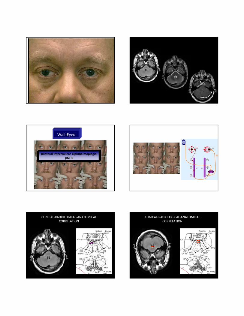
CLINICAL‐RADIOLOGICAL‐ANATOMICAL CORRELATION
CLINICAL‐RADIOLOGICAL‐ANATOMICAL CORRELATION
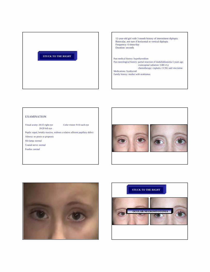
STUCK TO THE RIGHT
12-year-old girl with 3 month history of intermittent diplopia. Binocular, not sure if horizontal or vertical diplopiaFrequency: 6 times/dayDuration: seconds
Past medical history: hypothyroidismPast neurological history: partial resection of medullablastoma 5 years ago
craniospinal radiation: 5580 cGychemotherapy: cisplatin, CCNU and vincristine
Medications: SynthyroidFamily history: mother with strabismus
EXAMINATION
Visual acuity: 20/25 right eye Color vision: 9/10 each eye20/20 left eye
Pupils: equal, briskly reactive, without a relative afferent pupillary defect
Adnexa: no ptosis or proptosis
Slit-lamp: normal
Cranial nerve: normal
Fundus: normal
OCULAR NEUROMYOTONIA
STUCK TO THE RIGHT
OCULAR NEUROMYOTONIA
•Paroxysmal involuntary contraction of the extraocular muscles resulting in intermittent diplopia and ocular misalignment
•Tonic discharge of one of the ocular motor cranial nerves
•Most common ocular motor cranial nerve involved: CN III
•Intermittent diplopia with duration of seconds to minutes
•Precipitated (induced) by change in gaze (in some cases)
•Overaction of extraocular muscle resisting ipsilateral antagonist muscle (eye “stuck”)
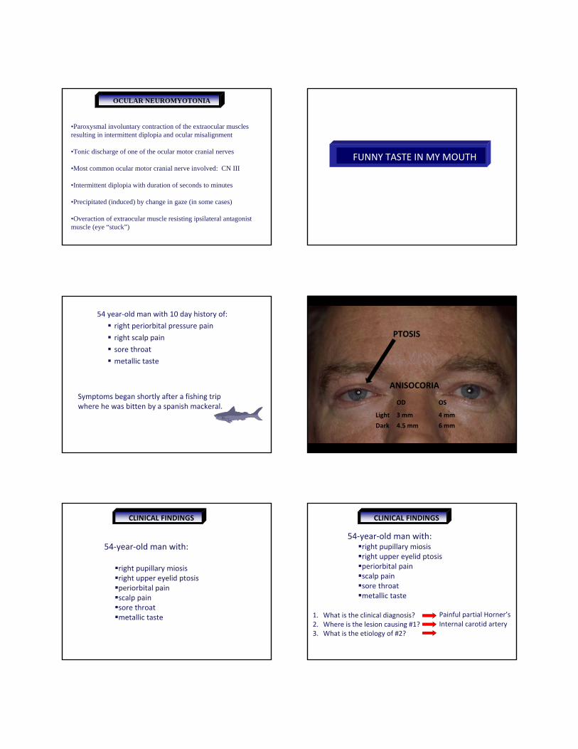
FUNNY TASTE IN MY MOUTH
54 year‐old man with 10 day history of:
right periorbital pressure pain
right scalp pain
sore throat
metallic taste
Symptoms began shortly after a fishing trip where he was bitten by a spanish mackeral.
ANISOCORIA
OD OS
Light 3 mm 4 mm
Dark 4.5 mm 6 mm
PTOSIS
CLINICAL FINDINGS
54‐year‐old man with:
right pupillary miosis right upper eyelid ptosisperiorbital painscalp painsore throat metallic taste 1. What is the clinical diagnosis?
2. Where is the lesion causing #1?3. What is the etiology of #2?
Painful partial Horner’s Internal carotid artery
CLINICAL FINDINGS
54‐year‐old man with:right pupillary miosis right upper eyelid ptosisperiorbital painscalp painsore throat metallic taste
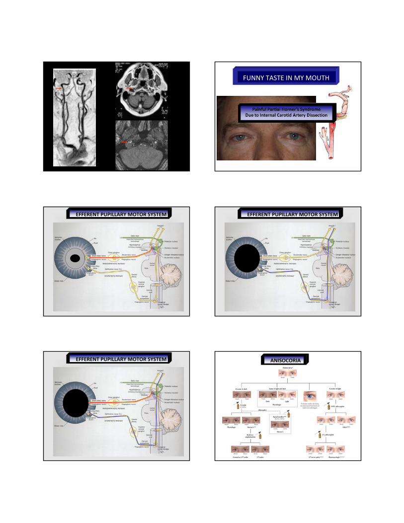
FUNNY TASTE IN MY MOUTH
EFFERENT PUPILLARY MOTOR SYSTEM EFFERENT PUPILLARY MOTOR SYSTEM
EFFERENT PUPILLARY MOTOR SYSTEM ANISOCORIA

• 1st order neuron
(brainstem + SC)
• 2nd order neuron
(preganglionic)
• 3rd order neuron
(postganglionic)
HORNER’S SYNDROME
Characterized by: ipsilateral ptosis miosis facial anhydrosis
DYSGUESIA
Chorda tympani
Oculosympathetic
Glossophyarngeal
THE TEACHER AND THE PUPIL
49 yo BF (kindergarten teacher) with 1 week history of left head pain radiating to left eye.
2 days later noticed left upper eyelid ptosis and double vision.
Primary physician ordered CT brain without contrast‐normal.
Past medical history: nonePast neurological history: migrainesMedications: Imitrex prnFamily history: None

THE TEACHER AND THE PUPIL
RE‐COILED•72‐year‐old previously healthy woman noted left eye pain.
•Ocular pain progressed to involve left head and face.
•One month later: noted ptosis of left upper eyelid.
•One month later: developed double vision.
•Diagnosed with a left third cranial nerve palsy.
•Cerebral angiogram confirmed an aneurysm at the junction of the left superior cerebellar and posterior cerebral arteries.
RE‐COILED

July 2001 One week following endovascular embolization treatment patient referred to the Neuro‐ophthalmology Clinic
October 2002
October 2002
July 2001
July 2002Feb 2002Cerebral Angiogram: October 2002
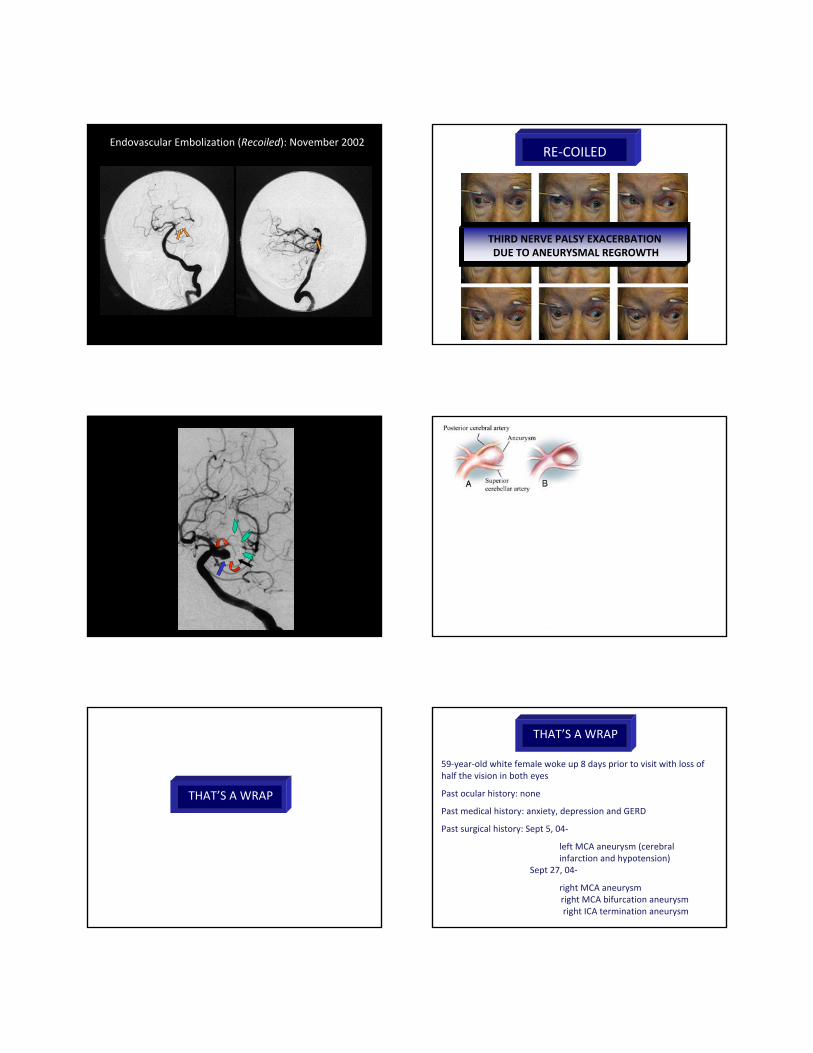
Endovascular Embolization (Recoiled): November 2002
THIRD NERVE PALSY EXACERBATION DUE TO ANEURYSMAL REGROWTH
RE‐COILED
THAT’S A WRAP
59‐year‐old white female woke up 8 days prior to visit with loss of half the vision in both eyes
Past ocular history: none
Past medical history: anxiety, depression and GERD
Past surgical history: Sept 5, 04‐
left MCA aneurysm (cerebral infarction and hypotension)
Sept 27, 04‐
right MCA aneurysm right MCA bifurcation aneurysmright ICA termination aneurysm
THAT’S A WRAP
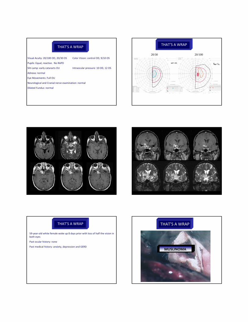
Visual Acuity: 20/100 OD, 20/30 OS Color Vision: control OD, 9/10 OS
Pupils: Equal, reactive. No RAPD
Slit‐Lamp: early cataracts OU Intraocular pressure: 10 OD, 12 OS
Adnexa: normal
Eye Movements: Full OU
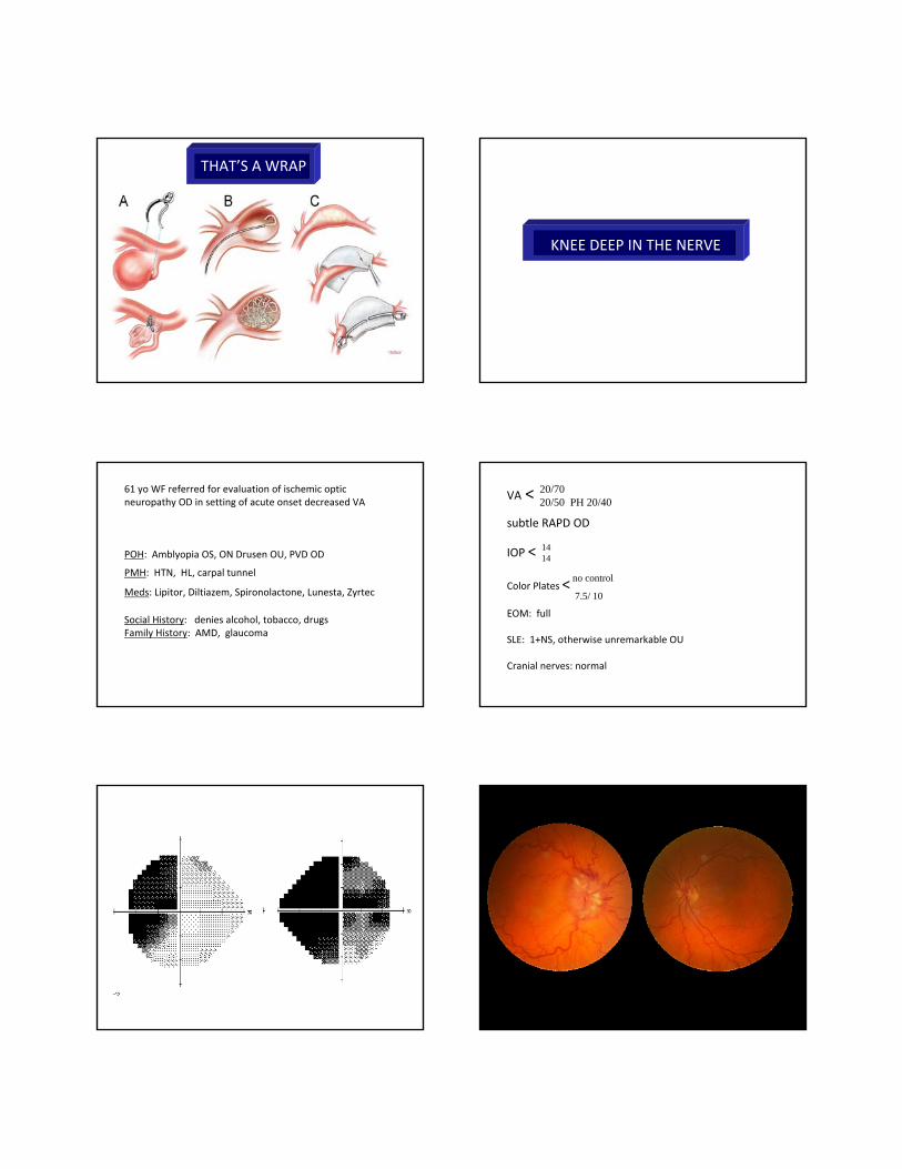
Neurological and Cranial nerve examination: normal
Dilated Fundus: normal
THAT’S A WRAP
20/10020/30
THAT’S A WRAP
59‐year‐old white female woke up 8 days prior with loss of half the vision in both eyes
Past ocular history: none
Past medical history: anxiety, depression and GERD
Past surgical history: Sept 5, 04‐
left MCA aneurysm: clipped Sept 27, 04‐
right MCA aneurysm: clippedright MCA bifurcation aneurysm: clippedright ICA termination aneurysm: wrapped
THAT’S A WRAP
MUSLINOMA
THAT’S A WRAP
THAT’S A WRAP
KNEE DEEP IN THE NERVE
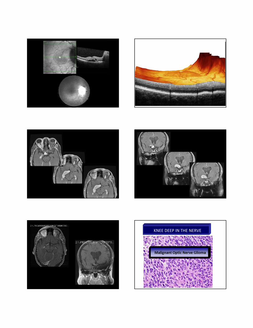
61 yo WF referred for evaluation of ischemic optic neuropathy OD in setting of acute onset decreased VA
POH: Amblyopia OS, ON Drusen OU, PVD OD
PMH: HTN, HL, carpal tunnel
Meds: Lipitor, Diltiazem, Spironolactone, Lunesta, Zyrtec
Social History: denies alcohol, tobacco, drugsFamily History: AMD, glaucoma
VA <
subtle RAPD OD
IOP <
Color Plates <
EOM: full
SLE: 1+NS, otherwise unremarkable OU
Cranial nerves: normal
20/7020/50 PH 20/40
1414
no control
7.5/ 10
KNEE DEEP IN THE NERVE
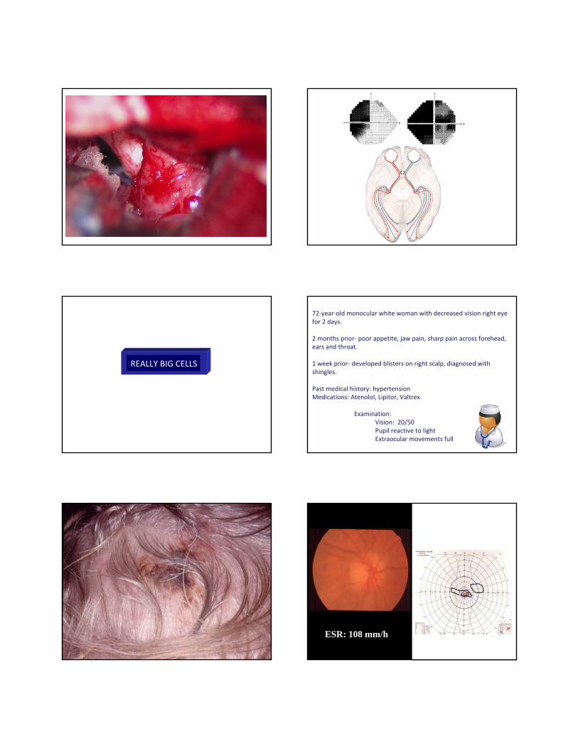
REALLY BIG CELLS
72‐year‐old monocular white woman with decreased vision right eye for 2 days.
2 months prior‐ poor appetite, jaw pain, sharp pain across forehead, ears and throat.
1 week prior‐ developed blisters on right scalp, diagnosed with shingles.
Past medical history: hypertensionMedications: Atenolol, Lipitor, Valtrex
Examination:Vision: 20/50Pupil reactive to light Extraocular movements full
ESR: 108 mm/h

GIANT CELL ARTERITIS
REALLY BIG CELLS
•Clinical Suspicion
•Systemic and Ocular manifestationsoccult GCA: 20%
•Laboratory datanormal ESR: 15‐20%
GIANT CELL ARTERITIS
Superficial temporal artery biopsy
Zygomatic arch
Superficial temporal artery
Skin
Superficial temporal fasciaTemporal branch of facial nerve
Temporalis muscle
Parotid gland
Loose areolar layer
Superficial layer,deep temporal fascia
Deep layer, deep temporal fascia
Deep temporal fat pad
Superficial temporal fat pad
Subdermal fatty layer
SUPERFICIAL TEMPORAL ARTERY BIOPSY
Danger zone
Parietal branch ofTemporal artery
Frontal branch ofTemporal artery
Facial nerve
1. Eye Movementsa. Wall-Eyed
b. Stuck to the right
2. Pupil
a. Funny taste in my mouth
b. The teacher and the pupil
3. Aneurysma. Re-coiled
b. That’s a wrap
4. Optic nerve
a. Knee deep in the nerve
b. Really big cells
1. Eye Movementsa. Bilateral INO
b. Ocular Neuromyotonia
2. Pupil
a. Internal carotid artery dissection
b. Aneurysmal third nerve palsy
3. Aneurysma. Recurrent aneurysm
b. Muslinoma
4. Optic nerve
a. Malignant optic nerve glioma
b. Giant cell arteritis
THE WILD AND WACKY WORLD OF NEURO‐OPHTHALMOLOGY


![Jacobo Grinberg.vision Extraocular [Articulo]](https://img.pdfslide.net/doc/110x75/577cde0c1a28ab9e78ae476f/jacobo-grinbergvision-extraocular-articulo.jpg)