Embed Size (px)
Citation preview
Photomedicine in Gynecology Photomedicine in Gynecology
Attila L. Major, MD, PhD
Training Course for Advanced Oncologic LaparoscopySt. Petersburg - February 17, 2006
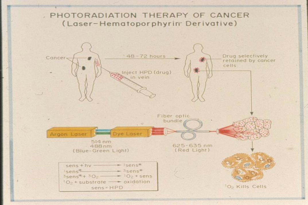
Photodynamic Principle Photodynamic Principle
• Use of a photo-enhancing or photo-sensitizing chemical to aid in the diagnosis or treatment of a target cell
• Use of a photo-enhancing or photo-sensitizing chemical to aid in the diagnosis or treatment of a target cell
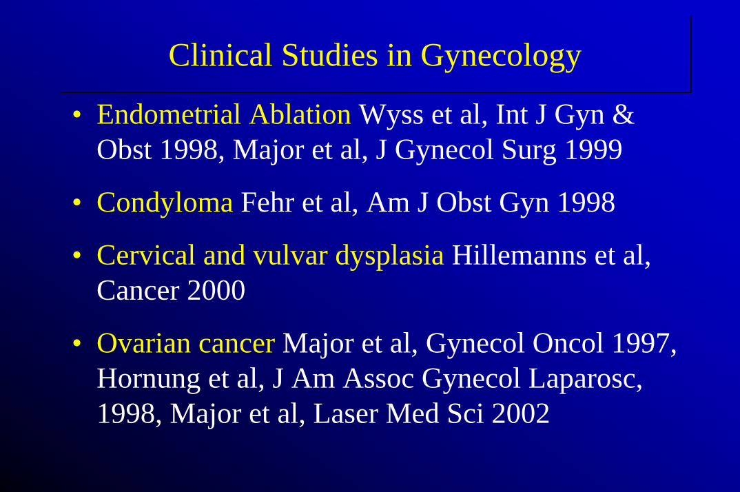
Clinical Studies in GynecologyClinical Studies in Gynecology
• Endometrial Ablation Wyss et al, Int J Gyn & Obst 1998, Major et al, J Gynecol Surg 1999
• Condyloma Fehr et al, Am J Obst Gyn 1998
• Cervical and vulvar dysplasia Hillemanns et al, Cancer 2000
• Ovarian cancer Major et al, Gynecol Oncol 1997, Hornung et al, J Am Assoc Gynecol Laparosc, 1998, Major et al, Laser Med Sci 2002
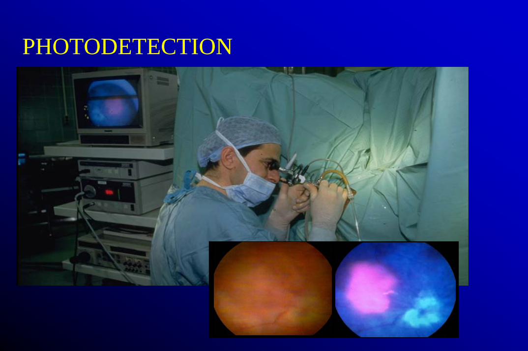
PHOTODETECTION
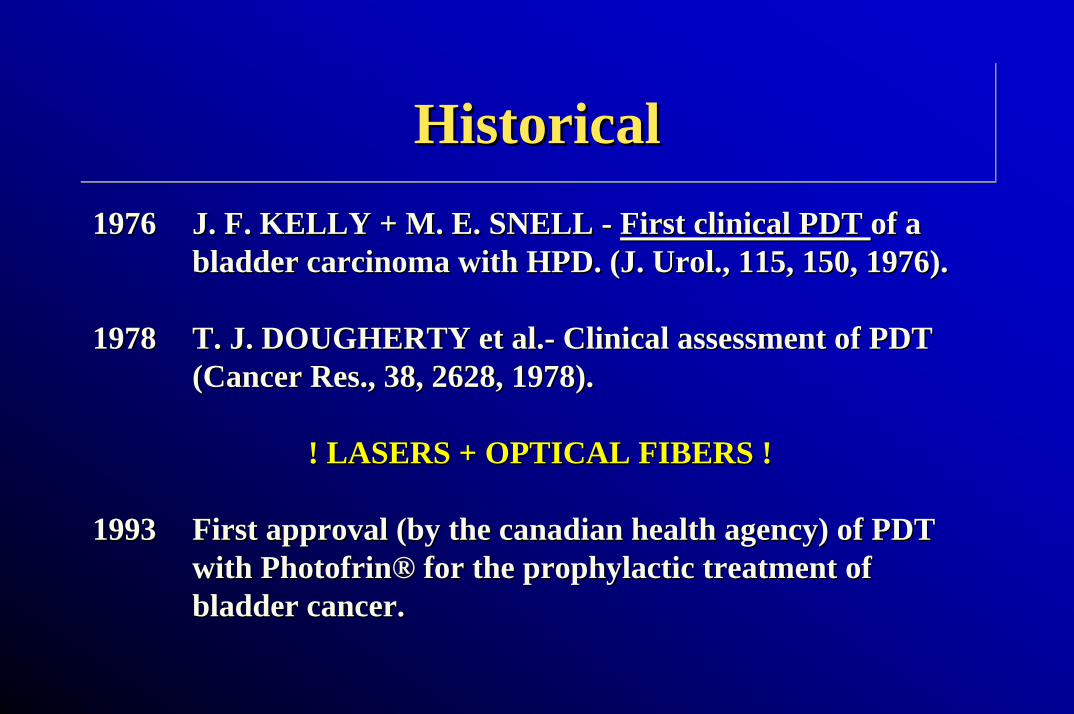
HistoricalHistoricalHistorical19761976 J. F. KELLY + M. E. SNELL J. F. KELLY + M. E. SNELL -- FirstFirst clinicalclinical PDT PDT ofof aa
bladderbladder carcinomacarcinoma withwith HPD. (J. HPD. (J. UrolUrol., 115, 150, 1976).., 115, 150, 1976).
19781978 T. J. DOUGHERTY et al.T. J. DOUGHERTY et al.-- ClinicalClinical assessmentassessment ofof PDTPDT(Cancer (Cancer ResRes., 38, 2628, 1978).., 38, 2628, 1978).
! LASERS + OPTICAL FIBERS !! LASERS + OPTICAL FIBERS !
19931993 FirstFirst approvalapproval (by (by thethe canadiancanadian healthhealth agencyagency) ) ofof PDTPDTwithwith PhotofrinPhotofrin®® for for thethe prophylacticprophylactic treatmenttreatment ofofbladderbladder cancer.cancer.
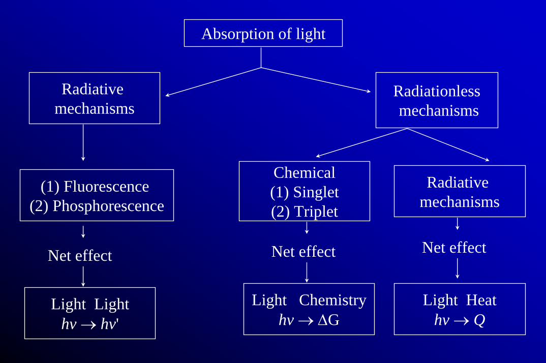
Absorption of light
Radiativemechanisms
Radiationlessmechanisms
(1) Fluorescence (2) Phosphorescence
Chemical(1) Singlet(2) Triplet
Radiativemechanisms
Light Lighthv → hv'
Light Chemistryhv → ∆G
Light Heathv → Q
Net effect Net effect Net effect
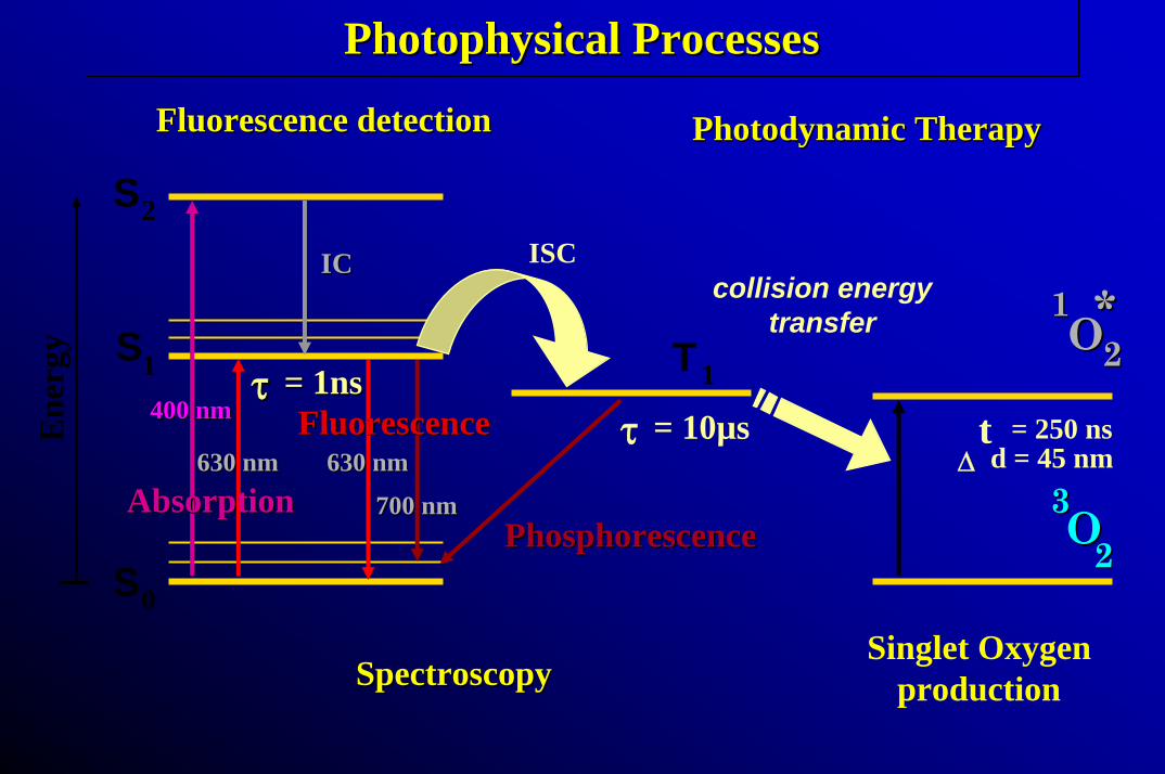
Photophysical ProcessesPhotophysicalPhotophysical ProcessesProcesses
Fluorescence Fluorescence detectiondetection PhotodynamicPhotodynamic TherapyTherapy
S0
S1
S2
T1
AbsorptionAbsorption
FluorescenceFluorescence630 nm630 nm
700 nm700 nm630 nm630 nm
400 nm400 nm
Ene
rgy
ISCICIC
= 1ns= 1nsττ
PhosphorescencePhosphorescence
= 10µsτ = 250 nstd = 45 nm∆
Singlet Oxygenproduction
OO3322
**22
11OO
collision energytransfer
SpectroscopySpectroscopy
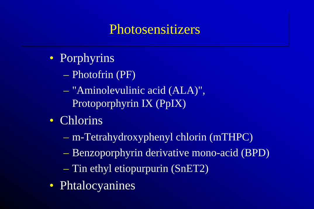
PhotosensitizersPhotosensitizers
• Porphyrins– Photofrin (PF) – "Aminolevulinic acid (ALA)",
Protoporphyrin IX (PpIX)• Chlorins
– m-Tetrahydroxyphenyl chlorin (mTHPC)– Benzoporphyrin derivative mono-acid (BPD)– Tin ethyl etiopurpurin (SnET2)
• Phtalocyanines
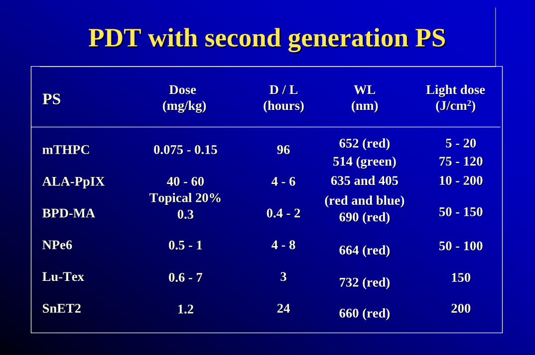
PDT with second generation PSPDT with second generation PSPDT with second generation PS
mTHPCmTHPC
ALAALA--PpIXPpIX
BPDBPD--MAMA
NPe6NPe6
LuLu--TexTex
SnET2SnET2
0.075 0.075 -- 0.150.15
40 40 -- 6060TopicalTopical 20%20%
0.30.3
0.5 0.5 -- 11
0.6 0.6 -- 7 7
1.21.2
9696
4 4 -- 66
0.4 0.4 -- 22
4 4 -- 88
3 3
2424
652 (652 (redred))514 (green)514 (green)635 635 andand 405 405
((redred andand blueblue))690 (690 (redred))
664 (664 (redred) )
732 (732 (redred))
660 (660 (redred))
Dose Dose (mg/kg)(mg/kg)PSPS D / LD / L
((hourshours))WLWL(nm)(nm)
Light doseLight dose(J/cm(J/cm22))
5 5 -- 202075 75 -- 12012010 10 -- 200200
50 50 -- 150150
50 50 -- 100100
150 150
200200
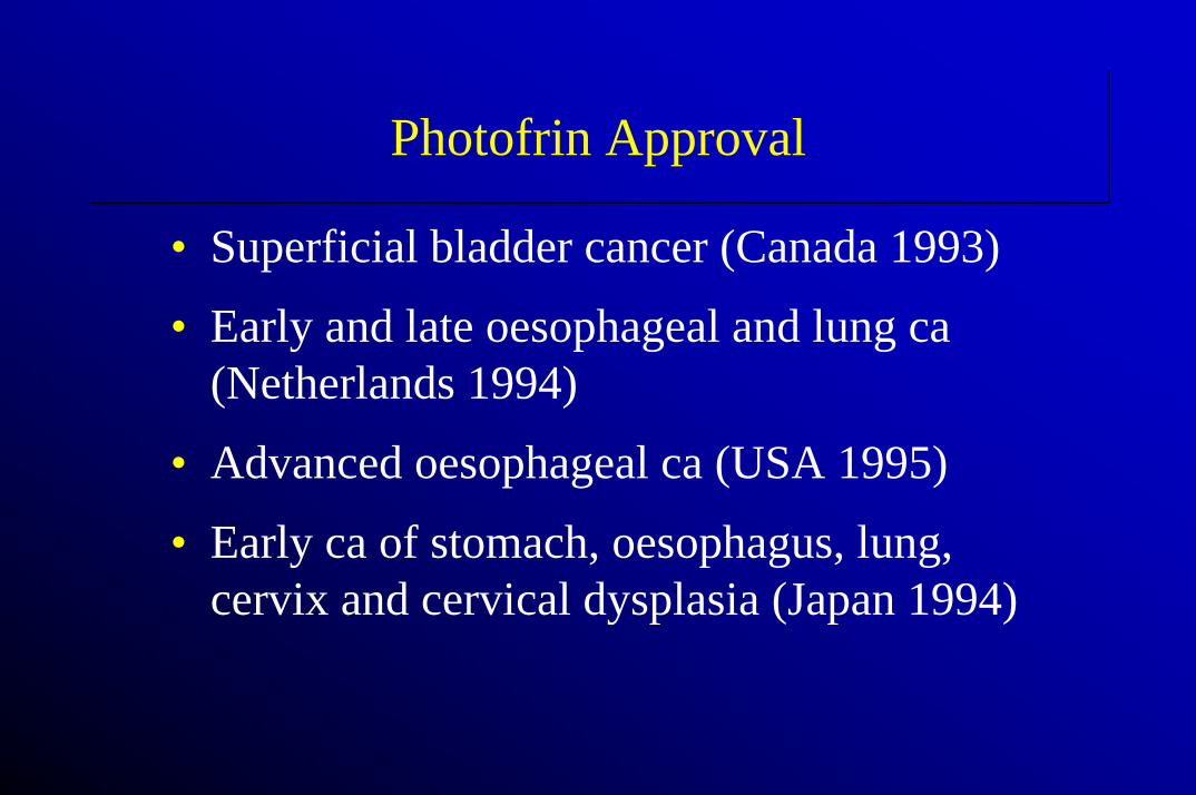
Photofrin ApprovalPhotofrin Approval
• Superficial bladder cancer (Canada 1993)
• Early and late oesophageal and lung ca (Netherlands 1994)
• Advanced oesophageal ca (USA 1995)
• Early ca of stomach, oesophagus, lung, cervix and cervical dysplasia (Japan 1994)
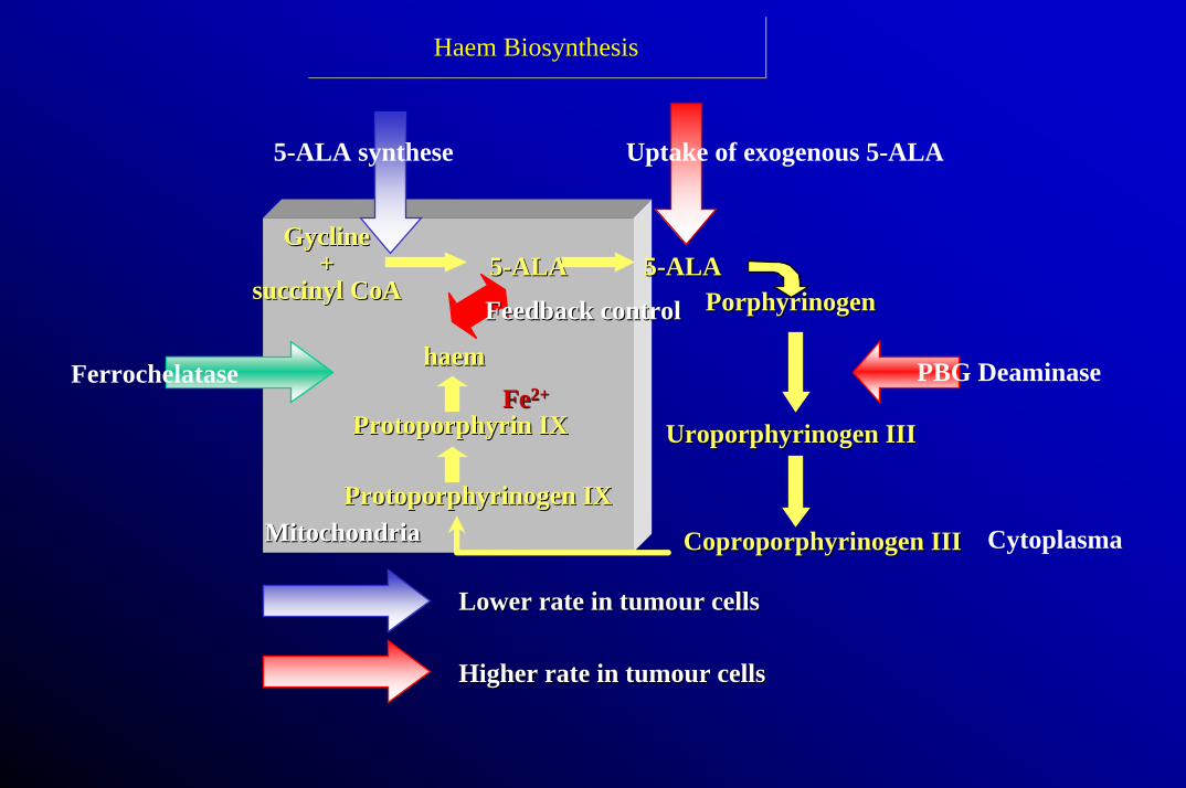
Haem BiosynthesisHaem Biosynthesis
5-ALA synthese Uptake of exogenous 5-ALA
PBG Deaminase
Cytoplasma
Lower rate in Lower rate in tumourtumour cellscells
Higher rate in Higher rate in tumourtumour cellscells
CoproporphyrinogenCoproporphyrinogen IIIIII
UroporphyrinogenUroporphyrinogen IIIIII
PorphyrinogenPorphyrinogen55--ALAALA55--ALAALA
GyclineGycline++
succinylsuccinyl CoACoA
haemhaem
ProtoporphyrinProtoporphyrin IXIX
ProtoporphyrinogenProtoporphyrinogen IXIX
Feedback controlFeedback control
MitochondriaMitochondria
FeFe2+2+Ferrochelatase
0
0.2
0.4
0.6
0.8
1
1.2
400 500 600 700 8000
0.2
0.4
0.6
0.8
1
1.2
Exc
itatio
n [a
.u.]
Fluorescence [a.u.]
Wavelength [nm]
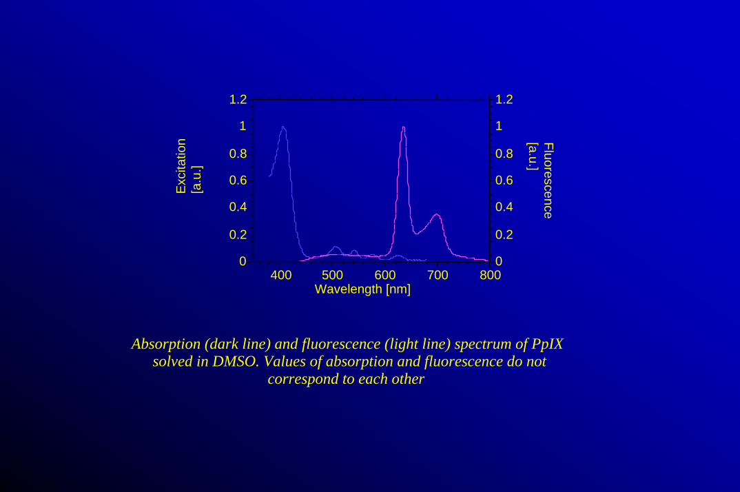
Absorption (dark line) and fluorescence (light line) spectrum of PpIXsolved in DMSO. Values of absorption and fluorescence do not
correspond to each other
]
400 600 800 1000 1200 1400 1600 1800 2000
5
4
3
2
1
0
[ mm
Pene
trat
ion
dept
h
Wavelength [ nm ]
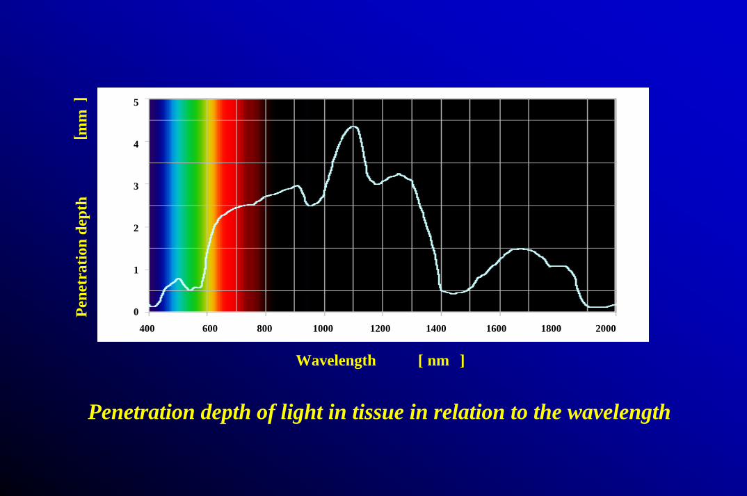
Penetration depth of light in tissue in relation to the wavelength
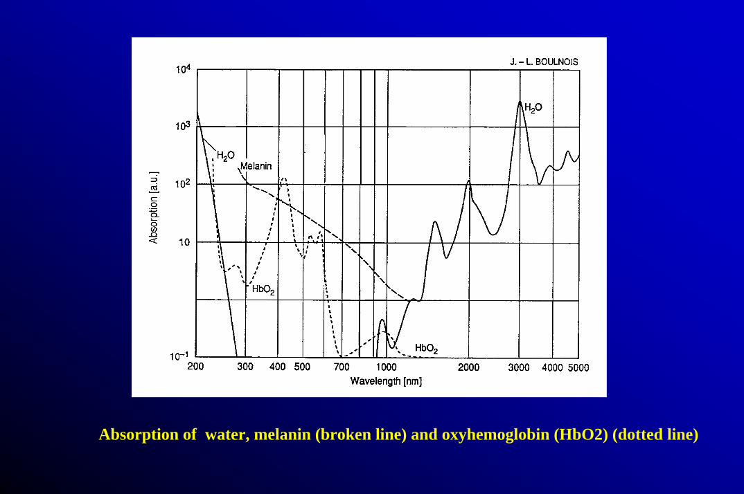
Absorption of water, melanin (broken line) and oxyhemoglobin (HbO2) (dotted line)
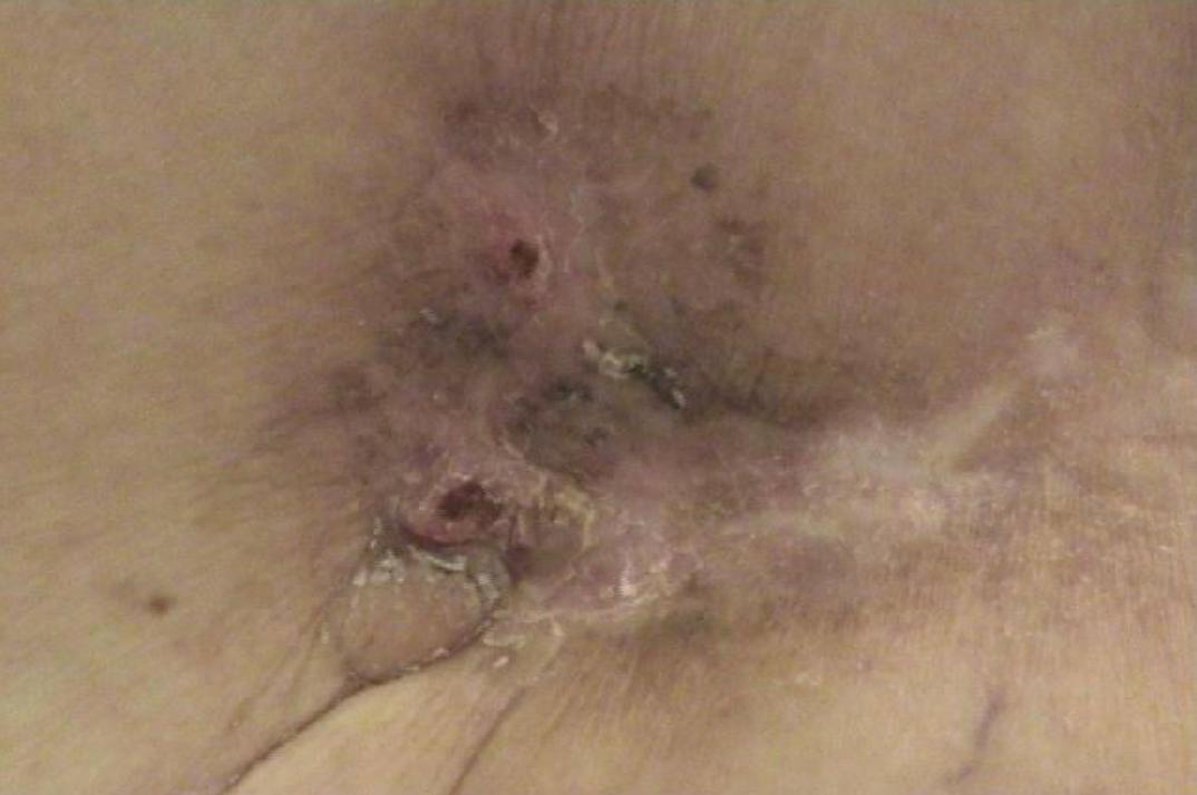
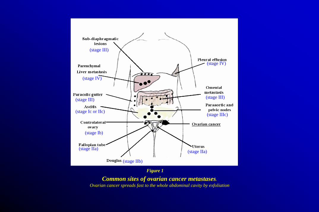
Figure 1
Common sites of ovarian cancer metastases. Ovarian cancer spreads fast to the whole abdominal cavity by exfoliation
(stage III)
(stage IV)
Parenchymal
(stage III)
(stage Ic or IIc)
(stage Ib)
(stage IIa)
(stage IIb)
(stage IIa)
(stage IIIc)
(stage III)
(stage IV)
0.00%
10.00%
20.00%
30.00%
40.00%
50.00%
60.00%
70.00%
80.00%
0-1st 0-2nd 0-3th 0-4th 0-5th
Other genital organs(vulva and vagina)
Cervix uteri
Corpus uteriBreast
Ovary
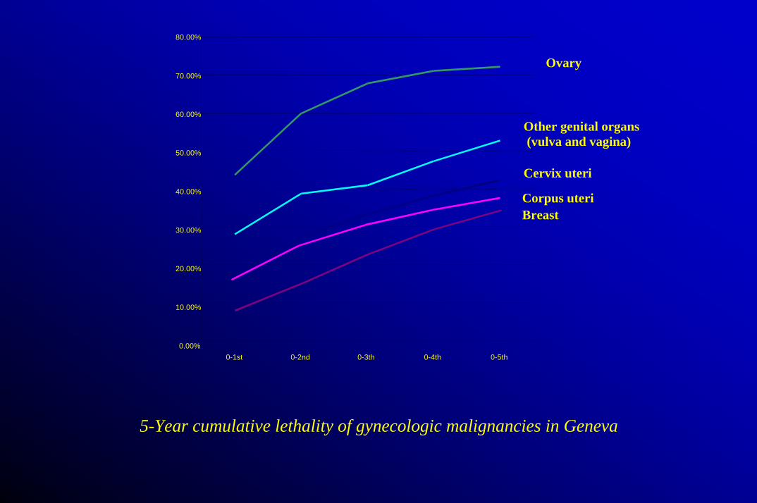
5-Year cumulative lethality of gynecologic malignancies in Geneva

Epithelial Ovarian CancerEpithelial Ovarian CancerEpithelial Ovarian Cancer
• Fourth most frequent cause of “cancer-related”death
• 65% diagnosed with stage III-IV disease• Initial response: 80% platinum sensitive• 5 year survival rate: 15-20%• Second look laparotomy
– Historically: no effect on survival• 1/3 macroscopic • 1/3 microscopic
• 1/3 negative– 50% of patients with a negative second look laparotomy
will recur
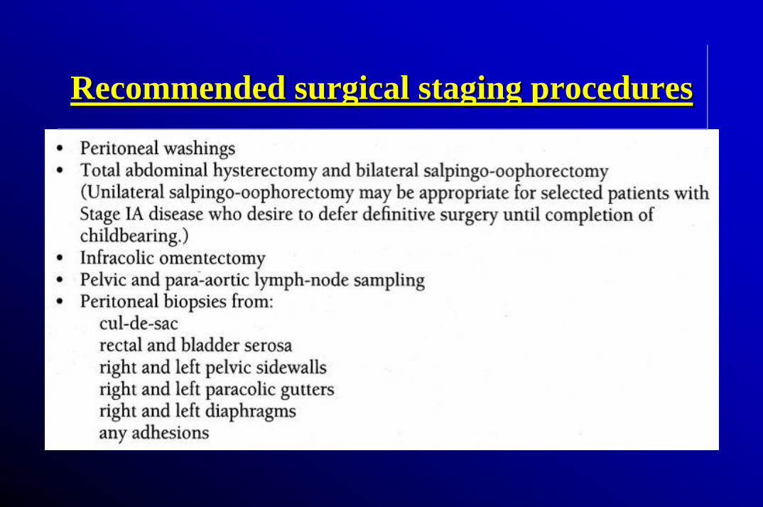
Recommended surgical staging proceduresRecommended surgical staging proceduresRecommended surgical staging procedures
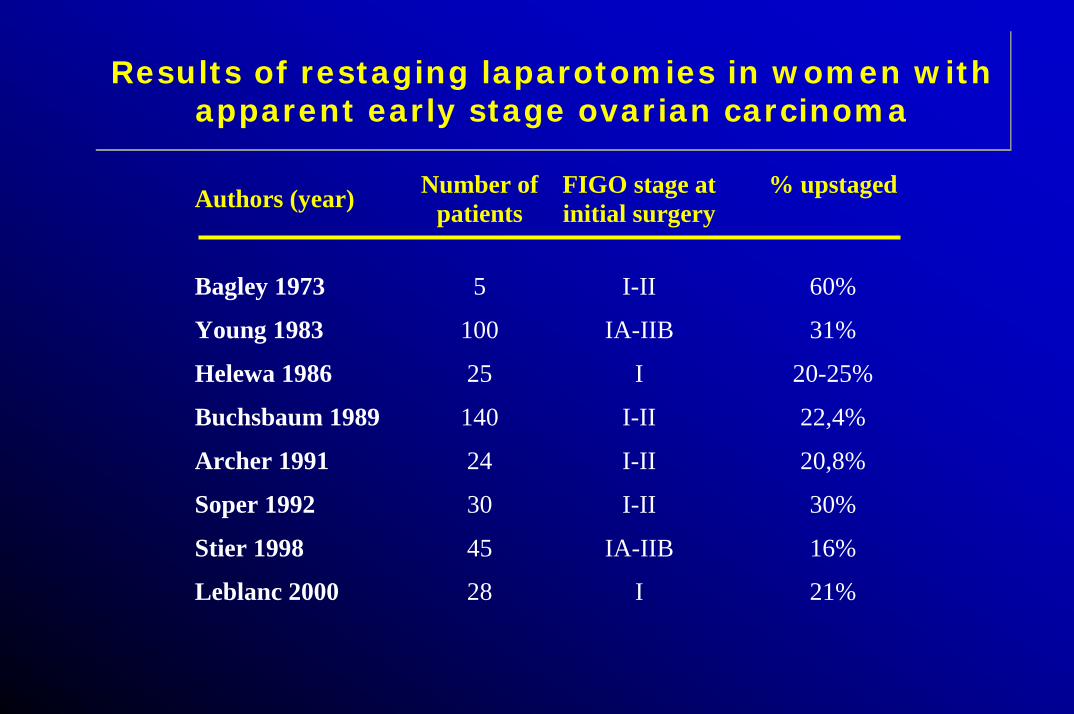
Results of restaging laparotomies in women with apparent early stage ovarian carcinoma
Results of restaging laparotomies in women with apparent early stage ovarian carcinoma
Authors (year) Number ofpatients
FIGO stage atinitial surgery
% upstaged
Bagley 1973 5 I-II 60%
Young 1983 100 IA-IIB 31%
Helewa 1986 25 I 20-25%
Buchsbaum 1989 140 I-II 22,4%
Archer 1991 24 I-II 20,8%
Soper 1992 30 I-II 30%
Stier 1998 45 IA-IIB 16%
Leblanc 2000 28 I 21%
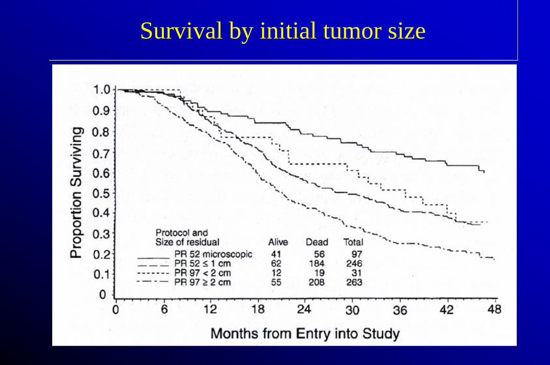
Survival by initial tumor sizeSurvival by initial tumor size
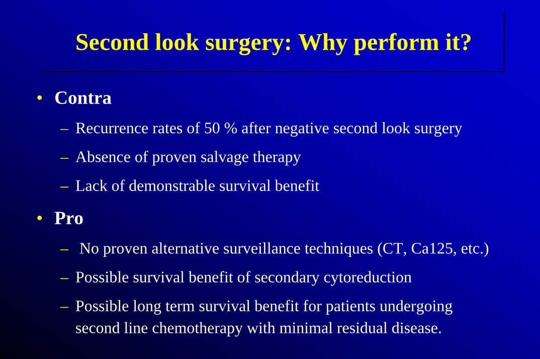
Second look surgery: Why perform it?Second look surgery: Why perform it?
• Contra– Recurrence rates of 50 % after negative second look surgery
– Absence of proven salvage therapy
– Lack of demonstrable survival benefit
• Pro– No proven alternative surveillance techniques (CT, Ca125, etc.)
– Possible survival benefit of secondary cytoreduction
– Possible long term survival benefit for patients undergoing second line chemotherapy with minimal residual disease.
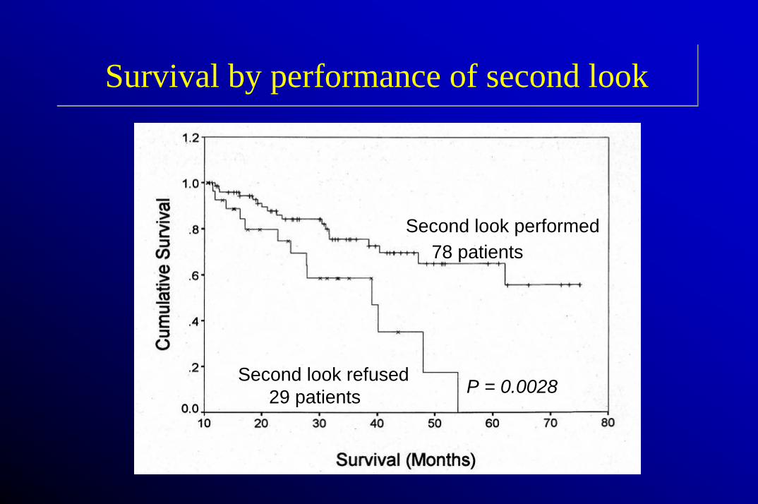
Survival by performance of second lookSurvival by performance of second look
Second look performed78 patients
Second look refused29 patients P = 0.0028P = 0.0028
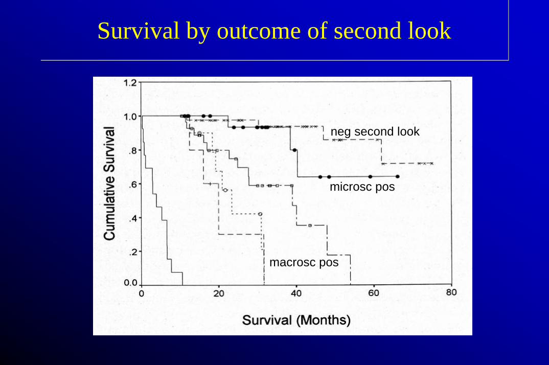
Survival by outcome of second lookSurvival by outcome of second look
neg second look
microsc pos
macrosc pos
Potential of In Vivo FluorescencePotential of In Vivo Fluorescence
• Staging laparotomy– 30% upstaged (Young RC, JAMA, 1983;
Zanetta G, Ann Oncol, 1998)• Second Look
– 50% recurrence of negative second-look after combination chemotherapy (DiSaia PJ, Mosby-Year Book, 1997)
AIMSAIMS
• To evaluate photodetection of ovarian cancer peritoneal implants in the animal model
• To study pharmacokinetics of the photosensitizerprecursor aminolevulinic acid (ALA)
• To evaluate photodetection of ovarian cancer peritoneal implants in patients
• To analyse toxicity of ALA photodynamic therapy(PDT) in the animal model
Enhanced diagnosis through photodetectionEnhanced diagnosis through photodetection
• Photodetection of ovarian cancer peritoneal implants in the animal model
• Determination of the best Photosensitizer
• Photodetection of ovarian cancer peritoneal implants in ovarian cancer patients
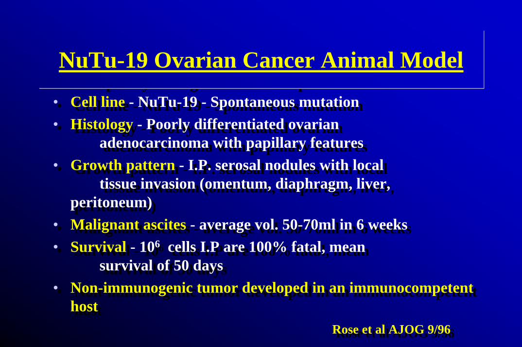
• Completely analogous to human epithelial ovarian cancer• Cell line - NuTu-19 - Spontaneous mutation• Histology - Poorly differentiated ovarian
adenocarcinoma with papillary features• Growth pattern - I.P. serosal nodules with local
tissue invasion (omentum, diaphragm, liver, peritoneum)
• Malignant ascites - average vol. 50-70ml in 6 weeks• Survival - 106 cells I.P are 100% fatal, mean
survival of 50 days• Non-immunogenic tumor developed in an immunocompetent
hostRose et al AJOG 9/96
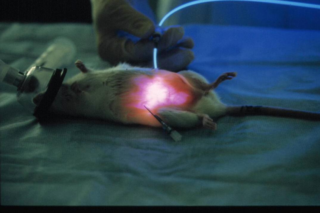
NuTu-19 Ovarian Cancer Animal ModelNuTu-19 Ovarian Cancer Animal Model

A
B
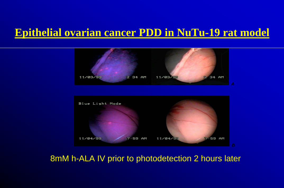
8mM h-ALA IV prior to photodetection 2 hours later
Epithelial ovarian cancer PDD in NuTu-19 rat modelEpithelial ovarian cancer PDD in NuTu-19 rat model
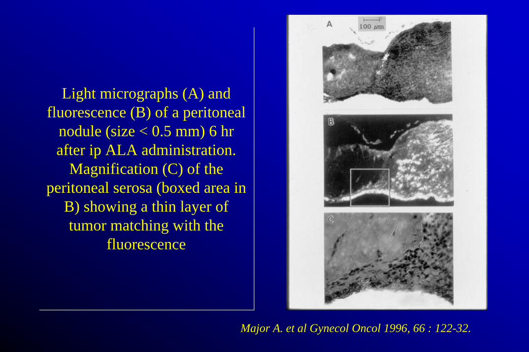
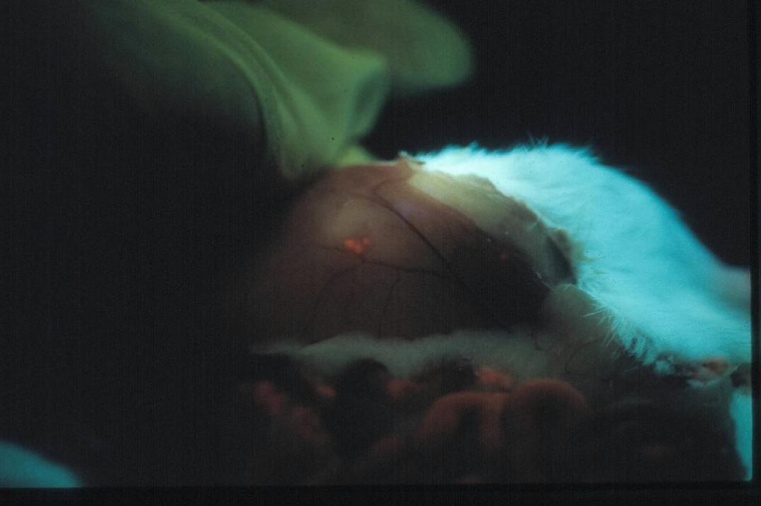
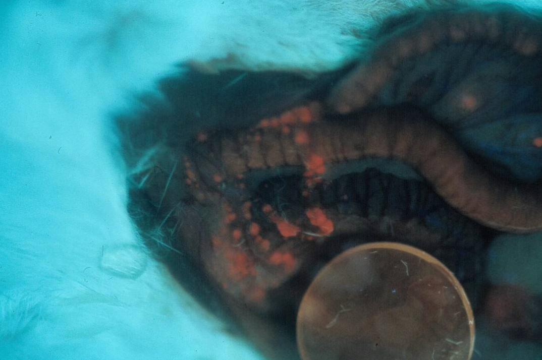
Light micrographs (A) and fluorescence (B) of a peritoneal
nodule (size < 0.5 mm) 6 hr after ip ALA administration.
Magnification (C) of the peritoneal serosa (boxed area in
B) showing a thin layer of tumor matching with the
fluorescence
Light micrographs (A) and fluorescence (B) of a peritoneal
nodule (size < 0.5 mm) 6 hr after ip ALA administration.
Magnification (C) of the peritoneal serosa (boxed area in
B) showing a thin layer of tumor matching with the
fluorescence
Major A. et al Gynecol Oncol 1996, 66 : 122-32.
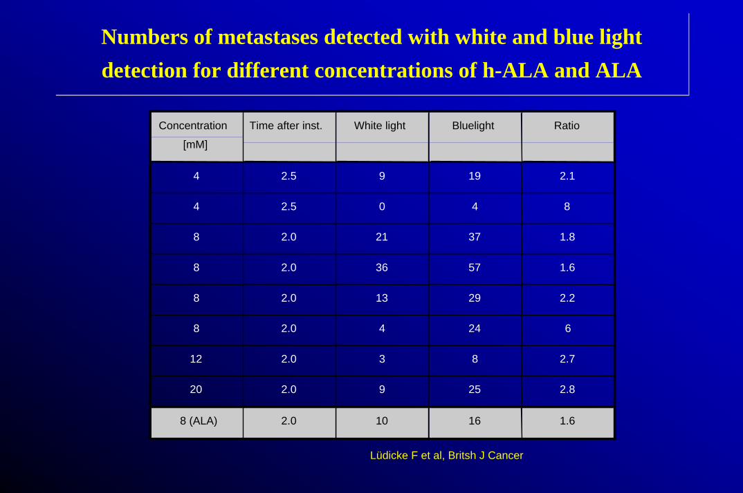
Numbers of metastases detected with white and blue light detection for different concentrations of h-ALA and ALANumbers of metastases detected with white and blue light detection for different concentrations of h-ALA and ALA
Concentration
[mM]
Time after inst. White light Bluelight Ratio
4 2.5 9 19 2.1
4 2.5 0 4 8
8 2.0 21 37 1.8
8 2.0 36 57 1.6
8 2.0 13 29 2.2
8 2.0 4 24 6
12 2.0 3 8 2.7
20 2.0 9 25 2.8
8 (ALA) 2.0 10 16 1.6
Lüdicke F et al, Britsh J Cancer
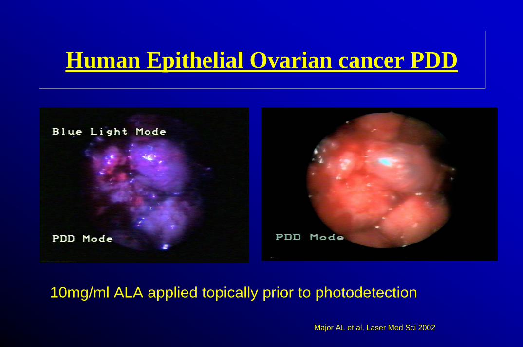
Human Epithelial Ovarian cancer PDDHuman Epithelial Ovarian cancer PDD
10mg/ml ALA applied topically prior to photodetection
Major AL et al, Laser Med Sci 2002
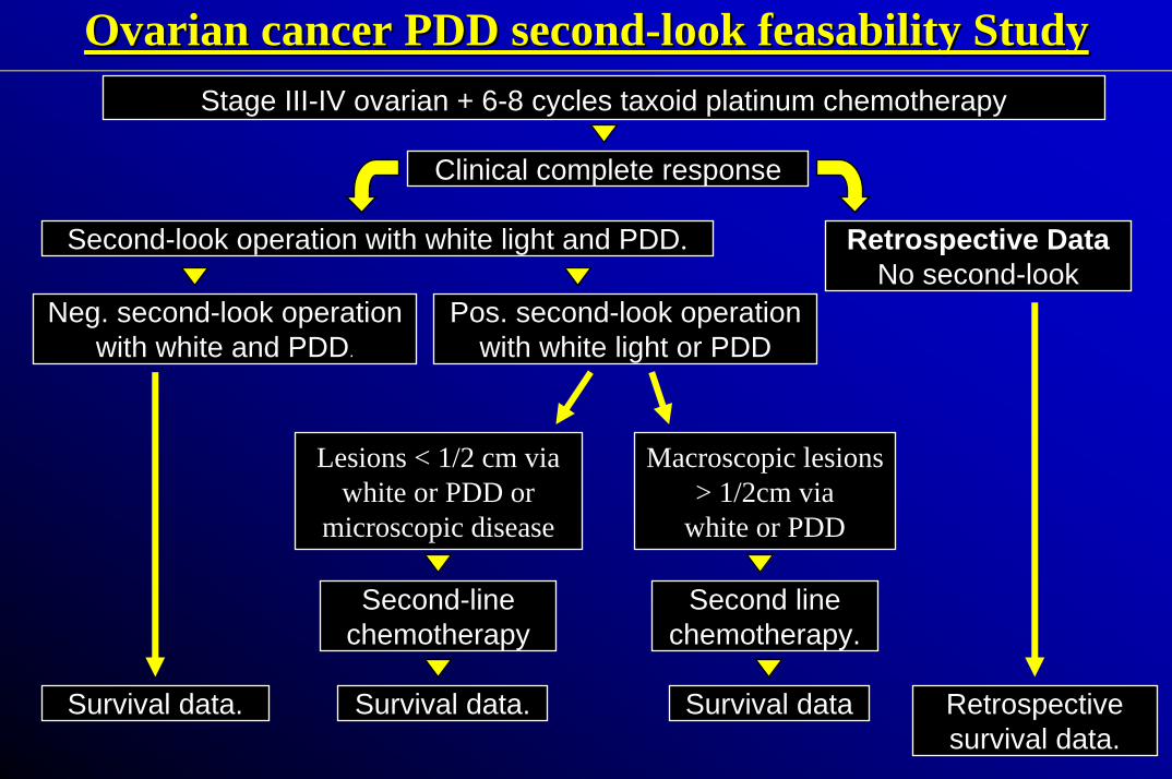
Stage III-IV ovarian + 6-8 cycles taxoid platinum chemotherapy
Second-look operation with white light and PDD.
Neg. second-look operationwith white and PDD.
Retrospectivesurvival data.
Retrospective DataNo second-look
Second-linechemotherapy
Survival data.
Clinical complete response
Survival data. Survival data
Second linechemotherapy.
Pos. second-look operationwith white light or PDD
Lesions < 1/2 cm via white or PDD or
microscopic disease
Macroscopic lesions > 1/2cm via
white or PDD
Ovarian cancer PDD second-look feasability StudyOvarian cancer PDD secondOvarian cancer PDD second--look look feasabilityfeasability StudyStudy
CONCLUSIONSCONCLUSIONS
• Photodetection has been shown to be efficient in the animal model and feasible in patients
• Photodetection of ovarian cancer peritoneal implants, not visible by other methods, is a conceivable goal for the future
• The impact on survival has to be demonstrated in further studies
“The facts remains that a large number of patients are being treated almost to the point of “cure” and an additional stroke of some sort is needed.”(DiSaia, Clinical Gynecological Oncology, Mosby-Year Book, 1997)
“The facts remains that a large number of patients are being treated almost to the point of “cure” and an additional stroke of some sort is needed.”(DiSaia, Clinical Gynecological Oncology, Mosby-Year Book, 1997)
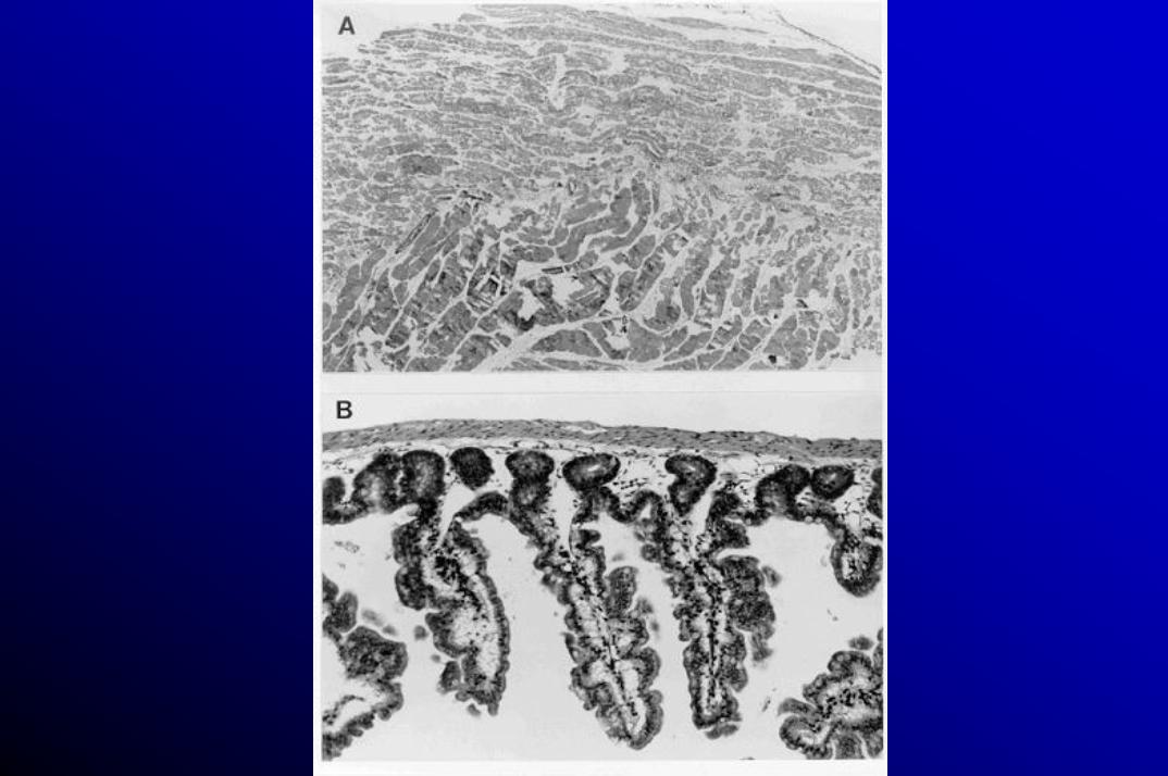
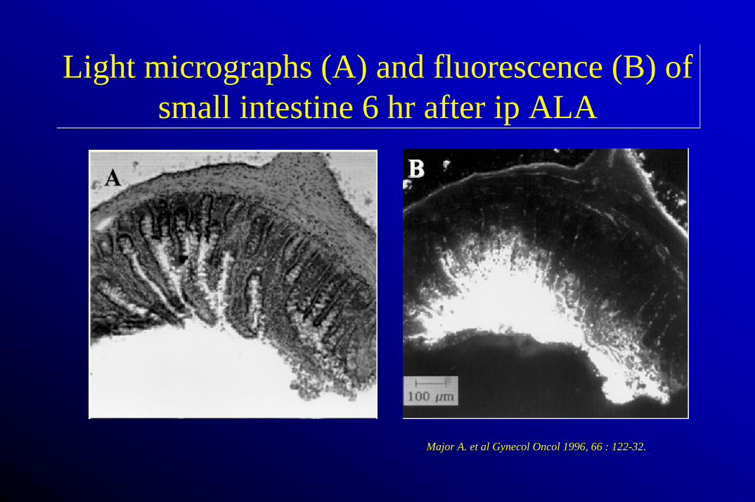
Light micrographs (A) and fluorescence (B) of small intestine 6 hr after ip ALA
Light micrographs (A) and fluorescence (B) of small intestine 6 hr after ip ALA
Major A. et al Gynecol Oncol 1996, 66 : 122-32.
AIMSAIMS
• Proof of principle of gene based photodynamic therapy of the peritoneal cavity after IP administration of ALA-S virus (establishment of a stable NuTu 19 ALA-S mutant cell line)
Problems in gene therapyProblems in gene therapy
• Transfection, transduction rate• Side effects• Tissue penetration• Immune reaction• Specificity
ResultsResults
• Pp IX production in the NuTu-19 ovarian cancer cell line after ALA and ALA-S mutant adenovirus application
• Toxicity (cell killing) of ALA, ALA-S virus and LacZ adenovirus in NuTu-19 cells
• Transduction rate of GFP adenovirus (CMV) in NuTu cells and in control cells (293T)
PerspectivePerspective
• Proof of efficient photodynamic therapy in the animal model after I.P ALA-S virus administration, impact on survival
• Increase transduction rate
• Achieve cancer specific expression of the transgene
PDT of cervical intraepithelial neoplasia
•Rationale•Introduction•Study design•Material and Methods•Results
Aim
•Determine if h-ALA is selectively absorbed by dysplastic cells at various times after topical application (5min to 7 hours)
Rationale
•Increasing incidence of cervical precancerous lesions in younger women•Treatment pf precancer of the cervix (conization) is an invasive procedure with its peri-operative risk and potential long term risk for fertility
Treatment of CIN: Excisional methods
•Cold knife conisation•Loop electrocurgical excision procedure (LEEP)
Treatment of CIN: Ablative methods
•Cryotherapy•Laser vaporisation•Photodynamic therapy
Advantages to treat CIN with PDT
•Outpatient clinic•Specificity (drug, light)•Tailored to the shape of the cervix•No stromal destruction (stenosis, cervix insufficiency)•Cell death by apoptosis (no inflammation, no scaring)•Specific HPV destruction (tetrapyrrol)•Repeatable
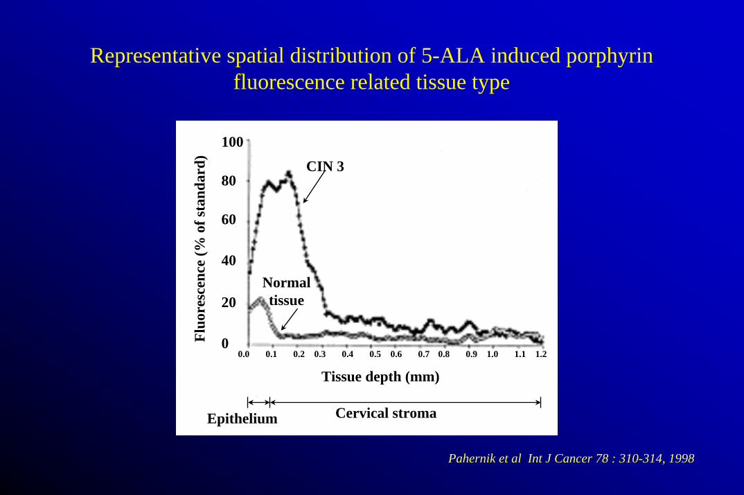
Representative spatial distribution of 5-ALA induced porphyrinfluorescence related tissue type
100
80
60
40
20
00.0 0.1 0.2 0.3 0.4 0.5 0.6 0.7 0.8 0.9 1.0 1.1 1.2
Tissue depth (mm)
Fluo
resc
ence
(% o
f sta
ndar
d) CIN 3
Normal tissue
Cervical stromaEpithelium
Pahernik et al Int J Cancer 78 : 310-314, 1998
Material and methods
•Phase I clinical trial involving 30 non-pregnant women with already biopsy-proven CIN 1-3•Application of a 0.5 % h-ALA•Random biopsies at time points ranging from 5min to 7 hours•Image analysis on frozen tissue sections using ZeissAxiophot image analysis system
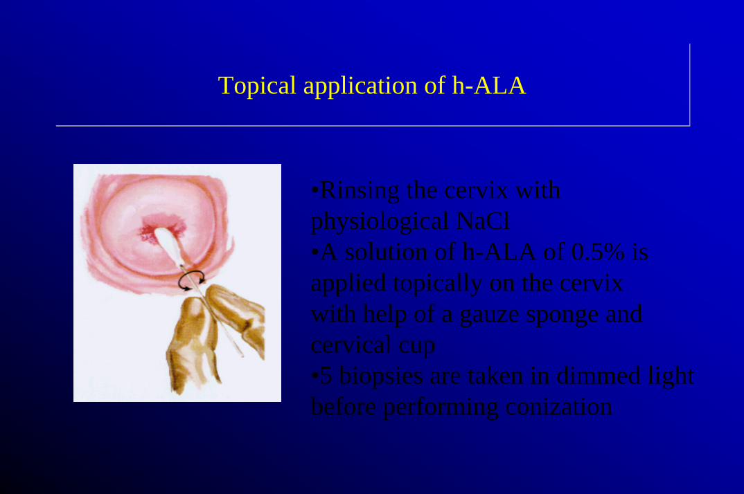
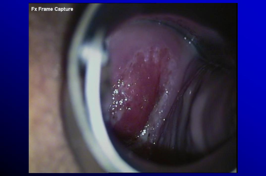
Topical application of h-ALATopical application of h-ALA
•Rinsing the cervix with physiological NaCl•A solution of h-ALA of 0.5% is applied topically on the cervixwith help of a gauze sponge and cervical cup•5 biopsies are taken in dimmed light before performing conization
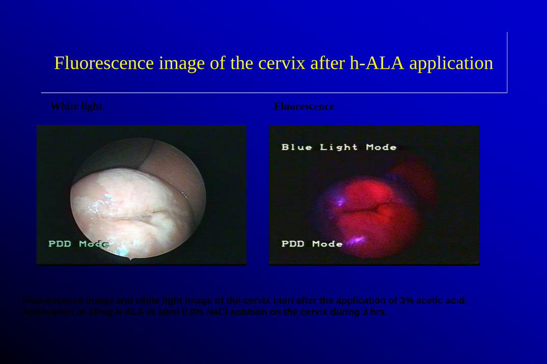
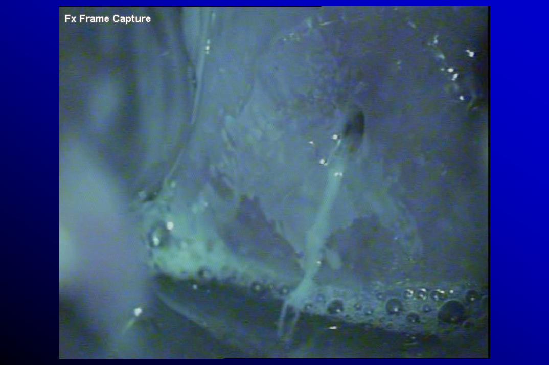
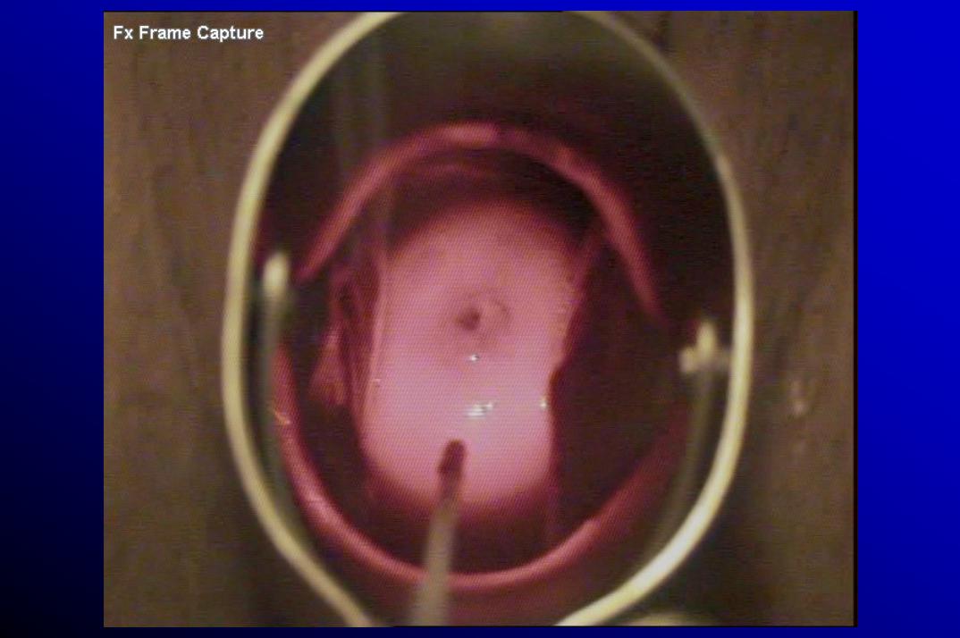
Fluorescence image of the cervix after h-ALA applicationFluorescence image of the cervix after h-ALA application
White light Fluorescence
Fluorescence image and white light image of the cervix uteri after the application of 3% acetic acid. Application of 10mg h-ALA in 10ml 0.9% NaCl solution on the cervix during 3 hrs.
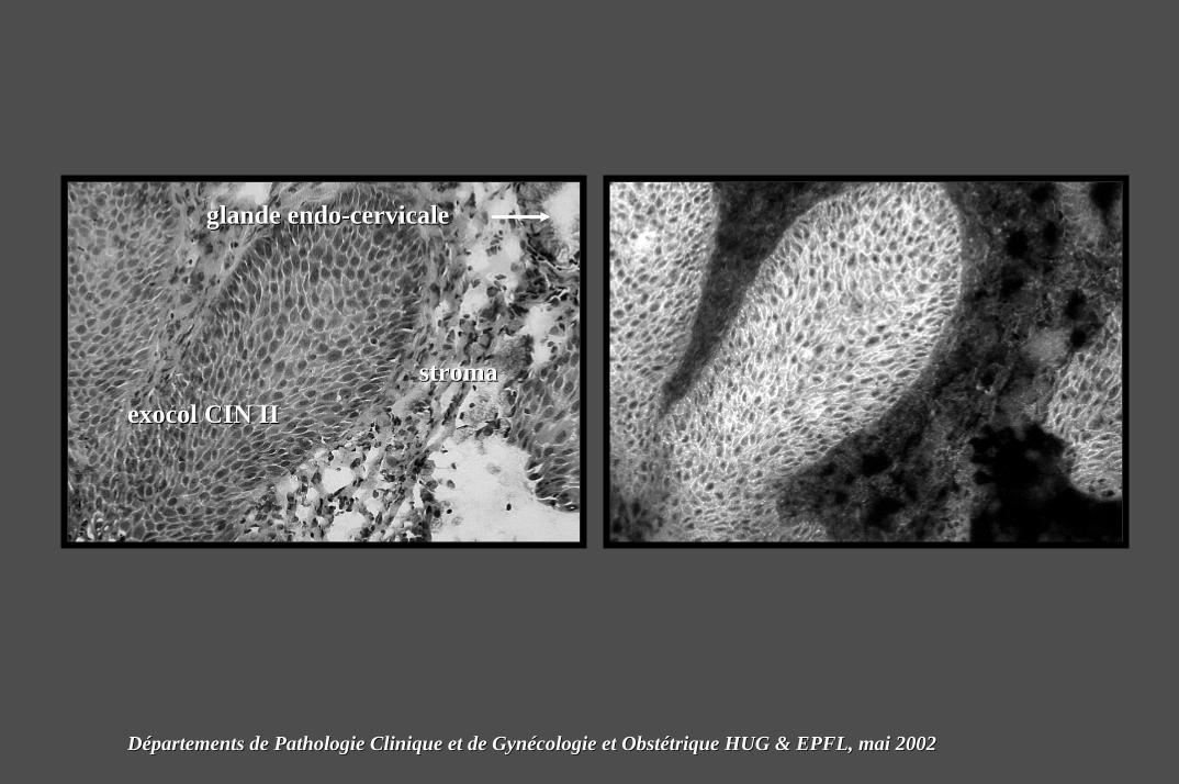
exocolexocol CIN IICIN II
glande glande endoendo--cervicalecervicale
stromastroma
DDéépartements de Pathologie Clinique et de Gynpartements de Pathologie Clinique et de Gynéécologie et Obstcologie et Obstéétrique HUG & EPFL, mai 2002trique HUG & EPFL, mai 2002
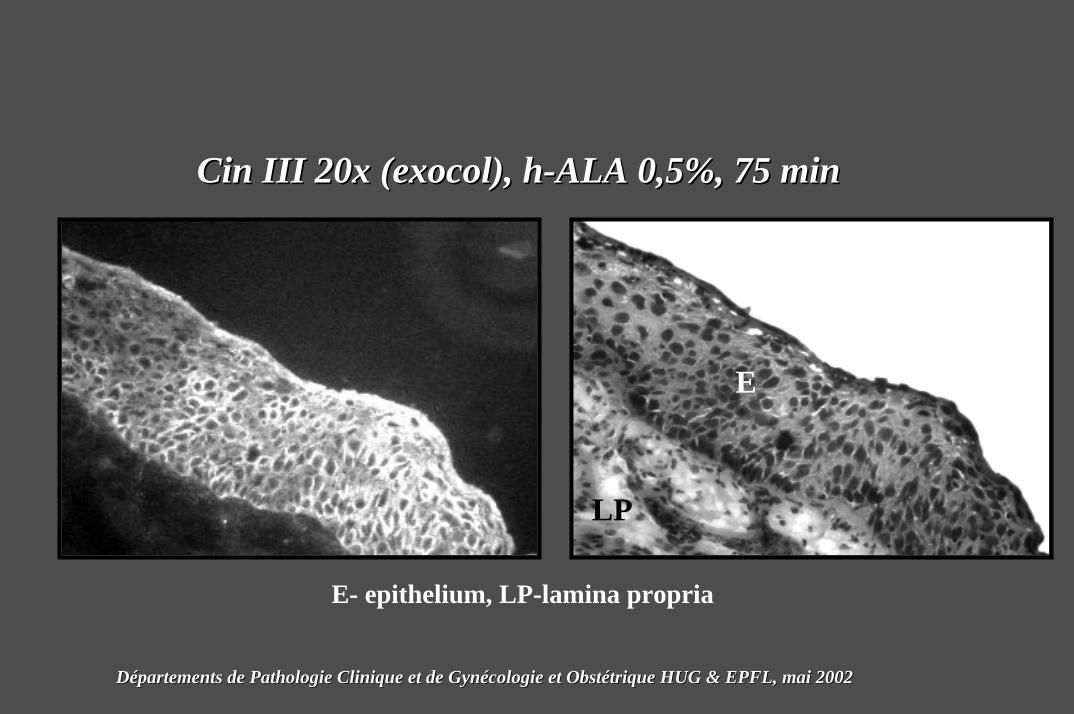
CinCin IIIIII 20x (20x (exocolexocol), ), hh--ALAALA 0,5%, 75 min0,5%, 75 min
E
LP
E- epithelium, LP-lamina propria
DDéépartements de Pathologie Clinique et de Gynpartements de Pathologie Clinique et de Gynéécologie et Obstcologie et Obstéétrique HUG & EPFL, mai 2002trique HUG & EPFL, mai 2002
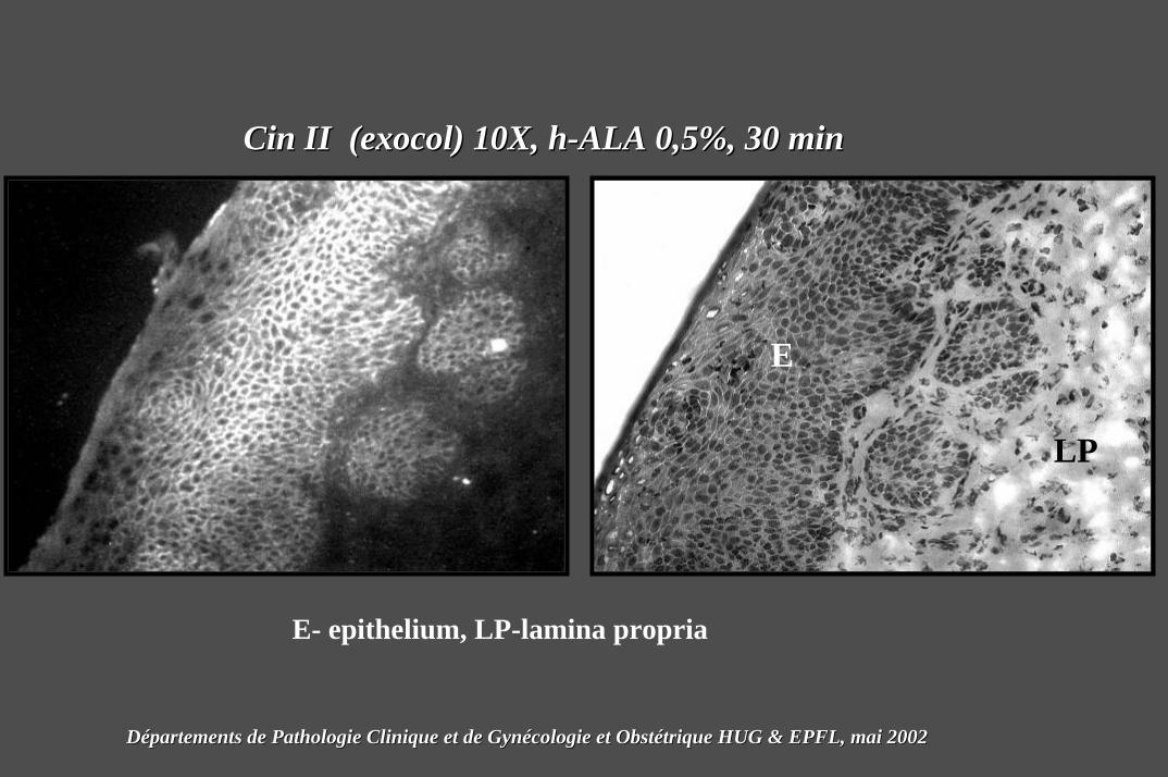
CinCin II (II (exocolexocol) 10X, ) 10X, hh--ALAALA 0,5%, 30 min0,5%, 30 min
E
LP
E- epithelium, LP-lamina propria
DDéépartements de Pathologie Clinique et de Gynpartements de Pathologie Clinique et de Gynéécologie et Obstcologie et Obstéétrique HUG & EPFL, mai 2002trique HUG & EPFL, mai 2002
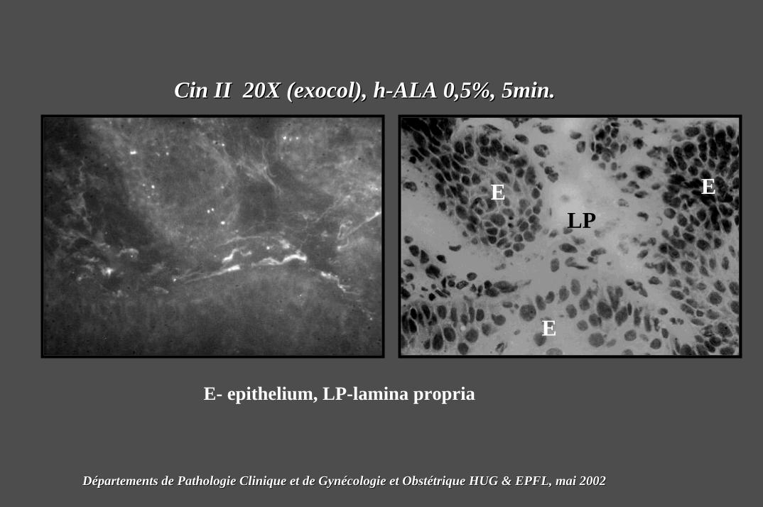
CinCin II 20X (II 20X (exocolexocol), ), hh--ALAALA 0,5%, 5min.0,5%, 5min.
E E
E
LP
E- epithelium, LP-lamina propria
DDéépartements de Pathologie Clinique et de Gynpartements de Pathologie Clinique et de Gynéécologie et Obstcologie et Obstéétrique HUG & EPFL, mai 2002trique HUG & EPFL, mai 2002
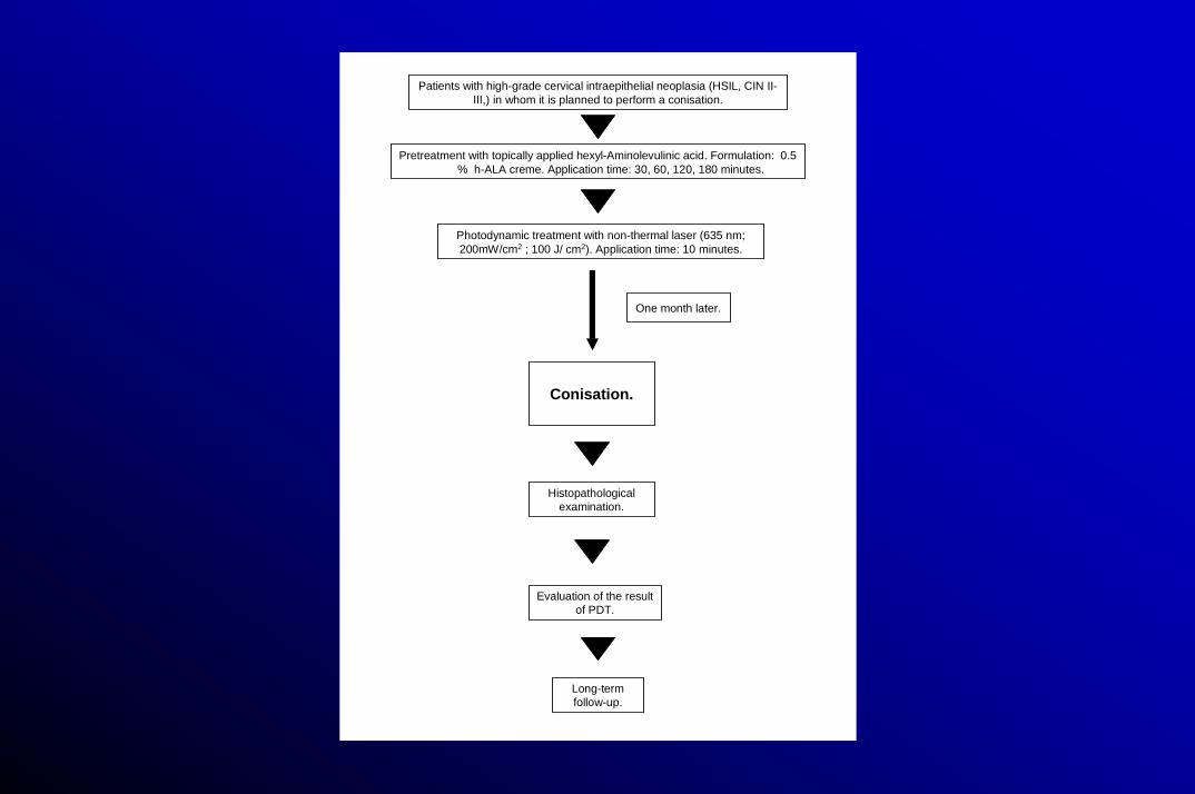
Patients with high-grade cervical intraepithelial neoplasia (HSIL, CIN II-III,) in whom it is planned to perform a conisation.
Pretreatment with topically applied hexyl-Aminolevulinic acid. Formulation: 0.5 % h-ALA creme. Application time: 30, 60, 120, 180 minutes.
Conisation.
Histopathological examination.
One month later.
Long-term follow-up.
Photodynamic treatment with non-thermal laser (635 nm; 200mW/cm2 ; 100 J/ cm2). Application time: 10 minutes.
Evaluation of the result of PDT.

Hubert van den Bergh
Georges Wagnières
Norbert LangeDidier GoujonTanja GabrechtThomas Stepinac
Attila L. Major
Frank LüdickeIvanna Mayboroda
Anis Feki