Embed Size (px)
Citation preview

Photosynthesis


Examples of photosynthetic organisms:
Leaves from higher plants flanked by colonies of
photosynthetic purple bacteria (left)
and
cyanobacteria (right)

plant cell
TEM
Chloroplasts are the organelles within plant and algal cells where photosynthesis occurs.The oxidation-reduction reactions involved in water oxidation, NADP+ reduction and synthesis of ATP via a chemiosmotic mechanism are located in the thylakoid membranes of the chloroplast.
The soluble enzymes involved in the synthesis of glucose from carbon dioxide and water, and utilizing the ATP and NADPH produced during the light reactions are in the stroma compartment of the chloroplast.

chloroplasts run on sunlightproducing sugars from carbon dioxide
thylakoid membrane
chloroplast
inner membranes (thylakoids)
TEM
grana stacks
stroma
To transduce light energy the photosynthetic membrane functions as a vesicle, with an inner (lumen) and outer (stromal) water phase

mitochondria run on coal (sugar)generation of energy in the form of adenosine triphosphate (ATP) by oxidative phosphorylation
mitochondrion
TEM
inner mitochondrial membrane
ATP production is a result of electron transportthe mitochondrion has been identified as playing a central role in apoptosis (cell death)

Light EnergyLight energy is absorbed by pigments in the thylakoids, and converted into forms of energy that are
used to synthesize glucose.
Light energy is a form of electromagnetic radiation.
Some properties are particle-like; some are wave-like.
Photon is term for a packet of light energy.
Energy is inversely proportional to wavelength.

Light HarvestingOverview of light harvesting
Molecules must absorb in a region of the spectrum where light reaches the surface of the
planet with sufficient intensity. Since CO2 and water vapor absorb much of the far IR, and oxygen
and ozone absorb all the far UV, this limits the range to the visible and near IR.
The light must have sufficient energy for useful chemistry,
< 1000 nm (the near IR),
but not be destructive
> 340 nm (the near UV).
For energy transfer, the molecules must:
the right distance apart
appropriately aligned
overlapping spectra for fluorescence and absorption

Light HarvestingOverview of light harvesting
For efficient energy transfer, the system is usually organized with a thermodynamic gradient from antenna to reaction center - “energy funnel".
The peripheral antenna absorbs light of higher energy (to the blue end of the spectrum), and transfers excitation to pigments with lower energy absorbance bands (further to the red), with the photochemical trap (the reaction center) at the lowest energy (furthest to the red).
Some photosynthetic systems compete for light, so an organism is at an advantage if it can find a niche in the spectrum underused by its competitors.
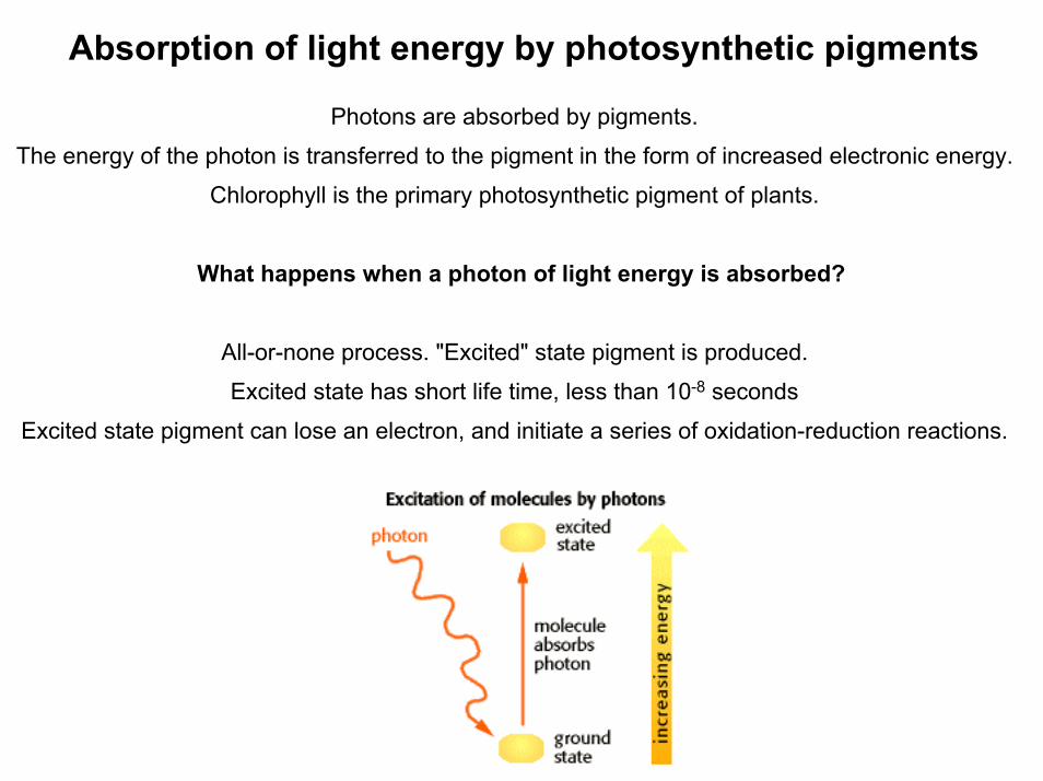
Absorption of light energy by photosynthetic pigments
Photons are absorbed by pigments.
The energy of the photon is transferred to the pigment in the form of increased electronic energy.
Chlorophyll is the primary photosynthetic pigment of plants.
What happens when a photon of light energy is absorbed?
All-or-none process. "Excited" state pigment is produced.
Excited state has short life time, less than 10-8 seconds
Excited state pigment can lose an electron, and initiate a series of oxidation-reduction reactions.

Light HarvestingFluorescence and light harvesting
Fluorescence from photosynthetic systems varies with the physiological state, and measurement of fluorescence is much used as a tool for exploring mechanism.

Fluorescencein photosynthesis
Pathways for loss of excitation from excited chlorophyll
1) Energy transfer to neighboring Chl of the same protein; rate constant ke1
2) Energy transfer to neighboring Chl of different protein; rate constant ke2
3) Fluorescence decay; rate constant kf
4) Non-radiative decay (loss as heat); rate constant kh
5) Formation of triplet state; rate constant kt
6) Energy transfer to an open photochemical trap, and photochemical conversion; rate constant kp

6) Energy transfer to an open photochemical trap, and photochemical conversion; rate constant kp
Energy level diagrams of Chl b and Chl a, indicating energy transfer from the lowest singlet excited state of Chl b to that of Chl a.
This transfer has a high probability because the process is "down-hill"

6) Electron transfer to an open photochemical trap, and photochemical conversion; rate constant kp
The reducing power of the electron is increased approximately by 1 volt.
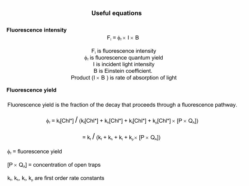
Useful equations
Fluorescence intensity
Fluorescence yield
FI = φf × I × B
FI is fluorescence intensityφf is fluorescence quantum yield
I is incident light intensityB is Einstein coefficient.
Product (I × B ) is rate of absorption of light
Fluorescence yield is the fraction of the decay that proceeds through a fluorescence pathway.
φf = kf[Chl*] / (kf[Chl*] + kh[Chl*] + kt[Chl*] + kp[Chl*] × [P × QA])
= kf / (kf + kh + kt + kp × [P × QA])
φf = fluorescence yield
[P × QA] = concentration of open traps
kf, kh, kt, kp are first order rate constants

Intrinsic fluorescence lifetime
is the lifetime fluorescence would have if there were no other competing processes.
τo = 1 / kf or kf = 1 / τo
Since probabilities of absorption and emission are related by the factor 8πhcν3,
1 / τo ~ 2.9 × 10⁻⁹ ⟨ν2⟩av ∫ ε × dν
ν is the wavenumber (cm-1) for the transition ⟨ν2⟩av is the mean wavenumber of the absorption band ε is the molar extinction coefficient∫ ε × dν is the area under the absorption band plotted as ε vs. ν.
Actual fluorescence lifetime
The actual or measured lifetime τ, is determined by all the decay pathways:
τ = 1 / k'
- d[Chl*] / dt = (kf + kh + kt + kp × [P × QA]) [Chl*]
= k' [Chl*] k' is the sum of the rate constants for all decay pathways
By substitution: φf = kf × τ = τ / τo and τ = φf × τo

The Calvin Cycle
3 ATP and 2 NADPH are required to reduce one carbon dioxide molecule to the level of hexose, a reaction with a net ∆G of +114 kcal/mol.

Light Harvesting in Bacterial Photosynthesis
Photochemistry begins at the reaction center (RC) where charges are separated.
The RC requires energy to perform, either from direct absorption of a photon, or energy transferred from a light-harvesting complex.
All purple bacteria produce a primary light-harvesting complex (LH-I).
Most purple bacteria produce a peripheral light-harvesting complex (LH-II), and some produce an additional peripheral complex (sometimes called LH-III).
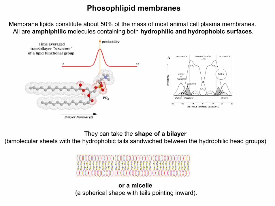
Phosophlipid membranes
Membrane lipids constitute about 50% of the mass of most animal cell plasma membranes. All are amphiphilic molecules containing both hydrophilic and hydrophobic surfaces.
They can take the shape of a bilayer(bimolecular sheets with the hydrophobic tails sandwiched between the hydrophilic head groups)
or a micelle(a spherical shape with tails pointing inward).

Membrane Protein Structural Motifs: Basic Principles.
A thermodynamic principle underlies the structure and stability of membrane proteins:The thermodynamic cost of transferring charged or highly polar uncharged compounds into the
oil-like hydrocarbon interior of bilayer membranes is very high.
Most of the amino acid sidechains of transmembrane segments must be non-polar(e.g. Ala, Val, Leu, Ile, Phe). Also, the very polar CONH groups (peptide bonds) of the poly-
peptide backbone of transmembrane segments must participate in hydrogen bonds (H-bonds) in order to lower the cost of transferring them into the hydrocarbon interior.
This H-bonding is accomplished with alpha-helices for which all peptide bonds are H-bonded internally. It can also be accomplished with beta-sheets provided that the beta-strands
form closed structures such as the beta-barrel.

Peripheral light harvesting complex - LH-II - from the photosynthetic bacteria Rps. acidophila
protein - ribbonscofactors - stick models
Mg atoms - magenta spheres
nonamer with ninefold rotational symmetry

an alpha and beta polypeptidethree bacteriochlorophyll a (Bchl a) moleculesone rhodopin glucosideone beta-octylglucoside molecule

9 carotenoids (rhodopin glucoside)
27 Bacteriochlorophyll a

Porphyrin Chlorin Bacteriochlorin
Chlorophyll a (R = phytyl) Chlorophyll b (R = phytyl)

Bchl a is a porphyrin like molecule with an asymmetric conjugated double bond system:this results in two resonant absorption bands.
This means that the position of the observed Qy absorption band at around 850nm (B850 band) depends on the size of the splitting due to coupling. The high energy compliment of this B850
band cannot be located - due to its negligible oscillator strength.

This is not the only mechanism of shifting the absorption of a Bchl a molecule.
• In organic solvent monomeric Bchl a absorbs at 777nm, this absorption shifts with the solvent.
• The keto and acetyl oxygens on rings C and A are conjugated into the chromophore. If they accept a Hydrogen bond we can expect an absorption shift.
• Perhaps the most profound change in absorption of Bchl a is induced by the deformation. The distortions can vary from expansions of the ion site to doming, ruffling etc. of the conjugated system.

The spectrum of the complex shows two BChl peaks in the near IRat 800nm and at 850 nm
These correspond to 1 B800 BChl, and two B850 BChls.

The LH-II complex is very efficient at harvesting available light:
The spectrum neatly brackets that of green plants etc. so that the photon energy available toLH-II is maximized.
Carotenoids in the complex (extended linear molecules with conjugated double bond systems) absorb in the visible region.
The carotenoid molecules in purple bacteria have a greater number of conjugated double bondsthan those found in higher plants
In fact purple bacteria are purple due to the absorption of carotenoids rather than Bchl a!

BChl of B850 pair
BChl of B800
These cylindrical assemblies allow the B850 BChls to form a closely coupled ring in whichexcitation transfer by an exciton mechanism is very rapid.
Excitation transfer in the B800 BChl ring, and between B800 and B850 molecules, is less rapid.

Each light-harvesting complex is an oligomer formed from a subunit consisting of two or more polypeptide chains with associated pigment molecules.
The pigments used in LH complexes are Bacteriochlorophyll a and carotenoid. BChla is the primary pigment, carotenoid molecules are primarily used for photo-
protection, although they do function as additional light-harvesting pigments.
The protein matrix which supports these molecules in each of the distinct LH complexes modulates the absorption of the chromophores.
This modulation results in a downward gradient of energy levels from
LH-III - LH-II - LH-I
Energy absorbed by any molecule in this antenna array is funneled down into LH-I .
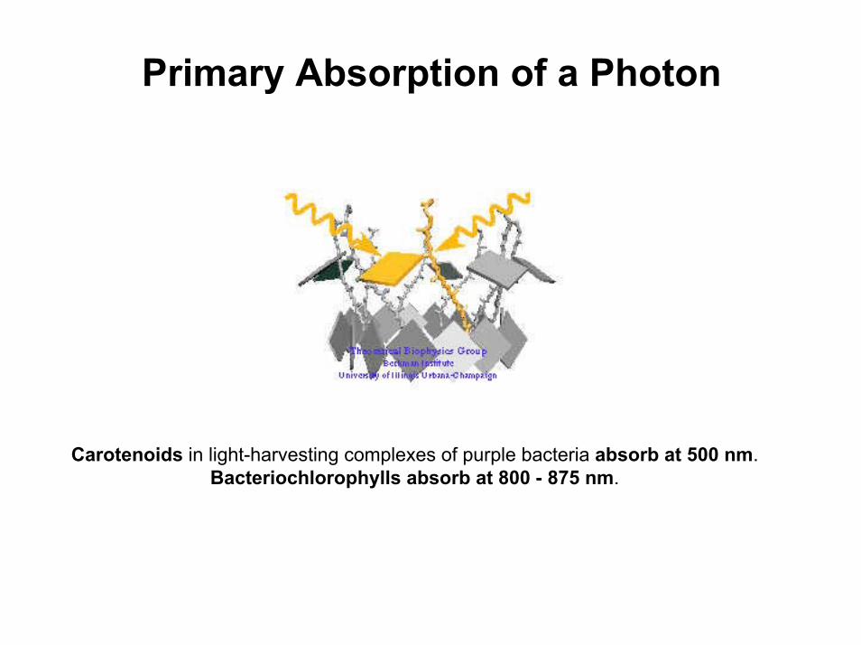
Primary Absorption of a Photon
Carotenoids in light-harvesting complexes of purple bacteria absorb at 500 nm. Bacteriochlorophylls absorb at 800 - 875 nm.

Carotenoid-Chlorophyll Excitation Transfer
Absorption of a photon is followed by rapid singlet excitation energy transfer tobacteriochlorophyll (BChl).
In addition to their light-harvesting role, carotenoids photoprotect antenna complexes
• prevent the formation of photo-oxidizing singlet oxygenby quenching BChl triplet states through triplet excitation transfer.

B800-B850 BChl Excitation Transfer
After primary absorption of a photon by either the B800 BChls or carotenoids its excitation energyis fueled into the B850 ring system.
The eight B800 BChls absorb light near 800 nm and transfer the resulting electronic excitationof the individual BChls through the Förster mechanism to the B850 BChl ring.
. Transfer proceeds within 700 femtoseconds.
Once the coupled ring of B850 BChls is excited a depolarization is observed. This suggests one of two things, the energy is either delocalized extensively, or rapidly migrates around the ring.
Extensive delocalization versus dimer-hopping

Excitation Transfer between Light-Harvesting Complexes
Once at the B850 BChl ring of LH-II the photon's excitation energy is transferred to the B875BChl ring of LH-I and from there on to the RC.
The excitation transfer occurs within 100 picoseconds and with near unit efficiency.
Time-resolved spectroscopy has revealed a 3-5 ps for the B850 -> LH-I transfer step.
The final LH-I -> RC transfer step was measured to require 35 ps, making it the slowest step.
The overall pigment organization is the coplanar arrangement of the B850 BChls of LH-II, the B875 BChls of LH-I, as well as the BChls inside the RC..

�α, β-subunit pairs
two BChls (B875)


bacteriochlorophyll aauxiliary bacteriochlorophyll - mediating electron transfercorresponding metal-free bacteriopheophytinstwo quinones (either ubiquinone or menaquinone)a non-heme Fe
cytochrome subunit

Light causes the "Special pair" of bacteriochlorophyll molecules to enter an excited state.
An electron leaves the special pair, possibly passing via an accessory bacteriochlorophyll.
This leaves a positive charge (electron hole) on the special pair.
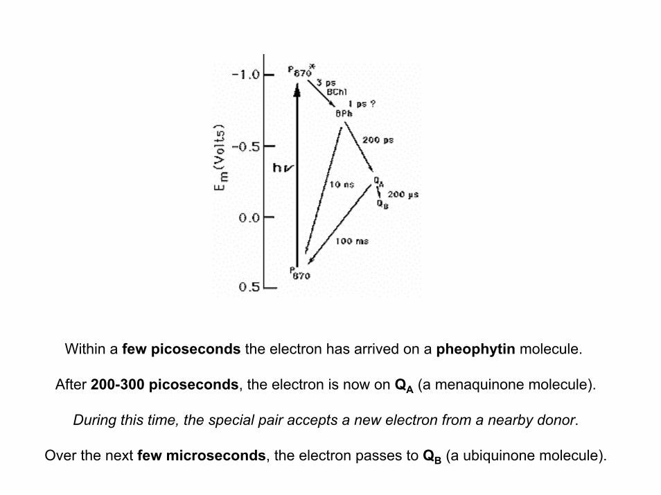
Within a few picoseconds the electron has arrived on a pheophytin molecule.
After 200-300 picoseconds, the electron is now on QA (a menaquinone molecule).
During this time, the special pair accepts a new electron from a nearby donor.
Over the next few microseconds, the electron passes to QB (a ubiquinone molecule).

Quinones
Biological transport of electrons within lipid membranes are carried out by hydrophobic quinones. They can undergo two successive one-electron reductions coupled with protonation:
Quinones act as free-diffusible electron-carriers within the lipid membrane. In mitochondrial respiration, this role is played by ubiquinone or coenzyme Q.
Other related quinones are plastoquinone (in the photosynthetic apparatus of plants) and menaquinone (in bateria).

forward electron transfer (1,2,3,4) - normal region - fastback electron transfer (6,7) - inverted region - slow

The photosynthetic apparatus of purple bacteria consists of two types of pigment-protein complexes:the reaction centers and the light-harvesting complexes.
In purple bacteria, the photosynthetic membranes contain two types of light-harvesting complexes: light harvesting complex I (LH-I) and light harvesting complex II (LH-II).
While LH-I is tightly bound to the RC, LH-II is not directly associatedwith the RC, but transfers energy to the reaction centers via LH-I.

The main function of the light-harvesting complexes is to gather light energy and to transferthis energy to the reaction centers for the photo-induced redox processes.

The role of the minor light harvesting complexes in photoprotection
In addition to their antenna function, the minor light harvesting complexes have an additional role in protection of the photosynthetic apparatus against bright light. Plants have evolved to operate most efficiently at light intensities corresponding to scattered sunlight. In full sunlight, the intensity is several-fold brighter than that necessary to saturate photosynthesis. In the absence of protective mechanisms, the photosystems get destroyed by photo-oxidative damage. Protection occursthrough a switch, which changes the function of the minor CPs from excitation transfer to exciton dumping; the energy is lost as heat rather than passed to the reaction center. The switching depends on two main factors:
1. The xanthophyll carotenoid, violaxanthin, is converted to antheraxanthin and zeaxanthin by de-epoxidation. The de-epoxidase is turned on by a fall in the lumenal pH.
2. In complexes which contain the de-epoxidation products, lowering of the lumenal pH causes the configuration of pigments to change so as to introduce the exciton dumping pathway.
The drop in lumenal pH is the feed-back signal from metabolism; as the light intensity delivers reducing power and ATP faster than the CO2-fixation reactions can handle it, the ATP synthasebacks-up, causing the ∆pH to build up, leading to the drop in lumenal pH which triggers the protective mechanism.

shorter carotenoids longer carotenoids
S2
S1
S0
protection to high energy fluxdrains out light energy