Embed Size (px)
Citation preview
HAL Id: hal-01974368https://hal.archives-ouvertes.fr/hal-01974368
Submitted on 8 Jan 2019
HAL is a multi-disciplinary open accessarchive for the deposit and dissemination of sci-entific research documents, whether they are pub-lished or not. The documents may come fromteaching and research institutions in France orabroad, or from public or private research centers.
L’archive ouverte pluridisciplinaire HAL, estdestinée au dépôt et à la diffusion de documentsscientifiques de niveau recherche, publiés ou non,émanant des établissements d’enseignement et derecherche français ou étrangers, des laboratoirespublics ou privés.
Phthalate Release from Plastic Fragments andDegradation in Seawater
Andrea Paluselli, Vincent Fauvelle, François Galgani, Richard Sempere
To cite this version:Andrea Paluselli, Vincent Fauvelle, François Galgani, Richard Sempere. Phthalate Release fromPlastic Fragments and Degradation in Seawater. Environmental Science and Technology, AmericanChemical Society, 2019, 53 (1), pp.166-175. �10.1021/acs.est.8b05083�. �hal-01974368�
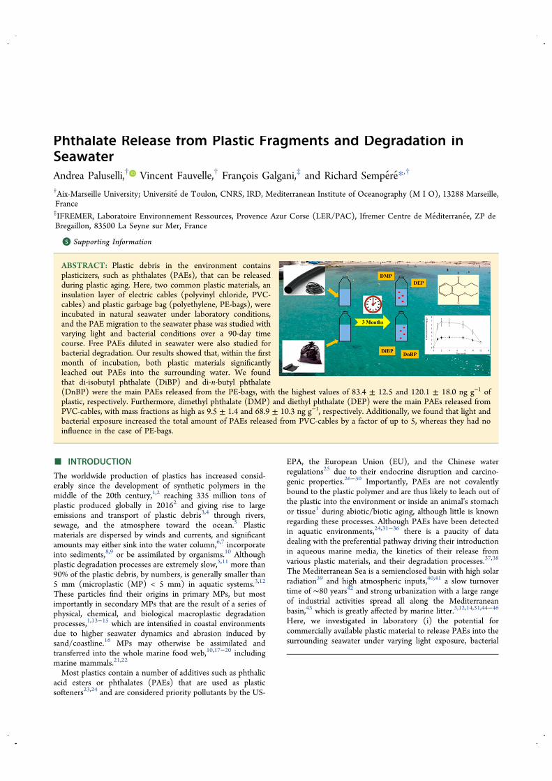
Phthalate Release from Plastic Fragments and Degradation inSeawaterAndrea Paluselli,† Vincent Fauvelle,† Francois Galgani,‡ and Richard Sempere*,†
†Aix-Marseille University; Universite de Toulon, CNRS, IRD, Mediterranean Institute of Oceanography (M I O), 13288 Marseille,France‡IFREMER, Laboratoire Environnement Ressources, Provence Azur Corse (LER/PAC), Ifremer Centre de Mediterranee, ZP deBregaillon, 83500 La Seyne sur Mer, France
*S Supporting Information
ABSTRACT: Plastic debris in the environment containsplasticizers, such as phthalates (PAEs), that can be releasedduring plastic aging. Here, two common plastic materials, aninsulation layer of electric cables (polyvinyl chloride, PVC-cables) and plastic garbage bag (polyethylene, PE-bags), wereincubated in natural seawater under laboratory conditions,and the PAE migration to the seawater phase was studied withvarying light and bacterial conditions over a 90-day timecourse. Free PAEs diluted in seawater were also studied forbacterial degradation. Our results showed that, within the firstmonth of incubation, both plastic materials significantlyleached out PAEs into the surrounding water. We foundthat di-isobutyl phthalate (DiBP) and di-n-butyl phthalate(DnBP) were the main PAEs released from the PE-bags, with the highest values of 83.4 ± 12.5 and 120.1 ± 18.0 ng g−1 ofplastic, respectively. Furthermore, dimethyl phthalate (DMP) and diethyl phthalate (DEP) were the main PAEs released fromPVC-cables, with mass fractions as high as 9.5 ± 1.4 and 68.9 ± 10.3 ng g−1, respectively. Additionally, we found that light andbacterial exposure increased the total amount of PAEs released from PVC-cables by a factor of up to 5, whereas they had noinfluence in the case of PE-bags.
■ INTRODUCTIONThe worldwide production of plastics has increased consid-erably since the development of synthetic polymers in themiddle of the 20th century,1,2 reaching 335 million tons ofplastic produced globally in 20162 and giving rise to largeemissions and transport of plastic debris3,4 through rivers,sewage, and the atmosphere toward the ocean.5 Plasticmaterials are dispersed by winds and currents, and significantamounts may either sink into the water column,6,7 incorporateinto sediments,8,9 or be assimilated by organisms.10 Althoughplastic degradation processes are extremely slow,5,11 more than90% of the plastic debris, by numbers, is generally smaller than5 mm (microplastic (MP) < 5 mm) in aquatic systems.3,12
These particles find their origins in primary MPs, but mostimportantly in secondary MPs that are the result of a series ofphysical, chemical, and biological macroplastic degradationprocesses,1,13−15 which are intensified in coastal environmentsdue to higher seawater dynamics and abrasion induced bysand/coastline.16 MPs may otherwise be assimilated andtransferred into the whole marine food web,10,17−20 includingmarine mammals.21,22
Most plastics contain a number of additives such as phthalicacid esters or phthalates (PAEs) that are used as plasticsofteners23,24 and are considered priority pollutants by the US-
EPA, the European Union (EU), and the Chinese waterregulations25 due to their endocrine disruption and carcino-genic properties.26−30 Importantly, PAEs are not covalentlybound to the plastic polymer and are thus likely to leach out ofthe plastic into the environment or inside an animal’s stomachor tissue1 during abiotic/biotic aging, although little is knownregarding these processes. Although PAEs have been detectedin aquatic environments,24,31−36 there is a paucity of datadealing with the preferential pathway driving their introductionin aqueous marine media, the kinetics of their release fromvarious plastic materials, and their degradation processes.37,38
The Mediterranean Sea is a semienclosed basin with high solarradiation39 and high atmospheric inputs,40,41 a slow turnovertime of ∼80 years42 and strong urbanization with a large rangeof industrial activities spread all along the Mediterraneanbasin,43 which is greatly affected by marine litter.3,12,14,31,44−46
Here, we investigated in laboratory (i) the potential forcommercially available plastic material to release PAEs into thesurrounding seawater under varying light exposure, bacterial
Received: September 10, 2018Revised: November 16, 2018Accepted: November 27, 2018Published: November 27, 2018
Article
pubs.acs.org/estCite This: Environ. Sci. Technol. 2019, 53, 166−175
© 2018 American Chemical Society 166 DOI: 10.1021/acs.est.8b05083Environ. Sci. Technol. 2019, 53, 166−175
Dow
nloa
ded
via
Ric
hard
Sem
pere
on
Janu
ary
4, 2
019
at 0
8:10
:07
(UTC
). Se
e ht
tps:
//pub
s.acs
.org
/sha
ringg
uide
lines
for o
ptio
ns o
n ho
w to
legi
timat
ely
shar
e pu
blis
hed
artic
les.
density, and temperature and (ii) the biodegradation of sevencommon PAEs diluted in Mediterranean coastal seawater.
■ EXPERIMENTAL PROCEDURESSeawater Sampling and Pretreatment. For all labo-
ratory experiments, a pool of 100 L of seawater was collectedin Marseille Bay (NW Mediterranean Sea: 43°16′N; 05°20′E)in June 2015 at a 3 m depth by using a 12 L GO-FLO (GeneralOceanics) bottle. The bottle was previously rinsed with 1%hydrochloric acid and ultrapure water (Milli-Q, resistivity >18.2 MΩ) to prevent contamination. The water was thentransferred in 5 and 10 L glass bottles and brought back in thelaboratory within 1 h. Then, the seawater was directly filteredin an ISO class 6 cleanroom (temperature, 22 °C; SASpressure, +15 Pa; SAS brewing rate, 30 vol h−1; lab pressure,+30 Pa; brewing rate, 50 vol h−1) through precombusted (450°C for 6 h) GF/C filters (1.2 μm retention size and 47 mmdiameter, which was rinsed with 2 L of Milli-Q and 150 mL ofsample prior to filtration) in a precombusted glass apparatus,transferred into 1 L glass bottles and stored for 2−3 h at 4 °Cfor further experiments. Physiochemical properties, bacterialabundance, and ΣPAEs concentration of the sample arereported in Table S1.PAE Release from Plastic Material Experiments. For
the PAE release experiments, two commercially availableplastic types were selected: one black plastic garbage bag (2fragments of 2 cm × 2 cm × 10 μm, total mass of 0.4 g, 8.1 cm2
surface area) and one insulation layer from an electrical cable(2 tube fragments of 1 cm length, 9 mm O.D., 5 mm I.D., totalmass of 1.5 g, 4.8 cm2 surface area). Both materials wereanalyzed by Fourier transform infrared spectroscopy (FTIRattenuated total reflectance, Thermo Scientific Nicolet iS50FT-IR, 4000−600 cm−1, 16 scans per sample, 0.5 cm−1
resolution, Figure S1), which allowed for identifying theplastic bag as polyethylene (PE) and the electric cable aspolyvinyl chloride (PVC). The plastic bag and electric cablewill hence be named “PE-bag” and “PVC-cable” in the rest ofthe document, respectively. PE is largely used for garbage bags,and is predominant among all plastic debris found in theocean, mainly at the ocean surface.12,15 Although less abundantthan PE,12 PVC is expected to sink rapidly through the watercolumn to the seafloor due to its density >1, therefore affectingits exposure to light and then colonization by biofilm. Eachtype of fragment was transferred into separate 1 L glass bottlesthat were previously filled with 600 mL of filtered seawater(1.2 μm GF/C filters, see “Seawater Sampling and Pretreat-ment” section) and each bottle corresponds to one incubationtime. The bottles were filled to 60% of the bottles’ volume toensure well-oxygenated conditions. Before the experiment,plastic surfaces were cleaned with Milli-Q and cut into pieceswith metal scissors that were previously cleaned with hexane,DCM, and Milli-Q water. The plastic fragments wereincubated for three months under various conditions of lightand bacteria content. Experimental details are given in Table 1.
The artificial light inside the thermostatic room was left onfor the light samples, whereas the dark samples were wrappedup with aluminum paper and kept in cardboard boxes. Then,all “light” samples were not subjected to radiation in the UVrange. The abiotic condition was obtained by poisoning thesamples with 1 mL of 10 g L−1 HgCl2 (17 mg L−1 in seawater),which has been successfully used to account for abioticconditions in a series of degradation study of a wide variety oforganic contaminants (e.g., pharmaceuticals, polycyclic hydro-carbon) in various matrices (e.g., soil, sewage effluent,estuarine waters).47−49 Temperature was controlled in athermostatic room. The bottle samples were gently swirledfor a few seconds three times a day and twice during theweekend. Duplicate samples were extracted for PAE after 0, 1,2, 4, 7.5, 10, and 12 weeks of exposure. Briefly, 400 mL of thetotal 600 mL were transferred to another clean glass bottle,poisoned with sulfuric acid to a pH ∼ 2 to avoid any biologicalactivity, closed with polytetrafluoroethylene-lined (PTFE)screw caps, and stored in the dark at 4 °C until analysis.The remaining 200 mL were used for dissolved organic carbon(DOC) measurements (10 mL in duplicate in glass vials,stored at 4 °C before analysis), and prokaryote abundancedetermination (1.8 mL transferred into cryovials and fixed with2% (w/v final dilution) formaldehyde solution and −80 °Cfrozen until analysis).
PAE Bacterial Degradation Experiment. For the PAEbiodegradation study, 700 mL of filtered seawater (1.2 μmGF/C filters, see “Seawater Sampling and Pretreatment”section) was transferred into precombusted 1 L glass bottle,spiked with a mixture of 7 PAEs’ solution (grade > 98%,Supelco, Bellefonte) to reach a final concentration of 1 μg L−1
in seawater, and incubated in duplicate at 22 °C for twomonths in the dark in a thermostated laboratory. Only 2-thirdsof the bottles were filled to ensure well-oxygenated conditions.The abiotic control samples were prepared in duplicate,poisoned with sulfuric acid to a pH ∼ 2 to avoid any biologicalactivity and measured at the end of the experiments to be ableto attribute all the PAE loss to biotic processes. Aliquots of allsamples were collected by using precombusted Pasteur pipettesat 0, 1, 2, 4, 7, 13, 21, 28, 35, 42, 49, and 60 days for the flowcytometry analysis, as detailed in the previous section.
Phthalate Analyses. For PAE analyses, seawater sampleswere performed following a method described elsewhere.33
Briefly, PAEs were extracted from seawater by solid phaseextraction (SPE) with a precombusted 6 mL-glass reactiontube and 200 mg of Oasis HLB sorbent (Waters Corporation,30 μm). After sample percolation, PAEs were eluted by 6 mLof ethyl acetate and then evaporated up to a final volume of200 μL under a gentle stream of nitrogen (purity > 99.995%).The extractions were carried out in controlled air conditions inan ISO class 6 chemistry cleanroom. The seven phthalates thatwere studied included dimethyl phthalate (DMP), diethylphthalate (DEP), dipropyl phthalate (DPP), di-isobutylphthalate (DiBP), di-n-butyl phthalate (DnBP), benzylbutylphthalate (BzBP) and di-(2-ethylhexyl) phthalate (DEHP).Before use, all the glassware was kept in an acid bath overnight(10% hydrochloric acid), combusted at 450 °C for 6 h andrinsed with methanol and dichloromethane. The analysis wasperformed using an Agilent Technologies 6850 gas chromato-graph system coupled to an Agilent Technologies 5975C massspectrometer (GC/MS) operated with electron impactionization (70 eV). Chromatographic separation was achievedusing an Agilent HP-5MS capillary column (30 m × 0.25 mm,
Table 1. Experimental Design of PE-Bag and PVC-CableExposure
experiment name irradiation biology temperature (°C)
LA22 light abiotic 22DA22 dark abiotic 22DB22 dark biotic 22
Environmental Science & Technology Article
DOI: 10.1021/acs.est.8b05083Environ. Sci. Technol. 2019, 53, 166−175
167
0.25 μm film thickness). PAEs average recovery ranged from90% (DEHP) to 108% (DiBP). Method detection limitsranged from 0.1 to 0.9 ng L−1 for DMP and DEHP,respectively. Although caution was paid to prevent contami-nation, DEP, DiBP, and occasionally DnBP were detected inthe procedural blanks at levels that remained below 0.4−2%,2−3%, and 0−4%, respectively, of the masses that weremeasured in different seawater samples.Heterotrophic Prokaryotes, DOC Analyses and Scan-
ning Electron Microscopy (SEM). For the heterotrophicprokaryote determination, seawater aliquots were analyzed byusing the flow cytometry core facility PRECYM of theMediterranean Institute of Oceanology (http://precym.mio.osupytheas.fr). Immediately after sampling, the samples werethawed at room temperature and stained using SYBR Green II(Molecular Probes). The analyses were performed on aFACSCalibur flow cytometer (BD Biosciences) equippedwith an air-cooled argon laser (488 nm, 15 mW).50 TheDOC concentrations were measured using a Shimadzu TOC-5000 carbon analyzer.51 The plastic pieces were analyzed withSEM at t0 and tf to obtain insights into the potential surfacemodification of the materials. To this end, the samples werecarbon-coated before being examined on two different zoneswith a Zeiss Supra 40VP microscope with an acceleratingvoltage set at 10 kV and a working distance of 9 mm.
■ RESULTS AND DISCUSSIONRelease from Plastic Fragments: Light Effect. Our
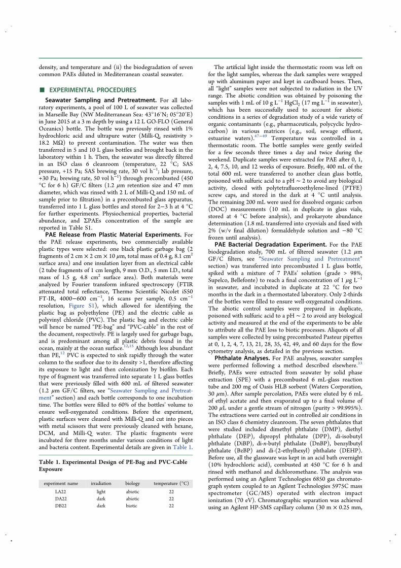
results indicated that, regardless of the indoor light/darkconditions, both PVC-cable and PE-bag leached specific PAEstoward the surrounding seawater, with higher release rates forthe latter. Only the DMP and DEP migrations (expressed as ngg−1 of plastic incubated) were detected from the PVC-cable,whereas only DiBP and DnBP were detected from the PE-bag
(Figure 1). The absence of other targeted PAEs may beexplained by (i) their absence from the selected polymers or(ii) the low release rate to the surrounding water phase due tohigh affinity with the polymer. In all experiments, the largermigration was measured within the first 2 weeks of incubationwith a specific magnitude and trend for each individualtreatment. LA22 were compared to DA22 treatment to isolatethe effect of the light (Table 1).Note that for the PVC-cable (Figure 1a,b), a higher
migration was observed during the first 1−2 weeks (up to6.6 ng g−1 and 23.2 ng g−1 for DMP and DEP, respectively),whereas the measured concentrations reached a plateau andremained stable in both the light- and dark-abiotic conditionsthroughout the following 6 weeks. After 8−10 weeks, themeasured concentrations started to slightly decrease, mostlikely due to the glass bottle adsorption or hydrolysis,52
although late prokaryotic development and subsequentbiodegradation cannot be precluded. Overall, our resultsshowed that (i) DEP was predominantly released from thePVC-cable over DMP (3.5 times more) and (ii) the indoorlight condition induced up to two times more DEP and DMPreleases compared to the dark condition. In contrast, for thePE-bag experiments, a higher amount of PAEs, including DiBPfollowed by DnBP, were released (up to 139 ng g−1) mainlyduring the first week. Differently from the PVC-cableexperiments, the PE-bag results indicated no significant releasedifferences between light- and dark-abiotic conditions (dark-abiotic) DiBP, 83.4 ± 12.5 ng g−1; and DnBP, 120.1 ± 18.0 ngg−1. (light-abiotic) DiBP, 103.6 ± 15.5 ng g−1; and DnBP,138.8 ± 20.8 ng g−1) during the time course experiment(Figure 1c,d), thus suggesting that only seawater leachingpromotes PAE release whatever the light conditions. Similardecreases for both dark and light conditions during the lastweeks of the experiment suggest that photodegradation in the
Figure 1. Graphical representation of the release kinetics of DMP (a) and DEP (b) from the PVC-cable experiments and of DiBP (c) and DnBP(d) from the PE-bag experiments. The two experimental conditions were dark abiotic (DA) and light abiotic (LA) incubated at 22 °C (in situtemperature). Curves are given to assist in the reading and do not represent data modeling.
Environmental Science & Technology Article
DOI: 10.1021/acs.est.8b05083Environ. Sci. Technol. 2019, 53, 166−175
168
visible radiation range was not a significant process on freelydissolved DiBP and DnBP destruction. Therefore, the differentpatterns observed for both PVC-cable and PE-bag could berather linked to the 3-dimension configuration of each plasticpiece (i.e., 2 mm vs 10 μm thicknesses, respectively). Indeed,the very thin PE-bag material could release a large part of itsPAE burden either with light or not. In contrast, photo-chemical oxidation reactions may alter the PVC-surface,thereby making more PAE quantities water-accessible.DOC leaching confirms the PAEs trend, with the PE-bag’s
highest release in the first week and small differences betweenthe dark and light conditions (24.4 × 10³ and 24.6 × 10³ ng Cg−1 of plastic bag) and with the PVC-cable’s highest releaseafter 1−2 weeks and higher release during the light experiment(13.4 × 10³ and 21.9 × 10³ ng C g−1 in the dark and lightconditions, respectively) (Figure S2). The PAE carbon contentreleased from the PE-bag and PVC cable thus represented asmall portion of the DOC that leached, that is, only 0.05−0.09% of the DOC released from the PVC-cable and 0.15−0.17% of the DOC from the PE-bag. In addition to PAEs,other groups of organic additive or oligomers could be leachedfrom this plastic during the experiment, thus increasing theconcentration of DOC in the surrounding water. The amountof DOC leached per surface area unit of the PE-bag in thisstudy (5.5 and 5.6 μg C cm−2 of the plastic surface in the darkand light conditions, respectively) are higher than themigration observed by Romera-Castillo et al. (2018) in PEfood packaging (0.26−0.31 μg C cm−2), which is probably dueto the lower amount of additives mixed in food plastic resins,but is in the same range of LDPE and HDPE pellets’ leaching(2.4−8.9 μg C cm−2). In the same study, similar leachingkinetics were reported, with the peak of leaching observed in
the first week of the experiment, which was followed by a sharpdecrease of DOC migration during the first month.Interestingly, we observed a second strong DOC leachingafter 10−12 weeks (83−96 × 103 ng C g−1 for the PE bag and28−38 × 103 ng C g−1 for the PVC-cable), which was probablydue to the initial degradation of the plastic surface. The lack ofa strong weathering such as UV-exposure or a strongmechanical abrasion induced a slow degeneration of thepolymers and thus, part of the organic matter pool morestrongly bounded to the polymer could be leached only whenthe fragments were affected by major surface modifications.
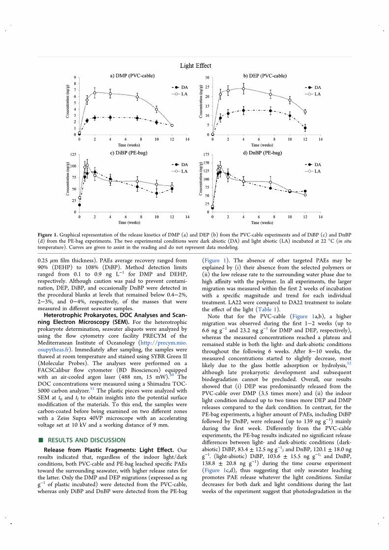
Release from Plastic Fragments: Biotic Effect. Bioticeffects were studied by comparing the results of the previousabiotic conditions with the PAE release kinetics from the sameplastic materials diluted in seawater comprising its naturalprokaryote assemblage (biotic conditions, seawater filteredthrough 1.2 μm GF/C and not poisoned with HgCl2; Figure2). The results indicated that DiBP and DnBP are more rapidlyreleased and in higher proportions (up to 122 ng g−1) from thePE-bag than the DMP and DEP from the PVC-cable (63.5 ngg−1). Globally, the same PAEs were detected for both light andbiotic experiments. However, 5-fold higher quantities of DMP/DEP were produced from the PVC-cable in the bioticconditions during the first month rather opposed to theabiotic conditions, thus indicating that PAE leachates werepromoted by prokaryotic activity. In contrast, no influence ofprokaryotes was observed on the initial release of DiBP andDnBP from the PE-bag. PAE release catalyzed by bacterialcommunities seemed to be more efficient for the PVC-cablethan for the PE-bag. The large difference in PAE releasebetween biotic and abiotic conditions observed in the case ofPVC-cable was not observable in the case of the PE-bag
Figure 2. Graphical representation of the release kinetics of DMP (a) and DEP (b) from the PVC-cable experiments and of DiBP (c) and DnBP(d) from the PE-bag experiments. The two experimental conditions were dark abiotic (DA) and dark biotic (DB) incubated at 22 °C (in situtemperature). Total bacteria include LNA and HNA cell abundance. The curves are given to assist in the reading and do not represent the datamodeling.
Environmental Science & Technology Article
DOI: 10.1021/acs.est.8b05083Environ. Sci. Technol. 2019, 53, 166−175
169
experiments. This could be attributed to (i) the low thicknessof the material, thus allowing for a complete release of PAEburden regardless of the conditions or (ii) the low PE agingunder the action of bacteria.Interestingly, for both materials incubated with seawater
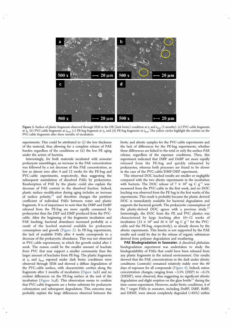
prokaryote assemblages, an increase in the PAE concentrationwas followed by a net decrease of this PAE concentration, aslow as almost zero after 4 and 12 weeks for the PE-bag andPVC-cable experiments, respectively, thus suggesting thesubsequent assimilation of dissolved PAEs by prokaryotes.Readsorption of PAE by the plastic could also explain thedecrease of PAE content in the dissolved fraction. Indeed,plastic surface modification during aging includes an increaseof surface polarity,53 and therefore changes the partitioncoefficient of individual PAEs between water and plasticfragments. It is of importance to note that the DiBP and DnBPreleased from the PE-bag are more rapidly consumed byprokaryotes than the DEP and DMP produced from the PVC-cable. After the beginning of the fragments incubation andPAE leaching, bacterial abundance increased probably as aresult of the leached material available for prokaryoteconsumption and growth (Figure 2). In PE-bag experiments,the lack of available PAEs after 4 weeks corresponds to adecrease of the prokaryotic abundance. This was not observedin PVC-cable experiments, in which the growth ended after 1week. The reason could be the smaller amount of leachatefrom PVC that may support a smaller community than thelarger amount of leachates from PE-bag. The plastic fragmentsat t0 and tfinal exposed under dark biotic conditions wereobserved through SEM and showed a diffuse degradation ofthe PVC-cable surface, with characteristic cavities along thefragments after 3 months of incubation (Figure 3a,b) and noevident differences on the PE-bag surface at the end of theincubation (Figure 3c,d). This observation seems to confirmthat PVC-cable fragments are a better substrate for prokaryotecolonization and subsequent degradation. This outcome mayprobably explain the large differences observed between the
biotic and abiotic samples for the PVC-cable experiments andthe lack of differences for the PE-bag experiments, whetherthese differences are linked to the total or only the surface PAErelease, regardless of the exposure conditions. Then, thisexperiment indicated that DiBP and DnBP are more rapidlyreleased from the PE-bag and quickly exhausted byprokaryotes, whereas both processes are found to be slowerin the case of the PVC-cable/DMP/DEP experiment.The observed DOC leached results are smaller or negligible
compared with the two abiotic experiments in the incubationwith bacteria. The DOC release of 7 × 10³ ng C g−1 wasmeasured from the PVC-cable in the first week, and no DOCleaching was observed from the PE bag in the first weeks of theexperiments. This result is probably because the plastic derivedDOC is immediately available for bacterial degradation andsupports the bacterial growth. The prokaryotic consumption ofthe plastic-derived DOC agrees with a previous study.15
Interestingly, the DOC from the PE and PVC plastics wascharacterized by large leaching after 10−12 weeks ofincubation (23 × 103 and 36 × 103 ng C g−1 for the PVC-cable and the PE-bag, respectively), as already shown by theabiotic experiments. This kinetic is not supported by the PAEresults and could be due to the release of organic substancesderived from polymer degradation and weathering.
PAE Biodegradation in Seawater. A dissolved phthalatebiodegradation experiment was undertaken to study thebiodegradability of PAEs that could have been released fromany plastic fragments in the natural environment. Our resultsshowed that the PAE concentrations in the dark under abioticconditions (controls) remained relatively stable over the 60days of exposure for all compounds (Figure 4). Indeed, minorconcentration changes, ranging from −3.5% (DEP) to −6.1%(DEHP), were observed, thus suggesting no significant abioticdegradation and slight sorption on the glass bottle52 during thetime course experiment. However, under biotic conditions, 4 ofthe 7 target PAEs in seawater, including DnBP, DiBP, BzBP,and DEHP, were almost completely degraded (>85%) within
Figure 3. Surface of plastic fragments observed through SEM in the DB (dark biotic) condition at t0 and tfinal (3 months). (a) PVC-cable fragmentsat t0, (b) PVC-cable fragments at tfinal, (c) PE-bag fragment at t0, and (d) PE-bag fragments at tfinal. The yellow circles highlight the cavities on thePVC-cable fragments after three months of incubation.
Environmental Science & Technology Article
DOI: 10.1021/acs.est.8b05083Environ. Sci. Technol. 2019, 53, 166−175
170
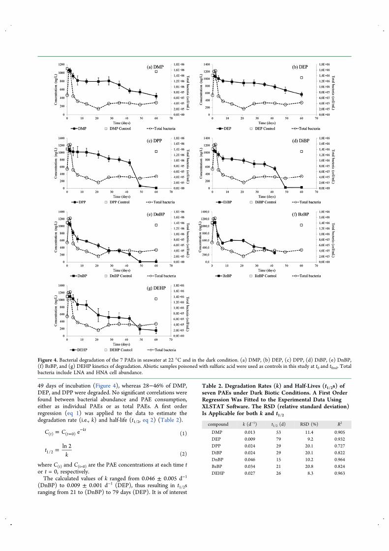
49 days of incubation (Figure 4), whereas 28−46% of DMP,DEP, and DPP were degraded. No significant correlations werefound between bacterial abundance and PAE consumption,either as individual PAEs or as total PAEs. A first orderregression (eq 1) was applied to the data to estimate thedegradation rate (i.e., k) and half-life (t1/2, eq 2) (Table 2).
= =−C C et t
kt( ) ( 0) (1)
=tk
ln 21/2 (2)
where C(t) and C(t=0) are the PAE concentrations at each time tor t = 0, respectively.The calculated values of k ranged from 0.046 ± 0.005 d−1
(DnBP) to 0.009 ± 0.001 d−1 (DEP), thus resulting in t1/2sranging from 21 to (DnBP) to 79 days (DEP). It is of interest
Figure 4. Bacterial degradation of the 7 PAEs in seawater at 22 °C and in the dark condition. (a) DMP, (b) DEP, (c) DPP, (d) DiBP, (e) DnBP,(f) BzBP, and (g) DEHP kinetics of degradation. Abiotic samples poisoned with sulfuric acid were used as controls in this study at t0 and tfinal. Totalbacteria include LNA and HNA cell abundance.
Table 2. Degradation Rates (k) and Half-Lives (t1/2s) ofseven PAEs under Dark Biotic Conditions. A First OrderRegression Was Fitted to the Experimental Data UsingXLSTAT Software. The RSD (relative standard deviation)Is Applicable for both k and t1/2
compound k (d−1) t1/2 (d) RSD (%) R2
DMP 0.013 53 11.4 0.905DEP 0.009 79 9.2 0.932DPP 0.024 29 20.1 0.727DiBP 0.024 29 20.1 0.822DnBP 0.046 15 10.2 0.964BzBP 0.034 21 20.8 0.824DEHP 0.027 26 8.3 0.963
Environmental Science & Technology Article
DOI: 10.1021/acs.est.8b05083Environ. Sci. Technol. 2019, 53, 166−175
171
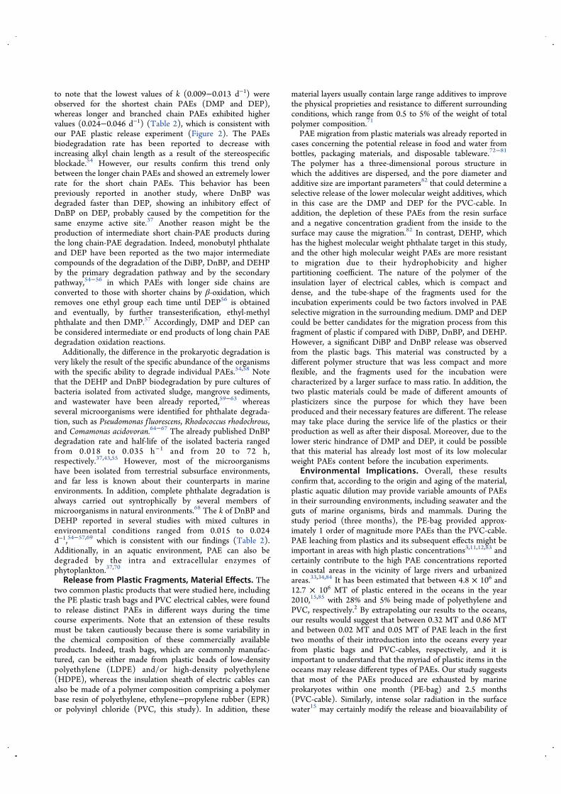
to note that the lowest values of k (0.009−0.013 d−1) wereobserved for the shortest chain PAEs (DMP and DEP),whereas longer and branched chain PAEs exhibited highervalues (0.024−0.046 d−1) (Table 2), which is consistent withour PAE plastic release experiment (Figure 2). The PAEsbiodegradation rate has been reported to decrease withincreasing alkyl chain length as a result of the stereospecificblockade.54 However, our results confirm this trend onlybetween the longer chain PAEs and showed an extremely lowerrate for the short chain PAEs. This behavior has beenpreviously reported in another study, where DnBP wasdegraded faster than DEP, showing an inhibitory effect ofDnBP on DEP, probably caused by the competition for thesame enzyme active site.37 Another reason might be theproduction of intermediate short chain-PAE products duringthe long chain-PAE degradation. Indeed, monobutyl phthalateand DEP have been reported as the two major intermediatecompounds of the degradation of the DiBP, DnBP, and DEHPby the primary degradation pathway and by the secondarypathway,54−56 in which PAEs with longer side chains areconverted to those with shorter chains by β-oxidation, whichremoves one ethyl group each time until DEP56 is obtainedand eventually, by further transesterification, ethyl-methylphthalate and then DMP.57 Accordingly, DMP and DEP canbe considered intermediate or end products of long chain PAEdegradation oxidation reactions.Additionally, the difference in the prokaryotic degradation is
very likely the result of the specific abundance of the organismswith the specific ability to degrade individual PAEs.54,58 Notethat the DEHP and DnBP biodegradation by pure cultures ofbacteria isolated from activated sludge, mangrove sediments,and wastewater have been already reported,59−63 whereasseveral microorganisms were identified for phthalate degrada-tion, such as Pseudomonas f luorescens, Rhodococcus rhodochrous,and Comamonas acidovoran.64−67 The already published DnBPdegradation rate and half-life of the isolated bacteria rangedfrom 0.018 to 0.035 h−1 and from 20 to 72 h,respectively.37,43,55 However, most of the microorganismshave been isolated from terrestrial subsurface environments,and far less is known about their counterparts in marineenvironments. In addition, complete phthalate degradation isalways carried out syntrophically by several members ofmicroorganisms in natural environments.68 The k of DnBP andDEHP reported in several studies with mixed cultures inenvironmental conditions ranged from 0.015 to 0.024d−1,54−57,69 which is consistent with our findings (Table 2).Additionally, in an aquatic environment, PAE can also bedegraded by the intra and extracellular enzymes ofphytoplankton.37,70
Release from Plastic Fragments, Material Effects. Thetwo common plastic products that were studied here, includingthe PE plastic trash bags and PVC electrical cables, were foundto release distinct PAEs in different ways during the timecourse experiments. Note that an extension of these resultsmust be taken cautiously because there is some variability inthe chemical composition of these commercially availableproducts. Indeed, trash bags, which are commonly manufac-tured, can be either made from plastic beads of low-densitypolyethylene (LDPE) and/or high-density polyethylene(HDPE), whereas the insulation sheath of electric cables canalso be made of a polymer composition comprising a polymerbase resin of polyethylene, ethylene−propylene rubber (EPR)or polyvinyl chloride (PVC, this study). In addition, these
material layers usually contain large range additives to improvethe physical proprieties and resistance to different surroundingconditions, which range from 0.5 to 5% of the weight of totalpolymer composition.71
PAE migration from plastic materials was already reported incases concerning the potential release in food and water frombottles, packaging materials, and disposable tableware.72−81
The polymer has a three-dimensional porous structure inwhich the additives are dispersed, and the pore diameter andadditive size are important parameters82 that could determine aselective release of the lower molecular weight additives, whichin this case are the DMP and DEP for the PVC-cable. Inaddition, the depletion of these PAEs from the resin surfaceand a negative concentration gradient from the inside to thesurface may cause the migration.82 In contrast, DEHP, whichhas the highest molecular weight phthalate target in this study,and the other high molecular weight PAEs are more resistantto migration due to their hydrophobicity and higherpartitioning coefficient. The nature of the polymer of theinsulation layer of electrical cables, which is compact anddense, and the tube-shape of the fragments used for theincubation experiments could be two factors involved in PAEselective migration in the surrounding medium. DMP and DEPcould be better candidates for the migration process from thisfragment of plastic if compared with DiBP, DnBP, and DEHP.However, a significant DiBP and DnBP release was observedfrom the plastic bags. This material was constructed by adifferent polymer structure that was less compact and moreflexible, and the fragments used for the incubation werecharacterized by a larger surface to mass ratio. In addition, thetwo plastic materials could be made of different amounts ofplasticizers since the purpose for which they have beenproduced and their necessary features are different. The releasemay take place during the service life of the plastics or theirproduction as well as after their disposal. Moreover, due to thelower steric hindrance of DMP and DEP, it could be possiblethat this material has already lost most of its low molecularweight PAEs content before the incubation experiments.
Environmental Implications. Overall, these resultsconfirm that, according to the origin and aging of the material,plastic aquatic dilution may provide variable amounts of PAEsin their surrounding environments, including seawater and theguts of marine organisms, birds and mammals. During thestudy period (three months), the PE-bag provided approx-imately 1 order of magnitude more PAEs than the PVC-cable.PAE leaching from plastics and its subsequent effects might beimportant in areas with high plastic concentrations3,11,12,83 andcertainly contribute to the high PAE concentrations reportedin coastal areas in the vicinity of large rivers and urbanizedareas.33,34,84 It has been estimated that between 4.8 × 10⁶ and12.7 × 106 MT of plastic entered in the oceans in the year2010,15,85 with 28% and 5% being made of polyethylene andPVC, respectively.2 By extrapolating our results to the oceans,our results would suggest that between 0.32 MT and 0.86 MTand between 0.02 MT and 0.05 MT of PAE leach in the firsttwo months of their introduction into the oceans every yearfrom plastic bags and PVC-cables, respectively, and it isimportant to understand that the myriad of plastic items in theoceans may release different types of PAEs. Our study suggeststhat most of the PAEs produced are exhausted by marineprokaryotes within one month (PE-bag) and 2.5 months(PVC-cable). Similarly, intense solar radiation in the surfacewater15 may certainly modify the release and bioavailability of
Environmental Science & Technology Article
DOI: 10.1021/acs.est.8b05083Environ. Sci. Technol. 2019, 53, 166−175
172
PAEs produced from plastics in the oceans, whereas highhydrostatic pressure in deep waters is able to modify theprokaryotic degradation of particulate organic matter86 andcertainly have a significant effect on the plastic aging depositedon the deep sediment. Considering that we found that PAEsthat were released ranged from 71 ng g−1 to 241 ng g−1 andthat plastics usually contain 0.5−5% of PAEs, our resultssuggest that, after three months, more than 90% of the PAEs inthe plastic remain and will ultimately leach out over a longerperiod of time.
■ ASSOCIATED CONTENT*S Supporting InformationThe Supporting Information is available free of charge on theACS Publications website at DOI: 10.1021/acs.est.8b05083.
FTIR-ATR analyses of PE-bag and PVC-cable frag-ments; kinetic of DOC leached; physiochemical proper-ties, bacterial abundance and ∑PAEs concentration othe sample (PDF)
■ AUTHOR INFORMATIONCorresponding Author*E-mail: [email protected] Paluselli: 0000-0002-4468-9802NotesThe authors declare no competing financial interest.
■ ACKNOWLEDGMENTSThis study was conducted as part of the PLASTOX-JPI Oceanand PlasticMicro-EC2CO/CNRS projects. We acknowledgethe financial support from the PACA region, which provided aPhD scholarship for A. Paluselli. The M I O flow cytometryand the SAM platforms are acknowledged for bacterialcounting and seawater sampling, respectively. The authorsare grateful to Francois-Xavier Perrin, Nathalie Patel, andAhmad Fahs from Toulon University for kindly providingFTIR and SEM analyses. Dr. M. Ourgaud is kindlyacknowledged for help during the experimental part of thestudy. The project leading to this publication has receivedfunding from the European FEDER Fund under Project 1166-39417. Reviewers and editor are kindly acknowledged forimproving the revised version of the MS.
■ REFERENCES(1) Andrady, A. L. Microplastics in the marine environment. Mar.Pollut. Bull. 2011, 62, 1596−1605.(2) Plastics Europe. PlasticsThe facts 2017: An Analysis ofEuropean Plastics Production, Demand and Waste Data; PlasticsEurope: Bruxelles, 2017.(3) Eriksen, M.; Lebreton, L. C. M.; Carson, H. S.; Thiel, M.;Moore, C. J.; Borerro, J. C.; Galgani, F.; Ryan, P. G.; Reisser, J. Plasticpollution in the World’s Ocean; More than 5 Trillion Plastic PeacesWeighing over 250.000 Tons Afloat at Sea. PLoS One 2014, 9 (12),e111913.(4) Saido, K.; Koizumi, K.; Sato, H.; Ogawa, N.; Kwon, B. G.;Chung, S. Y.; Kusui, T.; Nishimura, M.; Kodera, Y. New analyticalmethod for the determination of styrene oligomers formed frompolystyrene decomposition and its application at the coastlines of theNorth-West Pacific Ocean. Sci. Total Environ. 2014, 473−474, 490−495.
(5) Barnes, D. K. A.; Galgani, F.; Thompson, R. C.; Barlaz, M.Accumulation and fragmentation of plastic debris in global environ-ments. Philos. Trans. R. Soc., B 2009, 364 (1526), 1985−1998.(6) Ter Halle, A.; Ladirat, L.; Gendre, X.; Goudouneche, D.;Pusineri, C.; Routaboul, C.; Tenailleau, C.; Duployer, B.; Perez, E.Understanding the fragmentation pattern of marine plastic debris.Environ. Sci. Technol. 2016, 50, 5668−5675.(7) Moore, C. J.; Moore, S. L.; Leecaster, M. K.; Weisberg, S. B. Acomparison of plastic and plankton in the North Pacific Central Gyre.Mar. Pollut. Bull. 2001, 42 (12), 1297−1300.(8) Woodall, L. C.; Sanchez-Vidal, A.; Canals, M.; Paterson, G. L. J.;Coppock, R.; Sleight, V.; Calafat, A.; Rogers, A. D.; Narayanaswamy,B. E.; Thompson, R. C. The deep sea is a major sink for microplasticdebris. R. Soc. Open Sci. 2014, 1, 140317.(9) Cooper, D. A.; Corcoran, P. L. Effects of mechanical andchemical processes on the degradation of plastic beach debris on theisland of Kauai, Hawaii. Mar. Pollut. Bull. 2010, 60 (5), 650−654.(10) Neves, D.; Sobral, P.; Ferreira, J. L.; Pereira, T. Ingestion ofmicroplastics by commercial fish off the Portuguese coast. Mar. Pollut.Bull. 2015, 101, 119−126.(11) Browne, M. A.; Crump, P.; Niven, S. J.; Teuten, E. L.; Tonkin,A.; Galloway, T.; Thompson, R. C. Accumulations of microplastic onshorelines worldwide: sources and sinks. Environ. Sci. Technol. 2011,45, 9175−9179.(12) Suaria, G.; Avio, C. G.; Mineo, A.; Lattin, G. L.; Magaldi, M.G.; Belmonte, G.; Moore, C. J.; Regoli, F.; Aliani, S. TheMediterranean Plastic Soup: synthetic polymers in Mediterraneansurface waters. Sci. Rep. 2016, 6, 37551.(13) Andrady, A. L.; Neal, M. A. Applications and societal benefits ofplastics. Philos. Trans. R. Soc., B 2009, 364, 1977−1984.(14) Cozar, A.; Echavarria, F.; Gonzalez-Gordillo, J. I.; Irigoien, X.;Ubeda, B.; Hernandez-Leon, S.; Palma, A. T.; Navarro, S.; Garcia-de-Lomas, J.; Tuiz, A.; Fernandez-de-Puelles, M. L.; Duarte, C. M. Plasticdebris in the open ocean. Proc. Natl. Acad. Sci. U. S. A. 2014, 111(28), 10239−10244.(15) Romera-Castillo, C.; Pinto, M.; Langer, T. M.; Alvarez-Salgado,X. A.; Herndl, G. J. Dissolved organic carbon leaching from plasticsstimulates microbial activity in the ocean. Nat. Commun. 2018, 9,1430.(16) Corcoran, P. L.; Biesinger, M. C.; Grifi, M. Plastics andbeaches: a degrading relationship. Mar. Pollut. Bull. 2009, 58, 80−84.(17) Devriese, L. I.; van der Meulen, M. D.; Maes, T.; Bekaert, K.;Paul-Pont, I.; Frere, L.; Robbens, J.; Vethaak, A. D. Microplasticcontamination in brown shrimp(Crangon crangon, Linnaeus 1758)from coastal waters of the Southern North Sea and Channel area.Mar. Pollut. Bull. 2015, 98, 179−187.(18) Rochman, C. M.; Tahir, A.; Williams, S. L.; Baxa, D. V.; Lam,R.; Miller, J. T.; Teh, F.-C.; Werorilangi, S.; Teh, S. J. Anthropogenicdebris in seafood: plastic debris and fibers from textiles in fish andbivalves sold for human consumption. Sci. Rep. 2015, 5, No. 14340,DOI: 10.1038/srep14340.(19) Sussarellu, R.; Suquet, M.; Thomas, Y.; Lambert, C.; Fabioux,C.; Pernet, M. E. J.; LeGoïc, N.; Quillien, V.; Mingant, C.; Epelboin,Y.; Corporeau, C.; Guyomarch, J.; Robbens, J.; Paul-Pont, I.; Soudant,P.; Huvet, A. Oyster reproduction is affected by exposure topolystyrene microplastics. Proc. Natl. Acad. Sci. U. S. A. 2016, 113,2430−2435.(20) Van Cauwenberghe, L.; Claessens, M.; Vandegehuchte, M. B.;Janssen, C. R. Microplastics are taken up by mussels (Mytilus edulis)and lugworms (Arenicolamarina) living in natural habitats. Environ.Pollut. 2015, 199, 10−17.(21) Lusher, A. L.; Hernandez-Milian, G.; O’Brien, J.; Berrow, S.;O’Connor, I.; Officer, R. Microplastic and macroplastic ingestion by adeep diving, oceanic cetacean: the True’s beaked whale Mesoplodonmirus. Environ. Pollut. 2015, 199, 185−191.(22) Eriksson, C.; Burton, H. Origins and biological accumulation ofsmall plastic particles in fur seals from Macquarie island. Ambio 2003,32, 380−384.
Environmental Science & Technology Article
DOI: 10.1021/acs.est.8b05083Environ. Sci. Technol. 2019, 53, 166−175
173
(23) Serodio, P.; Nogueira, J. M. F. Considerations on ultra-traceanalysis of phthalates in drinking water. Water Res. 2006, 40 (13),2572−2582.(24) Net, S.; Sempere, R.; Delmont, A.; Paluselli, A.; Ouddane, B.Occurrence, Fate, Behavior and Ecotoxicological State of Phthalatesin Different Environmental Matrices. Environ. Sci. Technol. 2015, 49(7), 4019−4035.(25) Commission staff Working Document on the Implementation of theCommunity Strategy for Endocrine Disrupters-A Range of SubstancesSuspected of Interfering with the Hormone System of Humans andWildlife; CEC (Commission of the European Communities): Brussels,2007.(26) Crisp, T. M.; Clegg, E. D.; Cooper, R. L.; Wood, W. P.;Anderson, D. G.; Baetcke, K. P.; Hoffmann, J. L.; Morrow, M. S.;Rodier, D. J.; Schaeffer, J. E.; Touart, L. W.; Zeeman, M. G.; Patel, Y.M. Environmental endocrine disruption: an effects assessment andanalysis. Environ. Health Perspect. 1998, 106 (1), 11−56.(27) Latini, G. Monitoring phthalate exposure in humans. Clin.Chim. Acta 2005, 361 (1−2), 20−29.(28) Kamrin, M. A. Phthalate Risks, Phthalate Regulation, andPublic Health: A Review. J. Toxicol. Environ. Health, Part B 2009, 12(2), 157−174.(29) Meeker, J. D.; Sathyanarayana, S.; Swan, S. H. Phthalates andother additives in plastics: human exposure and associated healthoutcomes. Philos. Trans. R. Soc., B 2009, 364 (1526), 2097−2113.(30) Matsumoto, M.; Hirata-Koisumi, M.; Ema, M. Potentialadverse effects of phthalic acid esters on human health: a review ofrecent studies on reproduction. Regul. Toxicol. Pharmacol. 2008, 50,37−49.(31) Fossi, M. C.; Romeo, T.; Baini, M.; Panti, C.; Marsili, L.;Campani, T.; Canese, S.; Galgani, F.; Druon, J. N.; Airoldi, S.; Taddei,S.; Fattorini, M.; Brandini, C.; Lapucci, C. Plastic debris occurrence,convergence areas and fin whales feeding ground in theMediterranean marine protected area Pelagos Sanctuary: A modelingapproach. Front. Mar. Sci. 2017, 4, 1−15.(32) Net, S.; Dumoulin, D.; El-Osmani, R.; Rabodonirina, S.;Ouddane, B. Case study of PAHs, Me-PAHs, PCBs, Phthalates, andPesticides Contamination in the Somme River water, France. Int. J.Environ. Res. 2014, 8 (4), 1159−1170.(33) Paluselli, A.; Aminot, Y.; Galgani, F.; Net, S.; Sempere, R.Occurrence of phthalate acid esters (PAEs) in the northwesternMediterranean Sea and the Rhone River. Mermex special issue. Prog.Oceanogr. 2018, 163, 221−231.(34) Paluselli, A.; Fauvelle, V.; Schmidt, N.; Galgani, F.; Net, S.;Sempere, R. Seasonal distribution of phthalates in Mediterraneancoastal seawater. Sci. Total Environ. 2018, 621 (2018), 578−587.(35) Wang, I.; Lin, C.; Lin, Y.; Hsieh, W.; Chen, P. Early lifephthalate exposure and atopic disorders in children: A prospectivebirth cohort study. Environ. Int. 2014, 62, 48−54.(36) Xie, Z.; Ebinghaus, R.; Temme, C.; Lohmann, R.; Caba, A.;Ruck, W. Occurrence and Air-Sea Exchange of Phthalates in theArctic. Environ. Sci. Technol. 2007, 41, 4555−4560.(37) Gao, J.; Chi, J. Biodegradation of phthalate acid esters bydifferent marine microalgal species. Mar. Pollut. Bull. 2015, 99, 70−75.(38) Munshi, A. B.; Karim, N.; Shaukat, S.; Hashmi, D.; Boardman,G. D.; Flick, G. J. Toxicity of Phthalate Esters in Fish and Shellfishfrom Virginia Beach Using Matrix Solid Phase Dispersion (MSPD)and GC-MS. J. Chem. Soc. Pak. 2013, 35 (6), 1463−1471.(39) Sempere, R.; Para, J.; Tedetti, M.; Chattiere, B.; Mallet, M.Variability of Solar radiation and CDOM in Surface Coastal Waters ofthe Northwestern Mediterranean Sea. Photochem. Photobiol. 2015, 91(4), 851−561.(40) Castro-Jimenez, J.; Barhoumi, B.; Paluselli, A.; Tedetti, M.;Jimenez, B.; Munoz-Arnanz, J.; Wortham, H.; Driss, M. D.; Sempere,R. Occurrence, Loading, and Exposure of Atmospheric Particle-BoundPOPs at the African and European Edges of the WesternMediterranean Sea. Environ. Sci. Technol. 2017, 51, 13180.
(41) Theodosi, C.; Panagiotopoulos, C.; Nouara, A.; Zarmpas, P.;Violaki, K.; Kanakidou, M.; Sempere, R.; Mihalopoulos, N. Sugars inatmospheric aerosols over the Eastern Mediterranean. Prog. Oceanogr.2018, 163, 70−81.(42) The Mermex Group. Marine ecosystems responses to climaticand anthropogenic forcings in the Mediterranean. Progr. Oceanogr.2011, 91, 97−166.(43) Zorita, I.; Apraiz, I.; Ortiz-Zarragoitia, M.; Orbea, A.; Cancio,I.; Soto, M.; Marigomez, I.; Cajaraville, M. P. Assessment of biologicaleffects of environmental pollution along the NW Mediterranean Seausing mussels as sentinel organisms. Environ. Pollut. 2007, 148 (1),236−250.(44) Cozar, A.; Sanz-Martin, M.; Marti, E.; Gonzalez-Gordillo, J. I.;Ubeda, B.; Galvez, J. A.; Irigoien, X.; Duarte, C. M. Plasticaccumulation in the Mediterranean Sea. PLoS One 2015, 10 (4),e0121762.(45) Collignon, A.; Hecq, J. H.; Galgani, F.; Collard, F.; Goffart, A.Annual variation in neustonic micro- and meso-plastic particles andzooplankton in the Bay of Calvi (Mediterranean−Corsica). Mar.Pollut. Bull. 2014, 79, 293−298.(46) Fossi, M. C.; Marsili, L.; Baini, M.; Giannetti, M.; Coppola, D.;Guerranti, C.; Caliani, I.; Minutoli, R.; Lauriano, G.; Finoia, M. G.;Rubegni, F.; Panigada, S.; Berube, M.; Ramirez, J. U.; Panti, C. Finwhales and microplastics: The Mediterranean Sea and the Sea ofCortez scenarios. Environ. Pollut. 2016, 209, 68−78.(47) Wang, C. Y.; Wang, F.; Wang, T.; Yang, X. L.; Bian, Y. R.;Kengara, F. O.; Li, Z. B.; Jiang, X. Effects of Autoclaving and MercuricChloride Sterilization on PAHs Dissipation in a Two-Liquid-PhaseSoil Slurry. Pedosphere 2011, 21, 56−64.(48) Yu, J. T.; Bouwer, E. J.; Coelhan, M. Occurrence andbiodegradability studies of selected pharmaceuticals and personal careproducts in sewage effluent. Agric. Water Manag. 2006, 86, 72−80.(49) Aminot, Y.; Fuster, L.; Pardon, P.; Le Menach, K.; Budzinski,H. Suspended solids moderate the degradation and sorption of wastewater-derived pharmaceuticals in estuarine waters. Sci. Total Environ.2018, 15, 39−40.(50) Girault, M.; Arakawa, H.; Ceccaldi, H. J.; Hashihama, F.;Gregori, G. Heterotrophic prokaryote distribution along a 2300 kmtransect in the North Pacific subtropical gyre during a strong La Ninaconditions: relationship between distribution and hydrologicalconditions. Biogeosciences 2015, 12, 3607−3621.(51) Sempere, R.; Tedetti, M.; Panagiotopoulos, C.; Charriere, B.;Van Wambeke, F. Distribution and bacterial availability of dissolvedneutral sugars in the South East Pacific. Biogeosciences 2008, 5, 1165−1173.(52) Sempere, R.; Yoro, S. C.; Van Wambeke, F.; Charriere, B.Microbial decomposition of large organic particles in northwesternMediterranean Sea. Mar. Ecol.: Prog. Ser. 2000, 5 (198), 61−72.(53) Ter Halle, A.; Ladirat, L.; Martignac, M.; Mingotaud, A. F.;Boyron, O.; Perez, E. To what extent are microplastic from the openocean weathered? Environ. Pollut. 2017, 227, 167−174.(54) Liang, D.; Zhang, T.; Fang, H.; He, J. Phthalatesbiodegradation in the environment. Appl. Microbiol. Biotechnol.2008, 80, 183−198.(55) Kumar, V.; Sharma, N.; Maitra, S. S. Comparative study on thedegradation of dibutyl phthalate by two newly isolated Pseudomonassp. V21b and Comamonas sp. 51F. Biotechnol Rep. (Amst). 2017, 15,1−10.(56) Amir, S.; Hafidi, M.; Merlina, G.; Hamdi, H.; Jouraiphy, A.; ElGharous, M.; Revel, J. C. Fate of phthalic acid esters duringcomposting of both lagooning and activated sludges. Process Biochem.2005, 40, 2183−2190.(57) Cartwright, C. D.; Owen, S. A.; Thompson, I. P.; Burns, R. G.Biodegradation of diethyl phthalate in soil by a novel pathway. FEMSMicrobiol. Lett. 2000, 186, 27−34.(58) Kleerebezem, R.; Pol, L. W. H.; Lettinga, G. Anaerobicbiodegradation of phthalic acid and isomers and related compounds.Biodegradation 1999, 10, 63−73.
Environmental Science & Technology Article
DOI: 10.1021/acs.est.8b05083Environ. Sci. Technol. 2019, 53, 166−175
174
(59) Yuan, S. Y.; Liu, C.; Liao, C. S.; Chang, B. V. Occurrence andmicrobial degradation of phthalate esters in Taiwan river sediments.Chemosphere 2002, 49, 1295−1299.(60) Xu, X. R.; Li, H. B.; Gu, J. B.; Li, X. Y. Kinetics of n-butylbenzyl phthalate degradation by a pure bacterial culture from themangrove sediment. J. Hazard. Mater. 2007, 140, 194−199.(61) Jin, D.; Kong, X.; Cui, B.; Bai, Z.; Zhang, H. Biodegradation ofDi-n-Butyl Phthalate by a Newly Isolated Halotolerant Sphingobiumsp. Int. J. Mol. Sci. 2013, 14, 24046−24054.(62) Xu, X.-R.; Gu, J.-D.; Li, H.-B.; Li, X.-Y. Kinetics of di-n-ButylPhthalate Degradation by a Bacterium Isolated from MangroveSediment. J. Microbiol. Biotechnol. 2005, 15 (5), 946−951.(63) Kim, Y. H.; Lee, J. W.; Ahn, J. Y.; Gu, M. B.; Moon, S. H.Enhanced degradation of an endocrine-disrupting chemical, butylbenzyl phthalate, by Fusarium oxysporum f. sp pisi cutinase. Appl.Environ. Microbiol. 2002, 68, 4684−4688.(64) Iwaki, H.; Nishimura, A.; Hasegawa, Y. Tropicibacter phthalicussp. nov.; A Phthalate-Degrading Bacterium from Seawater. Curr.Microbiol. 2012, 64, 392−396.(65) Vamsee-Krishna, C.; Mohan, Y.; Phale, P. S. Biodegradation ofphthalate isomers by Pseudomonas aeruginosa PP4, Pseudomonas spPPD and Acinetobacter lwof f ii ISP4. Appl. Microbiol. Biotechnol. 2006,72, 1263−1269.(66) Xu, X. R.; Li, H. B.; Gu, J. D. Elucidation of n-butyl benzylphthalate biodegradation using high-performance liquid chromatog-raphy and gas chromatography−mass spectrometry. Anal. Bioanal.Chem. 2006, 386, 370−375.(67) Wang, Y. Y.; Fan, Y. Z.; Gu, J. D. Aerobic degradation ofphthalic acid by Comamonas acidovoran Fy-1 and dimethyl phthalateester by two reconstituted consortia from sewage sludge at highconcentrations. World J. Microbiol. Biotechnol. 2003, 19, 811−815.(68) Gu, J. D.; Li, J.; Wang, Y. Biochemical pathway and degradationof phthalate ester isomers by bacteria. Water Sci. Technol. 2005, 52,241−248.(69) Yuwatini, E.; Hata, N.; Taguchi, S. Behavior of di-(2-ethylhexyl) phthalate discharged from domestic waste water intoaquatic environment. J. Environ. Monit. 2006, 8, 191−196.(70) Li, B.; Chi, J.; Wu, W. X.; Wang, Z. K. Effects of nutrients andlight on biodegradation of dibuyl phthalate and di-2-ethylexylphthalate in haihe estuary. Bull. Environ. Contam. Toxicol. 2007, 79,80−83.(71) Hermabessiere, L.; Dehaut, A.; Paul-Pont, I.; Lacroix, C.;Jezequel, R.; Soudant, P.; Duflos, G. Occurrence and effects of plasticadditives on marine environments and organisms: A review.Chemosphere 2017, 182, 781−793.(72) Hahladakis, J. N.; Velis, C. A.; Weber, R.; Iacovidou, E.;Purnell, P. An overview of chemical additives present in plastics:Migration, release, fate and environmental impact during their use,disposal and recycling. J. Hazard. Mater. 2017, 344, 179.(73) Wagner, M.; Oehlmann, J. Endocrine disruptors in bottledmineral water: total estrogenic burden and migration from plasticbottles. Environ. Sci. Pollut. Res. 2009, 16, 278−286.(74) Montuori, P.; Jover, E.; Morgantini, M.; Bayona, J. M.; Triassi,M. Assessing human exposure to phthalic acid and phthalate estersfrom mineral water stored in polyethylene terephthalate and glassbottles. Food Addit. Contam., Part A 2008, 25, 511−518.(75) Fasano, E.; Bono-Blay, F.; Cirillo, T.; Montuori, P.; Lacorte, S.Migration of phthalates, alkylphenols, bisphenol A and di(2-ethylhexyl)adipate from food packaging. Food Control 2012, 27 (1),132−138.(76) Casajuana, N.; Lacorte, S. Presence and release of phthalicesters and other endocrine disrupting compounds in drinking water.Chromatographia 2003, 57, 649−655.(77) Simoneau, C.; Van den Eede, L.; Valzacchi, S. Identificationand quantification of the migration of chemicals from plastic babybottles used as substitutes for polycarbonate. Food Addit. Contam.,Part A 2012, 29, 469−480.(78) Xu, Q.; Yin, X.; Wang, M.; Wang, H.; Zhang, H.; Shen, Y.; Xu,S.; Zhang, L.; Gu, Z. Analysis of Phthalate Migration from Plastic
Containers to Packaged Cooking Oil and Mineral Water. J. Agric.Food Chem. 2010, 58, 11311−11317.(79) Bosnir, J.; Puntaric, D.; Galic, A.; Skes, I.; Dijanic, T.; Klaric,M.; Grgic, M.; Curkovic, M.; Smit, Z. Migration of Phthalates fromPlastic Containers into Soft Drinks and Mineral Water. Food Technol.Biotechnol. 2007, 45 (1), 91−95.(80) Li, C.; Chen, J.; Wang, J.; Han, P.; Luan, Y.; Ma, X.; Lu, A.Phthalate esters in soil, plastic film, and vegetable from greenhousevegetable production bases in Beijing, China: Concentration, sources,and risk assessment. Sci. Total Environ. 2016, 568, 1037−1043.(81) Suhrhoff, T. J.; Scholz-Bottcher, B. M. Qualitative impact ofsalinity, UV radiation and turbulence on leaching of organic plasticadditives from four common plastics- A lab experiment. Mar. Pollut.Bull. 2016, 102, 84−94.(82) Teuten, E. L.; Saquing, J. M.; Knappe, D. R. U.; Barlaz, M. A.;Jonsson, S.; Bjorn, A.; Rowland, S. J.; Thompson, R. C.; Galloway, T.S.; Yamashita, R.; Ochi, D.; Watanuki, Y.; Moore, C.; Viet, P. H.;Tana, T. S.; Prudente, M.; Boonyatumanond, R.; Zakaria, M. P.;Akkhavong, K.; Ogata, Y.; Hirai, H.; Iwasa, S.; Mizukawa, K.; Hagino,Y.; Imamura, A.; Saha, M.; Takada, H. Transport and release ofchemicals from plastics to the environment and to wildlife. Philos.Trans. R. Soc., B 2009, 364, 2027−2045.(83) Schmidt, N.; Thibault, D.; Galgani, F.; Paluselli, A.; Sempere,R. Occurrence of microplastics and potential contribution ofphthalates in the surface waters of the Gulf of Lion (NWMediterranean Sea). Prog. Oceanogr. 2018, 163, 214−220.(84) Sanchez-Avila, L.; Tauler, R.; Lacorte, S. Organic micro-pollutants in coastal waters from NW Mediterranean Sea: Sourcesdistribution and potential risk. Environ. Int. 2012, 46, 50−62.(85) Jambeck, L. R.; Geyer, R.; Wilcox, C.; Siegler, T. R.; Perryman,M.; Andrady, A.; Narayan, R.; Law, K. L. Plastic waste inputs fromland into the ocean. Science 2015, 347, 768−771.(86) Riou, V.; Para, J.; Garel, M.; Guigue, C.; Ali, B. A.; Santinelli,C.; Lefevre, D.; Gattuso, J. P.; Goutx, M.; Jacquet, S. H. M.; LeMoigne, F.; Tachikawa, K.; Tamburini, C. Biodegradation ofEmiliania huxleyi Aggregates by a Natural Mediterranean ProkaryoticCommunity under Increasing Hydrostatic Pressure. Prog. Oceanogr.2018, 163, 271−281.
Environmental Science & Technology Article
DOI: 10.1021/acs.est.8b05083Environ. Sci. Technol. 2019, 53, 166−175
175


![Electrochemical Degradation of Diethyl Phthalate under … · Vol. 11, 2016 5010 the degradation of PAEs [5−7]; hence, PAEs have been frequently detected in surface and groundwater](https://img.pdfslide.net/doc/110x75/60756ea84d114b2f783e4d77/electrochemical-degradation-of-diethyl-phthalate-under-vol-11-2016-5010-the-degradation.jpg)