Embed Size (px)
Citation preview

DISEASES OF AQUATIC ORGANISMSDis Aquat Org
Vol. 86: 235–243, 2009doi: 10.3354/dao02133
Published November 9
INTRODUCTION
Rainbow smelt Osmerus mordax is an anadromousfish that supports a commercial and recreational fish-ery in Prince Edward Island (PEI), Canada. Currentlythere is very little data available on the health of thesesmelt. In April 2008, smelt were caught from a fresh-water stream during their spawning migration andlarge parasitic cysts were observed within the gastro-intestinal tract. Wet-mount preparations of the contentof parasitic cysts demonstrated the presence of amicrosporidian parasite, likely Glugea hertwigi Weis-senberg, 1911. G. hertwigi is a microsporidian parasitereported in rainbow smelt throughout the Great Lakesand the Atlantic (Haley 1954, Chen & Power 1972),although the parasite was previously unidentified insmelt populations in PEI. Severe infections with theparasite were thought to be associated with mortality
of young-of-the-year smelt in the Great Lakes (Nepszyet al. 1978), adult smelt in Quebec (Delisle 1972), andEuropean smelt O. eperlanus in Europe (Ieshko et al.2000). Considering the disease implications that theparasite may have in both young-of-the-year and adultfish, it is of interest to document the prevalence ofG. hertwigi and the health of the rainbow smelt popu-lations in PEI.
Historically the classification of Glugea hertwigi wasbased on the identification of the microsporidian para-site in euryhaline smelt of the genus Osmerus and inpond smelt of the genera Hypomesus and Coregonus(Canning & Lom 1986). The original description of theparasite was based on spore size, host species, andanatomical location. In addition to host and anatomicalniches, a full characterization of microsporidian speciesis through morphological and molecular studies exa-mining relatedness to other species. Although G. hert-
© Inter-Research 2009 · www.int-res.com*Email: [email protected]
Phylogeny and morphology of Glugea hertwigifrom rainbow smelt Osmerus mordax found in
Prince Edward Island, Canada
J. Lovy1,*, M. Kostka2, I. Dyková2, G. Arsenault3, H. Pecková2, G. M. Wright1, D. J. Speare3
1Department of Biomedical Sciences, Atlantic Veterinary College, University of Prince Edward Island, 550 University Ave., Charlottetown, Prince Edward Island C1A 4P3, Canada
2Institute of Parasitology, Biology Centre, ASCR, Brani$ovská 31, 370 05 >eské Budejovice, Czech Republic3Department of Pathology and Microbiology, Atlantic Veterinary College, University of Prince Edward Island,
550 University Ave., Charlottetown, Prince Edward Island C1A 4P3, Canada
ABSTRACT: Infection of rainbow smelt Osmerus mordax with the microsporidian Glugea hertwigiwas diagnosed for the first time in Prince Edward Island, Canada. The prevalence of infection was24%, 45 infected out of 187 examined fish captured in February and March 2009. Both large andsmall xenomas of G. hertwigi observed within the submucosa of the gastrointestinal tract and alongthe mesentery of the host contained only mature spores. Advanced and degraded xenomas associ-ated with host reaction were described using light and transmission electron microscopy. The firstrDNA sequence of G. hertwigi prepared in the present study completed the set of sequences ofGlugea spp. available for comparison. The high level of rDNA sequence identity between Glugeaspp. suggests that these may be variants of a single species.
KEY WORDS: Glugea hertwigi · Microsporidia · Fish · Phylogeny · Morphology · Ultrastructure
Resale or republication not permitted without written consent of the publisher

Dis Aquat Org 86: 235–243, 2009
wigi has been documented in smelt populationsthroughout Europe and the Atlantic, morphological andmolecular data on the parasite are currently lacking.Microsporidia in the genus Glugea have been consid-ered problematic; however, it has been demonstratedthat G. anomala and G. stephani are likely the samespecies, whereas G. americanus belongs in fact to thegenus Spraguea (Pomport-Castillon et al. 2000, Free-man et al. 2004). In the present study, G. hertwigifrom O. mordax is characterized with transmission elec-tron microscopy (TEM) and ribosomal DNA (rDNA)sequences to determine its relatedness within thegenus Glugea.
MATERIALS AND METHODS
Prevalence data. During the spawning migration (inApril 2008), rainbow smelt were captured with a dipnet from a tributary of the North River, PEI. Approxi-mately 120 predominantly male fish were capturedand upon dissection, microsporidian xenomas wereobserved within the gastrointestinal tract in roughly20% of the fish. The content of xenomas was examinedin fresh fish samples to confirm the presence of micro-sporidian spores. Some xenomas were fixed in 70%ethanol and stored in a –80°C freezer for molecularanalysis, while other xenomas were fixed in 2% glu-taraldehyde and processed for TEM.
In order to collect more accurate data on the preva-lence of Glugea hertwigi infection in rainbow smelt,sampling was repeated in winter 2009 from Hillsbor-ough River, PEI, on 3 sample dates (February 25,March 5 and 9). Smelt were captured through the icewith a spear and were either frozen for future exami-nation or examined immediately after capture for thepresence of xenomas. From a total of 187 fish, sex wasnoted, total length was recorded (cm), and the pres-ence or absence of infection was noted includingobservations on size of xenomas and intensity of infec-tion. Upon visual inspection, the infections were desig-nated as light, moderate, or heavy, indicating an infec-tion with <5 xenomas, between 5 and 15, or >15respectively. Upon examination of fresh fish samples,any tissue that contained small xenomas was fixed inKarnovsky’s fixative, comprised of 4% paraformalde-hyde and 1% glutaraldehyde in phosphate buffer, forexamination with microscopy.
Light and electron microscopy. Samples for routinelight microscopy were dehydrated, infiltrated withxylene, and embedded in paraffin wax. Sections (5 µmthick) were cut, stained with hematoxylin and eosin(H&E), and examined under a light microscope. ForTEM, subsequent to fixation in Karnovsky’s fixative for24 h at 4°C, samples were washed in phosphate buffer
and postfixed in 1% osmium tetroxide for 1 h at roomtemperature. Samples were then washed in distilledwater and dehydrated through a graded series ofethanols. The tissue was then submerged in propyleneoxide and infiltrated in increasing ratios of Spurr’sresin and propylene oxide (Canemco-Marivac). Thesamples were kept in pure resin overnight in a vacuumdesiccator and finally embedded in pure fresh Spurr’sresin. Semi-thin (0.5 µm) sections were cut and stainedwith toluidine blue and examined with a light micro-scope. Ultra-thin (90 nm) sections were cut from areasof interest and examined and photographed with aHitachi 7500 TEM operated at 80 kV. Determination ofthe number of polar filament coils in mature sporesincluded counts of 10 spores that were cut along theirlong axis.
DNA extraction, PCR amplification, and sequencing.Spores collected from xenomas were transferred todouble-distilled water and washed several times withrepeated centrifugation at 4000 × g for 2 min. DNA wasextracted using the JETQUICK Tissue DNA Spin Kit(Genomed) after previous 0.5 mm glass-bead homoge-nization and disruption of spores using the FastPrep®-24Instrument (M.P. Biomedicals). The targeted DNA (par-tial small subunit [SSU] rDNA + internal transcribedspacer [ITS] + partial large subunit [LSU] rDNA) was am-plified by PCR in 2 overlapping fragments with primerpairs 18F (5’-CAC CAG GTT GAT TCT GCC-3’)–1492R(5’-GGT TAC CTT GTA CGA CTT-3’) and 530F (5’-GTGCCA GC(C/A) GCC GCG G-3’)–580R (5’-GGT CCGTGT TTC AAG ACG G-3’) (Vossbrinck et al. 1993,2004). PCR was carried out in a 25 µl reaction volume us-ing 10 pmol of each primer, 250 µM of each dNTP, 2.5 µlof 10× Taq polymerase buffer, and 1 unit of TaqDNApolymerase (Top-Bio). The reactions were run on a Tper-sonal Thermocycler (Biometra). The cycling conditionsconsisted of initial denaturation at 95°C (5 min), 35 cyclesof denaturation at 94°C (1 min), annealing at 50°C (30 s),and extension at 72°C (2 min) followed by a final exten-sion at 72°C (10 min). The amplified products were puri-fied from 1% agarose gel using JETQUICK Gel Extrac-tion Spin Kit (Genomed) and cloned into pDrive CloningVector using the QIAGEN PCR Cloning Kit (Qiagen).Both strands were sequenced on an automatic sequencerABI 3130x1 using the ABI PRISM BigDye Terminatorv3.1 Cycle Sequencing Kit (Applied Biosystems) withM13 forward and M13 reverse primers.
Phylogenetic analysis. An alignment of 8 sequencesdeposited in GenBank as sequences of Glugea spp.together with the newly prepared sequence of G. hert-wigi, 3 Loma spp. sequences, and 2 outgroups was pre-pared in ClustalX 2.0.6 (Larkin et al. 2007). The se-quences were carefully examined for ambiguouslyaligned positions, which were deleted manually inBioEdit (Hall 1999). Four different methods were used
236

Lovy et al.: Glugea hertwigi in rainbow smelt
to reconstruct the phylogenetic relationships amongthe included species. Maximum likelihood (ML) analy-ses were run in the program RAxML 7.0.3 (Stamatakis2006). We used the GTR + gamma model and rapidbootstrapping option (100 replicates). Maximum parsi-mony (MP) trees and analyses employing the Fitch-Margoliash method with LogDet distances (LD) werecomputed in PAUP* 4.0b10 (Swofford 2001). Heuristicsearches were conducted with 10 addition-sequencereplicates in which the starting tree was constructedby random taxa addition and swapped with the trun-cated balanced realization (TBR) algorithm. The num-ber of bootstrap replicates was 1000 for both MP andLD. The program MrBayes 3.1.2 (Ronquist & Huelsen-beck 2003) was used to perform a Bayesian analysis(BA) of the dataset. Four simultaneous Markov ChainMonte Carlo (MCMC) chains were run for 106 genera-tions with sampling frequency 100 generations and
burn-in 2500. The –ln L scores were plotted against thegeneration to determine appropriate burn-in. The per-centage of identity among Glugea species sequenceswere calculated in the program MegAlign (DNAS-TAR). A shorter alignment consisting of 1169 bp wasused because only this part of rDNA sequence wasavailable for the Glugea species under study.
RESULTS
Prevalence
The prevalence of Glugea hertwigi in the sampledfish was approximately 24% (45 infected/187 totalfish). The mean lengths and sex of fish in relation toseverity of infection are summarized in Table 1.
Morphology
Variably sized xenomas, ranging from<1 to 4 mm of Glugea hertwigi, were ob-served in the submucosa of the gastro-intestinal tract extending from the stom-ach to the posterior intestine and withinthe mesentery. Individual fish had uni-formly small xenomas (Fig. 1A), uni-formly large xenomas (Fig. 1B), or amixture of large and small xenomas(Fig. 1C). Histological examination of
237
G. hertwigi Prevalence Sex Mean ± SD length (mm)infection (n) Male/Female Male Female
Total fish 187 106/81 166.8 ± 14.7 174.2 ± 16.4Uninfected 142 82/60 167.9 ± 15.8 175.0 ± 17.3Infected 45 24/21 162.8 ± 9.0 172.0 ± 9.0Light 21 9/12 159.4 ± 9.2 172.7 ± 7.9Moderate 15 10/5 166.1 ± 8.9 178.0 ± 22.8Heavy 9 5/4 163.0 ± 8.4 162.5 ± 5.0
Table 1. Glugea hertwigi infecting Osmerus mordax. Prevalence of parasite inrainbow smelt sampled in February and March 2009 showing the relationship of
the severity of infection to sex and mean length
Fig. 1. Glugea hertwigi infecting Osmerus mordax. Variably sized xenomas in the gastrointestinal tract of infected rainbow smelt.(A) Heavily infected individual with uniformly small xenomas (^) (scale bar = 1 cm). (B) Moderate infection with uniformly large
xenomas (^) (scale bar = 1 cm). (C) Individual with a mixed infection of small and large xenomas (scale bar = 4 mm)

Dis Aquat Org 86: 235–243, 2009
H&E-stained sections demonstrated that xenomas of allsizes contained predominantly mature spores. Variousstages of xenoma degeneration were also observed.Within a single sample were intact xenomas sur-rounded by highly eosinophilic walls (Fig. 2A,B), xeno-mas with an increased number of host cells surround-ing the xenoma wall (Fig. 2C), xenomas which had lost
their eosinophilic wall and contained a heavy influx ofhost cells (Fig. 2A), and degraded granulomas, whichwere likely remnants of broken-down xenomas(Fig. 2D). TEM confirmed that only mature spores andno developmental stages were present within xenomas.In addition, they contained degenerated and emptyspores and infiltrated host inflammatory cells (Fig. 3A).
238
Fig. 2. Glugea hertwigi infecting Osmerus mordax. Various stages of xenoma degeneration in infected rainbow smelt. (A) Intactxenomas surrounded by highly eosinophilic walls (^) and a xenoma lacking the eosinophilic wall and containing a heavy infil-tration of host cells and fibrin (�) (scale bar = 200 µm). (B) Higher magnification of the xenoma wall, which consists of aneosinophilic layer composed of collagen (^) and a thin surrounding of host cells (scale bar = 10 µm). (C) Xenoma surrounded byan increased number of host cells (^) (scale bar = 100 µm). (D) Degraded xenoma (^) in the mesentary containing host
cells and amorphous material (*) (scale bar = 100 µm)

Lovy et al.: Glugea hertwigi in rainbow smelt 239
Fig. 3. Transmission electron micrographs of Glugea hertwigi. (A) Xenoma containing mature spores (S), empty spores (ES), de-generated spores (^), and host cells (N = nucleus). Collagen, which makes up the xenoma wall, can be observed in the bottomleft corner (scale bar = 2 µm). (B) Periphery of a xenoma with a defined collagenous wall (�) containing mature spores (S), emptyspores (ES), and numerous sections of the spore polar filament (^) within the xenoma, piercing the xenoma wall, and in the hosttissue outside the xenoma (scale bar = 2 µm). (C) Mature spore with the exospore (�), polaroplast (P), and posterior vacuole (PV).
The spore has 12 to 13 coils in the polar filament (^) (scale bar = 500 nm)
Dis Aquat Org 86: 235–243, 2009
Sections of extruded polar filaments extending beyondthe xenoma boundaries into the host tissue (Fig. 3B)were also observed. Longitudinally sectioned maturespores revealed 12 to 13 coils of polar filament (Fig. 3C).Frequently, host cells were observed containing maturespores, with no evidence of spore degeneration.
Phylogenetic analysis
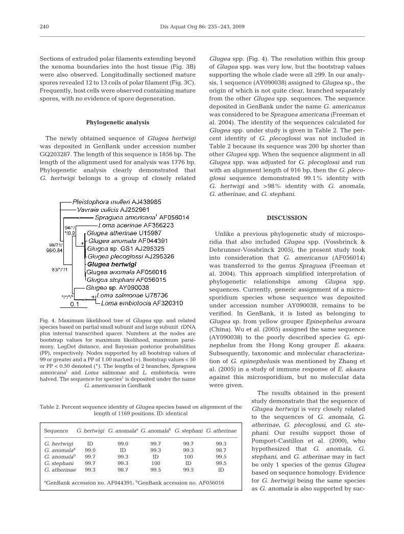
The newly obtained sequence of Glugea hertwigiwas deposited in GenBank under accession numberGQ203287. The length of this sequence is 1856 bp. Thelength of the alignment used for analysis was 1776 bp.Phylogenetic analysis clearly demonstrated thatG. hertwigi belongs to a group of closely related
Glugea spp. (Fig. 4). The resolution within this groupof Glugea spp. was very low, but the bootstrap valuessupporting the whole clade were all ≥99. In our analy-sis, 1 sequence (AY090038) assigned to Glugea sp., theorigin of which is not quite clear, branched separatelyfrom the other Glugea spp. sequences. The sequencedeposited in GenBank under the name G. americanuswas considered to be Spraguea americana (Freeman etal. 2004). The identity of the sequences calculated forGlugea spp. under study is given in Table 2. The per-cent identity of G. plecoglossi was not included inTable 2 because its sequence was 200 bp shorter thanother Glugea spp. When the sequence alignment in allGlugea spp. was adjusted for G. plecoglossi and runwith an alignment length of 916 bp, then the G. pleco-glossi sequence demonstrated 99.1% identity withG. hertwigi and >98% identity with G. anomala,G. atherinae, and G. stephani.
DISCUSSION
Unlike a previous phylogenetic study of microspo-ridia that also included Glugea spp. (Vossbrinck &Debrunner-Vossbrinck 2005), the present study tookinto consideration that G. americanus (AF056014)was transferred to the genus Spraguea (Freeman etal. 2004). This approach simplified interpretation ofphylogenetic relationships among Glugea spp.sequences. Currently, generic assignment of a micro-sporidium species whose sequence was depositedunder accession number AY090038, remains to beverified. In GenBank, it is listed as belonging toGlugea sp. from yellow grouper Epinephelus awoara(China). Wu et al. (2005) assigned the same sequence(AY090038) to the poorly described species G. epi-nephelus from the Hong Kong grouper E. akaara.Subsequently, taxonomic and molecular characteriza-tion of G. epinephelusis was mentioned by Zhang etal. (2005) in a study of immune response of E. akaaraagainst this microsporidium, but no molecular datawere given.
The results obtained in the presentstudy demonstrate that the sequence ofGlugea hertwigi is very closely relatedto the sequences of G. anomala, G.atherinae, G. plecoglossi, and G. ste-phani. Our results support those ofPomport-Castillon et al. (2000), whohypothesized that G. anomala, G.stephani, and G. atherinae may in factbe only 1 species of the genus Glugeabased on sequence homology. Evidencefor G. hertwigi being the same speciesas G. anomala is also supported by suc-
240
Fig. 4. Maximum likelihood tree of Glugea spp. and relatedspecies based on partial small subunit and large subunit rDNAplus internal transcribed spacer. Numbers at the nodes arebootstrap values for maximum likelihood, maximum parsi-mony, LogDet distance, and Bayesian posterior probabilities(PP), respectively. Nodes supported by all bootstrap values of99 or greater and a PP of 1.00 marked (+). Bootstrap values < 50or PP < 0.50 denoted (*). The lengths of 2 branches, Spragueaamericana1 and Loma salmonae and L. embiotocia, werehalved. The sequence for species1 is deposited under the name
G. americanus in GenBank
Sequence G. hertwigi G. anomalaa G. anomalab G. stephani G. atherinae
G. hertwigi ID 99.0 99.7 99.7 99.3G. anomalaa 99.0 ID 99.3 99.3 98.7G. anomalab 99.7 99.3 ID 100 99.5G. stephani 99.7 99.3 100 ID 99.5G. atherinae 99.3 98.7 99.5 99.5 ID
aGenBank accession no. AF044391; bGenBank accession no. AF056016
Table 2. Percent sequence identity of Glugea species based on alignment of the length of 1169 positions. ID: identical

Lovy et al.: Glugea hertwigi in rainbow smelt
cessful experimental infection of sticklebacks with G.hertwigi from the smelt Osmerus eperlanus (Weis-senberg 1968). Additionally, G. atherinae from thesand smelt Atherina boyeri can cross-infect the floun-der Platichthys flesus, suggesting a similarity toG. stephani (Mathieu-Daude et al. 1992). Cross-trans-mission studies in addition to rDNA comparisons havebeen used to establish species boundaries for thegenus Loma. L. embiotocia and L. salmonae were des-ignated as separate species based on their host speci-ficity to shiner perch Cymatogaster aggregata andsalmonids respectively (Kent et al. 1995), despite theirclose homologies of rDNA sequences showing only1.2% difference (Shaw et al. 1997). This suggests thatthere may be little variation in the rDNA sequencesacross species within a genus, although that work wasbased on the comparison of only 334 base pairs. Thenatural hosts for the various Glugea spp. includeG. anomala in sticklebacks Gasterosteus aculeatus,G. stephani in flatfishes such as flounder Pseudopleu-ronectes americanus, Glugea atherinae in sand smeltA. boyeri and the baitfish Dorosoma cepedianum, andG. hertwigi in smelts (Shaw & Kent 1999). Althoughphylogenetic analyses imply that close relatedness ofGlugea spp. sequences might be a consequence of theability of microsporidia to adapt to various hosts (asso-ciated with little changes in the genome), data onintragenomic variability, sequences of more genes,and data about cross-transmission are needed to verifythis hypothesis.
All of the smelts examined in the present study weresampled during the winter and spring months, whenthe water temperature was at its coldest. This wasdone because the winter and spring were the mostconvenient to capture smelt, since this is the time ofyear they enter freshwater rivers to spawn. Tempera-ture is a known factor for the development of micro-sporidia in the host, and low temperatures are knownto inhibit their development, especially in the earlystage of infection (Beaman et al. 1999, Speare et al.1999). In the present study, the finding of only maturespores and no other developmental stages in all xeno-mas examined, including the large and small xenomas,suggested that the parasite was no longer replicating.It is likely that the infections that we observed werexenomas that were over-wintering from the previoussummer. Through experimental infections of Glugeahertwigi it was determined that the parasite replicatesrapidly at 20°C and xenomas with mature spores werepresent 2 wk post-infection (Scarborough & Weidner1979), thus demonstrating that the life cycle of the par-asite can be completed within a relatively short time.It is unknown to what degree the spores from over-wintered xenomas can autoinfect the host in the fol-lowing summer. In the present study, the presence of
germinating spores and sections of everted polar fila-ments throughout the host tissue beyond the bound-aries of the xenomas suggests that the mature xenomasmay cause autoinfection in the host, although no earlystages of xenomas were seen in the area. A similarfinding was reported in three-spined sticklebacksinfected with G. anomala (Dykova & Lom 1978).Autoinfection has been proposed for other microspori-dia, such as Loma sp. in Atlantic cod Gadus morhua(Rodriguez-Tovar et al. 2003) and the Amazonian fishMyrophis platyrhynchus (Matos et al. 2003).
In addition to sequence data, the morphology ofGlugea hertwigi appeared similar to other species ofGlugea. G. hertwigi xenomas examined in the presentstudy were located predominantly in the submucosaand within the mesentery. The presence of the parasitein the submucosa is similar to findings reported byScarborough & Weidner (1979) of G. hertwigi in Osme-rus mordax, and also consistent with findings forG. stephani in winter flounder Pseudopleuronectesamericanus (Cali et al. 1986). The parasite was not ob-served in the ovaries, although previous reports de-monstrate the ovary as a common site of infection(Chen & Power 1972, Scarborough & Weidner 1979).Unfortunately, developmental stages of G. hertwigicould not be described because xenomas containedonly mature spores. In mature spores the number ofcoils in the polar filament is often diagnostic for micro-sporidian species. In the present study of G. hertwigithe polar filament had 12 to 13 coils. This is relativelyconsistent with those described for G. anomala, whichare reported to range between 12 and 14 coils, al-though 11 to 16 coils and 11 to 14 coils were observedin a microsporidian resembling G. anomala found inthe respective killifish Nothobranchius korthausae andFundulopanchax filamentosus (Lom et al. 1995). Basedon electron micrographs by Takvorian & Cali (1996), G.stephani contained approximately 12 coils of the polarfilament, and G. atheriane has been reported to have>10 coils of polar filament, although a range was notreported (Berrebi 1979). G. americanus, which hadbeen transferred to the genus Spraguea, had 6 to8 coils of polar filament (Keohane et al. 1996), thus outof the range of Glugea spp.
Rainbow smelt infected with Glugea hertwigi in PEIdemonstrated 24% prevalence in the present study.This is very similar to what has been reported in theGreat Bay region in New Hampshire, with 23.3%prevalence in Osmerus mordax sampled from October1951 to April 1952 (Haley 1954). A previous report sug-gested that females were more susceptible to infectionand carried heavier loads of G. hertwigi cysts thanmales (Chen & Power 1972). In the present study theinfection rates were marginally higher in females(26%) than in males (23%) and the infection intensity
241

Dis Aquat Org 86: 235–243, 2009
was similar in the 2 sexes, thus indicating that sex wasnot a factor for infection. This may be explained by thefact that the parasite was not found in ovaries in thepresent study, while others report the ovary as a com-mon site of infection. The affinity of the parasite toovarian tissue is unknown and it may vary dependingon host, parasite, or environmental factors. It is likelythat parasites with an affinity for ovarian tissue wouldresult in heavier and more frequent infections infemale fish. It is possible that our sampling in the win-ter and spring gave underestimates of the parasiteprevalence because the parasite likely most activelyreplicates in the summer when temperatures are attheir highest. Considering the 2 wk life cycle of theparasite, which was demonstrated in a laboratory in-fection at 20°C by Scarborough & Weidner (1979),xenomas may develop and degrade within a singlesummer. Histological examination demonstrated thepresence of remnant granulomas within the mesen-tery, which were suggestive of previously degradedxenomas and other xenomas with a heavy inflamma-tory infiltrate. The persistence of G. hertwigi xenomasin PEI smelt, however, is unknown and is most likelyrelated to environmental factors such as temperature.Xenoma size has been used as a predictor for age ofthe xenomas (Rodriguez-Tovar et al. 2004) and in thepresent study the finding of large, small, and degradedxenomas within an individual may reflect that infec-tion can occur continuously as opposed to a singleinfection time. We have demonstrated that the parasiteover-winters but does not seem to actively replicate infish through the winter, but other infection dynamics inPEI smelt are yet to be determined. Investigating thetotal amount of time that xenomas may persist and theorigins of infections, whether in freshwater or saltwa-ter, would be helpful in better understanding Glugeaspp. infections in PEI fish.
Acknowledgments. The authors gratefully thank D. Wad-owska for her help in preparing samples for TEM and N.Lewis for assistance in sampling and dissecting fish. Financialsupport was provided by research projects of the Institute ofParasitology, Biology Centre of the Academy of Sciences ofthe Czech Republic (Z60220518 and LC522), and a NaturalSciences and Engineering Research Council of CanadaStrategic Grant.
LITERATURE CITED
Beaman HJ, Speare DJ, Brimacombe M (1999) Regulatoryeffects of water temperature on Loma salmonae (Micro-spora) development in rainbow trout. J Aquat AnimHealth 11:237–245
Berrebi P (1979) Etude ultrastructurale de Glugea atherinaen. sp., microsporidie parasite de l’athérine Atherina boyeriRisso 1810 (poisson téléostéen) dans les lagunes duLanguedoc et de Provence. Z Parasitenkd 60:105–122
Cali A, Takvorian PM, Ziskowski JJ, Sawyer TK (1986) Exper-imental infection of American winter flounder (Pseudo-pleuronectes americanus) with Glugea stephani (Micro-sporida). J Fish Biol 28:199–206
Canning EU, Lom J (1986) The microsporidia of vertebrates.Academic Press, London
Chen M, Power G (1972) Infection of American smelt in LakeOntario and Lake Erie with the microsporidian parasiteGlugea hertwigi. Can J Zool 50:1183–1188
Delisle CE (1972) Monthly variations of Glugea hertwigi(Sporozoa: Microsporidia) in different tissues and organsof the freshwater smelt and the consequences of this infec-tion on the annual massive mortality of this fish. Can JZool 50:1589–1600
Dykova I, Lom J (1978) Tissue reaction of the three-spinedstickleback Gasterosteus aculeatus L. to infection withGlugea anomala (Moniez, 1887). J Fish Dis 1:83–90
Freeman MA, Yokoyama H, Ogawa K (2004) A microspori-dian parasite of the genus Spraguea in the nervous tissuesof the Japanese anglerfish Lophius litulon. Folia Parasitol(Praha) 51:167–176
Haley JA (1954) Microsporidian parasite, Glugea hertwigi inAmerican smelt from the Great Bay region, New Hamp-shire. Trans Am Fish Soc 83:84–90
Hall TA (1999) Bioedit: a user-friendly biological sequencealignment editor and analysis program for Windows95/98/NT. Nucleic Acids Symp Ser 41:95–98
Ieshko EP, Evseeva NV, Sterligova OP (2000) The role of fishparasite in fresh-water ecosystems exemplified by a parasiteof the smelt (Osmerus eperlanus). Parazitologiia 34:118–124
Kent ML, Dawe SC, Speare DJ (1995) Transmission of Lomasalmonae (Microsporea) to Chinook salmon in seawater.Can Vet J 36:98–101
Keohane EM, Takvorian PM, Cali A, Tanowitz HB, Wittner M,Weiss LM (1996) Identification of a microsporidian polartube protein reactive monoclonal antibody. J EukaryotMicrobiol 43:26–31
Larkin MA, Blackshields G, Brown NP, Chenna R and others(2007) Clustal W and Clustal X version 2.0. Bioinformatics23:2947–2948
Lom J, Noga EJ, Dykova I (1995) Ocurrence of a micro-sporean with characteristics of Glugea anomala in orna-mental fish of the family Cyprinodontidae. Dis Aquat Org21:239–242
Mathieu-Daude F, Faye N, Coste F, Manier F, Marques A,Bouix G (1992) Occurrence of microsporidiosis in marinecultured gilt-head sea bream from the Languedoc coast: aproblem of specificity in the genus Glugea (Protozoa,Microspora). Bull Eur Assoc Fish Pathol 12:67–70
Matos E, Corral L, Azevedo C (2003) Ultrastructural details ofthe xenoma of Loma myrophis (Phylum Microsporidia)and extrusion of the polar tube during autoinfection. DisAquat Org 54:203–207
Nepszy SJ, Budd J, Dechtiar AO (1978) Mortality of young-of-the-year rainbow smelt (Osmerus mordax) in Lake Erieassociated with the occurrence of Glugea hertwigi. J WildlDis 14:233–239
Pomport-Castillon C, DeJonckheere JF, Romestand B (2000)Ribosomal DNA sequences of Glugea anomala, G.stephani, G. americanus and Spraguea lophii (Microspori-dia): phylogenetic reconstruction. Dis Aquat Org 40:125–129
Rodriguez-Tovar LE, Wadowska DW, Wright GM, GromanDB, Speare DJ, Whelan DS (2003) Ultrastructural evi-dence of autoinfection in the gills of Atlantic cod Gadusmorhua infected with Loma sp. (phylum Microsporidia).Dis Aquat Org 57:227–230
242

Lovy et al.: Glugea hertwigi in rainbow smelt
Rodriguez-Tovar LE, Speare DJ, Markham RJF, Daley J(2004) Predictive modelling of post-onset xenoma growthduring microsporidial gill disease (Loma salmonae) ofsalmonids. J Comp Pathol 131:330–333
Ronquist F, Huelsenbeck JP (2003) MrBayes 3: Bayesian phy-logenetic inference under mixed models. Bioinformatics19:1572–1574
Scarborough A, Weidner E (1979) Field and laboratory studiesof Glugea hertwigi (Microsporidia) in the rainbow smeltOsmerus mordax. Biol Bull (Woods Hole) 157:334–343
Shaw RW, Kent ML (1999) Fish microsporidia. In: Whitner M,Weiss LM (eds) The microsporidia and microsporidiosis.ASM Press, Washington, DC, p 418–446
Shaw RW, Kent ML, Docker MF, Brown AMV, Devlin RH,Adamson ML (1997) A new species of Loma (Microsporea)in shiner perch (Cymatogaster aggregata). J Parasitol 83:296–301
Speare DJ, Beaman HJ, Daley J (1999) Effect of water temper-ature manipulation on a thermal unit predictive model forLoma salmonae. J Fish Dis 22:277–283
Stamatakis A (2006) RAxML-VI-HPC: maximum likelihood-based phylogenetic analyses with thousands of taxa andmixed models. Bioinformatics 22:2688–2690
Swofford DL (2001) PAUP: phylogenetic analysis using par-simony, Version 4.0b10. Sinauer Associates, Sunderland,MA
Takvorian PM, Cali A (1996) Polar tube formation and nucle-
oside diphosphatase activity in the microsporidian,Glugea stephani. J Eukaryot Microbiol 43:102S–103S
Vossbrinck CR, Debrunner-Vossbrinck BA (2005) Molecularphylogeny of the Microsporidia: ecological, ultrastructuraland taxonomic considerations. Folia Parasitol (Praha) 52:131–142
Vossbrinck CR, Baker MD, Didier ES, Debrunner-VossbrinckBA, Shadduck JA (1993) Ribosomal DNA sequences ofEncephalitozoon hellem and Encephalitozoon cuniculi:species identification and phylogenetic construction.J Eukaryot Microbiol 40:354–362
Vossbrinck CR, Andreadis TG, Vávra J, Becnel JJ (2004) Mol-ecular phylogeny and evolution of mosquito parasiticMicrosporidia (Microsporidia: Amblyosporidae). J Euka-ryot Microbiol 51:88–95
Weissenberg R (1968) Intracellular development of the micro-sporidian Glugea anomala Moniez in hypertrophyingmigratory cells of the fish Gasterosteus aculeatus, anexample of the formation of the xenoma tumors. J Proto-zool 15:44–57
Wu HB, Wu YS, Wu ZH (2005) Occurrence of a newmicrosporidium in the abdominal cavity of Epinephelusakaara. Acta Hydrobiol Sin 29:150–154
Zhang JY, Wu YS, Wu HB, Wang JG, Li AH, Li M (2005)Humoral immune responses of the grouper Epinephelusakaara against the microsporidium Glugea epinephelusis.Dis Aquat Org 64:121–126
243
Editorial responsibility: Dieter Steinhagen, Hannover, Germany
Submitted: June 2, 2009; Accepted: August 20, 2009Proofs received from author(s): November 9, 2009