Embed Size (px)
Citation preview
Phylogeny of the bee family Megachilidae (Hymenoptera: Apoidea)based on adult morphology
V I C T O R H . G O N ZA L E Z1, T E R RY G R I S WO L D
1, C H R I S T O P H E J. P RA Z
2,3
B RYA N N. D A N F O RT H2
1USDA-ARS, Bee Biology and Systematics Laboratory, Utah State University, Logan, UT, U.S.A., 2Department of Entomology,Cornell University, Ithaca, NY, U.S.A. and 3Laboratory of Evolutionary Entomology, University of Neuchatel, Neuchatel,Switzerland
Abstract. Phylogenetic relationships within the bee family Megachilidae are poorlyunderstood. The monophyly of the subfamily Fideliinae is questionable, the relation-ships among the tribes and subtribes in the subfamily Megachilinae are unknown,and some extant genera cannot be placed with certainty at the tribal level. Usinga cladistic analysis of adult external morphological characters, we explore the rela-tionships of the eight tribes and two subtribes currently recognised in Megachilidae.Our dataset included 80% of the extant generic-level diversity, representatives ofall fossil taxa, and was analysed using parsimony. We employed 200 charactersand selected 7 outgroups and 72 ingroup species of 60 genera, plus 7 species of 4extinct genera from Baltic amber. Our analysis shows that Fideliinae and the tribesAnthidiini and Osmiini of Megachilinae are paraphyletic; it supports the monophylyof Megachilinae, including the extinct taxa, and the sister group relationship ofLithurgini to the remaining megachilines. The Sub-Saharan genus Aspidosmia, arare group with a mixture of osmiine and anthidiine features, is herein removedfrom Anthidiini and placed in its own tribe, Aspidosmiini, new tribe. Protolithurginiis the sister of Lithurgini, both placed herein in the subfamily Lithurginae; theother extinct taxa, Glyptapina and Ctenoplectrellina, are more basally related amongMegachilinae than Osmiini, near Aspidosmia, and are herein treated at the tribal level.Noteriades, a genus presently in the Osmiini, is herein transferred to the Megachilini.Thus, we recognise four subfamilies (Fideliinae, Pararhophitinae, Lithurginae andMegachilinae) and nine tribes in Megachilidae. We briefly discuss the evolutionaryhistory and biogeography of the family, present alternative classifications, and providea revised key to the extant tribes of Megachilinae.
Introduction
Megachilidae is the second largest bee family, containingmore than 4000 described species worldwide (Michener, 2007;Ascher & Pickering, 2011). These solitary bees are bothecologically and economically relevant; they include manypollinators of natural, urban and agricultural vegetation. Forexample, Megachile rotundata (Fabricius) has been introducedto many parts of the world as a pollinator of alfalfa (Bohart,
Correspondence: Victor H. Gonzalez, USDA-ARS, Bee Biologyand Systematics Laboratory, Utah State University, Logan, UT 84322-5310, U.S.A. E-mail: [email protected]
1972; Michener, 2007; Pitts-Singer & Cane, 2011). Megachilidbees are found in a wide diversity of habitats on all conti-nents except Antarctica, ranging from lowland tropical rainforests to deserts to alpine environments. The diversity of nest-ing biology and floral relationships of Megachilidae is alsoastonishing. Diverse materials are used in nest building, includ-ing mud, petals, leaves (intact pieces or macerated to a pulp),resin, soil particles, gravel and plant trichomes. The inclusionof these foreign materials in nest construction may have pro-moted a massive range expansion and diversification withinthe family (Litman et al., 2011). Equally diverse are nestingsite choices: surfaces of walls, stones and tree branches; insidepre-existing cavities in the ground, wood, stems, galls and snail
,
1
shells; excavated in soil, wood or even in arboreal termitenests (e.g. Messer, 1984; Cane et al., 2007; Michener, 2007).Some megachilid genera, subgenera or species are oligolec-tic (e.g. Wcislo & Cane, 1996), collecting pollen exclusivelyfrom related plant species or genera, and frequently exhibitingunique associated morphological and behavioral adaptations(e.g. Muller, 1996; Thorp, 2000; Muller & Bansac, 2004).Other interesting adaptations are related to nesting biology.For example, females in several genera in the Anthidiini, com-monly known as wool carder bees, make cotton wool-likebrood cells from plant hairs or trichomes that they collect withtheir multidentate mandibles; also, some of them have a densetomentum on the outer surfaces of their basitarsi, which helpthem absorb extrafloral trichome secretions that are then addedto those plant hairs to waterproof the cell, facilitate manipula-tion, prevent microbial attack and to deter nest-robbing arthro-pods (Muller et al., 1996; Michener, 2007). Megachilidaealso contains a large number of cleptoparasitic bees (cuckoobees). Nineteen genera, including an entire tribe (Dioxyini),are cleptoparasites of other bees, thus making Megachilidaean important group for testing hypotheses on the origins of,and adaptations to, cleptoparasitic behaviour amongst bees.Megachilids are also notable as the primary source of inva-sive bees, including multiple species in the genera AnthidiumFabricius and Megachile Latreille (e.g. Cane, 2003; Michener,2007). For instance, A. manicatum (Linnaeus) is perhaps themost widely distributed unmanaged bee species in the world.It was unintentionally introduced to North America in the late1960s from Europe; and it is now established transcontinen-tally as well as in South America, New Zealand and the CanaryIslands (Jaycox, 1967; Smith, 1991; Miller et al., 2002; Hoe-beke & Wheeler, 2005; Maier, 2005; Zavortink & Shanks,2008; Gibbs & Sheffield, 2009; Strange et al., 2011). Despitethe ecological importance, evolutionary interest and economic
value of megachilid bees, their higher level classification andphylogeny remain poorly understood.
Classification and phylogeny
The most widely accepted classificatory proposal for bees(Michener, 2007) recognises seven extant families, althoughother proposals exist (for a discussion see Michener, 2007). InMichener’s classification, Megachilidae is organised into twosubfamilies, eight tribes (one extinct), two extinct subtribes and76 genera. An alternative proposal is that of Engel (2005), inwhich some tribes are given subfamilial rank, some tribes aretreated as subtribes and new tribes are proposed (Table 1). Twotaxa, now placed in the subfamily Fideliinae, were consideredfor a long time either as a distinct family or included within theApidae owing to a number of plesiomorphic features relativenot only to Megachilidae, but also to other long-tongued bees(Engel, 2002; Michener, 2007). However, the discovery of theimmature stages as well as studies on their nesting biology andethology suggested their closer affinity to the Megachilidae(Rozen, 1970, 1973, 1977; McGinley & Rozen, 1987).
Roig-Alsina & Michener (1993), in their study of the long-tongued bees using both larval and adult external and internalmorphological characters, provided the first exploration ofthe phylogenetic relationships of Megachilidae. Fideliinae wasrecovered as the sister group of the remaining megachilids intheir analyses, but the subfamily appeared paraphyletic whenboth larval and adult characters were combined. Fideliinae isa small group with a disjunct distribution in the xeric areas ofAsia, Africa and South America. The subfamily is segregatedinto two very distinct tribes: Fideliini containing 14 species intwo genera, and Pararhophitini with three species in a singlegenus (Engel, 2002, 2004; Michener, 2007).
The other six tribes are currently grouped into the subfamilyMegachilinae. The distinctive nature of one tribe, Lithurgini,
Table 1. Hierarchical suprageneric classifications of Megachilidae, including two proposals discussed in the text.
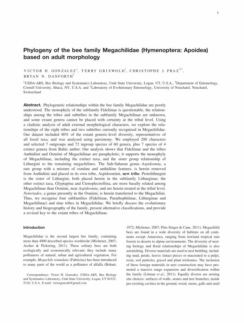
Michener (2007) Engel (2005) Proposal 1 Proposal 2 (preferred)
Subfamily Fideliinae Cockerell, 1932 Fideliinae Fideliinae FideliinaeTribe Fideliini Cockerell, 1932 Fideliini Pararhophitinae PararhophitinaeTribe Pararhophitini Popov, 1949 Neofideliini Engel, 2004 Megachilinae Lithurginae
Subfamily Megachilinae Latreille, 1802 Pararhophitinae Anthidiini LithurginiTribe Anthidiini Ashmead, 1899 Lithurginae Aspidosmiini, new tribe Protolithurginia
Tribe Dioxyini Cockerell, 1902 Lithurgini Ctenoplectrellinia MegachilinaeTribe Lithurgini Newman, 1834 Protolithurginia Dioxyini AnthidiiniTribe Megachilini Latreille, 1802 Megachilinae Glyptapinia Aspidosmiini, new tribeTribe Osmiini Newman, 1834 Anthidiini Lithurgini Ctenoplectrellinia
Subtribe Osmiina Newman, 1834 Anthidiina Megachilini DioxyiniSubtribe Ctenoplectrellina Engel, 2001a Dioxyina Osmiini Glyptapinia
Subtribe Glyptapina Cockerell, 1909a Ctenoplectrellinia Protolithurginia MegachiliniTribe Protolithurgini Engel, 2001a Glyptapinia Osmiini
MegachiliniOsmiini
OsmiinaHeriadina Michener, 1941
a Extinct taxa.
2
possessing numerous plesiomorphic as well as distinctive apo-morphic characters, resulted in subfamilial rank in the past (e.g.Michener, 1944, 1983; Engel, 2005). Lithurgini is a small tribe,with 61 species and 3 genera, found on all continents exceptAntarctica. The monophyly of the group is not questioned.Except for the sister group relationship of Lithurgini to allother extant tribes of Megachilinae, the phylogenetic study ofRoig-Alsina & Michener (1993) did not resolve the relation-ships among the tribes of Megachilinae. Relationships amongAnthidiini, Megachilini and Osmiini were unclear and neitherthe cleptoparasitic bee tribe Dioxyini nor any fossil taxa wereincluded in their analysis. Although they used a limited numberof taxa, their data suggested that all tribes are monophyleticexcept for Osmiini, which may be rendered paraphyletic byMegachilini (Michener, 2007).
Dioxyini is another unquestionably monophyletic tribe. Thissmall group of cleptoparasitic bees consists of 36 species andeight genera that attack species of Megachilini, Anthidiiniand Osmiini (Popov, 1936, 1953; Hurd, 1958). The mediantubercle on the metanotum and the extremely reduced sting(Popov, 1953), more reduced than that of the stingless bees(Apidae: Meliponini; Packer, 2003), are some of the charactersthat support the monophyly of Dioxyini. This tribe alsoshares some morphological characters with the Anthidiini, suchas the depression behind the propodeal spiracle, the shortstigma and prestigma (less than twice as long as broad),and the cleft pretarsal claws of the female (Michener, 1944,1996). Given the distinctive nature of these bees and thecharacters shared with Anthidiini, Dioxyini has been treatedas a separate subfamily or part of Anthidiini (Michener,1944; Engel, 2001). The characters shared with Anthidiinialso suggest that Dioxyini could be its sister group or derivedfrom it, making the former paraphyletic (Michener, 1996,2007). The only study exploring the relationships of Dioxyiniamong megachilids is that of Gogala (1995), using nine taxa,no outgroup and 11 morphological characters. In that study,Dioxys Lepeletier and Serville came out as the sister group toall other Megachilinae; however, as pointed out by Michener(2007), some of the characters used were highly variable andincorrectly polarised.
The remaining three tribes (Anthidiini, Osmiini andMegachilini) contain most of the species of Megachilidae.While the relationships among the few genera of Fideli-ini, Lithurgini and Dioxyini have been explored by Mich-ener (1983, 1996) and Engel (2001, 2002, 2004), relation-ships among the numerous and diverse genera of the remain-ing tribes have not been studied in detail, except for themolecular analysis of the Osmiini (Praz et al., 2008). The833 species of Anthidiini (Ascher & Pickering, 2011) havebeen grouped in 37 genera in the classification of Michener(2007) but numerous neotropical taxa assigned to the sub-generic level by him, many of them monotypic or consistingof a few unusual species, have been accorded generic sta-tus (Urban & Moure, 2007). For example, the Neotropicalgenus Hypanthidioides Moure (sensu Michener, 2007) con-tains 51 species grouped into ten subgenera (Michener, 2007;Ascher & Pickering, 2011) that are treated at the generic level
in the classification of Urban & Moure (2007). Some sub-genera are monotypic or contain a few species with unusualcharacters related to adaptations for pollen collecting (e.g.modified hairs on the mouthparts) or secondary sexual char-acters (e.g. spines on the hind coxa of the male) (Gonzalez &Griswold, 2011). Conversely, in a regional revisionary work,all nonparasitic Anthidiini were included in a single genusAnthidium (Warncke, 1980). A worldwide phylogenetic studyof the tribe is needed. The phylogenetic analysis of Anthidiiniby Muller (1996) only included western Palearctic nonpar-asitic species and its primary objective was to study floralassociations. A recent attempt to explore the generic relation-ships of the tribe from a global perspective is that of Combeyet al. (2010).
A total of 1074 species are currently included in theOsmiini (Ascher & Pickering, 2011). No morphologicalsynapomorphies are known for Osmiini and it has long beensuspected to be paraphyletic, from which one or all othermegachiline tribes originated (e.g. Engel, 2001; Michener,2007). A comprehensive molecular phylogenetic analysis ofthe tribe, including representative species of 18 of the 19currently recognised genera, supported the nonmonophyly ofthe tribe (Praz et al., 2008) with the genera AfroheriadesPeters, Noteriades Cockerell, Pseudoheriades Peters, andpossibly Ochreriades Mavromoustakis, excluded from thetribe. Interestingly, the resulting position of Noteriades withinthe Megachilini was first suggested by Griswold (1985); thephylogenetic placement of the other four genera remainsunclear (Griswold & Gonzalez, 2011).
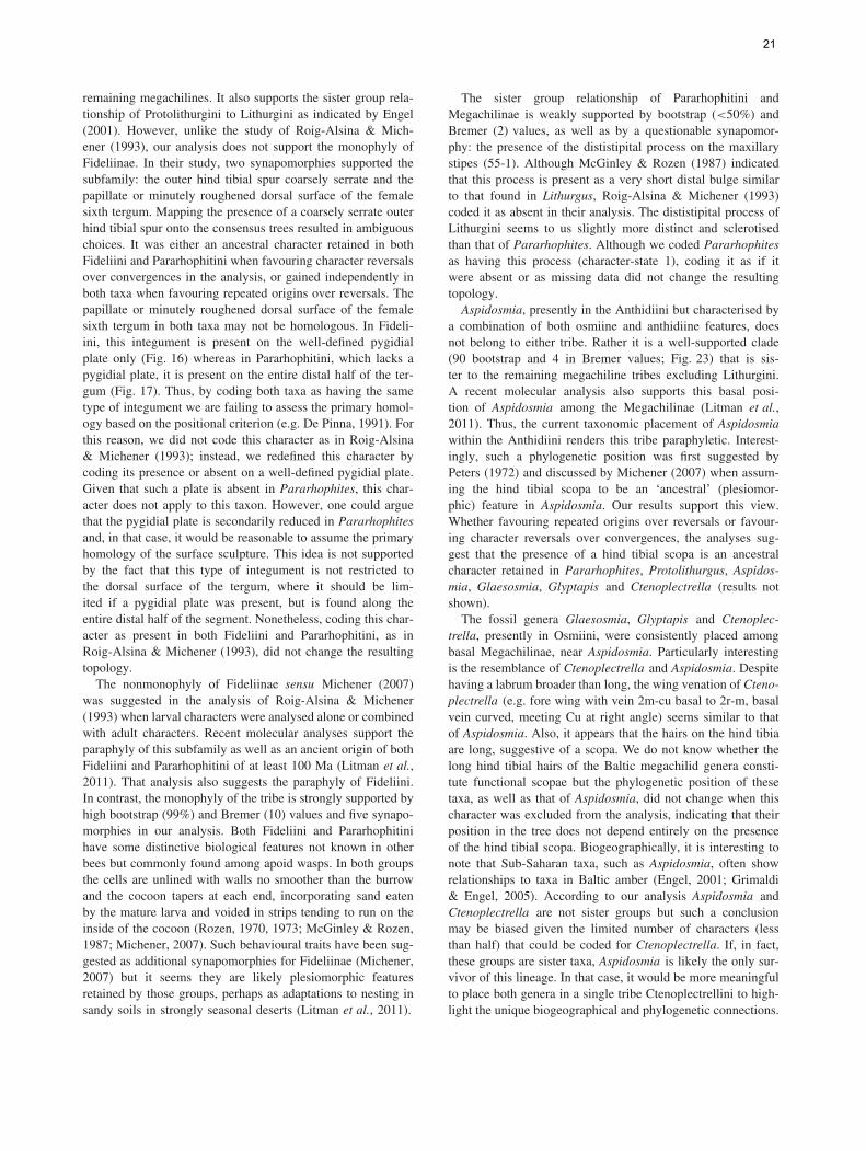
Another example of a genus that cannot be placed withconfidence at the tribal level is that of Aspidosmia Brauns(Figs 1–3). This Sub-Saharan genus, initially described as asubgenus of Osmia Panzer, and retained within the Osmiiniuntil recently (e.g. Griswold, 1985; Griswold & Michener,1997), is presently included within the Anthidiini (Michener,2007). Aspidosmia consists of two morphologically distinctspecies that exhibit a mixture of Osmiini and Anthidiinifeatures as well as unique characters found nowhere else inMegachilidae except in basal lineages. As in many osmiines,it lacks yellow integumental markings on the body (except forthe maculate clypeus of the male), the prestigma is longer thanthe stigma and the second recurrent vein (2m-cu) is basal to2r-m. However, the pretarsal claws are cleft and the shape ofthe male genitalia and associated sterna are similar to those ofAnthidiini (Peters, 1972; Michener, 2007). The presence of ahind tibial scopa (Fig. 1), in addition to the metasomal scopa ofall nonparasitic megachilids, is the most remarkable characterof Aspidosmia, shared only with Pararhophites Friese and thefossil taxa.
Accounting for 2021 species worldwide (Ascher & Picker-ing, 2011), the Megachilini is the most common and diverse ofall Megachilidae. Several genera have been traditionally recog-nised in this tribe but only three are currently accepted in theclassification of Michener (2007). Most species belong to thenonparasitic genus Megachile. The phylogenetic relationshipsamong bees in the tribe were recently explored by Gonzalez
3

1
2 3
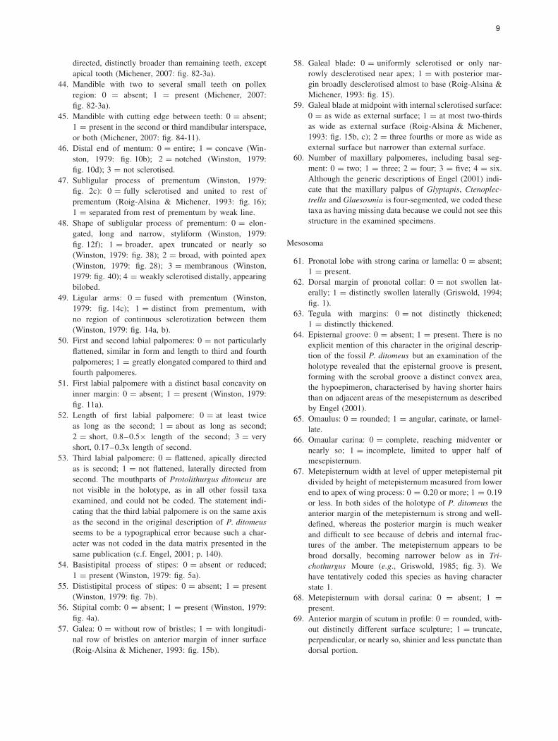
Figs 1–3. Lateral habitus (1) and frontal views of female Aspidosmia (2, 3) showing pollen on both sternal and tibial scopae. 1, 2, A. volkmanni ;3, A. arnoldi.
(2008). A new multigeneric classification and nomenclaturalchanges were proposed in that review.
A recent molecular phylogenetic analysis of the Megachili-dae, primarily done to trace the evolution of nesting biology,indicates that the family had a Gondwanan origin, arising rel-atively rapidly after the origin of bees about 100 Ma (Litmanet al., 2011). That study also suggests that Fideliinae, Fideliini,Anthidiini and Osmiini are not monophyletic.
Fossil record
The fossil record of Megachilidae is relatively good in com-parison to that for other bee families. The available materialis restricted to Cenozoic deposits of the Northern Hemisphere
(reviewed by Engel & Perkovsky, 2006). Particularly inter-esting, owing to the many plesiomorphic or unusual charac-ters, are the extinct genera from the Eocene Baltic amber:Protolithurgus Engel, Ctenoplectrella Cockerell, GlaesosmiaEngel and Glyptapis Cockerell. Protolithurgus has the dis-tinctive flattened first metasomal tergum that characterises theextant Lithurgini but the hind basitarsus is flattened, there arelong hairs on the outer surface of the hind tibia suggesting apollen collecting scopa, there are two rather than three teethon the mandible, and the typical spicules found on the outersurfaces of the lithurgine tibiae are absent. As shown by Engel(2001), Protolithurgus is likely the sister group to the remain-ing lithurgines.
The remaining genera are puzzling because unique combi-nations of features make them challenging to fit among their
4
extant relatives. Cockerell (1909), in erecting the subfamilyGlyptapinae for Glyptapis and Ctenoplectrella, commentedon its likely basal position within the Megachilidae and itsresemblance to the apid genus Ctenoplectra Kirby. The lat-ter comment was used by subsequent authors to erroneouslyinclude them in the same apine tribe, Ctenoplectrini Cock-erell. Engel (2001) was the first to recognise this error andtentatively transferred them to the Osmiini, placing Glyptapisin the subtribe Glyptapina and Ctenoplectrella and Glaesos-mia in the subtribe Ctenoplectrellina. All of these generahave aroliae and cleft pretarsal claws on all legs and, unlikeall members of the subfamily Megachilinae, an apparentlyshort labrum, much broader than long. The four species ofGlyptapis are remarkable in having hairy compound eyes (asin the megachiline genus Coelioxys Latreille and the anthidiinePachyanthidium of the subgenera Trichanthidium Cockerelland Trichanthidioides Michener and Griswold) and foveolatemesepisterna. Later, Engel (2005) elevated them to tribal rankwithin Megachilinae (Table 1). To date, the phylogenetic posi-tion of these genera remains unknown and their taxonomicplacement unresolved.
The purpose of this paper is to explore the relationshipsof the tribes in Megachilidae based on adult morphologicaldata, to validate the tribal assignment for included genera,and to develop a robust phylogeny-based classification ofthe family. Our phylogenetic analyses included adult externalmorphological characters for representative taxa of about80% of the extant generic-level diversity of the family. Weinclude in the taxa analysed representatives of all of thepuzzling extinct and extant genera of uncertain affinities(e.g. Aspidosmia) in an attempt to resolve their phylogeneticpositions and taxonomic placements. In addition we discuss theevolutionary history and biogeography of the family, proposealternative classifications, and provide a revised key to theextant tribes of Megachilinae.
Materials and methods
Taxon selection
We used species as terminals in all phylogenetic analyses.We attempted to include representatives of all megachilidgenera, choosing species that cover the maximal morphologicaland biogeographical diversity of the group. When possible,and to account for intraspecific variation, we studied morethan one specimen of each sex of each species, and otherspecies of the included genera. We examined the primary typesof Xeroheriades micheneri Griswold, Xenostelis polychromaBaker and all seven fossil taxa included in the analysis. Basedon the phylogeny of Roig-Alsina & Michener (1993), weused one species each of two genera of the family Melittidaeand eight genera of Apidae as outgroups (Table 2). Withthe exception of the holotype of X. polychroma in the SnowEntomological Museum, University of Kansas, Lawrence, KS,and the fossils in the American Museum of Natural History,New York, all specimens studied are in the U.S. National
Table 2. List of species used in the phylogenetic analysis of thefamily Megachilidae.
MELITTIDAEMacropis (Macropis) nuda (Provancher, 1882) [U.S.A.: Idaho]Melitta (Melitta) leporina (Panzer, 1799) [France; Spain; Iran]
APIDAEApinae
Apis mellifera Linnaeus, 1758 [U.S.A.: Utah]Exomalopsis (Stilbomalopsis) solani Cockerell, 1896 [Mexico:
Sinaloa; U.S.A.: Arizona]Diadasia (Coquillettapis) australis (Cresson, 1878) [U.S.A.:
Arizona, Utah]Nomadinae
Nomada utahensis Moalif, 1988 [U.S.A.: Utah]Xylocopinae
Ceratina calcarata Robertson, 1900 [U.S.A.: Illinois, North Carolina]MEGACHILIDAEFideliinaeFideliini
Fidelia (Parafidelia) pallidula (Cockerell, 1935) [South Africa:Cape Province]; F . (Fidelia) villosa Brauns, 1902 [SouthAfrica: Cape Province]; F . (Fideliana) braunsiana Friese,1905 [South Africa: Cape Province]; F . (Fideliopsis) majorFriese, 1911 [South Africa: Cape Province]
Neofidelia profuga Moure & Michener, 1955 [Chile: HuascoProvince]
PararhophitiniPararhophites orobinus (Morawitz, 1875) [Karakalpakstan;
Pakistan]; P. quadratus (Friese, 1898) [Egypt]MegachilinaeAnthidiini
Afranthidium (Capanthidium) capicola (Brauns, 1905) [SouthAfrica: Cape Province]
Anthidiellum (Loyolanthidium) robertsoni (Cockerell, 1904)[U.S.A.: Utah]
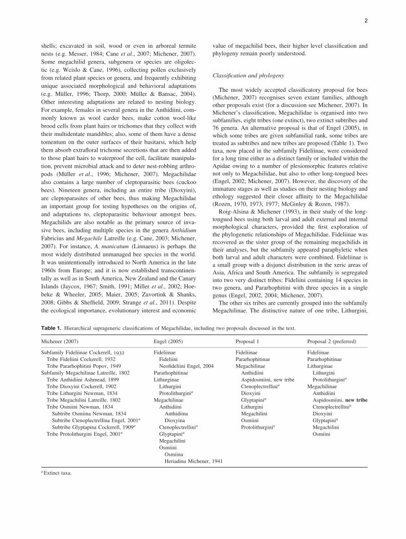
Anthidioma chalicodomoides Pasteels (1984) [South Africa: CapeProvince]
Anthidium (Anthidium) porterae Cockerell, 1900 [U.S.A.: Arizona,New Mexico]
Anthodioctes (Anthodioctes) calcaratus (Friese, 1921) [CostaRica: Guanacaste]
Aspidosmia arnoldi (Brauns, 1926); A. volkmanni (Friese, 1909)[South Africa: Cape Province]
Aztecanthidium tenochtitlanicum Snelling, 1987 [Mexico: Jalisco]Cyphanthidium intermedium Pasteels, 1969 [Namibia: Karibib]Dianthidium (Dianthidium) subparvum Swenk, 1914 [U.S.A.:
California, Utah]Duckeanthidium thielei Michener [Costa Rica: Heredia]Eoanthidium (Clistanthidium) rothschildi (Vachal, 1909) [South
Africa: Limpopo; Tanzania]Epanthidium (Epanthidium) bicoloratum (Smith, 1879) [Argentina:
Catamarca, Salta]Euaspis abdominalis (Fabricius, 1773) [Zambia]Gnathanthidium prionognathum (Mavromoustakis, 1935) [South
Africa: Cape Province]Hoplostelis (Hoplostelis) bivittata (Cresson, 1878) [Costa
Rica: Guanacaste]Hypanthidioides (Michanthidium) ferrugineum (Urban, 1992
[1994]) [Argentina: Tucuman]Hypanthidium (Hypanthidium) mexicanum (Cresson, 1878)
[Mexico: Jalisco]
5
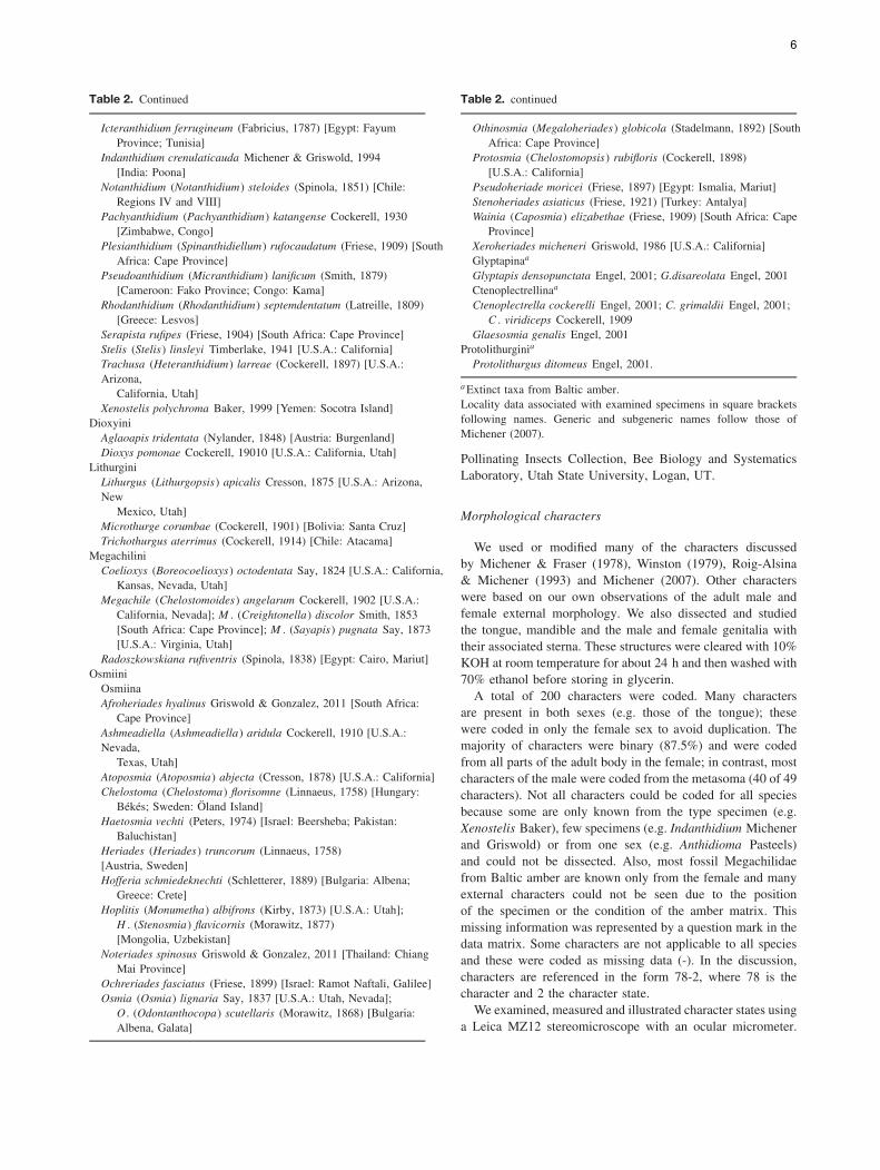
Table 2. Continued
Icteranthidium ferrugineum (Fabricius, 1787) [Egypt: FayumProvince; Tunisia]
Indanthidium crenulaticauda Michener & Griswold, 1994[India: Poona]
Notanthidium (Notanthidium) steloides (Spinola, 1851) [Chile:Regions IV and VIII]
Pachyanthidium (Pachyanthidium) katangense Cockerell, 1930[Zimbabwe, Congo]
Plesianthidium (Spinanthidiellum) rufocaudatum (Friese, 1909) [SouthAfrica: Cape Province]
Pseudoanthidium (Micranthidium) lanificum (Smith, 1879)[Cameroon: Fako Province; Congo: Kama]
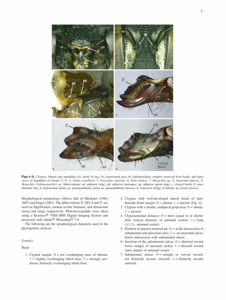
Rhodanthidium (Rhodanthidium) septemdentatum (Latreille, 1809)[Greece: Lesvos]
Serapista rufipes (Friese, 1904) [South Africa: Cape Province]Stelis (Stelis) linsleyi Timberlake, 1941 [U.S.A.: California]Trachusa (Heteranthidium) larreae (Cockerell, 1897) [U.S.A.:Arizona,
California, Utah]Xenostelis polychroma Baker, 1999 [Yemen: Socotra Island]
DioxyiniAglaoapis tridentata (Nylander, 1848) [Austria: Burgenland]Dioxys pomonae Cockerell, 19010 [U.S.A.: California, Utah]
LithurginiLithurgus (Lithurgopsis) apicalis Cresson, 1875 [U.S.A.: Arizona,New
Mexico, Utah]Microthurge corumbae (Cockerell, 1901) [Bolivia: Santa Cruz]Trichothurgus aterrimus (Cockerell, 1914) [Chile: Atacama]
MegachiliniCoelioxys (Boreocoelioxys) octodentata Say, 1824 [U.S.A.: California,
Kansas, Nevada, Utah]Megachile (Chelostomoides) angelarum Cockerell, 1902 [U.S.A.:
California, Nevada]; M . (Creightonella) discolor Smith, 1853[South Africa: Cape Province]; M . (Sayapis) pugnata Say, 1873[U.S.A.: Virginia, Utah]
Radoszkowskiana rufiventris (Spinola, 1838) [Egypt: Cairo, Mariut]Osmiini
OsmiinaAfroheriades hyalinus Griswold & Gonzalez, 2011 [South Africa:
Cape Province]Ashmeadiella (Ashmeadiella) aridula Cockerell, 1910 [U.S.A.:Nevada,
Texas, Utah]Atoposmia (Atoposmia) abjecta (Cresson, 1878) [U.S.A.: California]Chelostoma (Chelostoma) florisomne (Linnaeus, 1758) [Hungary:
Bekes; Sweden: Oland Island]Haetosmia vechti (Peters, 1974) [Israel: Beersheba; Pakistan:
Baluchistan]Heriades (Heriades) truncorum (Linnaeus, 1758)[Austria, Sweden]Hofferia schmiedeknechti (Schletterer, 1889) [Bulgaria: Albena;
Greece: Crete]Hoplitis (Monumetha) albifrons (Kirby, 1873) [U.S.A.: Utah];
H . (Stenosmia) flavicornis (Morawitz, 1877)[Mongolia, Uzbekistan]
Noteriades spinosus Griswold & Gonzalez, 2011 [Thailand: ChiangMai Province]
Ochreriades fasciatus (Friese, 1899) [Israel: Ramot Naftali, Galilee]Osmia (Osmia) lignaria Say, 1837 [U.S.A.: Utah, Nevada];
O . (Odontanthocopa) scutellaris (Morawitz, 1868) [Bulgaria:Albena, Galata]
Table 2. continued
Othinosmia (Megaloheriades) globicola (Stadelmann, 1892) [SouthAfrica: Cape Province]
Protosmia (Chelostomopsis) rubifloris (Cockerell, 1898)[U.S.A.: California]
Pseudoheriade moricei (Friese, 1897) [Egypt: Ismalia, Mariut]Stenoheriades asiaticus (Friese, 1921) [Turkey: Antalya]Wainia (Caposmia) elizabethae (Friese, 1909) [South Africa: Cape
Province]Xeroheriades micheneri Griswold, 1986 [U.S.A.: California]Glyptapinaa
Glyptapis densopunctata Engel, 2001; G.disareolata Engel, 2001Ctenoplectrellinaa
Ctenoplectrella cockerelli Engel, 2001; C. grimaldii Engel, 2001;C . viridiceps Cockerell, 1909
Glaesosmia genalis Engel, 2001Protolithurginia
Protolithurgus ditomeus Engel, 2001.
a Extinct taxa from Baltic amber.Locality data associated with examined specimens in square bracketsfollowing names. Generic and subgeneric names follow those ofMichener (2007).
Pollinating Insects Collection, Bee Biology and SystematicsLaboratory, Utah State University, Logan, UT.
Morphological characters
We used or modified many of the characters discussedby Michener & Fraser (1978), Winston (1979), Roig-Alsina& Michener (1993) and Michener (2007). Other characterswere based on our own observations of the adult male andfemale external morphology. We also dissected and studiedthe tongue, mandible and the male and female genitalia withtheir associated sterna. These structures were cleared with 10%KOH at room temperature for about 24 h and then washed with70% ethanol before storing in glycerin.
A total of 200 characters were coded. Many charactersare present in both sexes (e.g. those of the tongue); thesewere coded in only the female sex to avoid duplication. Themajority of characters were binary (87.5%) and were codedfrom all parts of the adult body in the female; in contrast, mostcharacters of the male were coded from the metasoma (40 of 49characters). Not all characters could be coded for all speciesbecause some are only known from the type specimen (e.g.Xenostelis Baker), few specimens (e.g. Indanthidium Michenerand Griswold) or from one sex (e.g. Anthidioma Pasteels)and could not be dissected. Also, most fossil Megachilidaefrom Baltic amber are known only from the female and manyexternal characters could not be seen due to the positionof the specimen or the condition of the amber matrix. Thismissing information was represented by a question mark in thedata matrix. Some characters are not applicable to all speciesand these were coded as missing data (-). In the discussion,characters are referenced in the form 78-2, where 78 is thecharacter and 2 the character state.
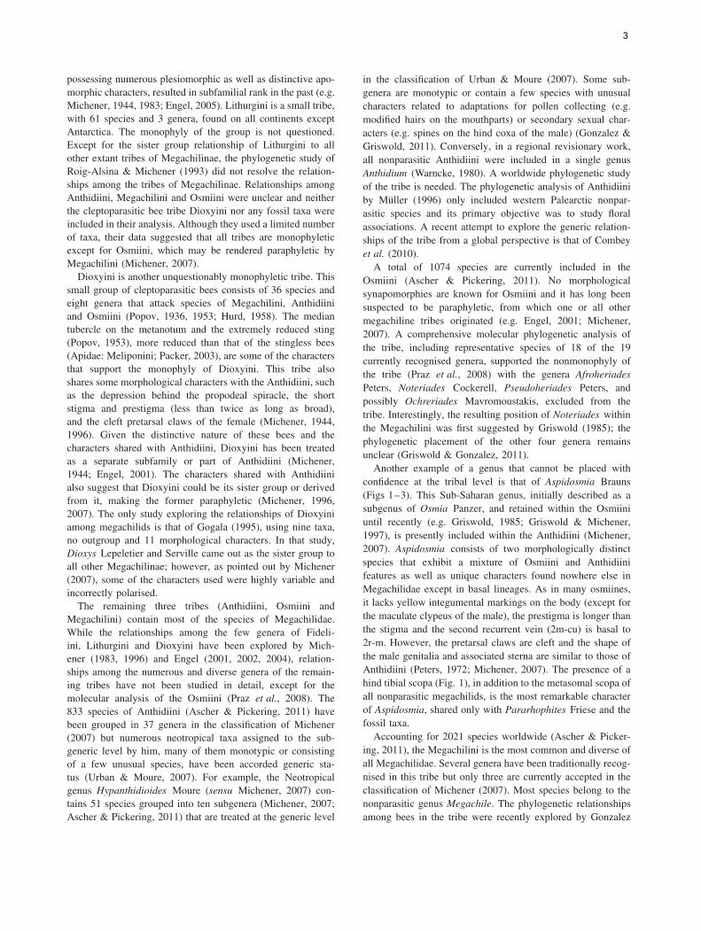
We examined, measured and illustrated character states usinga Leica MZ12 stereomicroscope with an ocular micrometer.
6
4 5
6 7
8 9
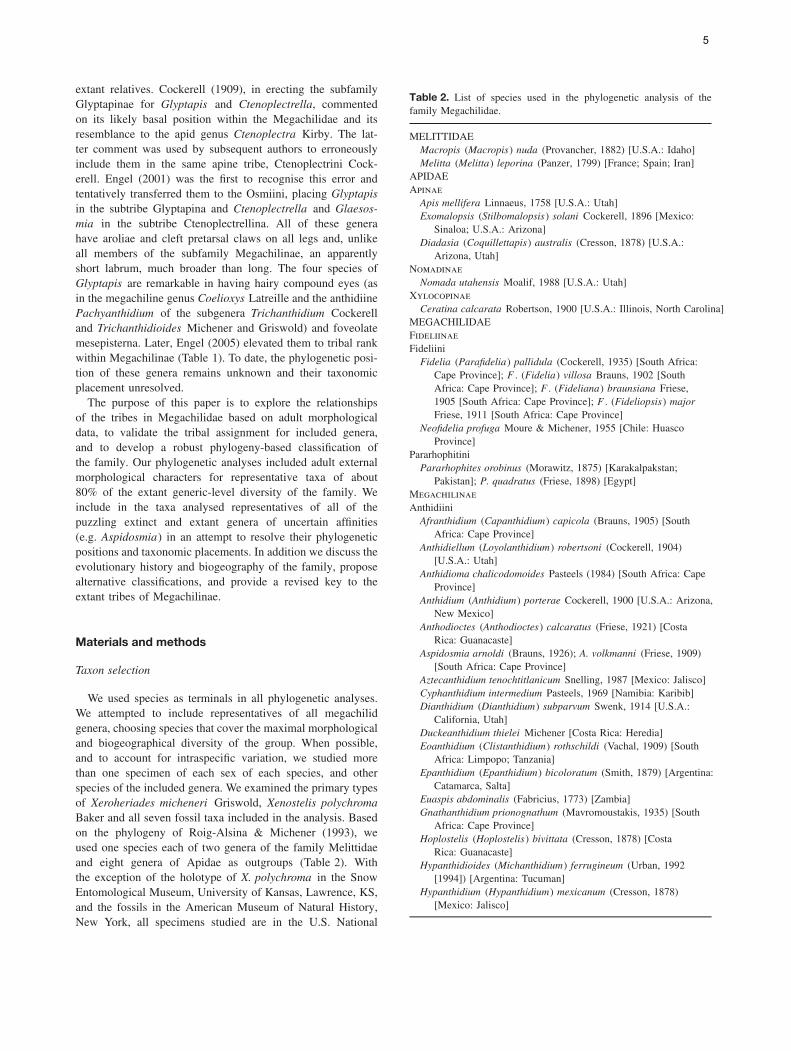
Figs 4–9. Clypeus, labrum and mandibles (4), detail of face (5), hypostomal area (6) (labiomaxillary complex removed from head), and innerviews of mandibles of female (7–9). 4, Osmia scutellaris; 5, Noteriades spinosus; 6, Stelis linsleyi ; 7, Megachile sp.; 8, Atoposmia abjecta; 9,Megachile (Callomegachile) sp. Abbreviations: ad, adductor ridge; adi, adductor interspace; ap, adductor apical ridge; c, clypeal brush; fl, innerfimbriate line; h, hypostomal carina; pc, paramandibular carina; pr, paramandibular process; tf, transverse fringe of labrum; tp, torular process.
Morphological terminology follows that of Michener (1944,2007) and Engel (2001). The abbreviations F, OD, S and T, areused for flagellomere, median ocellar diameter, and metasomalsterna and terga, respectively. Photomicrographs were takenusing a Keyence! VHX-500F Digital Imaging System andprocessed with Adobe! Photoshop! 7.0.
The following are the morphological characters used in thephylogenetic analysis.
Females
Head
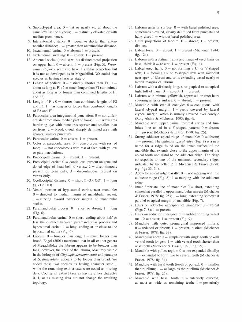
1. Clypeal margin: 0 = not overhanging base of labrum;1 = slightly overhanging labral base; 2 = strongly pro-duced, distinctly overhanging labral base.
2. Clypeus with well-developed lateral brush of hairsbeneath distal margin: 0 = absent; 1 = present (Fig. 4).
3. Clypeus with a slender, midapical projection: 0 = absent;1 = present.
4. Clypeoantennal distance: 0 = short (equal to or shorterthan vertical diameter of antennal socket); 1 = long(!1.2" antennal socket).
5. Position of anterior tentorial pit: 0 = at the intersection ofsubantennal and epistomal sulci; 1 = on epistomal sulcusbelow intersection with subantennal sulcus.
6. Insertion of the subantennal sulcus: 0 = directed towardlower margin of antennal socket; 1 = directed towardouter margin of antennal socket.
7. Subantennal sulcus: 0 = straight or curved inward,not distinctly arcuate outward; 1 = distinctly arcuateoutward.
7
8. Supraclypeal area: 0 = flat or nearly so, at about thesame level as the clypeus; 1 = distinctly elevated or withmedian prominence.
9. Interantennal distance: 0 = equal or shorter than anten-nocular distance; 1 = greater than antennocular distance.
10. Juxtantennal carina: 0 = absent; 1 = present.11. Juxtantennal swelling: 0 = absent; 1 = present.12. Antennal socket (torulus) with a distinct mesal projection
on upper half: 0 = absent; 1 = present (Fig. 5). Proto-smia rubifloris seems to have a similar projection butit is not as developed as in Megachilini. We coded thatspecies as having character state 0.
13. Length of pedicel: 0 = distinctly shorter than F1; 1 =about as long as F1; 2 = much longer than F1 (sometimesabout as long as or longer than combined lengths of F1and F2).
14. Length of F1: 0 = shorter than combined lengths of F2and F3; 1 = as long as or longer than combined lengthsof F2 and F3.
15. Paraocular area integumental punctation: 0 = not differ-entiated from more median part of frons; 1 = narrow areabordering eye with punctures sparser and smaller thanon frons; 2 = broad, ovoid, sharply delimited area withsparser, smaller punctures.
16. Paraocular carina: 0 = absent; 1 = present.17. Color of paraocular area: 0 = concolorous with rest of
face; 1 = not concolorous with rest of face, with yellowor pale maculations.
18. Preoccipital carina: 0 = absent; 1 = present.19. Preoccipital carina: 0 = continuous, present on gena and
dorsal edge of head behind vertex; 2 = discontinuous,present on gena only; 3 = discontinuous, present onvertex only.
20. Ocelloccipital distance: 0 = short (1–3" OD); 1 = long(!3.1" OD).
21. Ventral portion of hypostomal carina, near mandible:0 = directed to medial margin of mandibular socket;1 = curving toward posterior margin of mandibularsocket.
22. Paramandibular process: 0 = short or absent; 1 = long(Fig. 6).
23. Paramandibular carina: 0 = short, ending about half orless the distance between paramandibular process andhypostomal carina; 1 = long, ending at or close to thehypostomal carina (Fig. 6).
24. Labrum: 0 = broader than long; 1 = much longer thanbroad. Engel (2001) mentioned that in all extinct generaof Megachilidae the labrum appears to be broader thanlong; however, the apex of the labrum, obscurely visiblein the holotype of Glyptapis densopunctata and paratypeof G. disareolata, appears to be longer than broad. Wecoded those two species as having character state 1while the remaining extinct taxa were coded as missingdata. Coding all extinct taxa as having either character0, 1, or as missing data did not change the resultingtopology.
25. Labrum anterior surface: 0 = with basal polished area,sometimes elevated, clearly delimited from punctate andhairy disc; 1 = without basal polished area.
26. Basal projections of labrum: 0 = absent; 1 = present,distinct.
27. Labral fossa: 0 = absent; 1 = present (Michener, 1944:fig. 124).
28. Labrum with a distinct transverse fringe of erect hairs onbasal third: 0 = absent; 1 = present (Fig. 4).
29. Labral erect hairs: 0 = not forming a U- or V-shapedrow; 1 = forming U- or V-shaped row with midpointnear apex of labrum and arms extending basad nearly tolateral margins of labrum.
30. Labrum with a distinctly long, strong apical or subapicaltight tuft of hairs: 0 = absent; 1 = present.
31. Labrum with minute, yellowish, appressed or erect hairscovering anterior surface: 0 = absent; 1 = present.
32. Mandible with cranial condyle: 0 = contiguous withlateral clypeal margin; 1 = partly covered by lateralclypeal margin, which is usually elevated over condyle(Roig-Alsina & Michener, 1993: fig. 4).
33. Mandible with upper carina, trimmal carina and fim-briate line united in a Y-shaped pattern: 0 = absent;1 = present (Michener & Fraser, 1978: fig. 25).
34. Strong adductor apical ridge of mandible: 0 = absent;1 = present. The adductor apical ridge (Fig. 8) is a newname for a ridge found on the inner surface of themandible that extends close to the upper margin of theapical tooth and distal to the adductor ridge. The ridgecorresponds to one of the unnamed secondary ridgesindicated by the letter R in Michener & Fraser (1978:e.g. figs 33, 34).
35. Adductor apical ridge basally: 0 = not merging with theadductor ridge (Fig. 8); 1 = merging with the adductorridge.
36. Inner fimbriate line of mandible: 0 = short, extendingsomewhat parallel to upper mandibular margin (Michener& Fraser, 1978: fig. 25); 1 = long, extending somewhatparallel to apical margin of mandible (Fig. 7).
37. Hairs on adductor interspace of mandible: 0 = absent(Figs 7, 8); 1 = present.
38. Hairs on adductor interspace of mandible forming velvetmat: 0 = absent; 1 = present (Fig. 9).
39. Mandible with outer premarginal impressed fimbria:0 = reduced or absent; 1 = present, distinct (Michener& Fraser, 1978: fig. 33).
40. Mandibular apex: 0 = simple or with single tooth or withventral tooth longest; 1 = with ventral tooth shorter thannext tooth (Michener & Fraser, 1978: fig. 29).
41. Mandible with pollex region: 0 = not expanded distally;1 = expanded to form two to several teeth (Michener &Fraser, 1978: fig. 34).
42. Mandible with basal tooth (tooth of pollex): 0 = smallerthan rutellum; 1 = as large as the rutellum (Michener &Fraser, 1978: fig. 25).
43. Mandible with basal tooth: 0 = anteriorly directed,at most as wide as remaining teeth; 1 = posteriorly
8
directed, distinctly broader than remaining teeth, exceptapical tooth (Michener, 2007: fig. 82-3a).
44. Mandible with two to several small teeth on pollexregion: 0 = absent; 1 = present (Michener, 2007:fig. 82-3a).
45. Mandible with cutting edge between teeth: 0 = absent;1 = present in the second or third mandibular interspace,or both (Michener, 2007: fig. 84-11).
46. Distal end of mentum: 0 = entire; 1 = concave (Win-ston, 1979: fig. 10b); 2 = notched (Winston, 1979:fig. 10d); 3 = not sclerotised.
47. Subligular process of prementum (Winston, 1979:fig. 2c): 0 = fully sclerotised and united to rest ofprementum (Roig-Alsina & Michener, 1993: fig. 16);1 = separated from rest of prementum by weak line.
48. Shape of subligular process of prementum: 0 = elon-gated, long and narrow, styliform (Winston, 1979:fig. 12f); 1 = broader, apex truncated or nearly so(Winston, 1979: fig. 38); 2 = broad, with pointed apex(Winston, 1979: fig. 28); 3 = membranous (Winston,1979: fig. 40); 4 = weakly sclerotised distally, appearingbilobed.
49. Ligular arms: 0 = fused with prementum (Winston,1979: fig. 14c); 1 = distinct from prementum, withno region of continuous sclerotization between them(Winston, 1979: fig. 14a, b).
50. First and second labial palpomeres: 0 = not particularlyflattened, similar in form and length to third and fourthpalpomeres; 1 = greatly elongated compared to third andfourth palpomeres.
51. First labial palpomere with a distinct basal concavity oninner margin: 0 = absent; 1 = present (Winston, 1979:fig. 11a).
52. Length of first labial palpomere: 0 = at least twiceas long as the second; 1 = about as long as second;2 = short, 0.8–0.5" length of the second; 3 = veryshort, 0.17–0.3x length of second.
53. Third labial palpomere: 0 = flattened, apically directedas is second; 1 = not flattened, laterally directed fromsecond. The mouthparts of Protolithurgus ditomeus arenot visible in the holotype, as in all other fossil taxaexamined, and could not be coded. The statement indi-cating that the third labial palpomere is on the same axisas the second in the original description of P. ditomeusseems to be a typographical error because such a char-acter was not coded in the data matrix presented in thesame publication (c.f. Engel, 2001; p. 140).
54. Basistipital process of stipes: 0 = absent or reduced;1 = present (Winston, 1979: fig. 5a).
55. Dististipital process of stipes: 0 = absent; 1 = present(Winston, 1979: fig. 7b).
56. Stipital comb: 0 = absent; 1 = present (Winston, 1979:fig. 4a).
57. Galea: 0 = without row of bristles; 1 = with longitudi-nal row of bristles on anterior margin of inner surface(Roig-Alsina & Michener, 1993: fig. 15b).
58. Galeal blade: 0 = uniformly sclerotised or only nar-rowly desclerotised near apex; 1 = with posterior mar-gin broadly desclerotised almost to base (Roig-Alsina &Michener, 1993: fig. 15).
59. Galeal blade at midpoint with internal sclerotised surface:0 = as wide as external surface; 1 = at most two-thirdsas wide as external surface (Roig-Alsina & Michener,1993: fig. 15b, c); 2 = three fourths or more as wide asexternal surface but narrower than external surface.
60. Number of maxillary palpomeres, including basal seg-ment: 0 = two; 1 = three; 2 = four; 3 = five; 4 = six.Although the generic descriptions of Engel (2001) indi-cate that the maxillary palpus of Glyptapis, Ctenoplec-trella and Glaesosmia is four-segmented, we coded thesetaxa as having missing data because we could not see thisstructure in the examined specimens.
Mesosoma
61. Pronotal lobe with strong carina or lamella: 0 = absent;1 = present.
62. Dorsal margin of pronotal collar: 0 = not swollen lat-erally; 1 = distinctly swollen laterally (Griswold, 1994;fig. 1).
63. Tegula with margins: 0 = not distinctly thickened;1 = distinctly thickened.
64. Episternal groove: 0 = absent; 1 = present. There is noexplicit mention of this character in the original descrip-tion of the fossil P. ditomeus but an examination of theholotype revealed that the episternal groove is present,forming with the scrobal groove a distinct convex area,the hypoepimeron, characterised by having shorter hairsthan on adjacent areas of the mesepisternum as describedby Engel (2001).
65. Omaulus: 0 = rounded; 1 = angular, carinate, or lamel-late.
66. Omaular carina: 0 = complete, reaching midventer ornearly so; 1 = incomplete, limited to upper half ofmesepisternum.
67. Metepisternum width at level of upper metepisternal pitdivided by height of metepisternum measured from lowerend to apex of wing process: 0 = 0.20 or more; 1 = 0.19or less. In both sides of the holotype of P. ditomeus theanterior margin of the metepisternum is strong and well-defined, whereas the posterior margin is much weakerand difficult to see because of debris and internal frac-tures of the amber. The metepisternum appears to bebroad dorsally, becoming narrower below as in Tri-chothurgus Moure (e.g., Griswold, 1985; fig. 3). Wehave tentatively coded this species as having characterstate 1.
68. Metepisternum with dorsal carina: 0 = absent; 1 =present.
69. Anterior margin of scutum in profile: 0 = rounded, with-out distinctly different surface sculpture; 1 = truncate,perpendicular, or nearly so, shinier and less punctate thandorsal portion.
9
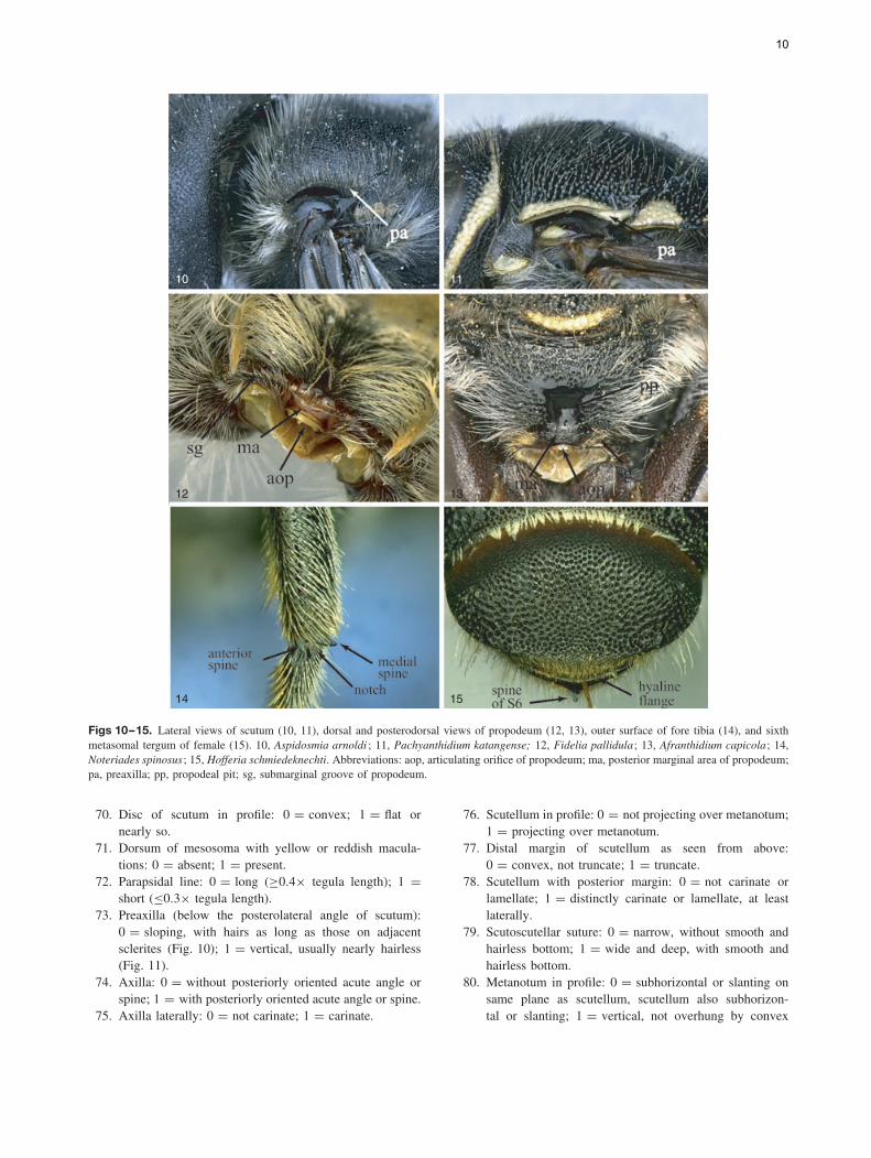
10 11
12 13
14 15
Figs 10–15. Lateral views of scutum (10, 11), dorsal and posterodorsal views of propodeum (12, 13), outer surface of fore tibia (14), and sixthmetasomal tergum of female (15). 10, Aspidosmia arnoldi ; 11, Pachyanthidium katangense; 12, Fidelia pallidula; 13, Afranthidium capicola; 14,Noteriades spinosus; 15, Hofferia schmiedeknechti. Abbreviations: aop, articulating orifice of propodeum; ma, posterior marginal area of propodeum;pa, preaxilla; pp, propodeal pit; sg, submarginal groove of propodeum.
70. Disc of scutum in profile: 0 = convex; 1 = flat ornearly so.
71. Dorsum of mesosoma with yellow or reddish macula-tions: 0 = absent; 1 = present.
72. Parapsidal line: 0 = long (!0.4" tegula length); 1 =short (#0.3" tegula length).
73. Preaxilla (below the posterolateral angle of scutum):0 = sloping, with hairs as long as those on adjacentsclerites (Fig. 10); 1 = vertical, usually nearly hairless(Fig. 11).
74. Axilla: 0 = without posteriorly oriented acute angle orspine; 1 = with posteriorly oriented acute angle or spine.
75. Axilla laterally: 0 = not carinate; 1 = carinate.
76. Scutellum in profile: 0 = not projecting over metanotum;1 = projecting over metanotum.
77. Distal margin of scutellum as seen from above:0 = convex, not truncate; 1 = truncate.
78. Scutellum with posterior margin: 0 = not carinate orlamellate; 1 = distinctly carinate or lamellate, at leastlaterally.
79. Scutoscutellar suture: 0 = narrow, without smooth andhairless bottom; 1 = wide and deep, with smooth andhairless bottom.
80. Metanotum in profile: 0 = subhorizontal or slanting onsame plane as scutellum, scutellum also subhorizon-tal or slanting; 1 = vertical, not overhung by convex
10

scutellum whose posterior margin is more or less verti-cal; 2 = vertical, strongly overhung by scutellum whoseposterior margin faces downward.
81. Metanotum with median tubercle or spine: 0 = absent;1 = present (Michener, 2007; fig. 83-1).
82. Propodeum in profile: 0 = with a nearly horizontal basalzone, behind which it rather abruptly turns downward toform the declivous posterior surface; 1 = with a steeplyslanting or sometimes convex basal zone or entirelydeclivous.
83. Basal zone of propodeum with distinct row of pits:0 = absent; 1 = present (Michener, 2007; fig. 82-14).
84. Propodeal basal zone: 0 = not bounded by a posteriorcarina or carina incomplete; 1 = bounded by a completeposterior carina.
85. Propodeal triangle with hairs: 0 = absent; 1 = present,widespread.
86. Propodeal triangle with integument: 0 = largely smoothand shiny; 1 = dull, lineolate, imbricate, minutely punc-tate or rugose.
87. Propodeal pit: 0 = rounded or elongate, but not linear;1 = linear.
88. Propodeum with a shiny fovea behind spiracle definedby posterior carina: 0 = absent; 1 = present (Michener,2007; fig. 82-8).
89. Posterior marginal area of propodeum dorsomedially:0 = rounded or flat, not carinate in lateral view, dis-tinctly projecting posteriorly, submarginal groove usuallyshallow (Fig. 12); 1 = carinate or sharply angulated, notdistinctly projecting posteriorly, submarginal groove usu-ally deep (Fig. 13).
90. Submarginal groove of propodeum: 0 = continuous, notsubmedially interrupted; 1 = not continuous, submedi-ally interrupted by a distinct wall, forming a deep pit.
91. Fore coxa with oblique carina or lamella medially:0 = absent; 1 = present.
92. Outer surfaces of fore and mid tibiae apically withacute angle and distinct notch anteriorly: 0 = absent;1 = present (Fig. 14). In some species the acute angle iswell-developed and it could be considered a spine, givingthe impression of having an anterior and medial spine onthe fore and mid tibiae. Some Anthidiini, such as Euaspisabdominalis, Pachyanthidium katangense and Stelis lins-leyi, also have two apical spines on the outer surface ofthe fore and mid tibiae; however, the other spine is onthe posterior margin. In Aztecanthidium tenochtitlanicumthe anterior margin is developed into a small spine sim-ilar to that of Megachilini. However, the notch is absentand the apical margin is densely covered by hairs; sucharea is usually bare in Megachilini. We coded all thesespecies as having character state 0.
93. Fore tibial spur (antennal cleaner) with malus: 0 = sim-ple, without a distinct projecting ridge on its anterior side;1 = with low expansion at right angle to velum, curvingapically into spine of malus; 2 = with strong expansionat right angles to velum, ending in strong angle or prong(Schonitzer & Renner, 1980; fig. 19).
94. Length of apical portion of malus of fore tibial spur:0 = long, at least half length of base of malus; 1 = short,less than half length of base of malus.
95. Fore tibial spur with apical row of teeth on apex of malus(along same margin of velum): 0 = absent; 1 = present(Schonitzer, 1986; figs 1–10).
96. Mid tibia outer apical margin: 0 = with a medialspine (Michener, 2007; fig. 82-6b); 1 = with medialand posterior spines (Michener, 2007; fig. 82-6a). InPlesianthidium rufocaudatum, Epanthidium bicoloratumand Cyphanthidium intermedium the medial spine isemarginate thus forming two spines; in Xenostelis thereare two spines, but one is anterior and the other medial.Thus, we coded these species as having character state0. The generic description of Ctenoplectrella in Engel(2001) indicates the absence of a spine on the outer sur-face of the mid tibia; however, such a spine is barelyvisible in the holotype of C. cockerelli and clearly visi-ble in that of C. viridiceps and C. phaeton Gonzalez &Engel (2011). We coded these species as having characterstate 1.
97. Mid tibial spur: 0 = finely serrate or ciliate; 1 = coarselyserrate.
98. Hind coxa with ventral carina: 0 = absent; 1 = present.99. Hind tibia with basitibial plate: 0 = absent; 1 = present.
100. Hind tibia with smooth, shiny, elevated bare area basally:0 = absent; 1 = present.
101. Hind tibia with distinct longitudinal carina on outer sur-face: 0 = absent; 1 = present.
102. Hind tibia with strong tubercles or spicules on outer sur-face that do not end in hairs or bristles: 0 = absent;1 = present (Michener, 2007; fig. 80-3b).
103. Hind tibial scopa consisting of uniformly dispersed longhairs on outer surface: 0 = absent; 1 = present. We donot know if the long tibial hairs of the examined fossiltaxa are functional scopae but we coded these species ashaving character state 1. Coding these species as havingcharacter state 0, as missing information, or excludingthis character from the analyses did not change the result-ing topology (see results).
104. Inner hind tibial spur: 0 = with apex straight or nearlyso; 1 = with apex strongly curved.
105. Outer hind tibial spur: 0 = about as long as inner spur;1 = distinctly shorter than inner spur.
106. Hind basitarsus: 0 = !3" longer than broad; 1 = #1.5"longer than broad; 2 = 1.6–2.9" longer than wide.
107. Hind basitarsus with distinctly long, simple hairs on pos-terior margin (as in Fidelia): 0 = absent; 1 = present.
108. Pretarsal claws: 0 = bifurcate or cleft, inner ramus some-times reduced; 1 = simple.
109. Arolia: 0 = present; 1 = absent.110. Wing vestiture: 0 = hairy throughout (Michener, 2007;
fig. 85-2); 1 = partly absent.111. Fore wing with distal papillae: 0 = absent; 1 = present.
In Anthidium porterae, Megachile pugnata andMegachile discolor the papillae are more pointed andapically curved than the typical papillae found in apids,
11
such as Anthophora Latreille. However, we coded thesespecies as having character state 1.
112. Prestigma: 0 = elongate, more than twice as long asbroad (Michener, 2007; fig. 76-1a); 1 = short, at mosttwice as long as broad (Michener, 2007; fig. 82-1c). Thewidth of the prestigma was measured to its margin, notto the wing margin as shown in fig. 10-8 of Michener(2007), that is, we excluded the width of the costal veinin this measurement. In Ctenoplectrella cockerelli theprestigma is more elongate than shown in fig. 43 of Engel(2001); we coded this species as having character state 1.
113. Stigma: 0 = longer than broad, length beyond vein r atleast half as long as margin basal to vein r, margin withinmarginal cell convex or sometimes straight (Michener,2007; fig. 68-1); 1 = longer than broad, length beyondvein r less than half as long as margin basal to vein r,margin within marginal cell concave (Michener, 2007;fig. 76-1b); 2 = not longer than broad, almost parallel-sided (Michener, 2007; fig. 82-1a); 3 = narrow, almostparallel-sided (as in Apis mellifera).
114. Apex of marginal cell: 0 = pointed, on wing margin;1 = separated from wing margin, pointed; 2 = separatedfrom wing margin, rounded.
115. Number of submarginal cells: 0 = three; 1 = two.116. Length of second submarginal cell on posterior mar-
gin: 0 = equal or longer than first on posterior margin;1 = shorter than first on posterior margin.
117. Basal vein: 0 = straight or nearly so, meeting Cu at acuteangle; 1 = curved, meeting Cu at right angle (Engel,2001; fig. 37).
118. Basal vein: 0 = confluent or distal to cu-v (Engel, 2001;fig. 37); 1 = basal to cu-v.
119. Vein 2m-cu (second recurrent vein): 0 = distinctly diago-nal, strongly or gently curved before meeting with 2r-m;1 = straight, or nearly so, for entire length (Michener,2007; fig. 81-1b).
120. Vein 2m-cu: 0 = basal to 2r-m (Michener, 2007; fig. 81-1b); 1 = confluent with, or distal to, 2r-m (Michener,2007; fig. 82-1). In species with only two submarginalcells we are assuming that either the second abscissa ofRs (first submarginal crossvein) or the first r-m (secondsubmarginal crossvein) is missing. Although the originaldescription of Ctenoplectrella cockerelli indicates that2m-cu is basal to 2r-m (Engel, 2001; fig. 43), a reexam-ination of the holotype revealed that these two veins arein fact confluent; thus, this species was coded as havingcharacter state 1.
121. Jugal lobe of hind wing: 0 = long, one-half or more van-nal lobe length; 1 = short, more than one-fourth but lessthan half, vannal lobe length; 2 = very short, less thanone-fourth vannal lobe length. Both lobes were measuredfrom the wing base to the apices of the lobes as indicatedin Michener (2007).
122. Hind wing with second abscissa of vein M + Cu:0 = short, #3.0" length of vein cu-v; 1 = long, !3.1"length of vein cu-v.
Metasoma
123. Coloration of metasomal terga: 0 = entirely of one color;1 = not entirely of one color, with distinct white, yellow,or reddish maculations.
124. T1: 0 = long, convex in profile, with posterior marginstraight or nearly so, and with distinct anterior and dor-sal surfaces; 1 = small, flattened, with posterior marginrounded, anterior and dorsal surfaces indistinguishable.
125. Junction of anterior and dorsal surfaces of T1; 0 =rounded; 1 = angled; 2 = carinate.
126. T2–T3 with deep postgradular grooves: 0 = absent;1 = present.
127. T2–T5 with distinct depressed marginal zones: 0 =absent; 1 = present.
128. T5 with polished apical margin: 0 = absent or narrowand parallel-sided; 1 = broad, wider in middle, basalmargin with long, stiff setae (Fig. 17).
129. T6 with transverse preapical carina: 0 = absent; 1 =present.
130. T6 in profile with dorsal surface: 0 = horizontal or sub-horizontal; 1 = vertical or convex.
131. T6 with wide apical hyaline flange: 0 = absent; 1 =present (Fig. 15).
132. Pygidial plate: 0 = present; 1 = absent.133. Integument of pygidial plate: 0 = not papillate; 1 =
papillate or minutely roughened (Fig. 16). The integu-ment of the pygidial plate of Melitta leporina is stronglyimbricate, thus somewhat resembling that of FideliaFriese and Neofidelia Moure and Michener. We codedthis species as having character state 0. This charac-ter does not apply to the two species of Pararhophitesbecause they do not have a distinct pygidial plate(Fig. 17).
134. Pygidial plate shape: 0 = triangular or nearly so, basallyoccupying about median one-third to one-fourth of ter-gal width (Michener, 2007; fig. 10-13); 1 = triangular ornearly so, basally occupying at least median two-thirdsof tergal width (Fig. 16); 2 = elongate, very narrow, nottriangular (Michener, 2007; fig. 80-2).
135. T6 with sublateral teeth on apical margin: 0 = absent;1 = present.
136. Metasomal sternal scopa: 0 = absent; 1 = present.137. S1 with subapical tooth, spine or projection: 0 = absent;
1 = present.138. Length of S6: 0 = short, about as long as wide or shorter
(length measured from apodeme to distal margin later-ally); 1 = elongate, !1.2" longer than wide.
139. Apodeme of S6: 0 = with distinct disc between marginalridge and transapodemal ridge (Fig. 18; see also Packer,2004; fig. 6a, d); 1 = with disc reduced or absent(Figs 19, 20; see also Packer, 2004; fig. 7f).
140. Pregradular area of S6: 0 = not elongate laterally(Fig. 18); 1 = elongate laterally (Figs 19, 20).
141. Width of elongated pregradular area of S6 (widthmeasured just posterior to apodeme, in ventral view):
12

16 17
20 21
18
19
Figs 16–21. Dorsal apex of metasoma (16, 17) and sixth metasomal sternum of female (18–20) and third sternum of male (21). 16, Fideliavillosa; 17, Pararhophites orobinus; 18, Hoplostelis bivittata; 19, Megachile pugnata; 20, Afranthidium capicola; 21, Stenoheriades asiaticus.Abbreviations: apo, apodeme; g, gradulus; lm, lamella of gradulus; pg, pregradular area; T6, sixth tergum.
0 = narrow, one-fourth or less of concavity width;1 = broad, at least half of concavity width (Figs 19, 20).
142. Pregradular area of S6: 0 = entirely or almost entirelysclerotised; 1 = entirely membranous or weakly scle-rotised (often easily broken during dissection)(Figs 19, 20). In Coelioxys octodentata the pregradulararea is membranous or weakly sclerotised only medially.We coded this species as having character state 0.
143. Concavity on basal margin of S6: 0 = deep, #2.0"wider than deep (Figs 19, 20); 1 = shallow, !2.1"wider than deep (Fig. 18).
144. Apex of S6: 0 = truncate; 1 = broadly to narrowlyrounded; 2 = pointed.
145. S6 with apical spine: 0 = absent; 1 = present.146. Stylet of sting apparatus: 0 = normal, distinct; 1 =
reduced or absent.
147. Pubescence of apex of sting gonostylus: 0 = nearly hair-less to sparsely covered by short hairs (# maximumgonostylus width in lateral view); 1 = densely cov-ered by long plumose hairs (!1.2" gonostylarwidth).
148. Medial and lateral portions of marginal ridge of T7hemitergite: 0 = not parallel-sided (e.g. Packer, 2003;fig. 5c); 1 = parallel to each other or nearly so (Packer,2003; fig. 5a, b).
149. Lamina spiracularis of T7 hemitergite: 0 = smooth andshiny, not sculptured; 1 = weakly to markedly sculp-tured (Packer, 2003; fig. 2e).
150. T7 hemitergite with strong protrusion on the laminaspiracularis, near base of lateral process: 0 = absent orweak; 1 = present (Packer, 2003; fig. 5b).
13
151. Spiracle of T7 hemitergite: 0 = located on the basal two-thirds of hemitergite length; 1 = located at or near apicalthird of hemitergite length.
Male
Head
152. Colour of clypeus: 0 = concolorous with rest of face;1 = yellow or pale, not concolorous with rest of face.
153. Median flagellomeres (F5–F7): 0 = short (#1.3" longerthan broad); 1 = long (!1.6" longer than broad).
154. Mandible: 0 = edentate (i.e. without subapical tooth);1 = with two teeth; 2 = with three or more teeth.
155. Basal tooth of mandible: 0 = pointed; 1 = truncate.156. Inferior process of mandible: 0 = reduced or absent;
1 = present (e.g., Gonzalez & Griswold, 2007; figs14, 17).
Mesosoma
157. Front coxal spine: 0 = absent; 1 = present (e.g., Gonza-lez & Griswold, 2007; figs 25–27).
158. Front tarsi: 0 = unmodified; 1 = modified, distinctivelyenlarged, excavated, inner surface frequently with darkspots (Michener, 2007; fig. 84-19a).
159. Hind basitarsus: 0 = elongate, !4.0" longer than broad;1 = not elongate, #3.5" longer than broad.
160. Arolia: 0 = absent or minute; 1 = present, large.
Metasoma
161. T5 with lateral spine: 0 = absent; 1 = present.162. T6 with transverse preapical carina: 0 = absent; 1 =
present, at least laterally.163. T6 with lateral spine: 0 = absent; 1 = present.164. T7: 0 = exposed, posteriorly directed; 1 = hidden, and/or
anteriorly or ventrally directed.165. T7 with pygidial plate: 0 = present, distinct; 1 = absent,
but sclerotised apical rim suggests apex of plate;2 = absent, without apical rim.
166. T7 with distinct lateral lobe: 0 = absent; 1 = present.167. T7 with median apical spine: 0 = absent; 1 = present.168. Number of fully exposed metasomal sterna: 0 = two or
three; 1 = four; 2 = five or six.169. S1 subapically: 0 = not produced, without a distinct dou-
ble margin; 1 = produced, forming a distinct doublemargin.
170. S3: 0 = not concealed by S2; 1 = concealed by S2.171. S3 with gradulus: 0 = not produced into a thin, hya-
line lamella; 1 = produced into a thin, hyaline lamella(Fig. 21). This is the same character referred to byGriswold (1985) and Michener (2007) as lateral hyalineflaps of S3. Lamella may cover most of disc.
172. S3 with disc: 0 = bare or sparsely pubescent; 1 = denselycovered with velvety pubescence (Fig. 21).
173. Fringe on apical margin of S3: 0 = absent; 1 = present,medially directed.
174. S5: 0 = well sclerotised as in the preceding sterna;1 = poorly sclerotised, translucent to membranous, notas preceding terga.
175. S5 length: 0 = #2" wider than long; 1 = !2.1" widerthan long. Because the midapical margin of S5 as wellas of S6 is highly variable, we measured the length ofthese sterna on the lateral margin, from the base of theapodeme to apical margin of the sternum.
176. S5 with hairs on disc: 0 = simple or plumose;1 = apically modified (e.g. lanceolate, ovate-acuminate,capitate or spatulate).
177. Pilose postgradular area of S5: 0 = large, maximumwidth !0.6" sternal width; 1 = small, #0.5" sternalwidth.
178. S5 with small lateral spine: 0 = absent; 1 = present.179. S5 with medial emargination: 0 = absent; 1 = present.180. S5 with fringe of modified hairs on margin lateral to
median emargination: 0 = absent; 1 = present. In Pseu-doanthidium lanificum S5 is deeply concave medially,with a distinct postgradular lateral extension distally,bearing a comb of ciliate cuticular processes, these arenot modified hairs. We coded this species as having char-acter state 0.
181. S5 with medial comb of heavily sclerotised, simple blacksetae on distal margin: 0 = absent; 1 = present (Pasteels,1984; fig. 266).
182. S6: 0 = well sclerotised as in the preceding sterna;1 = poorly sclerotised, translucent to membranous.
183. Length of S6: 0 = #2" wider than long; 1 = !2.1"wider than long.
184. Hairs of postgradular area of S6: 0 = sparse to dense,but not forming distinct sublateral patches; 1 = formingdistinct sublateral patches.
185. S6 with hairs on postgradular area: 0 = simple orplumose; 1 = apically modified (e.g. lanceolate, ovate-acuminate, capitate or spatulate).
186. S7: 0 = well sclerotised throughout, usually pilose(Michener, 2007; fig. 80-4e); 1 = weakly sclerotisedmedially, thus forming two separated, sclerotised scle-rites; 2 = mostly membranous, frequently hairless, atmost barely indicated by weakly sclerotised apodemes.
187. Length of S8: 0 = about as long as broad or shorter(Michener, 2007; fig. 82-2g); 1 = !1.3" longer thanbroad (Michener, 2007; fig. 81-11b).
188. Spiculum of S8: 0 = short, broad, parallel-sided (Mich-ener, 2007; fig. 88-6c); 1 = long, narrow, parallel-sided (Michener, 2007; fig. 82-2g, i); 2 = narrowlyrounded or V-shaped (Michener, 2007; fig. 81-11b); 3 =broadly rounded (Michener, 2007; fig. 77-1b); 4 = bifid.Character state 4 is only present in Plesianthid-ium rufocaudatum and Icteranthidium ferrugineum.
189. Distal margin of S8: 0 = not medially produced as alobe; 1 = medially produced as a lobe (Michener, 2007;fig. 81-11b).
14
190. Gonobase: 0 = present, ventrally absent (Michener,2007; fig. 88-6a ,b); 1 = present, forming a completering, ventrally narrower (Michener, 2007; fig. 84-10a);2 = present, forming a complete ring, well devel-oped ventrally (Michener, 2007; fig. 80-4c); 3 = entirelyreduced or absent (Michener, 2007; fig. 82-2h).
191. Gonocoxite length: 0 = subequal to, or longer than,gonostylus (Michener, 2007; fig. 77-1a); 1 = short,#0.5" gonostylar length (Michener, 2007; fig. 84-10a).
192. Volsella: 0 = absent; 1 = present.193. Apex of volsella with clearly differentiated medial digitus
and lateral cuspis: 0 = absent; 1 = present (Michener,2007; fig. 77-1a).
194. Volsella located: 0 = not in the same plane as gonocox-ite; 1 = in the same plane as gonocoxite.
195. Articulation between gonostylus and gonocoxite: 0 =fused, thus forming an unsegmented appendage; 1 = dis-tinct, at least ventrally.
196. Gonostylus with apex (in ventral view): 0 = reachingabout the same level as apex of penis valves; 1 = wellsurpassing apex of penis valves; 2 = short, not reachingapex of penis valves.
197. Apex of gonostylus (in ventral view): 0 = curved,laterally directed; 1 = curved, medially directed; 2 =straight, posteriorly directed.
198. Shape of gonostylus: 0 = not distinctly enlarged api-cally; 1 = distinctly enlarged apically.
199. Bridge of penis valves: 0 = absent; 1 = present.200. Apodemes of the penis valve: 0 = not projecting through
genital foramen; 1 = projecting through genital foramen(Michener, 2007; fig. 82-2a).
Phylogenetic analysis
We built a data matrix in WinClada (Nixon, 1999) con-sisting of 200 characters for seven outgroup species and 72representative ingroup taxa (Table 2, Appendix S1). Parsimonyanalyses were carried out in Tree Analysis Using New Technol-ogy (TNT; Goloboff et al., 2003). All characters were treatedas nonadditive and equally weighted. Tree search in TNT wasdone by implementing sectorial searches with tree drifting (TD)and tree fusing (TF) and ratchet runs with TD and TF. Weused the following search: keep a maximum of 10 000 ran-dom trees, 500 random addition sequences, and 1000 ratchetiterations, including 100 cycles of TD and 100 rounds of TFper iteration. Branch robustness was estimated with 10 000bootstrap replicates (Felsenstein, 1985) and absolute Bremersupport (Bremer, 1994) in TNT. The latter search was doneby withholding 10 000 suboptimal trees up to 10 steps longerthan the most parsimonious trees and plotting the values onthe strict consensus tree obtained from the final TNT par-simony run. We used the strict consensus tree to trace thepossible evolutionary pattern of those characters of particularevolutionary or taxonomic interest, namely the coarsely serratehind tibial spur and the female hind tibial scopa. Trees werevisualised and printed in WinClada, collapsing unsupported
nodes and using DELTRAN (slow) for character optimisa-tion; the latter favours, when the choices are equally parsi-monious, repeated origins of characters over reversals. Theabbreviations MPT, L, CI and RI are used for most parsimo-nious trees, tree length, and consistency and retention indices,respectively.
Results
The analysis of the data matrix including 79 taxa and200 characters yielded four most parsimonious trees (MPTs)(L = 1494, CI = 15, RI = 57); five nodes collapsed in theconsensus tree and most branches were poorly supported byboth bootstrap and Bremer values. The analysis recoveredthe subfamily Megachilinae, including the fossil taxa, butnot the Fideliinae (Figs 22, 23); the latter subfamily is para-phyletic as applied in Michener (2007) but both of its tribesare monophyletic. Fideliini is the sister group of the remain-ing megachilids. The following five putative synapomorphiessupport the monophyly of the Fideliini: mandible with uppercarina, trimmal carina and fimbriate line united in a Y-shapedpattern (33-1); maxillary galea without row of bristles on ante-rior margin of internal surface (57-0); maxillary galeal blade atmidpoint with internal sclerotised surface about three-quartersor more as wide as external surface but narrower than exter-nal surface (59-2); pygidial plate of female with papillateor minutely roughened integument (133-1) and triangular ornearly so, basally occupying at least median two-thirds oftergal width (134-1). A single putative synapomorphy, thepresence of dististipital process in the maxillary stipes (55-1),supports the sister group relationship of Pararhophites withMegachilinae. The monophyly of Megachilinae is supportedby the subantennal sulcus inserting on the outer margin of theantennal socket (6-1) and the volsella of the male genitalia,when present, located at the same level as the gonocoxite (194-1). Within Megachilinae, Protolithurgini + Lithurgini consti-tute the sister group of the remaining megachilines; Aspidosmiadid not cluster with the Anthidiini; Glaesosmia did not clus-ter with Ctenoplectrella; and Megachilini and Dioxyini werenested within the Osmiini. The genus Noteriades that has pre-viously been placed in Osmiini was nested within Megachilini;this clade is supported by the following three synapomorphies:antennal socket with a distinct mesal projection on upper half(12-1; Fig. 5); outer surfaces of fore and mid tibia apicallywith acute angle and distinct notch anteriorly (92-1; Fig. 14);and male S7 mostly membranous, frequently hairless, at mostbarely indicated by weakly sclerotised apodemes (186-2).
Discussion
Phylogenetic relationships
As in the phylogenetic study of Roig-Alsina & Michener(1993), our analysis supports the monophyly of Megachilinaeas well as the sister group relationship of Lithurgini to the
15
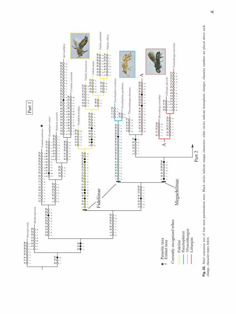
Fig.
22.
Stri
ctco
nsen
sus
tree
offo
urm
ost
pars
imon
ious
tree
s.B
lack
circ
les
indi
cate
uniq
uech
arac
ters
;w
hite
circ
les
indi
cate
hom
opla
stic
chan
ges;
char
acte
rnu
mbe
rsar
epl
aced
abov
eea
chch
ange
,cha
ract
er-s
tate
sbe
low
.
16

Fig.
22.
Con
tinue
d
17
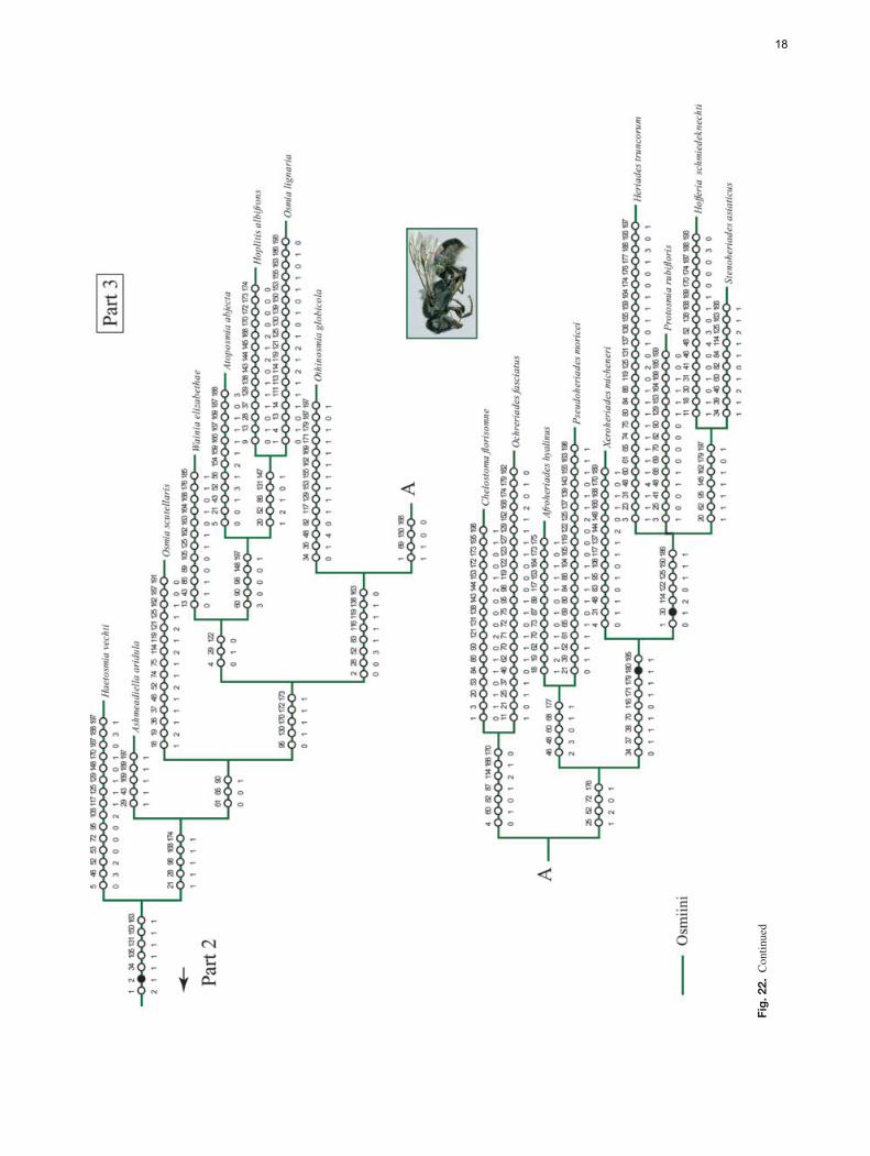
Fig.
22.
Con
tinue
d
18
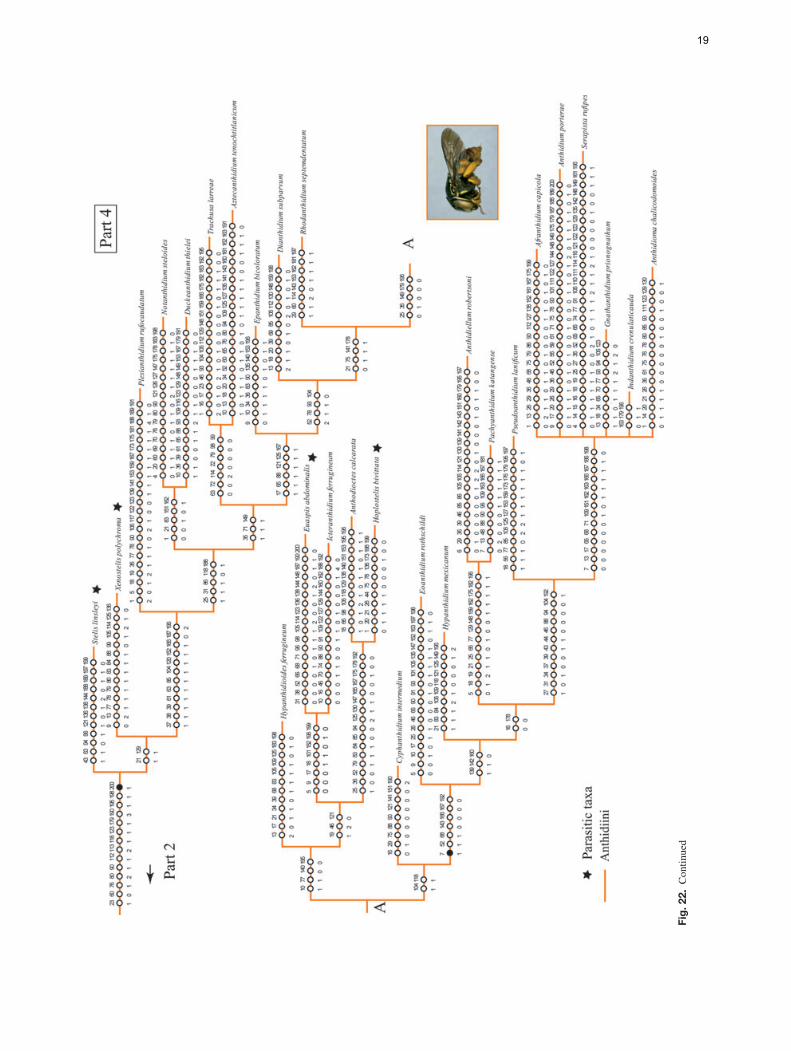
Fig.
22.
Con
tinue
d
19
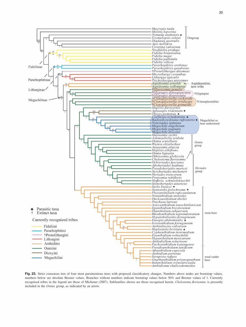
Fig. 23. Strict consensus tree of four most parsimonious trees with proposed classificatory changes. Numbers above nodes are bootstrap values,numbers below are absolute Bremer values. Branches without numbers indicate bootstrap values below 50% and Bremer values of 1. Currentlyrecognised tribes in the legend are those of Michener (2007). Subfamilies shown are those recognised herein. Chelostoma florisomne is presentlyincluded in the Osmia group, as indicated by an arrow.
20
remaining megachilines. It also supports the sister group rela-tionship of Protolithurgini to Lithurgini as indicated by Engel(2001). However, unlike the study of Roig-Alsina & Mich-ener (1993), our analysis does not support the monophyly ofFideliinae. In their study, two synapomorphies supported thesubfamily: the outer hind tibial spur coarsely serrate and thepapillate or minutely roughened dorsal surface of the femalesixth tergum. Mapping the presence of a coarsely serrate outerhind tibial spur onto the consensus trees resulted in ambiguouschoices. It was either an ancestral character retained in bothFideliini and Pararhophitini when favouring character reversalsover convergences in the analysis, or gained independently inboth taxa when favouring repeated origins over reversals. Thepapillate or minutely roughened dorsal surface of the femalesixth tergum in both taxa may not be homologous. In Fideli-ini, this integument is present on the well-defined pygidialplate only (Fig. 16) whereas in Pararhophitini, which lacks apygidial plate, it is present on the entire distal half of the ter-gum (Fig. 17). Thus, by coding both taxa as having the sametype of integument we are failing to assess the primary homol-ogy based on the positional criterion (e.g. De Pinna, 1991). Forthis reason, we did not code this character as in Roig-Alsina& Michener (1993); instead, we redefined this character bycoding its presence or absent on a well-defined pygidial plate.Given that such a plate is absent in Pararhophites, this char-acter does not apply to this taxon. However, one could arguethat the pygidial plate is secondarily reduced in Pararhophitesand, in that case, it would be reasonable to assume the primaryhomology of the surface sculpture. This idea is not supportedby the fact that this type of integument is not restricted tothe dorsal surface of the tergum, where it should be lim-ited if a pygidial plate was present, but is found along theentire distal half of the segment. Nonetheless, coding this char-acter as present in both Fideliini and Pararhophitini, as inRoig-Alsina & Michener (1993), did not change the resultingtopology.
The nonmonophyly of Fideliinae sensu Michener (2007)was suggested in the analysis of Roig-Alsina & Michener(1993) when larval characters were analysed alone or combinedwith adult characters. Recent molecular analyses support theparaphyly of this subfamily as well as an ancient origin of bothFideliini and Pararhophitini of at least 100 Ma (Litman et al.,2011). That analysis also suggests the paraphyly of Fideliini.In contrast, the monophyly of the tribe is strongly supported byhigh bootstrap (99%) and Bremer (10) values and five synapo-morphies in our analysis. Both Fideliini and Pararhophitinihave some distinctive biological features not known in otherbees but commonly found among apoid wasps. In both groupsthe cells are unlined with walls no smoother than the burrowand the cocoon tapers at each end, incorporating sand eatenby the mature larva and voided in strips tending to run on theinside of the cocoon (Rozen, 1970, 1973; McGinley & Rozen,1987; Michener, 2007). Such behavioural traits have been sug-gested as additional synapomorphies for Fideliinae (Michener,2007) but it seems they are likely plesiomorphic featuresretained by those groups, perhaps as adaptations to nesting insandy soils in strongly seasonal deserts (Litman et al., 2011).
The sister group relationship of Pararhophitini andMegachilinae is weakly supported by bootstrap (<50%) andBremer (2) values, as well as by a questionable synapomor-phy: the presence of the dististipital process on the maxillarystipes (55-1). Although McGinley & Rozen (1987) indicatedthat this process is present as a very short distal bulge similarto that found in Lithurgus, Roig-Alsina & Michener (1993)coded it as absent in their analysis. The dististipital process ofLithurgini seems to us slightly more distinct and sclerotisedthan that of Pararhophites. Although we coded Pararhophitesas having this process (character-state 1), coding it as if itwere absent or as missing data did not change the resultingtopology.
Aspidosmia, presently in the Anthidiini but characterised bya combination of both osmiine and anthidiine features, doesnot belong to either tribe. Rather it is a well-supported clade(90 bootstrap and 4 in Bremer values; Fig. 23) that is sis-ter to the remaining megachiline tribes excluding Lithurgini.A recent molecular analysis also supports this basal posi-tion of Aspidosmia among the Megachilinae (Litman et al.,2011). Thus, the current taxonomic placement of Aspidosmiawithin the Anthidiini renders this tribe paraphyletic. Interest-ingly, such a phylogenetic position was first suggested byPeters (1972) and discussed by Michener (2007) when assum-ing the hind tibial scopa to be an ‘ancestral’ (plesiomor-phic) feature in Aspidosmia. Our results support this view.Whether favouring repeated origins over reversals or favour-ing character reversals over convergences, the analyses sug-gest that the presence of a hind tibial scopa is an ancestralcharacter retained in Pararhophites, Protolithurgus, Aspidos-mia, Glaesosmia, Glyptapis and Ctenoplectrella (results notshown).
The fossil genera Glaesosmia, Glyptapis and Ctenoplec-trella, presently in Osmiini, were consistently placed amongbasal Megachilinae, near Aspidosmia. Particularly interestingis the resemblance of Ctenoplectrella and Aspidosmia. Despitehaving a labrum broader than long, the wing venation of Cteno-plectrella (e.g. fore wing with vein 2m-cu basal to 2r-m, basalvein curved, meeting Cu at right angle) seems similar to thatof Aspidosmia. Also, it appears that the hairs on the hind tibiaare long, suggestive of a scopa. We do not know whether thelong hind tibial hairs of the Baltic megachilid genera consti-tute functional scopae but the phylogenetic position of thesetaxa, as well as that of Aspidosmia, did not change when thischaracter was excluded from the analysis, indicating that theirposition in the tree does not depend entirely on the presenceof the hind tibial scopa. Biogeographically, it is interesting tonote that Sub-Saharan taxa, such as Aspidosmia, often showrelationships to taxa in Baltic amber (Engel, 2001; Grimaldi& Engel, 2005). According to our analysis Aspidosmia andCtenoplectrella are not sister groups but such a conclusionmay be biased given the limited number of characters (lessthan half) that could be coded for Ctenoplectrella. If, in fact,these groups are sister taxa, Aspidosmia is likely the only sur-vivor of this lineage. In that case, it would be more meaningfulto place both genera in a single tribe Ctenoplectrellini to high-light the unique biogeographical and phylogenetic connections.
21
Additional fossil material may resolve this question, but withavailable data such a decision would be premature. The posi-tion of Glaesosmia, as sister of Glyptapis, is doubtless due toabsence of data. Glaesosmia is only known from the femaleholotype, which is in poor condition. Additional material thatshows a large number of characters of these fossil taxa, includ-ing those of the male, will help to test their relationship toAspidosmia.
The remaining anthidiine taxa clustered in a clade withlow bootstrap (<50%) and Bremer (1) support values. Thepenis valves with long apodemes projecting through thegenital foramen in the male (200-1) was the single putativesynapomorphy supporting this clade. This character appearsto be secondarily lost in some Anthidiini genera such asAnthidium and Euaspis Gerstacker and is also present insome species of Megachile subgenus Chalicodoma (Gonzalez,2008). Our analysis shows that parasitism has evolved multipletimes in Anthidiini and that the wool carder bees or series Bof genera (sensu Michener, 2007) represent a derived cladewithin the paraphyletic resin bees or series A (Fig. 23). Theseries B clade includes the following eight genera AfranthidiumMichener, Anthidioma Pasteels, Anthidium, GnathanthidiumPasteels, Indanthidium Michener and Griswold, NeanthidiumPasteels, Pseudoanthidium Friese and Serapista Cockerell. It ischaracterised by the female mandible with at least four small,acute teeth on the distal margin and by the absence of velvetyhairs on the outer surface of the labrum and the inner surface ofthe mandible. These anthidiines are commonly known as woolcarder bees because such multidentate mandibles, as well asthe absence of velvety hairs, is presumably associated with theuse of plant hairs or trichomes to build their cotton-like broodcells (Michener, 2007).
The analysis also supports the long suspected nonmonophylyof Osmiini, which is mainly caused by the placement of Hopli-tis (in part) as sister group of the clade consisting of Dioxyini,Megachilini and Noteriades; no unique synapomorphies orhigh bootstrap and Bremer values support such a relationship(Figs 22, 23). Two groups of genera have traditionally beenrecognised in the 19 genera of Osmiini, sometimes treatedat the subtribal level: the Osmia group or subtribe Osmiinaand the Heriades group or subtribe Heriadina. Both groupscan be roughly characterised by a combination of characters,but intermediate taxa such as Protosmia Ducke and Othinos-mia Michener bridge the gap between the two (Griswold &Michener, 1997; Michener, 2007). Although we included arepresentative of each osmiine genus and scored the characterslisted by Griswold (1985) and Michener (2007) in their recog-nition of each group of genera, the analysis does not support themonophyly of either group. Yet neither were the results concor-dant with the molecular analysis of Praz et al. (2008), exceptfor Noteriades (see below); Afroheriades, Pseudoheriades andOchreriades appeared close to other ‘Osmiini’, within a cladeof most of the Heriades group. In fact, it is interesting thatresults place the rare genus Ochreriades as the sister groupof Chelostoma, a relationship previously suspected by Mavro-moustakis (1956) and Griswold (1985, 1994) but not supportedby molecular analyses (Praz et al., 2008).
The position of Dioxyini within the Osmiini, and sisterto the clade that includes Noteriades and Megachilini, wasan unanticipated result. Although the monophyly of Dioxyiniis unquestionable given a number of derived characters (e.g.reduced sting in the female, metanotum with median spine)and high bootstrap and Bremer values (Figs 22, 23), the phy-logenetic position of this parasitic tribe is puzzling. Dioxyinihas been long thought to be related to the Anthidiini becauseit exhibits typical features of that tribe, namely a depressionbehind the propodeal spiracle, short stigma and prestigma, andcleft pretarsal claws in the female (Michener, 1944, 1996,2007). The position of Dioxyini in our consensus tree suggeststhat it might have grouped with Megachilini because of theirmorphological similarity to Coelioxys and RadoszkowskianaPopov resulting from their shared cleptoparasitic lifestyle.Cleptoparasites do not collect pollen to feed their larvae, andtherefore the scopal hairs on abdomen and legs tend to bereduced or absent, their integument is also usually coarse, thepronotal lobe and omaulus are usually carinate or lamellate,and the axilla is usually strongly projected. Thus, cleptopara-sitic taxa that are not closely related might be clustered onthe basis of these characters. However, when we excludedeither the two parasitic megachiline genera or deactivated threecharacters related to cleptoparasitism from the analysis (char-acters 61, 66, and 74: pronotal lobe with strong carina orlamella, omaular carina, and axilla projected in acute angleor spine, respectively), we obtained a much higher numberof MPTs (59 and 81) and a large number of branches (25and 22) collapsed in the consensus trees; Dioxyini was thesister group of Xenostelis and was either in a large poly-tomy with other Osmiini and Anthidiini or was nested in thelatter tribe. Our results also contrast with those of Gogala(1995), the only other study exploring the relationships ofDioxyini among megachilids. In Gogala’s study Dioxys cameout as the sister group to all other Megachilinae but his anal-ysis included a limited number of taxa and characters (ninetaxa, no outgroup and 11 morphological characters). Whilethe phylogenetic relationships of Dioxyini need to be furtherexplored, our results support a distinct Dioxyini within theMegachilinae.
Another significant result of our analysis was the position ofNoteriades within Megachilini. Such a result confirms the ideaof its close relationship to this tribe first mentioned by Gris-wold (1985) and more recently supported by the molecularanalysis of Praz et al. (2008). In fact, when including Noteri-ades in the dataset of Gonzalez (2008) for the phylogeny ofMegachilini (6 outgroup species, 107 ingroup species and 231morphological characters), it was consistently included in thattribe but its position was not consistent among analyses. Note-riades appeared either as the sister group of Megachile s. l. orin a polytomy with Matangapis Baker & Engel and the cladethat contains Chelostomoda Michener and related Megachilesubgenera (V.H. Gonzalez and T.L. Griswold, in preparation).Independent of the position of Noteriades within Megachilini,its inclusion in Megachilini renders a more easily recognised
22

and diagnosed tribe. To date, there are no known synapomor-phies for Megachilini; however, at least one of the three puta-tive synapomorphies for the clade Noteriades + Megachilinifound in this analysis seems to be unambiguously present: thepresence of an anterior angle or spine followed by a notch onthe apical margin of the outer surfaces of both fore and mid tib-iae (92-1; Fig. 14). Thus, the recognition of Noteriades withinMegachilini is warranted and is formally established herein.Concerning the other two putative synapomorphies (both foundin Noteriades), the torular process (12-1; Fig. 5) is reduced tonearly absent in some Megachile (e.g. M. discolor) and themostly membranous S7 of the male (186-2) is a condition alsopresent in some Osmiini.
Classificatory considerations
Independently of the suprageneric classification followed,our analysis suggests four classificatory changes. First, ouranalysis indicates that Fideliini and Pararhophitini are notsister taxa but form successive sister taxa to the remainingMegachilidae. Thus, herein each is recognised in its ownsubfamily, as presently recognised by some authors (Table 1;Fig. 21). Second, the current placement of Aspidosmia withinthe Anthidiini would result in a paraphyletic taxon. Given thephylogenetic position and distinctive characters of Aspidosmia,such as the presence of a hind tibial scopa in the female, weherein place it in its own tribe, Aspidosmiini, new tribe (seebelow).
Third, our analysis indicates that the inclusion of Noteriadeswithin Megachilini strengthens the recognition and diagnosisof this tribe. Thus, it is formally transferred to that tribe fromOsmiini. We do not recommend synonymising Osmiini andDioxyini under Megachilini (the oldest name of these threetribes). That decision should await a more rigorous study ofthe osmiines, including more taxa and characters (molecularas well as morphological). Such a study could also shed lighton the monophyly of the Osmia and Heriades groups. Untilthen, we recommend continued treatment of them as informalunits rather than as subtribes.
Fourth, and finally, the current taxonomic placement ofthe fossil genera Glaesosmia, Glyptapis and Ctenoplectrellainto two subtribes of Osmiini, renders this tribe paraphyletic.Thus, herein they are treated at the tribal level, as presentlyrecognised by Engel (2005).
Given the results, two classificatory approaches are plausible(Table 1). The first one (Proposal 1) closely follows the currentclassification of Michener (2007) but with Pararhophitinielevated to the subfamily level and the fossil subtribesCtenoplectrellina and Glyptapina treated at the tribal level.The second approach (Proposal 2) follows that of Engel(2005) in recognising Fideliini, Pararhophitini and Lithurginiat the subfamilial level. It differs from Engel (2005) in notrecognizing two tribes within Fideliinae and in recognisingDioxyini at the tribal level. Of course, the number ofsuprageneric categories one wants to recognise in Megachilidaeis arbitrary; herein we prefer to follow Proposal 2 (Fig. 23).
Aspidosmiini, new tribe
Type genus: Aspidosmia Brauns(Figs 1–3, 10)
Diagnosis. This tribe can be readily separated from all othertribes of Megachilinae by the presence of scopal hairs on theouter surface of the female hind tibia (in addition to the sternalscopa; Fig. 1), and by the forewing with a long prestigma,about as long as the stigma, a short stigma (less than twice aslong as broad), and the vein 2m-cu basal to 2r-m.
Description. Female. Clypeus not covering labral base(Figs 2, 3); first labial palpomere short, about half lengthof second; maxillary palpi four-segmented. Preaxilla, belowposterolateral angle of scutum, sloping, with small patch ofhairs as long as those of adjacent sclerites (Fig. 10); propodealtriangle smooth and shiny, hairless; fore tibial spur (antennalcleaner) with malus simple, without a distinct projecting ridgeon its anterior side, apex long, at least half length of base ofmalus, with distinct row of teeth on inner and outer margins;outer surface of hind tibia with long simple hairs forminga distinct scopa; hind basitarsus elongate, more than fivetimes longer than broad; forewing with prestigma about aslong as stigma; stigma less than twice as long as broad;vein 2m-cu basal to 2r-m, basal vein curved, meeting Cu atright angle; hind wing with jugal lobe about half as long asvannal lobe. Male. T6 without preapical carina; T7 exposed,posteriorly directed, sparsely covered by long hairs; S7 well-sclerotised, not medially divided; gonoforceps dorsolaterallyweakly sclerotised; volsella present.
Comments. The novelty of this tribe was recognised by C.J. P. and B. N. D. based on the phylogenetic position ofAspidosmia in a recent molecular analysis of Megachilidae(Litman et al., 2011). Such a result is supported by the presentmorphological study. Therefore, the authorship of this tribe isattributed to all four authors of this paper to reflect the equalintellectual contribution in the recognition and diagnosis of thislineage.
Key to the extant tribes of the Megachilinae
(Modified from Michener, 2007)
1. Metanotum with median spine or tubercle (except inAllodioxys and Ensliniana); mandible of female slenderapically, bidentate, similar to that of male; pronotum (except inProdioxys) with prominent obtuse or right-angular dorsolateralangle, below which a vertical ridge extends downward; stingand associated structures greatly reduced (scopa absent) . . . . .. . . . . . . . . . . . . . . . . . . . . . . . . . . . . . . . . . . . . . . . . . . . . . . . . Dioxyini– Metanotum without median spine or tubercle; mandible offemale usually wider apically, with three or more teeth, exceptrarely bidentate when mandible is greatly enlarged and porrect
23
and clypeus is also modified; pronotum with dorsolateral angleweak or absent (or produced to a tooth in some Chelostoma butwithout vertical ridge below it); sting and associated structureswell developed . . . . . . . . . . . . . . . . . . . . . . . . . . . . . . . . . . . . . . . . . 22(1). Stigma less than twice as long as broad, inner marginbasal to vein r usually little if any longer than width, rarelyabout 1.5 times width; claws of female cleft or with an innertooth (except in Trachusoides); body commonly with yellowor white integumental marks . . . . . . . . . . . . . . . . . . . . . . . . . . . . .3– Stigma over twice as long as broad, inner margin basalto vein r longer than width; claws of female simple (exceptin Osmia subgenus Metalinella, Palaearctic); body withoutyellow or white integumental marks (except in Ochreriades) .. . . . . . . . . . . . . . . . . . . . . . . . . . . . . . . . . . . . . . . . . . . . . . . . . . . . . . . . 43(2). Outer surface of hind tibia with long hairs forming adistinct scopa; prestigma much more than twice as long asbroad; preaxilla, below posterolateral angle of scutum, slopingand with small patch of hairs, these as long as those of adjacentsclerites (Fig. 10) . . . . . . . . . . . . . . . . . . Aspidosmiini, new tribe– Outer surface of hind tibia usually with abundant simplebristles, not forming a distinct scopa; prestigma commonlyshort, usually less than twice as long as broad; preaxillavertical, smooth and shining, usually without hairs (Fig. 11) .. . . . . . . . . . . . . . . . . . . . . . . . . . . . . . . . . . . . . . . . . . . . . . . . Anthidiini4(2). Outer surfaces of fore and mid tibiae apically with anacute angle (usually produced into a spine) and distinct notchanteriorly (Fig. 14); arolia absent, except in a few tropical OldWorld taxa (Noteriades, Matangapis and Megachile subgenusHeriadopsis); body nonmetallic or nearly so . . . . Megachilini– Outer surfaces of fore and mid tibiae apically without anacute angle or spine and lacking distinct notch anteriorly; aroliapresent; body sometimes metallic green, blue, or brassy . . . . .. . . . . . . . . . . . . . . . . . . . . . . . . . . . . . . . . . . . . . . . . . . . . . . . . . Osmiini
Supporting Information
Additional Supporting Information may be found in the onlineversion of this article under the DOI reference:10.1111/j.1365-3113.2012.00620.x
Appendix S1. Character matrix used in cladistic analysisof the family Megachilidae. -, not applicable; ?, state ofcharacter that could not be recorded (see Materials andmethods); *, multistate (0,1).
Acknowledgements
We are grateful to David Grimaldi, American Museum ofNatural History, for kindly arranging the loan and allowingus to examine the Megachilidae fossils; Michael S. Engel forbringing to our attention the puzzling fossils and for valuablecomments on the manuscript; Michael S. Engel, Zackary Falin,Charles D. Michener and Jennifer Thomas for access to the
bee collection in their charge; Amy Comfort de Gonzalez,Charles D. Michener, Michael S. Engel, Laurence Packer, LarsVilhelmsen and two anonymous reviewers for their commentsand suggestions that improved this work. This study wassupported in part by National Science Foundation grant DEB-0742998 (to B.N.D and T.G).
References
Ascher, J. & Pickering, J. (2011) Bee Species Guide (Hymenoptera:Apoidea: Anthophila) [WWW document]. URL http://www.discoverlife.org/mp/20q?guide=Apoidea_species [accessed on 17February 2011].
Bohart, G.E. (1972) Management of wild bees for the pollination ofcrops. Annual Review of Entomology, 17, 287–312.
Bremer, K. (1994) Branch support and tree stability. Cladistics, 10,295–304.
Cane, J.H. (2003) Exotic nonsocial bees (Hymenoptera: Apiformes)in North America: ecological implications. For Non-Native Crops,Whence Pollinators of the Future? Thomas Say Publications inEntomology: Proceedings (ed. by K. Strickler and J.H. Cane),pp. 113–126. Entomological Society of America, Lanham,Maryland.
Cane, J.H., Griswold, T.L. & Parker, F.D. (2007) Substrates and mate-rials used for nesting by North American Osmia bees (Apiformes:Megachilidae). Annals of the Entomological Society of America, 100,350–358(9).
Cockerell, T.D.A. (1909) Descriptions of Hymenoptera from Balticamber. Schriften der Physikalische-Okononischen Gesellschaft zuKonigsberg, 50, 1–20.
Combey, R., Kwapong, P., Eardley, C.D. & Botchey, M. (2010) Phy-logenetic analysis of the bee tribe Anthidiini. Journal of GhanaScience Association, 12, 26–38.
De Pinna, M.G.G. (1991) Concepts and tests of homology in the cladis-tic paradigm. Cladistics, 7, 367–394.
Engel, M.S. (2001) A monograph of the Baltic amber bees and evo-lution of the Apoidea (Hymenoptera). Bulletin of the AmericanMuseum of Natural History, 259, 1–192.
Engel, M.S. (2002) Phylogeny of the bee tribe Fideliini (Hymenoptera:Megachilidae), with the description of a new genus from southernAfrica. African Entomology, 10, 305–313.
Engel, M.S. (2004) Fideliine phylogeny and classification revisited(Hymenoptera: Megachilidae). Journal of the Kansas EntomologicalSociety, 77, 821–836.
Engel, M.S. (2005) Family-group names for bees (Hymenoptera:Apoidea). American Museum Novitates, 3476, 1–33.
Engel, M.S. & Perkovsky, E.E. (2006) An Eocene bee in Rovnoamber, Ukraine (Hymenoptera: Megachilidae). American MuseumNovitates, 3506, 1–11.
Felsenstein, J. (1985) Confidence limits on phylogenies: an approachusing the bootstrap. Evolution, 39, 783–791.
Gibbs, J. & Sheffield, C.S. (2009) Rapid range expansion of thewool-carder bee, Anthidium manicatum (Linnaeus) (Hymenoptera:Megachilidae), in North America. Journal of the Kansas Entomo-logical Society, 82, 21–29.
Gogala, A. (1995) Phylogenetic relationships of the bee genusDioxys Lepeletier & Serville, 1825 (Hymenoptera: Megachilidae,Megachilinae). Acta Entomologica Slovenica, 3, 43–48.
24
Goloboff, P.A., Farris, J. & Nixon, K. (2003) T.N.T. Tree AnalysisUsing New Technology, Program and documentation [WWW docu-ment]. URL http://www.zmuc.dk/public/phylogeny/tnt [accessed on15 April 2011].
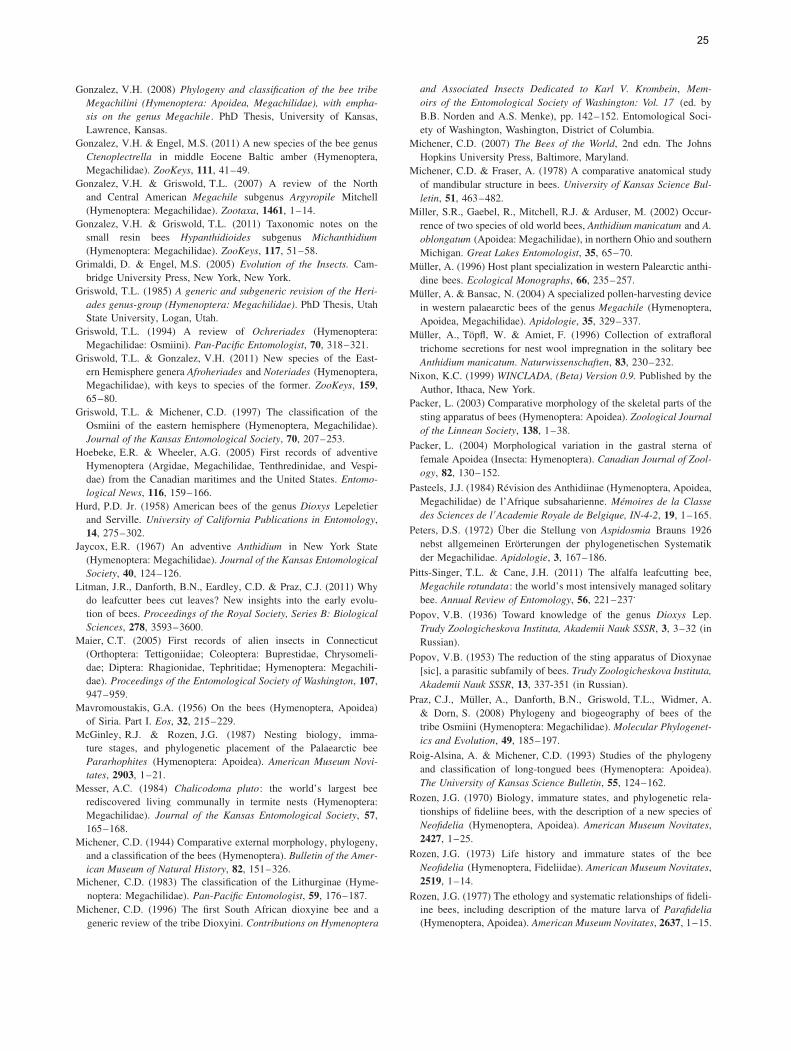
Gonzalez, V.H. (2008) Phylogeny and classification of the bee tribeMegachilini (Hymenoptera: Apoidea, Megachilidae), with empha-sis on the genus Megachile. PhD Thesis, University of Kansas,Lawrence, Kansas.
Gonzalez, V.H. & Engel, M.S. (2011) A new species of the bee genusCtenoplectrella in middle Eocene Baltic amber (Hymenoptera,Megachilidae). ZooKeys, 111, 41–49.
Gonzalez, V.H. & Griswold, T.L. (2007) A review of the Northand Central American Megachile subgenus Argyropile Mitchell(Hymenoptera: Megachilidae). Zootaxa, 1461, 1–14.
Gonzalez, V.H. & Griswold, T.L. (2011) Taxonomic notes on thesmall resin bees Hypanthidioides subgenus Michanthidium(Hymenoptera: Megachilidae). ZooKeys, 117, 51–58.
Grimaldi, D. & Engel, M.S. (2005) Evolution of the Insects. Cam-bridge University Press, New York, New York.
Griswold, T.L. (1985) A generic and subgeneric revision of the Heri-ades genus-group (Hymenoptera: Megachilidae). PhD Thesis, UtahState University, Logan, Utah.
Griswold, T.L. (1994) A review of Ochreriades (Hymenoptera:Megachilidae: Osmiini). Pan-Pacific Entomologist, 70, 318–321.
Griswold, T.L. & Gonzalez, V.H. (2011) New species of the East-ern Hemisphere genera Afroheriades and Noteriades (Hymenoptera,Megachilidae), with keys to species of the former. ZooKeys, 159,65–80.
Griswold, T.L. & Michener, C.D. (1997) The classification of theOsmiini of the eastern hemisphere (Hymenoptera, Megachilidae).Journal of the Kansas Entomological Society, 70, 207–253.
Hoebeke, E.R. & Wheeler, A.G. (2005) First records of adventiveHymenoptera (Argidae, Megachilidae, Tenthredinidae, and Vespi-dae) from the Canadian maritimes and the United States. Entomo-logical News, 116, 159–166.
Hurd, P.D. Jr. (1958) American bees of the genus Dioxys Lepeletierand Serville. University of California Publications in Entomology,14, 275–302.
Jaycox, E.R. (1967) An adventive Anthidium in New York State(Hymenoptera: Megachilidae). Journal of the Kansas EntomologicalSociety, 40, 124–126.
Litman, J.R., Danforth, B.N., Eardley, C.D. & Praz, C.J. (2011) Whydo leafcutter bees cut leaves? New insights into the early evolu-tion of bees. Proceedings of the Royal Society, Series B: BiologicalSciences, 278, 3593–3600.
Maier, C.T. (2005) First records of alien insects in Connecticut(Orthoptera: Tettigoniidae; Coleoptera: Buprestidae, Chrysomeli-dae; Diptera: Rhagionidae, Tephritidae; Hymenoptera: Megachili-dae). Proceedings of the Entomological Society of Washington, 107,947–959.
Mavromoustakis, G.A. (1956) On the bees (Hymenoptera, Apoidea)of Siria. Part I. Eos, 32, 215–229.
McGinley, R.J. & Rozen, J.G. (1987) Nesting biology, imma-ture stages, and phylogenetic placement of the Palaearctic beePararhophites (Hymenoptera: Apoidea). American Museum Novi-tates, 2903, 1–21.
Messer, A.C. (1984) Chalicodoma pluto: the world’s largest beerediscovered living communally in termite nests (Hymenoptera:Megachilidae). Journal of the Kansas Entomological Society, 57,165–168.
Michener, C.D. (1944) Comparative external morphology, phylogeny,and a classification of the bees (Hymenoptera). Bulletin of the Amer-ican Museum of Natural History, 82, 151–326.
Michener, C.D. (1983) The classification of the Lithurginae (Hyme-noptera: Megachilidae). Pan-Pacific Entomologist, 59, 176–187.
Michener, C.D. (1996) The first South African dioxyine bee and ageneric review of the tribe Dioxyini. Contributions on Hymenoptera
and Associated Insects Dedicated to Karl V. Krombein, Mem-oirs of the Entomological Society of Washington: Vol. 17 (ed. byB.B. Norden and A.S. Menke), pp. 142–152. Entomological Soci-ety of Washington, Washington, District of Columbia.
Michener, C.D. (2007) The Bees of the World, 2nd edn. The JohnsHopkins University Press, Baltimore, Maryland.
Michener, C.D. & Fraser, A. (1978) A comparative anatomical studyof mandibular structure in bees. University of Kansas Science Bul-letin, 51, 463–482.
Miller, S.R., Gaebel, R., Mitchell, R.J. & Arduser, M. (2002) Occur-rence of two species of old world bees, Anthidium manicatum and A.oblongatum (Apoidea: Megachilidae), in northern Ohio and southernMichigan. Great Lakes Entomologist, 35, 65–70.
Muller, A. (1996) Host plant specialization in western Palearctic anthi-dine bees. Ecological Monographs, 66, 235–257.
Muller, A. & Bansac, N. (2004) A specialized pollen-harvesting devicein western palaearctic bees of the genus Megachile (Hymenoptera,Apoidea, Megachilidae). Apidologie, 35, 329–337.
Muller, A., Topfl, W. & Amiet, F. (1996) Collection of extrafloraltrichome secretions for nest wool impregnation in the solitary beeAnthidium manicatum. Naturwissenschaften, 83, 230–232.
Nixon, K.C. (1999) WINCLADA, (Beta) Version 0.9. Published by theAuthor, Ithaca, New York.
Packer, L. (2003) Comparative morphology of the skeletal parts of thesting apparatus of bees (Hymenoptera: Apoidea). Zoological Journalof the Linnean Society, 138, 1–38.
Packer, L. (2004) Morphological variation in the gastral sterna offemale Apoidea (Insecta: Hymenoptera). Canadian Journal of Zool-ogy, 82, 130–152.
Pasteels, J.J. (1984) Revision des Anthidiinae (Hymenoptera, Apoidea,Megachilidae) de l’Afrique subsaharienne. Memoires de la Classedes Sciences de l $Academie Royale de Belgique, IN-4-2, 19, 1–165.
Peters, D.S. (1972) Uber die Stellung von Aspidosmia Brauns 1926nebst allgemeinen Erorterungen der phylogenetischen Systematikder Megachilidae. Apidologie, 3, 167–186.
Pitts-Singer, T.L. & Cane, J.H. (2011) The alfalfa leafcutting bee,Megachile rotundata: the world’s most intensively managed solitarybee. Annual Review of Entomology, 56, 221–237.
Popov, V.B. (1936) Toward knowledge of the genus Dioxys Lep.Trudy Zoologicheskova Instituta, Akademii Nauk SSSR, 3, 3–32 (inRussian).
Popov, V.B. (1953) The reduction of the sting apparatus of Dioxynae[sic], a parasitic subfamily of bees. Trudy Zoologicheskova Instituta,Akademii Nauk SSSR, 13, 337-351 (in Russian).
Praz, C.J., Muller, A., Danforth, B.N., Griswold, T.L., Widmer, A.& Dorn, S. (2008) Phylogeny and biogeography of bees of thetribe Osmiini (Hymenoptera: Megachilidae). Molecular Phylogenet-ics and Evolution, 49, 185–197.
Roig-Alsina, A. & Michener, C.D. (1993) Studies of the phylogenyand classification of long-tongued bees (Hymenoptera: Apoidea).The University of Kansas Science Bulletin, 55, 124–162.
Rozen, J.G. (1970) Biology, immature states, and phylogenetic rela-tionships of fideliine bees, with the description of a new species ofNeofidelia (Hymenoptera, Apoidea). American Museum Novitates,2427, 1–25.
Rozen, J.G. (1973) Life history and immature states of the beeNeofidelia (Hymenoptera, Fideliidae). American Museum Novitates,2519, 1–14.
25
Rozen, J.G. (1977) The ethology and systematic relationships of fideli-ine bees, including description of the mature larva of Parafidelia(Hymenoptera, Apoidea). American Museum Novitates, 2637, 1–15.
Schonitzer, K. (1986) Comparative morphology of the antenna cleanerin bees (Apoidea). Zeitschrift fur Zoologische Systematik und Evo-lutionsforschung, 24, 35–51.
Schonitzer, K. & Renner, M. (1980) Morphologie der Antennenputzap-parate bei Apoidea. Apidologie, 11, 113–130.
Smith, I.P. (1991) Anthidium manicatum (Hymenoptera, Megachili-dae), an interesting new Canadian record. Proceedings of the Ento-mological Society of Ontario, 122, 105–108.
Strange, J., Koch, J., Gonzalez, V.H., Nemelka, L. & Griswold, T.(2011) Global invasion by Anthidium manicatum (Linnaeus)(Hymenoptera: Megachilidae): assessing potential distribution inNorth America and beyond. Biological Invasions, 13, 2115–2133.
Thorp, R.W. (2000) The collection of pollen by bees. Plant Systematicsand Evolution, 222, 211–223.
Urban, D. & Moure, J.S. (2007) Anthidiini. Catalogue of Bees(Hymenoptera, Apoidea) in the Neotropical Region (ed. by
J.S. Moure, D. Urban and G.A.R. Melo), pp. 875–913. SociedadeBrasileira de Entomologia, Curitiba [WWW document]. URLhttp://www.moure.cria.org.br/catalogue [accessed on 6 June 2011].
Warncke, K. (1980) Die Bienengattung Anthidium Fabricius, 1804 inder Westpalaarktis und im turkestanischen Becken. Entomofauna, 1,119–209.
Wcislo, W.T. & Cane, J.H. (1996) Floral resource utilization bysolitary bees (Hymenoptera: Apoidea) and exploitation of theirstored foods by natural enemies. Annual Review of Entomology, 41,257–286.
Winston, M.L. (1979) The proboscis of the long-tongued bees: acomparative study. The University of Kansas Science Bulletin, 51,631–667.
Zavortink, T.J. & Shanks, S.S. (2008) Anthidium manicatum (Lin-naeus) (Hymenoptera: Megachilidae) in California. Pan-PacificEntomologist, 84, 238–241.
26