Embed Size (px)
Citation preview

Nova Southeastern University Nova Southeastern University
NSUWorks NSUWorks
Student Theses, Dissertations and Capstones College of Dental Medicine
2019
Physical and Optical Properties of Provisional Crown and Bridge Physical and Optical Properties of Provisional Crown and Bridge
Materials Fabricated Using CAD/CAM Milling or 3D Printing Materials Fabricated Using CAD/CAM Milling or 3D Printing
Technology Technology
Niranjan Joshi Nova Southeastern University
Follow this and additional works at: https://nsuworks.nova.edu/hpd_cdm_stuetd
Part of the Dentistry Commons
All rights reserved. This publication is intended for use solely by faculty, students, and staff of
Nova Southeastern University. No part of this publication may be reproduced, distributed, or
transmitted in any form or by any means, now known or later developed, including but not
limited to photocopying, recording, or other electronic or mechanical methods, without the prior
written permission of the author or the publisher.
NSUWorks Citation NSUWorks Citation Niranjan Joshi. 2019. Physical and Optical Properties of Provisional Crown and Bridge Materials Fabricated Using CAD/CAM Milling or 3D Printing Technology. Master's thesis. Nova Southeastern University. Retrieved from NSUWorks, College of Dental Medicine. (99) https://nsuworks.nova.edu/hpd_cdm_stuetd/99.
This Thesis is brought to you by the College of Dental Medicine at NSUWorks. It has been accepted for inclusion in Student Theses, Dissertations and Capstones by an authorized administrator of NSUWorks. For more information, please contact [email protected].

PHYSICAL AND OPTICAL PROPERTIES OF PROVISIONAL
CROWN AND BRIDGE MATERIALS FABRICATED USING
CAD/CAM MILLING OR 3D PRINTING TECHNOLOGY.
NIRANJAN JOSHI, B.D.S., M.D.S.
A Thesis presented to the Faculty of the College of Dental Medicine of
Nova Southeastern University in Partial Fulfillment of the Requirements
for the Degree of
MASTER OF SCIENCE
July 2019
© Copyright by Niranjan Joshi 2019 All Rights Reserved
Physical and optical properties of provisional crown and bridge materials
fabricated using CAD/CAM milling or 3D printing technology.
By
NIRANJAN JOSHI, B.D.S., M.D.S.
A thesis submitted to the College of Dental Medicine of Nova
Southeastern University in partial fulfillment of the
requirements for the degree of
MASTER OF SCIENCE
Department of Prosthodontics
College of Dental Medicine
Nova Southeastern University
July 2019
Approved as to style and content by:
APPROVED BY:
___________________________________________________________
Jeffrey Thompson, B.S., Ph.D., FADM (Committee Chair) Date
APPROVED BY:
___________________________________________________________
Jack Piermatti, D.M.D., FACP (Committee Member) Date
APPROVED BY:
___________________________________________________________
Leila Ahmadian, D.D.S., M.S., FACP (Committee Member) Date
APPROVED BY:
___________________________________________________________
Steven Kaltman, D.M.D., M.D., F.A.C.P. Date
(Dean, College of Dental Medicine)
Health Professions Division
Department of Prosthodontics College of Dental Medicine
STUDENT NAME: Niranjan Joshi B.D.S., M.D.S.
STUDENT E-MAIL ADDRESS: [email protected]
STUDENT TELEPHONE NUMBER: (918) 413-7207
COURSE DESCRIPTION: Master of Science in Dentistry with specialty
certificate in postgraduate Prosthodontics
TITLE OF SUBMISSION: Physical and optical properties of provisional crown and
bridge materials fabricated using CAD/CAM milling or 3D printing technology
DATE SUBMITTED: July 2019
I certify that I am the sole author of this thesis, and that any assistance I received
in its preparation has been fully acknowledged and disclosed in the thesis. I have
cited any sources from which I used ideas, data, or words, and labeled as
quotations any directly quoted phrases or passages, as well as providing proper
documentation and citations. This thesis was prepared by me, specifically for the
M.S. degree and for this assignment.
STUDENT SIGNATURE:
________________________________________________________________
Niranjan Joshi B.D.S., M.D.S. Date

Dedication
I would like to dedicate this thesis to my parents Dr. Ratnakar Joshi and Chhaya
Joshi, to my wife Dr. Vedavati Joshi, my daughter Eva and all my close friends
who stood by me during these three years.

v
Acknowledgements
• First, I would like to thank my mentor Dr. Jeffrey Thompson who helped me
and guided me throughout the study. I would also like to thank him for being
patient with my project and helping me navigate this journey.
• I would like to express my gratitude to Dr. Jack Piermatti for being a great
teacher, mentor and for his guidance and his suggestions throughout the
study.
• I am grateful to Dr. Thomas Balshi for his valuable suggestions and mentoring
this study.
• I am thankful to Dr. Leila Ahmadian for her extremely valuable and helpful
suggestions.
• I am thankful to Mr. Anthony Smithswick and Mr. Matt Winstead from Oral Arts
Dental Laboratory, for their generous support in providing samples.
• I am grateful to Dr. Max Nahon for always supporting me in this study.
• I am thankful to Tariq Rehman for his support and suggestions during the
study.
• I would also like to thank Dr. Patrick Hardigan for providing with the statistical
analysis.
• I appreciate the help from Ria Achong-Bowe for her help in the Bioscience
Research Center. Without her help this study wouldn’t have been possible.

vi
ABSTRACT
PHYSICAL AND OPTICAL PROPERTIES OF PROVISIONAL CROWN AND
BRIDGE MATERIALS FABRICATED USING CAD/CAM MILLING OR 3D
PRINTING TECHNOLOGY.
DEGREE DATE: JULY 2019
NIRANJAN JOSHI, B.D.S., M.D.S.
COLLEGE OF DENTAL MEDICINE NOVA SOUTHEASTERN UNIVERSITY
Thesis Directed by:
• Jeffrey Thompson, B.S., Ph.D., FADM (Committee Chair)
• Jack Piermatti, D.M.D., FACP (Committee Member)
• Leila Ahmadian, D.D.S, M.S., FACP (Committee Member)
Objective:
This study compared the physical and optical properties of provisional crown and
bridge materials fabricated using CAD/CAM or 3D printing technology.
Aim:
To compare the biaxial flexural strength, microhardness, translucency parameter
and gloss of provisional resin discs fabricated by milling PMMA blocks, versus two
different resins printed using 3D printers.

vii
Hypothesis:
There is no difference in the flexural strength, hardness, translucency and gloss of
provisional resins fabricated by different digital technologies.
Materials and methods:
90-disc shaped specimens for provisional resins were fabricated using a common
digital file. These samples were equally distributed in three groups of 30 each.
Group I samples were fabricated by milling specimens from a polymethyl
methacrylate (PMMA). Group II samples were fabricated by printing urethane
methacrylate resin using a 3D DLP printer. Group III samples were fabricated by
printing acrylic ester resin using a 3D SLA printer. All samples were tested for
biaxial flexural strength, translucency parameter, microhardness and gloss.
Results
A one-way ANOVA with Tukey HSD pair-wise comparisons was employed to
analyze the data and answer the research questions. The mean values for biaxial
flexural strength for milled polymethyl methacrylate (PMMA), urethane
methacrylate resin and acrylic ester resin were 136.9 MPa, 101.6 MPa and 98.4
MPa respectively. The mean hardness values for the groups in the same order
were 28.5, 9.7 and 14.8 respectively. The mean translucency parameter values for
the groups in the same order were 3.8, 6.3 and 4.4 respectively. The mean gloss
values for the groups in the same order were 3.9, 28.8 and 1.7 respectively. There
viii
was a statistically significant difference amongst the groups for all parameters
tested.
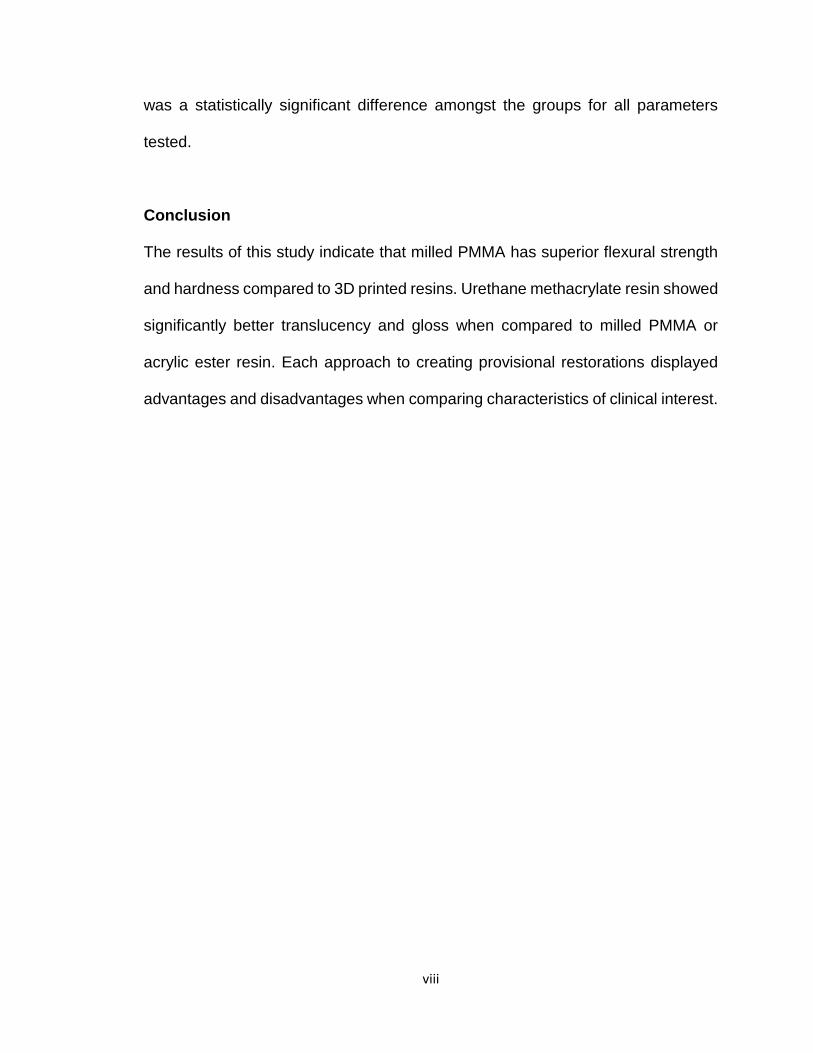
Conclusion
The results of this study indicate that milled PMMA has superior flexural strength
and hardness compared to 3D printed resins. Urethane methacrylate resin showed
significantly better translucency and gloss when compared to milled PMMA or
acrylic ester resin. Each approach to creating provisional restorations displayed
advantages and disadvantages when comparing characteristics of clinical interest.

ix
Table of Contents Title page………………………………………………………………………………….i
Signature Page ………………………………………………………………………… .ii
Disclosure Page ……………………………………………….……………………… .iii
Dedication Page…………………………………………...…………………….…..… iv
Acknowledgements …………………………………………………………….……… v
Abstract …………………………………………………………...……………………..vi
Table of Contents ……………………………………………………………...…..… ..ix
List of Tables ………………………………………...………………...…………..…. .xi
List of Figures …………………………………………………………….………….. ..xii
Chapter 1: Introduction…………………………………………………………………1
1.1 Provisional restorations …………………………………………………………..1
1.2 Types of provisional restorations ……………………………………………….2
1.3 Techniques of fabricating provisional restorations …………………………….2
1.4 Provisional restorative materials ………………………………………………..3
1.5 CAD/CAM applications……………………………………………………………4
1.5.1 Advantages of CAD CAM technology.………………………………..5
1.5.2 Dis-advantages of CAD CAM technology..…...……………………...5
1.6 3D printing applications…………………………………………………………...6
1.6.1 Advantages of 3D printing technology………………………………...6
1.6.2 Dis-advantages of 3D printing technology…..………………………..6
1.7 Strength of provisional restorative materials…………………………………….7
1.7.1 Biaxial flexural strength…………………………………………………7

x
1.7.2 Microhardness…...……………………………………………………..7
1.8 Optical properties of dental materials…………………………………………..8
1.8.1 Translucency…………………………………………………………....8
1.8.2 Gloss……………………………………………………………………..8
1.9 Purpose specific aims and hypothesis………………………………………..…9
1.9.1 Purpose…………………………………………………………………..9
1.9.2 Specific aims and hypothesis……………………………………….....9
Chapter 2: Materials and Methods………………………………………………….11
2.1 Specimen fabrication……………………………………………………………..11
2.2 Flexural strength measurement…………………………………………………16
2.3 Microhardness measurement…………………………………………………...19
2.4 Translucency parameter…………………………………………………………20
2.5 Gloss testing………………………………………………………………………22
Chapter 3: Results…………………………………………………………………….23
Chapter 4: Discussion………………………………………………………………..32
Chapter 5: Conclusion………………………………………………………………..41
Chapter 6: Bibliography………………………………………………………………42

xi
List of Tables
Table 1. Summary Statistics for Flexural Strength (MPa)…………………….......23
Table 2. Pairwise Comparisons for Flexural Strength (MPa) …………………….24
Table 3. Summary Statistics for Translucency Parameter ………………..……...26
Table 4. Pairwise Comparisons for Translucency Parameter ……………………26
Table 5. Summary Statistics for Hardness Values ……………………………..…28
Table 6. Pairwise Comparisons for Hardness Values …………………………….28
Table 7. Summary Statistics for Gloss Values (GU) ………………………………30
Table 8. Pairwise Comparisons for Gloss ………………………………………….30

xii
List of Figures
Figure 1. STL file common for milling or printing (Meshmixer, Autodesk
Inc, California, USA)…………………………….….................................................11
Figure 2. Cara 4.0 digital light processing (DLP) printer (Kulzer
GmbH, Indiana,USA)…………………………………………………………………..12
Figure 3. Dymax ECE 5000 UV light curing unit (Dymax corporation,
Torrington, USA)……………………………………………………………………….13
Figure 4. PMMA billet manufactured by Aidite®,(Qinhuangdao)
Technology Co., Ltd, China ………………….......................................................14
Figure 5. Zenotec® mini Technik GmbH & Co. KG, Germany …………………….14
Figure 6. Formslab2 3D printer (Formslab, USA)…………………………….. …...15
Figure 7. LC- 3D Print Box (Nextdent® by 3D systems, USA)…………………….15
Figure 8. Samples printed with Formslab 2 with excess resin
prior to cleaning………………………………………………………………………...16
Figure 9. Acrylic ester resin (Nextdent C&B MFH, Nextdent® by
3D systems, USA) 3D printed sample after cleaning and curing……………….…16
Figure 10. Universal testing machine (Instron 8871, Instron Corporation,
Norwood, USA)………………………………………………………………………...18

xiii
Figure 11. Thin mylar sheet was placed between the specimen surface
and the loading cylinder ………………………………………………………………18
Figure 12. Fractured milled PMMA specimen …………………………………..…19
Figure 13. The Buehler micro hardness tester ………………………………..……20
Figure 14. Scanning spectrophotometer (Color-Eye 7000A,
Gretag Macbeth Instruments Corp., New Windsor, N.Y., USA)…...………………21
Figure 15. Gloss meter (Novo-Curve, Rhopoint Instruments,
East Sussex, UK) ………………………………………………….…………………22
Figure 16. Specimens prepared for scanning electron microscopy………………22
Figure 17. Confidence intervals for the means of the biaxial
flexural strength values………………………………………………………………..25
Figure 18. Confidence intervals for the means of the Translucency Parameter
values ………………………………………………………….……………….………27
Figure 19. Confidence intervals for the means of the
hardness values………………………………………………….…………………….29
Figure 20. Confidence intervals for the means of
the gloss values………………………………………………………………………..31
Figure 21. SEM image of the fractured surface of the milled
PMMA specimen………………………………………………………………………33

xiv
Figure 22. SEM image of the fractured surface of the printed acrylic ester
resin specimen (Nextdent C&B MFH, Nextdent® by 3D systems, USA)………...34
Figure 23. SEM image of the fractured surface of the urethane methacrylate
group (Dentca™ Inc, California, USA)………………………………………………35
Figure 24. SEM image of the surface of the urethane methacrylate group
(Dentca™ Inc, California, USA)…………………….……………..………………….36
Figure 25. SEM image of the surface of the printed acrylic ester resin
specimen (Nextdent C&B MFH, Nextdent® by 3D systems, USA)……………….37
Figure 26. SEM image of the surface of the milled PMMA specimen
(Aidite® Technology Co., Ltd, China)…………………………………………..……38

1
CHAPTER 1
INTRODUCTION
1.1 Provisional restorations
Provisional restorations are an integral part of fixed prosthodontics as they provide
protection and function to the prepared teeth while the definitive restorations are
fabricated. The ability to easily modify these provisional restorations provides
diagnostic value. More complex rehabilitations need provisional restorations to
provide a blue print for fabricating definitive restorations. Esthetics, contours and
comfort are all assessed in provisional restorations and modified accordingly.1,2
Provisional restorations mimic definitive restorations in all aspects of form, function
and appearance. The major difference that separates the two is durability. While
definitive restorations are designed to last a very long time, provisional restorations
need to be functional for shorter durations. However, these shorter durations vary
according to treatment timelines, complexity of any specific case, and healing time
of any surgeries performed during treatment. Certain requirements for good
provisional restorations have been suggested. These include pulpal protection,
positional stability, occlusal function, ease of cleaning, non-impinging margins,
strength, retention and esthetics.1

2
1.2 Types of provisional restorations
Provisional restorations can be classified as prefabricated or custom-made ones.
Prefabricated restorations are more commonly prefabricated tooth colored
polycarbonate crowns with the ability to be relined for fit. These come in multiple
shapes, shades and forms and the clinician can select one which fits best for the
given situation. Although they provide excellent esthetics, they are limited in use
for single crowns.1
More complex restorations need customized provisional restorations. Custom
made provisional restorations can be made by multiple techniques with different
materials. These choices increase treatment versatility.
1.3 Techniques for fabricating provisional restorations.
Custom made provisional restorations can either be fabricated directly or indirectly.
In the direct technique acrylic resin is mixed with a monomer to make a doughy
mix which is placed directly on the tooth preparation. A silicone/ alginate matrix
from a wax up or a free form block can be used to deliver the material to the tooth
preparation. After it is set, it is removed from the mouth, contoured and shaped.1,2
Polymethyl methacrylate (PMMA) as a material has a setting reaction which is
highly exothermic. The direct method is thereby extremely technique sensitive with
temperature regulation and cooling being vital during the process. There is also an
increased amount of direct contact of the free monomer with the freshly cut dentin
3
and surrounding soft tissues. Chances of the acrylic getting stuck interproximally
is also significantly higher with this technique.1
The indirect technique involves fabricating the provisional from a model of the
prepared teeth. Since it needs to be done with patient seated at the operatory, it is
a less common method of fabricating provisional restorations.
More often an indirect-direct technique is used to fabricate provisional restorations.
A shell of acrylic is pre-fabricated from a mock preparation of the teeth and a wax
up. This shell is hollowed out and relined using acrylic directly on the preparation.
This minimizes the amount of acrylic in direct contact with the teeth. Since the
reline material occupies minimal space, the heat from the exothermic reaction can
be managed in a more controlled way.2
Since the advent of CAD/CAM in dentistry, provisional restorations can now be
fabricated by milling of high-density acrylic billets.3 The CAD software allows for
superimposition of the virtual wax up and the tooth preparation. A provisional
restoration can then be designed and milled from the generated CAD file.3 More
recently, the same CAD file can also be used to 3D print provisional restorations.4
1.4 Provisional restorative materials.
Different materials have been used to fabricate provisional restorations. Acrylic
resins have been by far the most commonly used material. Acrylic resins are
relatively inexpensive and easy to use. Different acrylic materials available
include:2

4
1. Poly methyl methacrylate: Offer good strength, polishability and durability,
but also exhibits high polymerization shrinkage, high free monomer content
and a highly exothermic reaction.
2. Poly ethyl methacrylate: Has minimal exothermic reaction and shrinkage,
but tends to have lower fracture toughness and durability.
3. Poly vinylethyl methacrylate: Has good polishability and stain resistance,
but has poor fracture toughness and esthetics.
4. Bisacryl composites: Have good strength, low exothermic heat generation,
but are limited by shade selection, polishability and brittle nature.
5. Visible light cure urethane dimethacrylates: Have a controllable working
time, good strength and abrasion resistance, but are expensive, and have
poor marginal fit and are brittle.
Use of metal to reinforce acrylics has substantially increased their longevity.5 This
requires additional procedures to fabricate and incorporate metal within a
provisional restoration, increasing fabrication time and cost of a restoration.
Addition of metal also increases the overall bulk of the restoration, reducing
comfort, and can also change the color, altering esthetics.
1.5 CAD CAM applications
Use of CAD/CAM milling technology has improved the physical properties of
provisional restorations.6,7 Acrylic in the form of PMMA is condensed under heat
and pressure to fabricate highly dense “billets”.8 These billets are then used to mill

5
different structures based on a CAD file.6 Although the strength of these dense
acrylics is increased, milling from a “block” of material remains a subtractive
process. It involves an increased waste of material as well as the increased cost
of a milling unit and cutting burs.9 The shapes fabricated by these milling units are
limited by the size of the puck and by the burs used. Milling units are often limited
by their axes of rotation, further reducing the range of shapes that can be created.
1.5.1 Advantages:
a. Material properties can be predictably controlled thus producing denser
structures.
b. Precision and accuracy in reproducing shapes.
c. Saves significant time and manual labor.
d. Potentially any material can be used.
1.5.2 Dis-advantages:
a. Subtractive process which creates a lot of waste.
b. Limited in complexity of shapes produced by the axis of the mill and bur
head size.
c. Increased cost of equipment and maintenance.
d. Difficult to have it available in-office.

6
1.6. 3D printing applications
More recently, the use of 3D printing has emerged as another option to fabricate
provisional restorations.10 3D printing involves curing of liquid resin with light or
lasers in an incremental fashion to produce complex structures. 3D printing can be
achieved by fused deposition modelling (FDM), where a solid resin filament is
passed through a nozzle to be ejected as fine lines, or by a laser stereolithographic
apparatus (SLA), where a laser beam cures liquid resin in incremental lines.9,11
SLA printers are more commonly used as they produce structures with resolutions
as low as 25 microns as opposed to FDM printers, which are limited to
approximately 100 microns. Resin structures fabricated by 3D printing use as
much as 40% less material when compared to milling technologies.12
Structures printed with an SLA technique need to be post processed to achieve
the final product. This post-processing includes washing the structure in isopropyl
alcohol and then curing in UV light for up to three hours.11 3D printers range from
inexpensive ones designed for in-house use to higher-end laboratory models.
1.6.1 Advantages
a. Complex geometrical structures can be produced.
b. Minimizes waste as uncured resin can be re-used.
c. Ability to have in-office printers.
1.6.2 Dis-advantages
a. Interfaces between printed layers could be weak links.
b. Difficult to produce dense structures.

7
c. Limited by the material choices which can be cured by light/laser.
d. May take significant time to print large and complex structures.
1.7 Strength of provisional materials
1.7.1 Biaxial flexural strength
Even though provisional restorative materials are less durable than definitive ones,
they still do need to possess adequate mechanical properties to withstand the
forces of mastication.13 These restorations tend to have thicknesses in the range
of 0.5 mm to 2 mm. Materials with adequate bulk will naturally have an increased
strength, but it is critical to know the strength of these materials in thinner sections.
Earlier studies have shown that biaxial flexural strength analysis shows more
differences amongst materials tested and has strength value estimates closer to
those obtained by finite element analysis (FEA).14-16
1.7.2 Microhardness
Microhardness is another important parameter to consider with provisional
restorative materials. Hardness of a material is dependent upon other physical
properties of strength, elastic limit, abrasion resistance, ductility and malleability.14-
17 Hardness of a material also influences wear resistance, which is a critical
parameter to retain the shape of a provisional restoration throughout the duration
of its use.7,14,18

8
1.8 Optical properties of dental materials
Provisional restorations should have optical properties very similar to the final
restorations. This allows the clinician and the patient to visualize the esthetics of
final restorations prior to being fabricated.
1.8.1 Translucency
Translucency of these acrylic materials gives vitality and life-like appearance to a
provisional restoration. To achieve the best esthetics, a restorative material should
interact with light in a manner similar to a natural tooth.19 The translucency
parameter of 1 mm sections of human dentin and enamel have been reported to
be 16.4 and 18.7.20 These values should define the translucency target for
potential provisional restorative materials.
1.8.2 Gloss
Gloss measures the reflectivity of a surface. Gloss unit (GU) values range from 0-
100 with less than 10 GU indicating low gloss and 70 GU or higher considered high
gloss.21 Previous studies measured enamel gloss to be around 6 GU22 and our
study aimed to evaluate gloss values of provisional acrylic resins.
9
1.9 Purpose, Specific Aims and Hypothesis
1.9.1 Purpose:
The overall purpose of this study was to compare physical and optical properties
of provisional crown and bridge materials fabricated using CAD/CAM or 3D printing
technology.
1.9.2 Specific aims and hypothesis
Specific aim 1: To compare the biaxial flexural strength of provisional resin discs
fabricated by milling PMMA blocks, versus 2 different resins printed using 3D
printers.
Hypothesis: There is no difference in the biaxial flexural strength values of
provisional resins fabricated by different digital technologies.
Specific aim 2: To compare the microhardness of provisional resin discs
fabricated by milling PMMA blocks, versus 2 different resins printed using 3D
printers.
Hypothesis: There is no difference in the microhardness values of provisional
resins fabricated by different digital technologies.
Specific aim 3: To compare the translucency parameter of provisional resin discs
fabricated by milling PMMA blocks, versus 2 different resins printed using 3D
printers.

10
Hypothesis: There is no difference in the translucency parameter (TP) of
provisional resins fabricated by different digital technologies.
Specific aim 4: To compare gloss of provisional resin discs fabricated by milling
PMMA blocks, versus 2 different resins printed using 3D printers.
Hypothesis: There is no difference in the gloss of provisional resins fabricated by
different digital technologies.

11
Chapter 2
Materials and Methods
Approach
2.1 Specimen fabrication
Ninety samples were fabricated by 3 different techniques to obtain 30 disc shaped
specimens (9.4 mm x 1.0 mm) per group. These specimens were used to measure
the following physical and optical properties: Biaxial flexural strength, Vickers
microhardness, translucency parameter and surface gloss. An initial stl (standard
tessellation language) file was created using a free software called Meshmixer
(Autodesk Inc, California, USA). This computer-generated CAD file is a common
file that can be used for milling or 3D printing (Figure 1).
Figure 1. STL file common for milling or printing. (Meshmixer, Autodesk Inc,
California, USA).

12
The specimens were grouped and fabricated as follows:
Group I:
Disc shaped specimens were printed using a 3D printer and a resin. Liquid acrylic
resin containing urethane methacrylate and an acrylic monomer was obtained
(Dentca™ Inc, California, USA) (Shade A1). The 3D printer used was a Cara 4.0
digital light processing (DLP) printer (Kulzer GmbH, Indiana, USA) (Figure 2) and
the specimens were printed in increments of 50µm. After printing, the specimens
were soaked in an alcohol bath for 15 min. The specimens were then completely
submerged in a glycerol container and the container was placed for 30 min in a
blue ultra violet light curing system with wavelength of 410nm (Dymax ECE 5000,
Dymax corporation, Torrington, CT, USA) (Figure 3). Post-processed specimens
were snapped off the build platform and sprue areas were smoothed with a rubber
wheel.
Figure 2. Cara 4.0 digital light processing (DLP) printer (Kulzer GmbH, Indiana,
USA).

13
Figure 3. Dymax ECE 5000 UV light curing unit (Dymax corporation, Torrington,
CT, USA).
Group II:
A polymethyl methacrylate (PMMA) billet with A1 shade was selected. The PMMA
billet was manufactured by Aidite® (Qinhuangdao) Technology Co., Ltd, China
(Figure 4). The billets are fabricated using high-temperature injection modeling
technology. The specimens were nested within the dense PMMA billet using the
CAD file. The specimens were then milled in a 4-axis milling unit called Zenotec®
mini Technik GmbH & Co. KG, Germany (Figure 5). After milling the discs were
snapped off and edges smoothened with a rubber wheel.

14
Figure 4. PMMA billet manufactured by Aidite® (Qinhuangdao) Technology Co.,
Ltd, China.
Figure 5. Zenotec® mini Technik GmbH & Co. KG, Germany.
Group III:
30-disc shaped specimens were printed using a stereolithography (SLA) 3D printer
(Formslab2 printer, Formslab, USA) (Figure 6 & 8) and acrylic ester resin used for
fabricating provisional restorations of shade A1 (Nextdent C&B MFH, Nextdent®
by 3D systems, USA). Post processing was done using an 96% alcohol solution in

15
an ultrasonic bath for 3 minutes. Post cleansing, curing was carried out for 30 mins
using a blue UV light chamber with a wavelength of 315-400 nm using the (LC- 3D
Print Box, Nextdent® by 3D systems, USA) (Figure 7 & 9).
Figure 6. Formslab2 3D printer (Formslab, USA.)
Figure 7. LC- 3D Print Box (Nextdent® by 3D systems, USA).

16
Figure 8. Samples printed with SLA printer with excess resin prior to cleaning.
Figure 9. Acrylic ester resin (Nextdent C&B MFH, Nextdent® by 3D systems,
USA) 3D printed sample after cleaning and curing.
2.2 Flexural strength measurement
Specimens were evaluated for biaxial flexural strength as follows. Disc specimens
were centered and supported on three steel spheres of 1.6 mm diameter
positioned 120 degrees apart on 8 mm diameter circle. The load was applied to
the center of the specimen using a circular cylinder of hardened steel. The loading
cylinder had a diameter of 1.2 mm with a flat end perpendicular to the axis of
attachment to the upper member of the universal testing machine (Instron 8841,

17
Instron Corporation, Norwood, MA, USA) (Figure 10). A thin mylar sheet was
placed between the specimen surface and the loading cylinder to distribute the
load uniformly (Figure 11). The specimens were then be loaded at a cross head
speed of 0.5 mm/min till the specimen fracture occurs9 (Figure 12). The testing was
performed at room temperature conditions using an universal testing machine
(Instron 8871, Instron Corporation, Norwood, MA, USA). The maximal tensile
strength which corresponds to the biaxial flexural strength was calculated
according to the equation suggested by the test standard (ASTM F394-78) as
follows.
S = -0.2387 P(X-Y)d2
S = Maximal tensile strength (MPa)
P = Load at fracture
d = Specimen thickness (mm) at fracture origin
X = (1+) In (B/C)2 + [(1-)/2](B/C)2
Y = (1+)[1+In(A/C)2] +(1-)(A/C)2
= Poissons ratio
A= Radius of the support sphere (mm)
B= Radius of the tip of the piston (mm)
C= Radius of the specimen (mm).
The Poisson’s ratio was assumed to be 0.24 for dental acrylics.

18
Figure 10. Universal testing machine (Instron 8871, Instron Corporation, Norwood,
MA, USA).
Figure 11. Thin mylar sheet was placed between the specimen surface and the
loading cylinder.

19
Figure 12. Fractured milled PMMA specimen.
2.3 Microhardness Measurement
The Vickers microhardness was used to determine the hardness number for each
specimen group.10 A force of 50g was applied via a diamond indenter at three
distinct points on each specimen surface for 10 seconds. The indenter is a square
based- pyramidal shaped diamond with a face angle of 136°. After force removal
the impression diagonals are measured with light microscopy. The Vickers
hardness values were calculated using the following formula, and the average

20
group value were calculated for analysis. The Buehler micro hardness tester (Lake
Bluff, Illinois, Chicago) was used for the measurement (Figure13).
HV= 1854 (p/d2)
Where:
1854 = constant value of equation based upon the specific geometry of the
indenter.
p = applied force (Kg)
d = mean diagonal of the impression (mm)
Figure 13. The Buehler micro hardness tester.
2.4 Translucency Parameter (TP)
Once the samples were obtained, translucency was measured using CIE L*a*b*
(Commission International L’ Eclairage) parameters against white and black
backgrounds using a scanning spectrophotometer (Color-Eye 7000A, Gretag
Macbeth Instruments Corp., New Windsor, N.Y., USA) (Figure 14). The aperture

21
size of the device was a 3 x 8 mm rectangular and the measuring geometry was
8°. The translucency parameter was calculated for each specimen using the
formula below.11
TP= [(L*b - L*w)2 + (a*b - a*w)2 + (b*b - b*w)2] ½
Where L*w, a*w and b*w belong to the white and L*b, a*b and b*b belong to the
black background respectively. L* indicates lightness, a* indicates the green-red
axis (-a: green and +a: red) and b* indicates the yellow-blue axis (-b: blue and +b:
yellow) of each specimen.
The difference between the two readings gives the translucency parameter (TP)
for each sample.11 The TP readings obtained were compared with different groups
to determine significance.
Figure 14. Scanning spectrophotometer (Color-Eye 7000A, Gretag Macbeth
Instruments Corp., New Windsor, N.Y., USA).

22
2.5 Gloss
The gloss measurements of the samples were measured using a gloss meter
(Novo-Curve, Rhopoint Instruments, East Sussex, UK) (Figure 15) using a 60-
degree geometry. Three readings were made at 90-degree orientations and
average value was calculated.12
Figure 15. Gloss meter (Novo-Curve, Rhopoint Instruments, East Sussex, UK).
After testing, selected specimens were prepared to be observed under scanning
electron microscope (Figure 16).
Figure 16. Specimens prepared for scanning electron microscopy.

23
Chapter 3
Results
Univariate and bivariate statistics were calculated for all study variables. The data
was reviewed for outliers and missing data. Appropriate data transformations were
applied as necessary. Prior to the analysis the assumption of equal variance and
normality was tested, and appropriate adjustments were made as necessary. A
fixed-effect, one-way ANOVA with Tukey HSD pair-wise comparisons was
employed to answer the research question. R 3.4.6 statistical software (RStudio,
Inc, Boston USA) was used for the analysis of data. Results of the statistical tests
were considered significant when p values were <0.05.
Fracture toughness:
In this study, the biaxial flexural strength was recorded and compared amongst
different groups and tested for significance. The descriptive statistics are
presented in Table 1.
Table 1. Summary Statistics for Flexural Strength (MPa).
Mean St. Dev. Min Max
Printed Urethane Methacrylate 101.61 18.75 52.00 120.00
Milled Polymethyl Methacrylate (PMMA). 136.94 18.02 110.10 183.70
Printed Acrylic Ester Resin 98.37 6.52 84.30 108.50
Printed Urethane Methacrylate = Dentca™; Milled Polymethyl Methacrylate (PMMA) = Aidite®;
Printed Acrylic Ester Resin = Nextdent®.

24
There was a significant effect of milling (PMMA group) on flexural strength at the
p<.05 level for the three conditions [F(2, 82) = 55.45, p < 0.001]. There was no
significant effect amongst the two groups which had printed provisionals on the
flexural strength. Table 2 depicts the pairwise comparisons for the flexural
strengths. Figure 17 depicts the blue bars as confidence intervals for the means
for the flexural strength, and the red arrows are for the comparisons among them.
If an arrow from one mean overlaps an arrow from another group, the difference
is not significant.
Table 2. Pairwise Comparisons for Flexural Strength (MPa).
Group Group Difference Lower
95% CI
Upper
95% CI P-Value
Printed Acrylic
Ester Resin vs
Milled
Polymethyl
Methacrylate
(PMMA)
-38.56 -46.43 -30.69 <.0001
Printed Acrylic
Ester Resin vs
Printed
Urethane
Methacrylate
-3.23 -11.18 4.71 0.705
Milled Polymethyl
Methacrylate
(PMMA).
vs
Printed
Urethane
Methacrylate
35.33 27.25 43.41 <.0001
Printed Urethane Methacrylate = Dentca™; Milled Polymethyl Methacrylate (PMMA) = Aidite®;
Printed Acrylic Ester Resin = Nextdent®.

25
Figure 17. Confidence intervals for the means of the biaxial flexural strength
values.
Translucency Parameter (TP):
There was a significant effect of milling (PMMA group) on translucency at the p <
0.05 level for the three conditions [F(2, 87) = 373.6, p < 0.001]. Descriptive
statistics are presented in Table 3. Table 4 shows the Tukey pairwise comparisons
for translucency parameter. Figure 18 depicts the blue bars as confidence intervals
for the means for Translucency, and the red arrows are for the comparisons among
them. If an arrow from one mean overlaps an arrow from another group, the
difference is not significant.

26
Table 3. Summary Statistics for Translucency Parameter.
Mean St. Dev. Min Max
Printed Urethane Methacrylate 6.30 0.5 4.4 7.10
Milled Polymethyl Methacrylate (PMMA).
3.80 0.2 3.40 4.10
Printed Acrylic Ester Resin 4.35 0.3 3.80 5.20
Printed Urethane Methacrylate = Dentca™; Milled Polymethyl Methacrylate (PMMA) = Aidite®; Printed Acrylic Ester Resin = Nextdent®.
Table 4. Pairwise Comparisons for Translucency Parameter.
Group Group Difference Lower
95% CI Upper
95% CI P-Value
Printed acrylic ester
resin vs
Milled Polymethyl
Methacrylate (PMMA).
0.60 0.41 0.78 <.0001
Printed acrylic ester
resin vs
Printed Urethane
Methacrylate -1.91 -2.10 -1.72 <.0001
Milled Polymethyl
Methacrylate (PMMA).
vs Printed
Urethane Methacrylate
-2.51 -2.69 -2.32 <.0001
Printed Urethane Methacrylate = Dentca™; Milled Polymethyl Methacrylate (PMMA) = Aidite®; Printed Acrylic Ester Resin = Nextdent®.

27
Figure 18. Confidence intervals for the means of the Translucency Parameter
values.
Hardness:
There was a significant effect of milling (group) on hardness at the p<.05 level for
the three conditions [F(2, 87) = 99.19, p < 0.001]. Descriptive statistics are
presented in Table 5. Table 6 shows the Tukey pairwise comparisons for hardness
values. Figure 19 depicts the blue bars as confidence intervals for the means for
hardness, and the red arrows are for the comparisons among them. If an arrow
from one mean overlaps an arrow from another group, the difference is not
significant.

28
Table 5: Summary Statistics for Hardness Values
Mean St. Dev. Min Max
Printed Urethane Methacrylate 9.71 1.2 7.1 11.00
Milled Polymethyl Methacrylate (PMMA).
28.5 9.1 18.30 58.10
Printed Acrylic Ester Resin 14.82 1.40 12.30 18.30
Printed Urethane Methacrylate = Dentca™; Milled Polymethyl Methacrylate (PMMA) = Aidite®; Printed Acrylic Ester Resin = Nextdent®.
Table 6: Pairwise Comparisons for Hardness Values
Group Group Difference Lower
95% CI Upper
95% CI P-
Value
Printed Acrylic Ester Resin
vs
Milled Polymethyl
Methacrylate (PMMA).
-13.67 -16.38 -10.97 <.0001
Printed Acrylic Ester Resin
vs Printed
Urethane Methacrylate
5.11 2.41 7.81 0.001
Milled Polymethyl
Methacrylate (PMMA).
vs Printed
Urethane Methacrylate
18.78 16.08 21.49 <.0001
Printed Urethane Methacrylate = Dentca™; Milled Polymethyl Methacrylate (PMMA) = Aidite®; Printed Acrylic Ester Resin = Nextdent®.

29
Figure 19. Confidence intervals for the means of the hardness values.
Gloss:
There was a significant effect of milling (group) on Gloss at the p<.05 level for the
three conditions [F(2, 87) = 281.3, p < 0.001]. Descriptive statistics are presented
in Table 7. Table 8 shows the Tukey pairwise comparisons for Gloss. Figure 20
depicts the blue bars as confidence intervals for the means for Gloss, and the red
arrows are for the comparisons among them. If an arrow from one mean overlaps
an arrow from another group, the difference is not significant.

30
Table 7: Summary Statistics for Gloss Values (GU).
Mean St. Dev. Min Max
Printed Urethane Methacrylate 28.8 8.5 15.9 48.10
Milled Polymethyl Methacrylate (PMMA).
3.9 0.6 2.7 5.7
Printed Acrylic Ester Resin 1.65 0.16 1.40 2.00
Printed Urethane Methacrylate = Dentca™; Milled Polymethyl Methacrylate (PMMA) = Aidite®; Printed Acrylic Ester Resin = Nextdent®.
Table 8. Pairwise Comparisons for Gloss.
Group Group Difference Lower 95% CI
Upper 95% CI
P-Value
Printed Acrylic Ester Resin
vs
Milled Polymethyl Methacrylate
(PMMA).
-2.25 -4.74 0.23 0.184
Printed Acrylic Ester Resin
vs Printed Urethane
Methacrylate -27.11 -29.60 -24.62 <.0001
Milled Polymethyl Methacrylate
(PMMA).
vs Printed Urethane
Methacrylate -24.86 -27.34 -22.37 <.0001
Printed Urethane Methacrylate = Dentca™; Milled Polymethyl Methacrylate (PMMA) = Aidite®; Printed Acrylic Ester Resin = Nextdent®.

31
Figure 20. Confidence intervals for the means of the gloss values.

32
Chapter 4
Discussion
This study was intended to evaluate physical and optical properties of provisional
resin materials fabricated by CAD CAM technology. As milling technology is
increasingly becoming the norm, the world of 3D printing is ever expanding as well.
According to the 2018 Wohler’s23 report 135 companies produced and sold
industrial 3D printing systems in 2017, up from 97 companies in 2016. As of 2018,
528,952 desktop 3D printers have been sold, compared to 278,000 in 2015. 3D
printing has expanded from being used for light activated resins to now being
capable of printing certain metals and ceramics. As the dental profession is
increasingly incorporating digital technology, our study was designed to better
understand the physical and optical properties of provisional restorative resins
fabricating by these methods.
Provisional restorations have been fabricated from different materials, but acrylic
has been by far the most common. Digholkar et al15 in their study found that heat
processed acrylic resins have shown to have strength in the range of 100 MPa.
Provisionals restorations fabricated by milling prefabricated PMMA billets have
strengths in a similar range. In the same study, provisionals fabricated using rapid
prototyping technology yielded inferior results as compared to conventional and
milled restorations with strengths in range of 80 MPa. The results of our study
yielded similar results, with provisionals fabricated by 3D printing yielding lower
strength values as opposed to the milled group. However, the strength values
obtained in our study were in range of 100 MPa for the printed groups and 138

33
MPa for milled group, with the latter being significantly higher. Scanning electron
microscopic imaging of the fractured surfaces of the specimens from our study
corroborated this finding (Figure 21). Our study however, found no statistically
significant difference in the ultimate fracture strength values between provisionals
printed using acrylic ester resin or urethane methacrylate.
Figure 21. SEM image of the fractured surface of the milled PMMA specimen. The
tortuous morphology seen on the fracture surface is a result of higher levels of
energy absorption during fracture, which translates into higher measured strength.
On examination of the stress strain curves, it was noticed that the acrylic ester
resin group (Nextdent C&B MFH, Nextdent® by 3D systems, USA) showed higher
elastic deformation and little plastic deformation prior to fracture. This suggested a
more brittle tendency of the resin during fracture. Scanning electron microscopy of

34
the fractured surface of these specimens revealed multiple cracks propagating
through the specimen and an overall smoother fracture surface (Figure 22).
Figure 22. SEM image of the fractured surface of the printed acrylic ester resin
specimen (Nextdent C&B MFH, Nextdent® by 3D systems, USA).
The urethane methacrylate group (Dentca™ Inc, California, USA) however,
showed a lower elastic deformation and prolonged plastic deformation with an
abrupt increase in elastic deformation just prior to fracture. This suggests a more
resilient material with a tendency to deform before breaking. Scanning electron
microscopy of the fractured surface of these specimens revealed surface cracks
propagating superficially through the specimen with the bulk of the structure
remaining unaffected (Figure 23).

35
Figure 23. SEM image of the fractured surface of the urethane methacrylate group
(Dentca™ Inc, California, USA). The smooth fracture surface morphology is
indicative of a low strength fracture.
The milled polymethyl methacrylate (PMMA) (Aidite® Technology Co., Ltd, China)
group showed a similar stress strain curve when compared to the urethane
methacrylate group, except for having higher fracture loads. Similar results were
seen from a study by Tahayeri et al,4 who also compared the degree of conversion
of printed resins and reported it to be higher than auto polymerizing resins.
Translucency of a restoration is its ability to partially allow light to pass through it.
It thus lies in an area of complete transparency to total opacity. Thickness,
refractive index, and filler particles (structure) are a few variables that affect the
translucency of dental materials.24 This property allows the restoration to blend in
with the adjacent teeth or restorations. Since provisional restorations serve as an

36
esthetic blueprint of the final restorations, the translucency should bear close
resemblance to the teeth and/or final restorations. Hasegawa et al25 in their study
reported translucency to be around 5 in the cervical region and around 15 in the
incisal region of natural incisors. Translucency parameter (TP) is a measure of
translucency where color difference of the sample is measured against a white and
a black background. A zero value would indicate a completely opaque material
while a value of 100 denotes a completely transparent material.
Our study showed a statistically significant difference in translucency amongst all
three groups with printed urethane methacrylate group (Dentca™ Inc, California,
USA) being the most translucent with a mean of 6.3 followed by printed acrylic
ester resin (Nextdent C&B MFH, Nextdent® by 3D systems, USA) with a mean
value of 4.4. Scanning electron microscopy of the surface of these specimens
revealed a homogenous surface for the urethane methacrylate group (Dentca™
Inc, California, USA; Figure 24).
Figure 24. SEM image of the surface of the urethane methacrylate group (Dentca™
Inc, California, USA).

37
The printed acrylic ester resin (Nextdent C&B MFH, Nextdent® by 3D systems,
USA) surface however was rougher & more irregular (Figure 25). This would cause
considerable scatter of light as it travels through it thereby reducing the
translucency.
Figure 25. SEM image of the surface of the printed acrylic ester resin specimen
(Nextdent C&B MFH, Nextdent® by 3D systems, USA).
Milled polymethyl methacrylate (PMMA) (Aidite® Technology Co., Ltd, China) was
the most opaque amongst the three with a mean value of 3.8. Although scientific
proof is lacking, it is our opinion, that the incremental layering of printed resins
permits more light transmission as opposed to a billet which is highly dense with
intertwined long polymeric chains. The dense and long polymeric chains tightly
intertwined with each other allow for more scatter thereby limiting the light

38
transmission. A scanning electron microscopic image of the surface of a milled
PMMA specimen shows considerable irregularity on the milled surface, with little
structural imperfections observable, perhaps as a result of dense structure of the
starting billet (Figure 26).
Figure 26. SEM image of the surface of the milled PMMA specimen (Aidite®
Technology Co., Ltd, China).
Hardness is another property of a material which can be related to density,
especially for polymers. It can also be assumed that a harder material could be
more wear resistant. Our study indicated that there was a significant difference
between the microhardness values tested for all the groups. The milled polymethyl
methacrylate (PMMA) (Aidite® Technology Co., Ltd, China) group had the highest
39
hardness value, with a mean of 28.1. This could be attributed to the dense
polymeric structure formed during manufacturing of the billet. These values are
similar to a previously reported study.15 Printed acrylic ester resin (Nextdent C&B
MFH, Nextdent® by 3D systems, USA) was second hardest in the groups tested
with an average microhardness value of 14.8. This can also be linked to its stress
strain graph indicating a brittle, stiff nature, which translates into higher hardness.
The urethane methacrylate group (Dentca™ Inc, California, USA) had the lowest
microhardness value with an average of 9.7. The stress strain graph indicates it as
a more resilient, but less stiff material, supporting this finding. These hardness
findings are contradictory to the findings by Digholkar et al.15 The difference could
be attributed to different fabrication methodology and different materials tested.
The property of a material to reflect light is termed gloss. Gloss is an important
characteristic which provides a life like appearance to a restoration. In our study,
we aimed to study the gloss of the surface of the specimens, without any finishing
and polishing procedures post fabrication. Gloss values are expressed as GU units
and range from 0-100, with zero denoting a non-reflective surface and 100 being
that of a reflective glass with refractive index of 1.567.26 Our study indicated a
statistically significant difference between the gloss values of urethane
methacrylate resin group (Dentca™ Inc, California, USA) and the printed acrylic
ester resin (Nextdent C&B MFH, Nextdent® by 3D systems, USA) and the milled
polymethyl methacrylate (PMMA) resin. Difference in gloss values of printed acrylic
ester resin (Nextdent C&B MFH, Nextdent® by 3D systems, USA) and milled
polymethyl methacrylate (PMMA) were not significant, with mean values of 1.7 and

40
3.9 respectively. Since the milled PMMA group samples were fabricated by milling,
the cut of the bur would dictate the surface reflectivity, resulting in a low gloss
value. The printed acrylic ester resin (Nextdent C&B MFH, Nextdent® by 3D
systems, USA) samples are kept in an ultrasonic alcohol bath which would
effectively remove any uncured resin from the surface. This would possibly be the
reason for low gloss values. The urethane methacrylate group (Dentca™ Inc,
California, USA) group showed high gloss values with a mean of 28.8. Since this
group employed a curing process under glycerol, it could leave a thin skin of
uncured resin on the surface which gets cured in a more protected environment
leading to the higher gloss value.
The aim of this study was to compare the physical properties of these materials as
they are manufactured. Further testing to simulate oral conditions would give us
additional insight on the practical applications of these materials.

41
Chapter 5
Conclusion
Within the limitations of this study, the following conclusions were drawn.
1. Results indicate that milled polymethyl methacrylate (PMMA) has superior
flexural strength compared to 3D printed resins.
2. Results indicate that milled polymethyl methacrylate (PMMA) has superior
microhardness compared to 3D printed resins.
3. Printed urethane methacrylate showed significantly better translucency when
compared to milled PMMA or resins with acrylic esters.
4. Printed urethane methacrylate also showed significantly better gloss values
when compared to milled PMMA or resins with acrylic esters.
Each approach to creating provisional restorations displayed advantages and
disadvantages when comparing characteristics of clinical interest. Although the
specimens fabricated by milling technology have superior mechanical properties
now, 3D printing is a vastly growing field and continued research and innovations
are emerging rapidly.
Our study aims to create a benchmark for future studies on the subject of 3D
printing.

42
Bibliography
1. Rosenstiel SF, Land MF, Fujimoto J, Lang SC. Contemporary Fixed
Prosthodontics. 2001.
2. Shillingburg HT, Sather DA, Wilson EL, et al. Fundamentals of Fixed
Prosthodontics. Quintessence Publishing Company; 2012.
3. Guth JF, Almeida E Silva JS, Ramberger M, Beuer F, Edelhoff D. Treatment
Concept with CAD/CAM-Fabricated High-Density Polymer Temporary
Restorations. Journal of Esthetic & Restorative Dentistry. 2012;24(5):310-318.
4. Tahayeri A, Morgan M, Fugolin AP, et al. 3D printed versus conventionally cured
provisional crown and bridge dental materials. Dental Materials. 2018;34(2):192-
200.
5. Binkley CJ, Irvin PT. Reinforced heat-processed acrylic resin provisional
restorations. The Journal of Prosthetic Dentistry. 1987;57(6):689-693.
6. Alt V, Hannig M, Wöstmann B, Balkenhol M. Fracture strength of temporary
fixed partial dentures: CAD/CAM versus directly fabricated restorations. Dental
Materials. 2011;27(4):339-347.
7. Rayyan MM, Aboushelib M, Sayed NM, Ibrahim A, Jimbo R. Comparison of
interim restorations fabricated by CAD/CAM with those fabricated manually.
Journal of Prosthetic Dentistry. 2015;114(3):414-419.

43
8. Wiegand A, Stucki L, Hoffmann R, Attin T, Stawarczyk B. Repairability of
CAD/CAM high-density PMMA- and composite-based polymers. Clinical Oral
Investigations. 2015;19(8):2007-2013.
9. van Noort R. The future of dental devices is digital. Dental Materials.
2012;28(1):3-12.
10. Mai H-N, Lee K-B, Lee D-H. Fit of interim crowns fabricated using
photopolymer-jetting 3D printing. Journal of Prosthetic Dentistry. 2017;118(2):208-
215.
11. Ferguson R. 2018--the year of 3-D printing in the dental office? Dental
Economics. 2018;108(4):44-47.
12. Nikzad S, Azari A. The evolution of rapid prototyping in dentistry: a review.
Rapid Prototyping Journal. 2009;15(3):216-225.
13. Romil S, Kumar AS, Kumar AS, Saurabh G, Praveen G, Siddhi T. An
Evaluation Of Flexural Strength Of Different Provisional Restorative Materials- An
In-Vitro Study. Indian Journal of Dental Sciences. 2012;4(3):17-19.
14. Diaz-Arnold AM, Dunne JT, Jones AH. Microhardness of provisional fixed
prosthodontic materials. The Journal of Prosthetic Dentistry. 1999;82(5):525-528.
15. Digholkar S, Madhav VNV, Palaskar J. Evaluation of the flexural strength and
microhardness of provisional crown and bridge materials fabricated by different
methods. Journal of Indian Prosthodontic Society. 2016;16(4):328-334.

44
16. Yap AUJ, Mah MKS, Lye CPW, Loh PL. Influence of dietary simulating solvents
on the hardness of provisional restorative materials. Dental Materials.
2004;20(4):370.
17. Yazici AR, Tuncer D, Antonson S, Onen A, Kilinc E. Effects of delayed
finishing/polishing on surface roughness, hardness and gloss of tooth-coloured
restorative materials. European Journal of Dentistry. 2010;4(1):50-56.
18. Alhavaz A, Rezaei Dastjerdi M, Ghasemi A, Ghasemi A, Alizadeh Sahraei A.
Effect of untreated zirconium oxide nanofiller on the flexural strength and surface
hardness of autopolymerized interim fixed restoration resins. Journal of Esthetic &
Restorative Dentistry. 2017;29(4):264-269.
19. Quek SHQ, Yap AUJ, Rosa V, Tan KBC, Teoh KH. Effect of staining beverages
on color and translucency of CAD/CAM composites. Journal of Esthetic &
Restorative Dentistry. 2018;30(2): E9-E17.
20. Yu B, Ahn J-S, Lee Y-K. Measurement of translucency of tooth enamel and
dentin. Acta Odontologica Scandinavica, 2009; 67: 57-64.
21. Vance M, Lawson NC, Rupal M, Beck P, Burgess JO. Color and Gloss of Nano-
Filled Resin-Modified Glass Ionomers and Resin Composites. Journal of Esthetic
& Restorative Dentistry. 2015;27(5):293-299.
22. Sifakakis I, Zinelis S, Eliades G, Koletsi D, Eliades T. Enamel gloss changes
induced by orthodontic bonding. Journal of Orthodontics. 2018;45(4):269-274.

45
23. Wohlers Report 2018: 3D Printer Industry Tops $7 Billion. (2019). Retrieved
17 July 2019, from https://www.forbes.com/sites/tjmccue/2018/06/04/wohlers-
report-2018-3d-printer-industry-rises-21-percent-to-over-7-billion/#5c43340f2d1a.
24. Çelik EU, Aladag A, Turk LŞ, Yilmaz G. Color Changes of Dental Resin
Composites before and after Polymerization and Storage in Water. Journal of
Esthetic & Restorative Dentistry. 2011;23(3):179-188.
25. Hasegawa A, Ikeda I, Kawaguchi S. Color and translucency of in vivo natural
central incisors. The Journal of Prosthetic Dentistry. 2000;83(4):418-423.
26. Paravina RD, Roeder L, Lu H, Vogel K, Powers JM. Effect of finishing and
polishing procedures on surface roughness, gloss and color of resin-based
composites. American Journal of Dentistry. 2004;17(4):262-266.