Embed Size (px)
Citation preview
RESEARCH Open Access
Physical basis of the ‘magnification rule’ forstandardized Immunohistochemical scoringof HER2 in breast and gastric cancerAndreas H. Scheel1*, Frédérique Penault-Llorca2, Wedad Hanna3, Gustavo Baretton4, Peter Middel5,6,Judith Burchhardt5, Manfred Hofmann5, Bharat Jasani7 and Josef Rüschoff5,7
Abstract
Background: Detection of HER2/neu receptor overexpression and/or amplification is a prerequisite for efficientanti-HER2 treatment of breast and gastric carcinomas. Immunohistochemistry (IHC) of the HER2 protein is themost common screening test, thus precise and reproducible IHC-scoring is of utmost importance. Interobserver variancestill is a problem; in particular in gastric carcinomas the reliable differentiation of IHC scores 2+ and 1+ is challenging.Herein we describe the physical basis of what we called the ‘magnification rule’: Different microscope objectivesare employed to reproducibly subdivide the continuous spectrum of IHC staining intensities into distinctcategories (1+, 2+, 3+).
Methods: HER2-IHC was performed on 120 breast cancer biopsy specimens (n = 40 per category). Widthand color-intensity of membranous DAB chromogen precipitates were measured by whole-slide scanningand digital morphometry. Image-analysis data were related to semi-quantitative manual scoring accordingto the magnification rule and to the optical properties of the employed microscope objectives.
Results: The semi-quantitative manual HER2-IHC scores are correlated to color-intensity measured by image-analysisand to the width of DAB-precipitates. The mean widths ±standard deviations of precipitates were: IHC-score 1+, 0.64± 0.1 μm; score 2+, 1.0 ± 0.23 μm; score 3+, 2.14 ± 0.4 μm. The width of precipitates per category matched the opticalresolution of the employed microscope objective lenses: Approximately 0.4 μm (40×), 1.0 μm (10×) and 2.0 μm (5×).
Conclusions: Perceived intensity, width of the DAB chromogen precipitate, and absolute color-intensity determined byimage-analysis are linked. These interrelations form the physical basis of the ‘magnification rule’: 2+ precipitates are toonarrow to be observed with 5× microscope objectives, 1+ precipitates are too narrow for 10× objectives. Thus, the ruleuses the optical resolution windows of standard diagnostic microscope objectives to derive the width of theDAB-precipitates. The width is in turn correlated with color-intensity. Hereby, the more or less subjective estimation ofIHC scores based only on the staining-intensity is replaced by a quasi-morphometric measurement. Theprinciple seems universally applicable to immunohistochemical stainings of membrane-bound biomarkersthat require an intensity-dependent scoring.
Keywords: HER2/neu, Immunohistochemistry, Breast cancer, Gastric cancer, Magnification rule, Predictive biomarker
* Correspondence: [email protected] of Pathology, University Hospital Cologne, Kerpener Str. 62, 50937Cologne, GermanyFull list of author information is available at the end of the article
© The Author(s). 2018 Open Access This article is distributed under the terms of the Creative Commons Attribution 4.0International License (http://creativecommons.org/licenses/by/4.0/), which permits unrestricted use, distribution, andreproduction in any medium, provided you give appropriate credit to the original author(s) and the source, provide a link tothe Creative Commons license, and indicate if changes were made. The Creative Commons Public Domain Dedication waiver(http://creativecommons.org/publicdomain/zero/1.0/) applies to the data made available in this article, unless otherwise stated.
Scheel et al. Diagnostic Pathology (2018) 13:19 https://doi.org/10.1186/s13000-018-0696-x

BackgroundTargeting the HER2/neu pathway [1] has shown re-markable efficiency in the treatment of breast and gas-tric cancer [2, 3]. A prerequisite for specific treatmentis the demonstration of HER2 receptor overexpressionby immunohistochemistry (IHC) and/or HER2/neugene amplification by in-situ hybridization (ISH) [4–6].Although advanced DNA-sequencing techniques havebeen demonstrated to analyze panels of oncogenic gen-omic aberrations including amplification of HER2/neu[7], current testing guidelines are based on IHC andISH only [4, 5]. Most algorithms use IHC as firstscreening test and ISH as second test for the confirm-ation of equivocal cases (IHC 2+). Thus, IHC plays akey-role for HER2 testing in the routine diagnostics ofbreast and gastroesophageal cancer.Interpretation of HER2-IHC is, however, more or less
subjective which causes overall disagreement rates ofaround 10% [8]. The main issue in breast cancer is falsepositive scoring while in gastric cancer false negativescoring is the major problem. In a retrospective centralreview of 187 HER2 stained breast cancer specimensfrom 10 pathological institutions 9.5% of the negativecases were reclassified as positive and 31.7% of the posi-tive cases as negative [9]. In gastric cancer, a central re-view of 394 HER2 stained specimens from 19 Frenchpathological institutions revealed a false positive rate of5% but a false negative rate of 27.4% [10]. This problemhas recently also been addressed by the panelists of thenew HER2 testing guideline for gastric and gastroesoph-ageal cancer [5]. It is stated that in particular reproduci-bility of 1+ and 2+ IHC scores can be low and thedistinction between 1+ and 2+ is “challenging”. However,it remains unclear to the reader how this particular scor-ing problem can be resolved in clinical practice.From the perspective of our long-standing experi-
ence with HER2 testing, e.g., as the central lab forHERA [2] and ToGA [3] trials, we consider subjectiv-ity in IHC-scoring as major source of discordant re-sults between local and central testing. This isparticularly true for false negative HER2 testing ingastric cancer. In contrast to breast cancer wherering-shaped membranous staining is crucial to score acase either positive (IHC 3+) or potentially positive(IHC2+), scoring in gastric cancer is solely based onintensity assessment by eye. Due to neurophysiologicallimitations it is practically impossible to objectivelyassess color-intensities alone unless other structuralcriteria, e.g. such as ring-shaped staining, are included[11–13].In the context of the ToGA-study [3] we therefore de-
veloped a semiquantitative approach called ‘magnifica-tion rule’ (MR) that relates staining-intensity to themicroscope magnification used to perceive it: Any
membranous staining that can be recognized at lowmagnification (2.5-5× objective lens) corresponds toIHC3+; if higher magnification (10×-20×) is needed tounequivocally identify stained membranes, IHC2+ is di-agnosed. Any staining visible only at 40× objective lensrepresents an IHC1+ score [14, 15].By using this rule the inter-observer consensus raised
significantly from κ< 0.5 to κ=0.805 in a study on 547gastric cancer specimens evaluated by six pathologists[15]. The finding was confirmed by a recent study whichcompared HER2 scoring by conventional light micros-copy and by virtual microscopy and yielded inter-observer concordance values of up to κ=0.811 [16].Thus, the MR has already been incorporated in nationalrecommendations on HER2-testing in gastric cancer [6,17]. This quasi-morphometric semiquantitative approachapplies also to HER2-IHC scoring in breast cancer whereit is used for the first step of scoring, i.e. the estimationof the color-intensity, before the second criterion, thering-shape pattern of the staining, is assessed [15, 17].The present study analyses the physical background of
the MR using a series of 120 breast cancer samples im-munostained for HER2. The data provide a physicalbasis of how the MR works to overcome subjectivity inthe scoring of membrane-bound IHC-biomarkers.
MethodsBreast cancer biopsy specimensOne hundred and twenty specimens of invasive breastcarcinoma (no special subtype; NST) were retrospect-ively investigated using routinely HER2 stained biopsiesdiagnosed within one year at the Institute of PathologyNordhessen, Kassel, Germany (Example photomicro-graphs: Fig. 1, Additional file 1: Figure S1). HER2 statuswas determined according to the 2013 updated ASCO/CAP recommendations [4]. Accordingly, carcinomasclassified as IHC 2+ were subsequently tested with dual-color chromogenic in situ hybridization (ISH) for ampli-fication of the HER2/Neu Gene (INFORM HER2 DualISH DNA Probe Cocktail Assay, Ventana Medical Sys-tems Inc., Tucson, USA). Anonymized cases were scoredby three pathologists and the consensus score for eachtaken as the final IHC HER2 status.
IHC-staining and digital quantificationImmunohistochemistry (IHC) was performed using the4B5 anti-HER2 primary antibody and a polymer-baseddetection system (UltraView DAB) on a BenchMarkautomated staining system (all by Ventana Medical Sys-tems Inc., Tucson, USA). Peroxidase-conjugated second-ary antibodies were used for chromogenic detecting byoxidizing 3,3′-Diaminobenzidin according to the manu-factures protocol.
Scheel et al. Diagnostic Pathology (2018) 13:19 Page 2 of 7

IHC HER2 stained slides were digitized using aPannoramic P250 whole slide scanner (3D Histech,Budapest, Hungary) at 5.11 pixel/μm. DAB-precipitatethickness was measured with ‘ImageJ’ image-analysissoftware [18]. The regions of interest (ROIs) weremanually defined according to the following rules: 10non-adjacent tumor cells were measured per specimen.For each cell, ROIs perpendicular to the precipitate weredrawn at 4 positions, i.e. 40 measurements perspecimen, 4800 measurements in total. DAB-precipitateintensities were calculated using a color deconvolution al-gorithm [19]. The mean ROI-length and color intensitywas calculated for each cell. The 8bit grey-scale intensity-
values (0 = black, 256 = white) are as stated inverted,relative values to facilitate interpretation (0% =white, nostaining; 100% black, full staining-intensity).
Microscope resolutionThe resolution (d) of the objective of a light microscopeis the minimum distance required to distinguish twoadjacent points on a focal plane. In light microscopy d isdetermined by the numerical apertures (NA) of themicroscope objectives and condenser and thewavelength of the employed light (λ) through Abbe’s law¼ λ
2NA . NA is defined as NA = n ∗ sin(α) by the half-angle
Fig. 1 Her2-IHC scoring categories reflect DAB-precipitate widths. Table: Microscope objectives have a fixed resolution that depends on the numericalaperture (Range: Values of common objectives). DAB-precipitates in HER2-IHC differ in width according to the intensity score. a histogram: Summary of1200 DAB-precipitate-width measurements in μm. b bar chart: Mean DAB-width (bars) ±SD (antennae); resolution of standard microscope objectives(dashed lines). c images: Representative HER2-IHC stainings of invasive ductal breast carcinomas according to intensity score
Scheel et al. Diagnostic Pathology (2018) 13:19 Page 3 of 7
of the maximum cone of light that can pass through theobjective (α) and the index of refraction (n) of the mediumin which the objective is used, d ¼ λ
2n sinðαÞ . For
λ=600 nm, standard diagnostic microscope objectivesyield the resolutions 5×: 2.0 μm (NA=0.14), 10×: 1.0 μm(NA=0.3), 20×: 0.6 μm (NA=0.5), 100×: 0.4 μm (NA=0.75).
StatisticsStatistics and statistical testing were performed using ‘R’statistical programming language (http://www.r-project.org/). The data were found normally distributed and were testedusing the Welch two-sample t-test. In all tests, the signifi-cance level was set to α = 1%.
ResultsHER2-IHC scoring categories reflect the width of DAB-precipitatesIn total, n = 120 cases of invasive breast carcinoma (nospecial subtype; NST) were analyzed which yielded 4800individual measurements. The linear DAB-precipitatesformed by the HER2-IHC were quantified. Plotting thewidth of the precipitates per cells as continuous histo-gram yielded a biphasic distribution (Fig. 1a). However,if the cells per case are aggregated by the arithmeticmean, three groups emerged that matched the manualscoring categories (Fig. 1b, Additional file 2: Figure S2).The mean widths were found to be: IHC-score 1+, 0.64± 0.1 μm; score 2+, 1.0 ± 0.23 μm; score 3+, 2.14 ±0.4 μm. The differences between the three groups arestatistically significant (p < 0.01). Thus, the scoringcategories indicate groups of cases with perceivabledifferences in the widths of the DAB-precipitates.The values were related to the optical resolutions of
diagnostic microscope objectives. As predicted by the MR,precipitates of the scoring category 1+ are too narrow tobe observable with a 10× microscopic lens and are delin-eated best by a 40× objective. Moreover, 2+ precipitatesare broad enough to be visible at 10× but to narrow to bevisible at 5×. Only 3+ precipitates were found broadenough to be readily recognizable if a 5× (or even a 2.5×)objective lens is used (Fig. 1). The forth scoring category,‘0’ was omitted, as the DAB-precipitates were found ab-sent or insufficient for quantification.
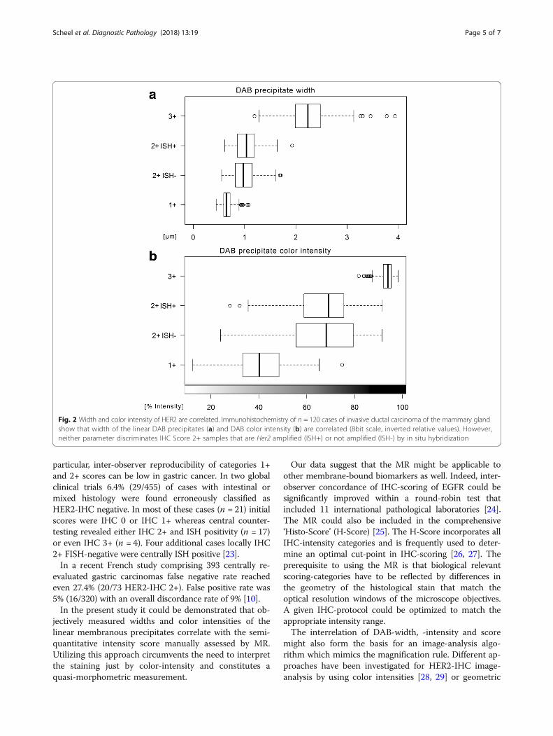
Precipitate width and color intensity are correlatedColor intensity of the DAB-precipitates was determinedusing color deconvolution [19]. Similar to the precipitatewidth, the intensities of the three scoring categories weresignificantly different (p < 0.01) (Fig. 2). Moreover, agood linear correlation between width and intensity wasnoticed among scoring categories 1+ and 2+ (Pearson’sr = 0.73). The intensity in scoring category 3+ was satu-rated (Additional file 3: Figure S3).
Precipitate width and color intensity do not differentiatebetween amplified and non-amplified cases in the IHC2+ scoring categoryAll cases classified as IHC 2+ were subsequently testedfor HER2/neu gene amplification by in situ hybridization(ISH). Among the study cases, 20 were ISH positive and20 were ISH negative. The DAB-precipitate widthshowed a non-significant difference between the ISHpositive cases (1.02 ± 0.23 μm) and the ISH negativecases (0.98 ± 0.22 μm, p = 0.02485). Indeed, histogramsof the individual cells showed that the IHC 2+ ISHpositive and IHC 2+ ISH negative cases feature almostcompletely overlapping precipitate widths (Additionalfile 2: Figure S2). No difference in the HER2-IHC colorintensities was noticed between ISH negative and ISHpositive cases either (p = 0.7493) (Fig. 2).
DiscussionHER2-IHC scores determined according to the ‘magnifi-cation rule’ (MR) were compared to image-analysis ofwidth and color intensity of the DAB chromogen precip-itates along the tumor cell membranes. The parameterswere closely correlated and matched the opticalresolutions of the employed microscope objectives. Thisprovides a physical basis of the MR which was originallyestablished as an empirical rule for standardized HER2-IHC scoring in gastric cancer.HER2-IHC assays are based on peroxidase-coupled
secondary antibodies that oxidize 3,3′-diaminobenzidine(DAB) into an insoluble, brownish precipitate at the spotof the bound epitope. As HER2 is confined to the cell-membrane, the reaction yields linear precipitates at thecell-boundaries. Technical aspects of HER2-IHC arerobust and can be standardized by validated protocols,on-slide control tissue and external quality assessment[20–22] but interpretation of the resulting staining pat-terns may be challenging [4]: HER2-IHC scoring relies onsubdividing the cases into categories based on staining-intensity (0, 1+, negative; 2+, equivocal, requires ISH-testing; 3+ positive) (Fig. 1, Additional file 1: Figure S1).The human optical system is optimized to notice rela-
tive differences in color-intensity rather than absolutevalues. Visual stimuli are precortically processed in theretina through lateral inhibition which underlies variesoptical illusions first described by Ernst Mach in 1865 as‘Mach bands’ [12]. A given surface might appear brighteror darker depending on the luminosity of its surround-ings [11, 13]. Accordingly, intensity-scores in histopath-ology are in general prone to subjectivity.This is of particular importance in HER2-IHC scoring
in gastric cancer, which is based solely on staining-intensity. In contrast, HER2-IHC scoring in breast canceralso includes the staining-pattern as the DAB-precipitateshave to be ring-shaped to be considered as IHC 3+. In
Scheel et al. Diagnostic Pathology (2018) 13:19 Page 4 of 7
particular, inter-observer reproducibility of categories 1+and 2+ scores can be low in gastric cancer. In two globalclinical trials 6.4% (29/455) of cases with intestinal ormixed histology were found erroneously classified asHER2-IHC negative. In most of these cases (n = 21) initialscores were IHC 0 or IHC 1+ whereas central counter-testing revealed either IHC 2+ and ISH positivity (n = 17)or even IHC 3+ (n = 4). Four additional cases locally IHC2+ FISH-negative were centrally ISH positive [23].In a recent French study comprising 393 centrally re-
evaluated gastric carcinomas false negative rate reachedeven 27.4% (20/73 HER2-IHC 2+). False positive rate was5% (16/320) with an overall discordance rate of 9% [10].In the present study it could be demonstrated that ob-
jectively measured widths and color intensities of thelinear membranous precipitates correlate with the semi-quantitative intensity score manually assessed by MR.Utilizing this approach circumvents the need to interpretthe staining just by color-intensity and constitutes aquasi-morphometric measurement.
Our data suggest that the MR might be applicable toother membrane-bound biomarkers as well. Indeed, inter-observer concordance of IHC-scoring of EGFR could besignificantly improved within a round-robin test thatincluded 11 international pathological laboratories [24].The MR could also be included in the comprehensive‘Histo-Score’ (H-Score) [25]. The H-Score incorporates allIHC-intensity categories and is frequently used to deter-mine an optimal cut-point in IHC-scoring [26, 27]. Theprerequisite to using the MR is that biological relevantscoring-categories have to be reflected by differences inthe geometry of the histological stain that match theoptical resolution windows of the microscope objectives.A given IHC-protocol could be optimized to match theappropriate intensity range.The interrelation of DAB-width, -intensity and score
might also form the basis for an image-analysis algo-rithm which mimics the magnification rule. Different ap-proaches have been investigated for HER2-IHC image-analysis by using color intensities [28, 29] or geometric
Fig. 2 Width and color intensity of HER2 are correlated. Immunohistochemistry of n = 120 cases of invasive ductal carcinoma of the mammary glandshow that width of the linear DAB precipitates (a) and DAB color intensity (b) are correlated (8bit scale, inverted relative values). However,neither parameter discriminates IHC Score 2+ samples that are Her2 amplified (ISH+) or not amplified (ISH-) by in situ hybridization
Scheel et al. Diagnostic Pathology (2018) 13:19 Page 5 of 7

properties of the staining pattern [30]. Recent advancesin digital image analysis have shown to increase of inter-observer agreement and decrease of the number ofequivocally scored cases [31, 32].
ConclusionsIHC scoring by using the ‘magnification rule’ is a semi-quantitative procedure that circumvents neurophysio-logical restrictions of our visual system. It is based onphysical interrelations and can be used to overcomesubjectivity in HER2 IHC-testing, particularly in gastriccancer. It might also be applicable to other membrane-bound IHC-stainings. As a practical and easy-to-usemethod it has found wide application and was incorpo-rated into national and international recommendationon HER2-IHC [6, 15, 17].
Additional files
Additional file 1: Figure S1. Example photomicrographs of HER2-IHC.Images depict scoring categories 1+, 2+ and 3+ at magnifications reflectingdifferent microscope objectives (2.5× - 63×. Inserts: Magnified details, 4×additional magnification). Note that the linear DAB-precipitates in categories1+ and 2+ are not perceivable at low power magnification (2.5×, 5×).(TIFF 47314 kb)
Additional file 2: Figure S2. Width of HER2 DAB-precipitates and resultof in situ hybridization (ISH). Histograms of n = 1200 measurements in 40cases per scoring category; estimated density (graphs). (TIFF 1103 kb)
Additional file 3: Figure S3. Scatter-plot of HER2 DAB-precipitates widthand color intensity. For scoring intensities 1+ and 2+ (grey), width andintensity show a linear correlation (r = 0.73, dashed lined). Scoring category3+ shows saturated intensity (n = 1200 measurements in 40 cases perscoring category). (JPEG 585 kb)
AbbreviationsASCO/CAP: American Society of Clinical Oncology / College of AmericanPathologists; DAB: Diaminobenzidine; HER2: The HER2 protein, i.e. humanepidermal growth factor receptor 2; HER2/neu: The HER2/neu gene whichencodes the HER2 protein; HERA: Acronym of the clinical phase 3 trial thattested adjuvant Trastuzumab in breast cancer, cf. Lancet. 2017; 389:1195-1205; IHC: Immunohistochemistry; ISH: In-situ hybridization;MR: Magnification rule; NA: Numerical aperture; ROI: Region of interest;SD: Standard deviation; ToGA: Acronym of the clinical phase 3 trial thattested adjuvant Trastuzumab in gastric cancer, cf. Lancet. 2010; 376:687-97; 0,1+, 2+, 3 + : Categories of the scoring system for HER2-IHC interpretation;4B5: Clone of the primary anti-HER2 antibodies used for HER2-IHC
AcknowledgementsWe are much obliged to Sysmex (Norderstedt, Germany) for kindly providingus with the Pannoramic P250 whole slide scanner. We would like to thankUlrike Hampacher and Heike Fliegel (Institute of Pathology Nordhessen, Kassel)for excellent technical assistance.
FundingThe study did not receive funding.
Availability of data and materialsPlease contact author for data requests.
Authors’ contributionsThe magnification rule (MR) was conceived by JR. Usage of the MR in practicalHER2-testing was tested and discussed by FPL, WH, GB, PM, MH, BJ and JR. Thestudy was designed by AHS and JB under guidance of JR, PM and MH withsupport of FPL, WH and GB. Whole slide scanning was performed by JB and
AHS. Manual HER2-scoring was done by JR, MH and PM. Image-analysis wasdone by AHS. Data-analysis was done by AHS under guidance of JR, PM, andBJ. The manuscript was drafted by AHS and JR. The manuscript was discussedwith all coauthors and revised accordingly by AHS. The final version of themanuscript was agreed by all authors.
Ethics approval and consent to participateThe study was approved by the responsible local ethical committee of theLandesärztekammer Hess, Frankfurt, Germany (file number FF 135/2013). Thecommittee decided that additional informed consent was not required forthis retrospective analysis.
Consent for publicationNot applicable.
Competing interestsThe authors declare that they have no competing interests.
Publisher’s NoteSpringer Nature remains neutral with regard to jurisdictional claims in publishedmaps and institutional affiliations.
Author details1Institute of Pathology, University Hospital Cologne, Kerpener Str. 62, 50937Cologne, Germany. 2Département de Pathologie, Centre Jean-Perrin, 58, rueMontalembert, 392, 63011 Clermont-Ferrand cedex 1, BP, France.3Department of Laboratory Medicine and Pathobiology, University ofToronto, Toronto, Canada. 4Institute of Pathology, University HospitalDresden, Fetscherstr, 74, 01307 Dresden, Germany. 5Institute of PathologyNordhessen, Germaniastraße 7, 34119 Kassel, Germany. 6Institute ofPathology, University Hospital Göttingen, Robert-Koch-Str. 40, 37075Göttingen, Germany. 7Targos Molecular Pathology GmbH, Germaniastraße 7,34119 Kassel, Germany.
Received: 15 October 2017 Accepted: 1 March 2018
References1. Yarden Y, Pines G. The ERBB network: at last, cancer therapy meets systems
biology. Nat Rev Cancer. 2012;12:553–63.2. Cameron D, Piccart-Gebhart MJ, Gelber RD, et al. 11 years' follow-up of
trastuzumab after adjuvant chemotherapy in HER2-positive early breastcancer: final analysis of the HERceptin adjuvant (HERA) trial. Lancet. 2017;389:1195–205.
3. Bang YJ, Van Cutsem E, Feyereislova A, et al. Trastuzumab in combinationwith chemotherapy versus chemotherapy alone for treatment of HER2-positive advanced gastric or gastro-oesophageal junction cancer (ToGA): aphase 3, open-label, randomised controlled trial. Lancet. 2010;376:687–97.
4. Wolff AC, Hammond ME, Hicks DG, et al. Recommendations for humanepidermal growth factor receptor 2 testing in breast cancer: AmericanSociety of Clinical Oncology/College of American Pathologists clinicalpractice guideline update. J Clin Oncol. 2013;31:3997–4013.
5. Bartley AN, Washington MK, Ismaila N, Ajani JA. HER2 testing andclinical decision making in gastroesophageal adenocarcinoma: guidelinesummary from the College of American Pathologists, American Societyfor Clinical Pathology, and American Society of Clinical Oncology. JOncol Pract. 2017;13:53–7.
6. Baretton G, Dietel M, Gaiser T, et al. HER2 testing in gastric cancer : resultsof a meeting of German experts. Pathologe. 2016;37:361–6.
7. Ross DS, Zehir A, Cheng DT, et al. Next-generation assessment of humanepidermal growth factor receptor 2 (ERBB2) amplification status: clinicalvalidation in the context of a hybrid capture-based, comprehensive solidtumor genomic profiling assay. J Mol Diagn. 2017;19:244–54.
8. Piccart-Gebhart MJ. St.Gallen International Breast Cancer Conference PrimaryTherapy of Early Breast Cancer Evidence, Controversies, Consensus; 11 - 14Mar 2009; St. Gallen, Switzerland.
9. Orlando L, Viale G, Bria E, et al. Discordance in pathology report after centralpathology review: implications for breast cancer adjuvant treatment. Breast.2016;30:151–5.
10. Monges G, Terris B, Chenard M-P, et al. Assessment of HER2 status from anepidemiology study in tumor tissue samples of gastric and gastro-
Scheel et al. Diagnostic Pathology (2018) 13:19 Page 6 of 7

esophageal junction cancer: Results from the french cohort of the HER-EAGLE study. JCO. 2013;31(4, Supplement S):26. http://ascopubs.org/doi/abs/10.1200/jco.2013.31.4_suppl.26.
11. Goldstein EB, editor. Blackwell handbook of perception. 4th ed. USA:Blackwell Publishers Inc; 2001. p. 53 ff.
12. Lotto RB, Williams SM, Purves D. Mach bands as empirically derivedassociations. Proc Natl Acad Sci U S A. 1999;96:5245–50.
13. Adelson EH. Perceptual organization and the judgment of brightness.Science. 1993;262:2042–4.
14. Rüschoff J, Dietel M, Baretton G, et al. HER2 diagnostics in gastric cancer-guideline validation and development of standardizedimmunohistochemical testing. Virchows Arch. 2010;457:299–307.
15. Rüschoff J, Hanna W, Bilous M, et al. HER2 testing in gastric cancer: apractical approach. Mod Pathol. 2012;25:637–50.
16. Behrens HM, Warneke VS, Böger C, et al. Reproducibility of Her2/neuscoring in gastric cancer and assessment of the 10% cut-off rule.Cancer Med. 2015;4:235–44.
17. Rakha EA, Starczynski J, Lee AH, Ellis IO. The updated ASCO/CAP guidelinerecommendations for HER2 testing in the management of invasive breastcancer: a critical review of their implications for routine practice.Histopathology. 2014;64:609–15.
18. Schneider CA, Rasband WS, Eliceiri KW. NIH image to ImageJ: 25 years ofimage analysis. Nat Methods. 2012;9:671–5.
19. Ruifrok AC, Johnston DA. Quantification of histochemical staining by colourdeconvolution. Anal Quant Cytol Histol. 2001;23:291–9.
20. Choritz H, Büsche G, Kreipe H, et al. Quality assessment of HER2 testing bymonitoring of positivity rates. Virchows Arch. 2011;459:283–9.
21. Vyberg M, Nielsen S, Røge R, et al. Immunohistochemical expression ofHER2 in breast cancer: socioeconomic impact of inaccurate tests. BMCHealth Serv Res. 2015;15:352.
22. Rüschoff J, Lebeau A, Kreipe H, et al. Assessing HER2 testing quality inbreast cancer: variables that influence HER2 positivity rate from a large,multicenter, observational study in Germany. Mod Pathol. 2017;30:217–26.
23. Cunningham D, Shah MA, Smith D, et al. False-negative rate for HER2testing in 738 gastric and gastroesophageal junction cancers from twoglobal randomized clinical trials. J Clin Oncol. 2015;33(Supplement 3):16.http://ascopubs.org/doi/abs/10.1200/jco.2015.33.3_suppl.16.
24. Rüschoff J, Kerr KM, Grote HJ, et al. Reproducibility of immunohistochemicalscoring for epidermal growth factor receptor expression in non-small celllung cancer: round robin test. Arch Pathol Lab Med. 2013;137:1255–61.
25. Hirsch FR, Varella-Garcia M, Bunn PA Jr, et al. Epidermal growth factorreceptor in non-small-cell lung carcinomas: correlation between gene copynumber and protein expression and impact on prognosis. J Clin Oncol.2003;21:3798–807.
26. Garon EB, Rizvi NA, Hui R, et al. Pembrolizumab for the treatment of non-small-cell lung cancer. N Engl J Med. 2015;372:2018–28.
27. Dolled-Filhart M, Roach C, Toland G, et al. Development of a companiondiagnostic for Pembrolizumab in non-small cell lung cancer usingimmunohistochemistry for programmed death Ligand-1. Arch Pathol LabMed. 2016;140:1243–9.
28. Ali HR, Irwin M, Morris L, et al. Astronomical algorithms for automated analysisof tissue protein expression in breast cancer. Br J Cancer. 2013;108:602–12.
29. Jeung J, Patel R, Vila L, Wakefield D, Liu C. Quantitation of HER2/neuexpression in primary gastroesophageal adenocarcinomas usingconventional light microscopy and quantitative image analysis. Arch PatholLab Med. 2012;136:610–7.
30. Laurinaviciene A, Dasevicius D, Ostapenko V, Jarmalaite S, Lazutka J,Laurinavicius A. Membrane connectivity estimated by digital image analysisof HER2 immunohistochemistry is concordant with visual scoring andfluorescence in situ hybridization results: algorithm evaluation on breastcancer tissue microarrays. Diagn Pathol. 2011;6:1746–596.
31. Helin HO, Tuominen VJ, Ylinen O, Helin HJ, Isola J. Free digital imageanalysis software helps to resolve equivocal scores in HER2immunohistochemistry. Virchows Arch. 2016;468:191–8.
32. Nielsen SL, Nielsen S, Vyberg M. Digital image analysis of HER2Immunostained gastric and gastroesophageal junction adenocarcinomas.Appl Immunohistochem Mol Morphol. 2017;25:320–8.
• We accept pre-submission inquiries
• Our selector tool helps you to find the most relevant journal
• We provide round the clock customer support
• Convenient online submission
• Thorough peer review
• Inclusion in PubMed and all major indexing services
• Maximum visibility for your research
Submit your manuscript atwww.biomedcentral.com/submit
Submit your next manuscript to BioMed Central and we will help you at every step:
Scheel et al. Diagnostic Pathology (2018) 13:19 Page 7 of 7