Embed Size (px)
Citation preview
PART
IV
PHYSICAL EXAMINATION OF PATIENTS
WITH HEADACHE
52835_CH13_Fernandez.qxd 1/2/09 3:26 PM Page 131
© Jones and Bartlett Publishers, LLC. NOT FOR SALE OR DISTRIBUTION

52835_CH13_Fernandez.qxd 1/2/09 3:26 PM Page 132
© Jones and Bartlett Publishers, LLC. NOT FOR SALE OR DISTRIBUTION
133
CHAPTER
13
Clinical Reasoning in theDiagnosis: HistoryTaking in Patients
with Headache
Peter A. Huijbregts, PT, MSc, MHSc, DPT, OCS, MTC, FAAOMPT, FCAMT
ROLE OF HISTORY TAKING IN THE CLINICAL EXAMINATION
In physical therapy, as in other health-care professions, there are five el-ements to patient management. The examination is followed by evalu-ation of the examination findings, establishing a diagnosis, producing aprognosis and developing a plan of care, and, finally, performing the in-terventions (American Physical Therapy Association [APTA], 2001). Theexamination element of this process of care usually consists of historytaking, systems review, and tests and measures. Unique to professionssuch as physical therapy with a limited scope of practice, a systems re-view is a brief history and physical examination specifically reviewing the cardiopulmonary, integumentary, musculoskeletal, and neuromus-cular systems, but also meant to get an impression of a patient’s com-munication ability, affect, cognition, language, and learning style (APTA,2001).
Generally, the role of the examination process is twofold. First, intended to be comprehensive with regard to screening and specifictesting, this process ideally leads to diagnostic classification. A second im-portant role of the physical therapy examination and related to the lim-ited neuromusculoskeletal scope of practice of physical therapy is theidentification of problems outside this scope of practice that require a
CHAPTER OUTLINE
Role of History Taking in theClinical Examination
DemographicsLocation of PainOnset and Course of HeadacheCharacter and Intensity of
HeadacheAggravating and Easing FactorsNeurologic SymptomsOtolaryngologic SymptomsSystemic SymptomsMedical HistoryMedication HistoryFamily HistoryPrevious Diagnostic TestsPrognostic IndicatorsSystems ReviewClinical Prediction Rules for
DiagnosisOutcome Measures
Pain MeasuresDisability Measures
Red FlagsConclusions and Implications of
History FindingsAcknowledgmentsReferences
52835_CH13_Fernandez.qxd 1/2/09 3:26 PM Page 133
© Jones and Bartlett Publishers, LLC. NOT FOR SALE OR DISTRIBUTION
efit from pharmacologic management as well, indicat-ing the (potential) need for medical comanagement.
This limited number of headache types potentiallyamenable to physical therapy management means thatone of the main objectives of the examination will be toidentify whether the presenting headache complaintcan in fact be classified as one of these five headachetypes. Tables 13.1 to 13.5 provide the diagnostic criteriafor these headache types. We should note that for cer-vicogenic headache there are two different sets of diag-nostic criteria (Table 13.3). In addition to the criteria inthe International Classification of Headache Disorders(IHS, 2004), Sjaastad et al. and the CervicogenicHeadache International Study Group (1998) establishedcriteria that are in fact used more frequently in the clin-ical situation.
Granella et al. (1994) demonstrated that diagnosis of primary headache types using the InternationalClassification of Headache Disorders had adequate inter-rater reliability for clinical use (k = 0.74). However, beforewe become too confident in our ability to diagnoseheadaches using this classification system, we have torealize that this system may seem simple to use but thatthe absence of confirming imaging, electrophysiologic, orlaboratory tests for the primary headache disordersmeans that the clinician is frequently in the position ofneeding to consider or rule out a secondary headachetype that may be similar in appearance to the primaryheadache disorder based on history and examinationfindings alone (Bartleson, 2006). For an extended out-line of headache diagnosis, see Chapter 3.
Some of these secondary headache disorders mim-icking the headache types that therapists can, in fact,manage may be serious or even life threatening and re-quire urgent medical or surgical referral. Because the diagnostic procedures confirming such secondaryheadaches are outside the diagnostic scope of practice ofthe therapist, the second main objective of the history—and of the examination process in general—becomesnot only the identification of a headache (or other undiagnosed) disorder not amenable to physical ther-apy intervention but also and more specifically the identification of those headache emergencies that re-quire urgent referral (Chapter 3). Within the context ofhistory taking, the clinician should seek to identify redflag symptoms indicating serious pathology. The historycomponent of the systems review may also yield indica-tions of pathology that may or may not be related to the presenting complaint of headache but that still require referral.
referral for medical or surgical diagnosis and, perhaps, co-management. The systems review component of theexamination plays an important role in detecting indica-tions for such referral (APTA, 2001).
Obtaining a comprehensive history is paramount toany diagnostic process but perhaps even more so in pa-tients presenting with headache. Bartleson (2006) notedthat in patients with complaint of headache, history tak-ing in combination with the general and neurologic ex-aminations constitutes the diagnostic gold standard.Indeed, the primary headache disorders defined in the International Classification of Headache Disorders(International Headache Society [IHS], 2004) have noavailable confirming diagnostic tests or procedures.Welch (2005) further emphasized the importance of his-tory taking by noting that most patients presenting withheadache often have few signs on physical examina-tion. In children, the role of the history is perhaps evenmore important. Dooley et al. (2003) reported that thehistory provided the correct diagnosis and managementin 100% of 150 children presenting with headaches.
Although there is some variation in a worldwide context, physical therapy interventions are generallylimited to manual therapy, education, exercise, andmodality-based treatments, thereby excluding pharma-cologic and surgical interventions. In this aspect, physi-cal therapy is similar to other professions that might beinvolved in the diagnosis and management of patientswith headache, such as chiropractic and massage therapy.As discussed in this chapter, this makes the diagnosticprocess in such professions a bit easier.
There is mounting research evidence (or at the veryleast a plausible pathophysiologic rationale) that manualtherapy interventions and other modalities within thescope of practice of physical therapy are effective in the management of five distinct types of headaches. Ofthe primary headaches, the scientific literature indicatesthat tension-type headache, and to a lesser extent mi-graine, may have an underlying neuromusculoskeletalcontribution (Fernández-de-las-Peñas et al., 2006a,2006b, 2006c; Tuchin et al., 2000; Simons et al., 1999;Tuchin, 1999). Secondary headaches with a neuro-musculoskeletal etiology that are, therefore, potentiallyamenable to interventions within the physical therapyscope of practice include cervicogenic headache, occipi-tal neuralgia, and headache associated with temporo-mandibular disorder (Issa & Huijbregts, 2006; Bronfort et al., 2001; Jull et al., 2002; Okeson, 1996; Bogduk,1986). It should be noted here that these headachetypes—and especially migraine headache—all may ben-
134 | Part IV | PHYSICAL EXAMINATION OF PATIENTS WITH HEADACHE
52835_CH13_Fernandez.qxd 1/2/09 3:26 PM Page 134
© Jones and Bartlett Publishers, LLC. NOT FOR SALE OR DISTRIBUTION
In summary, although mirroring the two main goals ofthe examination in general as outlined earlier (APTA,2001), the three main objectives of history taking in pa-tients with headache disorders are more specific:
1. Confirm the diagnosis of tension-type headache,migraine headache, cervicogenic headache,occipital neuralgia, or headache associated withtemporomandibular disorder, according to
Chapter 13 | Clinical Reasoning in the Diagnosis: History Taking in Patients with Headache | 135
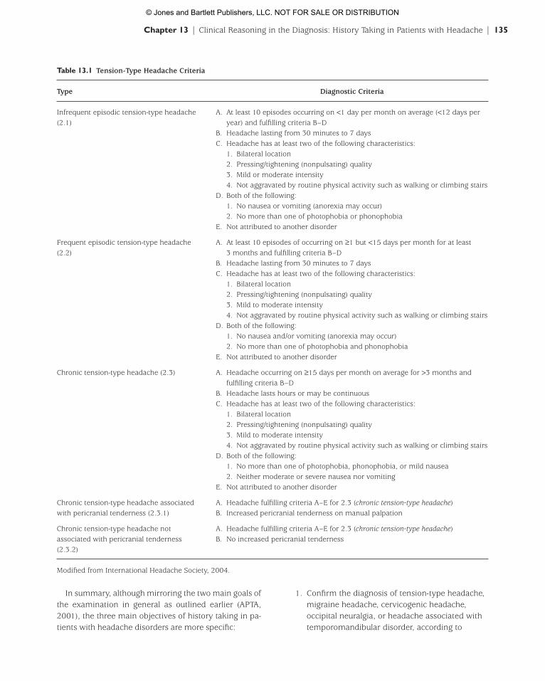
Table 13.1 Tension-Type Headache Criteria
Type Diagnostic Criteria
Infrequent episodic tension-type headache A. At least 10 episodes occurring on <1 day per month on average (<12 days per(2.1) year) and fulfilling criteria B–D
B. Headache lasting from 30 minutes to 7 daysC. Headache has at least two of the following characteristics:
1. Bilateral location2. Pressing/tightening (nonpulsating) quality3. Mild or moderate intensity4. Not aggravated by routine physical activity such as walking or climbing stairs
D. Both of the following:1. No nausea or vomiting (anorexia may occur)2. No more than one of photophobia or phonophobia
E. Not attributed to another disorder
Frequent episodic tension-type headache A. At least 10 episodes of occurring on ≥1 but <15 days per month for at least (2.2) 3 months and fulfilling criteria B–D
B. Headache lasting from 30 minutes to 7 daysC. Headache has at least two of the following characteristics:
1. Bilateral location2. Pressing/tightening (nonpulsating) quality3. Mild to moderate intensity4. Not aggravated by routine physical activity such as walking or climbing stairs
D. Both of the following:1. No nausea and/or vomiting (anorexia may occur)2. No more than one of photophobia and phonophobia
E. Not attributed to another disorder
Chronic tension-type headache (2.3) A. Headache occurring on ≥15 days per month on average for >3 months andfulfilling criteria B–D
B. Headache lasts hours or may be continuousC. Headache has at least two of the following characteristics:
1. Bilateral location2. Pressing/tightening (nonpulsating) quality3. Mild to moderate intensity4. Not aggravated by routine physical activity such as walking or climbing stairs
D. Both of the following:1. No more than one of photophobia, phonophobia, or mild nausea2. Neither moderate or severe nausea nor vomiting
E. Not attributed to another disorder
Chronic tension-type headache associated A. Headache fulfilling criteria A–E for 2.3 (chronic tension-type headache)with pericranial tenderness (2.3.1) B. Increased pericranial tenderness on manual palpation
Chronic tension-type headache not A. Headache fulfilling criteria A–E for 2.3 (chronic tension-type headache)associated with pericranial tenderness B. No increased pericranial tenderness(2.3.2)
Modified from International Headache Society, 2004.
52835_CH13_Fernandez.qxd 1/2/09 3:26 PM Page 135
© Jones and Bartlett Publishers, LLC. NOT FOR SALE OR DISTRIBUTION
13.6) with specific attention to these three main objec-tives. The following sections discuss the various items inthis format.
DEMOGRAPHICS
In patients older than 50, a new onset of migraineheadache is unlikely (Gladstein, 2006). In fact, if a newheadache suddenly appears in a patient older than 50, it should be considered a secondary headache untilcauses such as tumor, cerebrovascular disease, or tem-poral arteritis are excluded (Gentile, 2005). Temporal or
established diagnostic classification systems(IHS, 2004; Okeson, 1996; Sjaastad et al., 1998).
2. Identify diagnostic indicators of headache typesnot amenable to physical therapy management,and specifically red flag symptoms indicatingserious pathology requiring urgent referral.
3. Identify the presence—but not the exactnature—of pathology not necessarily related tothe presenting headache complaint that requiresreferral for medical or surgical evaluation.
This chapter presents a suggested format for historytaking in patients presenting with headache (Table
136 | Part IV | PHYSICAL EXAMINATION OF PATIENTS WITH HEADACHE
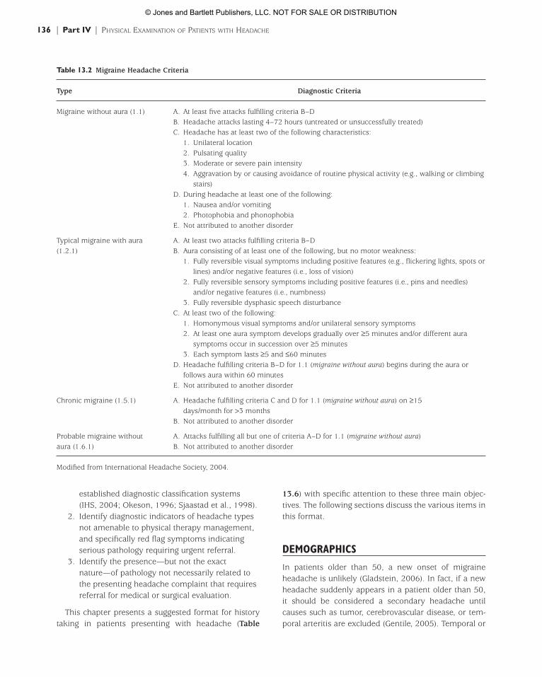
Table 13.2 Migraine Headache Criteria
Type Diagnostic Criteria
Migraine without aura (1.1) A. At least five attacks fulfilling criteria B–DB. Headache attacks lasting 4–72 hours (untreated or unsuccessfully treated)C. Headache has at least two of the following characteristics:
1. Unilateral location2. Pulsating quality3. Moderate or severe pain intensity4. Aggravation by or causing avoidance of routine physical activity (e.g., walking or climbing
stairs)D. During headache at least one of the following:
1. Nausea and/or vomiting2. Photophobia and phonophobia
E. Not attributed to another disorder
Typical migraine with aura A. At least two attacks fulfilling criteria B–D(1.2.1) B. Aura consisting of at least one of the following, but no motor weakness:
1. Fully reversible visual symptoms including positive features (e.g., flickering lights, spots orlines) and/or negative features (i.e., loss of vision)
2. Fully reversible sensory symptoms including positive features (i.e., pins and needles)and/or negative features (i.e., numbness)
3. Fully reversible dysphasic speech disturbanceC. At least two of the following:
1. Homonymous visual symptoms and/or unilateral sensory symptoms2. At least one aura symptom develops gradually over ≥5 minutes and/or different aura
symptoms occur in succession over ≥5 minutes3. Each symptom lasts ≥5 and ≤60 minutes
D. Headache fulfilling criteria B–D for 1.1 (migraine without aura) begins during the aura orfollows aura within 60 minutes
E. Not attributed to another disorder
Chronic migraine (1.5.1) A. Headache fulfilling criteria C and D for 1.1 (migraine without aura) on ≥15days/month for >3 months
B. Not attributed to another disorder
Probable migraine without A. Attacks fulfilling all but one of criteria A–D for 1.1 (migraine without aura)aura (1.6.1) B. Not attributed to another disorder
Modified from International Headache Society, 2004.
52835_CH13_Fernandez.qxd 1/2/09 3:26 PM Page 136
© Jones and Bartlett Publishers, LLC. NOT FOR SALE OR DISTRIBUTION
Chapter 13 | Clinical Reasoning in the Diagnosis: History Taking in Patients with Headache | 137
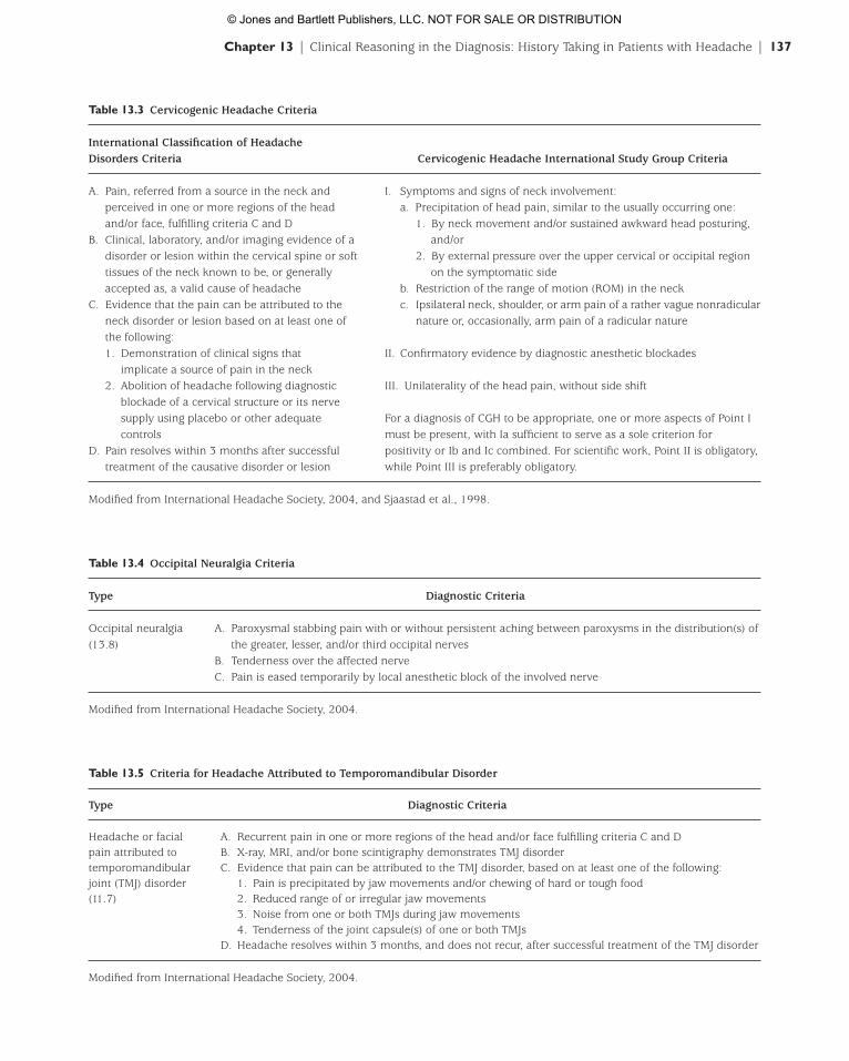
Table 13.3 Cervicogenic Headache Criteria
International Classification of Headache Disorders Criteria Cervicogenic Headache International Study Group Criteria
A. Pain, referred from a source in the neck and I. Symptoms and signs of neck involvement:perceived in one or more regions of the head a. Precipitation of head pain, similar to the usually occurring one:and/or face, fulfilling criteria C and D 1. By neck movement and/or sustained awkward head posturing,
B. Clinical, laboratory, and/or imaging evidence of a and/ordisorder or lesion within the cervical spine or soft 2. By external pressure over the upper cervical or occipital region tissues of the neck known to be, or generally on the symptomatic sideaccepted as, a valid cause of headache b. Restriction of the range of motion (ROM) in the neck
C. Evidence that the pain can be attributed to the c. Ipsilateral neck, shoulder, or arm pain of a rather vague nonradicular neck disorder or lesion based on at least one of nature or, occasionally, arm pain of a radicular naturethe following:1. Demonstration of clinical signs that II. Confirmatory evidence by diagnostic anesthetic blockades
implicate a source of pain in the neck2. Abolition of headache following diagnostic III. Unilaterality of the head pain, without side shift
blockade of a cervical structure or its nervesupply using placebo or other adequate For a diagnosis of CGH to be appropriate, one or more aspects of Point I controls must be present, with Ia sufficient to serve as a sole criterion for
D. Pain resolves within 3 months after successful positivity or Ib and Ic combined. For scientific work, Point II is obligatory,treatment of the causative disorder or lesion while Point III is preferably obligatory.
Modified from International Headache Society, 2004, and Sjaastad et al., 1998.
Table 13.4 Occipital Neuralgia Criteria
Type Diagnostic Criteria
Occipital neuralgia A. Paroxysmal stabbing pain with or without persistent aching between paroxysms in the distribution(s) of (13.8) the greater, lesser, and/or third occipital nerves
B. Tenderness over the affected nerveC. Pain is eased temporarily by local anesthetic block of the involved nerve
Modified from International Headache Society, 2004.
Table 13.5 Criteria for Headache Attributed to Temporomandibular Disorder
Type Diagnostic Criteria
Headache or facial A. Recurrent pain in one or more regions of the head and/or face fulfilling criteria C and Dpain attributed to B. X-ray, MRI, and/or bone scintigraphy demonstrates TMJ disordertemporomandibular C. Evidence that pain can be attributed to the TMJ disorder, based on at least one of the following:joint (TMJ) disorder 1. Pain is precipitated by jaw movements and/or chewing of hard or tough food(11.7) 2. Reduced range of or irregular jaw movements
3. Noise from one or both TMJs during jaw movements4. Tenderness of the joint capsule(s) of one or both TMJs
D. Headache resolves within 3 months, and does not recur, after successful treatment of the TMJ disorder
Modified from International Headache Society, 2004.
52835_CH13_Fernandez.qxd 1/2/09 3:26 PM Page 137
© Jones and Bartlett Publishers, LLC. NOT FOR SALE OR DISTRIBUTION
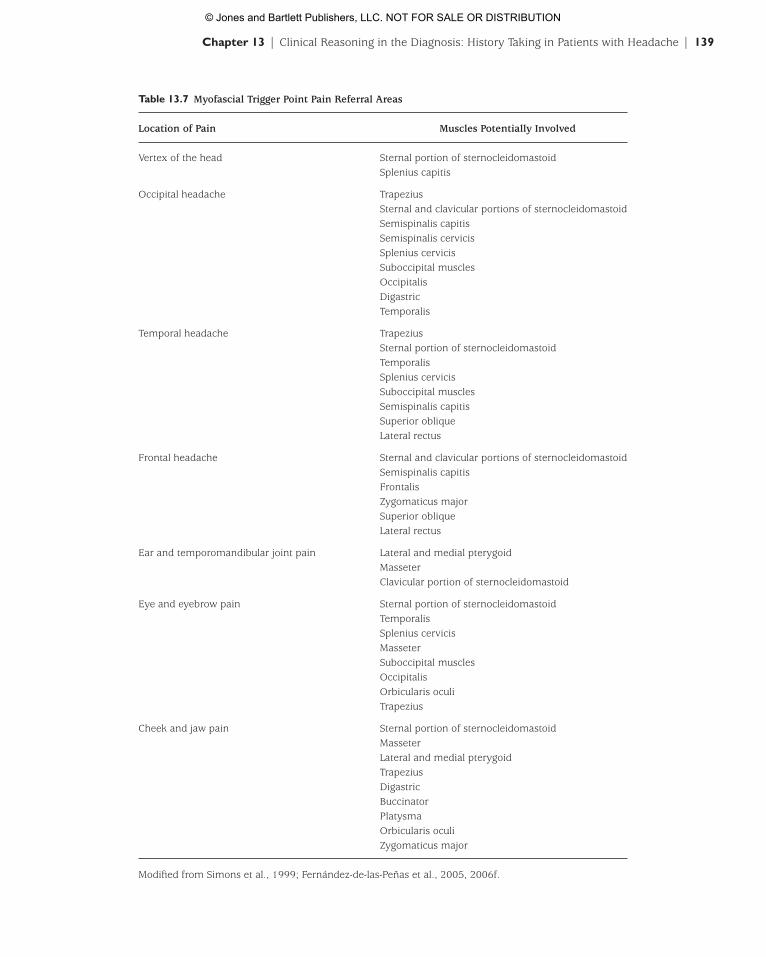
sion-type headache (Fernández-de-las-Peñas et al.,2006a, 2006b, 2006d, 2006e). This means that specificpain locations may be matched with established painreferral patterns from myofascial trigger points, whichin turn will guide the subsequent palpatory examina-tion. Headache location is matched to specific muscletrigger points in Table 13.7 (Fernández-de-las-Peñas et al., 2005, 2006f; Simons et al., 1999).
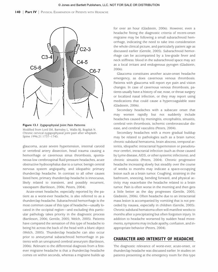
Further, with cervical zygapophysial joints implicatedas the major etiologic factor in cervicogenic headache, re-ferral patterns of these joints as established by way ofjoint infiltration can also guide subsequent examina-tion (Dwyer et al., 1990). Figure 13.1 provides zy-gapophysial joint referral patterns.
Headache occurring exclusively on one side may indi-cate a need for referral. Note that unilateral headaches arealso likely in patients with more innocuous headachetypes such as cervicogenic headache; occipital neural-gia; headaches due to temporomandibular disorder;cluster headaches; paroxysmal hemicrania; short-lasting, unilateral, neuralgiform headache attacks withconjunctival injection and tearing (SUNCT); and withmigraine. However, in a patient with a new onset of uni-lateral headache that does not have the features of oneof these headache disorders—and of course for allheadache types not amenable to (sole) physical therapymanagement—referral for further medical diagnosis isrequired (Bartleson, 2006).
ONSET AND COURSE OF HEADACHE
Headache onset can be qualified as acute, subacute, andchronic. In addition, patients may report a course of in-termittent, episodic, or continuous pain that is stable or progressive in intensity. The five headache typesamenable to physical therapy intervention are generallyinsidious in their onset (with the exception of a first-ever migraine headache) and of an intermittent, non-progressive nature. Cluster headaches are—as the nameimplies—episodic in nature. Especially if a constantheadache is progressive and worsening or if the patientpresents with an acute-onset, worst-ever headache, theclinician should be attentive to a serious underlyingpathology. Mills et al. (1986) reported abnormal find-ings on computed tomography (CT) scan for 29% of patients presenting with sudden-onset, reported worst-ever, or severe persistent headaches.
Causes of acute-onset headache include intracranialor intracerebral hemorrhage, acute subdural or epiduralhematoma, pituitary apoplexy, acute closed-angle
giant cell arteritis can lead to blindness due to occlusionof the ocular arteries. Patients may report severe unilat-eral or bilateral throbbing pain in the temporal regionthat is worse at night. Patients may also report scalptenderness, jaw claudication, visual complaints, fatigue orjoint pain associated with polymyalgia rheumatica,weight loss, fever, and night sweats. They may also havenoted a tender, red, thickened, and nonpulsatile tempo-ral artery (Peters, 2004; Welch, 2005). The clinicianshould not get too fixated on an age limit of 50 years, be-cause values reported in the literature as red flags forthe onset of a new headache vary between 40 and 60years (Bartleson, 2006).
Gender also plays a role in diagnosis in that variousheadaches are more prevalent in women. European andAmerican studies have showed a 1-year prevalence of6% to 8% in men but 15% to 18% in women for migraine headaches. In developed countries, tension-type headache affects two-thirds of men and over 80% of women (World Health Organization, 2004).Musculoskeletal pain due to temporomandibular disor-der is nine times more common in women than in men(Türp et al., 2002). In contrast, cluster headaches aremore common in men, most typically smokers in theirlate 20s (Welch, 2005).
LOCATION OF PAIN
Characteristic locations of pain for the five headachetypes amenable to physical therapy management areindicated to some extent in the diagnostic criteria pro-vided in Tables 13.1 to 13.5. Myofascial trigger pointshave been suggested as a main etiologic factor in ten-
138 | Part IV | PHYSICAL EXAMINATION OF PATIENTS WITH HEADACHE
Table 13.6 Suggested Format for History Taking in Patientswith Headache
DemographicsLocation of painOnset and course of headacheCharacter and intensity of headacheAggravating and easing factorsNeurologic symptomsOtolaryngologic symptomsSystemic symptomsMedical historyMedication historyFamily historyPrevious diagnostic testsPrognostic indicatorsSystems review
52835_CH13_Fernandez.qxd 1/2/09 3:26 PM Page 138
© Jones and Bartlett Publishers, LLC. NOT FOR SALE OR DISTRIBUTION
Chapter 13 | Clinical Reasoning in the Diagnosis: History Taking in Patients with Headache | 139
Table 13.7 Myofascial Trigger Point Pain Referral Areas
Location of Pain Muscles Potentially Involved
Vertex of the head Sternal portion of sternocleidomastoidSplenius capitis
Occipital headache TrapeziusSternal and clavicular portions of sternocleidomastoidSemispinalis capitisSemispinalis cervicisSplenius cervicisSuboccipital musclesOccipitalisDigastricTemporalis
Temporal headache TrapeziusSternal portion of sternocleidomastoidTemporalisSplenius cervicisSuboccipital musclesSemispinalis capitisSuperior obliqueLateral rectus
Frontal headache Sternal and clavicular portions of sternocleidomastoidSemispinalis capitisFrontalisZygomaticus majorSuperior obliqueLateral rectus
Ear and temporomandibular joint pain Lateral and medial pterygoidMasseterClavicular portion of sternocleidomastoid
Eye and eyebrow pain Sternal portion of sternocleidomastoidTemporalisSplenius cervicisMasseterSuboccipital musclesOccipitalisOrbicularis oculiTrapezius
Cheek and jaw pain Sternal portion of sternocleidomastoidMasseterLateral and medial pterygoidTrapeziusDigastricBuccinatorPlatysmaOrbicularis oculiZygomaticus major
Modified from Simons et al., 1999; Fernández-de-las-Peñas et al., 2005, 2006f.
52835_CH13_Fernandez.qxd 1/2/09 3:26 PM Page 139
© Jones and Bartlett Publishers, LLC. NOT FOR SALE OR DISTRIBUTION
for over an hour (Gladstein, 2006). However, even aheadache fitting the diagnostic criteria of recent-onsetmigraine may be following a small subarachnoid hem-orrhage, indicating the need to take into considerationthe whole clinical picture, and particularly patient age asdiscussed earlier (Gentile, 2005). Subarachnoid hemor-rhage can be accompanied by a low-grade fever andneck stiffness: blood in the subarachnoid space may actas a local irritant and endogenous pyrogen (Gladstein,2006).
Glaucoma constitutes another acute-onset headacheemergency, as does cavernous venous thrombosis.Patients with glaucoma will report eye pain and visionchanges. In case of cavernous venous thrombosis, pa-tients usually have a history of ear, nose, or throat surgeryor localized nasal infection, or they may report usingmedications that could cause a hypercoagulable state(Gladstein, 2006).
Secondary headaches with a subacute onset that may worsen rapidly but not suddenly includeheadaches caused by meningitis, encephalitis, sinusitis,cerebral vein thrombosis, ischemic cerebrovascular dis-ease, and cerebral vasculitis (Peters, 2004).
Secondary headaches with a more gradual buildupmay be related to pathologies such as a brain tumor,chronic subdural hematoma, brain abscess, temporal ar-teritis, idiopathic intracranial hypertension or pseudotu-mor cerebri, intracranial infection (such as those causedby Lyme disease, AIDS, or other systemic infections), andchronic sinusitis (Peters, 2004). Chronic progressiveheadache increasing slowly but steadily over the courseof weeks to months may indicate a space-occupying lesion such as a brain tumor. Coughing, straining in thebathroom, sneezing, bending forward, and physical ac-tivity may exacerbate the headache related to a braintumor. Pain is often worse in the morning and then getsa little better as the day progresses (Gentile, 2005;Gladstein, 2006). Often headache due to an intracranialmass lesion is accompanied by vomiting that is not pre-ceded by nausea, especially in children (Gentile, 2005).Chronic subdural hematoma often will manifest weeks tomonths after a precipitating but often forgotten injury. Inaddition to headache worsened by sudden head move-ments, symptoms may include apathy, confusion, and in-appropriate behavior (Peters, 2004).
CHARACTER AND INTENSITY OF HEADACHE
The diagnostic relevance of worst-ever, acute-onset, orthunderclap headache was discussed earlier. In studies onpatients presenting at the emergency room for this type
glaucoma, acute severe hypertension, internal carotidor vertebral artery dissection, head trauma causing ahemorrhage or cavernous sinus thrombosis, sponta-neous low cerebrospinal fluid pressure headaches, acuteobstructive hydrocephalus due to a tumor, benign centralnervous system angiopathy, and idiopathic primarythunderclap headache. In contrast to all other causeslisted here, primary thunderclap headache is innocuous,likely related to transient, and possibly recurrent, vasospasm (Bartleson, 2006; Peters, 2004).
Acute-onset headache, especially reported by the pa-tient as a worst-ever headache, is also referred to as athunderclap headache. Subarachnoid hemorrhage is themost common cause of this type of headache—usually lo-cated in the occipital region—and excluding this partic-ular pathology takes priority in the diagnostic process(Bartleson, 2006; Gentile, 2005; Welch, 2005). Patientshave compared the sensation of this type of headache tobeing hit across the back of the head with a blunt object(Welch, 2005). Thunderclap headache can also occurprior to aneurysmal subarachnoid hemorrhage in pa-tients with an unruptured cerebral aneurysm (Bartleson,2006). Relevant to the differential diagnosis from a first-ever migraine headache is that a thunderclap headachecomes on within seconds, whereas a migraine builds up
140 | Part IV | PHYSICAL EXAMINATION OF PATIENTS WITH HEADACHE
Figure 13.1 Zygapophysial Joint Pain Patterns
Modified from Lord SM, Barnsley L, Wallis BJ, Bogduk N.Chronic cervical zygapophyseal joint pain after whiplash.Spine 1996;21:1737–1745.
52835_CH13_Fernandez.qxd 1/2/09 3:26 PM Page 140
© Jones and Bartlett Publishers, LLC. NOT FOR SALE OR DISTRIBUTION
of headache, the combined prevalence of significant intracranial pathology was 43% (95% CI: 20–68%)(Detsky et al., 2006).
The International Classification of Headache Disorders(IHS, 2004) classifies cluster headache as a primaryheadache. Cluster-type headaches are excruciating or-bital and temporal headaches that come and go in aclusterlike pattern. Although this pattern is characteristicof the primary cluster headaches, it may also serve asan indicator of significant secondary headache types:the presence of cluster-type headaches carries a posi-tive likelihood ratio (LR) of 10.7 (95% CI: 2.2–5.2) forthe presence of serious intracranial abnormalities(Detsky et al., 2006).
Relevant to this portion of the history-taking process isthat frequently headaches are of a mixed nature: mi-graine and tension-type headache frequently occur to-gether, and they can be combined later in their course with headaches related to medication overuse. Of pa-tients with migraine, 83% also have had tension-typeheadaches, whereas 23% of patients with tension-typeheadache also report migraine headaches (De Jongh et al.,2001). Every headache type requires a separate history(Welch, 2005). Tables 13.1 to 13.5 provide further detailson the character and intensity of the headache typesamenable to physical therapy management.
AGGRAVATING AND EASING FACTORS
Headaches amenable to physical therapy managementshould have underlying neuromusculoskeletal dysfunc-tions that can usually be aggravated or eased by me-chanical factors such as those occurring with movementand position. Effects differ between these headachetypes: migraine may be differentiated from tension-type headache in that migraine headache is exacer-bated by physical activity, whereas tension-typeheadache is not. Smetana (2000) established a positiveLR of 3.7 (95% CI: 3.4–4.0) and a negative LR of 0.24(95% CI: 0.23–0.26) for exacerbation by physical activ-ity in the differential diagnosis of these two primaryheadache types. In fact, patients with tension-typeheadache may even seek out physical activity to de-crease pain by way of distraction (Weeks & Weier,2006). Patients with migraine tend to retreat to a darkand quiet environment, whereas patients with clusterheadache are agitated and often will pace the room orrock back and forth holding their head (Gladstein, 2006;Weeks & Weier, 2006). However, some movement- andposition-related effects on headache may be indicative ofsecondary headaches requiring (urgent) referral.
A more benign secondary headache is one related tocerebrospinal fluid hypotension. This type of headache isclassically postural in that it occurs or worsens within15 minutes of going from a lying to an upright position.It generally disappears within 30 minutes of again lyingdown. Usually bilateral and frontal in location, thisheadache is commonly associated with nausea, vomiting,dizziness, and tinnitus. One of the most commoncauses is a persistent cerebrospinal fluid leak after alumbar puncture, but in the absence of such a medicalhistory, it may be indicative of spontaneous low cere-brospinal fluid pressure syndrome (Bartleson, 2006;Peters, 2004).
Idiopathic intracranial hypertension usually causes ageneralized frontal headache that may be unilateral orbilateral and is often worsened by bending over orstraining. Patients may note a progressive loss of vision.This secondary headache is most common in obesewomen; average age of onset is 30 years (Peters, 2004).
It was noted earlier that the headache related to abrain tumor is exacerbated with coughing, straining inthe bathroom, sneezing, bending forward, and physicalactivity. Pain is often worse in the morning and thengets a little better as the day progresses (Gentile, 2005;Welch, 2005; Gladstein, 2006). Coughing or posturechanges may also cause the onset of headache if there isan underlying structural cause, such as Arnold-Chiarimalformation type I, a colloid cyst of the third ventricle,or a tumor growing in the ventricular system (Gentile,2005). Although exertional and cough headache canoccur as an innocuous primary headache disorder,headache aggravated by exertion or a Valsalva-type ma-neuver carries a positive LR of 2.3 (95% CI: 1.4–3.8) forthe presence of serious intracranial pathology (Detskyet al., 2006). In patients with a normal neurologic ex-amination, headache aggravated by a Valsalva maneu-ver occurred significantly more often in those withsecondary headaches than in patients with primaryheadaches (OR = 3.4) (Duarte et al., 1996). Bartleson(2006) noted that headache initiated by exertion orValsalva maneuver (or cough) and associated neurologicsymptoms or signs that are not consistent with the cri-teria for migraine with typical aura indicate the need tosearch for underlying intracranial disease.
Headache brought on by sexual activity can alsooccur as a relatively innocuous primary headache dis-order. Similar to the exertional headache discussed ear-lier, it can be associated with migraine. Nevertheless,headache associated with sexual activity can also occursecondary to subarachnoid hemorrhage and arterial dis-section and should always be considered a red flag
Chapter 13 | Clinical Reasoning in the Diagnosis: History Taking in Patients with Headache | 141
52835_CH13_Fernandez.qxd 1/2/09 3:26 PM Page 141
© Jones and Bartlett Publishers, LLC. NOT FOR SALE OR DISTRIBUTION
cervical degenerative joint disease as implicated in cer-vicogenic headache, allowing for differential diagnosis(Davenport, 2002). However, it should be noted thatnuchal rigidity has a sensitivity of only 30% for menin-gitis, making a negative test relatively meaningless whenit comes to excluding serious causative pathology (Peters,2004). In patients with brain tumors, the headache usually is preceded by neurologic symptoms, including,as noted earlier, seizure but also hemiparesis, ataxia, andcognitive or speech impairment (Peters, 2004).
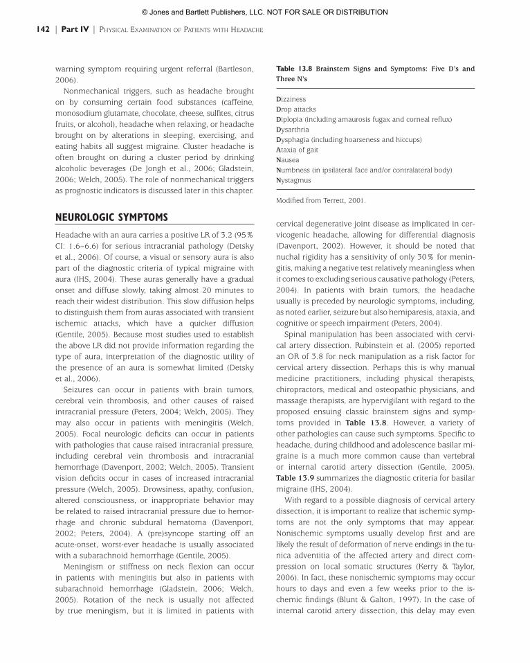
Spinal manipulation has been associated with cervi-cal artery dissection. Rubinstein et al. (2005) reportedan OR of 3.8 for neck manipulation as a risk factor forcervical artery dissection. Perhaps this is why manualmedicine practitioners, including physical therapists,chiropractors, medical and osteopathic physicians, andmassage therapists, are hypervigilant with regard to theproposed ensuing classic brainstem signs and symp-toms provided in Table 13.8. However, a variety ofother pathologies can cause such symptoms. Specific toheadache, during childhood and adolescence basilar mi-graine is a much more common cause than vertebral or internal carotid artery dissection (Gentile, 2005).Table 13.9 summarizes the diagnostic criteria for basilarmigraine (IHS, 2004).
With regard to a possible diagnosis of cervical arterydissection, it is important to realize that ischemic symp-toms are not the only symptoms that may appear.Nonischemic symptoms usually develop first and arelikely the result of deformation of nerve endings in the tu-nica adventitia of the affected artery and direct com-pression on local somatic structures (Kerry & Taylor,2006). In fact, these nonischemic symptoms may occurhours to days and even a few weeks prior to the is-chemic findings (Blunt & Galton, 1997). In the case ofinternal carotid artery dissection, this delay may even
warning symptom requiring urgent referral (Bartleson,2006).
Nonmechanical triggers, such as headache broughton by consuming certain food substances (caffeine,monosodium glutamate, chocolate, cheese, sulfites, citrusfruits, or alcohol), headache when relaxing, or headachebrought on by alterations in sleeping, exercising, andeating habits all suggest migraine. Cluster headache isoften brought on during a cluster period by drinking alcoholic beverages (De Jongh et al., 2006; Gladstein,2006; Welch, 2005). The role of nonmechanical triggersas prognostic indicators is discussed later in this chapter.
NEUROLOGIC SYMPTOMS
Headache with an aura carries a positive LR of 3.2 (95%CI: 1.6–6.6) for serious intracranial pathology (Detsky et al., 2006). Of course, a visual or sensory aura is alsopart of the diagnostic criteria of typical migraine withaura (IHS, 2004). These auras generally have a gradualonset and diffuse slowly, taking almost 20 minutes toreach their widest distribution. This slow diffusion helpsto distinguish them from auras associated with transientischemic attacks, which have a quicker diffusion(Gentile, 2005). Because most studies used to establishthe above LR did not provide information regarding thetype of aura, interpretation of the diagnostic utility ofthe presence of an aura is somewhat limited (Detsky et al., 2006).
Seizures can occur in patients with brain tumors,cerebral vein thrombosis, and other causes of raised intracranial pressure (Peters, 2004; Welch, 2005). Theymay also occur in patients with meningitis (Welch,2005). Focal neurologic deficits can occur in patientswith pathologies that cause raised intracranial pressure,including cerebral vein thrombosis and intracranialhemorrhage (Davenport, 2002; Welch, 2005). Transientvision deficits occur in cases of increased intracranialpressure (Welch, 2005). Drowsiness, apathy, confusion,altered consciousness, or inappropriate behavior maybe related to raised intracranial pressure due to hemor-rhage and chronic subdural hematoma (Davenport,2002; Peters, 2004). A (pre)syncope starting off anacute-onset, worst-ever headache is usually associatedwith a subarachnoid hemorrhage (Gentile, 2005).
Meningism or stiffness on neck flexion can occur in patients with meningitis but also in patients with subarachnoid hemorrhage (Gladstein, 2006; Welch,2005). Rotation of the neck is usually not affected by true meningism, but it is limited in patients with
142 | Part IV | PHYSICAL EXAMINATION OF PATIENTS WITH HEADACHE
Table 13.8 Brainstem Signs and Symptoms: Five D’s andThree N’s
DizzinessDrop attacksDiplopia (including amaurosis fugax and corneal reflux)DysarthriaDysphagia (including hoarseness and hiccups)Ataxia of gaitNauseaNumbness (in ipsilateral face and/or contralateral body)Nystagmus
Modified from Terrett, 2001.
52835_CH13_Fernandez.qxd 1/2/09 3:26 PM Page 142
© Jones and Bartlett Publishers, LLC. NOT FOR SALE OR DISTRIBUTION
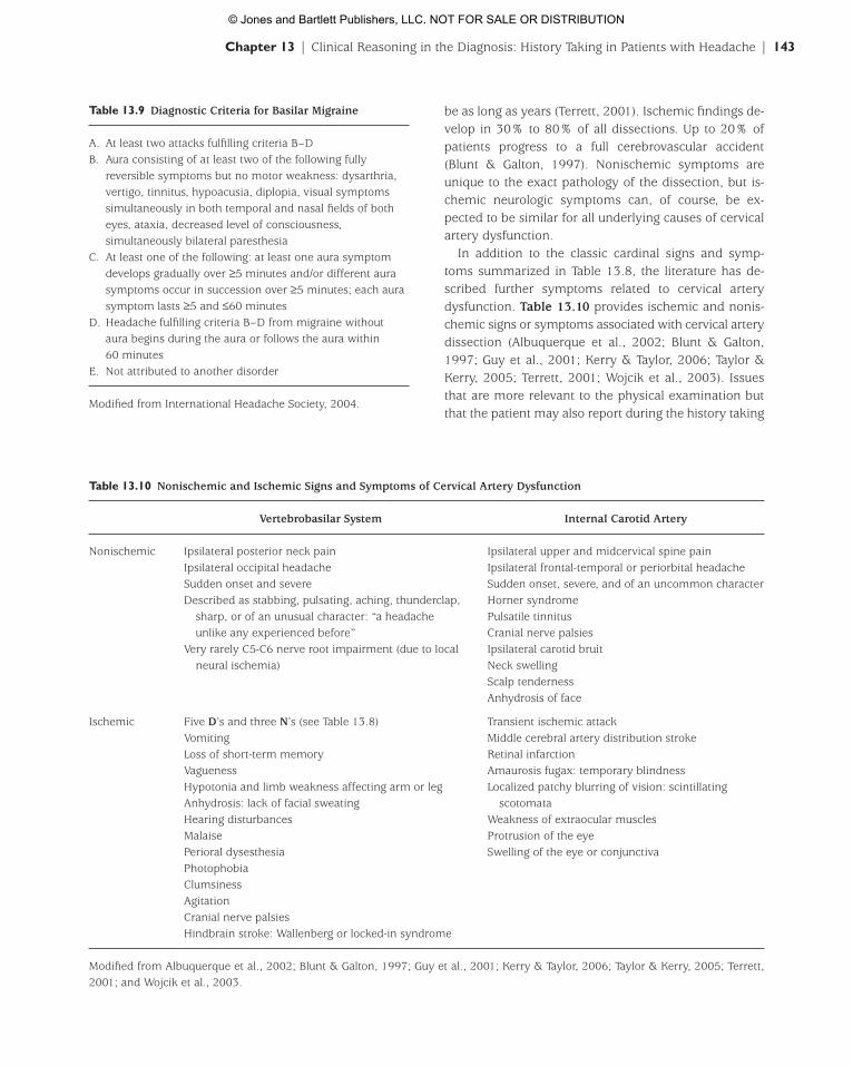
be as long as years (Terrett, 2001). Ischemic findings de-velop in 30% to 80% of all dissections. Up to 20% of patients progress to a full cerebrovascular accident(Blunt & Galton, 1997). Nonischemic symptoms areunique to the exact pathology of the dissection, but is-chemic neurologic symptoms can, of course, be ex-pected to be similar for all underlying causes of cervicalartery dysfunction.
In addition to the classic cardinal signs and symp-toms summarized in Table 13.8, the literature has de-scribed further symptoms related to cervical arterydysfunction. Table 13.10 provides ischemic and nonis-chemic signs or symptoms associated with cervical arterydissection (Albuquerque et al., 2002; Blunt & Galton,1997; Guy et al., 2001; Kerry & Taylor, 2006; Taylor &Kerry, 2005; Terrett, 2001; Wojcik et al., 2003). Issuesthat are more relevant to the physical examination butthat the patient may also report during the history taking
Chapter 13 | Clinical Reasoning in the Diagnosis: History Taking in Patients with Headache | 143
Table 13.9 Diagnostic Criteria for Basilar Migraine
A. At least two attacks fulfilling criteria B–DB. Aura consisting of at least two of the following fully
reversible symptoms but no motor weakness: dysarthria, vertigo, tinnitus, hypoacusia, diplopia, visual symptoms simultaneously in both temporal and nasal fields of both eyes, ataxia, decreased level of consciousness, simultaneously bilateral paresthesia
C. At least one of the following: at least one aura symptom develops gradually over ≥5 minutes and/or different aura symptoms occur in succession over ≥5 minutes; each aura symptom lasts ≥5 and ≤60 minutes
D. Headache fulfilling criteria B–D from migraine without aura begins during the aura or follows the aura within 60 minutes
E. Not attributed to another disorder
Modified from International Headache Society, 2004.
Table 13.10 Nonischemic and Ischemic Signs and Symptoms of Cervical Artery Dysfunction
Vertebrobasilar System Internal Carotid Artery
Nonischemic Ipsilateral posterior neck pain Ipsilateral upper and midcervical spine painIpsilateral occipital headache Ipsilateral frontal-temporal or periorbital headacheSudden onset and severe Sudden onset, severe, and of an uncommon characterDescribed as stabbing, pulsating, aching, thunderclap, Horner syndrome
sharp, or of an unusual character: “a headache Pulsatile tinnitusunlike any experienced before” Cranial nerve palsies
Very rarely C5-C6 nerve root impairment (due to local Ipsilateral carotid bruitneural ischemia) Neck swelling
Scalp tendernessAnhydrosis of face
Ischemic Five D’s and three N’s (see Table 13.8) Transient ischemic attackVomiting Middle cerebral artery distribution strokeLoss of short-term memory Retinal infarctionVagueness Amaurosis fugax: temporary blindnessHypotonia and limb weakness affecting arm or leg Localized patchy blurring of vision: scintillating Anhydrosis: lack of facial sweating scotomataHearing disturbances Weakness of extraocular musclesMalaise Protrusion of the eyePerioral dysesthesia Swelling of the eye or conjunctivaPhotophobiaClumsinessAgitationCranial nerve palsiesHindbrain stroke: Wallenberg or locked-in syndrome
Modified from Albuquerque et al., 2002; Blunt & Galton, 1997; Guy et al., 2001; Kerry & Taylor, 2006; Taylor & Kerry, 2005; Terrett,2001; and Wojcik et al., 2003.
52835_CH13_Fernandez.qxd 1/2/09 3:26 PM Page 143
© Jones and Bartlett Publishers, LLC. NOT FOR SALE OR DISTRIBUTION
toms and signs can be explained by another static and al-ready medically diagnosed condition in the patient’smedical history (Bartleson, 2006).
OTOLARYNGOLOGIC SYMPTOMS
Earlier we discussed how headache related to temporo-mandibular disorders could present with neuromuscu-loskeletal dysfunctions amenable to physical therapy.Temporomandibular disorders can present with anycombination of headache, earache, or facial pain. Thepatient may also note limited opening of the jaw, jointnoises, and localized muscle tenderness that is oftenunilateral (Cosenza, 2000; Türp et al., 2002). Patientsmay also report parafunctional habits such as teethgrinding, clenching, nail biting, and chewing gum(Royal College of Dental Surgeons of Ontario, 2002).This topic is addressed in detail in Chapter 17.
As discussed earlier, one of the pathologies resulting ina subacute-onset headache is acute sinusitis. Patientswith sinusitis usually have purulent discharge from thenose, pain on palpation over the involved sinuses,cough, halitosis, and low-grade fever. However temptingto attribute all such symptoms to sinus pathology, weshould assume that patients who present with lacrima-tion, rhinorrhea, sinus tenderness, and nasal conges-tion usually have migraine and not sinusitis (Gladstein,2006; Peters, 2004). In case of autonomic symptomsand an acute and recurrent pattern, the likelihood of mi-graine headache is further increased. Of differential di-agnostic importance is that sinus disease does not remitand return as migraine does (Gladstein, 2006).
SYSTEMIC SYMPTOMS
Nausea is a symptom helpful in distinguishing migrainefrom tension-type headache. Smetana (2000) reported apositive LR of 19 (95% CI: 15–25) and a negative LR of0.19 (95% CI: 0.18–0.20). Photophobia and phonopho-bia also have good diagnostic utility for the purpose ofdistinguishing these two primary headache types, with apositive LR of 5.8 (95% CI: 5.1–6.6) and 5.2 (95% CI:4.5–5.9) for the presence of migraine, respectively. Thenegative LR is 0.24 (95% CI: 0.23–0.26) and 0.38 (95%CI: 0.36–0.40) for photophobia and phonophobia, re-spectively (Smetana, 2000). Headache associated withvomiting has limited diagnostic utility for detecting serious intracranial pathology, with a positive LR of 1.8 (95% CI: 1.2–2.6) (Detsky et al., 2006). Vomiting, together with fatigue, diarrhea, and poor appetite, may
are the cranial nerve palsies that can occur with cervicalartery dissection. Dissection of the internal carotid ar-tery mainly causes cranial nerve IX to XII dysfunction,with the hypoglossal nerve initially affected and thenthe other three nerves; eventually, all cranial nerves ex-cept the olfactory can be affected (Blunt & Galton, 1997;Kerry & Taylor, 2006; Rubinstein et al., 2005).
Table 13.10 indicates that Horner syndrome is fre-quently associated with ipsilateral carotid artery dissec-tion. The patient may have noted these physical signsand therefore may report these during the history tak-ing. However, Horner syndrome is also common in clus-ter headaches. When the patient suffers symptoms andsigns suggestive of a cluster headache, including Hornersyndrome, but the pain is constant rather than intermit-tent and episodic, the possibility of arterial dissectionneeds to be considered, and, of course, more urgent re-ferral is indicated (Gentile, 2005).
Arnold-Chiari type I malformation is a structuralcause for headache with coughing or posture changesthat requires surgical intervention once symptomatic(Gladstein, 2006). This pathology involves downwarddisplacement of the cerebellar tonsils through the fora-men magnum, causing symptoms of cerebellar involve-ment and brainstem compression. Ataxia in thismalformation affects gait and is bilateral. Resultant hy-drocephalus may also cause headache and vomiting.Brainstem compression can be associated with vertigo,nystagmus, and cranial nerve palsies (Simon et al.,1999). Other symptoms demonstrated in patients withcompression at the level of the foramen magnum in-clude suboccipital or neck pain (described as a tight col-lar) (in 65% of patients), often exacerbated by neckmovement; pain in the hand (59%) or arm (55%), espe-cially burning along the ulnar border of the contralateralarm in unilateral lesions; pain in the leg (26%) and face(7%); weak arm (40%) or leg (30%); hand clumsiness(27%); bladder dysfunction (22%); dysphagia (13%);dysarthria (3%); and paresthesia along the spine withtrunk and neck flexion (positive Lhermitte sign) (3%). Inaddition to Horner syndrome occurring with internalcarotid artery dissection and cluster headaches as dis-cussed earlier, Arnold-Chiari malformation may also bethe cause (Cross & Coles, 2002).
In summary, the neurologic symptoms associatedwith headaches can be varied and confusing. In general,we can state, though, that patients with nonmigrainoustransient or persistent neurologic symptoms, seizures,or persistent neurologic signs most certainly require re-ferral for medical diagnosis unless the neurologic symp-
144 | Part IV | PHYSICAL EXAMINATION OF PATIENTS WITH HEADACHE
52835_CH13_Fernandez.qxd 1/2/09 3:26 PM Page 144
© Jones and Bartlett Publishers, LLC. NOT FOR SALE OR DISTRIBUTION
also be the presenting picture of meningitis, especially inelderly patients and infants (Welch et al., 2005).
Up to 30% of acute headaches may be secondary toviral infections (Brna & Dooley, 2006). This may beeven more relevant for children presenting withheadaches. Burton et al. (1997) reported that viral ill-ness accounted for 39% of headaches in children pre-senting to a U.S. emergency room. Fever and headachemay indicate headache due to viral syndrome and needcause little concern. However, headache and fever asso-ciated with neck stiffness increases the suspicion ofmeningitis (Gladstein, 2006). The clinician should keep inmind the low sensitivity for meningism in the diagnosisof meningitis, as discussed previously. Usually meningi-tis is also associated with photophobia and a rash(Welch, 2005). Headache associated with fever and a change in mental status requires evaluation for encephalitis. A low-grade fever may also occur in a subarachnoid hemorrhage (Gladstein, 2006). This chap-ter has already discussed the fever, weight loss, andnight sweats associated with temporal arteritis (Peters,2004).
Night wakening due to headache has a high likelihoodratio (LR = 98) for serious intracranial pathology(Bartleson, 2006). Classically, headache associated witha brain tumor will disturb sleep (Peters, 2004). However,night wakening is also common with less worrisomeheadaches such as migraine, paroxysmal hemicrania, orcluster headache. Therefore, night wakening needs tobe assessed in the context of the patient’s total presen-tation. However, the clinician should keep in mind thatwaking from sleep because of headache is more worri-some in children (Bartleson, 2006). History-based indi-cations for further diagnostic testing in children includerecent onset of headache, seizures, and night wakening,especially in the absence of a family history of migraine(Bartleson, 2006).
MEDICAL HISTORY
Red flags in the medical history include history ofhuman immunodeficiency virus (HIV) infection or cancer,because these diagnoses increase the chance of second-ary headaches due to brain tumor, meningitis, and opportunistic infections (Bartleson, 2006; Clinch, 2001;Detsky et al., 2006). However, even a patient with canceror HIV infection may require no further medical diagno-sis if the presenting headache complaint is long-standing,stable, and consistent with a primary headache disorder(Bartleson, 2006).
Uncontrolled hypertension may lead to spontaneousintracerebral hemorrhage. Cerebral vein thrombosisshould be considered in patients with hypercoagulablestates, trauma, or rheumatologic disorders (Peters,2004). In patients with headache due to cavernous ve-nous thrombosis, there is usually a previous medicalhistory of ear, nose, and/or throat surgery, localizednasal infection, or the use of medications that couldcause a hypercoagulable state (Gladstein, 2006).Temporal arteritis is more common in patients withpolymyalgia rheumatica (Welch, 2005). Cerebrospinalfluid hypotension headache is most commonly caused bya persistent leak after lumbar puncture (Peters, 2004).
A patient reporting a history of head and neck traumaindicates the need to look for epidural, subdural, sub-arachnoid, or other intracranial hemorrhage and post-traumatic dissection of the carotid and vertebral arteries(Bartleson, 2006). Acute subdural hematoma usuallycomes on after head trauma and is seen commonly in al-coholic patients who have frequent falls (Peters, 2004).With subdural hematoma and arterial dissection, there isoften a delay between the precipitating trauma and theonset of headache. A relatively minor head injury, whichmay have occurred weeks or even months before theonset of the presenting headache, may be forgotten butsignificant in an older patient or a patient on anticoagu-lant medication (Bartleson, 2006). Prior facial injury,jaw fracture, or stressful life situations can all indicatethe presence of headache related to temporomandibulardisorders (Cosenza, 2000).
MEDICATION HISTORY
Collecting information on medications taken by the patient and on their effect allows the therapist to double-check the medical history provided for its comprehen-siveness and accuracy. Asking about recreational or illicit substances may uncover underlying causes ofheadaches. Alcohol, caffeine, cannabis, and cocaine areexamples of substances implicated with regard to pro-ducing headaches (Katsarava et al., 2006).
Medications that cause hypercoagulable states havebeen implicated in the etiology of cavernous venousthrombosis. In contrast, anticoagulant medications(e.g., warfarin, heparin, or aspirin) may lead to hemor-rhage and hematoma even after minor trauma(Bartleson, 2006; Gladstein, 2006). Oral contraceptives;vitamin A; antibiotics, including tetracycline, minocy-cline, trimethoprim and sulfamethoxazole, and nalidixicacid; corticosteroids; and other drugs (e.g., isotretinoin or
Chapter 13 | Clinical Reasoning in the Diagnosis: History Taking in Patients with Headache | 145
52835_CH13_Fernandez.qxd 1/2/09 3:26 PM Page 145
© Jones and Bartlett Publishers, LLC. NOT FOR SALE OR DISTRIBUTION
PROGNOSTIC INDICATORS
Perhaps most relevant to physical therapists are symp-toms that indicate a good prognosis (or absencethereof) with interventions within the physical therapyscope of practice. Although likely already collected aspart of a comprehensive history taking, such prognosticindicators deserve to again be highlighted here.
Niere (1998) reported that patients noting diet as an aggravating factor, those who used affective and autonomic pain descriptors (i.e., sickening, fearful, nau-seating, punishing, splitting), those who had unilateralheadaches, and those with a low headache frequency allhad a negative response to manipulative physiotherapytreatment, the content of which was left to the discretionof the treating clinician. In contrast, Niere found that highheadache frequency predicted a positive outcome.
Jull and Stanton (2005) aimed to identify those pa-tients with cervicogenic headache who did or did notachieve a 50% to 79% or 80% to 100% reduction inheadache immediately and 12 months postinterven-tion. Only the absence of lightheadedness indicatedhigher odds of achieving either a 50% to 79% (OR =5.45) or 80% to 100% (OR = 5.7) reduction in headachefrequency in the long term. Of note was that headachesof at least moderate intensity, patient age, andheadache chronicity did not preclude a successful out-come with physical therapy consisting of combinationsof manipulation and specific exercise.
Fleming et al. (2007) found that increased patientage, provocation or relief of headache with movement,and being gainfully employed were all patient factorsthat were significantly related to improved outcomeswith physical therapy consisting of manual therapy andspecific exercise aimed at specific impairments identi-fied during the examination in patients with cervico-genic headache.
Clinical prediction rules are decision-making toolsthat contain predictor variables obtained from patienthistory, examination, and simple diagnostic tests. Theycan assist in making a diagnosis, establishing prognosis,or determining appropriate management (Laupacis et al., 1997). Fernández-de-las-Peñas et al. (2008) havedeveloped a clinical prediction rule to identify patientswith chronic tension-type headache apt to have short-term benefit from manual trigger point therapy consist-ing of varied combinations of pressure release, muscleenergy, and soft-tissue techniques combined with pro-gressive, low-load deep cervical flexor and extensormuscle strengthening exercises. Relevant improvement
Accutane, tamoxifen, cimetidine or Tagamet) have allbeen implicated in the etiology of idiopathic intracranialhypertension (Brna & Dooley, 2006; Clinch, 2001).
Perhaps most relevant to the etiology of chronic dailyheadache is the fact that overuse of analgesic medica-tion—including most medications prescribed for acuteattacks of migraine or tension-type headache—puts patients at risk of developing a daily, oppressive, dullheadache that is worse in the mornings and is unrelievedby analgesics. This type of headache is known as a medication-overuse headache (Welch, 2005). In the con-text of medication-overuse headache, taking an acute at-tack medicine more than twice a week should beconsidered overuse. Usually patients have a headachethat is improved by the acute attack medicine, only to re-turn (rebound headache) as the effects of the analgesicwear off. Patients take the medicine again and thus starta vicious cycle. The main medications implicated with re-gard to medication-overuse headache are over-the-counter analgesics that combine Tylenol or aspirin withcaffeine or codeine phosphate, or both. Relevant pre-scription analgesics include caffeine opiate agonists, er-gotamine, and triptans (Goadsby & Boes, 2002).
Other medications can produce a headache mimickingeither migraine or tension-type headaches. Nitrates,phosphodiesterase inhibitors, and histamine can cause a bilateral, frontotemporal headache that is aggravated by physical activity similar to migraine. In contrast, atropine, digitalis, imipramine, nifedipine, and nimodi-pine produce generalized headaches similar to tension-type headaches (Katsarava et al., 2006).
FAMILY HISTORY
Migraine headaches usually have a strong genetic com-ponent (Gladstein, 2006). In addition, taking a familyhistory may reveal rheumatologic, collagenous tissue,and cardiovascular disorders that have been implicatedin the etiology of secondary headaches.
PREVIOUS DIAGNOSTIC TESTS
Imaging findings and laboratory test results may indicate the presence of an underlying pathology re-sponsible for a secondary headache disorder. TheInternational Classification of Headache Disorders (IHS,2004) notes imaging findings of a disorder or lesionwithin the cervical spine or soft tissues of the neckknown to be, or generally accepted as, a valid cause ofheadache as one of the diagnostic criteria for cervico-genic headache.
146 | Part IV | PHYSICAL EXAMINATION OF PATIENTS WITH HEADACHE
52835_CH13_Fernandez.qxd 1/2/09 3:26 PM Page 146
© Jones and Bartlett Publishers, LLC. NOT FOR SALE OR DISTRIBUTION
was defined in this study as an at least 50% reduction inheadache intensity, frequency, or duration and an in-crease of 5 or more points on a 15-point global rating ofchange or patient satisfaction scale. Four variables con-stituted the clinical prediction rule for benefit at 1 weekafter discharge: headache duration less than 8.5 hoursper day, headache frequency less than 5.5 days perweek, SF-36 bodily pain domain score less than 47, andSF-36 vitality domain score less than 47.5. If the patienthad three of these four variables, the positive LR forshort-term benefit at 1 week postdischarge was 3.4(95% CI: 1.4–8.0). If all four variables were present, thepositive LR increased to 5.9 (95% CI: 0.8–42.9). Twovariables made up the clinical prediction rule for benefit1 month postdischarge: headache frequency less than5.5 days per week, and SF-36 bodily pain domain scoreless than 47. With one of these two variables present,the positive LR was 2.2 (95% CI: 1.2–3.8); two variablespresent yielded a positive LR of 4.6 (95% CI: 1.2–17.9).
SYSTEMS REVIEW
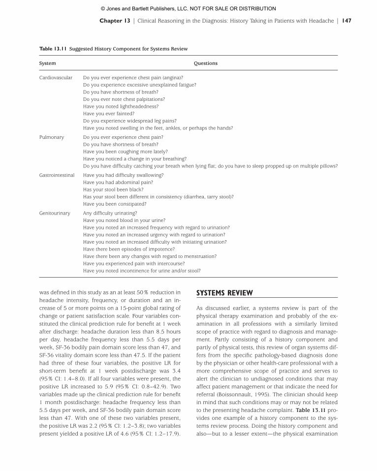
As discussed earlier, a systems review is part of thephysical therapy examination and probably of the ex-amination in all professions with a similarly limitedscope of practice with regard to diagnosis and manage-ment. Partly consisting of a history component andpartly of physical tests, this review of organ systems dif-fers from the specific pathology-based diagnosis doneby the physician or other health-care professional with amore comprehensive scope of practice and serves toalert the clinician to undiagnosed conditions that mayaffect patient management or that indicate the need forreferral (Boissonnault, 1995). The clinician should keepin mind that such conditions may or may not be relatedto the presenting headache complaint. Table 13.11 pro-vides one example of a history component to the sys-tems review process. Doing the history component andalso—but to a lesser extent—the physical examination
Chapter 13 | Clinical Reasoning in the Diagnosis: History Taking in Patients with Headache | 147
Table 13.11 Suggested History Component for Systems Review
System Questions
Cardiovascular Do you ever experience chest pain (angina)?Do you experience excessive unexplained fatigue?Do you have shortness of breath?Do you ever note chest palpitations?Have you noted lightheadedness?Have you ever fainted?Do you experience widespread leg pains?Have you noted swelling in the feet, ankles, or perhaps the hands?
Pulmonary Do you ever experience chest pain?Do you have shortness of breath?Have you been coughing more lately?Have you noticed a change in your breathing?Do you have difficulty catching your breath when lying flat; do you have to sleep propped up on multiple pillows?
Gastrointestinal Have you had difficulty swallowing?Have you had abdominal pain?Has your stool been black?Has your stool been different in consistency (diarrhea, tarry stool)?Have you been constipated?
Genitourinary Any difficulty urinating?Have you noted blood in your urine?Have you noted an increased frequency with regard to urination?Have you noted an increased urgency with regard to urination?Have you noted an increased difficulty with initiating urination?Have there been episodes of impotence?Have there been any changes with regard to menstruation?Have you experienced pain with intercourse?Have you noted incontinence for urine and/or stool?
52835_CH13_Fernandez.qxd 1/2/09 3:26 PM Page 147
© Jones and Bartlett Publishers, LLC. NOT FOR SALE OR DISTRIBUTION
the patient, and other interested parties (such as third-party payers) with a reliable, valid, and responsivegauge of the effect of treatment.
Pain Measures
The Numeric Pain Rating Scale (NPRS) and the VisualAnalogue Scale (VAS) for pain both are reliable, valid,and easy-to-apply outcome measures with regard toheadache pain parameters. The VAS may be the pre-ferred measure because research into its responsive-ness has greater external validity to headache patientsthan research available on responsiveness for the NPRS.Kelly (2001) determined the minimal clinically impor-tant difference (MCID) for the VAS in a varied populationof patients presenting to the emergency departmentwith pain as 10 to 12 (mm), depending on but not significantly different between groups for severity of re-ported pain.
Disability Measures
The Headache Disability Inventory (HDI) is a 25-itemquestionnaire that seeks to measure the self-perceiveddisabling effects of headache on daily life. The question-naire contains two subgroups of questions, thereby cre-ating emotional and functional subscale scores and atotal score. Two additional items on the questionnaire askpatients to rate the severity of their headache as mild, mod-erate, or severe, and the frequency as less than or equalto one per month, more than one but less than four permonth, or four or more times per month. The HDI hasgood internal consistency reliability; correlations be-tween the emotional and functional subscale scores andthe total score were both excellent (r = 0.89) (Jacobson et al., 1994). It also has good short-term (1-week) (r =0.93–0.95) (Jacobson et al., 1995) and generally goodlong-term (2-month) test-retest reliability (r = 0.83) forthe total scores. The HDI also exhibits good internal con-struct validity (P <0.001) (Jacobson et al., 1994). A min-imal detectable change (MDC95) score at 1-week retest is16 points. This value for the MDC95 indicates that a clini-cian can be 95% confident that a true change has oc-curred with a change in the HDI score of 16 or morepoints (Jacobson et al., 1995). Similarly, a 29-point scoreimprovement constitutes the MDC95 over a 2-monthtime period (Jacobson et al., 1994). The HDI test is sim-ple to administer and takes little time to complete.
With physical therapy interventions for the fiveheadaches amenable to physical therapy management
component of the systems review depends on the infor-mation obtained in the rest of the history-takingprocess, meaning that all questions are not mandatorybut rather at the discretion of the clinician.
CLINICAL PREDICTION RULES FOR DIAGNOSIS
Research-based data on the diagnostic utility of the history-taking process in patients with headache is limited. Most of the evidence relevant to the diagnosis ofpatients with headaches comes from uncontrolled stud-ies, case series, and expert opinion. The over 300 po-tential causes for headache and the benign nature ofmost headaches, especially the most common types of headache such as migraine and tension-typeheadache, may explain this dearth of research evidence(Bartleson, 2006). However, in their review of the litera-ture, Detsky et al. (2006) identified one clinical predictionrule relevant to diagnosis. This clinical prediction ruleconsists of the following five questions:
1. Is it a pulsating headache?2. Does it last between 4 and 72 hours without
medication?3. Is it unilateral?4. Is there nausea?5. Is the headache disabling (with disabling
headaches defined as headaches that disrupt apatient’s daily activities)?
When the patient answers yes to four or more ofthese five questions, the positive LR for a diagnosis ofmigraine headache is 24 (95% CI: 1.5–388). With a yesanswer to three questions, the positive LR is 3.5 (95% CI: 1.3–9.2). For a yes answer to one or two of these cri-teria, the positive LR is 0.41 (95% CI: 0.32–0.52). Themnemonic POUNDing (pulsating, duration of 4–72hours, unilateral, nausea, disabling) may be helpful forclinicians when using this clinical prediction rule(Detsky et al., 2006).
OUTCOME MEASURES
The information discussed so far can serve to producequestions in the history-taking process that provide thetherapist with diagnostic information. However, historytaking also should include outcome measures. The in-tent of outcome measures is not the collection of data tohelp with diagnosis but rather to provide the therapist,
148 | Part IV | PHYSICAL EXAMINATION OF PATIENTS WITH HEADACHE
52835_CH13_Fernandez.qxd 1/2/09 3:26 PM Page 148
© Jones and Bartlett Publishers, LLC. NOT FOR SALE OR DISTRIBUTION
largely geared to affecting underlying neuromuscu-loskeletal impairments in the cervical spine, the NeckDisability Index (NDI) is potentially a useful outcomemeasure in that this 10-item questionnaire aims tomeasure the self-perceived disabling effects of neckpain on daily life. Interpretation is possible throughscoring intervals as follows: 0–4 = no disability, 5–14 =mild, 15–24 = moderate, 25–34 = severe, and above 34= complete disability (Vernon & Mior, 1991). To arrive ata percentage disability, the total score is multiplied bytwo. The NDI has moderate test-retest reliability (ICC =0.68) (Cleland et al., 2006). Construct validity of the NDIas an outcome measure for neck pain has been demon-strated by comparing it with other tests or measures.Cleland et al. (2006) showed that a 7-point (14%)change in the NDI constituted a minimally clinically im-portant difference for the NDI, albeit only in patients
with cervical radiculopathy and not specifically in pa-tients reporting headaches.
RED FLAGS
One of the main objectives of the physical therapy history-taking process in patients presenting with acomplaint of headaches is to identify red flag findingsthat indicate the need for urgent referral. Table 13.12provides a list of such red flag indicators based on the in-formation presented in this chapter.
CONCLUSIONS AND IMPLICATIONS OFHISTORY FINDINGS
The history-taking process in patients with headachespresents unique challenges, particularly to health-care
Chapter 13 | Clinical Reasoning in the Diagnosis: History Taking in Patients with Headache | 149
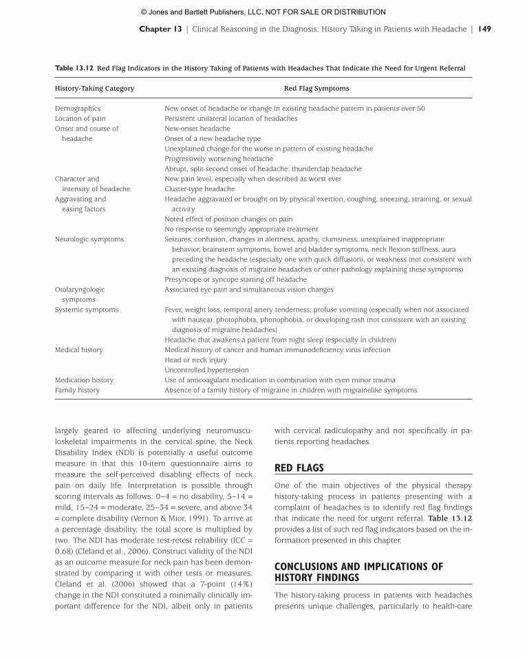
Table 13.12 Red Flag Indicators in the History Taking of Patients with Headaches That Indicate the Need for Urgent Referral
History-Taking Category Red Flag Symptoms
Demographics New onset of headache or change in existing headache pattern in patients over 50Location of pain Persistent unilateral location of headachesOnset and course of New-onset headache
headache Onset of a new headache typeUnexplained change for the worse in pattern of existing headacheProgressively worsening headacheAbrupt, split-second onset of headache: thunderclap headache
Character and New pain level, especially when described as worst everintensity of headache Cluster-type headache
Aggravating and Headache aggravated or brought on by physical exertion, coughing, sneezing, straining, or sexual easing factors activity
Noted effect of position changes on painNo response to seemingly appropriate treatment
Neurologic symptoms Seizures, confusion, changes in alertness, apathy, clumsiness, unexplained inappropriate behavior, brainstem symptoms, bowel and bladder symptoms, neck flexion stiffness, aura preceding the headache (especially one with quick diffusion), or weakness (not consistent with an existing diagnosis of migraine headaches or other pathology explaining these symptoms)
Presyncope or syncope starting off headacheOtolaryngologic Associated eye pain and simultaneous vision changes
symptomsSystemic symptoms Fever, weight loss, temporal artery tenderness, profuse vomiting (especially when not associated
with nausea), photophobia, phonophobia, or developing rash (not consistent with an existing diagnosis of migraine headaches)
Headache that awakens a patient from night sleep (especially in children)Medical history Medical history of cancer and human immunodeficiency virus infection
Head or neck injuryUncontrolled hypertension
Medication history Use of anticoagulant medication in combination with even minor traumaFamily history Absence of a family history of migraine in children with migrainelike symptoms
52835_CH13_Fernandez.qxd 1/2/09 3:26 PM Page 149
© Jones and Bartlett Publishers, LLC. NOT FOR SALE OR DISTRIBUTION
Potentially most relevant to history taking and the di-agnostic process is the identification of red flag indicators.The relevance and implications of finding such red flag in-dicators should always be considered in the context of thewhole presenting clinical picture. However, the principleof primum non nocere clearly indicates the need at alltimes for referral in case of even minimal diagnostic uncertainty.
ACKNOWLEDGMENTS
I would like to thank Eugene Barsky, MILS, at the IrvingK. Barber Learning Center of the University of BritishColumbia, who serves as the outreach librarian for thePhysiotherapy Association of British Columbia, for hishelp with collecting some of the more relevant up-to-dateresearch references upon which this chapter is based.
providers with a limited scope of practice, such as phys-ical therapists, chiropractors, and massage therapists.The information provided in this chapter should allowthe clinician to develop a history-taking process that al-lows him or her to meet the three main objectives notedin the beginning of this chapter, namely, diagnosingthose headaches amenable to physical therapy inter-ventions, identifying diagnostic indicators of headachenot amenable to sole physical therapy management oreven those that require urgent referral, and, finally,identifying undiagnosed pathology that may or may notbe related to the presenting complaint of headache butthat requires referral nevertheless for further diagnosisand management. History taking and systems review as discussed here are only part of the examinationprocess, and other chapters address the other and inter-related portions in more detail.
150 | Part IV | PHYSICAL EXAMINATION OF PATIENTS WITH HEADACHE
REFERENCES
Albuquerque FC, Han PH, Spetzler RF, Zabramski JM,McDougall CG. Carotid dissection: technical factor affectingendovascular therapy. Can J Neurol Sci 2002;29:54–60.
American Physical Therapy Association. Guide to physicaltherapist practice: second edition. Phys Ther2001;81:9–744.
Bartleson JD. When and how to investigate the patient withheadache. Semin Neurol 2006;26:163–170.
Blunt SB, Galton C. Cervical carotid or vertebral arterydissection. Br Med J 1997;314:243.
Bogduk N. Cervical causes of headache and dizziness. In:Grieve G, ed. Modern Manual Therapy of the VertebralColumn. New York: Churchill Livingstone; 1986.
Boissonnault WG. Examination in Physical Therapy Practice:Screening for Medical Disease. New York: ChurchillLivingstone; 1995.
Brna PM, Dooley JM. Headaches in the pediatric population.Semin Pediatr Neurol 2006;13:222–230.
Bronfort G, Assendelft WJ, Evans R, Haas M, Bouter L. Efficacy of spinal manipulation for chronic headache: asystematic review. J Manipulative Physiol Ther2001;24:457–466.
Burton LJ, Quinn B, Pratt-Cheney JL, et al. Headache etiologyin a pediatric emergency department. Pediatr Emerg Care1997;13:1–4.
Cleland JA, Fritz JM, Whitman JM, Palmer JA. The reliabilityand construct validity of the Neck Disability Index andpatient-specific functional scale in patients with cervicalradiculopathy. Spine 2006;31:598–602.
Clinch CR. Evaluation of acute headaches in adults. Am FamPhysician 2001;63:685–692.
Cosenza MJ. Headache as a manifestation of otolaryngologicdisease. J Am Osteopath Assoc 2000;100(suppl):S1–S5.
Cross J, Coles A. Foramen magnum. Adv Clin Neurosci Rehabil2002;2:16–17.
Davenport R. Acute headache in the emergency department. J Neurol Neurosurg Psychiatry 2002;72(suppl II):ii33–ii37.
De Jongh TOH, Knuistingh-Neven A, Couturier EGM.Hoofdpijn. Huisarts Wetenschap 2001;44:615–619.
Detsky ME, McDonald DR, Baerlocher MO, et al. Does this patient with headache have a migraine or needneuroimaging? JAMA 2006;296:1274–1283.
Dooley JM, Gordon KE, Wood EP, et al. The utility of thephysical examination and investigations in the pediatricneurology consultation. Pediatr Neurol 2003;28:96–99.
Duarte J, Sempere AP, Delgado JA, et al. Headache of recentonset in adults: a prospective population-based study. ActaNeurol Scand 1996;94:67–70.
Dwyer A, Aprill C, Bogduk N. Cervical zygapophyseal jointpain patterns. I: a study in normal volunteers. Spine1990;15:453–457.
Fernández-de-las-Peñas C, Cuadrado ML, Gerwin RD, ParejaJA. Referred pain from the trochlear region in tension-typeheadache: a myofascial trigger point from the superioroblique muscle. Headache 2005;45:731–737.
Fernández-de-las-Peñas C, Alonso-Blanco C, Cuadrado ML,Gerwin R, Pareja J. Myofascial trigger points and theirrelationship to headache clinical parameters in chronictension-type headache. Headache 2006a;46:1264–1272.
Fernández-de-las-Peñas C, Alonso-Blanco C, Cuadrado ML,Gerwin RD, Pareja JA. Trigger points in the suboccipitalmuscles and forward head posture in tension-typeheadache. Headache 2006b;46:454–460.
Fernández-de-las-Peñas C, Cuadrado ML, Pareja JA. Myofascial trigger points, neck mobility and forward headposture in unilateral migraine. Cephalalgia 2006c;26:1061–1070.
Fernández-de-las-Peñas C, Arendt-Nielsen L, Simons DG.Contributions of myofascial trigger points to chronic
52835_CH13_Fernandez.qxd 1/2/09 3:26 PM Page 150
© Jones and Bartlett Publishers, LLC. NOT FOR SALE OR DISTRIBUTION
tension-type headache. J Manual Manipulative Ther2006d;14:222–231.
Fernández-de-las-Peñas C, Alonso-Blanco C, Cuadrado ML,Pareja J. Myofascial trigger points in the suboccipitalmuscles in episodic tension-type headache. Man Ther2006e;11:225–230.
Fernández-de-las-Peñas C, Cuadrado ML, Gerwin RD, ParejaJA. Referred pain from the lateral rectus muscle in subjectswith chronic tension-type headache [abstract]. Headache2006f;46:880.
Fernández-de-las-Peñas C, Cleland JA, Cuadrado ML, Pareja JA.Predictor variables for identifying patients with chronictension type headache who are likely to achieve short-termsuccess with muscle trigger point therapy. Cephalalgia2008;28:264–275.
Fleming R, Forsythe S, Cook C. Influential variables associatedwith outcomes in patients with cervicogenic headache.J Manual Manipulative Ther 2007;15:155–164.
Gentile S. Indications for the diagnosis and treatment of acuteheadaches correlated with neurological pathologies.J Headache Pain 2005;6:290–293.
Gladstein J. Headache. Med Clin North Am 2006;90:275–290.Goadsby P, Boes C. Chronic daily headache. J Neurol Neurosurg
Psychiatry 2002;72(suppl II):ii2–ii5.Granella F, D’Alessandro R, Manzoni GC, Cerbo R, Colucci
D’Amato C. International Headache Society classification:inter-observer reliability in the diagnosis of primaryheadaches. Cephalalgia 1994;14:16–20.
Guy N, Deffond D, Gabrillargues J, et al. Spontaneous internalcarotid artery dissection with lower cranial nerve palsy. CanJ Neurol Sci 2001;28:265–269.
International Headache Society. The InternationalClassification of Headache Disorders: 2nd ed. Cephalalgia2004;24(suppl):1–150.
Issa TS, Huijbregts PA. Physical therapy diagnosis andmanagement of a patient with chronic daily headache: acase report. J Manual Manipulative Ther 2006;14:E88–E123.
Jacobson GP, Ramadan NM, Aggarwal SK, Newman CW. TheHenry Ford Hospital Headache Disability Inventory (HDI).Neurology 1994;44:837–842.
Jacobson GP, Ramadan NM, Norris L, Newman CW. HeadacheDisability Inventory (HDI): short-term test-retest reliabilityand spouse perceptions. Headache 1995;35:534–539.
Jull GA, Stanton WR. Predictors of responsiveness tophysiotherapy management of cervicogenic headache.Cephalalgia 2005;25:101–108.
Jull G, Trott P, Potter H, et al. A randomized controlled trial ofexercise and manipulative therapy for cervicogenicheadache. Spine 2002;27:1835–1843.
Katsarava Z, Bartsch T, Diener H. Dermedikamenteninduzierte Kopfschmerz. Akta Neurol2006;33:28–43.
Kelly AM. The minimum clinically significant difference invisual analogue scale pain score does not differ withseverity of pain. Emerg Med J 2001;18:205–207.
Kerry R, Taylor AJ. Cervical arterial dysfunction assessmentand manual therapy. Man Ther 2006;11:243–253.
Laupacis A, Sekar N, Stiell I. Clinical prediction rules: a reviewand suggested modification of methodological standards.JAMA 1997;277:488–494.
Mills ML, Russo LS, Vines FS, Ross BA. High-yield criteria forurgent cranial computed tomography scans. Ann Emerg Med1986;15:1167–1172.
Niere KR. Can subjective characteristics of benign headachepredict manipulative physiotherapy treatment outcome?Aust J Physiother 1998;44:87–93.
Okeson JP. Orofacial Pain: Guidelines for Assessment,Classification, and Management. Hanover Park, IL:Quintessence Publishing; 1996.
Peters KS. Secondary headache and head pain emergencies.Prim Care 2004;31:381–393.
Royal College of Dental Surgeons of Ontario. Guidelines:Diagnosis and Management of Temporomandibular Disorders.Toronto, ON: Author; 2002.
Rubinstein SM, Peerdeman SM, Van Tulder M, Riphagen I,Haldeman S. A systematic review of the risk factors forcervical artery dissection. Stroke 2005;36:1575–1580.
Simon RP, Aminoff MJ, Greenberg DA. Clinical Neurology. 4thed. Stanford, CT: Appleton & Lange; 1999.
Simons DG, Travell J, Simons LS. Travell and Simons’Myofascial Pain and Dysfunction: The Trigger Point Manual.Vol. 1. Upper Half of the Body. 2nd ed. Baltimore, MD:Williams & Wilkins; 1999.
Sjaastad O, Fredriksen TA, Pfaffenrath V, for the CervicogenicHeadache International Study Group. Cervicogenicheadache: diagnostic criteria. Headache 1998;38:442–445.
Smetana GW. The diagnostic value of historical features inprimary headache syndromes. Arch Intern Med 2000;160:2729–2737.
Taylor AJ, Kerry R. Neck pain and headache as a result ofinternal carotid artery dissection: implications for manualtherapists. Man Ther 2005;10:73–77.
Terrett AGJ. Current Concepts in Vertebrobasilar ComplicationsFollowing Spinal Manipulation. 2nd ed. Norwalk, CT:Foundation for Chiropractic Education and Research; 2001.
Tuchin PJ. A twelve-month clinical trial of chiropractic spinalmanipulative therapy for migraine. Australas ChiropracticOsteopathy 1999;8:61–65.
Tuchin PJ, Pollard H, Bonello R. A randomized controlled trialof chiropractic spinal manipulative therapy for migraine.J Manipulative Physiol Ther 2000;23:91–95.
Türp JC, et al. Schmerzen im Bereich der Kaumuskulatur undKiefergelenke. Manuelle Medizin 2002;40:55–67.
Vernon H, Mior S. The Neck Disability Index: a study ofreliability and validity. J Manipulative Physiol Ther1991;14:409–415.
Weeks R, Weier Z. Psychological assessment of the headachepatient. Headache 2006;46(suppl 3):S110–S118.
Welch E. Headache. Nursing Standard 2005;19:45–52.Wojcik W, Pawlak JK, Knaus R. Doctor! I can’t stand the noise
in my ear! Can J Diagnosis 2003;20:55–59.World Health Organization. Headache disorders. 2004.
Available at: http://www.who.int/mediacentre/factsheets/fs277/en/index.html. Accessed September 20, 2007.
Chapter 13 | Clinical Reasoning in the Diagnosis: History Taking in Patients with Headache | 151
52835_CH13_Fernandez.qxd 1/2/09 3:26 PM Page 151
© Jones and Bartlett Publishers, LLC. NOT FOR SALE OR DISTRIBUTION

52835_CH13_Fernandez.qxd 1/2/09 3:26 PM Page 152
© Jones and Bartlett Publishers, LLC. NOT FOR SALE OR DISTRIBUTION