Embed Size (px)
Citation preview

1
Kendrick, B.S., Li, T., and Chang, B.S. 2002. Physical stabilization of proteins in aqueous solution. in "Rationale Design of stable protein formulations-theory and practice" (J.F. Carpenter and M.C. Manning eds.) Kluwer Academic/Plenum publishers, New York, pp. 61-84.
PHYSICAL STABILIZATION OF PROTEINS IN AQUEOUS SOLUTION
Brent S. Kendrick,1 Tiansheng Li,1 and Byeong S. Chang1 1Amgen, Inc. Thousand Oaks, CA 91320
INTRODUCTION The formulation scientist’s key goal is to achieve long-term stability of a drug compound. In the case of protein drugs, stabilization means not only maintaining the native chemical structure, but the native secondary and higher order structures necessary for biological activity (Cleland et al., 1993; Manning et al., 1989). Denaturation, as it is defined in this context, will be any non-native physical or chemical state of the protein. Physical and chemical denaturations are often accompanied by covalent and non-covalent aggregates that not only can destroy the activity of the drug, but also cause adverse side effects (Carpenter and Chang, 1996; Thornton and Ballow, 1993). Without the ability to stabilize native protein structures, even the most efficacious protein therapeutics will fail to make viable drug products.
How does a formulation scientist develop a formulation that stabilizes native protein structure against physical and chemical stresses in solution, and what are the relevant stresses that cause denaturation? These are the questions that this chapter will address.
Although the chemical and physical stabilities of a protein may seem separate parameters, they are actually closely tied to one another (Brange, 1992; Khossravi et al., 2000; McCrossin et al., 1998; RahuelClermont et al., 1997). Physical degradation of a protein can lead to covalent changes (oxidation, hydrolysis, disulfide scrambling). The reverse is also true; reduction of disulfide bonds, hydrolysis, and other covalent changes can cause a loss of the protein native state. For chemical degradations that are linked to denaturation, formulations that stabilize the native state will necessarily stabilize against the chemical degradation. Understanding these relationships between physical and chemical stabilities are currently a major goal in formulation research. OVERVIEW OF PHYSICAL STABILITY
2
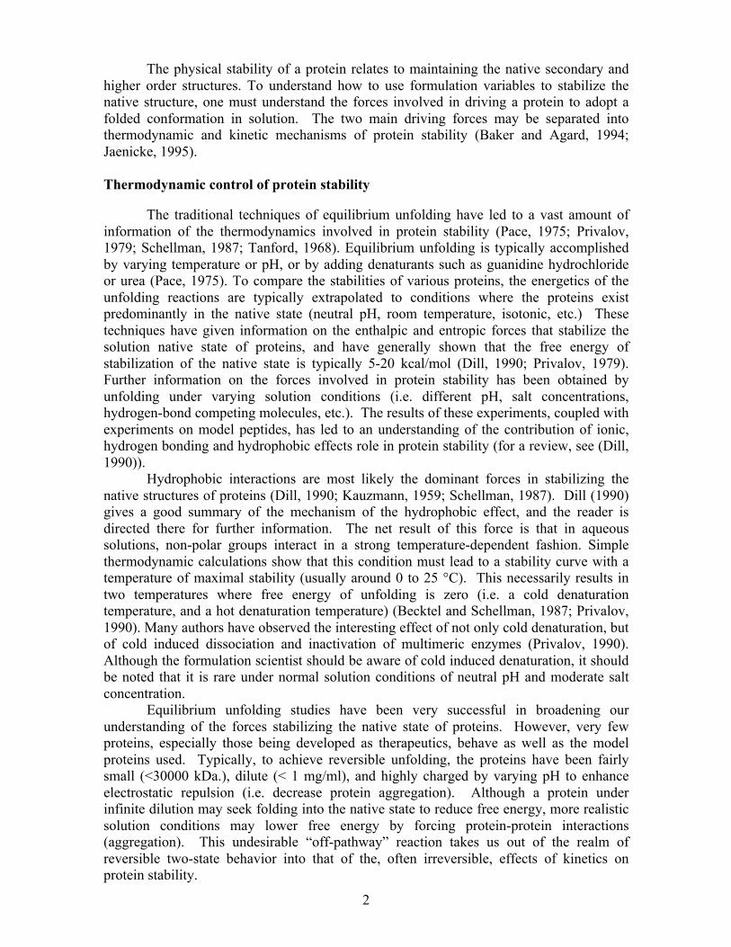
The physical stability of a protein relates to maintaining the native secondary and
higher order structures. To understand how to use formulation variables to stabilize the native structure, one must understand the forces involved in driving a protein to adopt a folded conformation in solution. The two main driving forces may be separated into thermodynamic and kinetic mechanisms of protein stability (Baker and Agard, 1994; Jaenicke, 1995).
Thermodynamic control of protein stability
The traditional techniques of equilibrium unfolding have led to a vast amount of information of the thermodynamics involved in protein stability (Pace, 1975; Privalov, 1979; Schellman, 1987; Tanford, 1968). Equilibrium unfolding is typically accomplished by varying temperature or pH, or by adding denaturants such as guanidine hydrochloride or urea (Pace, 1975). To compare the stabilities of various proteins, the energetics of the unfolding reactions are typically extrapolated to conditions where the proteins exist predominantly in the native state (neutral pH, room temperature, isotonic, etc.) These techniques have given information on the enthalpic and entropic forces that stabilize the solution native state of proteins, and have generally shown that the free energy of stabilization of the native state is typically 5-20 kcal/mol (Dill, 1990; Privalov, 1979). Further information on the forces involved in protein stability has been obtained by unfolding under varying solution conditions (i.e. different pH, salt concentrations, hydrogen-bond competing molecules, etc.). The results of these experiments, coupled with experiments on model peptides, has led to an understanding of the contribution of ionic, hydrogen bonding and hydrophobic effects role in protein stability (for a review, see (Dill, 1990)).
Hydrophobic interactions are most likely the dominant forces in stabilizing the native structures of proteins (Dill, 1990; Kauzmann, 1959; Schellman, 1987). Dill (1990) gives a good summary of the mechanism of the hydrophobic effect, and the reader is directed there for further information. The net result of this force is that in aqueous solutions, non-polar groups interact in a strong temperature-dependent fashion. Simple thermodynamic calculations show that this condition must lead to a stability curve with a temperature of maximal stability (usually around 0 to 25 °C). This necessarily results in two temperatures where free energy of unfolding is zero (i.e. a cold denaturation temperature, and a hot denaturation temperature) (Becktel and Schellman, 1987; Privalov, 1990). Many authors have observed the interesting effect of not only cold denaturation, but of cold induced dissociation and inactivation of multimeric enzymes (Privalov, 1990). Although the formulation scientist should be aware of cold induced denaturation, it should be noted that it is rare under normal solution conditions of neutral pH and moderate salt concentration.
Equilibrium unfolding studies have been very successful in broadening our understanding of the forces stabilizing the native state of proteins. However, very few proteins, especially those being developed as therapeutics, behave as well as the model proteins used. Typically, to achieve reversible unfolding, the proteins have been fairly small (<30000 kDa.), dilute (< 1 mg/ml), and highly charged by varying pH to enhance electrostatic repulsion (i.e. decrease protein aggregation). Although a protein under infinite dilution may seek folding into the native state to reduce free energy, more realistic solution conditions may lower free energy by forcing protein-protein interactions (aggregation). This undesirable “off-pathway” reaction takes us out of the realm of reversible two-state behavior into that of the, often irreversible, effects of kinetics on protein stability.
3
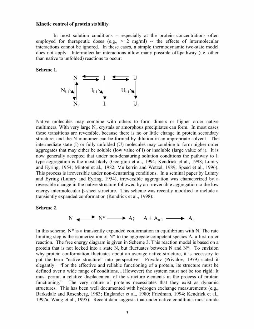
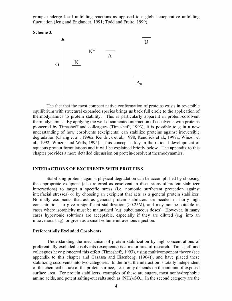
Kinetic control of protein stability In most solution conditions -- especially at the protein concentrations often employed for therapeutic doses (e.g., > 2 mg/ml) -- the effects of intermolecular interactions cannot be ignored. In these cases, a simple thermodynamic two-state model does not apply. Intermolecular interactions allow many possible off-pathway (i.e. other than native to unfolded) reactions to occur: Scheme 1. Native molecules may combine with others to form dimers or higher order native multimers. With very large Ni, crystals or amorphous precipitates can form. In most cases these transitions are reversible, because there is no or little change in protein secondary structure, and the N monomer can be formed by dilution in an appropriate solvent. The intermediate state (I) or fully unfolded (U) molecules may combine to form higher order aggregates that may either be soluble (low value of i) or insoluble (large value of i). It is now generally accepted that under non-denaturing solution conditions the pathway to Ii type aggregation is the most likely (Georgiou et al., 1994; Kendrick et al., 1998; Lumry and Eyring, 1954; Minton et al., 1982; Mulkerrin and Wetzel, 1989; Speed et al., 1996). This process is irreversible under non-denaturing conditions. In a seminal paper by Lumry and Eyring (Lumry and Eyring, 1954), irreversible aggregation was characterized by a reversible change in the native structure followed by an irreversible aggregation to the low energy intermolecular β-sheet structure. This scheme was recently modified to include a transiently expanded conformation (Kendrick et al., 1998): Scheme 2. In this scheme, N* is a transiently expanded conformation in equilibrium with N. The rate limiting step is the isomerization of N* to the aggregate competent species A, a first order reaction. The free energy diagram is given in Scheme 3. This reaction model is based on a protein that is not locked into a state N, but fluctuates between N and N*. To envision why protein conformation fluctuates about an average native structure, it is necessary to put the term “native structure” into perspective. Privalov (Privalov, 1979) stated it elegantly: “For the effective and reliable functioning of a protein, its structure must be defined over a wide range of conditions…(However) the system must not be too rigid: It must permit a relative displacement of the structure elements in the process of protein functioning.” The very nature of proteins necessitates that they exist as dynamic structures. This has been well documented with hydrogen exchange measurements (e.g., Barksdale and Rosenberg, 1983; Englander et al., 1980; Friedman, 1994; Kendrick et al., 1997a; Wang et al., 1995). Recent data suggests that under native conditions most amide
N I U Ni-1 Ii-1 Ui-1
Ni Ii UI
N N* A; A + An-1 An
4
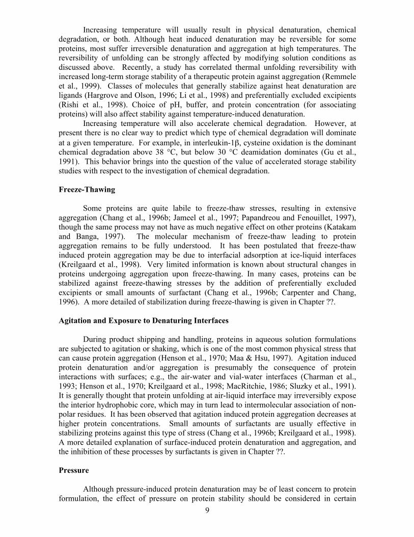
groups undergo local unfolding reactions as opposed to a global cooperative unfolding fluctuation (Jeng and Englander, 1991; Todd and Freire, 1999). Scheme 3. The fact that the most compact native conformation of proteins exists in reversible equilibrium with structural expanded species brings us back full circle to the application of thermodynamics to protein stability. This is particularly apparent in protein-cosolvent thermodynamics. By applying the well-documented interaction of cosolvents with proteins pioneered by Timasheff and colleagues (Timasheff, 1993), it is possible to gain a new understanding of how cosolvents (excipients) can stabilize proteins against irreversible degradation (Chang et al., 1996a; Kendrick et al., 1998; Kendrick et al., 1997a; Winzor et al., 1992; Winzor and Wills, 1995). This concept is key in the rational development of aqueous protein formulations and it will be explained briefly below. The appendix to this chapter provides a more detailed discussion on protein-cosolvent thermodynamics. INTERACTIONS OF EXCIPIENTS WITH PROTEINS Stabilizing proteins against physical degradation can be accomplished by choosing the appropriate excipient (also referred as cosolvent in discussions of protein-stabilizer intreractions) to target a specific stress (i.e. nonionic surfactant protection against interfacial stresses) or by choosing an excipient that acts as a general protein stabilizer. Normally excipients that act as general protein stabilizers are needed in fairly high concentrations to give a significant stabilization (>0.25M), and may not be suitable in cases where isotonicity must be maintained (e.g. subcutaneous doses). However, in many cases hypertonic solutions are acceptable, especially if they are diluted (e.g. into an intravenous bag), or given as a small volume intravenous injection. Preferentially Excluded Cosolvents Understanding the mechanism of protein stabilization by high concentrations of preferentially excluded cosolvents (excipients) is a major area of research. Timasheff and colleagues have pioneered this effort (Timasheff, 1993), using multicomponent theory (see appendix to this chapter and Casassa and Eisenberg, (1964)), and have placed these stabilizing cosolvents into two categories. In the first, the interaction is totally independent of the chemical nature of the protein surface, i.e. it only depends on the amount of exposed surface area. For protein stabilizers, examples of these are sugars, most nonhydrophobic amino acids, and potent salting-out salts such as (NH4)2SO4. In the second category are the
G N
An
A N*
U

5
cosolvents that act in a manner dependent on the chemical nature of the protein surface and the cosolvents. Examples of these types of cosolvents are polyethylene glycols, certain polyols (e.g., ethylene glycol), and alcohols. For these cosolvents, it is difficult to predict their effect on protein stability, especially at elevated temperatures (i.e., greater than room temperature). These cosolvents have substantial hydrophobic character that can foster greater binding with protein molecules, especially the denatured state, as temperature is increased. The result is that a compound (e.g., ethanol) may be a protein stabilizer at low temperature, but favor the denatured state at higher temperatures.
In terms of stabilizing interactions with proteins sucrose is the most studied cosolvent/excipient (Kendrick et al., 1998; Kendrick et al., 1997a; Lee and Timasheff, 1981; Liu and Bolen, 1995; Wang et al., 1995). It is a good model to explain the protective effect of the first category of preferentially excluded cosolvents, those for which the degree of preferential exclusion is a direct function of protein surface area. The interaction is thermodynamically unfavorable, primarily due to sucrose’s repulsion from the protein backbone (Liu and Bolen, 1995), resulting in an increase in protein chemical potential (see Appendix). Thus, in the presence of sucrose, the protein state with the least surface area will be thermodynamically favored; sucrose stabilizes proteins by driving them towards a compact native state. As a result sucrose will stabilize the protein against any stress that perturbs the native conformation towards a more expanded state. In Scheme 1, in the presence of sucrose the equilibrium would be shifted towards N and away from I or U. In Schemes 2 and 3, the ΔG of the N to N* transition would increase in the presence of sucrose. As a result the level of N* would be reduced and the aggregation would be inhibited. Thus, thermodynamic stabilization of the native state can increased the kinetic stability of a protein.
It is through the preferential exclusion mechanism that sucrose and other excluded excipients stabilize proteins against thermal unfolding (Lee and Timasheff, 1981; Santoro et al., 1992), denaturant unfolding (Foord and Leatherbarrow, 1998), aggregation (Kendrick et al., 1997b), and random conformational fluctuations from the native state (Foord and Leatherbarrow, 1998; Kendrick et al., 1997a; Wang et al., 1995). Excluded excipients may also reduce the rates chemical degradation reactions that are coupled to protein conformation, e.g. they have the ability to reduce the chemical reactivity of buried side chains such as cysteine (Kapoor and Parfett, 1977; Kendrick et al., 1997a; Yancey and Somero, 1979). Buffers/Salts The choice of a buffer may not depend only on having the appropriate pKa for the formulation. Buffers with similar pKas can have profoundly different effects on protein stability, depending on the buffer’s interaction with the protein. Salts and buffers can interact with proteins through three mechanisms: Changing the enthalpy of ionization of various side chains, a cosolvent exclusion mechanism, and a Debye screening of charge fluctuations. Preferential Exclusion of Salts. Many buffers and/or salts at high concentrations can also stabilize proteins through the preferential exclusion mechanism. The ranking in effectiveness of stabilization follows the well-known Hofmeister series for anions (Collins and Washabaugh, 1985; Hofmeister, 1888): citrate3- / citrate2- > PO4
3- ≅ HPO42- ≅ SO4
2- > OAc-, F- > Cl- > Br- >I- > ClO4
-. Ions above Br- in this series have an increasing ability to stabilize proteins (Jensen et al., 1995; von Hippel and Schleich, 1969; von Hippel and Wong, 1965). For example, the Tm of ribonuclease A increases by 8 °C in 0.5M potassium phosphate, relative to a solution without phosphate (von Hippel and Wong, 1965). The β-

6
lactoglobulin dimer is similarly stabilized against urea and temperature induced denaturation by salts in the trend predicted by the Hofmeister series (Jensen et al., 1995; Kella and Kinsella, 1988). Interestingly, cations have little effect on protein stability relative to the anions.
Although salts can have a remarkable effect on protein stability, they must be used with care, since they can also dramatically affect protein solubility. The stabilizing salts, through the preferential exclusion mechanism, can drive the minimization of protein surface area exposed to solvent to the extreme by “salting-out” the protein (Arakawa and Timasheff, 1982). The influence of various formulation conditions on protein solubility is discussed in a review by Middaugh and Volkin (Middaugh and Volkin, 1992). It should be noted that under conditions of increasing or decreasing pH from the pI of the protein, solubility of the native state increases dramatically. However, at pH extremes, the protein may unfold and irreversibly precipitate from solution through the formation of non-native intermolecular β-sheet structure. Debye Screening. Even salts that interact weakly through the preferential exclusion mechanism, or salts in relatively low concentrations (< 0.15 M) can have a strong influence on the physical state of the protein. This is primarily due to the effects of salts on electrostatic interactions between protein molecules that are governed by charge fluctuations. Charge fluctuations in proteins are caused by dipole moment fluctuations, which are in turn caused by configurational fluctuations of the protein (Kirkwood and Shumaker, 1952a). In salt free solutions, especially those close to the isoionic point of the protein, fluctuations of charge produce a long-range attractive force with a potential diminishing as 1/R2 (Kirkwood and Shumaker, 1952b). Under this solution condition, protein solubility is very small. As salt is added, the charge fluctuations contribute to a binding interaction between the protein and the salt ions (Kirkwood and Shumaker, 1952b; Scatchard, 1943). This charge screening interferes with the attractive force, and the net result is a significant increase in protein solubility (Melander and Horvath, 1977). Ionization Enthalpy. The free energy of unfolding is by definition affected by an enthalpic transition. Buffer choice can have a strong influence on the magnitude of the enthalpy of unfolding. The mechanism for this is rooted in the cause of pH induced unfolding of a protein. The dependence of the unfolding temperature on pH is determined by the number of groups ionized upon denaturation, γDNΔ , through the relation (Privalov, 1990):
(1) The enthalpy of the unfolding reaction, and thus the free energy of the
transition (see (Todd et al., 1998)), is strongly dependent on the choice of buffer, since the ionization of the buffer occurs in conjunction with the ionization of protein side groups. For example, the Tm for myoglobin is 59.2 °C in piperazine buffer but 73.7 °C in acetate buffer at the same pH (Privalov et al., 1986). In a pharmaceutically relevant study, it was found that interferon-gamma degraded faster in succinate buffer than in acetate buffer at the same pH (Lam et al., 1997). Specific binding of ligands
dpHdT
T3.2)H(T m
2mD
N ⋅Δ
− = Δ ΝD
γ

7
Protein-ligand interactions by their very nature have long been known to affect the free energy of the native state through thermodynamic linkage functions (Wyman, 1964; Wyman and Gill, 1990). Linkage functions simply state that if a protein in its native state binds a ligand with more affinity than it would in a denatured state, the free energy of the native state is decreased more than free energy of the denatured state (see Appendix). As a result the free energy barrier between native and denatured states is increased; the native state is stabilized. More specifically, for a protein with a single binding site in the presence of a ligand L, the free energy of stabilization (ΔGb) is given by:
[L])K (1[L])K (1
ln - G Gd
n0bb +
+Δ=Δ RT (2)
where Kn is the binding constant to the native state, Kd is the binding constant to the denatured state, R is the gas constant, T is the absolute temperature, and [L] is the free ligand concentration (Freire, 1999; Schellman, 1987). The increase in Tm can be approximated by (Schellman, 1975):
⎟⎟⎠
⎞⎜⎜⎝
⎛
+
++Δ
Δ=
[L]K 1[L]K 1
ln T H
H T T
n
d00
00
m
R (3)
As an example, let the ligand bind only to the native state (Kn = 106 M-1, 298
K). At a free ligand concentration of 28 µM, the free energy of stabilization is 2 kcal/mol, and the corresponding increase in Tm is approximately 18 °C (Figure 1).
A few applications of ligand binding will illustrate the large increases in protein
stability gained by the use of ligands. One example is the use of polyanions in the formulation of acidic fibroblast growth factor (aFGF) (Tsai et al., 1993), a protein that naturally binds heparin. Although aFGF is normally unstable and aggregates extensively after only weeks of storage at 4 °C, in the presence of heparin it is stable at this temperature for at least one year. Another example is the use of divalent cation metals in proteins with binding sites for such ligands. In the presence of calcium, the increase in free energy of unfolding for βγ-crystallin homolog spherulin 3a is 13.9 kcal/mol, with a corresponding Tm increase of 20 °C (Kretschmar et al., 1999). Protein-receptor complexes are also stabilized through binding interactions (Li et al., 1997; Li et al., 1998).

8
Figure 1. Hypothetical free energy of stabilization (filled circles) and corresponding increase in Tm (open circles) due to ligand binding to the native state (Kn = 106 M-1)
Protein Self-Stabilization Proteins that undergo dimerization or higher-order reversible association are stabilized by such interactions. In low molecular weight proteins this is especially apparent, with most of the stabilization against unfolding arising from interactions at the interface between subunits (Johnson and Freire, 1996; Todd et al., 1998). For example, in the simplest case where a protein dimer unfolds with no intermediates the stabilization against unfolding is given directly through the free energy equation:
2U N lnK - G dK2d ⎯→←=Δ RT (4)
A striking example of the increase in stability through protein association is
seen from the protein p53tet, which has a native tetramer:unfolded monomer equilibrium in solution. As protein concentration is increased from 0.5 to 150 µM, the Tm increases from about 39 to 75 °C (Johnson and Freire, 1996). Although significant gains in protein stability can be realized by increasing protein concentration, the therapeutic dose of the multimeric protein is going to have some influence on acceptable protein concentrations in the formulation. PHYSICAL FACTORS AFFECTING PROTEIN STABILITY Temperature
Free ligand concentration (M)0.02.0e-54.0e-56.0e-58.0e-51.0e-41.2e-4
Δ G of stabilization (kcal/mol)
-3
-2
-1
0 Tm (o C)65
70
75
80
85
90
95
100
9
Increasing temperature will usually result in physical denaturation, chemical degradation, or both. Although heat induced denaturation may be reversible for some proteins, most suffer irreversible denaturation and aggregation at high temperatures. The reversibility of unfolding can be strongly affected by modifying solution conditions as discussed above. Recently, a study has correlated thermal unfolding reversibility with increased long-term storage stability of a therapeutic protein against aggregation (Remmele et al., 1999). Classes of molecules that generally stabilize against heat denaturation are ligands (Hargrove and Olson, 1996; Li et al., 1998) and preferentially excluded excipients (Rishi et al., 1998). Choice of pH, buffer, and protein concentration (for associating proteins) will also affect stability against temperature-induced denaturation. Increasing temperature will also accelerate chemical degradation. However, at present there is no clear way to predict which type of chemical degradation will dominate at a given temperature. For example, in interleukin-1β, cysteine oxidation is the dominant chemical degradation above 38 °C, but below 30 °C deamidation dominates (Gu et al., 1991). This behavior brings into the question of the value of accelerated storage stability studies with respect to the investigation of chemical degradation. Freeze-Thawing Some proteins are quite labile to freeze-thaw stresses, resulting in extensive aggregation (Chang et al., 1996b; Jameel et al., 1997; Papandreou and Fenouillet, 1997), though the same process may not have as much negative effect on other proteins (Katakam and Banga, 1997). The molecular mechanism of freeze-thaw leading to protein aggregation remains to be fully understood. It has been postulated that freeze-thaw induced protein aggregation may be due to interfacial adsorption at ice-liquid interfaces (Kreilgaard et al., 1998). Very limited information is known about structural changes in proteins undergoing aggregation upon freeze-thawing. In many cases, proteins can be stabilized against freeze-thawing stresses by the addition of preferentially excluded excipients or small amounts of surfactant (Chang et al., 1996b; Carpenter and Chang, 1996). A more detailed of stabilization during freeze-thawing is given in Chapter ??. Agitation and Exposure to Denaturing Interfaces During product shipping and handling, proteins in aqueous solution formulations are subjected to agitation or shaking, which is one of the most common physical stress that can cause protein aggregation (Henson et al., 1970; Maa & Hsu, 1997). Agitation induced protein denaturation and/or aggregation is presumably the consequence of protein interactions with surfaces; e.g., the air-water and vial-water interfaces (Charman et al., 1993; Henson et al., 1970; Kreilgaard et al., 1998; MacRitchie, 1986; Sluzky et al., 1991). It is generally thought that protein unfolding at air-liquid interface may irreversibly expose the interior hydrophobic core, which may in turn lead to intermolecular association of non-polar residues. It has been observed that agitation induced protein aggregation decreases at higher protein concentrations. Small amounts of surfactants are usually effective in stabilizing proteins against this type of stress (Chang et al., 1996b; Kreilgaard et al., 1998). A more detailed explanation of surface-induced protein denaturation and aggregation, and the inhibition of these processes by surfactants is given in Chapter ??. Pressure Although pressure-induced protein denaturation may be of least concern to protein formulation, the effect of pressure on protein stability should be considered in certain
10
delivery devices and certain manufacturing process. In the last several years, there is an increased interest in the study of pressure denaturation of proteins. A few reported studies have shown that pressure of a few hundred MPa can induce unfolding intermediates or molten globules in protein solutions (Clery et al., 1995; Muller et al., 1981; Wong and Heremans, 1988; Yamasaki et al., 1998). Often it was found that pressure induced protein denaturation is reversible within certain range of pressure limits. It has been shown that unlike thermally denatured proteins, proteins denatured by pressure can retain their compact structures (Zhang et al., 1995). It has been proposed that pressure induced denaturation may result from the water transfer into the hydrophobic core and that the destabilization of the hydrophobic core by the penetrating water molecule leads to protein denaturation (Hummer et al., 1998; Wolfenden and Radzicka, 1994). The penetration of water molecule into protein hydrophobic core could loosen the compactness of protein interior and causes protein to swell under high pressure. Also, in general any increase in the surface area of hydrophobic residues exposed to solvent will be favored under pressure. This is because pressure favors reactions that reduce the overall system volume. Exposure of hydrophobic residues to solvent reduces volume because the density of water near these groups is greater than that of water in bulk solution. Protein structural expansion under pressure is documented by the experimental observation that lysozyme, Arc repressor, and RNase exhibit increased hydrodynamic radius upon pressure denaturation and higher hydrogen-exchange rates (Chryssomallis et al., 1981; Clery et al., 1995; Peng et al., 1994). Therefore, preferentially excluded cosolvents such as sugars and polyols will stabilize proteins against pressure-induced denaturation (e.g., Oliveira et al., 1994). CONCLUSIONS Long-term storage stability of proteins can be achieved through understanding of protein degradation pathways and how excipients interact with proteins. This chapter has covered many aspects of these concepts, but is by no means a comprehensive compendium. The reader is directed to other Chapters in this book, resources such as the Series in Pharmaceutical Biotechnology (Ahern and Manning, 1992; Pearlman and Wang, 1996; Wang and Pearlman, 1993) and the review article by Cleland et al. (Cleland et al., 1993). It is hoped that the pharmaceutical scientist can take these concepts and effectively apply them towards the rational design of stable aqueous formulations.
11
APPENDIX: DERIVATION OF THE WYMAN LINKAGE FUNCTION AND APPLICATION TO THE TIMASHEFF PREFERENTIAL EXCLUSION MECHANISM
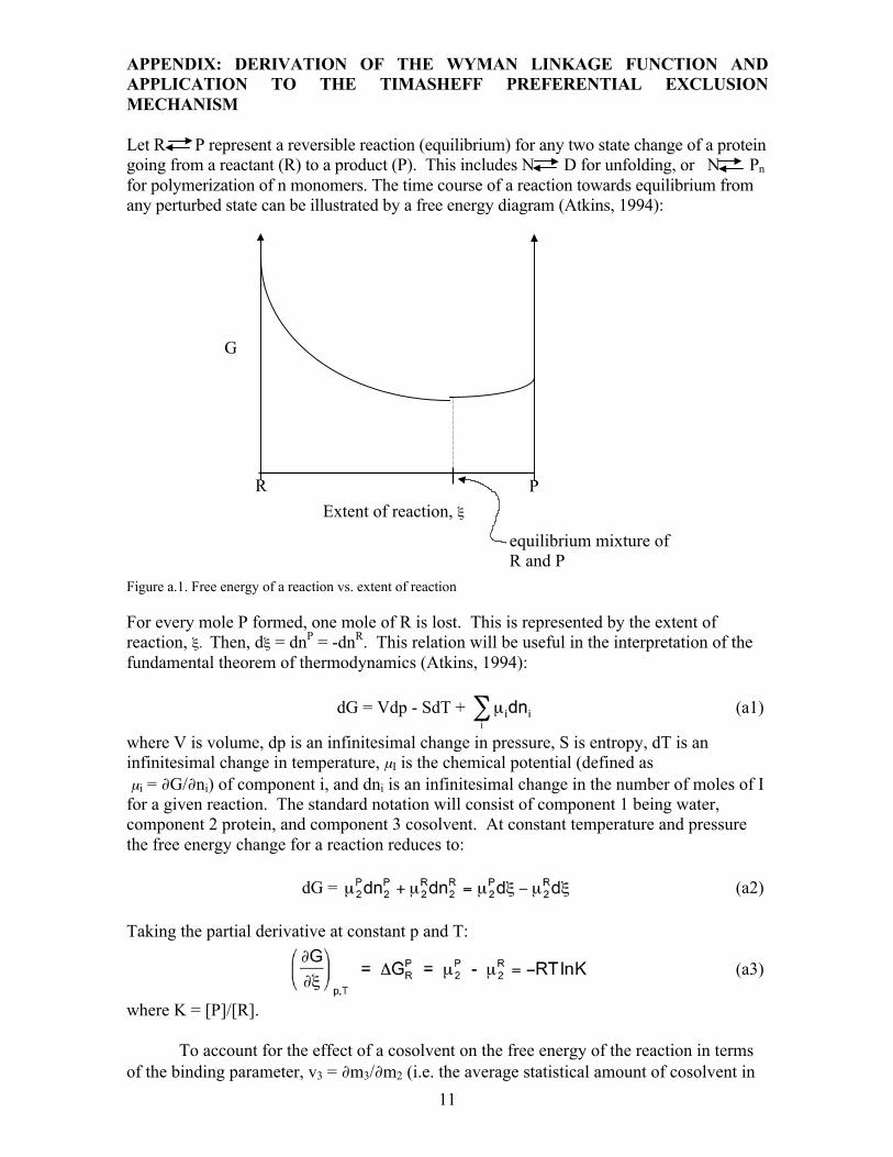
Let R P represent a reversible reaction (equilibrium) for any two state change of a protein going from a reactant (R) to a product (P). This includes N D for unfolding, or N Pn for polymerization of n monomers. The time course of a reaction towards equilibrium from any perturbed state can be illustrated by a free energy diagram (Atkins, 1994):
Figure a.1. Free energy of a reaction vs. extent of reaction
For every mole P formed, one mole of R is lost. This is represented by the extent of reaction, ξ. Then, dξ = dnP = -dnR. This relation will be useful in the interpretation of the fundamental theorem of thermodynamics (Atkins, 1994):
dG = Vdp - SdT + (a1)
where V is volume, dp is an infinitesimal change in pressure, S is entropy, dT is an infinitesimal change in temperature, µI is the chemical potential (defined as µi = ∂G/∂ni) of component i, and dni is an infinitesimal change in the number of moles of I for a given reaction. The standard notation will consist of component 1 being water, component 2 protein, and component 3 cosolvent. At constant temperature and pressure the free energy change for a reaction reduces to: dG = (a2) Taking the partial derivative at constant p and T:
(a3)
where K = [P]/[R]. To account for the effect of a cosolvent on the free energy of the reaction in terms of the binding parameter, v3 = ∂m3/∂m2 (i.e. the average statistical amount of cosolvent in
G
Extent of reaction, ξ R P
equilibrium mixture of R and P

12
the vicinity of the protein surface), take the partial derivative of the free energy equation with respect to the cosolvent (Wyman, 1964):
(a4)
Since the definition of chemical potential depends on the activity of the solute from a given reference state by: µ3 = µ3
* + RT ln(a3) (a5) Then: dµ3 = RT d[ln(a3)] Substituting into equation a4 for chemical potential perturbation upon introduction of cosolvent gives the Wyman linkage function in terms of the activity of component 3:
(a6)
Dividing the top and bottom of the right hand side by ∂m3 gives:
(a7)
Now, given a general function z(x,y): dz = (a8a)
Take partial derivative of dz with respect to y, at constant z it will equal 0.
Let:
Then: (a9)
This is the relation between binding and chemical potential perturbation.
From this relation, the Wyman linkage function becomes(Timasheff, 1993; Wyman, 1964):
(a8b) (a8c)

13
(a10)
In terms of free energy:
(a11)
From this relationship it is apparent that a greater exclusion (negative binding) of a solute from the product (unfolded protein) than from the reactant (native protein) will result in a positive slope of free energy vs. solute concentration. Although the Wyman equation gives an indication of the direction (see graph below) that ΔG changes by the addition of a cosolvent, to obtain absolute ΔGN-D values directly it is necessary to measure them by standard unfolding experiments.
Figure a.2. Difference in ΔG (ΔΔG) of a process upon introduction of a cosolvent. It is important to note that this mechanism of salting-out and protein stabilization
depends on the extent of exclusion being proportional to the exposed surface area of the protein (i.e. ∂m3/∂m2 becomes a larger negative value as solvent exposed protein surface increases). As a technical note, it must be realized that measurement of a preferential interaction parameter for a protein in the R state under a given solution condition makes it extremely difficult to measure the preferential interaction parameter for the P state due to the direct effect of solution conditions on the equilibrium of the R P transition. Preferential interaction theory allows a direct calculation of transfer free energies of R and P from one cosolvent concentration to another. For this, knowledge of ∂µ2/∂m3 is necessary:
From Equation a9:
ΔG (kcal/mol)
Solute concentration
Stabilized ΔG of unfolding
Native ΔG of unfolding
ΔG < 0; crystallization or salting-out is favorable
N U
N Pn
15
10
0

14
This can be rearranged to: (∂µ2/∂m3)m2 = (a12)
The binding interaction, , can be measured experimentally at equilibrium
dialysis. The chemical potential perturbation of the cosolvent with itself (self interaction
parameter), , can be obtained through the equation:
µ3 = µ3
* + RTln(a3) = µ3* + RTln(m3γ3) = µ3
* + RTln(m3) + RTln(γ3) (a13) This relationship can be transformed into:
= RT/m3 + RT (a14)
The activity coefficient increment, is readily obtained from osmotic pressure
data, and is usually insignificant for non-polymer cosolvents relative to RT/m3. The transfer free energy of R or P from water into the cosolvent system can now be obtained by (Timasheff, 1993):
(a15a)
(a15b)
Again, this parameter is difficult to obtain for U and Pn under the same solution conditions as N.
The classical “thermodynamic box” can be closed by the following relationship for
N to D equilibria: δΔG = - = - = free energy of stabilization due to cosolvent molality m3 (Figures a.3 and a.4). The subscripts m3 and w for ΔGN-D refer to the change in free energy of unfolding in the presence of cosolvent (molality m3) and the free energy of unfolding in the absence of cosolvent respectively. These values are typically measured directly from unfolding experiments.

15
0 Increasing cosolvent concentration (m3)
Figure a.3. Free energy of N to D transition in the absence and presence of an excluded cosolvent.
Figure a.4. Classical thermodynamic box for N to D transition in the presence and absence of an excluded cosolvent.
The second group of chemical potential perturbation terms, Δµ 2,tr , refers to the free energy of transfer of D or N respectively from pure water to a cosolvent of molality m3. This cycle has been recently completed by Timasheff’s laboratory (Xie and Timasheff, 1997) for ribonuclease A in sorbitol, and can be illustrated by Figure a.4. REFERENCES
G
D
N
N3
D3
Nw
Dw
16
Ahern, T. J. and Manning, M. C. 1992. Stability of protein pharmaceuticals. Plenum Press, New York. Arakawa, T. and Timasheff, S. N. 1982. Preferential interactions of proteins with salts in concentrated
solutions. Biochemistry 21:6545-6552. Atkins, P. W. 1994. Physical chemistry. W. H. Freeman and Company, New York. Baker, D. and Agard, D. A. 1994. Kinetics versus thermodynamics in protein folding. Biochemistry
33:7505-7509. Bam, N. B., Cleland, J. L. and Randolph, T. W. 1996. Molten globule intermediate of recombinant human
growth hormone: stabilization with surfactants. Biotechnol Prog 12:801-9. Barksdale, A. D. and Rosenberg, A. 1983. Aquisition and interpretation of hydrogen exchange data from
peptides, polymers, and proteins. Methods Biochem. Anal. 28:1-113. Becktel, W. J. and Schellman, J. A. 1987. Protein stability curves. Biopolymers 26:1859-1877. Brange, J. 1992. Chemical stability of insulin. 4. Mechanisms and kinetics of chemical transformations in
pharmaceutical formulation. Acta Pharm Nord 4:209-22. Carpenter, J. F. and Chang, B. S. 1996. Lyophilization of protein pharmaceuticals, in: Biotechnology and
Biopharmaceutical Manufacturing, Processing, and Preservation K. E. Avis and V. L. Wu, eds., vol. 2, pp. 199-264. Interpharm. Press, Inc., Buffalo Grove, IL.
Casassa, E. F. and Eisenberg, H. 1964. Thermodynamic analysis of multicomponent systems. Adv. Protein Chem. 19:287-395.
Chang, B. S., Beauvais, R. M., Dong, A. and Carpenter, J. F. 1996a. Physical factors affecting the storage stability of freeze-dried interleukin-1 receptor antagonist: glass transition and protein conformation. Arch. Biochem. Biophys. 331:249-58.
Chang, B. S., Kendrick, B. S. and Carpenter, J. F. 1996b. Surface-induced denaturation of proteins during freezing and its inhibition by surfactants. J Pharm Sci 85:1325-30.
Charman, S. A., Mason, K. L. and Charman, W. N. 1993. Techniques for assessing the effects of pharmaceutical excipients on the aggregation of porcine growth hormone. Pharm Res 10:954-62.
Chryssomallis, G. S., Torgerson, P. M., Drickamer, H. G. and Weber, G. 1981. Effect of hydrostatic pressure on lysozyme and chymotrypsinogen detected by fluorescence polarization. Biochemistry 20:3955-9.
Cleland, J. L., Powell, M. F. and Shire, S. J. 1993. The development of stable protein formulations: a close look at protein aggregation, deamidation, and oxidation [published erratum appears in Crit Rev Ther Drug Carrier Syst 1994;11(1):60]. Crit Rev Ther Drug Carrier Syst 10:307-77.
Cleland, J. L. and Randolph, T. W. 1992. Mechanism of polyethylene glycol interaction with the molten globule folding intermediate of bovine carbonic anhydrase B. J Biol Chem 267:3147-53.
Clery, C., Renault, F. and Masson, P. 1995. Pressure-induced molten globule state of cholinesterase. FEBS Lett 370:212-4.
Collins, K. D. and Washabaugh, M. W. 1985. The Hofmeister effect and the behaviour of water at interfaces. Quarterly Rev. Biophys. 18:323-422.
Cumper, C. and Alexander, A. 1950. The surface chemistry of proteins. Trans. Faraday Soc. 46:235. Dill, K. A. 1990. Dominant forces in protein folding. Biochemistry 29:7133-7155. Donaldson, T., Boonstra, E. and JM, H. 1980. Kinetics of protein denaturation at gas-liquid interfaces. J.
Coll. Int. Sci. 74:443. Englander, S. W., Calhoun, D. B., Englander, J. J., Kallenbach, N. R., Liem, R. K., Malin, E. L., Mandal,
C. and Rogero, J. R. 1980. Individual breathing reactions measured in hemoglobin by hydrogen exchange methods. Biophys. J. 32:577-89.
Foord, R. L. and Leatherbarrow, R. J. 1998. Effect of osmolytes on the exchange rates of backbone amide protons in proteins. Biochemistry 37:2969-2978.
Freire, E. 1999. The propogation of binding interactions to remote sites in proteins: Analysis of the binding of the monoclonal antibody D1.3 to lysozyme. Proc. Natl. Acad. Sci. USA 96:10118-10122.
Friedman, J. M. 1994. Time-resolved resonance Raman spectroscopy as probe of structure, dynamics, and reactivity in hemoglobin. Methods Enzymol. 232:205-31.
Georgiou, G., Valax, P., Ostermeier, M. and Horowitz, P. M. 1994. Folding and aggregation of TEM beta-lactamase: analogies with the formation of inclusion bodies in Escherichia coli. Protein Sci. 3:1953-60.
Gu, L. C., Erdos, E. A., Chiang, H. S., Calderwood, T., Tsai, K., Visor, G. C., Duffy, J., Hsu, W. C. and Foster, L. C. 1991. Stability of interleukin 1 beta (IL-1 beta) in aqueous solution: analytical methods, kinetics, products, and solution formulation implications. Pharm Res 8:485-90.
Hargrove, M. S. and Olson, J. S. 1996. The stability of holomyoglobin is determined by heme affinity. Biochemistry 35:11310-8.
Henson, A. F., Mitchell, J. R. and Musselwhite, P. R. 1970. The surface coagulation of proteins during shaking. J Colloid Interface Sci 32:162-5.
17
Hofmeister, F. 1888. On the understanding of the effect of salts. Second report. On regularities in the precipitating effect of salts and their relationship to their physiological behavior. Naunyn-Schmiedebergs Archiv fuer Experimentelle Pathologie und Pharmakologie 24:247-260.
Hummer, G., Garde, S., Garcia, A. E., Paulaitis, M. E. and Pratt, L. R. 1998. The pressure dependence of hydrophobic interactions is consistent with the observed pressure denaturation of proteins. Proc Natl Acad Sci U S A 95:1552-5.
Jaenicke, R. 1995. Folding and association versus misfolding and aggregation of proteins. Phil. Trans. R. Soc. Lond. B. 348:97-105.
Jameel, F., Bogner, R., Mauri, F. and Kalonia, D. 1997. Investigation of physicochemical changes to L-asparaginase during freeze-thaw cycling. J Pharm Pharmacol 49:472-7.
Jeng, M. F. and Englander, S. W. 1991. Stable submolecular folding units in a non-compact form of cytochrome c. J Mol Biol 221:1045-61.
Jensen, W. A., Armstrong, J. M., DeGiorgio, J. and Hearn, M. T. W. 1995. Stability studies on maize leaf phosphoenolpyruvate carboxylase: The effect of salts. Biochemistry 34:472-480.
Johnson, C. R. and Freire, E. 1996. Structural stability of small oligomeric proteins. Techniques Protein Chem. VII:459-467.
Kapoor, M. and Parfett, C. L. 1977. Ligand-induced alterations in the reactivity of sulfhydryl groups and the structure of bovine liver glutamate dehydrogenase. Arch. Biochem. Biophys. 184:518-528.
Katakam, M. and Banga, A. K. 1997. Use of poloxamer polymers to stabilize recombinant human growth hormone against various processing stresses. Pharm Dev Technol 2:143-9.
Kauzmann, W. 1959. Some factors in the interpretation of protein denaturation. Adv. Protein Chem. 14:1-63.
Kella, N. K. D. and Kinsella, J. E. 1988. Structural stability of b-lactoglobulin in the presence of kosmotropic salts. Int. J. Peptide Protein Res. 32:396-405.
Kendrick, B. S., Carpenter, J. F., Cleland, J. L. and Randolph, T. W. 1998. A transient expansion of the native state precedes aggregation of recombinant human interferon-g. Proc. Natl. Acad. Sci., USA 95:14142-14146.
Kendrick, B. S., Chang, B. S., Arakawa, T., Peterson, B., Randolph, T. W., Manning, M. C. and Carpenter, J. F. 1997a. Preferential exclusion of sucrose from recombinant interleukin-1 receptor antagonist: Role in restricted conformational mobility and compaction of native state. Proc. Natl. Acad. Sci. USA 94:11917-11922.
Kendrick, B. S., Cleland, J. L., Lam, X., Nguyen, T., Randolph, T. W., Manning, M. C. and Carpenter, J. F. 1997b. Aggregation of recombinant human interferon gamma: Kinetics and structural transitions. submitted to J. Pharm. Sci. .
Khossravi, M., Shire, S. J. and Borchardt, R. T. 2000. Evidence for the involvement of histidine A(12) in the aggregation and precipitation of human relaxin induced by metal-catalyzed oxidation. Biochemistry. 39.:5876-5885.
Kirkwood, J. G. and Shumaker, J. B. 1952a. Proc. Natl. Acad. Sci. USA 38:855-862. Kirkwood, J. G. and Shumaker, J. B. 1952b. Forces between protein molecules in solution arising from
fluctuations in proton charge and configuration. Proc. Natl. Acad. Sci. USA 38:863-871. Kreilgaard, L., Jones, L. S., Randolph, T. W., Frokjaer, S., Flink, J. M., Manning, M. C. and Carpenter, J.
F. 1998. Effect of Tween 20 on freeze-thawing- and agitation-induced aggregation of recombinant human factor XIII. J Pharm Sci 87:1597-603.
Kretschmar, M., Mayr, E. M. and Jaenicke, R. 1999. Kinetic and thermodynamic stabilization of the betagamma-crystallin homolog spherulin 3a from Physarum polycephalum by calcium binding. J Mol Biol 289:701-5.
Lam, X. M., Patapoff, T. W. and Nguyen, T. H. 1997. The effect of benzyl alcohol on recombinant human interferon-gamma. Pharm. Res. 14:725-729.
Lee, J. C. and Timasheff, S. N. 1981. The stabilization of proteins by sucrose. J. Biol. Chem. 256:7193-7201.
Levine, H. L., Ransohoff, T. C., Kawahata, R. T. and McGregor, W. C. 1991. The use of surface tension measurements in the design of antibody-based product formulations. J Parenter Sci Technol 45:160-5.
Li, T., Horan, T., Osslund, T., Stearns, G. and Arakawa, T. 1997. Conformational changes in G-CSF/Receptor complex as investigated by isotope-edited FTIR spectroscopy. Biochemistry 36:8849-57.
Li, T., Narhi, L. O., Wen, J., Philo, J. S., Sitney, K., Inoue, J., Yamamoto, T. and Arakawa, T. 1998. Interactions between NFkappaB and its inhibitor ikappaB: biophysical characterization of a NFkappaB/ikappaB-alpha complex. J Protein Chem 17:757-63.
Liu, Y. and Bolen, D. W. 1995. The peptide backbone plays a dominant role in protein stabilization by naturally occurring osmolytes. Biochemistry 34:12884-12891.

18
Lumry, R. and Eyring, H. 1954. Conformation changes of proteins. J. Phys. Chem. 58:110-120. Macritchie, F. 1978. Proteins at interfaces. Adv Protein Chem 32:283-326. MacRitchie, F. 1986. Spread monolayers of proteins. Adv Colloid Interface Sci 25:341-85. Manning, M. C., Patel, K. and Borchardt, R. T. 1989. Stability of protein pharmaceuticals. Pharm Res
6:903-18. McCrossin, L. E., Charman, W. N. and Charman, S. A. 1998. Degradation of recombinant porcine growth
hormone in the presence of guanidine hydrochloride. Int J Pharm. 173.:157-170. Melander, W. and Horvath, C. 1977. Salt effects on hydrophobic interactions in precipitation and
chromatography of proteins: An interpretation of the lyotropic series. Arch. Biochem. Biophys. 183:200-215.
Middaugh, C. R. and Volkin, D. B. 1992. Protein solubility, in: Stability of protein pharmaceuticals T. J. Ahern and M. C. Manning, eds., vol. 2, pp. 109-134. Plenum Press, New York.
Minton, K. W., Karmin, P., Hahn, G. M. and Minton, A. P. 1982. Nonspecific stabilization of stress-susceptible proteins by stress- resistant proteins: a model for the biological role of heat shock proteins. Proc. Natl. Acad. Sci. USA 79:7107-11.
Mulkerrin, M. G. and Wetzel, R. 1989. pH dependence of the reversible and irreversible thermal denaturation of gamma interferons. Biochemistry 28:6556-61.
Muller, K., Ludemann, H. D. and Jaenicke, R. 1981. Pressure-induced structural changes of pig heart lactic dehydrogenase. Biophys Chem 14:101-10.
Oliveira, A. C., Gaspar, L. P., Da Poian, A. T. and Silva, J. L. 1994. Arc repressor will not denature under pressure in the absence of water. J Mol Biol 240:184-7.
Pace, C. N. 1975. The stability of globular proteins. CRC Critical Reviews in Biochemistry 3:1-41. Papandreou, M. J. and Fenouillet, E. 1997. Effect of various glycosidase treatments on the resistance of
the HIV-1 envelope to degradation. FEBS Lett 406:191-5. Pearlman, R. and Wang, Y. J. 1996. Formulation, characterization, and stability of protein drugs. Plenum
Press, New York. Peng, X., Jonas, J. and Silva, J. L. 1994. High-pressure NMR study of the dissociation of Arc repressor.
Biochemistry 33:8323-9. Privalov, P. L. 1979. Stability of proteins. Adv. Protein Chem 33:167-241. Privalov, P. L. 1990. Cold denaturation of proteins. Crit. Rev. Biochem. Mol. Biol. 25:281-306. Privalov, P. L., Griko, Y. V. and Venyaminov, S. Y. 1986. Cold denaturation of myoglobin. J. Mol. Biol.
190:487-498. RahuelClermont, S., French, C. A., Kaarsholm, N. C. and Dunn, M. F. 1997. Mechanisms of stabilization
of the insulin hexamer through allosteric ligand interactions. Biochemistry. 36.:5837-5845. Remmele, R. L. J., Bhat, S. D., Phan, D. H. and Gombotz, W. R. 1999. Minimization of recombinant
human Flt3 ligand aggregation at the Tm plateau: a matter of thermal reversibility. Biochemistry 38:5241-5247.
Rishi, V., Anjum, F., Ahmad, F. and Pfeil, W. 1998. Role of non-compatible osmolytes in the stabilization of proteins during heat stress. Biochem J 329:137-43.
Santoro, M. M., Liu, Y., Khan, S. M., Hou, L. X. and Bolen, D. W. 1992. Increased thermal stability of proteins in the presence of naturally occurring osmolytes. Biochemistry 31:5278-5283.
Scatchard, G. 1943. Chapter 3, in: Proteins, amino acids and peptides C. a. Edsall, eds. Reinhold, New York.
Schellman, J. A. 1975. Macromolecular binding. Biopolymers 14:999-1018. Schellman, J. A. 1987. The thermodynamic stability of proteins. Ann. Rev. Biophys. Biophys. Chem.
16:115-137. Sluzky, V., Tamada, J. A., Klibanov, A. M. and Langer, R. 1991. Kinetics of insulin aggregation in
aqueous solutions upon agitation in the presence of hydrophobic surfaces. Proc Natl Acad Sci U S A 88:9377-81.
Speed, M. A., Wang, D. I. C. and King, J. 1996. Specific aggregation of partially folded polypeptide chains: The molecular basis of inclusion body composition. Nature Biotechnology 14:1283-1287.
Sukow, W. W., Sandberg, H. E., Lewis, E. A., Eatough, D. J. and Hansen, L. D. 1980. Binding of the Triton X series of nonionic surfactants to bovine serum albumin. Biochemistry 19:912-7.
Tanford, C. 1968. Protein denaturation: Part A. Characterization of the denatured state: Part B. The transition from the native to denatured state. Adv. Protein Chem. 23:121-275.
Thornton, C. A. and Ballow, M. 1993. Safety of intravenous immunoglobulin. Arch. Neurol. 50:135-136. Timasheff, S. N. 1993. The control of protein stability and association by weak interactions with water:
How do solvents affect these processes? Annu. Rev. Biophys. Biomol. Struct. 22:67-97. Todd, M. J. and Freire, E. 1999. The effect of inhibitor binding on the structural stability and
cooperativity of the HIV-1 protease. Proteins 36:147-56.
19
Todd, M. J., Semo, N. and Freire, E. 1998. The structural stability of the HIV-1 protease. J Mol Biol 283:475-88.
Tsai, P. K., Volkin, D. B., Dabora, J. M., Thompson, K. C., Bruner, M. W., Gress, J. O., Matuszewska, B., Keogan, M., Bondi, J. V. and Middaugh, C. R. 1993. Formulation design of acidic fibroblast growth factor. Pharm Res 10:649-59.
von Hippel, P. H. and Schleich, T. 1969. The effects of neutral salts on the structure and conformational stability of macromolecules in solution, in: Structure and stability of biological macromolecules S. N. Timasheff and G. D. Fasman, eds., pp. 417-574. Marcel Dekker, New York.
von Hippel, P. H. and Wong, K. Y. 1965. On the conformational stability of globular proteins. J. Biol. Chem. 240:3909-3923.
Wang, A., Robertson, A. D. and Bolen, D. W. 1995. Effects of a naturally occurring compatible osmolyte on the internal dynamics of ribonuclease A. Biochemistry 34:15096-104.
Wang, Y. J. and Pearlman, R. 1993. Stability and characterization of protein and peptide drugs: Case histories. Plenum Press, New York.
Winzor, C. L., Winzor, D. J., Paleg, L. G., Jones, G. P. and Naidu, B. P. 1992. Rationalization of the effects of compatible solutes on protein stability in terms of thermodynamic nonideality. Arch. Biochem. Biophys. 296:102-7.
Winzor, D. J. and Wills, P. R. 1995. Thermodynamic nonideality of enzyme solutions supplemented with inert solutes: yeast hexokinase revisited. Biophys. Chem. 57:103-10.
Wolfenden, R. and Radzicka, A. 1994. On the probability of finding a water molecule in a nonpolar cavity. Science 265:936-7.
Wong, P. T. and Heremans, K. 1988. Pressure effects on protein secondary structure and hydrogen deuterium exchange in chymotrypsinogen: a Fourier transform infrared spectroscopic study. Biochim Biophys Acta 956:1-9.
Wyman, J. 1964. Linked functions and reciprocal effects in hemoglobin: A second look. Adv. Protein Chem. 19:223-286.
Wyman, J. and Gill, S. J. 1990. Binding and linkage: Functional chemistry of biological macromolecules. University Science Books, Mill Valley, CA.
Xie, G. and Timasheff, S. N. 1997. Mechanism of the stabilization of ribonuclease A by sorbitol: preferential hydration is greater for the denatured then for the native protein. Protein Sci. 6:211-21.
Yamasaki, K., Taniguchi, Y., Takeda, N., Nakano, K., Yamasaki, T., Kanaya, S. and Oobatake, M. 1998. Pressure-denatured state of Escherichia coli ribonuclease HI as monitored by Fourier transform infrared and NMR spectroscopy. Biochemistry 37:18001-9.
Yancey, P. H. and Somero, G. N. 1979. Counteraction of urea destabilization of protein structure by methylamine osmoregulatory compounds of elasmobranch fishes. Biochem. J. 183:317-323.
Zhang, J., Peng, X., Jonas, A. and Jonas, J. 1995. NMR study of the cold, heat, and pressure unfolding of ribonuclease A. Biochemistry 34:8631-41.