Embed Size (px)
Citation preview

1
PHYSICS OF ULTRASOUNDEmergency Ultrasound Course
Justin Bowra

Ultrasound
RapidNon-invasive
PainlessLook inside the patient
But it needs fluid!

1-20 MHz

1-20 MHz


7
Theoretical Stuff

FREQUENCY
No of wavelengths per second
1 cycle = 1 hertz (Hz)
Diagnostic US is 1-20MHz

As frequency increases…Wavelength decreases

High frequency waves (eg 10 MHz)

High frequency waves (eg 10 MHz)

High frequency waves (eg 10 MHz)
Better resolution
Less penetration
Skinny people, vascular access, kids…

Low frequency waves (eg 3 MHz)

Low frequency waves (eg 3 MHz)
Better penetration
But less resolution!
Fat people / greater depth
How does it work?

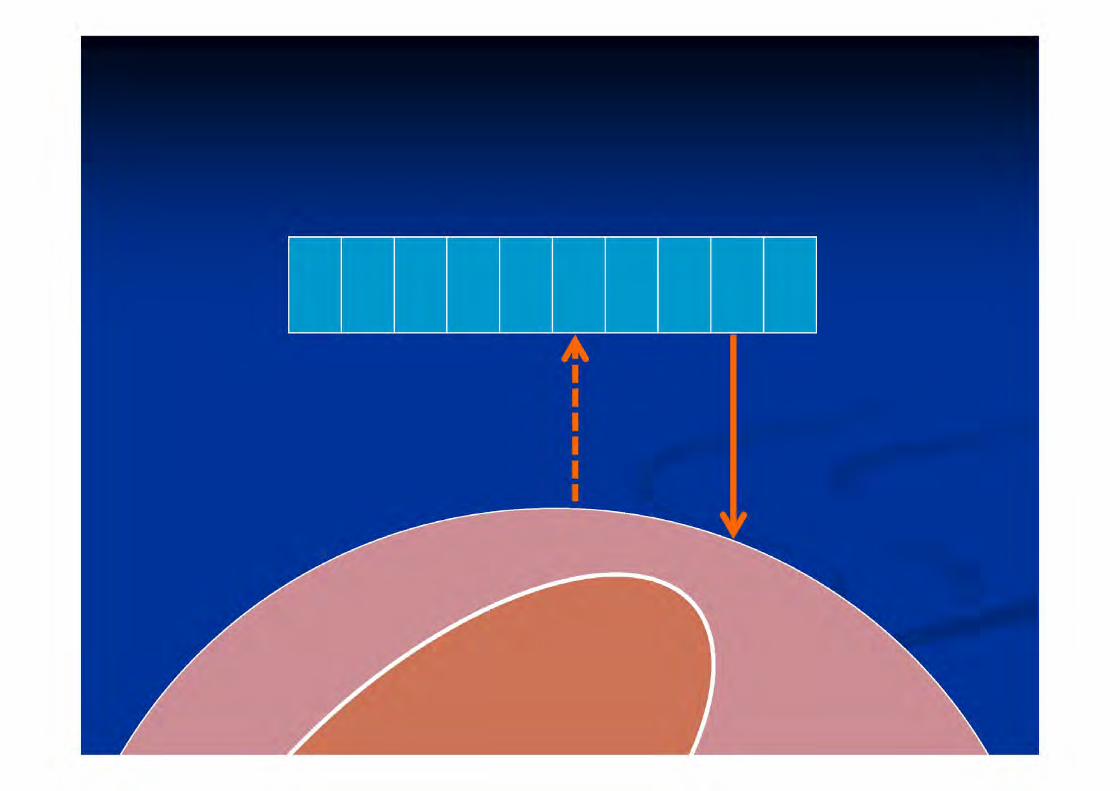
Piezoelectric effect
Electricity sound waves

1. Electrons hit the Piezoelectric crystal
2. Transducer (Probe) transmits sound wave
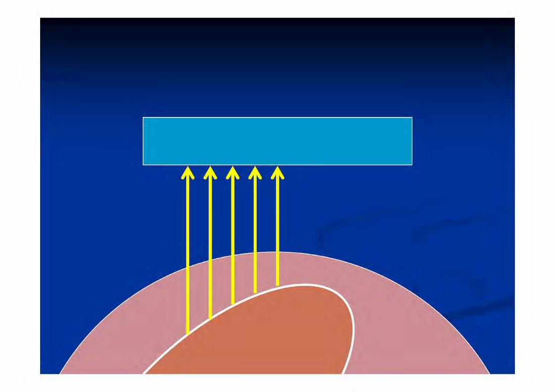
3. Patient reflects sound wave
4. Wave hits the piezoelectric crystal
5. Electrons travel to processor
6. Image is formed!
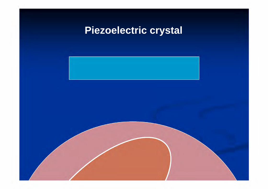
Piezoelectric crystal


25


36

37vvv


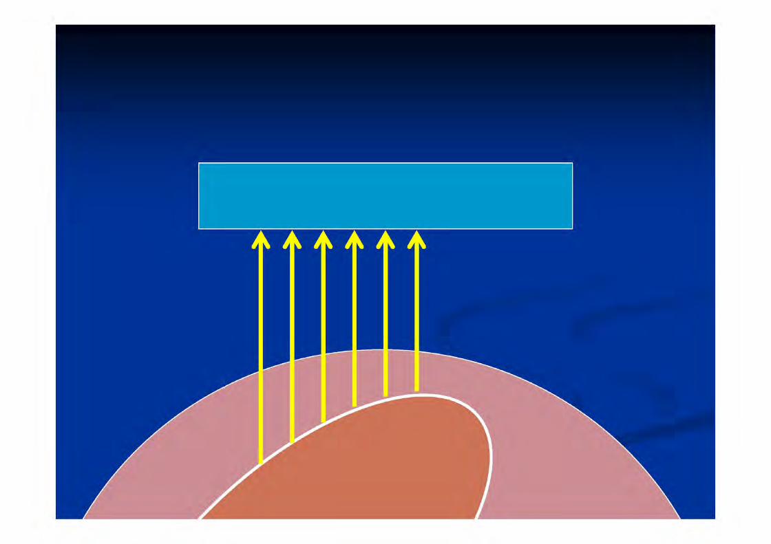


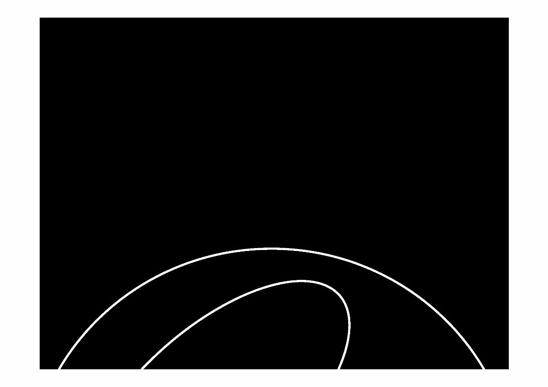
How is the image formed?Dots on screen
Brightness proportional to strength of returning echoes


But…
Location of the dots is determined by travel time, NOT DISTANCE

Interfaces
This is where the action is!
At each interface:
Transmitted
Attenuated

Example

Absorbed

Tissue


Transmitted

Fluid (GB)

Fluid (GB)

Fluid (GB)

Posterior enhancement
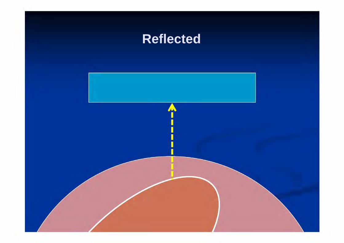
Reflected

Something very dense (gallstone)


Posterior shadowing

Refracted

Refracted

Edge shadowing

Edge shadowing

Edge shadowing

Scattered e.g. air


78
Artefacts a product of artificial character due to
extraneous (as human) agency… caused by manipulation not indicative of actual structural
relationships

How many artefacts can you see?

Artefacts
Mirror
Reverberation
‘Dirty’ shadowing (bowel gas)
Enhancement

How does reverberation happen?

True image of pleural line

But… sound wave bounces off probe!

…and off pleura… again

True path

‘Ghost image’ appears below real image
True image of pleura
‘Ghost’ image of pleura= reverberation artifact

Remember?
Location of the dots is determined by travel time, NOT DISTANCE

‘Bad’ acoustic shadowing (rib)

‘Good’ acoustic shadowing (gallstone)

Acoustic enhancement (GB)

Edge enhancement (GB)

Mirror image

Mirror image 1. (true image)

Mirror image 1. (true image)

Mirror image 2. (false image)

Mirror image 2. (false image)

Mirror image 2. (false image)

Mirror image 2. (false image)

Mirror image 2. (false image)

LIVER
MIRROR IMAGEOF LIVER DIAPHRAGM
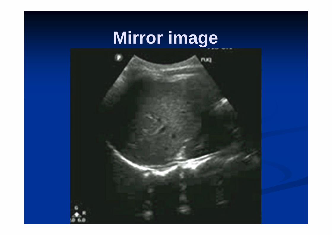
Mirror image

Mirror bladder

Mirror heart

Basic artifacts
Reverberation Posterior shadowing Posterior enhancement Edge artifact Mirror B lines (later)


SUMMARY
Fluid is dark Solid organs are grey Air and dense things are bright Not everything onscreen is actually there!

Thanks
Dr Anjana Amarasekarahttp://images.tutorvista.com/cms/images/83/freque
ncy-wavelength-image1.PNG