Embed Size (px)
Citation preview

1
2
3
4
3
2
5
6
7
8
9
10
11
12
13
14

Day 1

Retakes can be done here in class! (No books, no notes!)

1. What makes up the blood?
2. What role does hemoglobin play in red blood cells?
3. What do cells get their oxygen from?
4. Describe Sickle Cell Disease
Unit Cat Title Page #
Comp Check
Due Date
8 Act Blood Video 12/9

Here are the %’s:
Erythrocytes (Red Blood Cells): 94%
Leukocytes (White Blood Cells): .2%
Thrombocytes (Platelets): 6%
What is main function of each?
Platelet
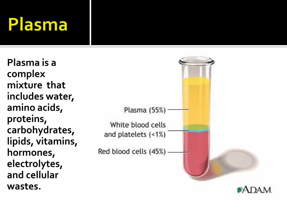
Plasma
Plasma is a complex mixture that includes water, amino acids, proteins, carbohydrates, lipids, vitamins, hormones, electrolytes, and cellular wastes.

You struggled on the parts of the last exam that required you to read for meaning!
Thus we will practice together! Assignment: 1. Read “Characteristics of Red Blood Cells” pgs
530-533, then answer the section review questions 1-3 on page 533.
2. Read “Types of White Blood Cells” pgs 537-539, then answer the section review questions 1-4 on page 539.
We will grade the questions in 30 minutes!
Unit Cat Title Page #
Comp Check
Due Date
8 Act Red & White Blood Cells Reading Activity 11/14

On a separate sheet of paper that you will attach in your lab book later complete the following assignment:
Assignment:
1. Read “Functions of White Blood Cells”pgs 539-540, then answer the section review questions 1-3 on page 540.
Unit Cat Title Page #
Comp Check
Due Date
8 HW Functions of White Blood Cells 11/16 12/9

Day 2

Know your White Blood Cell Characteristics and functions!
Don’t touch the tape!

Nose bleed
http://www.youtube.com/watch?v=yuODCgpMmos&feature=related&safety_mode=true&persist_safety_mode=1
How does blood clot?
http://www.youtube.com/watch?v=--bZUeb83uU&feature=related&safety_mode=true&persist_safety_mode=1
Get with a partner you can work with! Move if you have to!

This lab will involve 2 parts: 1- Notes and figures from the powerpoint 2. Lab and Analysis Activity Using
Instructions and your Textbook.
Hemostasis – refers to the stoppage of bleeding. There are 3 main mechanisms (see figure in cut-n-paste)
Unit Cat Title Page #
Comp Check
Due Date
8 Act Hemostasis Candy Lab 12/9


Platelet Plug Formation
Write in steps in correct order! Platelets adhere to each other, to end of broken vessel, and to exposed collagen.
Blood escaping through leak.
Platelet plug helps control blood loss.
Break in vessel wall.


If you are doing step #5 at home, complete the activity, take a picture of it and email or show it to me in class.

Using your awesome textbook reading and learning skills, use chapter 14 to learn the difference between a thrombus and embolus, then explain the difference in a paragraph of 25+ words or more.
Unit Cat Title Page #
Comp Check
Due Date
8 HW Thrombus vs. Embolus 11/22 12/9

Day 3

We had a substitute today so I assigned the following: Read pages 550-555 in your text and then write each question and answer the following?
1. Distinguish between antigens and antibodies.
2. What is the main concern when blood is transfused from one individual to another?
3. Why is a Type AB person called a Universal Recipient?
4. Why is a Type O person called a Universal Donor?
5. What is the role of the Rh blood group?
6. What are two ways that Rh incompatability can arise?
Unit Cat Title Page #
Comp Check
Due Date
8 HW Blood Groups and Transfusions 11/22 12/9

Day 4
Thrombus vs. Embolus – everyone should have this complete.
Blood Groups and Transfusions Reading Assignment – Those who were here and have it done will get credit, those who have not done it yet need to get it done!

Lets begin by watching some Seinfeld! Write notes in Activity Sheet Margins!
Unit Cat Title Page #
Comp Check
Due Date
8 Act ABO Blood Typing & Genetics 12/9
Major Blood Type Classifications
•ABO Grouping •Rh Grouping
Antigens Surface molecules on erythrocytes by which blood groups are classified

Gene •A section of DNA which determines traits
Alleles Alternative forms of a gene that code for the same trait. Ex. Brown Eye allele and Blue Eye Allele
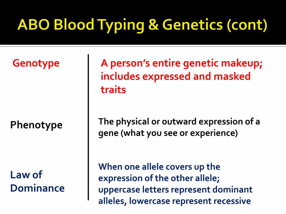
Genotype A person’s entire genetic makeup; includes expressed and masked traits
Phenotype The physical or outward expression of a gene (what you see or experience)
Law of Dominance
When one allele covers up the expression of the other allele; uppercase letters represent dominant alleles, lowercase represent recessive

Homozygous A person with the same alleles for a trait from both parents. Ex. PP, pp, etc.
Heterozygous A person who has received two different alleles from their parents. Ex. Pp, Hh, Tt, etc.


Each parent contributes an A, B or, i allele which determines the antigens (agglutinogens) or lack of antigens to their offspring
When determining blood genotype possibilities, a lower case i is used rather than the letter O indicating that blood type O is recessive.

i + i forms ? blood type
A + i forms ? blood type
A + A forms ? blood type
B + i forms ? blood type
B + B forms ? blood type
A + B forms ? blood type
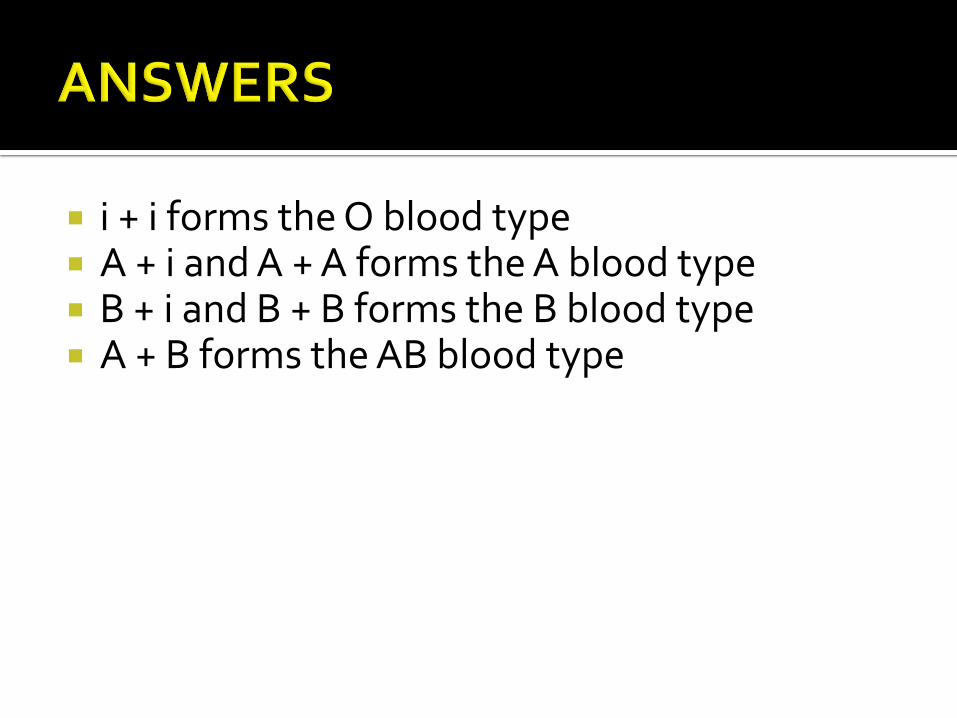
i + i forms the O blood type A + i and A + A forms the A blood type B + i and B + B forms the B blood type A + B forms the AB blood type

Father: Type B heterozygous Mother: Type A heterozygous Using a Punnett Square, determine the:
Genotypic Ratios: Phenotypic Ratios:

B i
A
i
Fathers Genotype
Mo
ther
s G
eno
typ
e
Genotype: AB Phenotype: AB
Genotype: Ai Phenotype: A
Genotype: Bi Phenotype: B
Genotype: ii Phenotype: O

od Type Percentage
A 40% B 11% AB 4% O 45% Rh+ 85% Rh- 15% NOTE: Distribution of blood types varies among different races and ethnic backgrounds

Antigen Surface molecule on red blood cells.
Antibodies Immune System Proteins carried in the plasma.
Agglutination Clumping of Red Blood cells

Because agglutination may cause Hemolysis!
rupturing of blood cells
If blood types are not matched may have antigen - antibody reaction
Could result in kidney damage
Could result in death
Must match blood between donor and recipient when performing blood transfusions


Let’s play a game first! Highest scoring table gets Extra Credit!
Seats 1 & 2 make Type A Blood
Seats 3&4 Make Type B Blood
Seat 5make Type AB Blood
Seat 6 make Type O blood


Based upon antigens (agglutinogens) located on the surface of erythrocytes
Named because it was discovered from the blood of Rhesus monkeys
Rh+ indicates people have Rh agglutinogens (D antigens)
Rh- indicates people lack Rh agglutinogens

If an Rh- person receives blood from an Rh+ donor, the body will start to make Rh+ antibodies (agglutinins)
If during a second transfusion, Rh+ blood is again given, the antibodies produced after receiving the first transfusion will cause hemolysis of the blood from the second transfusion which may result in death
Works the same way with pregnancies! (Rh- mom and Rh+ Baby: must protect baby! RhoGam shot)

Person with type A blood
may receive blood from type A or O donor
may not receive type B or AB blood
Person with type B blood
may receive blood from type B or O donor
may not receive type A or AB blood
Person with type AB blood
may receive blood from type A, type B, or type O donors
(universal recipient)

Person with type O blood
May only receive blood from type O donors
May donate blood to all other blood types
(universal donors)
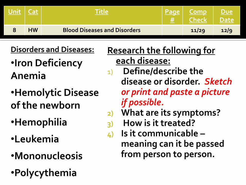
Research the following for each disease:
1) Define/describe the disease or disorder. Sketch or print and paste a picture if possible.
2) What are its symptoms? 3) How is it treated? 4) Is it communicable –
meaning can it be passed from person to person.
Disorders and Diseases:
•Iron Deficiency Anemia
•Hemolytic Disease of the newborn
•Hemophilia
•Leukemia
•Mononucleosis
•Polycythemia
Unit Cat Title Page #
Comp Check
Due Date
8 HW Blood Diseases and Disorders 11/29 12/9

Day 5

Know your blood groups/transfer rules and diseases!
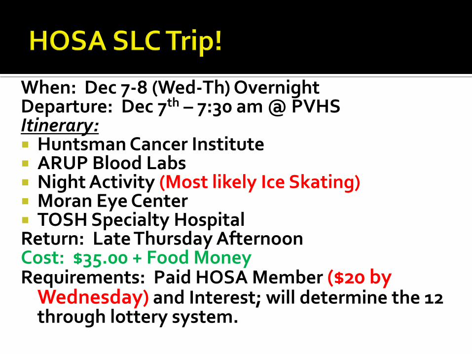
When: Dec 7-8 (Wed-Th) Overnight Departure: Dec 7th – 7:30 am @ PVHS Itinerary: Huntsman Cancer Institute ARUP Blood Labs Night Activity (Most likely Ice Skating) Moran Eye Center TOSH Specialty Hospital Return: Late Thursday Afternoon Cost: $35.00 + Food Money Requirements: Paid HOSA Member ($20 by
Wednesday) and Interest; will determine the 12 through lottery system.

Unit Cat Title Page #
Comp Check
Due Date
8 Act Immunity Notes 12/9
Immunity •Resistance to particular pathogens or their toxins or metabolic byproducts.
Antigens Surface molecules on cells that can elicit an immune response
Antibodies Large proteins that help fight off infection

T-Cells •Immune cells that interact directly with antigen bearing agents to destroy them.
B-Cells Immune Cells interact indirectly with antigen bearing agents by producing antibodies that help destroy them.

Follow the directions given on the webquest sheet for today.
If viewing this on the website, download the webquest
from the link below!
Unit Cat Title Page #
Comp Check
Due Date
8 Act Immune Responses 12/9

Copy the following figure in your notes and then use the internet or your text (Chapter 16) to explain the meaning and/or provide examples of each of the types of immunity that are listed.
Unit Cat Title Page #
Comp Check
Due Date
8 HW Distinguishing Immunity 12/9

Day 6
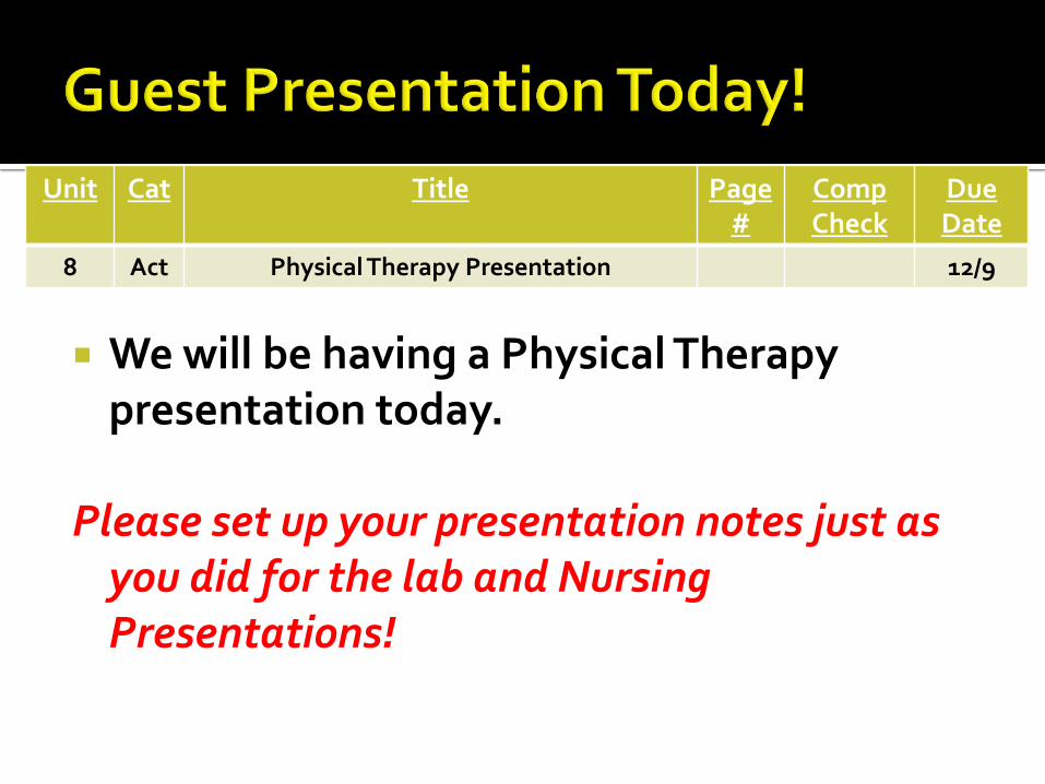
We will be having a Physical Therapy presentation today.
Please set up your presentation notes just as you did for the lab and Nursing Presentations!
Unit Cat Title Page #
Comp Check
Due Date
8 Act Physical Therapy Presentation 12/9

Unit Cat Title Page #
Comp Check
Due Date
8 Act Where is my pulse? 12/9
Pulse •Alternating expansion and recoiling of an arterial wall near the surface of the skin. •What causes it to expand and recoil?
Draw and label figure 15.34 (p. 590) in your book here!
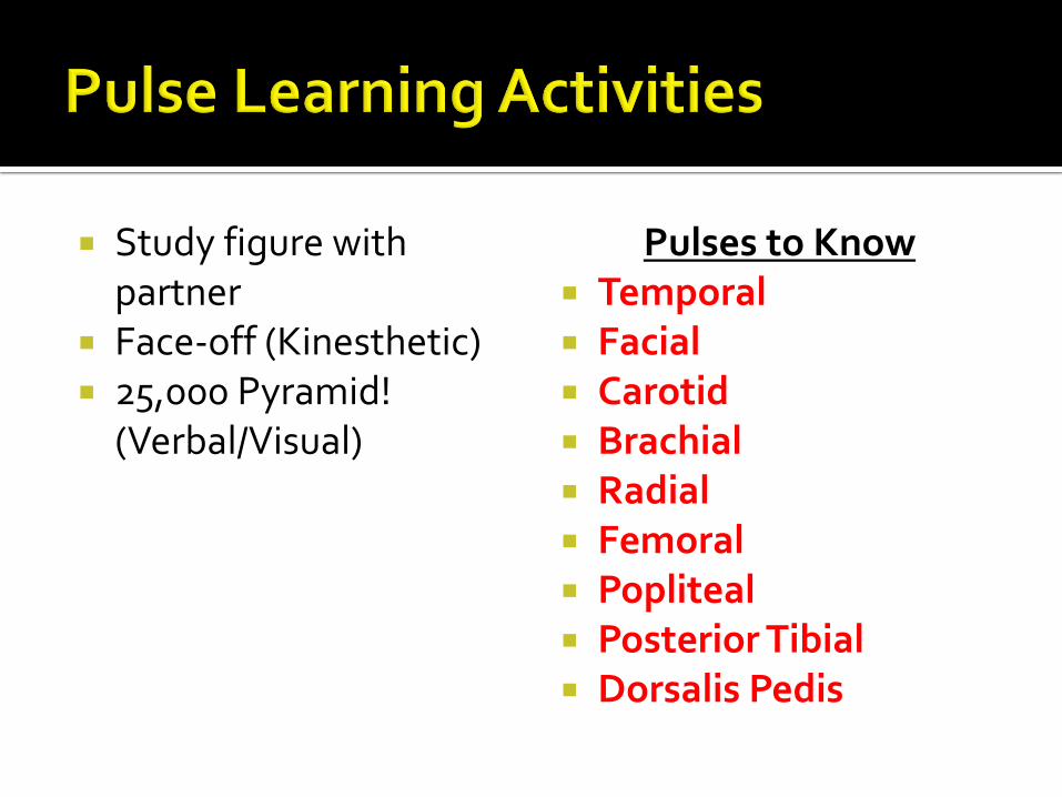
Study figure with partner
Face-off (Kinesthetic) 25,000 Pyramid!
(Verbal/Visual)
Pulses to Know Temporal Facial Carotid Brachial Radial Femoral Popliteal Posterior Tibial Dorsalis Pedis

Research the following for each disease:
1) Define/describe the disease or disorder. Sketch or print and paste a picture if possible.
2) What are its symptoms? 3) How is it treated? 4) Is it communicable –
meaning can it be passed from person to person.
Disorders and Diseases:
•Aneurysm
•Arteriosclerosis
•Atherosclerosis
•Stroke
•Coronary Artery Disease
•Hypertension
•Murmur
•Myocardial Infarction
Unit Cat Title Page #
Comp Check
Due Date
8 HW Cardiovascular Diseases and Disorders 12/5 12/9

Day 7

Today you and your group (which I will choose) will be going to six different stations and completing activities that will teach you about the heart and its functions.
You will only have 11 minutes at each station to
complete the work to be done there so work together and efficiently!
I will also be coming around and quizzing you for
points so be ready!
Unit Cat Title Page #
Comp Check
Due Date
8 Act Cardiovascular System Stations 12/9

Deoxygenated Blood – blood with a low concentration of oxygen; needs to be taken to the lungs; usually drawn as blue on diagrams.
Oxygenated blood – blood with a high
concentration of oxygen; needs to be circulated throughout the body; usually drawn as red on diagrams.
Artery – moves blood away from the heart. Vein – moves blood toward the heart


1. Write the title of this station big in your notes to provide a separation between each station.
2. Take the notes
3. Quiz each other on what is transported by the blood to prepare for Friday’s exam.

Cardiovascular System
•Composed of the heart and a closed system of blood vessels through which blood is circulated.
Primary Function
To circulate (transport) the blood through the body.
Transportation Needs
•Oxygen •Carbon Dioxide •Heat •Nutrients
•Hormones •Waste •Enzymes •Electrolytes

1. Write the title of this station big in your notes to provide a separation between each station.
2. Take the notes
3. Label on your heart diagram the different layers of the heart using the pages referenced below.
Figure 15.4 and Table 15.1 on page 563
4. Quiz each other on the layers of the heart.

Epicardium •The outermost layer. Composed of epithelial and connective tissue. Provides a small amount of protection.
Myocardium Middle, muscular layer. Composed of cardiac muscle, blood vessels and nerves. Responsible for pumping the blood.
Endocardium
•Inner layer of the heart. Composed of epithelial tissue and is very smooth. Blood makes contact with this layer.

1. Write the title of this station big in your notes to provide a separation between each station.
2. Take the notes
3. Label on your heart diagram the different chambers of the heart using the pages referenced below.
Pages 563-567
4. Indicate which chambers move deoxygenated blood and which chambers move oxygenated blood.
5. Indicate where each chamber receives blood from and where each chamber sends blood to.
6. Put on gloves and explore the anatomy of these chambers on the Elk Heart provided.
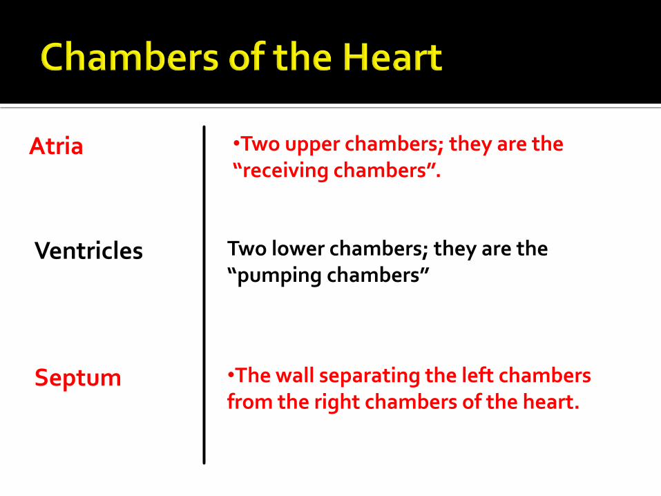
Atria •Two upper chambers; they are the “receiving chambers”.
Ventricles Two lower chambers; they are the “pumping chambers”
Septum •The wall separating the left chambers from the right chambers of the heart.

1. Write the title of this station big in your notes to provide a separation between each station.
2. Take the notes
3. Label on your heart diagram the different great vessels of the heart using the pages referenced below.
Pages 563-567
4. Indicate which chamber of the heart each vessel is connected to.
5. Quiz each other on the vessels using the giant heart model provided..

Superior Vena Cava
•Drains deoxygenated blood from the veins of the head , neck and arms into the right atrium.
Inferior Vena Cava
Drains deoxygenated blood from the veins in the abdomen and legs into the right atrium.
Pulmonary Trunk
•Arises from the right ventricle after the pulmonary semilunar valve. Branches into the pulmonary arteries; contains deoxygenated blood.

Pulmonary Arteries
•Branch from pulmonary trunk; take deoxygenated blood to the lungs.
Pulmonary Veins
Take oxygenated blood from the lungs into the left atrium of the heart.
Aorta •Largest artery in the body; extends from the left ventricle after the aortic semilunar valve; contains oxygen rich blood to be taken to the rest of body

1. Write the title of this station big in your notes to provide a separation between each station.
2. Take the notes
3. Label on your heart diagram the different valves of the heart using the pages referenced below.
Pages 563-567
4. Indicate which chamber of the heart each vessel is connected to.
5. Quiz each other on the vessels using the giant heart model provided..

Valve •Flap-like structures that permit the flow of blood in one direction only.
Tricuspid Valve
Located between the right atrium and right ventricle. Composed of three flaps.
Pulmonary Semilunar Valve
•Located between right ventricle and the pulmonary trunk vessel. Composed of three flaps.

Bicuspid (Mitral) Valve
•Located between the left atrium and left ventricle. Composed of two flaps.
Aortic Semilunar Valve
•Located between left ventricle and the aorta. Composed of three flaps.

1. Write the title of this station big in your notes to provide a separation between each station.
2. In your notes, use figure 15.11 in your text (p. 569) to create a flow chart indicating the path of blood through the right side of the heart, to the lungs, back to the heart, and out to the body.
3. Starting with one of the vena cavae, use the yarn and pins provided to thread the yarn through the heart in the direction that blood would flow into the heart, out to the lungs, back to the heart, and then out to the body.
4. Using arrows, label on your heart diagram the blood flow through the heart starting with the vena cavae vessels and ending with the aorta.

Color code your heart diagram using Blue for the deoxygenated blood vessels and chambers. Then use red for the oxygenated blood vessels and chambers.
Unit Cat Title Page #
Comp Check
Due Date
8 Hw Heart color coding 12/7 12/9