Embed Size (px)
Citation preview

Physiological Factors Affecting Testing
ByProf. Dr. Samy A. Nasef
Muscle Tissue
One of 4 primary tissue types, divided into:
skeletal muscle
cardiac muscle
smooth muscle
Without these muscles, nothing in the body would move and no body movement would occur
6 Functions of Skeletal Muscles
1. Produce skeletal movement
2. Maintain body position and posture
3. Support soft tissues
4. Guard body openings (entrance/exit)
5. Maintain body temperature
6. Store Nutrient reserves
Organization of Connective Tissues
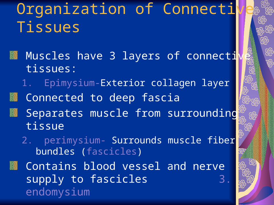
Muscles have 3 layers of connective tissues:
1. Epimysium-Exterior collagen layer
Connected to deep fascia
Separates muscle from surrounding tissue2. perimysium- Surrounds muscle fiber bundles
(fascicles)
Contains blood vessel and nerve supply to fascicles 3. endomysium

3 .Endomysium
Surrounds individual muscle cells (muscle fibers)
Contains capillaries and nerve fibers contacting muscle cells
Contains satellite cells (stem cells) that repair damage
Muscle Attachments
Endomysium, perimysium, and epimysium come together:
at ends of muscles
to form connective tissue attachment to bone matrix
i.e., tendon (bundle) or aponeurosis (sheet)
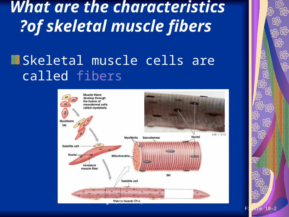
What are the characteristics of skeletal muscle fibers?
Skeletal muscle cells are called fibers
Figure 10–2فاروس سنتر صوتك اسرة

Skeletal Muscle Fibers
Are very long
Develop through fusion of mesodermal cells (myoblasts- embryonic cells))
Become very large
Contain hundreds of nuclei –multinucleate
Unfused cells are satellite cells- assist in repair after injury
The Sarcolemma
The cell membrane of a muscle cell
Surrounds the sarcoplasm (cytoplasm of muscle fiber)
A change in transmembrane potential begins contractions
All regions of the cell must contract simultaneously
Transverse Tubules (T tubules)
Transmit action potential – impulses through cell
Allow entire muscle fiber to contract simultaneously
Have same properties as sarcolemma
Filled with extracellular fluid
Myofibrils- 1-2um in diameter Lengthwise subdivisions within muscle fiberMade up of bundles of protein filaments (myofilaments)Myofilaments - are responsible for muscle contraction
2 Types of MyofilamentsThin filaments:
made of the protein actin
Thick filaments: made of the protein myosin
Sarcoplasmic Reticulum (SR)
A membranous structure surrounding each myofibril Helps transmit action potential to myofibrilSimilar in structure to smooth endoplasmic reticulumForms chambers (terminal cisternae) attached to T tubules
فاروس سنتر صوتك اسرة

A Triad
Is formed by 1 T tubule and 2 terminal cisterna
CisternaeConcentrate Ca2+ (via ion pumps)
Release Ca2+ into sarcomeres to begin muscle contraction
فاروس سنتر صوتك اسرة
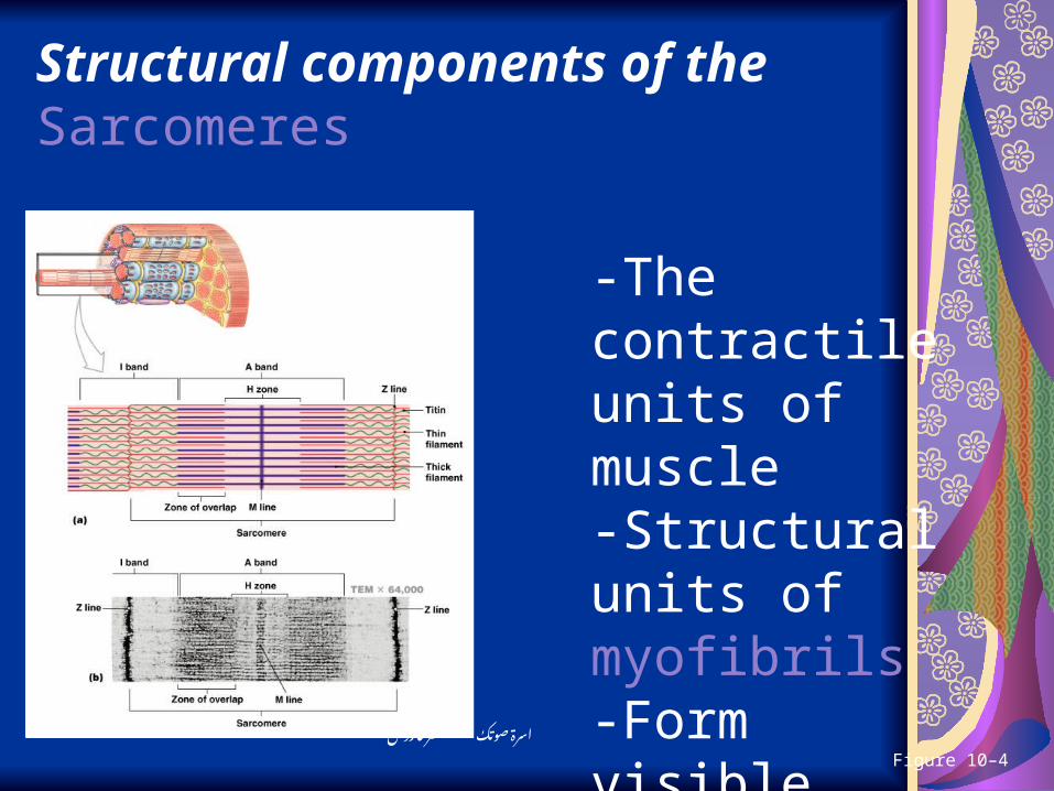
Structural components of the Sarcomeres
Figure 10–4
-The contractile units of muscle-Structural units of myofibrils -Form visible patterns within myofibrils
فاروس سنتر صوتك اسرة
InterActive Physiology: Contraction of Whole MusclePLAYPLAY
Tension Produced by Whole Skeletal Muscles
Depends on:internal tension produced by muscle fibers
external tension exerted by muscle fibers on elastic extracellular fibers
total number of muscle fibers stimulated
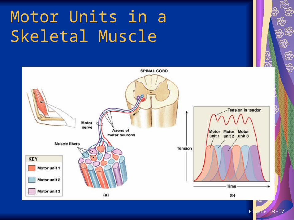
Motor Units in a Skeletal Muscle
Figure 10–17
Motor Units in a Skeletal Muscle
Contain hundreds of muscle fibers
That contract at the same time
Controlled by a single motor neuron
InterActive Physiology: Contraction of Motor UnitsPLAYPLAY
Recruitment (Multiple Motor Unit Summation)
In a whole muscle or group of muscles, smooth motion and increasing tension is produced by slowly increasing size or number of motor units stimulated
Maximum Tension
Achieved when all motor units reach tetanus
Can be sustained only a very short time
Sustained Tension
Less than maximum tension
Allows motor units to rest in rotation