Embed Size (px)
Citation preview
PlantPhysUW425.doc 5/24/2008 23:32 Page 1 of 138
The Physiology Of Higher PlantsAn Outline
Prepared by Michael McGoodwin (MCM)Winter/Spring 2008
TABLE OF CONTENTSIntroduction........................................................................................................................................................................................6Instructor And Course Website ........................................................................................................................................................7Sugar Chemistry Highlights..............................................................................................................................................................7Chapter I. Plant Cells Review of Plant Anatomy and Cellular Components................................................................................8
Meristems And Vessels; Anatomy Of Stems and Trunks ........................................................................................................8Cell Wall and Intracellular Components ..................................................................................................................................9Plasmodesmata, Symplast, and Apoplast ...............................................................................................................................11
Chapter II. Energy And Enzymes: Entropy, Free Energy, Redox And Electrochemical Potentials ........................................11Thermodynamics: Work, Free Energy, Enthalpy, and Entropy .............................................................................................12
First Law of Thermodynamics .......................................................................................................................................12Second Law of Thermodynamics ...................................................................................................................................12Gibb’s Free Energy and Enthalpy (Heat Content)..........................................................................................................13
Oxidation / Reduction (Redox) reactions ...............................................................................................................................14Electrochemical Potential.......................................................................................................................................................15Proteins and Enzymes ............................................................................................................................................................16
Chapter III. Water And Plant Cells ...............................................................................................................................................18Water: Heat Properties ...........................................................................................................................................................18Water: Cohesion, Adhesion, Surface Tension, and Capillarity ..............................................................................................18Diffusion Rates of Dissolved Substances...............................................................................................................................19Bulk Flow of Water, Water Potential, and Osmosis...............................................................................................................20
Chapter IV. Water Balance Of Plants............................................................................................................................................21Soil Water...............................................................................................................................................................................22Root Absorption And Water Transport ..................................................................................................................................22Water Transport Through The Xylem....................................................................................................................................23Water Movement From The Leaf To The Atmosphere..........................................................................................................23Summary Of Water Movement In The Soil-Plant-Atmosphere Continuum ..........................................................................24
Chapter V. Mineral Nutrition: Soil Nutrients And Their Absorption By Roots ........................................................................24Essential Nutrients And Deficiencies.....................................................................................................................................24Soil, Roots, and Microbes ......................................................................................................................................................25
Chapter VI. Solute Transport .........................................................................................................................................................27Passive Versus Active Transport, Nernst Equation, Ion Concentrations................................................................................27Membrane Transport Processes..............................................................................................................................................28
Channels .........................................................................................................................................................................29Carriers and Pumps ........................................................................................................................................................29
Ion Transport In Roots ...........................................................................................................................................................31Chapter VII. Photosynthesis Light (Thylakoid) Reactions ..........................................................................................................31
Light Physics..........................................................................................................................................................................31Photosynthesis Pigments And Complexes .............................................................................................................................32Photosystems I And II And The Synthesis of NADPH and ATP...........................................................................................34Protection And Regulation of Photosynthetic Machinery ......................................................................................................35Genetics Of The Photosynthetic Systems...............................................................................................................................36
Chapter VIII. Photosynthesis Carbon (Stromal) Reactions .........................................................................................................36The Calvin C3 Cycle...............................................................................................................................................................36
Energy Conversion Efficiency Of Photosynthesis Using The Calvin Cycle ..................................................................37Regulation Of The Calvin C3 Cycle ...............................................................................................................................37
The C2 Oxidative Photosynthetic Carbon Cycle (Photorespiration) ......................................................................................38Mechanisms For Suppressing Photorespiration Or Preventing Photo-oxidation ...................................................................38
Suppressing Photorespiration By Concentrating CO2 and HCO3- ..................................................................................38
The C4 Carbon Cycle-Plants...........................................................................................................................................38Crassulacean Acid Metabolism (CAM) .........................................................................................................................39

PlantPhysUW425.doc 5/24/2008 23:32 Page 2 of 138
Synthesis of Starch and Sucrose.............................................................................................................................................39Chapter IX. Photosynthesis Physiology And Ecology...................................................................................................................40
Adaptations Of Plants To Bright Light And Shade................................................................................................................40Bright Light Adaptations................................................................................................................................................40Shade Adaptations and Light Concentrating Mechanisms .............................................................................................41
Quantitation Of Photosynthetic Responses To Light, Temperature, And Other Factors .......................................................41Dark Respiration Regime, Light Compensation Point, Light-Limited Regime, And CO2 Limited Regime..................41Effect On Quantum Yields By O2, CO2, Temperature, And Light Levels .....................................................................41Mechanisms of Protection Against Excess Light ...........................................................................................................42Effects of Temperature And Heat On PS .......................................................................................................................42Effects of CO2 on PS and Evolution of C4 versus C3 Plants...........................................................................................43Summary of PS As A Function Of CO2 Versus Temperature ........................................................................................44Crassulacean Acid Metabolism (CAM) .........................................................................................................................44Use Of Stable Carbon Isotope Ratios To Study C3, C4, and CAM Plants ......................................................................44
Chapter X. Translocation In The Phloem......................................................................................................................................45Phloem Vessels, Interconnections, Damage Responses, Companion Cells ...........................................................................45
Protective Mechanisms...................................................................................................................................................45Companion Cells ............................................................................................................................................................45
Sources and Sinks ..................................................................................................................................................................46Materials Transported In The Phloem; Sap............................................................................................................................46Rates And Mechanisms Of Phloem Transport .......................................................................................................................47Phloem Loading: Apoplastic Versus Symplastic ...................................................................................................................47Phloem Unloading And Transition From Source To Sink .....................................................................................................48
Transition From Source To Sink ....................................................................................................................................48Photosynthate Distribution And Regulation...........................................................................................................................48Transport of Signaling Molecules In The Phloem..................................................................................................................49
Chapter XI. Respiration And Lipid Metabolism...........................................................................................................................49Glycolysis...............................................................................................................................................................................49Fermentation ..........................................................................................................................................................................49Control Of Glycolysis ............................................................................................................................................................50The Oxidative Pentose Phosphate Pathway ...........................................................................................................................50The Citric Acid Cycle, Oxidative Phosphorylation, and Mitochondria .................................................................................50Methods Of Lowering ATP Yield, Generating Heat, Or Reducing Oxidative Stress ............................................................52Respiration In Intact Plants ....................................................................................................................................................52Lipid Metabolism ...................................................................................................................................................................53
Chapter XII. Assimilation Of Mineral Nutrients And Oxygen....................................................................................................54Nitrogen .................................................................................................................................................................................54
Biological nitrogen fixation and associated symbiotic relationships..............................................................................55Toxicity of ammonium and nitrate .................................................................................................................................57Nitrate assimilation ........................................................................................................................................................57Ammonium assimilation ................................................................................................................................................58Amino acid biosynthesis ................................................................................................................................................58
Sulfur Assimilation ................................................................................................................................................................59Phosphate Assimilation ..........................................................................................................................................................59Metal Cation Assimilation .....................................................................................................................................................60Oxygen Assimilation..............................................................................................................................................................60Energetics Of Nutrient Assimilation And Impact Of Rising CO2 ..........................................................................................61
Chapter XIII. Secondary Metabolites And Plant Defenses ..........................................................................................................61Waxes, Cutins, and Suberin ...................................................................................................................................................62Secondary Metabolites Defined .............................................................................................................................................62Allelopathy Defined ...............................................................................................................................................................63Terpenes, Terpenoids and Isoprenoids ...................................................................................................................................63
Classification and biosynthesis.......................................................................................................................................63Terpene-derived compounds that are primary metabolites.............................................................................................64Terpenes that protect against herbivores ........................................................................................................................64Terpenes that confer scents and flavors potentially useful to humans............................................................................65
Phenolics ................................................................................................................................................................................66Biosynthesis ...................................................................................................................................................................66Simple phenolics ............................................................................................................................................................66Lignin .............................................................................................................................................................................67Flavonoids ......................................................................................................................................................................67
PlantPhysUW425.doc 5/24/2008 23:32 Page 3 of 138
Tannins...........................................................................................................................................................................68Nitrogen-Containing Secondary Metabolites .........................................................................................................................68
Alkaloids ........................................................................................................................................................................68Cyanogenic glycosides ...................................................................................................................................................70Glucosinolates (AKA Mustard oil glycosides, Thioglycosides, or Thioglucosides) ......................................................71Nonproteinogenic (nonprotein) amino acids ..................................................................................................................71
Induced Plant Defenses Against Insect Herbivores................................................................................................................72Induction of Jasmonic Acid, a plant defense hormone...................................................................................................72Plant proteins that inhibit insect herbivore digestion .....................................................................................................73Herbivore damage induces systemic defenses................................................................................................................73
Plant Defenses Against Pathogenic Microorganisms .............................................................................................................74Antimicrobial compounds synthesized before pathogen attack .....................................................................................74Hypersensitive response, reactive oxygen species, and nitric oxide ..............................................................................74Compounds increasing polymeric physical barriers to spread .......................................................................................74Hydrolytic enzymes directed against the cell walls of pathogens ..................................................................................74Phytoalexins ...................................................................................................................................................................75Some plants respond rapidly to specific substances (elicitors) released from pathogens...............................................75Systemic acquired resistance..........................................................................................................................................75
Chapter XIV. Gene Expression And Signal Transduction...........................................................................................................76Genome Size and Complexity................................................................................................................................................76Prokaryotic Gene Expression .................................................................................................................................................76Eukaryotic Gene Expression ..................................................................................................................................................77Signal Transduction................................................................................................................................................................78
Chapter XV. Cell Walls: Structure, Biogenesis, And Expansion.................................................................................................78Key Properties And Benefits Of Cell Walls...........................................................................................................................78Cellulose.................................................................................................................................................................................79Primary And Secondary Cell Walls .......................................................................................................................................79Patterns of Cell Expansion .....................................................................................................................................................80Wall Degradation and Plant Defenses....................................................................................................................................81
Chapter XVI. Growth and Development .......................................................................................................................................81Sporophytic Development......................................................................................................................................................81Embryogenesis And The Origins Of Polarity.........................................................................................................................81The Shoot Apical Meristem (SAM) .......................................................................................................................................82The Root Apical Meristem (RAM) ........................................................................................................................................83Vegetative Organogenesis......................................................................................................................................................83Senescence And Programmed Cell Death ..............................................................................................................................83
Chapter XVII. Phytochrome And The Light Control Of Plant Development............................................................................84Phytochrome Biochemical and Photochemical Properties .....................................................................................................84Phytochrome Biological Responses Categorized By Fluence And Irradiance.......................................................................85Circadian Rhythms.................................................................................................................................................................86Ecological Functions ..............................................................................................................................................................86
Chapter XVIII. Blue-Light Responses: Stomatal Movements And Morphogenesis ..................................................................87Photophysiology of Blue Light Responses.............................................................................................................................87The Blue Light Photoreceptors: Cryptochromes, Phototropins, And Zeaxanthin ..................................................................88
Chapter XIX. Auxin: The Growth Hormone.................................................................................................................................89Biochemistry, Biosynthesis and Metabolism of Auxin ..........................................................................................................89Transport of Auxin .................................................................................................................................................................90
Polar Transport (Cell-To-Cell) .......................................................................................................................................91Non-Polar Transport (In Phloem)...................................................................................................................................91
Actions of Auxin ....................................................................................................................................................................92Cell Elongation...............................................................................................................................................................92Control Of Plant Orientation ..........................................................................................................................................92Phototropism ..................................................................................................................................................................92Gravitropism...................................................................................................................................................................93Thigmotropism...............................................................................................................................................................93
Developmental Effects Of Auxin ...........................................................................................................................................94Auxin Regulates Apical Dominance ..............................................................................................................................94Auxin Transport Regulates Floral Bud Development And Phyllotaxy ..........................................................................94Auxin Promotes The Formation Of Lateral And Adventitious Roots ............................................................................94Auxin Induces Vascular Differentiation.........................................................................................................................94Auxin Delays Leaf Abscission .......................................................................................................................................94
PlantPhysUW425.doc 5/24/2008 23:32 Page 4 of 138
Auxin Promotes Fruit Development...............................................................................................................................94Commercial Uses Of Synthetic Auxins..........................................................................................................................95
Auxin Signal Transduction Pathways ....................................................................................................................................95Chapter XX. Gibberellins: Regulators Of Plant Height And Seed Germination .......................................................................95
Discovery and Chemical Structure Of Bioactive Forms ........................................................................................................95Discovery .......................................................................................................................................................................95Structure and Nomenclature ...........................................................................................................................................96
Biosynthesis And Catabolism Pathways, Regulation, Organ Distribution.............................................................................96Specific Growth And Development Effects of Endogenous And Commercial (Exogenous) Gibberellins............................97
Promote Stem (And Root) Growth.................................................................................................................................97Regulate Transition From Juvenile To Adult Phases .....................................................................................................97Influence Floral Initiation And Flower Sex Determination............................................................................................97Promote Pollen Development And Tube Growth...........................................................................................................98Promote Fruit Growth And Parthenocarpy.....................................................................................................................98Promote Seed Development and Germination ...............................................................................................................98
Gibberellin Signaling .............................................................................................................................................................98Details of Certain Gibberellin Responses...............................................................................................................................98
The Aleurone Layer In Seed Endosperm .......................................................................................................................98Flowering In Long-Day Plants .......................................................................................................................................99Stem Growth ..................................................................................................................................................................99
Chapter XXI. Cytokinins: Regulators Of Cell Division................................................................................................................99Cell Division and Plant Development ..................................................................................................................................100Discovery, Detection, And Properties Of Cytokinins ..........................................................................................................100
Pathogenic Secretion ....................................................................................................................................................101Biosynthesis, Metabolism, and Transport ............................................................................................................................101Biological Roles of Cytokinins ............................................................................................................................................102
Cytokinins regulate cell division in shoots and roots ...................................................................................................102Cytokinins regulate specific components of the cell cycle...........................................................................................102The Auxin:Cytokinin ratio regulates morphogenesis in cultured tissues .....................................................................102Cytokinins modify apical dominance and promote lateral bud growth........................................................................103Cytokinins induce bud formation in a moss .................................................................................................................103Cytokinin overproduction has been implicated in genetic tumors................................................................................103Cytokinins delay leaf senescence .................................................................................................................................103Cytokinins promote movement of nutrients into leaves ...............................................................................................103Cytokinins promote chloroplast development ..............................................................................................................104Cytokinins promote cell expansion in leaves and cotyledons ......................................................................................104Cytokinin-regulated processes are revealed in plants that overproduce cytokinins .....................................................104
Cellular And Molecular Modes Of Cytokinin Action ..........................................................................................................104Chapter XXII. Ethylene: The Gaseous Hormone .......................................................................................................................104
Early Observations ...............................................................................................................................................................104Structure, Biosynthesis, and Measurement of Ethylene .......................................................................................................105More Details On Developmental and Physiological Effects of Ethylene.............................................................................106
Ethylene promotes ripening of some fruits...................................................................................................................106Leaf Epinasty................................................................................................................................................................107Ethylene induces lateral cell expansion........................................................................................................................107Ethylene induces and maintains the hook of etiolated seedlings..................................................................................107Ethylene breaks seed and bud dormancy in some species............................................................................................107Ethylene promotes the elongation growth of submerged aquatic species ....................................................................107Ethylene induces the formation of roots and root hairs ................................................................................................107Ethylene is used by crown galls ...................................................................................................................................108Ethylene induces flowering or alters flower sex in some species.................................................................................108Ethylene enhances the rate of leaf senescence .............................................................................................................108Ethylene is involved in defense responses to wounding ..............................................................................................108Ethylene and auxin regulate abscission ........................................................................................................................108Commercial uses of ethylene-related products.............................................................................................................109
Ethylene Signal Transduction Pathways ..............................................................................................................................109Chapter XXIII. Abscisic Acid: Seed Maturation And AntistressSignal...................................................................................109
Effects Of ABA On Seeds....................................................................................................................................................110Other Effects Of ABA..........................................................................................................................................................111ABA Signal Transduction Pathways ....................................................................................................................................111
Chapter XXIV. Brassinosteroids ..................................................................................................................................................111
PlantPhysUW425.doc 5/24/2008 23:32 Page 5 of 138
Chapter XXV. The Control Of Flowering ...................................................................................................................................112Review Of Flower Anatomy And Fertilization ....................................................................................................................112Floral Meristems and Floral Organ Development ................................................................................................................113Floral Competence, Induction, Determined State, And Expression .....................................................................................113Circadian Rhythms And Photoperiodism.............................................................................................................................114Vernalization ........................................................................................................................................................................115Biochemical Signaling In Flowering....................................................................................................................................116
Chapter XXVI. Physiology of Environmental Stresses...............................................................................................................116Water Deficit and Drought Tolerance ..................................................................................................................................117
Decreased leaf area.......................................................................................................................................................117Leaf abscission .............................................................................................................................................................118Increased root growth (which may be in competition with reproduction)....................................................................118Stomatal closure from Abscisic acid ABA...................................................................................................................118Limiting photosynthesis ...............................................................................................................................................118Osmotic adjustment of cells helps to maintain water balance ......................................................................................119Increased resistance to water flow................................................................................................................................119Increased leaf wax deposition ......................................................................................................................................119Alteration in energy dissipation from leaves by altering leaf angle, geometry, or barriers to sunlight ........................119Crassulacean acid metabolism (CAM) photosynthesis ................................................................................................120Osmotic stress and altered gene expression .................................................................................................................120
Heat Stress and Heat Shock .................................................................................................................................................120Levels of heat tolerance................................................................................................................................................120Mechanisms of heat dissipation and tolerance .............................................................................................................120Effects of heat stress.....................................................................................................................................................121Role of membranes in plant adaptation and acclimation to high temperatures ............................................................121Leaf adaptations against high temperatures..................................................................................................................121Protective heat shock proteins and other heat tolerance agents....................................................................................121
Chilling And Freezing..........................................................................................................................................................122Membrane properties in chilling injury and resistance.................................................................................................122Ice crystal formation and protoplast dehydration kills cells .........................................................................................122Limitation of ice formation (or of ice recrystallization) contributes to freezing tolerance...........................................123Some woody plants can acclimate to very low temperatures .......................................................................................124Additional factors in freezing responses ......................................................................................................................125
Saline Stress and Toxic Trace Elements ..............................................................................................................................125Salt accumulates in irrigated soils and impairs plant function .....................................................................................126Plants vary considerably in salt tolerance.....................................................................................................................127The mechanisms of salt stress ......................................................................................................................................127Plant strategies for reducing salt stress.........................................................................................................................128Plant adaptations to toxic trace elements......................................................................................................................128
Oxygen Deficiency...............................................................................................................................................................129Anaerobic microorganisms are active in water-saturated soils and impart toxins........................................................129Roots are damaged in anoxic environments and this can injure shoots........................................................................129Submerged organs can acquire O2 through specialized structures including aerenchyma ...........................................130Mechanisms for adaptation to anaerobic conditions ....................................................................................................131Anaerobic sensing, signal transduction, and anaerobic stress proteins in acclimation to oxygen deficit .....................131
Air Pollution.........................................................................................................................................................................132Plant Neurobiology.........................................................................................................................................................................133Possible Corrections or Clarifications For The Textbook ..........................................................................................................135

PlantPhysUW425.doc 5/24/2008 23:32 Page 6 of 138
Introduction
All animal life on this planet depends ultimately on plants—especially the green autotrophic plants—for theoxygen we breathe and the food we eat. There could hardly be a more important group of organisms tostudy and attempt to understand.
This compilation is derived from the University of Washington Plant Physiology (Biology 425) courselectures given by Professor Elizabeth Van Volkenburgh and from the textbook, Lincoln Taiz and EduardoZeiger, Plant Physiology, 4th Ed., publ. Sinauer Associates, Inc., 2006. Online components of this textbookincluding “Web Topics” and “Web Essays” referenced throughout this outline may be found athttp://4e.plantphys.net/index.php. Page numbers expressed as TZ252 refer to pages in this edition of thistextbook. Although most of this document follows the chapter and section sequences of the textbook,rearrangements have occasionally been made, sections have been freely retitled for clarity, and additionalmaterials have been included from other sources where appropriate and feasible.
The subject matter of this course and textbook includes how plants work: the mechanical, physical, andbiochemical functioning of living plants, primarily the vascular seed-bearing higher plants (mostlyangiosperms and gymnosperms), but some references are made to physiology of other eukaryotic plantssuch as algae, fungi, and mosses, and to prokaryotes such as bacteria, in order to contrast or showsimilarities. Another definition of plant physiology is “What plants do: Metabolism, Assimilation andtransport of water and nutrients, Growth and development, and Responses to the environment”.1Compared to my previous experiences with human physiology, this course involves considerably moredetails at the biochemical level.
These notes present selected facts and concepts that are especially interesting or fundamental, much ofwhich I was not previously well versed on. I am a retired physician—not a biologist, botanist, orphysiologist—and I don’t claim authoritative knowledge of this material. (Disclaimer: I am not in a positionto evaluate or make medical recommendations regarding various health-related claims made aboutpurportedly beneficial or harmful phytochemicals such as isoflavonoids, carotenoids, tannins, or otherherbal substances. You should seek qualified advice before deciding to ingest or be treated with any of thebiochemicals described in this outline.)
I am preparing these notes primarily as a learning aid for myself, but some of the material could be usefulto others. In some cases, I have added basic review material on subjects for which prior knowledge ispresumed for the proper understanding of plant physiology, particularly topics in biochemistry, physics,and geobiology. To add personal interest, I have also tried to at least mention many of the specific plants,plant etymologies, plant compounds, plant phenomena, and other plant trivia and lore that my wife or Ihave had direct or indirect experience with, or that seem to be of general or medical interest (for example,the psychoactive and poisonous plant products).
The superb textbook is extremely detailed and technical, so these notes only scratch the surface of thisamazingly complex subject. For the sake of brevity, many of the experimental details and qualifyingcomments that make scientific assertions more accurate have often been omitted. Also for brevity and toavoid copyright problems, I have not included illustrations or diagrams in this document, only textparaphrases and (hopefully) “fair use” quoted text and formulas. Many of the cited references includesome of the diagrams needed to understand this material, but there is no substitute for having access tothe textbook—you should buy this book!
Viewing this document in a Web browser or in a recent generation Adobe Acrobat Reader allows followingthe Web URL hyperlinks, just as with a normal Web page. In some instances, multiple separate sourcesare combined into one footnote (in which case the individual sources are demarcated by a bullet “• ”).Inclusion of material from Wikipedia and other Web sources does not imply any assumption ofauthoritativeness of these resources.
1 The website http://www.uwyo.edu/botany4400/ using the same textbook to present a quite differentplant physiology course outline.
PlantPhysUW425.doc 5/24/2008 23:32 Page 7 of 138
All text and Web resources were accessed in January to June 2008 unless otherwise stated. Correctionsand comments are welcome—send these to mcgoo at u period washington period edu [after converting thiseaddress to standard format].
Instructor And Course Website
Professor: Elizabeth “Liz” Van Volkenburgh, Ph.D. = “EVV”Websites:
Course Website: http://courses.washington.edu/biol425/index.html [expired]Course Web Forum: https://catalysttools.washington.edu/gopost/board/lizvanv/4598
(restricted access)Textbook Website: http://4e.plantphys.net/index.php (or http://www.plantphys.net/)
My wife and I both express our thanks to Professor Van Volkenburgh for allowing us to audit her excellentcourse.
Sugar Chemistry Highlights
This review2 is intended to aid the study and understanding of sucrose, cellulose, starch, and the carbonsugar-related produts of photosynthesis. (For a general look at bioactive chemicals, see web essay 1.1,Exploring Chemical Space in the Plant World. See also Web Topic 8.11 Glossary of carbohydratebiochemistry. Proteins and Enzymes are discussed in chapter 2.)
• The CO or C=O group is called a carbonyl group3. It appears in aldehydes RCHO, ketones RCOR′,Carboxylic acids RCOOH, esters RCOOR', and amides RCONR'R''
• Monosaccharides have the chemical formula (CH2O)n + m with the chemical structureH(CHOH)nC=O(CHOH)mH. If n or m is zero, it is an aldehyde and is termed an aldose, otherwise it is aketone and is termed a ketose. Monosaccharides contain either a ketone or aldehyde functionalgroup, and hydroxyl groups on most or all of the non-carbonyl carbon atoms.4 [This describes thelinear form of a monosaccharide.]
• A monosaccharide is a triose, tetrose, pentose, or hexose, etc. based on the number of carbons (e.g.,3, 4, 5, or 6). Sedoheptulose is one of the few heptoses, sialose is a nonose.
• Linear (chain) forms of monosaccharides are aldehydes [e.g., glucose = C1 aldose] or ketones [e.g.,fructose= C2 ketose]. Carbon numbering begins at the end closest to the aldehyde or ketone C.
• The linear stereoisomers (in this case, mirror images = enantiomers) of monosaccharides are D- or L-based on the asymmetric C furthest from the aldehyde or ketone group, e.g., C5 [not C6] for glucose, incomparison to D-glyceraldehyde or L-glyceraldehyde. Most natural sugars are D-stereoisomers.
• The aldohexose stereoisomers5 are glucose, mannose, galactose (plus 5 uncommon or unnaturalforms—allose, altrose, gulose, idose, and talose). The common ketohexose stereoisomers are D-fructose and D-sorbose
• Ring formation (Cyclization): The aldehyde and ketone functional groups in monosaccharides arereactive and react reversibly with nearby hydroxyl functional groups to form intramolecularhemiacetals [R-CHOH-O-R′] or hemiketals, respectively. The resulting 6 member ring structure isrelated to pyran [more closely to saturated tetrahydropyran], and is termed a pyranose. If the ring has5 members (similar to furan), it is a furanose. (For example, arabinose may occur in furanose andpyranose forms.)6 As a monosaccharide in solution, the ring spontaneously opens and closes, allowingrotation to occur about the bond between the carbonyl group and the neighboring carbon atom,yielding two distinct stereoisomers or anomers (α and β). This process is termed mutarotation. Forinstance, in the familiar “chair” form of glucose in cellulose, the β-D-glucopyranose has the -OH arisingat the anomeric carbon [C1] pointing out rather than down.
2 Sugar chemistry: http://www.rpi.edu/dept/bcbp/molbiochem/MBWeb/mb1/part2/sugar.htm3 Carbonyl: http://en.wikipedia.org/wiki/Carbonyl4 Monosaccharide: http://en.wikipedia.org/wiki/Monosaccharide5 Aldohexoses: http://en.wikipedia.org/wiki/Hexose6 Arabinose forms: http://www-jmg.ch.cam.ac.uk/data/molecules/nameindex.html
PlantPhysUW425.doc 5/24/2008 23:32 Page 8 of 138
• The aldehyde [glucose = C1 aldose] or ketone [fructose= C2 ketose] linear forms of monosaccharides(glucose, fructose) are energetically disfavored over the cyclized forms—only 0.02% of glucose insolution is in chain form.
• Glycosidic bonds and Polymers: In a 1→4-O-glycosidic bond7, a C1-O-C4 bond is made involving theC1 of one sugar molecule and the C4 of the other. Likewise a C1-O-C6 bond involving the C1 of onesugar molecule and the C6 of the other is called a 1→6-O-glycosidic bond. Glycosidic bonds allowformation of disaccharides such as sucrose [α-D-glucopyranosyl-(1↔2)-β-D-fructofuranoside],polysaccharides such as cellulose and starch, and other sugar polymers. Examples include:• Starch (amylose): α-D-1,4 glycosidic bonds, or (1→4)α-D glycosidic bonds
[forming linear chains]• Starch (amylopectin): α-D-1,6 glycosidic bonds, or (1→6)α-D glycosidic bonds
[forming side chains]• Cellulose: β-D-1,4 glycosidic bonds, or (1→4)β-D glycosidic bonds
[forming linear chains]• In sugar acids, the aldehyde at C1, or the hydroxyl on the terminal carbon, is oxidized to a carboxylic
acid. Examples are gluconic acid and glucuronic acid.• A sugar alcohol is a hydrogenated form of carbohydrate, whose carbonyl group (aldehyde or ketone,
reducing sugar) has been reduced to a primary or secondary hydroxyl group. Examples include glycol,glycerol, sorbitol, mannitol.
• Polysaccharides: A glycan is a polysaccharide polymer. A glucan is a glycan of glucose, a xylan is axylose polymer, galactan is a glycan of galactose, etc. Xyloglucan is a primary glucan with side chainsof one or more xyloses. Arabinoxylan is a xylan with arabinose side chains (apparently in furanose 5-member ring form). Naming is not always consistent. In polysaccharides, the anomeric form (α or β) isstabilized and does not mutarotate.
Chapter I. Plant CellsReview of Plant Anatomy and Cellular Components
Meristems And Vessels; Anatomy Of Stems and Trunks
Above the soil level is the major vegetative organ, the shoot, and below is the root system. Theattachment or transition point between the stem or shoot and the root is termed the base. (See Web essay19.2 regarding confusion in the terminology for root apex and base in embryos versus seedlings.) The termvegetative signifies that part of a plant’s life other than the reproductive phase. The major plant tissuesare dermal, ground, and vascular.
Plant tissues grow at rapidly dividing tissue sites called meristems, including apical meristems at shootapex, root apex, and axillary budTZ2. Meristematic tissue, which contain totipotent undifferentiated cellsanalogous to animal stem cells, may also be found in stems (as intercalary meristems arising at nodes) andin reproductive structures such as floral (inflorescence) meristems. Vegetative plant growth isindeterminate, due to the permanent growth of meristem tissues in the shoot and root apices (butreproductive growth is determinate). The vascular cambium is a later meristem that generates the xylemand phloem. These vascular structures are positioned in a ring located inside the cortex and outside thepith.
Woody trees and shrubs:8 the vascular cambium initially produces primary xylem (inside) and primaryphloem (outside). In subsequent years the vascular cambium adds growth rings: additional layers ofsecondary xylem (which forms immediately inside the vascular cambium) and of secondary phloem(which forms immediately outside the vascular cambium). Older (more centrally positioned) layers of xylembecome the annual tree rings—these stop conducting water but provide mechanical support. The olderlayers of primary and secondary phloem are crushed by outward expansion of the secondary xylem. (fig.10.2) “Since the phloem is constantly being pushed outward and crushed, only the innermost layers [of
7 Glycosidic bond: http://en.wikipedia.org/wiki/Glycosidic_bond8 Anatomy of trunks of trees and shrubs: http://plantphys.info/plants_human/secondary.html
PlantPhysUW425.doc 5/24/2008 23:32 Page 9 of 138
secondary phloem] adjacent to the vascular cambium are functional phloem.”9 The secondary xylem cellsproduced in the spring and early summer of the year are larger in radial dimension. In late summer, fall,and winter the secondary xylem cells are much smaller in the radial dimension, thus generating thealternating bands of growth rings. Girdling a tree destroys the active phloem layer preventing distributionof sugars, but does not stop transpiration from water transport located in the more deeply positionedxylem.
Cork cambium10 (phellogen, bark cambium) generates cork (phellem) to the outside (as part of the barkor periderm) and in some plants the phelloderm or phellem layer (inside the cork cambium).
The plant circulatory system (in vascular tissues) is not a closed loop as in animals with hearts. It may beconsidered divided into:• Xylem vessels: which transport water and soil nutrients from the roots to the shoot, and, separated
from the xylem by the vascular cambium, the• Phloem vessels: which transports photosynthate (organic products of photosynthesis, also called
photoassimilate) from sources to sinks (such as meristems) regardless of the up/down direction offlow in the plant.
The axially (longitudinally) elongated vessels of the xylem consist of tracheids (found in allspermatophytes) and vessel elements (found primarily in angiosperms). These tracheary elements arecomposed of apoptotically dead cells with rigid perforated walls stiffened with lignin, thereby capable ofremaining open at negative pressure (called “tension” throughout this book). They are interconnected bysimple pits and end wall perforations. They have no cytoplasm, organelles, or plasma membranes. (Seefurther discussion in Chapter IV.)
In contrast, the phloem vessels consist of living sieve cells interconnected through sieve areas and (inangiosperms only) sieve plates, with adjacent companion cells. (See Chapter X)
For flower parts and the angiosperm life cycle, see TZ Webtopic 1.2
For more details on plant tissue systems, see TZ Webtopic 1.3 and chapters pertaining to xylem andphloem.
Cell Wall and Intracellular Components
The cell wall is semi-rigid and consists of a matrix of cellulose [(1→4)β-D-glucan] microfibrils interspersedwith hemicelluloses and pectins (sugar acids). It is relatively porous to water and many molecules, andadjacent cell walls are glued together by the middle lamella. See chap 15 for full description. Because ithas great tensile strength and limited distensibility, it serves to limit cell expansion, allowing hydrostaticpressure and turgor to build up. The cell wall is of course outside the plasma membrane and it can bedebated whether it is extracellular or an intrinsic part of the cell—EVV and many authors [and MCM]consider it to be a part of the cell (as it is with bacteria). The primary cell wall does not contain lignin,but the secondary cell wall does, and is the basis of wood formation.
The plasma membrane (a term favored for plants over cell membrane) consists of an 8 nm-thickphospholipid bilayer which is hydrophilic on both surfaces and hydrophobic inside. (Many membranesare phospholipids, but the bounding membrane of chloroplasts and other plastids is a glycosylglyceridewhich does not contain phosphate). Proteins embedded in or attached to this membrane can be integral,anchored, or peripheral.TZ6 Membranes are maintained in a semi-permeable state by living organismsthrough the expenditure of energy—this property disappears in dead cells. Inside the plasma membrane isthe cytosol and organelles.
The nucleus is bounded by the nuclear membrane, which has nuclear pores that allow selective passageof ribosomes (synthesized in the nucleolus), transcribed mRNA, and certain proteins, etc. The geneticmaterial of condensing chromatin at mitosis is hierarchically organized as
9 Secondary and primary phloem: http://arnica.csustan.edu/boty1050/Secondary/secondary.htm10 Cork cambium: http://en.wikipedia.org/wiki/Cork_cambium

PlantPhysUW425.doc 5/24/2008 23:32 Page 10 of 138
• DNA double helix of hydrogen bonded single strands (overall 2 nm diameter)• Nucleosomes (“beads on a string”) of DNA wrapped around histone proteins (11 nm diam)• Chromatin fibers of closely packed nuclesomes, 6 nucleosomes per turn (overall 30 nm diam)• Looped domains of chromatin (300 nm)• Condensed chromatid of chromosomes (700 nm)• Condensed metaphase chromosome with 2 chromatids joined at the centromere (1400 nm diamter
for both chromatids combined)
Protein synthesis takes place by translation of mRNA and amino acids at free or bound ribosomes in thecytoplasm, especially on the rough endoplasmic reticulum (RER). (Lipids are synthesized on smooth ER(SER).TZ11
The Golgi apparatus, body, complex, or stacks (named after Camillo Golgi) create secretory vesicleswhich discharge to the extracellular space at the plasma membrane.
Other membrane-bound organelles include:• MitochondriaTZ17, the sites of intracellular respiration, in which energy is released from sugar
metabolism and H+ gradient with creation of ATP from ADP and inorganic phosphate Pi. (See chapter 11for more details regarding mitochondria.)
• ChloroplastsTZ18 have grana (which are stacks of membranes called thylakoids having an internallumen), and stroma (the region lying outside the thylakoid lumen), and are where photosynthesisoccurs.Both organelles are probably endosymbionts (based on the presence of extra bounding membranes,metabolism, intrinsic circular chromosome of DNA typical of bacteria, eubacterial RNA polymerases,etc.. They are hypothesized to have derived from alpha-proteobacteria and cyanobacteria, resp.11 Other plastids besides chloroplasts include non-pigmented leucoplasts, chloroplast precursors(etioplasts), starch-storing amyloplasts, and pigmented chromoplasts. Plastids can in certaincircumstances interconvert.TZ20 (See also web essay 7.1 on a novel view of chloroplast structure.)
The central vacuole,TZ17 which is bounded by the tonoplast membrane, may occupy up to 95% of thevolume of the mature cell. It has many functions: stores water, nutrients, inorganic ions including saltsand Ca+2, sugars, malate, organic acids, enzymes, proteases, toxic metals including cadmium, wasteproducts, secondary compounds or metabolites such as proteins, pigments, oxalic acid, toxic plantalkaloids (e.g., strychnine, cocaine, and caffeine), nucleic acids, and major lipids. The vacuole, bydistending the cell against the restraining cell wall, helps to maintain the hydrostatic pressure and turgorof the cell, thereby contributing to a dynamic stiffening of plant parts such as the stem or leaf, etc. in theabsence of rigid substances such as lignin. Smaller vacuoles may store protein (in seeds) or hydrolyticenzymes (lytic vacuoles).
Microbodies play a role in leaves and seeds and include peroxisomes (which remove O2 in PS cells fromorganic substrates producing H2O2, which is broken down) and glyoxysomes (for converting fatty acidsinto sugars in oil-storing seeds) and oleosomes (oil bodies, for oil storage).
The intracellular cytoskeletonTZ23 consists of• Microtubules: 25 nm diameter composed of tubulin subunits α and β. These play a role in mitosis
(forming the box-like mitotic spindle, as plants do not have centrosomes from which the spindleradiates), and in cytokinesis (the partitioning of the cell in late mitosis after nuclear division at the siteof the phragmoplast and subsequent cell plate, see web topic 1.5),
• Microfilaments, 7 nm diameter, composed of globular actin (G-actin). These combine with myosins(motor proteins) to cause cytoplasmic streaming (as in Chara with high speeds of up to 75 μm/sec;and
• Intermediate filaments, 10 nm diameter, composed of various linear polypeptide monomers, etc.The first two of these listed can dynamically assemble and disassemble to meet the cell’s minute by minuteneeds.
11 For Woese’s phylogenetic tree of life of eubacteria, archaea, and eucarya and a vascular plantphylogenetic trees, see TZ web topic 1.1
PlantPhysUW425.doc 5/24/2008 23:32 Page 11 of 138
The cell cycle is regulated by cyclin dependent protein kinases (CDKs) and consists of the longerInterphase (during which DNA transcription is possible and optimized) and a shorter phase, Mitosis(during which transcription is thought to cease):• G1 part of Interphase: The letter G = Gap. START is passed committing the cell, and CDK, activated
by ATP, will stimulate DNA synthesis during early S.• S part of Interphase: The letter S = Synthesis. DNA of chromatin is replicated into 2 identical sister
chromatin strands (terminology varies, but generally these strands are not termed chromatids at thispoint, though they are sometimes called chromosomes).
• G2 part of Interphase• M : The letter M = Mitosis. Chromatin condenses to chromosomes in prophase, with each
chromosome consisting of 2 identical sister chromatids connected at the centromere. In metaphase,the chromosomes line up along the metaphase plate. In anaphase, the proteins binding sisterchromatids at the centromere are cleaved, and microtubules connected to kinetochores in thecentromere region pull the two chromatids of a chromosome apart (at which point the strands arecalled “daughter chromosomes”)...
The karyotypes12 of humans and other eukaryote organisms diplay chromosomes at arrested cell divisionusing colchicine. Humans are diploid organisms and somatic and zygote human cells (i.e., not gametes,the sperm or egg) have 22 pairs of homologous but non-identical chromosomes plus the XX or XY pair—each of these 23 pairs consisting of a chromosome derived from each parent [aside from the mixing ofcrossover that occurs during Prophase 1 of meiosis]. For humans, the monoploid number ofchromosomes in each mutually homologous set is said to be 23. Each single chromosome of a 46-chromosome karyotype consists of 2 identical sister chromatids united at the centromere.
Plant cells like animal cells are frequently diploid (having 2 sets of homologous but non-identicalchromosomes), but plants may be tetraploid (4 sets of homologous chromosomes) or polyploid (manysets) after 2 or more cycles of DNA replication occur without intervening mitosis (i.e., cell division).Diploidy, which applies to homologous nonidentical chromosomes, is not the same as the replication ofidentical sister chromatids which can be seen in metaphase chromosomes. (Polyploidy is uniformly fatal inhumans.)
Plasmodesmata, Symplast, and Apoplast
PlasmodesmataTZ29 are tubular extensions of the plasma membrane, 40-50 nm diameter, which cross thecell walls and provide cytoplasmic interconnections between cells. The continuous cytoplasm of two ormore such interconnected cells is called the symplast and movement of water and solutes in this space iscalled symplastic transport. The connected cells may or may not be clonally related. A plasmodesmahas 2 channels:• the desmotubule: a complex centrally positioned inner membranous structure connecting the ER of
adjacent cells (of uncertain actual functionality, since it is so narrow)• the cytoplasmic sleeve interconnecting the cytosol of adjacent cells. It is a narrow space between the
desmotubule and the plasma membrane, with a size exclusion limit SEL of c. 1.5 to 2 nm and c. 700to 1000 daltons.13 Actin and myosin may regulate the actual pore size.
Movement in the symplast through plasmodesmata allows intercellular signaling but also transmission ofviruses.
Apoplast: The space within the plant but outside the regions bounded by plasma membranes—much ofthis space being within or adjacent to the porous cell walls—is called the apoplast. According to thetextbook, the apoplast includes the air spaces between cells, and the xylem vessels of dead cells.
Chapter II. Energy And Enzymes:Entropy, Free Energy, Redox And Electrochemical Potentials
12 Karyotype: http://en.wikipedia.org/wiki/Karyotype13 Dalton: 1 dalton or Da = 1 atomic mass unit, 1/12 of the mass of 12C
PlantPhysUW425.doc 5/24/2008 23:32 Page 12 of 138
This entire chapter is available online at http://4e.plantphys.net/pdf/ch2.pdf
The purpose of this chapter is to gather together and explain recurring thermodynamic concepts found inthe text.
Thermodynamics: Work, Free Energy, Enthalpy, and Entropy
Energy is the capacity to do work, which may be mechanical, osmotic, chemical, or electrical, etc.
W = F ΔlMechanical work W is the product of the opposing force F exerted while displacing an object by distance Δl.It has dimensions m l2 t-2. Expansion during a reaction of an initial volume against atmospheric pressureto volume V + ΔV performs work PΔV. In biology, work is any displacement performed against an opposingforce: electric, osmotic, chemical. It consists of a product of a potential/intensity factor independent of size(such as force) and a capacity factor proportional to size (such as distance). These potential|capacityfactor pairs include: Force|Distance, Pressure|Volume, Electric Potential|Charge, ChemicalPotential|Mass, Concentration|Mass, and Temperature|Entropy. Work or Energy may be expressed ascalories or Joules (1 calorie = 4.186 Joule or kg m2/s2). 1 mol charge moved against a potential of 1 voltinvolves 96,500 J work.
First Law of Thermodynamics14
Total energy of a closed system is always conserved. Some work goes to heat, so in generalΔU = ΔQ + ΔW where
ΔU is the net internal energy15 added to a systemΔQ is the amount of heat absorbed by the system from its surroundingsΔW is the amt. of work done on the system by its surroundings
For a leaf, the energy absorbed from light equals the amount of energy stored plus the amount emitted.Alternatively ΔW and ΔU may be expressed as:
ΔU = ΔQ - ΔW whereΔU is the net internal energy added to a systemΔQ is the amount of heat absorbed by the system from its surroundingsΔW is the amt. of work done by the system on its surroundings
For a thermodynamically reversible process (one in which the opposing forces are so nearly balancedthat an infinitesimal change in one or the other could reverse the direction of the process, and defined interms of exact differentials), a maximum amount of work is performed.
Second Law of Thermodynamics16
The entropy17 represents the amount of energy in a system not available for doing work, and correspondsto the degree of randomness in the system. Systems do not always move spontaneously to lower internalenergy—e.g., melting ice in water yields a higher internal energy (at the expense of increasing entropy).The direction of all spontaneous processes moves a system plus its surroundings to a state of higherentropy—this is the Second Law. Mathematically, the isothermally unavailable energy of a system (thatenergy which cannot be given up to do work without a drop in temperature) is ST, where T is temperatureºK and S is entropy (e.g., in J·K−1). It represents the part of the system’s energy required just to be at its 14 First law of thermodynamics: http://en.wikipedia.org/wiki/First_law_of_thermodynamics15 Internal energy:“In thermodynamics, the internal energy of a thermodynamic system, or a body with well-definedboundaries, denoted by U ... is the total of the kinetic energy due to the motion of molecules(translational, rotational, vibrational) and the potential energy associated with the vibrational and electricenergy of atoms within molecules or crystals. It includes the energy in all the chemical bonds, and theenergy of the free, conduction electrons in metals... Internal energy does not include the translational orrotational kinetic energy of a body as a whole.”http://en.wikipedia.org/wiki/Internal_energy16 Second law of thermodynamics: http://en.wikipedia.org/wiki/Second_law_of_thermodynamics17 Entropy: http://en.wikipedia.org/wiki/Entropy
PlantPhysUW425.doc 5/24/2008 23:32 Page 13 of 138
current temperature. This may also be stated: “In any process where the system gives up energy ΔE, andits entropy falls by ΔS, a quantity at least TRΔS of that energy must be given up to the system'ssurroundings as unusable heat (where TR is the temperature of the system's external surroundings).”18
Clausius stated his law as follows: ∫ ∂Q/T ≥ 0 for real spontaneous cyclic processes.
Approaching absolute zero 0ºK, the entropy of a pure [?crystalline] substance approaches zero (otherwiseit approaches at least a minimum value dependent only on the temperature)—this is sometimes called theThird Law).19
Gibb’s Free Energy and Enthalpy (Heat Content)In order to express the work that chemical reactions perform within organisms, use Gibbs’ Free Energy:the energy in a system that is available for doing work under isothermal conditions:
ΔH = ΔG + TΔS, orΔG = ΔH - TΔS where
ΔH = enthalpy20 (heat content), the total energy changeincluding possible volume changes
ΔG = Gibbs’ free energyThe relationship between H and U is given by:
dH = dU + (PdV + VdP)But when volume changes are negligible,
ΔU ≈ ΔG + TΔS, andΔG ≈ ΔU - TΔS
For all spontaneous changes at constant temperature, ΔG < 0. This can be used as a criterion offeasibility. When this condition is met,
ΔU - TΔS < 0, orTΔS > ΔU, orTΔS > 0
[If we may assume that ΔU = 0 in the case of spontaneous isolated reactions occurring within anorganism?]A chemical reaction cannot proceed spontaneously unless ΔG is negative. A chemical reaction for whichΔG = 0 is at chemical equilibrium.
The relationship between the maximum amt. of work that a reversible reaction at constant temp andpressure can perform and the change in free energy ΔG is given by
ΔWmax = -ΔGSince ΔG must be negative for all spontaneous reactions (at constant temperature and pressure), ΔWmax isa positive quantity as expected. If the reaction in not reversible, the work yield will be lower. If there ispressure-volume work, the work available otherwise is lower.
A process for which ΔG < 0 is energy releasing (exergonic), and can proceed spontaneously.A process for which ΔG > 0 is energy-consuming (endergonic), and cannot proceed spontaneously unlessit is coupled to (i.e., accompanied by) an exergonic reaction, so that net ΔG < 0.
Standard free energy change ΔGº is the change of free energy when the reactants and products are at1M. The related ΔG0’ is the change of free energy when the reactants and products are at 1M and atpH=7. These conditions are highly non-physiologic.
The equilibrium constant K for a reaction αA + βB + ... ↔ σS + τT + ... at equilibrium is given by
K = {A}α{B}β... / {S}σ{T}τ...and relates to ΔGº as follows:
ΔGº = -RT ln Kq = [C][D]/[A][B] (the mass action ratio)ΔG = ΔGº + RT ln q = -2.3 RT log (K/q)
18 Entropy: http://en.wikipedia.org/wiki/Entropy19 Third law of thermodynamics: http://en.wikipedia.org/wiki/Third_law_of_thermodynamics20 Enthalpy: http://en.wikipedia.org/wiki/Enthalpy

PlantPhysUW425.doc 5/24/2008 23:32 Page 14 of 138
Thus ΔG is determined by the displacement from equilibrium, and work must be done to displace thesystem from equilibrium.
The enthalpy change measures the energy transferred as heat.Processes that generate heat are said to be exothermic (e.g., oxidation of glucose to CO+ and H20).21
Processes that absorb heat are said to be endothermic.
Oxidation / Reduction (Redox) reactions
For Oxidation/Reduction Redox reactions, the Free energy change is expressed as the standard Redoxpotential in electrochemical units.
Oxidation is the transfer of electrons from an electron donor (reductant or reducing agent or reducer)to an electron acceptor (oxidant or oxidizing agent or oxidizer). The donor is oxidized by losingelectrons and the acceptor is reduced by gaining electrons. In this textbook, oxidation is also used to referto the removal of hydrogen atoms from a substance, and reduction to the incorporation of hydrogen by asubstance.
Donor of Electrons (e.g., Fe2+)or of Hydrogen (e.g., H2 orNADPH)
Acceptor of Electrons (e.g., O2)or of Hydrogen (e.g., C6H6)
Pre-Reaction ReactantSynonyms
Reductant, Reducing Agent,Reducer, Electron Donor(or Hydrogen Donor)
Oxidant, Oxidizing Agent,Oxidizer, Electron Acceptor(or Hydrogen Acceptor)
Reaction Effect on Reactant • Oxidized (loses electrons, endswith higher oxidation number),or in some cases• Diminished in hydrogen (e.g.,NADPH→NADP+) or reactanthydrogen has been consumed
• Reduced (gains electrons,ends with lower oxidationnumber), or in some cases• Increased in hydrogen (e.g.,C6H6 + 3 H2 → C6H12)
E.g., for the inorganic oxidation of ferrous ion to ferric:2Fe2+ + 1/2O2 + 2H+ ↔ 2Fe3+ + H20
• Oxidation Half Reaction: 2Fe2+ ↔ 2 Fe3+ + 2e− [Fe2+ or Fe(II) is oxidized by losing electrons to formFe3+ ferric or Fe(III)]
• Reduction Half Reaction: 1/2O2 + 2H+ + 2e− → H2O− [O0 is reduced to O-2 by gaining electrons]
In organic chemistry (and this textbook), reduction also refers to the addition of hydrogen to an organicmolecule by a reducing agent which provides those hydrogens.22 For example, benzene is reduced tocyclohexane by the reducing agent H2 in the presence of a platinum catalyst:
C6H6 + 3 H2 → C6H12
The tendency of a substance to donate electrons (its “electron pressure”) is measured by its E0 or Eø
(superscript should be a circle with a horizontal bar through it), which is its standard redox potential (orStandard Electrode Potential SEP) measured at 1M concentrations and pH = 0. It may be expressed asE´0, the standard redox potential measured at 1M concentrations and at pH = 7.
SEP is measured in a half-cell by comparison with the voltage in a separate half cell connected by a saltbridge and containing a standard hydrogen electrode23 (of platinum surrounded by hydrogen bubbles at1 atmosphere) which can reversibly accept electrons and has reference redox potential always defined aszero.24 Standard electrode potentials are expressed as standard reduction potentials with the reduced
21 Enthalpy of reactions: http://webbook.nist.gov/chemistry/name-ser.html22 Reduction is gain of electons or of hydrogen: http://en.wikipedia.org/wiki/Reductant23 Hydrogen electrode: http://en.wikipedia.org/wiki/Hydrogen_electrode24 Absolute electode potential of Standard Hydrogen Electrode is given as either• 4.44 V at 298.15K: http://goldbook.iupac.org/S05917.html and

PlantPhysUW425.doc 5/24/2008 23:32 Page 15 of 138
species on the right of the reaction equation. For example, , with Fe2+ and ferrous sulfate in the other half-cell, and the reaction written as a reduction:
Fe2+ (aq) + 2e- ↔ Fe0 (solid)the measured Eo = -0.42 V. This means that the ferrous Fe(II) half-cell has a more negative voltage (electricpotential) than the standard hydrogen electrode half cell, so that the Fe(II) here acts as an electron donor(reductant) to the other cell.25 For ferric Fe(III), the Eo = -0.04 V, a much weaker reductant.
Similarly, with Zn and zinc sulfate in the other half-cell, and the reaction written as a reduction:Zn2+ (aq) + 2e- ↔ Zn0 (solid)
the measured Eo = -0.786 V. This means that the zinc half-cell has a more negative voltage (electricpotential) than the standard hydrogen electrode half cell, so that the zinc here acts as an electron donor(reductant) to the standard half cell.26
When comparing the redox potential of reactants, the more negative the value of E0, the greater thereducing strength (ability to donate electrons) of this reactant. Thus, Fe(III) has a SEP of -0.04 V. Its E0 isless negative than that of Fe(II). The redox potential of the reduction of oxygen to water is +0.82 V. The E0
of the reduction of Fe(III) to Fe(II) is +0.77 [at pH = 1].... [MCM: some aspects of these numbers are unclearand appear to be inconsistent.] A solution of Fe(II) and Fe(III) and O2 is not at equilibrium, and has freeenergy given (see below) by:
ΔG0’ = -nF ΔE’0 wheren = number of electrons transferredF = Faraday constant.
Thus the redox potential can give the change in free energy on a redox reaction.
For nonstandard conditions and a redox pair, the measured voltage Eh relates to E´0 as follows:Eh = E´0 + (2.3RT/nF) log ([Oxidant]/[Reductant]) wheren = number of electrons transferredR = Ideal Gas ConstantF = Faraday constant.
The mid-point potential Em is defined as the electrochemical potential for which the concentrations of theoxidized and reduced forms of the redox pair are equal though not necessarily 1 molar.27 This measure isuseful in biological systems for which attaining 1M concentrations may not be feasible. When conditionsare nonstandard, the measured redox potential will differ substantially from standard reduction potential.Em (like Eh) measures the tendency of a compound or ion to donate or receive electrons from othercompounds/ions. A large positive Em means that the compound/ion is a strong oxidant and attracts andwants to accept electrons, whereas a large negative value means that the compound/ion is a strongreductant and repels and wants to donate electrons. In either case, Em are measured relative to thestandard hydrogen electrode.28
When the oxidant and reductant concentrations are equal (but not necessarily 1M each) and pH = 7,Eh = E´0 = Em
Electrochemical Potential
Transporting an uncharged solute uphill against its concentration gradient (from lower to higherconcentration) decreases entropy and requires the input of free energy. To transfer 1 mol of solute requires
ΔG = 2.3RT log ([C2]/[C1])If C2 > C1, then ΔG > 0 and work is required to make this transfer. However, movement such as bydiffusion can proceed spontaneously from C2 to C1 when C2> C1, since this increases entropy and ΔG < 0.
http://en.wikipedia.org/wiki/Hydrogen_electrode• possibly -0.42 V according to the textbook25 Redox measurement: Clugston and Flemmin, Advanced Chemistry, 200026 Ibid.27 Midpoint potential: Plant Energetics, Octavian S. Ksenzhek, Alexander G. Volkov, 199828 Midpoint potential: TZ web topic 7.6

PlantPhysUW425.doc 5/24/2008 23:32 Page 16 of 138
If the solute is also charged, the work done to move it across a membrane electric potential depends on themembrane potential ΔE. This is defined as the work required by an agent to move a test charge from oneside of a membrane to the other. The change in free energy when moving one mole or ion against amembrane potential ΔE is given by:
ΔG = zF ΔE whereΔG = change in free energy of the ionΔE = membrane potentialz = valence or charge of the ionF = Faraday’s constant
Moving a cation (positively charged ion such as Ca+2) to a higher (more positive) voltage requires input ofwork from another source (e.g., “pumping”, as with moving H+ outside the relatively electronegative cellcytosol, a process coupled with ATP hydrolysis)Moving an anion (negatively charged such as Cl-) to a more positive voltage can proceed spontaneously andoutput (do) work.
The Electrochemical Potential Difference Δµ [printed mu with a tilde over it] for a particular ion takesinto consideration the work required or performed arising from both the voltage (“electro-“) andconcentration (“-chemical”) gradients, and is given by
Δµ = ΔWmax = ΔG (kJ mol-1) = zF ΔE + 2.3RT log ([C2]/[C1])It is the partial molar Gibbs free energy of the substance at the specified electric potential [andconcentrations].29 Ions tend to flow from areas of higher to lower Electrochemical Potential so that ΔG isnegative. If ΔG is negative, it defines the maximal work output a reaction can perform; if negative, itdefines the minimum work input a reaction requires to proceed. To express Δµ in terms of mV, use
Δµ/F (in mV) = z ΔE + 2.3(RT/F) log ([C2]/[C1])In the special case of protons, the total energy available for ATP synthesis is given by the proton motiveforce (see TZ p. 106 and 148):
Δp (in mV) = ΔE -59(pHi - pHo)
For permeant ions distributed at equilibrium across a membrane, ΔG = Δµ = 0 and we haveΔE = - 2.3(RT/zF) log ([C2]/[C1])
which is the Nernst equation for the permeant ionic species. At equilibrium, a permeant ion will be sodistributed across the membrane that the chemical driving force will be balanced by and equal magnitudebut opposite electric driving force.
A transmembrane electric potential may arise merely from a diffusion (Donnan) potential down apreexisting concentration gradient (when the counterion is impermeant), but more commonly arises fromdiffusion plus electrogenic ion pumps that require an external energy source.
Proteins and Enzymes
The alpha amino acids have an -NH2 amino group and a carboxyl group (-COOH) attached to the sameterminal carbon (called the α-carbon), plus an organic substituent R group also attached to this alphacarbon (simply -H for glycine, -CH3 for alanine, -CH2SH for cysteine, etc). The 20 usual proteinogenicamino acids may be divided into 4 groups based on the chemical behavior of the R group:• Hydrophobic (nonpolar) R group: Alanine, Valine, Leucine, Isoleucine, Proline, Phenylalanine,
Tryptophan, Methionine, Cysteine, Glycine• Hydrophilic (polar) Neutral R group: Asparagine, Glutamine, Serine, Threonine, Tyrosine• Hydrophilic (polar) Basic R group: Lysine, Arginine, Histidine• Hydrophilic (polar) Acidic R group: Aspartate (and Aspartic Acid), Glutamate (and Glutamic Acid)
Proteins are made of polypeptides consisting of chains of amino acids. Amino acids are linked in peptidebonds, which form when the carboxyl group of one molecule reacts with the amino group of the othermolecule, releasing a molecule of water (H2O)—a dehydration synthesis reaction (also known as acondensation reaction)—which usually but not always occurs between amino acids. The resulting -C(=O)NH- bond is called a peptide bond and the resulting group an amide group or peptide group.
29 Electrochemical potential: http://goldbook.iupac.org/E01945.html
PlantPhysUW425.doc 5/24/2008 23:32 Page 17 of 138
Peptide bonds may be broken by amide hydrolysis, the adding of water, to reform the -NH2 and -C(=O)OHgroups.
Peptide bonds have two resonance forms and have a partial double bond character and can formhydrogen bonds.30 Because the bond between the carbonyl carbon and the nitrogen (-CO-NH-) has apartial double bond character, rotation around this bond is restricted. “Thus, the peptide unit is a planar,rigid structure and rotation in the peptide backbone is restricted to the bonds involving the α- carbon.”31
Enzymes are proteins that have high specificity and high efficiency for catalyzing (accelerating) specificreactions. (Some RNAs and protein-RNA complexes also catalyze reactions, so-called ribozymes.) Theyincrease rates of exergonic reactions by lowering the free energy barrier between reactants and products(the energy of the transition state), usually by forming complexes and intermediates. This effectively raisesthe ground state ΔG and lowers the ΔG of the transition state. Enzymes however cannot convert anoverall-endergonic reaction to an overall-exergonic one. Enzymes often couple a favorable exergonicreaction (ΔG < 0) with an unfavorable endergonic (ΔG > 0) reaction, where the coupled reactions have netΔG < 0. The commonest protein on earth is the enzyme rubisco (used in C3 photosynthesis), comprising50% or more of plant leaf proteins. Many enzymes function optimally in narrow pH and temperatureranges, some in extreme conditions.
The levels of protein and specifically enzyme organization are as follows:• Primary structure: polypeptide chain are comprised of amino acids linked by covalent peptide bonds.• Secondary structure: The polypeptide chain folds spontaneously into a 3-dimensional conformations
linked by noncovalent bonding (hydrogen, electrostatic, and van der Waals bonds, and hydrophobic“bonds”, etc.), initially as α-helices (singular: helix) and β-pleated sheets. There may also be covalentdisulfide bonds (-C-S-S-C-) formed usually by the oxidation of sulhydryl (thiol) -SH groups fromcysteine (but not methionine).
• Tertiary structures: The secondary structures pack together via further conformational changes• Quaternary structure: The final form of enzymes consists of combining one or more protein
polypeptide tertiary structures as subunits (protomers) in an oligomeric complex.
The active site of an enzyme is often located at the interface of two or more independently folding unitscalled domains, typically of size c. 104 daltons. Binding of the substrate is non-covalent. The catalyticgroups may be -NH2, -COOH, imidazole, -S-, -OH, or cofactors such as metal ions etc. A conformationalchange may result from substrate binding, affecting the binding of a 2nd substrate (e.g., hexokinase).Isoenzymes (isozymes) are enzymes in a family having similar catalytic properties but different structures.Enzymes frequently require cofactors:• coenzymes such as metal ions (Mo or Zn ions) or small non-protein non-permanently-bound organiccompounds such as Coenzyme A and NAD+/NADH, or• prosthetic groups (non-protein components that are bound tightly to enzymes, such as heme and Ca2+)Enzymes are typically highly stereospecific—e.g., β-glucosidase hydrolyzes organic β-glucosides formed byβ-glycosidic bonds to D-glucose but not α- anomers or L-glucose compounds.
Enzyme kinetics often follow Michaelis-Menten kinetics:v (initial velocity) = (Vmax [S]) / (Km + [S])
where Vmax is the asymptotic maximum velocity attained at saturation by substrate S. A small Km isassociated with a high affinity of the enzyme for the substrate.
These kinetics are also seen by some membrane transport processes such as (reversible) mitochondrialor chlorplastic ATP-ase pumps. The reactants are the solute outside (or inside) the membrane and thecarrier [pump] and the products are the solute inside (or outside) the membrane and the carrier [pump]. Amaximum rate of carrier mediated transport is exhibited.
Enzymes are subject to activation and inactivation including inhibition. The Lineweaver-Burk doublereciprocal plots allow graphical determination of 1/Km and of 1/Vmax. These graphs may also be used todemonstrate competitive inhibition (Km increased but Vmax unchanged) versus noncompetitiveinhibition (Km unchanged but Vmax reduced). Competitive inhibition often results direct competition froma resemblance of the inhibitor to the substrate, or indirectly in allosteric enzymes for which binding of an 30 Peptide bonds and hydrogen bonding: http://en.wikipedia.org/wiki/Peptide_bond31 Rotation at peptide bonds: http://webhost.bridgew.edu/fgorga/proteins/resonance.htm

PlantPhysUW425.doc 5/24/2008 23:32 Page 18 of 138
effector distant from the active site alters the conformation of the active site. In noncompetitive inhibition,the inhibitor does not compete for binding with the substrate for the active site, but may block thesubstrate’s access, etc. Inhibition may also be mixed.
Enzyme kinetics may be strongly dependent on pH—typically bell-shaped curves having a peak optimalvalue of pH—or temperature (in which a peak occurs at an optimal temperature and increasingdenaturation at temps typically of 40 - 50 ºC. Tightly regulated enzymes are often cooperative systemsin which a small change such as in an inhibitor or activator concentration causes a large change inreaction velocity. These are often allosteric systems with oligomeric enzymes, in which the inhibitorinduces a conformational change in the enzyme. The cooperation may arise from binding of a ligand at thesubstrate binding site (homoallostery) or at other sites (heteroallostery).
Enzyme activity may also be controlled by altering:• concentrations controlled by genetic processes• phosphorylation or adenylylation• compartmentalization in vacuoles, chloroplasts, mitochondria, membranes, cell walls, etc.• feedback inhibition in which intermediate or final products negatively feedback on the initial
committed step (as in glycolysis and gluconeogenesis)
Chapter III. Water And Plant Cells
Water is frequently a limiting resource for plants. Crop yields are directly affected by the number of daysof high water availability during the growing season, or by annual precipitation. Plants must transpirewater at the leaves as a consequence or tradeoff needed for obtaining atmospheric CO2—the H20:CO2ratiois 500:1 in some healthy well-watered plants. The cell wall is porous to water.
Water moves passively in the plant down gradients of water potential—there are no water “pumps”.
Water: Heat Properties
Water is a polar molecule. The hydrogens form an angle of 105º with the oxygen. The O is moreelectronegative than the hydrogens. The weak separation of partial charges and resulting electrostaticattraction between opposites allows water to form hydrogen bonds. Water is an excellent solvent due to itssmall size and polar nature, especially for ionic substances and molecules with -OH or -NH2 groups. Theelectrostatic attraction increases the solubility of polar substances in water.
Water requires an unusually large energy input to raise its temperature (high specific heat capacity cp).32
Water (liquid) cp = 4.1855 J g–1 K–1 (or about 1 calorie / gram -°C)Water (liquid) Cp = 75.327 J mol−1 K−1 (or about
The latent heat of vaporization for water33 is c. 44 kJ mol-1 or 2272 J/g, a very high value (e.g., ethylalcohol is 855 J/g, and ammonia is 1369 J/g).These properties help plants buffer temperature fluctuations intrinsic heat capacity and by evaporation.
Water: Cohesion, Adhesion, Surface Tension, and Capillarity
Water has a high cohesivity due to the mutual attraction of its polar molecules—this can be seen withwater droplets.
32 Specific heat capacity: Specific heat capacity cp is defined as (T/N) (∂S/∂T) where T is the temperature, Nis the number of molecules being heated, and ∂S/∂T is the ratio of heat input to temperature change:http://en.wikipedia.org/wiki/Heat_capacity#Heat_capacity33 Latent heat of vaporization J/g: http://en.wikipedia.org/wiki/Latent_heat
PlantPhysUW425.doc 5/24/2008 23:32 Page 19 of 138
At an air-water interface, there is a greater attraction between water molecules compared to between airand water molecules, and the lowest energy configuration is one which minimizes the surface area of theinterface.
Water’s adhesivity (attraction to an adjacent solid phase) varies with the solid material and is quantifiedvia the contact angle. A low contact angle (θ < 90º) indicates relatively high adhesion to a hydrophilicsubstance such as glass (higher “wettability”) and higher capillarity (i.e., higher level in a capillary tubecompared to the baseline water level). A high contact angle θ > 90º indicates relatively low adhesion to ahydrophobic substances such as a waxed surface, lower wettability, and lower capillarity (i.e., lower levelin a capillary tube compared to the baseline water level).
In order to overcome the force of cohesion and adhere to a larger curved surface area interface with ahydrophilic substance, additional energy is required to exceed the minimum energy configuration. Thisquantity is called surface tension,34 and is measured in energy/area J/m2 or in Force/distance N/m)
The surface tension produces a net pressure differential at a curved air-water interface. The pressure is afunction of the surface tension and the radii of curvature of the surface, and is given by35
ΔP = γ ( 1/Rx + 1/Ry) whereΔP is pressure difference in the waterγ is surface tension [72.0 mN m-1 for water at 25 ºC]36
Rx and Ry are radii of curvature in each of the axes that are parallel to the surfaceand positive when directed into the fluid.
When Rx and Ry are positive (as with hydrophobic surfaces), the internal liquid pressure is increasedallowing the surface to bow out in the center and thereby diminish contact with a hydrophobic surface.When Rx and Ry are negative, the internal liquid pressure is decreased, allowing the water to extend furtherup the hydrophilic surface.
The pressure differential that develops in water from an air-water interface due to surface tensionincreases with decreasing radius. For interstices between hydrophilic surfaces (such as inside leaves orsoils), lower water content translates to smaller water collections having smaller radii of curvature, leadingto a greater tension (more negative pressure change, ΔP<0) within this water.
Pressure Unit Comparisons:1 atmosphere = 14.7 PSI or 0.1013 MPa1 MPa = 9.9 atmospheresTire pressure and home plumbing pressure are c. 0.2 MPaA water column of 15 feet has positive pressure at the bottom of 0.05 MPa.
Water has a high tensile strength arising from its surface tension. This allows water to be pulled up a tubeby tension above. Capillarity in xylem vessels contributes < 1 meter rise, so cannot explain watertransport in tall trees (Web topic 3.1). In a thin capillary, the negative pressure (tension) sustainablebefore cavitation (breaking into bubbles) occurs can be as high as -30 MPa. Microbubbles greatlyincrease the likelihood of cavitation. (see chapter 4)
Diffusion Rates of Dissolved Substances
Substances diffuse down a concentration gradients according to Fick’s first law:37
J = - D ∂φ/∂x whereJ is the diffusion flux density (rate of transport) in (amount of substance) length−2 time-1,D is the diffusion coefficient or diffusivity in length2 time−1
φ (for ideal mixtures) is the concentration in (amount of substance) length−3
Diffusion rate is proportional to concentration gradient. D depends on the substance diffusing, themedium through which it diffuses, and Temperature. Diffusion can be rapid over short distances, but is
34 Surface tension: http://en.wikipedia.org/wiki/Surface_tension35 Laplace-Young equation for surface tension: http://en.wikipedia.org/wiki/Young-Laplace_equation36 Water/air surface tension: http://www.kayelaby.npl.co.uk/general_physics/2_2/2_2_5.html37 Fick’s law: http://en.wikipedia.org/wiki/Fick's_law_of_diffusion
PlantPhysUW425.doc 5/24/2008 23:32 Page 20 of 138
very slow over long distances and not adequate, for instance, to move glucose between parts of a plant(which would require 32 years to diffuse 1 meter—see web topic 3.2).38
Bulk Flow of Water, Water Potential, and Osmosis
Water moves passively in the plant down the gradient of water potential (if semi-permeable membranes areinvolved) or the gradient of pressure potential (if bulk flow not involving semi-permeable membranes).Because diffusion would be so slow, water containing solutes generally moves by bulk (mass) flow. Thevolume flow rate is given by Poiseuille’s equation:
Volume flow rate (in m3 s-1) = (πr4/8η) (ΔΨp/Δx) wherer = radius of tubeη = viscosityΔΨp/Δx = gradient of hydrostatic pressure potential
Thus such flow is very dependent on radius of the conducting tube. Bulk flow accounts for long-distancetransport in the xylem.
While chemical potential expresses free energy per mole, water potential is usually used, and is given interms of the partial molal volume of water (thus a measure of the free energy of water per unit volume, inunit of J m-3). 1 mole of water occupies 18x10-6 m3 mol-1. Water potential in plants is primarily a sumgiven by
Ψw = Ψs + Ψp + Ψg whereΨw = water potentialΨs = solute potentialΨp = hydrostatic pressure potentialΨg = gravity potential
The units of these potentials are therefore energy per unit volume J m-3 = N-m m-3 = N m-2 = Pa. (Otherpressure units such as MPa may also be used). The potentials must be given relative to a reference,usually pure water at ambient temperature and atmospheric pressure and at some defined height relativeto the plant. (See Topic 3.3 for Alternative Conventions for Components of Water Potential and Topic 3.6on measuring water potential.)
Ψg gravity potential is given byΨg = ρwgh whereg = gravitational constant = 9.8 m s-2
ρw = water density = 1000 kg/m3
ρwg = water density times gravitational acceleration (9800 Pa = 0.0098 MPa ≈ 0.01 MPa m-1)h = height (m)
For a height of 1 m, the Ψg is 9800 Pa or 0.0098 MPa or c. 0.01 MPa.For a height of 10 m, the Ψg is c. 0.1 MPa.For a height of 100 m, the Ψg is c. 1 MPa.
Solute or osmotic potential Ψs is usually negative because solutes reduce the free energy of water bydiluting it and increasing entropy. It is given by the van ’t Hoff equation:
Ψs = -RTcs whereR = gas constant (8.32 J mol-1 K-1)T = degrees Kelvincs = solute concentration in osmolality (moles of osmotically active solute such as ions
per liter of water, or mol L-1)
Hydrostatic Pressure Ψp: Inside cells it is called turgor pressure and is usually positive. (It mightconceivably be negative at times in lignified living cells, but not in more flexible living cells, see Web Topic3.4.) It is often negative in the xylem.
It can be hard to distinguish between solute potential Ψs and hydrostatic potential Ψp in dry soils and dryplant tissues such as seeds, and these terms are sometimes combined as matric potential Ψm (Ψm = Ψs +Ψp, see web topic 3.5).
38 Diffusion rates: see TZ Webtopic 3.2
PlantPhysUW425.doc 5/24/2008 23:32 Page 21 of 138
Measure water potential with psychrometer or pressure chambers, solute potential with cryoscopicosmometer, pressure potential with pressure probes,
Water moves across a semi-permeable (selectively permeable) membranes from areas of higher waterpotential to areas of lower water potential—this process is called osmosis. (NOTE: In the absence of themembrane, the movement of water is controlled simply by hydrostatic pressure differences.) Cells receivewater by reducing their internal water potential.
The transport flow rate of water across a membrane Jv is given byJv = Lp ΔΨw whereJv = transport flow rate of water across a membrane (m3 m-2 s-1 or m s-1)Lp = hydraulic conductivity of the membrane (m s-1 MPa-1)ΔΨw = transmembrane water potential difference ΔΨw
Osmosis across semi-permeable membranes is driven by water potential gradients, combining both thesolute potential Ψs and the hydrostatic pressure Ψp. Plants do not have active transmembrane waterpumps, but can change intracellular (or intraluminal xylem) solute potentials in order to influence waterflow. Water can move in a plant against its water potential only when coupled to the movement of solutes,in which case the decrease of free energy of the solute exceeds the gain in the water’s free energy—theoverall net change of free energy is negative as expected.TZ47 Flow across membranes is passive inresponse to water potential differences, and occurs primarily through water-selective aquaporins (integralmembrane proteins) rather than directly through the otherwise nearly impermeable membrane.Aquaporins can be gated reversibly, so that plants may be able to control their plasma membrane waterpermeability.
(For full details of experiments that follows, see fig. 3.9) A wilted or flaccid plant cell has low turgor(hydrostatic) pressure, though its negative solute potential contributes to a typically negative waterpotential. The cell wall collapses along with the plasma membrane and remains in contact with it. Aflaccid cell with high internal solute content placed in 0.1 M sucrose will attract water to its interior andinto the vacuole, and may end up with increased hydrostatic pressure as a result of distension, especiallyof the vacuole, pressing against the restraining cell membrane. Cells placed in unusually highconcentrations of glucose may exhibit plasmolysis from water loss—a separation of the plasma membranefrom the cell wall with interposed fluid [or air, somewhat like a pneumothorax]. (This phenomenon rarelyoccurs in nature except in extreme conditions such as air entry through the cell wall. See web topic 3.8)External pressure applied to a cell may squeeze water out of it, make its internal solute concentrationhigher and its solute potential more negative. Small changes in water content of a cell may result in largechanges in hydrostatic pressure, as depicted in Höffler diagrams. If cell volume decreases by only 10-15%, the turgor pressure may drop to 0.
In cacti, the water-storing cells are much more distensible than the photosynthetic cells, due to moreflexible walls, so are less prone to have rising hydrostatic pressure.TZ48 These cells give up their watermore readily, allowing the photosynthetic cells to maintain higher water content during water stress.
The Ψs of the apoplast is usually very low.
Well watered plants have water potentials of 0 to -1 MPa, but with higher degrees of water stress developwater potentials down to -3 of -4 MPa, with extreme cases down to -10. Halophytes, which grow in salineconditions, are able to survive by generating a very low intracellular Ψs, allowing them to maintain a lowerintracellular water potential to attract water. Water stress affects many processes as follows (listed inorder from earlier to later to occur based on degree of water stress):
Cell expansion < Wall synthesis < Protein synthesis < Stomatal Conductance <Photosynthesis < Solute accumulation < Abscisic acid accumulation
Positive turgor pressure is important to stretch the cell walls, provide support for nonlignified tissues, andfacilitate plant growth.
Chapter IV. Water Balance Of Plants
PlantPhysUW425.doc 5/24/2008 23:32 Page 22 of 138
The need for CO2 gas exchange exposes plants to risk of dehydration. The plant does not use metabolicenergy to pump water in from the soil, but rather uses spontaneous movement of water to regions of lowerfree energy and water potential.
Soil Water
Soils range from sandy soils (with c. 1 mm particles, low overall surface area, and low “field capacity” toretain water) to clays (with particles < 1 μm, high overall surface area, and high capacity to retain water).Soil has air in between the particles. Soils typically have a nearly zero water potential Ψw due to lowconcentrations of dissolved solutes (but may be more negative if solutes are high). In addition, Ψp is nearzero for wet soils, but more negative for dry soils. The negative potential arises from the relationship ofsurface tension and curvature of the water/air interface in the soil interstices as previously discussed. Soilwater tension may be estimated at
Ψp = -2T/r whereΨp = hydrostatic pressure (tension in this case)T = surface tension of water (7.28 x 10⁻⁸ MPa m)r = radius of curvature at air/water interstitial interfaces
As the soil dries out and water recedes into the smallest cracks, the radii of curvature become smaller (c. 1μm) and soil Ψp becomes more negative to as low as -1 to -2 MPa.
Water moves in soil by bulk flow along pressure gradients ΔΨp, and also by diffusion, especially in driersoils. Plants deplete the water (and nutrients) next to the roots and root hairs, locally reducing Ψp near theroot surface. This water deficit is replenished at a rate dependent on the hydraulic soil conductivity (seeweb topic 3.7). This quantity decreases as water content decreases, due primarily to presence of soil airand narrowing of conduction channels. In very dry soils, the Ψw may fall to the permanent wilting point(c. 1.5 MPa, see chapter 26) at which point the plant cannot maintain turgor pressure (web topic 4.2).
For more on irrigation, see Web topic 4.1
Root Absorption And Water Transport
Root hairs greatly increase the surface area available for absorption. Water enters primarily near the roottip. Older root areas are sealed off by hydrophobic materials and protective outer tissues (exodermis,etc.)—this facilitates water entry preferentially from new regions that have not already been depleted ofnutrients. The delicate connections between new root tips and hairs and soil water are easily disrupted, aswith transplanting seedlings.
Water enters the root via both apoplastic and symplastic pathways under the driving force of differences inΨp. In the apoplast pathway, water travels across the epidermis and along the root cortex. At theendodermis (surrounding the vascular bundle), apoplastic water encounters the Casparian strip, animpervious barrier of suberin, forcing this water to cross a cell membrane (through aquaporins) and enterthe symplast. This transport process requires actively metabolizing root cells and is inhibited when thesecells are not receiving adequate O2 (such as when the root is submerged, since water diffusion of oxygen ismuch slower). The permeability of aquaporins may decrease from increased pH [sic, may actually beacidosis]39 resulting from decreased rates of respiration. Thus shoots may paradoxically wilt when rootsare flooded with water and hypoxic.
Water crossing a cell membrane and entering directly into the symplast traverses the cytosol of cells viatheir desmoplasts until the xylem conduits are reached. There is also a possible “transmembrane”pathway involving multiple membrane crossings, including the tonoplasts. Both of these pathways dependon well-oxygenated root cells that can be impaired by root hypoxia/anoxia.
Roots can generate “root pressure”, a positive Ψp in the root xylem as high as 0.05 to 0.5 MPa. Thisoccurs primarily at night and early morning when soil water potential is high, humidity is high and
39 Effect on aquaporins of cell acidosis in anoxia: p. 757 ofhttp://www.biolcell.org/boc/097/0749/0970749.pdf
PlantPhysUW425.doc 5/24/2008 23:32 Page 23 of 138
transpiration rates are low—absorbed solutes build up in the xylem, lowering Ψs and decreasing Ψw in thexylem, providing a driving force like an osmotic cell for water absorption. Positive root pressure isassociated with guttation or “dewdrops” of xylem sap forming at hydathodes at vein endings at the tips ofleaves. Positive root pressure may help to dissolve xylem bubbles developed during the day.
Water Transport Through The Xylem
See web essay 4.1 for a history of the study of water movement in the xylem.
Xylem consists of dead lignified (and thus rigid) tracheary elements: tracheids and vessel elements (seealso TZ Chapter I discussion). The simple pits connect adjacent tracheids and vessel elements, and thelatter also have connecting end wall perforations (which provide a larger and lower resistanceconnection). The simple pits have a porous pit membrane (with a thickened torus in conifers) that acts asa safety valve which closes in the event of gas bubbles and air embolism.
Water movement in xylem is by bulk flow along pressure gradients ΔΨp, and is facilitated by the relativelylow resistance. A velocity of 4 mm s-1 and vessel radius 40 μm requires by Poiseuille’s equation a gradientof 0.02 MPa m-1, but actual resistance may be twice as high. To move water across cell membranes at thisrate would require 2x10⁸ MPa m-1—thus xylem transport is vastly more efficient. (See web topic 4.3 formore on calculating velocities of water movement in the xylem.)
The pressure differential required to lift water 100 m from the base to the top of a tall tree is estimated at 2MPa, or 3 MPa allowing for friction—the top of the tree must have a water potential of c. -3 MPa (see webessay 4.3). The still somewhat debated cohesion-tension theory of sap ascent explains how thispressure differential is achieved (web essay 4.2). Root pressure (0.05 to 0.5 MPa maximal under optimalconditions) is not sufficient, and most of the pressure differential arises from tension (negative pressure)that develops in the leaves and that “pulls” the water up. The water in the interstices between thehydrophilic mesophyll cell cell walls is like a fine capillary network, forming curved air-water interfaceswhich induce a tension transmitted to the cells. The sun provides the energy by heating and evaporatingwater that is transpired.
But can xylem cells really maintain such high tensions without cavitation? Apparently so, though somevessels do cavitate (see web essay 4.4) The lignin prevents collapse of the xylem vessels under tension, anddenser wood is required to sustain higher tensions. The tension creates a metastable state in the xylem,since the saturated vapor pressure of water is c. 0.002 MPa at 20 ºC40 [MCM: how can this data beapplied?]. Why does the water not boil? The cohesion and adhesion make the activation energy of thetransition to the vapor phase very high, and the xylem structure minimizes the existence of nucleatingsites for gas bubbles. [In contrast, the highest sustainable water column at 1 atmosphere (0.1013 MPa) isabout 10.3 m =33.9 ft in larger-diameter non-biological pipes41—above this height, the water boils or“cavitates”.]
Water Movement From The Leaf To The Atmosphere
The waxy impervious cuticle prevents water diffusion across much of the leaf surface—95% of water exitsby diffusion via the stomata. The factors affecting this transpiration movement are(1) a gradient in water vapor concentration between the internal leaf air spaces (5-40% of the leaf) and theair in the boundary layer immediately outside the stomata, and(2) Diffusion resistance
The internal surface area inside the leaf and from which water evaporates is quite large, 7-30x the leafsurface area. This allows water potential equilibration inside the leaf between the air spaces and the cellwall spaces. The water potential for inner air is c. -1.38 MPa, for inside air next to the pore is -7 MPa, andfor air just outside the pore is -103 MPa. Thus water vapor wants to move down this potential gradient tothe outside.
40 Water vapor pressure by temperature: http://en.wikipedia.org/wiki/Vapor_pressure_of_water41 Maximum sustainable in vitro water column: http://en.wikipedia.org/wiki/Atmospheric_pressure
PlantPhysUW425.doc 5/24/2008 23:32 Page 24 of 138
The boundary layer water content, and thus the potential gradient, can be strongly influenced by windspeed (“still air” diminishes exchange), leaf size and shape, fluttering of leaves (as in quaking aspen), leafhairs, and sunken configuration of stomata. The opening of the stomata is done to lower resistance to CO2
diffusion and increase uptake, but water loss inevitably accompanies this. When a plant attempts toprevent dehydration by keeping its stomata closed, the availability of CO2 inside the leaf diminishes, andtherefore photosynthesis diminishes. Movement of water in outside air away from the boundary layer is byconvection, a type of bulk flow.
The stomata pore size is regulated by guard cells, which are found in liverworts, mosses, and higherplants. They guard cells are flanked by subsidiary cells. The cellulose microfibrils of guard cells vary withspecies but tend to fan out radially from the pore [or are oriented like barrel hoops per LVV], so that whenthe cells enlarge from increased turgor pressure, they primarily elongate Since the ends are tetheredtogether, and the inner and outer cell wall may differ in thickness, elongation results in bowing outward,opening the pore. Swelling results from lowering of Ψw inside the cell compared to adjacent cells resultingfrom lowering of Ψs by ion uptake and biosynthesis of organic molecules in the guard cell. (Guards cellsclose in water stress conditions under the influence of the stress hormone, abscisic acid ABA.)
The transpiration ratio expresses water moles transpired / CO2 moles assimilated. This ratio is up to 500in C3 plants, as a result of:• higher water than CO2 gradients• lower CO2 concentrations compared to water• slower rate of CO2 diffusion• CO2 must cross several membranes to be assimilated.The C4 plants have more favorable transpiration ratios of c. 250, and CAM desert plants achieve muchmore efficient use of scarce water, with ratios of c. 50.
For more on leaf transpiration and water vapor gradients, see web topic 4.4
Summary Of Water Movement In The Soil-Plant-Atmosphere Continuum
A tug-of-war for water exists in the soil-plant-atmosphere continuum as follows:• Soil: Dry soils have very negative water potential Ψw and may attract water from the roots.• Root xylem: Lowered solute potential Ψs and lowered overall Ψw by solute accumulation in the root
xylem attracts water into the roots (sometimes increasing xylem Ψp in the roots under optimalconditions).
• Shoot xylem and leaves: Tension (low Ψp) and resulting low leaf Ψw draw water up the xylem to theleaves.
Chapter V. Mineral Nutrition:Soil Nutrients And Their Absorption By Roots
Although they can manufacture carbohydrates and other related organic compounds from water and CO2,plants require mineral nutrients (N, P, K, other inorganic ions) from the soil. They are like miners of theearth, and are aided in this process by mycorrhizal fungi and nitrogen fixing bacteria. (Some insectivorousplants such as the pitcher plants receive nutrients from insects.)
Essential Nutrients And Deficiencies
Essential nutrients are those minerals (elemental or inorganic ions, customarily excluding C H O in waterand CO2) for which the absence leads to severe abnormalities in growth, development, or reproduction.The essential nutrients include:
Essential Nutrients:
PlantPhysUW425.doc 5/24/2008 23:32 Page 25 of 138
• Derived from Water and CO2 (soil concentrations are 6%, 45%, 45%):C H O
• Soil Macronutrients (typically concentration 1.5 - 0.1 % of dry soil, listed in decreasing %):N K Ca Mg P S Si
• Soil Micronutrients (typically 100 - 0.1 ppm in dry soil, listed in decreasing abundance):Cl Fe B Mn Na Zn Cu Ni Mo
Nutrients By Biochemical Function:Another classification of Nutrients is by biochemical function as follows:• Part of Carbon compounds (other than C H O): N S• Important in Energy Storage or Structural Integrity: P Si B• Remain in Ionic Form: K Ca Mg Cl Mn Na• Involved in Redox Reactions: Fe Zn Cu Ni Mo
The essentialness of a nutrient can be determined with hydroponic growing conditions. Hoaglandsolution (p. 78) was formulated to allow rapid plant growth, and contains all the known essential mineralelements. Here is a sample recipe (all concentrations in units of millimoles/liter):42
0.4 NH4H2PO4 [Ammonium phosphate]2.4 KNO3
1.6 Ca(NO3)20.8 MgSO4
0.1 Fe as Fe-chelate [e.g., DTPA]0.023 B as B(OH)3 [boric acid]0.0045 Mn as MnCl20.0003 Cu as CuCl20.0015 Zn as ZnCl20.0001 Mo as MoO3 or (NH4)6Mo7O24 [Ammonium heptamolybdate]Cl as chlorides of Mn, Zn, and Cu listed above
N must be supplied both as ammonium (NH4+) and as nitrate (NO3-) for optimal growth. Iron tends toprecipitate as insoluble iron hydroxide (Fe(OH)2 or Fe(OH)2) or iron phosphate (Fe3(PO4)2 or FePO4), and Femust be present in “available” form, typically chelated with citric acid, tartaric acid, or improved chelatorssuch as EDTA or DTPA.
For a discussion of plant deficiency disorders, see textbook and web topic 5.1. Common symptomsinclude chlorosis (leaf yellowing, sometimes in characteristic patterns involving new vs. old, intervenousareas, etc.) or leaf whitening; purple, dark green, bronze discoloration, growth stunting (sometimeslocalized, such as to internodal area), necrotic areas, susceptibility to root rot, leaf curl and crinkle, bentstem (lodging), premature abscission, etc.
Analysis of plant tissues, not just of soil, provides the most useful data for assessing possible deficiencies.
Treatment of deficiencies may involve soil application (rarely, leaf application) as follows:• Correcting soil pH—some nutrients are less available at altered pH, typically raised pH.• Inorganic fertilizer addition (including micronutrients, especially in leached sandy acid soils)• Organic fertilizers (these require breaking down, or mineralization before they are fully available)• Mycorrhizal fungi
Soil, Roots, and Microbes
Soil particles tend to have negative charges on their surfaces, due to exchange of Al3+ and Si4+ cations withcations of lower charge: K+ Mg2- Ca2+. Organic particles also have negative charge due to dissociation of H+
from carboxylic and phenolic groups. Mineral cations such as NH4+ and K+ adsorb to these negativecharges, preventing their ready leaching and maintaining their availability. “The degree to which a soil canadsorb and exchange ions is termed its cation exchange capacity (CEC)...” Soils with higher CEC tend to
42 Hoagland solution: http://www.soils.wisc.edu/~barak/soilscience326/hydropon.htm
PlantPhysUW425.doc 5/24/2008 23:32 Page 26 of 138
have a larger reserve of mineral nutrients. But anions such as NO3- are easily leached out and can becomedepleted. Phosphate (H2PO4-) can be tightly bound to Fe2+, Fe3+, and Al3+, and therefore of limitedavailability.
Soil pH affects availability and growth. Root growth is usually favored in slightly acid soils (which fungiprefer) compared to more alkaline soils (which bacteria like). Acidity promotes weathering of rock andrelease of K+, Mg2+, and Mn2+... Acidity arises from CO2 conversion to carbonic acid. Bacterialdecomposition can convert ammonia and hydrogen sulfide to nitric and sulfuric acids. In arid climates, K+,Mg2+, and Ca2+, and Mn2+ are released but do not leach out, leaving the soil alkaline...
Soils are categorized by particle size:• Gravel < 2 mm• Coarse sand: 0.2 - 2 mm• Fine sand: 0.02 - 0.2 mm• Silt: 0.002 - 0.02 mm• Clay: < 0.002 mm
Excess soil minerals, such as salinization arising from repeated irrigation, can cause salt stress and limitplant growth, though some halophytes can tolerate high salt levels. Salts (or heavy metals) may besequestered in the vacuole, and some plants can secrete salt from salt glands.
Roots have extensive surface area, mostly due to the billions of root hairs. Roots of the desert-dwellingmesquite may reach to 50 m depth. The rhizosphere is the domain of roots in soil. Root growth requiresnutrients and water from the soil, carbohydrate from the phloem, and oxygen from the soil. Well fertilizedand watered plants may have relatively small root systems that are meeting the plant’s needs. (See webtopic 5.2 for observing roots below ground).
Roots may grow in one of two patterns: (1) multiple primary roots with branching nodal or brace roots(monocots), or (2) a single taproot with many lateral roots (dicots). The most rapid rate of cell divisiontakes place at the apical meristematic zone, protected by the root cap and the quiescent center, andfacilitated by secretion of a lubricating mucigel. Behind the meristematic zone is the zone of elongation(a region of rapid cell growth and eventual differentiation into the endodermis and ectodermis, etc.) and,more proximally, the maturation zone (where root hairs grow). Main root growth is gravitropic. Rootsgenerate the Casparian strip from suberin to isolate the endodermis from the surrounding tissue and forceentry of water and nutrients into the symplast. The endodermis divides the root cortex (outside theendodermis) from the stele (vascular bundle inside the endodermis, containing the phloem and xylem).Hexoses in the phloem provide nourishment to the root but also provide osmotically active solute particlesto the root xylem that lower the solute potential and attract water.
Absorption of nutrients may take place mainly at growing root hair tips, because of a nutrient depletionzone around established roots. Thus roots must grow continuously, and selectively reduce relative waterabsorption along the more proximal parts of the roots which are in the nutrient-depleted zone. (However,in some organisms, some nutrients such as iron are absorbed along the entire root surface...)
Soil mycorrhizal fungi are found in association with 83% of dicots, 79% of monocots, and allgymnosperms. These fungi have filaments called hyphae, which form a mycelium network. Ectotrophicmycorrhizal fungi form a sheath around the roots, and some penetrate the cortex of the root to surroundbut not penetrate into the cortical cells, in an internal network called a Hartig net. In contrast, thevesicular-arbuscular mycorrhizal fungi more extensively penetrate into the root, and were once thoughtto actually enter into individual cells of the cortex in the form of vesicles and arborized arbuscules. Moreaccurately, these structures are now thought to invaginate the cortical cell plasma membrane andtonoplast, but not break them.43
Because of their greater extension into soil and much finer caliber with resulting large surface area,mycorrhizal fungi facilitate absorption of water and nutrients, especially the uptake of phosphorus andtrace metals such as Zn and Cu, for plants in nutrient poor soils. Though usually a symbioticrelationship—the fungi assist the plant’s nutrient absorption in exchange for receiving CHO—the
43 Mycorrhizae: http://en.wikipedia.org/wiki/Mycorrhizal_fungi

PlantPhysUW425.doc 5/24/2008 23:32 Page 27 of 138
association can at times shift to a parasitic one, and the plant may then develop defenses against them asfor other pathogens.
EVV also mentions the work of Regina Redman and Russell Rodriguez on fungi that appear to have havesymbiotic endophytic relationships with plants, for instance conferring salt tolerance.
Chapter VI. Solute Transport
This chapter deals with the physical and chemical principles governing the movements of ions and smallmolecules (“transport” of solutes) across semipermeable membranes, specifically the plasma membraneand the tonoplast. Transport takes place to maintain optimal intracellular conditions (osmolality, turgorpressure, ion concentrations, etc.) despite substantially varying extracellular conditions, as well as totransduce signals, etc.
Concentrations are measured asmolarity (moles / L solution or mol L-1, often ambiguously abbreviated M)molality (moles / kg solvent or mol kg-1, sometimes ambiguously and incorrectly abbreviated m)osmolarity (osmoles / L solution)osmolality (osmoles / kg solvent)
The equation to determine the osmolality44 of a solution is given by Osm = φ n C, where• φ is the osmotic coefficient, which accounts for the degree of non-ideality of the solution. In the
simplest case it is the degree of dissociation of the solute, between 0 and 1, where 1 indicates 100%dissociation...
• n is the number of particles (ions) into which a molecule dissociates. For example: Glucose equals 1and NaCl equals 2.
• C is the molal concentration of the solution
Passive Versus Active Transport, Nernst Equation, Ion Concentrations
Diffusion of substances is governed by Fick’s law (TZ Chap. 3), and occurs down a gradient of free energy(potential gradient) until equilibrium is reached. This passive movement is called passive transport.Movement of a substance against its chemical potential gradient is called active transport, and requiresthe input of work or energy from another energy source such as a coupled reaction (e.g., the hydrolysis ofATP).
The chemical potential µj for a specified solute j (TZ equation 6.1 p. 96, as in TZ Chap. 3, ignoring gravity)is given by the sum of contributions from• Chemical potential under standard conditions = µj*
• Concentration component = RT ln Cj
• Electric potential = zjFE• Hydrostatic pressure component = VjPThe term VjP, relating to the partial molal volume of j, is usually much smaller than the other terms (exceptfor osmotic water movements) and is ignored in the following analysis.
For passive movement of j from compartment A to B, µjA > µjB.For active transport of j from A to B, µjA < µjB.
For a non-charged solute like sucrose, the Δµ between outside (o) and inside (i) a cell is given byΔµs = RT ln (Csi/Dso)
If this is Δµs negative, sucrose can spontaneously diffuse inward since the inside concentration is lower.
For a charged ionic solute like K+ with charge z=1, the Δµ between outside (o) and inside (i) a cell is givenby
Δµs = RT ln (Csi/Cso) + zF(Ei - Eo)
44 Osmolality: http://en.wikipedia.org/wiki/Osmolarity
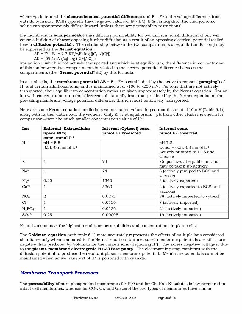
PlantPhysUW425.doc 5/24/2008 23:32 Page 28 of 138
where Δµs is termed the electrochemical potential difference and Ei - Eo is the voltage difference fromoutside to inside. (Cells typically have negative values of Ei - Eo.) If Δµs is negative, the charged ionicsolute can spontaneously diffuse inward (unless there are permeability restrictions).
If a membrane is semipermeable (has differing permeability for two different ions), diffusion of one willcause a buildup of charge opposing further diffusion as a result of an opposing electrical potential (calledhere a diffusion potential). The relationship between the two compartments at equilibrium for ion j maybe expressed as the Nernst equation:
ΔE = Ei - Eo = 2.3(RT/zjF) log ([Cjo]/[Cji])ΔE = (59.1mV)/zj) log ([Cjo]/[Cji])
For an ion j, which is not actively transported and which is at equilibrium, the difference in concentrationof this ion between two compartments is related to the electric potential difference between thecompartments (the “Nernst potential” ΔE) by this formula.
In actual cells, the membrane potential ΔE = Ei - Eo is established by the active transport (“pumping”) ofH+ and certain additional ions, and is maintained at c. -100 to -200 mV. For ions that are not activelytransported, their equilibrium concentration ratios are given approximately by the Nernst equation. For anion with concentration ratio that diverges substantially from that predicted by the Nernst equation at theprevailing membrane voltage potential difference, this ion must be actively transported.
Here are some Nernst equation predictions vs. measured values in pea root tissue at -110 mV (Table 6.1),along with further data about the vacuole. Only K+ is at equilibrium. pH from other studies is shown forcomparison—note the much smaller concentration values of H+:
Ion External (ExtracellularSpace ECS)conc. mmol L-1
Internal (Cytosol) conc.mmol L-1 Predicted
Internal conc.mmol L-1 Observed
H+ pH = 5.53.2E-06 mmol L-1
pH 7.2Conc. = 6.3E-08 mmol L-1
Actively pumped to ECS andvacuole
K+ 1 74 75 (passive, at equilibrium, butmay be taken up actively)
Na+ 1 74 8 (actively pumped to ECS andvacuole)
Mg2+ 0.25 1340 3 (actively exported)Ca2+ 1 5360 2 (actively exported to ECS and
vacuole)NO3- 2 0.0272 28 (actively imported to cytosol)Cl- 1 0.0136 7 (actively imported)H2PO4- 1 0.0136 21 (actively imported)SO42- 0.25 0.00005 19 (actively imported)
K+ and anions have the highest membrane permeabilities and concentrations in plant cells.
The Goldman equation (web topic 6.1) more accurately represents the effects of multiple ions consideredsimultaneously when compared to the Nernst equation, but measured membrane potentials are still morenegative than predicted by Goldman for the various ions (if ignoring H+). The excess negative voltage is dueto the plasma membrane electrogenic H+-ATPase pump. The electrogenic pump combines with thediffusion potential to produce the resultant plasma membrane potential. Membrane potentials cannot bemaintained when active transport of H+ is poisoned with cyanide.
Membrane Transport Processes
The permeability of pure phospholipid membranes for H20 and for Cl-, Na+, K+ solutes is low compared tointact cell membranes, whereas for CO2, O2, and Glycerol the two types of membranes have similar
PlantPhysUW425.doc 5/24/2008 23:32 Page 29 of 138
permeabilities. The differences are due to presence of transport proteins which are relatively specific forcertain solutes, and which are subdivided into:
Channels: Simple diffusion, passive transportCarriers: Facilitated diffusion, passive transport (secondary active transport)Pumps: Primary active transport using energy source such as ATP hydrolysis
ChannelsChannels act like selective pores to enhance diffusion across membranes for specific substances.Transport is limited to ions and water, and is always passive transport. They conduct up to 108 ions ormolecules per second. Pores may be gated in response to signals such as ion concentrations like K+ orCa2+, voltage changes such as depolarization or hyperpolarization, hormones, cyclic nucleotides, light,phosphorylation, etc. Studied by patch clamp electrophysiology (web topic 6.2). An ion species may movethrough different channels with differing gating conditions, allowing fine tuning of passage—the membranepermeability depends on the mix of differing channels.
Cations may move in to cytosol through PM channels—K+, Na+, Ca2+ (however, Ca2+ must be activelypumped back out of the cytosol). K+ can move either direction into or out of the vacuole via channels.Ca2+ can move from vacuole to cytosol via channels. Anions (Malate2-, Cl-, NO3-) move via channelsthrough PM out of cytosol or into vacuole. As previously stated, K+ moves in or out to maintain the Ernstequilibrium potential for potassium Ek equal to the membrane potential ΔE:
ΔE = (59.1mV)/zj) log ([Cjo]/[Cji]) Nernst equationlog ([CKo]/[CKi]) = ΔE / 59.1 for K+
[CKo]/[CKi] = 10^(-110 / 59.1) = 0.014 for measured ΔE = -110 mV[CKi] = 73 mmol L-1 for measured Cko = 1 mmol L-1
Some K+ channels are rectifying in that they open only above or below the Nernst potential for K+ (see fig.6.9) and thus allow movement only in or only out:Inwardly rectifying channels or inward channels open at potentials more negative than the prevailingNernst potential for K+ (so positive charges are attracted inwardly). These are found in guard cells thattake up K+ from the apoplast during stomatal opening.Outwardly rectifying channels or outward channels open at potentials more positive than the prevailingNernst potential for K+ (so positive charges are repelled from the inside). These function in the closing ofthe stomata, and the release of K+ into the xylem or apoplast.
Carriers and PumpsCarrier proteins do not have pores, and are much slower in transporting than channels, typically only 100to 1000 ions or molecules per second. They bind the substance to a specific site, giving them highspecificity. Binding causes a conformational change, releasing the substance to the other side. Kinetics issimilar to enzyme kinetics. Transport through carriers may be:I. Passive Transport (“facilitated diffusion”, somewhat of a misnomer); [example may be the PM’ssucrose efflux carrier] orII. Active TransportActive transport requires coupling (directly or indirectly) the energetically uphill transport of the solute (i.e.,against its electrochemical potential gradient) with an energy-releasing event so that the overall free energychange is negative. When a carrier performs active transport, it may be either:II.A. Primary Active Transport: the transport is coupled directly to a source of energy such as ATPhydrolysis. A carrier that performs primary active transport is called a pump. Most pumps use ATP totransport ions such as H+ - ATPase or Ca2+ - ATPase (H+ is pumped out of plasma membrane or into vacuole,Ca2+ is pumped out of plasma membrane), but some pumps can pump large organic molecules. Pumpsmay be electrogenic (leading to a net movement of charge across the membrane) or electroneutral (suchas the H+/K+-ATPase pump of animal gastric mucosa). Electrogenic pumps include:
In Animals: the Na+/K+-ATPase of animal cellsIn Plants:
the plasma membrane H+-ATPase pump;the vacuolar H+-ATPase (V-ATPase) pump;the vacuolar H+- pyrophosphatase pump

PlantPhysUW425.doc 5/24/2008 23:32 Page 30 of 138
[the vacuolar Anthocyanin-GS - ATPase ABC transporter]the Golgi cisternae H+ - pyrophosphatase pump
II.B. Secondary Active Transport: the uphill transport of a solute is coupled to the downhill transport ofanother. “This type of carrier-mediated cotransport is termed secondary active transport and is drivenindirectly by pumps.” (p. 105) It uses stored energy in the form of protons that have been previouslypumped out of the cytosol, a process which produces a pH gradient relative to the ECS and the vacuole,yielding a proton motive force, the stored free energy in the form of the H+ gradient (available for ATPsynthesis):
Δp (in mV) = ΔE -59(pHi - pHo)This force drives the transport of many other ions against their gradients. Such coupled cotransport maybe eitherIII.B.1 Symport, in which the two substances move in the same direction via a symporter protein (someauthors use “cotransport” as synonymous with symport).45 Examples of Symports:
Plasma Membrane:Fe, Mn, Zn, Cd in [text does not state what moves with these]NO3- - H+
PO43- - H+
H+ - K+
H+ - Na+
Sucrose - H+
Amino Acid - H+
Peptide - H+
Tonoplast: None are shown in fig. 6.14III.B.2 Antiport, in which the two substances move in opposite directions via an antiporter protein (someauthors use “countertransport” as synonymous with symport). Examples of Antiports:
Plasma Membrane:Na+ out - H+ in
Tonoplast (directions are given with respect to the vacuole):H+ in - Na+ out;Ca2+ in - H+ out;Cd2+ in - H+ out;Mg2+ in - H+ out;Hexose in - H+ out;Sucrose in - H+ out;
Kinetic analysis can elucidate transport mechanisms, similar to the analysis with enzyme kinetics. Theconstant Km is equal to the solute concentration that yields half the maximal rate of transport. When Km islow, there is a high affinity of the transport site. Transport often displays evidence of multiple channels ofboth high and low affinity.
The membrane transport proteins and the genes coding for them have in many cases been identified.[Details not reviewed here.] See diagram fig. 6.14 for summary of the various transport processes acrossthe plasma and vacuole membranes. Cation transporters are diverse. Some anion transporters have beenidentified—for NO3-. Cl-, and PO42-. Metals are transported by high affinity ZIP proteins, including Fe, Zn,Mn. Toxic metals such as Cd, As, Zn, Pb, U, Hg, and Se can be taken up by some plants, and this may beuseful for phytoremediation.46 (For more on metals phytoremediation, see web essay 5.1 and chapter 26.)
Water-selective channels, aquaporins, in some cases transport uncharged solutes. They may function assensors of gradients in osmotic potential and turgor pressure. They are regulated by proteinphosphorylation, pH, calcium concentration, heteromerization, and reactive oxygen species. Plant cellscan rapidly alter their water permeability in response to various stresses.
Plasma membrane H+-ATPase has several functional domains...
The tonoplast H+-ATPase (V-ATPase) drives solute accumulation into vacuoles... The vacuole is typically 20- 30 mV more positive than the cytoplasm.
45 Cotransport and countertransport:http://www.lib.mcg.edu/edu/eshuphysio/program/section7/7ch05/7ch05p13.htm46 Phytoremediation: http://en.wikipedia.org/wiki/Phytoremediation
PlantPhysUW425.doc 5/24/2008 23:32 Page 31 of 138
Although the vacuole is usually more acidic than the cytosol, some plant species make the vacuolehyperacidic—e.g., lime pH 1.7, lemons pH 2.5, cherry 2.5, grapefruit 3.0, rhubarb [3.2 petioles], pricklypear in AM 1.4, oxalis sp. 1.5, begonia ‘lucerna’ 0.9, etc.
Ion Transport In Roots
Root ion transport is highly specific and tightly regulated. In the plant, solutes move through both theapoplast (5-20% of the total plant volume) and the symplast via plasmodesmata. Cells that secrete ionssuch as floral nectaries and salt gland have high densities of plasmodesmata. Symplastic flow of ions isprimarily by diffusion through plasmodesmata.
Ion absorption in the root is primarily in the root hair zone. Like water absorbed in a root, ions may moveinitially(1) in the apoplast (eventually crossing, due to the blocking Casparian strip, over to the symplast beforeentering the stele [central part of the root containing the vascular tissue and occasionally a pith]), or(2) immediately enter the symplast across a membrane.
The living xylem parenchyma cell participate in xylem loading—injection of ions and solute into the deadxylem tracheary elements to lower osmotic potential in the root xylem and promote water flow up the plant.
Chapter VII. Photosynthesis Light (Thylakoid) Reactions
The overall result of the photosynthesis [PS] reaction may be summarized in one form as6 CO2 + 6 H2O + light + plant → C6H12O6 + 6 O2
Thus solar energy is used to oxidize water and reduce carbon dioxide to produce carbohydrate (via c. 50intermediate steps). This reaction to form CHO cannot occur to any significant effect without PS, and byoxidizing water it is also the only significant source of atmospheric O2 on Earth.
PS is divided into light (thylakoid) reactions and carbon fixation (stroma) reactions. The latter are alsodirectly regulated by light, and so the term “dark reactions” is considered inaccurate.
Original experiments in PS by Van Helmont in the 1600s, then Engelman (working with Spirogyra), etc.
The light reactions begin with the excitation of chlorophyll by light and culminate in the synthesis of ATP(via the photophosphorylation of ADP) and NADPH (via the reduction of NADP+).
Most PS in higher plants takes place in the chloroplasts of the mesophyll cells of the interior of leaves.The light reactions occur in association with the thylakoid membranes of the chloroplasts, whereas thecarbon reactions occur in the stroma of the chloroplasts.
Light Physics
Light is wavelike (having mutually perpendicular magnetic and electric fields, both of which oscillateperpendicular to the direction of propagation) and is also particle-like. The relationship of light wavelengthto frequency is given by
c = λ ν wherec = speed of light, 3 x 10⁸ m s-1
λ = wavelength (m)ν = frequency (Hz or s-1)
Photons are the particles or wave packets that make up bulk light. The energy of one photon depends onthe frequency:
E = h ν whereE = energy (Joules)ν = frequency (s-1)
PlantPhysUW425.doc 5/24/2008 23:32 Page 32 of 138
h = Planck constant (or Planck’s constant) = 6.626 x 10⁻³⁴ J sVisible light for humans falls between c. 400 - 700 nm. The spectrum of sunlight (TZ fig. 7.3) at the sunsurface and just above our atmosphere has a peak irradiance (measured in W m-2 nm-1) in bluewavelengths of c. 450 - 480 nm. The solar spectrum above our atmosphere is that expected from a 5525 K(5250 °C) blackbody47, which has a theoretical peak by Wien’s law at c. 525 nm.48 Passage through theEarth’s atmosphere attenuates the light a little and alters the spectrum somewhat, mostly by introducingseveral absorption bands, including one at c. 700 nm (far red) arising from water vapor, and moreabsorption bands at longer infrared wavelengths, but the peak does not shift significantly, still being at c.530 nm at the surface of the Earth.
Photosynthetically active light falling on leaves may be quantitated by irradiance W m-2 ofPhotosynthetically Active Radiation PAR (400 nm - 700 nm) or photosynthetic photon (quantum)irradiance in mol m-2 s-1 of PAR [also called somewhat ambiguously photosynthetic photon fluxdensity]. Measures such as lux, candela, and foot-candles are human-oriented and not suitable for plantphysiology. (Web topic 9.1) Spectral irradiance expresses W m-2 nm-1, thus the irradiance measured atdefined points in the spectrum. (See also web topic 7.1 principles of spectrophotometry.)
The photosynthesis experimenter much choose the geometry of the light sensor to most closely match thetype of plant part being studied:• Flat—having uniform irradiance across its surface from unidirectional light, but total is proportional to
the cosine of the angle with respect to perpendicular; or• Spherical or Omnidirectional—having varying irradiance across its curving surface from
unidirectional light but total not varying with angle of incidence. In this case, a fluence rate49 [theirradiance which is incident from all angles] is measured.
On a sunny day in direct (unidirectional) sunlight at sea level, PAR irradiance and PAR fluence are both400 W m-2 or 2,000 μmol m-2 s-1 [These figures are stated in Web topic 9.1. On p. 200 of the textbook, aprobably incorrect figure of 900 W m-2 is given. “Sun” is said to have irradiance of 920 μmol m-2 s-1 in fig.9.10] Completely diffuse light has irradiance equal to 25% of fluence. PAR is “38% (21-46%) of the extra-terrestrial solar irradiance”, “46% to 50% global solar radiation at ground level.”50 The solar constant,which measures total solar radiation arriving at the upper atmosphere of all wavelengths, is c. 1366 W m-2,but of course is not actually constant and varies with the time of the year due to Earth’s orbital position,etc.]
The Quantum yield (quantum efficiency) is variously and rather confusingly defined for photosynthesisas:• “The number of photochemical products” [such as moles of CO2 assimilated or O2 evolved] divided by
the “Total number of quanta absorbed”TZ132, or• “The fraction of excited molecules” [i.e., excited by the absorption of a photon] that decay via a
designated pathway [such as via photochemistry, apparently no matter how small the effect producedper quantum] (web topic 7.3), or
• The number of times that a defined event occurs per photon absorbed by the system.51
Photosynthesis Pigments And Complexes
Free Chlorophyll a (Chla), as extracted with nonpolar diethyl ether, has greatest absorption at blue (c. 430nm) and red (c. 662 nm) wavelengths, thereby having absorption maxima straddling the peak of 525 nmfor solar irradiance, and reflecting green light c. 550 nm. Chlorophyll b (Chlb) similarly extracted has
47 Solar spectral irradiance:• http://en.wikipedia.org/wiki/Sunlight• http://rredc.nrel.gov/solar/spectra/am1.5/ASTMG173/ASTMG173.html48 Wien’s law of black body radiation: http://en.wikipedia.org/wiki/Black_body49 Fluence rate: http://omlc.ogi.edu/classroom/ece532/class1/fluencerate.html50 Solar PAR irradiance at ground level:http://www.apesimulator.it/help/models/solarradiation/Photosynthetically_active_radiation.html51 Quantum yield: http://en.wikipedia.org/wiki/Quantum_yield

PlantPhysUW425.doc 5/24/2008 23:32 Page 33 of 138
greatest absorption at blue (c. 453 nm) and red (c. 642 nm) wavelengths.52 Light excites chlorophyll fromthe ground state to a short-lived metastable excited state designated by Chl*, a state having a potential lifespan of only a few nanoseconds. After absorbing blue light, Chl can give up its energy and drop to lowerenergy states by• Heat loss from the higher excited state to the lowest excited state (with no photon emission)• Fluorescence from the lowest excited state to ground level (radiates a 673 nm photon, in the red
region)• Energy transfer to another molecule• Photochemistry reactions (the redox reactions that are useful in PS—these are extremely fast
reactions)
Certain bacteria including cyanobacteria, plus diatoms, dinoflagellates, brown algae, red algae can alsophotosynthesize and have various chlorophyll combinations including Chl c and d. Sulfur purplebacteria, nonsulfur purple bacteria, green bacteria, and heliobacteria can have various combinations ofbacteriochlorophylls a - g (see web topic 7.2)
Chlorophylls have two major components:• Porphyrin-like ring with a centrally coordinated Mg in the N4 cavity. (The porphyrin is where
excitation initially occurs.)• Phytol tail (which anchors the molecule to a hydrophobic part of the environment)
Carotenoids (e.g., β-carotene) have long linear molecules with multiple conjugated double bonds. Theseare alternating single and double bonds with delocalized electrons (similar to the benzene ring), yieldingchromophores (the part or moiety of a molecule responsible for its color). Carotenoids absorb light in theblue 400-500 nm range and reflect a characteristic orange color.
Joseph Priestly first discovered O2 and found that plants evolved it. Jan Ingenhousz showed that light isessential to photosynthesis in 1779, and others demonstrated the need in PS for H20 and CO2. C. van Nielfound that PS is a redox process.
The action spectrum of PS (e.g., the O2 evolution rate graphed as a function of wavelength) closely relatesto the absorption spectrum of chloroplasts (with the exception that light absorbed by carotenoids in 450 -550 nm, which somewhat widens the blue absorption spectrum compared to Chl, is not as efficientlyconverted via PS—see TZ fig. 7.8).
Engelmann showed that O2-seeking (aerotactic) bacteria were attracted to the segments of Spirogyra spiralchloroplast that were irradiated with blue or red light more than the parts irradiated with green,confirming that PS makes O2 where light is most strongly absorbed (the blue peak is broadened bycarotene).
Photosynthetic complexes consist of:• Light-harvesting antennas, which assist in the absorption of light• Photochemical reaction centers (discussed further below)
In intact plants, absorption of light is assisted in light-harvesting antennas containing• Photosynthetic pigments include chlorophyll a and b (along with c and d in diatoms and algae).• Accessory pigments include and carotenoids (such as β-carotene). In addition, bilin pigments or
phycobiliproteins such as phycoerythrobilin are found in cyanobacteria and red algae but not invascular plants.
• Photoprotective pigments, which protect from excess light and photoinhibition, includinganthocyanin and xanthophylls.
• Light-harvesting complex proteins (LHCI and LHCII), which aid in the efficient transfer of excitationenergy.
52 Chlorophyll absorption spectrum:• http://www.biologie.uni-hamburg.de/b-online/e24/3.htm• http://en.wikipedia.org/wiki/Chlorophyll
PlantPhysUW425.doc 5/24/2008 23:32 Page 34 of 138
The use of antenna structures (comprising hundreds or thousands of Chl and accessory pigmentmolecules) makes for a more favorable allocation of energy, since many pigment molecules are needed todrive a single reaction center. Experiments by Emerson and Arnold with brief light flashes showed that atmaximum PS yield, there are one O2 molecule generated for every c. 2,500 chlorophyll molecules per highintensity flash. This is because(1) several hundred pigment molecules are associated with a single chloroplast reaction center (in plants,200-300 chlorophylls per PSII reaction center, and c. 100 core antenna chlorophylls or 200 overall for PSIcenters); and(2) Each reaction center must operate multiple times to produce just 1 molecule of O2.
The excitation energy in antenna pigments is transferred to the reaction center by fluorescence resonanceenergy transfer, a non-radiative process with up to 95 to 99% energy transfer efficiency.
At much lower flash intensities, Emerson and Arnold found that the quantum yield of chloroplasts was0.95 (versus 0.05 for absorbed photons whose energy is wasted by fluorescence). 1 molecule of O2
molecule was generated for every 9-10 photon absorbed (i.e., not reflected or transmitted). These twovalues are not discordant, since each photon absorbed and yielding a photochemical effect exerts only afractional effect with respect to generating O2, but is fully counted as part of the quantum yield.
The following simplified reaction expresses overall PS:CO2 + H2O + c. 10 hν → (CH2O) + O2where (CH2O) is 1/6 of a glucose molecule
This reaction requires a theoretical minimum free energy change of 467 kJ/mol O2 evolved in synthesis ofglucose, but in practice requires 1760 kJ of absorbed red light in the plant per mol O2 evolved, giving anefficiency for conversion of absorbed light energy to chemical energy as glucose in overall PS = 27%overall. (However, only a small part of this chemical energy goes to formation of biomass; see TZ p. 132and fig. 9.3 and below). The remaining 73% of the energy entering photochemistry is consumed in cellularmaintenance and ultimately ends as heat.
The light reactions as studied first by Robert Hill in 1939 are given by redox reactions with compoundssuch as Fe or Mn:
4 Fe3+ + 2 H20 → 4 Fe2+ + O2 + 4 H+
Fe3+ is the oxidant or oxidizing agent and is reduced to Fe2+
H20 is the ultimate electron donor (reductant or reducing agent) and is oxidized to O2 + 4 H+
(in the sense that water loses hydrogen and O changes oxidation number from -2 to 0).It has been proven that the O2 evolved in PS originates from H2O, not from CO2.
The thylakoid reactions, in addition to oxidation of H20, include the reduction of NADP+ to NADPH andthe phosphorylation of ADP to ATP.
Despite the substantially lower overall absorption of photons in the green c. 550 nm for chloroplasts, thosephotons that are absorbed show a much flatter curve for quantum yield from 400 to 700 nm—there is anabrupt “red drop” at ≥ c. 700 nm. This is because the accessory pigments help to make more efficient useof photons with wavelengths other than at the optimal values for chlorophyll.
Photosystems I And II And The Synthesis of NADPH and ATP
[This is a complex topic not fully summarized here, see also Web Topic 7.8] Photosystems I and II (PSI andPSII) are found in chloroplasts of plants and algae (and also cyanobacteria). The reaction center of PSII(termed P680) has chlorophylls which absorb maximally at red 680 nm, whereas the PSI reaction centerP700 has chlorophylls absorbing maximally at far red > 680 nm. Absorption at both wavelengths isneeded for the most efficient PS. In chloroplasts, these photosystems are spatially separated: PSII and itsantenna pigments are located primarily in the stacked thylakoid membranes (grana), whereas PSI is mainlylocated in the nonstacked stroma lamellae protruding into the chloroplast stroma from the grana lamellae.The proposed chemical sequence is depicted in the “Z scheme” (a term referring to the graphicalappearance of vertical jumps in redox potentials and the sloping electron transport chains).
The P680 center of PSII contains a dimer of “specialized chlorophylls” (not otherwise specified in TZ,though EVV states they are chlorophyll a) along with other pigments, several proteins including D1 and
PlantPhysUW425.doc 5/24/2008 23:32 Page 35 of 138
D2, and other factors. Red light excites the reaction center directly, or more commonly an antennacomplex, which efficiently funnels this excitation energy from carotenoids to Chlb and Chla and finally toP680 in the reaction center. [MCM: The following redox sequence is somewhat confusing, and I may nothave all the details correct.] The P680 is excited to P680*, the latter having a relatively negative redoxpotential Em (and thus is mildly reductant) compared to the ground state. Prior to excitation, P680 hasrelative positive Em, and acts as a strong oxidant (electron acceptor), altering the state of the oxygenevolving complex (OEC), which ultimately leads to the oxidization of water (at the inner thylakoidmembrane): 2 H20 → 2 O2 + 4 H+ + 4 e-. The products O2 and H+ are released into the thylakoid lumen, thelatter creating a high H+ concentration and resulting high proton motive force (an electrochemicalpotential gradient, representing stored chemical energy) across the thylakoid membrane. (Oxidation ofwater at the OEC also requires presence of Mn2+ ions and a series of oxidations called S states probablyinvolving a manganese containing enzyme—the mechanisms are not well understood.) The electrons enterthe non-cyclic electron transport chain extending from P680* to P700.
The relatively negative redox potential Em of the excited P680* state indicates that it can serve as a weakreductant (electron donor). P680* can transfer an electron to the first electron acceptor in a transportchain, pheophytin—this transfer is the first event in PS where light excitation energy has been convertedto chemical energy. An electron transport chain including plastoquinones and the cytochrome b6fcomplex carries electrons along states of increasingly more positive Em to plastocyanin (PC) and ending inthe P700 center of PSI.
The P700 center is excited by photons absorbed at far red 700 nm. P700 is a weak oxidant, but P700* is astrong reductant (electron donor, with very negative Em). P700* passes electrons (received at P700 fromPSII) down a series of membrane-bound iron-sulfur proteins (again, down states of increasingly morepositive Em) to soluble ferredoxin (Fd), which is acted on by ferredoxin-NADP-reductase (FNR) to reducethe ultimate electron acceptor, NADP+, to NADPH in the stroma. The PSI reaction center is a largemultisubunit complex with (in addition to P700 and chlorophylls) has several proteins, as well asphylloquinone (Vitamin K1). Reduced ferredoxin is also used by the plant to reduce nitrate.
Chloroplast ATP synthase (also called CFO-CF1 or CFO-CF1 ATP Synthase, resembling mitochondrial FO-F1 ATP synthase, see Chap. 11) is located in the chloroplast stroma lamellae. It couples the passage of H+
ions down their electrochemical gradient (from the thylakoid lumen to the stroma) with thephosphorylation of ADP via Pi to ATP in the chloroplast stroma (a process termedphotophosphorylation). This ATP (along with the NADPH) will be used to drive the carbon reactions ofPS. The coupling between the electrochemical potentials from proton pumping—the proton motive forcearising from the chemical pH gradient and the membrane electric potential—and the performance of work,as with CFO-CF1 ATP synthase, is termed chemiosmosis by Peter Mitchell (web topic 6.3; see also webtopic 7.9 on ATP Synthase).
In some cases, “cyclic electron flow” in the chloroplast can be utilized to generate ATP withoutcorresponding reduction of NADP+ to NADPH. In other cases, the electron flow in the chloroplast mayoccur without corresponding photophosphorylation of ADP (an uncouplingTZ148 apparently analogous tothe uncoupling of oxidative phosphorylation from electron transport that can occur in mitochondria—seeTZ269 and Web topic 11.5).
The herbicides paraquat and DCMU act by blocking electron transport of PSII or PSI (see web topic 7.10).
Protection And Regulation of Photosynthetic Machinery
[Brief summary] Toxic photoproducts can form in excess light conditions, including triplet state of Chl(3Chl*) and reactive oxygen species such as the superoxide anion (O2•-), singlet oxygen (1O2*), hydrogenperoxide (H2O2), and hydroxyl radical (•OH). (“Singlet oxygen is the common name used for the twometastable states of molecular oxygen O2 with higher energy than the ground state triplet oxygen.”)53
Singlet oxygen can damage many cellular components including lipids. The PSII reaction center is easilydamaged by excess light, especially the D1 core protein. Carotenoids, superoxide dismutase, and ascorbateserve as photoprotective agents, helping to prevent photoinhibition (a reduction in a plant's capacity forPS caused by exposure to strong light, which may be reversible or irreversible) and damaging effects of 53 Singlet oxygen: http://en.wikipedia.org/wiki/Singlet_oxygen
PlantPhysUW425.doc 5/24/2008 23:32 Page 36 of 138
excess light. Carotenoids can quench the excess energy of singlet oxygen by converting it back to tripletoxygen releasing heat. Non-photochemical quenching of excess energy (conversion to heat withoutinducing photochemistry) can be done by xanthophylls. These are yellow pigments that are oxidizedcarotenoid derivatives (listed in order of least to greatest protectiveness: violaxanthin < antheraxanthin <zeaxanthin). The least protective in the xanthophyll cycle, violaxanthin, converts to the most protective,zeaxanthin, when light is intense and protection is needed.
Thylakoid stacking permits energy partitioning between the photosystems, allowing the most efficient useof the available energy...
Chloroplasts can reposition themselves along the side walls of cells, so that the more superficial onesprovide shade to deeper chloroplasts along the same wall, in response to excessively intense light (TZ fig.9.4).
Chloroplasts sometimes extend stromules, fine tubular interconnections with nearby chloroplasts andplastids that allow transfer of proteins etc., but the ultimate purpose is unknown.54
Genetics Of The Photosynthetic Systems
[Brief summary] Chloroplasts derive from endosymbiosis of cyanobacteria. Chloroplast DNA is circular,and codes for proteins synthesized in the chloroplast. But much of the DNA needed to synthesize thechloroplast photosynthetic apparatus has been transferred to the parent cell nucleus. Proteinssynthesized from such nuclear DNA are synthesized in the cytoplasm and imported into the chloroplasts.Chloroplasts reproduce by division, not de novo synthesis, and they can no longer survive outside theireukaryotic host cell. Only the maternal plant contributes chloroplasts to the zygote, and therefore theinheritance of chloroplast intrinsic DNA in not Mendelian.
Some of the precursors of Chla are glutamic acid, 5-aminolevulinic acid (ALA), porphobilinogen (PBG),protoporphyrin IX, etc.
Chapter VIII. Photosynthesis Carbon (Stromal) Reactions
The carbon PS reactions fix carbon to biomass from inorganic CO2, about “200 billion tons of CO2 eachyear”TZ159 (elsewhere estimated at 60 Giga metric tons Carbon per year), 40% of which is performed bymarine phytoplankton. (Much of this fixed carbon is returned to the atmosphere through aerobicrespiration, decomposition, methanogenesis, etc.) In plants, these reactions occur in the chloroplaststroma.
The Calvin C3 Cycle
This cycle is the core of PS as found in almost all autotrophic photosynthetic (i.e., photoautotrophic)organisms. (Some photosynthetic prokaryotes such as green sulfur bacteria and Chloroflexi do not use theCalvin cycle, while some chemoautotrophic bacteria or Archaea such as methanogens and halophiles donot use PS at all.) It transforms CO2 by reducing (“fixing”) the C in CO2 (which is in the most heavilyoxidized state for C, oxidation number 4, thus C+4O2-2 ) to C0 (oxidation number 0) as found in covalentlylinked organic compounds. [MCM: absolute oxidation numbers or oxidation states for C in organiccompounds seem to be controversial, complex, or unknowable, but “only the changes in oxidation stateduring a reaction are important.”]55
The Calvin cycle was elucidated by in the 1950s by Melvin Calvin, James Bassham, Andrew Benson, andcolleagues at the University of California, Berkeley. Using 2D chromatograms and autoradiography, theywere able to show that 3-phosphoglycerate is the first stable intermediate formed (Web Topic 8.1).
54 Stromules: http://jxb.oxfordjournals.org/cgi/reprint/56/413/78755 Organic carbon oxidation states: http://pages.towson.edu/ladon/orgrxs/reagent/redox.htm
PlantPhysUW425.doc 5/24/2008 23:32 Page 37 of 138
This cycle has 3 phases [Limited summary, abbreviations for molecules are my own]:
(1) Carboxylation of R15B to PGA: The starting 5-carbon CO2 acceptor is Ribulose-1-5-biphosphate[R15B], which is carboxylated with CO2 and H20 by Ribulose-1,5-biphosphate carboxylase-oxygenase(“rubisco”) to an unstable 6-carbon intermediate, which rapidly decays to 2 molecules of 3-phosphoglycerate [PGA], the first stable intermediate of the Calvin cycle. (The C in PGA has been reducedto oxidation number +3 per TZ.) (See Web Topics 8.2 and 8.3 on Rubisco)
(2) Reduction of PGA to GA3P: PGA is reduced using with ATP, NADPH, and H+ culminating inGlyceraldehyde-3-phosphate [GA3P], a triose phosphate carbohydrate, plus ADP, Pi, and NADP+. (TheC in GA3P has been further reduced to oxidation number +1 per TZ.)
(3) Regeneration of R15B: Ribulose-1-5-biphosphate is regenerated in a complex set of reactions,consuming 5 out of 6 of the GA3Ps that were formed.
The net stoichiometry of the C3 Calvin cycle reactions (including the regeneration of R15B) may besummarized as follows:
3 CO2 + 5 H20 + 6 NADPH + 9 ATP → G3P + 6 NADP+ + 3 H+ + 9 ADP + 8 Pi
where the NADPH and ATP come from the light thylakoid reactions.
The Calvin cycle intermediates must first be generated before the Calvin cycle can fully resume after aperiod of darkness—the delay required is called the induction period. Once the intermediates have builtup, the Calvin cycle can begin generating net reserves of carbohydrates (ultimately, starch or sucrose, etc.)
Energy Conversion Efficiency Of Photosynthesis Using The Calvin Cycle
The free energy released by the complete oxidation of a hexose sugar to CO2 and H2O is 2804 kJ mol-1—thus the absolute minimum energy to synthesize one mole of fructose must be 2804 kJ. In the plant,synthesis of 1 mole of fructose-6-phosphate from 6 moles of CO2 takes 3126 kJ from oxidizing the neededNADPH and hydrolyzing the needed ATP. Thereforethe thermodynamic efficiency for conversion of the chemical energy in NADPH and ATP to hexoseenergy via the Calvin Cycle is 90%. 83% of the energy required comes from the reductant NADPH.
Incident red light photons at 680 nm contain energy of c. 175 kJ per quantum mole, computed as follows: E = hc/λ
= (6.626 x 10⁻³⁴ J s) x (3 x 10⁸ m s-1) x (109 nm/m) x (6.023 x 10²³ mol-1) / 680 nm= 176 kJ mol-1
At least 8 absorbed photons are needed to reduce 1 mole of CO2 to hexose. Thus, the minimum lightenergy required to reduce 6 moles of CO2 to one mole of hexose is 6 x 8 x 175 = 8400 kJ. Therefore, theoverall thermodynamic efficiency of PS for the conversion of absorbed photon energy to hexose is2804/8400 = 33%. [This calculation from TZ p. 165 appears to consider only the photons that areabsorbed and used for photochemistry, not those that are reflected or transmitted.] Under normal growingconditions, plants are much less efficient: crops such as potatoes, corn, rice, etc. typically yield only 0.1 to0.4% conversion efficiencies, while sugarcane approaches 2%. (See also estimates in Chap. IX)
Regulation Of The Calvin C3 Cycle
[Limited summary] Light regulates the Calvin cycle, modulating the activity of target stromal Calvin cycleenzymes including rubisco (which increases in light), fructuse-1,6-bisphosphate phosphatase, etc.TZ165 Theferredoxin-thioredoxin system contributes to this regulation. Movement of ions such as H+ and Mg2+ alsomodify the activity of key regulatory enzymes.
PlantPhysUW425.doc 5/24/2008 23:32 Page 38 of 138
The C2 Oxidative Photosynthetic Carbon Cycle (Photorespiration)
[Limited summary] Rubisco can catalyze either a carboxylase reaction of R15B using CO2 (the desired entrypathway of the Calvin cycle), or it can cause oxygenation of R15B using O2, leading to the formation of a 2-carbon product, 2-phosphoglycolate [2PG] and a loss of efficiency of PS. This product is not useful for PS,and appears to be, in most cases, a waste product which must be scavenged and reconverted to PGA in theenergy-consuming C2 process called photorespiration. This takes place in a series of steps starting withconversion to glycolate and involving additional organelles, the peroxisome and mitochondrion. Ultimately,2 moles of 2PG are converted to one of 3-carbon PGA and one of CO2, consuming 3 moles of O2 and 2moles of ATP as well as 2 moles of reducing equivalents (2 Fdred + 2 H+)...
CO2 is relatively less soluble in water at higher temperatures than is O2 (web topic 8.6), so the ratio ofCO2/O2 decreases with rising temperature. In the plant, both increased temperature and lower CO2/O2ratio increase the relative rate of oxidation over carboxylation (and thus lowers the efficiency with whichCO2 is utilized). Water stress also increases the rate of C2 photorespiration.
A possible useful role for C2 photorespiration is still under investigation. It may help to protect plants fromphoto-oxidation and photoinhibition when illumination is high and CO2 low, and it may also assist withnitrate assimilation.
Mechanisms For Suppressing Photorespiration Or Preventing Photo-oxidation
Suppressing Photorespiration By Concentrating CO2 and HCO3-
Some aquatic organisms such as cyanobacteria and algae can accumulate HCO3- or concentrate CO2 nearrubisco, suppressing photorespiration and favoring carboxylation of R15P.
The C4 Carbon Cycle-Plants
[Limited summary] This cycle is superimposed on the Calvin Cycle, and is found in monocots and dicots,particularly in sugarcane, corn (maize), millet, sorghum, sedges, switchgrass, and other hot weatherplants. It is most common in sedges and grasses, and is found less commonly in shrubs and herbs, and inonly one tree species. (Se web topic 9.3 on geographic distributions, and topic 8.7 on three variations of c4metabolism). It was originally discovered by Kortschack and Karpilov, and further elucidated by Hatch andSlack. It separates initial CO2 processing from the Calvin cycle, allowing CO2 to be internally concentratedat the site of rubisco, suppressing photorespiration.
Leaf mesophyll cells receive the atmospheric CO2, convert it to HCO3-, and use HCO3- to carboxylatephosphoenolpyruvate (a C3 compound) using phosphoenolpyruvate carboxylase (PEPcase). This leadsto the formation of the first stable intermediates malate- or aspartate-, both of which are 4 carbon acidcompounds (thus the name C4). One or the other of these C4 acids pass through a “diffusion barrier” (doesit prevent back-migration of CO2 from the bundle sheath cells?) to the mitochondrion of the bundle sheathcells (found in the vascular region of the leaf). An NAD-dependent malic enzyme NAD-ME (or other variantenzymes, NADP-ME, etc.TZ176) decarboxylates the malate or aspartate, releasing concentrated CO2 that ispresented to the nearby chloroplast for Calvin cycle processing into carbohydrates. Pyruvate or alanine(both are C3 compounds) is returned to the mesophyll cell. Only the bundle sheath cells have thedecarboxylases for regenerating CO2 and the enzymes of the Calvin cycle. In many (but not all) C4 plants,a ring of mesophyll cells surrounds the large bundle sheath cell in an arrangement termed Kranzanatomy—this arrangement that ensures proper compartmentalization and contiguity of the twoprocesses. The chloroplasts of the mesophyll and bundle sheath cells differ in several ways, including agreater concentration of grana in the former...
Some plants employ C4 PS implemented within single chlorenchyma (chloroplast-containing parenchymal)cells. Diatoms are unicellular protists that also exhibit a C4 pathway. This provides them with more than

PlantPhysUW425.doc 5/24/2008 23:32 Page 39 of 138
one CO2 concentrating mechanism, so that they are capable of responding to varying environmentalconditions.
The C4 cycle, which is superimposed on C3, requires greater energy than use of only C3, specifically anadditional 2 ATP per CO2 concentrated. However, PEPcase has a high affinity for HCO3- and thus indirectlyfor CO2, and this affinity is not competed for by O2 (such a competition is found with rubisco). The higherefficiency of utilizing HCO3- and thus of CO2 allows C4 plants to reduce stomatal aperture at hightemperatures while fixing CO2. In general, therefore,• C4 plants in high CO2/O2 ratio environments and/or in lower temperature non-water-stressed
conditions are less efficient at PS than C3 plants, because they need more light quanta per CO2 used.• C4 plants in lower CO2/O2 ratio environments and/or at higher temperatures and/or water stress
conditions have more efficient PS and greater water-stress tolerance due to reduced photorespirationand more efficient use of CO2 relative to water loss.
(These relationships are seen in part in TZ figures 9.9, 9.17, and 9.18, see also Chap 9 discussion.)
Light is necessary for the regulation of the C4 enzymes...
Crassulacean Acid Metabolism (CAM)
This is a third mechanism used for concentrating CO2 near rubisco for the Calvin cycle, as well as forpreventing photo-oxidation (photorespiration). It is employed by xerophytic plants living in arid hotenvironments with seasonal water availability. Examples include pineapple, agave, bromeliads, cacti andother succulents, and orchids. These plants tend to have desert adaptations such as low surface-to-volume ratios, thick cuticles, large vacuoles, reduced stomatal size, leaf hairs, etc. It is named after themember (Bryophyllum calycinum) of the Crassulaceae56 succulent family (a family which includes jadeplants and sedums) in which it was first studied, as well as the acidity of the leaves that forms at night.
[Limited summary] In CAM plants:
• Night: the stomata of single leaf cells open at night when water loss is minimal, and CO2 enters thecytosol. CO2 converts to HCO3- in the cytosol, which is combined with phosphoenolpyruvate (derivedfrom chloroplast-stored starch) via PEPcase to form oxaloacetate. Oxaloacetate is ultimatelyconverted to malic acid (a C4 compound), which is typically stored in the vacuole. The accumulation ofmalic acid leads to a nocturnal acidification of the leaf, thus the “acid” part of the name in CAM.
• Day: During the day, the stomata close, further entry of external CO2 stops, and water loss isminimized. The malic acid stored in the vacuole is released to the cytosol, is decarboxylated withNADP+-dependent malic enzyme, etc. This releases localized CO2 into the chloroplast near rubisco,where it enters the Calvin cycle, as well as pyruvate (which is converted to starch stored in thechloroplast). Because their stomata are closed in the daytime, CAM plants lose only “50 to 100 gram ofwater for every gram of CO2 gained” (mostly at night), compared to 250 to 300 g water for C4 plants and400 - 500 g water for C3 plants.TZ180 The internal O2 concentration rises while the stomata are closed,and is processed by photorespiration. Here, photorespiration is thought to be a beneficial response inthat it reduces oxygen concentration and therefore oxygen toxicity (photo-oxidation), and generatesadditional CO2. (see Web Topic 8.8).
Synthesis of Starch and Sucrose
For sugar chemistry, see also “Sugar Chemistry Review” above.
Sucrose is the nonreducing disaccharide glucose + fructose, also written as α-D-glucopyranosyl-(1↔2)-β-D-fructofuranoside. Both constituent sugars are 6-carbon hexoses, but fructose appears in a furanose 5-member ring form whereas the glucose is in the form of a 6-member glucopyranose ring. In plants,sucrose is synthesized in the cytosol of the leaf mesophyll cells. Sucrose is exported into the phloem. It isthe main form of photoassimilated carbon (“photoassimilate” or “photosynthate”) that is exported to thephloem, but other sucrose-galactosyl oligosaccharides are also formed from PS—raffinose (3 sugars),
56 Crassulaceae: http://en.wikipedia.org/wiki/Crassulaceae
PlantPhysUW425.doc 5/24/2008 23:32 Page 40 of 138
stachyose (4 sugars), verbascose (5 sugars)—as well as sugar alcohols such as sorbitol and mannitol, andother sugar compounds.
[Limited summary] Starch is a mixture of mostly linear amylose (glucan polymers with α-D-1,4 glycosidicbonds) and branched amylopectin (glucan polymers like amylose but with branches formed by α-D-1,6glycosidic bonds). Amylopectin is firmer, more waxy, and ? less readily digested compared to amylose.Starch accumulates as dense granules in the chloroplasts of the leaf cell during the day (“transitorystarch” storage when photosynthesis is maximal), and is mobilized to hexose phosphates at night (whenphotosynthesis has ceased)—this tends to smooth out the diurnal variation in levels of phloem sugars.Some plants also store sugars as fructans (polysaccharide polymers of fructose, such as the linear forminulin), which can for example be found in onion bulbs. Amylose starch is assembled by adding units ofADP-glucose to (α-D-1,4 glucosyl)n polymer chains, using starch synthase... Amylopectin synthesisrequires the starch-branching enzyme and other enzymes. Starch degradation requires phosphorylation,debranching enzyme, β-Amylases and α-amylases (both of these act on starch α-1,4 glycosidic bonds),etc...
The Calvin cycle C3 products synthesized in the chloroplast—GA3P and another triose phosphatedihydroxyacetone-phosphate—are readily interconvertible. They are converted in the cytosol to fructose1,6 bisphosphate, and thence to 3 hexose monophosphates (fructose-6-phosphate, glucose-6-phosphate, and glucose-1-phosphate). Fructose 2,6 bisphosphate is an important regulatorycompound affecting the pool of hexose monophosphates and the synthesis of sucrose: increased levels ofFructose 2,6 bisphosphate inhibits sucrose formation during the daytime but not at night. Fructose-6-phosphate combines with UDP-glucose to form Sucrose-6F-phosphate, which is converted to sucrose inthe cytosol. The formation of sucrose-6F-phosphate is catalyzed by sucrose-6F-phosphate synthetase,and is modulated by glucose-6-phosphate: increased glucose-6-phosphate increases sucrose-6F-phosphateand therefore sucrose synthesis. The final dephosphorylation of sucrose-6F-phosphate to sucrose, which iscatalyzed by sucrose phosphate synthetase-phosphatase, is decreased by increased Pi, therebydecreasing sucrose synthesis.
PS is most active in mature leaves. Photosynthate, mostly sucrose, is transported in the phloem tomeristems and developing leaves and other organs.
Chapter IX. Photosynthesis Physiology And Ecology
This chapter deals with current (and fossil) photosynthetic responses of intact plants to environmentfactors such as light, temperature, CO2, moisture, etc., and their ultimate effects on plant productivity andcrop yields. For a given set of conditions, what are the limiting factors in PS?—is it light or CO2 limited?[In fact, several other factors contribute to the PS rate, including temperature and O2.]
The supply of CO2 to the leaf is regulated by the stomata. (Chap 4 etc.)
The measurement of light is summarized in Chap. 7.
Adaptations Of Plants To Bright Light And Shade
Bright Light AdaptationsSun-exposed plant leaves tend to grow thicker than shaded leaves of the same plant. Desert plants, toprevent harm by excess light (and dessication), develop various defense including hairs, salt glands,epicuticular wax, all of which increase reflection of light from the leaf surface and reduce absorption oflight by up to 40%. Some plants utilize paraheliotropic tracking to turn away from direct sun and therebyreduce leaf exposure to light.

PlantPhysUW425.doc 5/24/2008 23:32 Page 41 of 138
Shade Adaptations and Light Concentrating MechanismsColumnar superficial palisade cells allow efficient capturing of light despite the sieve effect (which arisesfrom gaps between chloroplasts that reduce absorption compared to chlorophyll in solution). Lightchanneling may also divert light through the vacuole or the cell wall areas to facilitate transmission intodeeper layers of the leaf. The spongy mesophyll in the leaf interior has many reflecting interfaces whichcause light scattering and increase the probability for light absorption. Leaves of plants living in theunderstory in dim light may have focusing mechanisms (convex epidermal cells) that focus the lightonto the chloroplasts. Trees have elaborate branching structures of leaves, which tend to maximize overalllight absorption by the plant. Plants competing for light in the understory may receive up to 50% of theirlight as sunflecks—transient sun exposures—and can often rapidly ramp up PS and stomatal openingduring these brief events. Light reaching the understory is least depleted in the far red wavelengths (TZ fig.9.5) compared to blue or red etc. Many plants alter their leaf angles to track the position of the sun (solartracking), including alfalfa, cotton, soybean, lavatera, and lupine. This is a blue light response (seeChapter 18), which is often controlled at the pulvinus found at the junction of the blade and the petiole,and such leaves are called diaheliotropic. Such plants are often competing in short growing seasons.Plants in deep shade often acclimate, but the plant (or at least the shade-adapted leaves) may not be ableto survive if the habitat becomes sunny, due to photoinhibition.
Here are general properties and tendencies of shade leaves compared to sun leaves:• have more total chlorophyll per reaction center• tend to be thinner, with thinner palisades• have less rubisco and less xanthophyll (which is photoprotective)• have higher ratio of PSI to PSII (3:1 compared to 2:1), or have more antenna chlorophyll in PSII. These
adaptations “enhance light absorption and energy transfer” to make better use of the relatively moreabundant far red light. [MCM: I am not sure how this is accomplished.]
• have lower rates of respiration (“dark respiration”) and lower Light Compensation Point (see below)• have lower maximum PS rates (saturation) that sun plants (see below)
Quantitation Of Photosynthetic Responses To Light, Temperature, And OtherFactors
Of 100% incident solar energy arriving at the leaf , 60% is non-PAR photons, 8% is reflected ortransmitted, 8% is dissipated as heat, 19% is consumed in metabolism, and the remaining 5% of incidentenergy is utilized for the production of CHO. [MCM: However, the efficiency of utilization of this photonenergy may be much lower, see estimates in Chap. 8.] 85-90% of PAR incident on the leaf is absorbed,strongest in blue and red (fig. 9.4) and lowest in green.
Light response curves typically plot PS O2 production rate or CO2 assimilation rate (measured in μmol ofO2 or CO2 m-2 s-1) against absorbed light or PAR irradiance, etc.
Dark Respiration Regime, Light Compensation Point, Light-Limited Regime, And CO2 Limited Regime
In C3 plants, below a certain value of absorbed light (the Light Compensation Point), the plant evolvesmore CO2 from mitochondrial respiration than it assimilates and fixes by PS (i.e., CO2 assimilation isnegative). Above the Light Compensation Point, the plant exhibits net positive assimilation of CO2, takingin more and converting it through PS to photosynthate than it gives off. Above but near the LightCompensation Point, increases in light substantially increase CO2 assimilated, a regime termed “light-limited”. Eventually the curve levels off with increasing light and becomes “CO2 limited”. (fig. 9.7) Sunplants have Light Compensation Points typically of 10 - 20 μmol m-2 s-1 CO2 assimilation, whereas shadeplants have lower, c. 1 - 5 μmol m-2 s-1. (Shade plants have low rates of respiration and make moreefficient use of light for PS.) For increased light levels, shade plants have lower maximum PS rates thatsun plants.
Effect On Quantum Yields By O2, CO2, Temperature, And Light Levels

PlantPhysUW425.doc 5/24/2008 23:32 Page 42 of 138
Under current atmospheric conditions with 380 ppm CO2, the quantum yield of C3 and C4 leaves aresimilar at about 0.04 to 0.06 mole of CO2 assimilated per mole of photons absorbed. The reduction fromthe theoretical quantum yield of c. 0.125 is due to losses from photorespiration (in C3 plants) or to energyloss from CO2 concentrating mechanisms (in C4 plants). (See web topic 9.5 on Prehistoric changes inatmospheric CO2)
Effect of O2, Temp, and CO2 may be summarized:• Oxygen: C3 plants in low O2 environments have lower photorespiration and higher photosynthetic
quantum yield, whereas C4 plant do not improve in lower O2. (see Chap. 8 regarding CO2/O2 ratios)• Temperature: C3 plants in lower temperature environments have higher quantum yield than C4 plants,
whereas at higher temperatures, C4 plants have higher quantum yield, although these differences aremodest over moderate temperature ranges. (fig. 9.9) The quantum yield of C4 plants in almost constantover temperature range of 10 - 40 ºC
• CO2: C4 plants in lower CO2/O2 ratio environments have more efficient PS, whereas the opposite is truein higher CO2/O2 ratio environments. (see Chap. 8)
Sun and shade plants “show very similar quantum yields”TZ205 [for quantum yields expressed in terms ofmol CO2 fixation per absorbed quantum]. However, the maximal PS CO2 assimilation of sun plants atsaturation (example: c. 35 μmol CO2 m-2 s-1) is substantially higher than for shade plants (example: c. 5μmol CO2 m-2 s-1, see fig. 9.10). [MCM: this is apparently not contradictory, since quantum yield at leastby some definitions does not quantitate how much effect each of the absorbed photons utilized has, seeearlier discussion of definitions of quantum yield.] This indicates that earlier growing conditions affect howmuch capacity for PS the leaf develops. Above saturation, the leaf is said to be “CO2 limited”—the Calvincycle processes cannot keep up with the absorbed light energy producing ATP and NADPH.
Mechanisms of Protection Against Excess Light
Most leaves can utilize only as many as 500 - 1000 μmol m-2 s-1 of photons out of full sunlight’s maximal2,000. Most plant leaves are partially shaded so a plant as a whole is rarely saturated with light.Although individual needles or leaves may be saturated, whole trees and the forest canopy as a whole israrely saturated
Leaves must dissipate excess light energy (to prevent photoinhibition), often as heat. An importantmechanism is the use of the xanthophyll cycle (see Chapter VII comments), which employs violaxanthin,antheraxanthin, and zeaxanthin. Zeaxanthin is the most effective at dissipating heat, and, along withantheraxanthin, rises in concentration as sunlight becomes more intense, while violaxanthin declinescorrespondingly. This cycle of waxing and waning concentrations is diurnal in summer, but in conifers inwinter, zeaxanthin stays high all day apparently to prevent photo-oxidation. Xanthophyll may also protectchloroplasts against the effects of excess heat.
Chloroplasts also protect themselves against excess light by shifting in distribution within cells, moving tothe cytoplasmic margins and thereby increasing their overlap. This phenomenon is seen in algae, mosses,and higher plants, and is a blue-light response related to phytochrome and using actin microfilaments.
Leaf orientation, overlap, and wilting also help plants to regulate excess light and heat by reducingincident heat and light load.
Too much light can lead to photoinhibition.TZ208 This may take the form of• Dynamic photoinhibition—a reversible effect from moderately excess light in which PS efficiency
decreases (slope of light-response curve) but the maximum PS rate is not significantly changed. Someof these changes are photoprotective, and can occur normally at midday and at colder temperatures.
• Chronic photoinhibition—irreversible reduction of maximum PS rate (O2 evolved per quantum mol)from excess light.
Cumulative effects of recurring photoinhibition over a growing season can reduce crop yield.
Effects of Temperature And Heat On PS
PlantPhysUW425.doc 5/24/2008 23:32 Page 43 of 138
Leaves must dissipate large amounts of heat, but are aided in this by absorbing only about 50% of theincident solar energy in the 300 - 3000 nm range (most of this absorption is in the visible spectrum).Absorbed energy is dissipated by• Re-radiation as long wavelength infrared (typically c. 10,000 nm)• Sensible heat loss by air circulation, convection, and conduction• Latent heat loss as evaporation (evapo-transpiration)The Bowen ratio (web topic 9.2) is the ratio of sensible heat loss to evaporative heat loss. It is higher indesert plants with little water loss, and lower in tropical rain forests and well-watered crops with highevapotranspiration.
PS is sensitive to temperature. At normal “ambient” CO2 concentrations, C3 plants have a lower PS versustemp curve than C4 plants (see fig. 9.17), and show peak PS rate μmol [?CO2] m-2 s-1 (optimal temperatureresponse) at lower temperatures. (In other words, C4 plants in ambient CO2 conditions and at highertemperatures are more efficient at PS than C3 plants.) But at high CO2, the curves of PS rate versustemperature are almost identical. (See also Chapter 7 comments) The declines of PS at highertemperatures are due mainly to instability of membrane bound electron transport, not to photo-oxidation.Different plants have different adaptations and optimal temperature response—some are able tophotosynthesize at 0 ºC and others as high as 50 ºC.
When considered as a function of latitude, C3 grasses in savanna and steppe ecosystems are predicted tobe more productive (and in fact are found to be more common) in higher latitudes above 45 degrees (wherelower temperatures prevail and adequate water supplies are perhaps more likely). C4 grasses are predictedto be more productive (and are found to be more common) in semi-arid latitudes from 20 to 40 degrees andwhich have warm wet summers. [Latitudes closer to the equator are not included in this analysis becausetropical forests are more common, and would shade C4 grasses.] (See TZ fig. 9.18 and web topic 9.3 forfurther details). In modern agriculture, C4 plants such as corn, sugarcane, and sorghum are being grownoutside their customary geographic ranges.
Effects of CO2 on PS and Evolution of C4 versus C3 Plants
Atmospheric CO2 has evolved over the millennia, and is currently at c. 380 ppm, or a partial pressure of 38Pa. These values are twice the value prevailing during most of the past 420,000 years, probably higherthan any time since 2 million years. Thus, recently evolved plants developed in low CO2 conditions.Concentrations > 1000 ppm have not existed since the Cretaceous > 70 Ma. Rising CO2 contributes to anincreasing greenhouse effect. Current C3 PS is CO2 limited, and C3 plant growth would have been fasterwhen CO2 was c. 600 ppm.
Diffusion through the stomata and on to the chloroplasts can be limited at several “points of resistance”.Keeping the stomata less open minimizes water loss but raises stomatal resistance, decreasing CO2
uptake, as previously discussed—stomatal resistance therefore is often the single greatest point ofresistance. Leaves in still air develop low CO2 and higher H20 in the boundary layer (Chap 4), impairingentry of CO2 into the leaf. Stirring the leaves and the boundary air (as with quaking aspen leaves) helps tomaintain a higher CO2 gradient in the boundary layer, promoting greater entry into the leaf. Desert leafadaptations tend to produce thicker boundary layers. Resistance to CO2 diffusion within the leaf to thechloroplast is relatively low, and this diffusion is facilitated by the generally peripheral position ofchloroplasts within the mesophyll cells. PS within a leaf cross-section is maximal in its midportion wherethe mesophyll cells are concentrated, and is lower closer to the dorsal surface (light side where palisadecells dominate) and also lower closer to the ventral surface (undersurface or dark stomatal side). This bell-shaped distribution is similar to the concentration of chlorophyll in the leaf cross-section.
The current relatively low concentration of CO2 limits PS, and higher levels would increase yields of lettuce,tomatoes, cucumbers, etc. In C3 plants, below a certain partial pressure of intercellular CO2,(the CO2Compensation Point), the plant evolves more CO2 from mitochondrial respiration than it assimilates andfixes by PS (i.e., CO2 assimilation is negative). The CO2 Compensation Point is higher for C3 (c. 10 Pa) thanfor C4 plants (c. 0 Pa) because C4 plants use CO2 more efficiently in low CO2 conditions.
As CO2 increases, CO2 assimilation remains higher for C4 than C3 plants (for C3, it is rubisco carboxylationcapacity limited). However, C4 PS attains a maximum at a low CO2 partial pressure of c. 15 Pa. At higherCO2 partial pressure, a crossover point is reached (c. 50 Pa CO2) where C3 CO2 assimilation becomes
PlantPhysUW425.doc 5/24/2008 23:32 Page 44 of 138
greater than that of C4. (fig. 9.22) C3 plants would benefit by rising CO2 levels (if temperatures and wateravailability were unchanged). Most plants (70% of current “productivity”) are C3, and probably evolved longago during times of higher CO2 concentration. In contrast, C4 plants evolved more recently, perhaps 10 -15 million years ago, in part to adapt to falling ambient CO2, such as during the glacial periods (when CO2
was below 200 ppm), probably however in the warmer areas of the Earth during these times (see alsobelow).
C4 plants can use CO2 and N more efficiently, and require less N to grow, but C3 are better shade adapted.
Summary of PS As A Function Of CO2 Versus Temperature
(See fig. 9.23 and web topic 9.8)Lower temperatures and higher CO2 conditions (and shade) favor C3 plants.Higher temperatures and lower CO2 conditions favor C4 plants.
Crassulacean Acid Metabolism (CAM)
Cacti, succulents, etc. use CAM (see Chap 8) to maximally conserve water while using CO2 efficiently—i.e.,the ratio of water loss to CO2 uptake is the lowest compared to C3 or C4 plants. CAM plants can employ“CAM idling” with closed stomata night and day to survive prolonged severe drought.
Use Of Stable Carbon Isotope Ratios To Study C3, C4, and CAM Plants
The stable carbon isotope ratio57 δ13C ‰ (“delta”) expresses the ratio of abundances R of stable Cisotopes 13C in CO2 compared to 12C in CO2:
δ13C ‰ = ((Rsample/Rstandard)-1) x 1000 whereδ13C ‰ is the carbon isotope ratio expressed as parts per mil (or per mill, or per thousand)Rstandard is based on abundances of stable C isotopes found in [Vienna] Pee Dee Belemnite
(a fossil limestone from S. Carolina)In the present atmosphere, δ13C is -8 ‰, so there is relatively less 13C than in the fossil carbonatestandard. Such a negative value is variously called “lower”, “lighter”, “more negative”, or “depleted in 13C”compared to the standard (which would itself yield 0 ‰). Plants have differing δ13C due to differences inhow much they discriminate against 13C during PS:
C3 plants δ13C = -28 ‰ (or -26‰ with range -23 to -33‰)58
C4 plants δ13C = -14 ‰ (or - 13‰, with a range -9 to -16‰)CAM plants δ13C are more variable (range -10 to -31‰, with most between -17 and -22‰)
Most of our foods come from C3 plants, including fruits and beets, but corn, sugarcane, and sorghum areC4, so that sucrose derived from the latter is distinguishable from sucrose derived from the former by theuse of the δ13C. (See Web Topic 9.7) The discrimination of PS plants favoring 12C from CO2 is based ondifferences in:• diffusion rates of CO2 across the stomata: 12C is lighter and diffuses faster, producing δ13C = -4.4‰• carboxylation rates:
rubisco of C3 has a high discrimination for CO2 producing δ13C = -30 ‰PEP-carboxylase of C4 has much less marked δ13C = -2 ‰
C3 values of δ13C are more positive when growing under water stress conditions.
Mammalian Herbivore Dental Enamel: The δ13C ‰ of C in tooth enamel of modern herbivores reflectsthe sources of dietary C from C3 or C4 plants. [The values must be adjusted for a preferential enrichmentof 13C in formation of carbonate.] (See web topic 9.8 figure 9.8.d). Carbon isotope ratios in tooth enamel offossil herbivores has been used to help in reconstructing the relative proportions of C3 and C4 plant PSfrom past geologic periods and therefore the onset of C4 PS—one study showed inconspicuous C4 before8 Ma and substantial C4 by about 6 Ma. 57 Stable carbon isotope ratio: http://wwwrcamnl.wr.usgs.gov/isoig/res/funda.html58 Stable carbon isotope ratio in C3 and C4 and CAM plants:http://ethomas.web.wesleyan.edu/ees123/carboniso.htm
PlantPhysUW425.doc 5/24/2008 23:32 Page 45 of 138
Glacial Versus Inter-Glacial Period δ13C ‰: An African tropical bog carbon study (applying to a muchmore recent time than above) showed a change from C4-dominated plants to C3-dominated plants between10,000 - 12,000 ka. This is thought to reflect the time when the last (most recent) Pleistocene glacialperiod ended—it was variously named Wisconsin, Fraser, Würm, etc. During this same time period,Antarctic ice core CO2 rose from c. 180 to c. 280 ppm, consistent with the onset of the current HoloceneInter-glacial period.
Chapter X. Translocation In The Phloem
This chapter focuses on movement of products in the phloem in angiosperms. The word translocation isused by the textbook authors for several different types of chemical movements, but EVV suggests it bestapplies to movement of photosynthate and other compounds in the phloem.
Phloem Vessels, Interconnections, Damage Responses, Companion Cells
Phloem vessels consist of living sieve elements (sieve tube elements in angiosperms, sieve cells ingymnosperms), which are cells interconnected through lateral sieve areas and (in angiosperms only) byterminal sieve plates. Phloem vessels in leaves and stems are commonly positioned adjacent to morecentrally positioned xylem vessels, separated by the vascular cambium (a lateral meristem)—the resultingvascular bundle is enclosed by bundle sheaths of sclerenchyma cells. Mature sieve elements arespecialized for fluid conduction:• They lose their nuclei and tonoplasts, and many other cellular organelles.• They retain their plasma membranes, mitochondria, plastids, and SER.• The walls are non-lignified but may be secondarily thickened.(In contrast, xylem tracheids are dead, have no PM or organelles, and are often lignified.)
Phloem vessels are partially summarized in Chapter 1. For a discussion of anatomy of trunks and stems ofwoody trees and shrubs, see also the Chapter I summary. The prominent lateral sieve areas have poresinterconnecting adjacent sieve elements, and sieve plates connect sieve elements end-to-end (but inangiosperms only).
Protective Mechanisms
Damaged sieve elements are sealed off acutely by the plugging up during a sudden surging flow of theaffected sieve plate pores by one or more of:• P-proteins (a kind of “slime”, found only in angiosperm sieve tube elements but synthesized in
companion cells)• occluding protein bodies• SER membranes (possibly; in gymnosperms only)Longer term sealing of injuries involves closing the sieve pores with callose (a glucose polymer, β-1,3-glucan, synthesized in sieve elements.
Companion Cells
Sieve elements have adjacent companion cells connected via plasmodesmata and allowing exchange ofsolutes, performance of critical metabolic functions such as provision of ATP, etc. Types of companioncells include:• Ordinary companion cells: Have chloroplasts, few connections to cells other than its own sieve
element.• Transfer cells: Have fingerlike walls facing away from the sieve element which increase surface area of
the PM available for transfer of solutes to/from the sieve element. Probably specialized (like ordinarycompanion cells) for taking up solute such as sugars from the apoplast in sources, and transferring itsymplastically to the sieve element. (Xylem companion cells also can do this.)
PlantPhysUW425.doc 5/24/2008 23:32 Page 46 of 138
• Intermediary cells: Well suited for symplastic transport of sugars from mesophyll cells to sieveelements.
Sources and Sinks
Movement of photosynthate from CO2 in leaves into phloem sieve elements can be shown with 14CO2
studies (web topic 10.1). Photosynthate (photoassimilate) and other nutrients in phloem moves from sitesof synthesis (sources) to sites of consumption by metabolism or storage (sinks). Unlike xylem, whichusually moves from the roots to the leaves, phloem can move up or down the plant, distributed to where itis needed—for example, to roots, tubers, developing fruits, and immature leaves.
The patterns of distribution follow anatomic and developmental patterns. Upper mature leaves are sourcesfor growing shoot tip and young immature leaves. Lower leaves are sources for the roots. Developingfruits and flowers are major sinks for nearby leaves.
Leaves tend to be most interconnected vertically on the same side of the plant (orthostichy). However,there are anastomoses that interconnect other vessels in alternate pathways—these pathways are plastic,and can adapt to injury such as leaf loss, etc.
Materials Transported In The Phloem; Sap
Water is the most plentiful phloem substance. Constituents of phloem (here, in castor bean exudate) alsoincludeSugars 80 - 106 mg mL-1
[concentrations up to 0.3 - 0.9M; cf. only c. 1.0 mg/mL in humans]Amino Acids 5 mg mL-1
Organic Acids 2 - 3 mg mL-1
Protein 1.5 - 2.2 mg mL-1 (esp. P-Protein)Potassium 2.3 - 4.4 mg mL-1
Chloride 0.36 - 0.68 mg mL-1
Phosphate 0.35 - 0.55 mg mL-1
Magnesium 0.11 - 0.12 mg mL-1
Nitrogen is carried in AAs and their amides (especially glutamate or its amide glutamine, and aspartate orits amide asparagine), and may be transferred from the xylem to the phloem. (See web topic 10.2) Inplants that fix N with root nodules, N is also transported in the form of ureides: allantoic acid, allantoin,and citrulline.
Also present are hormones (including auxin, GAs, cytokinins, and ABA), various other signalingmolecules, nucleotide phosphates, and small amounts of RNAs.
It is technically difficult to harvest pure phloem fluid or sap (see web topic 10.3 for use of aphid stylets).The term “sap” in plants typically refers to a mixture combining sieve element phloem fluid, xylem fluid,and cell vacuole fluid.
Sugars are translocated as non-reducing sugars (which are less reactive), especially sucrose, rather thanglucose, mannose, fructose, etc. (which in the linear forms expose aldehyde or ketone groups). Othertranslocated phloem sugar derivatives include the sucrose-galactosyl oligosaccharides59 raffinose (3
59 Raffinose family of oligosaccharides RFOs:“Humans ... do not possess the α-GAL [α-galactosidase] enzyme to break down RFOs and theseoligosaccharides pass undigested through the stomach and upper intestine. In the lower intestine, they arefermented by gas-producing bacteria which do posses the α-GAL enzyme and make carbon dioxide,methane, and/or hydrogen—leading to the flatulence commonly associated with eating beans and othervegetables. α-GAL is present in digestive aids such as the product Beano.”http://en.wikipedia.org/wiki/Raffinose

PlantPhysUW425.doc 5/24/2008 23:32 Page 47 of 138
sugars), stachyose (4 sugars), and verbascose (5 sugars), as well as sugar alcohols such as sorbitol andmannitol. [MCM: Presumably these are also non-reducing products.]
Rates And Mechanisms Of Phloem Transport
Phloem transport rates are much slower than xylem, and are expressed as either of• Mass transfer rate: c. 1 - 15 g h-1 cm-2 of sieve element cross sectional area (see web topic 10.4), or• Linear velocity: c. 30 - 150 cm h-1
Linear velocities are much faster than diffusion would provide, consistent with a mass or bulk transferprocess in which no membranes are crossed (except through pores). Rates are determined with radioactivetracers (web topic 10.4).
The flow of phloem is explained best by the pressure-flow model, first elaborated by Ernst Münch in 1930,and experimentally confirmed. Unlike with pure xylem flow (which depends on a hydrostatic pressuredifference arising from negative tension generated in the leaves, and does not require a connection with thephloem), with phloem the bulk water flow is produced by an osmotically generated positive hydrostaticpressure gradient produced between the source and sink by phloem loading and unloading, and whichrequires water flows out of and into the xylem.
Water exhibits bulk flow due to differences of water potential Ψw = Ψs + Ψp + Ψg (if semi-permeablemembranes are involved) or differences of hydrostatic pressure potential (if not involving semi-permeablemembranes). The latter is the case with phloem flow. In phloem, energy-driven phloem loading at thesource generates a higher solute concentration in the source sieve elements (thus a lower more negativesolute potential Ψs and resulting negative Ψw). This negative Ψw causes water to enter the source sieveelements by osmosis across a membrane from the xylem, raising hydrostatic pressure Ψp locally in thesource sieve elements. This creates a hydrostatic pressure gradient (higher Ψp at the source than the sink)that drives phloem solution bulk flow across open sieve plate pores toward the lower hydrostatic pressuresink sieve elements. (In the example shown in fig. 10.10, the water potential Ψw is actually higher at thesink than the source—this is not paradoxical, since semi-permeable membranes are not involved in thisbulk flow, and the condition is maintained by the active transport mechanisms of loading and unloading.)At the sink sieve element, phloem unloading of solute is performed, lowering the solute concentration inthe sink sieve element, raising the Ψs in the sink sieve element. As a result, when the Ψw of the phloemrises above the Ψw of the adjacent xylem, water flows from the phloem across a membrane into the xylem,lowering the hydrostatic pressure Ψp in the sink sieve element, thus helping to maintain the drivinghydrostatic pressure gradient. (See fig. 10.10)
The direction of flow in any single sieve element is one-way and not bi-directional
The phloem solute loading at the source occurs into companion cells, which connect to sieve elements.
Some substances are probably not actively loaded into the phloem, but simply diffuse in—e.g., organicacids, some plant hormones, herbicides, fungicides, etc.—and are then carried along with bulk water flow.Calcium does not enter the phloem. (Web Topic 10.6)
The energy requirements for phloem transport are low. (fig. 10.12)
Some of the aspects of phloem flow remain controversial—for instance, do osmotic differences play agreater role compared to pressure differences instead? The mechanisms of phloem flow in gymnospermsare less clear, especially because the pores appear to be covered by membranes.
Phloem Loading: Apoplastic Versus Symplastic
[Limited summary] Factors involved in moving photosynthate from mesophyll cells to the sieve elements ofmature leaves:• Triose phosphate is converted to sucrose in mesophyll cells.• Sucrose moves to various cells in the vicinity of sieve elements.
PlantPhysUW425.doc 5/24/2008 23:32 Page 48 of 138
• Sugars are transported into (and usually become more concentrated in) the sieve elements (phloemloading) and companion cells.
Once loaded into the sieve elements, translocation over long distances can occur.
Phloem loading can occur via1) Apoplastic route: In this route (fig. 10.14), sugar passes symplastically from mesophyll cells acrossplasmodesmata to bundle sheath cells. However, sugars then can move either
apoplastically from bundle sheath cells across a PM to enter ordinary companion cells (which thenconnect symplastically to sieve elements), or
symplastically from bundle sheath cells to phloem parenchyma cells, and then apoplasticallyacross a PM to the ordinary companion cells (which then connect symplastically to sieve elements).
2) Symplastic route: The initial short-distance route is always symplastic. The entire route however maybe symplastic. In this route, sugar passes entirely symplastically from mesophyll cells acrossplasmodesmata to bundle sheath cells and intermediary companion cells and on into the sieve elements.
Apoplastic phloem loading requires energy and ATP, which is used to concentrate sucrose in the sieveelement and its companion cell. (Verification is demonstrated in web topic 10.5, for instance using PCMBSwhich inhibits transport of sucrose from the apoplast across plasma membranes.) It involves a sucrose-H+
symporter (a type of Secondary Active Transport using proton pumping ATP-ases; see fig. 10.16). In someplants, apoplastic movement occurs directly into the sieve element (Table 10.4). Factors regulating sucroseloading apoplastically are summarized on p. 238.
Symplastic phloem loading occurs in plant with intermediary companion cells. (fig. 10.17) Themechanism requires conversion in the IC of sucrose with galactose to form raffinose (and stachyose).These larger molecules can freely enter the sieve element through large connecting pores, but they are toolarge to return to the bundle sheath cells because the connecting pores are smaller.
Phloem Unloading And Transition From Source To Sink
This is performed at sinks, such as tubers, growing root tips, reproductive structures, and young leaves.Requires short-distance transport to cells, then storage and/or metabolism. It can be apoplastic orsymplastic:1) Apoplastic route: In this route (fig. 10.18), sugar pass apoplastically across at least one PM2) Symplastic route: The entire pathway can be symplastic, from companion cell and sieve elementcomplex to sink cell.
Transport into sink tissues requires metabolic energy and is thought to be done with a sucrose-H+
antiporter.
Transition From Source To Sink
Young leaves are sinks. As they mature, beginning first at the tips (using autoradiographs) they stop beingsinks and becomes sources. With further maturation, the entire leaf becomes a source. (fig. 10.19)Export as a source in a leaf is initiated when the unloading pathway is closed, minor veins responsible forloading have matured, and the leaf is adequately photosynthesizing. At a transitional stage, parts of theleaf are sources while parts are sinks, and the vessels serving these opposite functions are not the samevessels. (fig. 10.20)
Photosynthate Distribution And Regulation
[Limited summary] The allocation or partitioning of photosynthate involves synthesis of storagecompounds (starch), temporary storage in the vacuole, metabolic utilization, and synthesis of transportcompounds. Sinks compete for the photosynthate, and the distribution to various sinks must bebalanced. Source leaves regulate the allocation... Sink strength depends on sink size and activity... Therate of source PS adjusts to changes in the source to sink ratio... Possible mechanisms linking sources
PlantPhysUW425.doc 5/24/2008 23:32 Page 49 of 138
and sinks are shown in web topic 10.10, including phosphate availability in the chloroplast, and feedbackby sugar levels on gene transcription and expression...
Transport of Signaling Molecules In The Phloem
The phloem also plays an important role in transporting signaling molecules such as proteins, hormones,mRNA, and small RNAs... The SEL (size exclusion limit) of the plasmodesmata limits passive nonselectivemovement of larger molecules, but selective active movement can occur of larger molecules and even ofviruses traveling in the phloem. Signals may also be transmitted by changes in turgor pressure.
Chapter XI. Respiration And Lipid Metabolism
[Chapter not studied in UW Plant Physiology Biol 425, somewhat limited summary follows.]
“Aerobic respiration is the biological process by which reduced carbon compounds are mobilized andsubsequently oxidized in a controlled manner.” Overall redox reactions for fully oxidizing sucrose (andreducing oxygen) may be expressed as follows:
Reaction Components:Oxidation of Sucrose: C12H22O11 + 13 H20 → 12 CO2 + 48 H+ + 48 e-
Reduction of Oxygen: 12 O2 + 48 H+ + 48 e- → 24 H2OATP Synthesis: 60 ADP + 60 Pi → 60 ATP + 60 H2O
Overall reaction: C12H22O11 + 12 02 → 12 CO2 + 11 H2O
This is more or less the reverse of PS. The standard free energy released by the full oxidation of sucrose towater and CO2 is 5,760 kJ mol-1 (1380 kCal per 342 gm = 4 kCal/gm).
Respiration proceeds in controlled steps to prevent cellular damage and to efficiently capture the energyreleased in the form of ATP.
Normal plant respiration takes place in glycolysis, the citric acid cycle, and oxidative phosphorylation.
Glycolysis
Sugar such as sucrose is broken down and partially oxidized in the cytosol and plastid to yield an organicacid pyruvate or malate—this process is called glycolysis. (fig. 11.3) The sequence of intermediatesinclude UDP-Glucose, Fructose, Glucose, Hexose phosphates (Glucose-6-P, Fructose-6-P), Fructose-1,6-bisphosphate, Triose Phosphates (Glyceraldehyde-3-phosphate [GA3P] or Dihydroxyacetonephosphate), 1,3-Bisphosphoglycerate, 3-Phosphoglycerate, 2-Phosphoglycerate, andPhosphoenolpyruvate (PEP), finally Pyruvate-. (This sequence can be reversed as gluconeogenesis,though uncommon in plants...) In the process, 4 ATP are synthesized directly per sucrose (a cytosolicprocess termed “substrate-level phosphorylations”, in contrast to oxidative phosphorylation andphotophosphorylation). The 4 NADH, synthesized from NAD+ (if not consumed in fermentation) areultimately converted to 6 ATP (Table 11.2)—thus a total of 10 ATP per sucrose result from glycolysiswithout fermentation.
If PEP is converted to Malate- instead of Pyruvate, the intermediate is Oxaloacetate. Malate can be storedin the vacuole or transported to mitochondria, but pyruvate usually dominates over malate in glycolysis.
Fermentation
If oxygen is lacking, the citric acid cycle and oxidative phosphorylation cannot proceed (the supply of NAD+
rapidly becomes limited by conversion to the reduced form NADH). In hypoxic conditions, plants gain
PlantPhysUW425.doc 5/24/2008 23:32 Page 50 of 138
limited additional energy and regeneration of NAD+ by fermentative metabolism (also sometimes calledanaerobic respiration). Fermentation can take place by either of the following pathways:
• Alcoholic fermentation: Pyruvate is further oxidized to acetaldehyde and (via alcohol dehydrogenase)to ethanol. This releases CO2 and oxidizes one NADH back to NAD+. Ethanol is a relatively nontoxicend-product.
• Lactic acid fermentation: Pyruvate is converted to Lactate via lactate dehydrogenase, with oxidationof one NADH back to NAD+. Lactate is more toxic, as it acidifies the cytosol.
This process of fermentative metabolism (glycolysis followed by fermentation) is energetically inefficient—it does not efficiently utilize the remaining energy derived from sucrose which still remains in alcohol orlactic acid. Although the NAD+ is recovered, the net result of glycolysis followed by fermentation is only 4ATP molecules per molecule of sucrose oxidized. Assuming the nonstandard typical conditions thatcommonly exist in plants, the free energy that must be added to ADP to synthesize ATP is 50 kJ mol–1, andthe overall efficiency is 4 * 50 / 5,760 = 3.5%. As a result, plants depending on fermentation require amuch faster glycolysis rate (the Pasteur Effect).
Control Of Glycolysis
The products of glycolysis control glycolysis, including Fructose-6-P and especially PEP (details omitted) ...Analysis of metabolites (“metabolic profiling”) is discussed in Web essay 11.2.
The Oxidative Pentose Phosphate Pathway
Glycolysis accounts for 80 - 95% of the total sugar respiration compared to the oxidative pentosephosphate pathway. The latter pathway is an alternative to glycolysis for complete oxidation of sugarssuch as Glucose-6-Phosphate in plant cells to CO2, etc. It is also called the hexose monophosphateshunt. (fig. 11.4) It is found in the cytosol and in plastids. It acts on a Hexose-P such as Glucose-6-Phosphate, passing through intermediates 6-Phosphogluconate to Ribulose-5-phosphate (a Pentose-P)... Fructose-6-P or Erythrose-4-P), ending with the triose phosphate glyceraldehyde-3-P (GA3P) andreleasing CO2 and converting NADP+ to NADPH (this is not the same molecule as NAPH). The GA3P re-enters the normal glycolytic pathway. The net result of this shunt pathway is:
6 Glucose-6-P + 12 NADP+ + 7 H2O → 5 Glucose-6-P + 6 CO2 + Pi + 12 NADPH + 12 H+
Thus, “for 6 turns of the cycle” (in which glucose-6-P is regenerated from GA3P and Fructose-6-P), onemolecule of Glucose-6-P is fully oxidized. [MCM: this is a little hard to follow.]
The roles of this pathway in plant metabolism are summarized as follows:• NADPH supply for biosynthetic redox reactions (including lipid biosynthesis, nitrogen assimilation, etc.)• NADPH supply for respiration (used in generating ATP and reducing O2)• Supply of biosynthesis substrates (such as ribose-5-P needed for DNA and RNA synthesis, and
erythrose-4-P needed in synthesis of phenolics such as lignin and flavonoids, etc.)• Generation of Calvin Cycle intermediates
Control of this pathway is inhibition via a high ratio of NADPH to NADP+ (details omitted)...
The Citric Acid Cycle, Oxidative Phosphorylation, and Mitochondria
[Limited summary] The Citric acid cycle is also known as the Krebs Cycle after its 1937 discoverer HansKrebs, or the Tricarboxylic Acid Cycle (named after citric and isocitric acids). It is intimately combinedwith oxidative phosphorylation, both of which take place in the mitochondria, and these complete thebreakdown of pyruvate to CO2 and H2O. The Citric acid cycle deals with breakdown of pyruvate to CO2
and H2O and the generation of the reduced forms NADH and FADH2. Oxidative phosphorylation deals with

PlantPhysUW425.doc 5/24/2008 23:32 Page 51 of 138
utilization of the energy stored in NADH, FADH2, and the electrochemical proton gradient for the synthesisof ATP using FOF1-ATP synthase.
Mitochondria have an outer membrane (which is smoothly contoured and quite porous for molecules upto 10,000 Dalton) and a highly convoluted osmotically semi-permeable inner membrane that is folded intocristae. The space inside the inner membrane is the matrix, whereas the space between the membranesis termed the intermembrane space. Mitochondria are typically 1 to 3 μm in greatest axis and 0.5 to 1μm in short axis diameter. They are quite dynamic in shape, size, and location, and move about in the cellaided by the cytoskeleton (see Web essay 11.3 on mitochondrial dynamics, including a movie showing theirmovement). They divide to reproduce themselves. Plant cells typically have fewer mitochondria thananimal cells, though some plant cells such as guard cells and anthers are more richly endowed. Themitochondrial genome varies from 200 to 2,400 kilobase pairs in plants (encoding c. 35 proteins, plusrRNAs and tRNAs, along with substantial non-coding sequences)—this compares to a more compact 16kbp in animals (which encode 13 proteins plus rRNAs and tRNAs).
Pyruvate enters the mitochondrion and is oxidized via the citric acid cycle as follows. (fig. 11.6) Pyruvateis joined with coenzyme A (CoA) to yield Acetyl-CoA, converting NAD+ to NADH and yielding one CO2. TheCoA is removed as 2-C Acetyl-CoA is combined with 4-C oxaloacetate OAA, yielding Citrate (a 6 carbontricarboxylic acid), and then Isocitrate (also a 6 carbon tricarboxylic acid). Other intermediates of thiscycle are 2-Oxoglutarate, Succinyl-CoA, Succinate, Fumarate, Malate, Oxaloacetate, and back toCitrate. In the process, NAD+ is reduced to NADH and FAD is reduced to FADH2... (In animals, some ofthe energy is stored in QH2 = Ubiquinol, the reduction product of Ubiquinone = Coenzyme Q = CoQ10.)No free oxygen enters as a reactant in the citric acid cycle. There are alternate pathways available for thecitric acid cycle... (details omitted)
The high energy stored in electrons in the NADH and FADH2 must be converted to ATP for ultimate use inliving processes. This O2 dependent mitochondrial process is called Oxidative Phosphorylation (fig.11.8). An electron transport chain in the inner membrane catalyzes a flow of electrons from NADH to O2
(fig. 11.8), which brings about the oxidation of NADH to NAD+ and FADH2 to FAD, the reduction of O2, andthe generation through coupling with proton pumping of the electrochemical proton gradient needed forATP synthase... The electron transport chain reactions and other inner membrane components include:
• Complex I (NADH Hydrogenase): This pumps 4 protons out of the matrix for every electron pair (onepair per oxygen) transported through this complex.
• Ubiquinone UQ: a lipid soluble and diffusible electron and proton carrier located inside the membrane.• Complex II (Succinate dehydrogenase): Oxidizes succinate but does not pump protons• Complex III (Cytochrome BC1 complex): This oxidizes reduced UQ (QH2) and pumps 4 more protons
out of the matrix per electron pair, setting up a proton gradient (proton motive force) created by boththe ΔE and the ΔpH. ...
• Complex IV (Cytochrome C oxidase): This pumps 2 protons out of the matrix and reduces O2 to H20.• Complex V (ATP Synthase): FOF1-ATP Synthase, the site of ATP synthesis, accompanied by re-entry
of 3 H+ into the matrix. [The subscript for FO is the letter O, not a zero, and stands for oligomycin]Thus, ATP synthesis in the mitochondrion is coupled with electron transport. This complex is a “rotarymotor” whose rotation has been made visible experimentally. (web topic 11.4)60
• Alternative Oxidase (AOX): accepts electrons from Ubiquinone (see below)• Uncoupling Protein (see below): Directly transports H+ through the membrane back to the matrix.
Some aspects of this cycle are unique to plants. For instance, the ADP is phosphorylated to ADP in plantsin the step acting on succinyl-CoA that is catalyzed by succinyl-CoA synthetase, whereas phosphorylationof GDP to GTP occurs in animals at the corresponding step. Plants have significant activity of NAD+malic enzyme, which catalyzes the conversion of malate and NAD+ to pyruvate, CO2, and NADH, apathway used in CAM plants... (further details omitted here)
ATP synthesis in mitochondria at FO-F1 ATP synthase is coupled (as in chloroplasts) to the proton motiveforce, resulting from both the voltage difference across the membrane and (less significantly inmitochondria) the difference in H+ concentrations. (In chloroplasts, the difference in H+ concentration
60 FoF1-ATP Synthase as a rotary motor:http://www.res.titech.ac.jp/~seibutu/main.html?right/~seibutu/projects/f1_e.html
PlantPhysUW425.doc 5/24/2008 23:32 Page 52 of 138
across the thylakoid membrane is the more important of the two factors.) This coupling is based on thechemiosmotic mechanism postulated by Peter Mitchell.
The citric acid cycle itself (table 11.2) yields approximately 4 ATPs directly per sucrose oxidized, as well as6 ATPs derived indirectly from 4 FADH2, and 40 ATPs from 16 NADH.
Overall, the total ATP production from the complete oxidation of sucrose to CO2 by glycolysis plus the citricacid cycle plus oxidative phosphorylation is 60 ATPs. This represents about 3,010 kJ mol-1 of conservedfree energy in ATP (each ATP has effectively about 50 kJ mol-1 free energy under typical plant conditions).Sucrose has 5,760 kJ mol-1, so the free energy conversion efficiency from sucrose to ATP is c. 52%.
Several transporters as well as the electrochemical gradient across the mitochondrial inner membranemove substrates and products in and out: these include ADP in/ATP out, Pi in, Pyruvate in, Malate outcoupled with Citrate in, the electron transport complexes which move H+ out, etc. (details omitted, see fig.11.10)
Methods Of Lowering ATP Yield, Generating Heat, Or Reducing Oxidative Stress
Plants use several processes to lower ATP yield, probably as protective mechanisms, or to generate heat.These mechanisms include:
Alternative Oxidase (AOX)... This may be useful during floral development in Araceae—such as theSauromatum venosum (voodoo lily), Amorphophallus titanum (Titan Arum), Symplocarpus foetidus(Eastern Skunk Cabbage), and Philodendron selloum—as well as lotus, palms, magnolias, cycads. It isused to raise the rate of respiration and resulting temperature (thermogenesis up to a 40 ºC increment overambient), thus increasing the emanations of volatile aromatic amines, indoles, and terpenes that attractinsect pollinators. The flower may in some cases be providing warmth to insect, or keeping snow away andkeeping itself from freezing... This involves especially the pollen-containing male parts—e.g., the maleflorets of the spadix of Araceae such as Philodendron selloum, etc. (See web essay 11.4 TemperatureRegulation By Thermogenic Flowers) This response may also be protective in that it is used to drain offover-reduction of the ubiquinone pool, thus preventing generation of reactive oxygen species (see Essay11.5)... In extreme instances, the respiratory rate may be as high as 400 mW g-1, higher even than a flyinghummingbird (240 mW g-1). Such rapid respiration and heat generation requires a rapid oxygen deliverysupply. Some plants like Nelumbo nucifera (sacred lotus) regulate the flower temperature closely.
Uncoupling Protein: : Permits H+ to re-enter the matrix without generating ATP, helping to dissipate theproton gradient, and increasing heat production. This mechanism is used in animals for heat generationand therefore thermoregulation, but in plants it serves possibly to protect against oxidative stress, orover-reduction of the electron transport chain. (Web topic 11.3)
Internal Rotenone-Insensitive NADH Dehydrogenase... Its role is unclear
Respiration In Intact Plants
[Limited summary] Substrates of ATP synthesis, namely ADP and Pi, are the major control factors in thecytosol and mitochondria... Respiration is tightly coupled to other pathways...
Plants respire roughly half of the daily PS yield in “maintenance respiration”, merely what is needed tokeep mature cells in a viable state without growth. (Web topic 11.7) Tropical plants may lose up to 70 to80% of their daily photosynthetic gain, due to the high dark respiration rate due to higher nighttemperatures. Mitochondrial respiration is usually much slower than the maximum rate ofphotosynthesis, whereas photorespiration can approach 20-40% of the gross PS rate... (details omitted)Mitochondria are a major supplier of ATP even in actively photosynthesizing leaves. Mitochondrialrespiration during PS also supplies precursors such as 2-oxoglutarate needed for N assimilation...Mitochondrial function is crucial during pollen development... Plants with cytoplasmic male sterilityCMS (a property of the maternal mitochondrial genome) are useful in hybridization programs because thepollen is sterile. (A mistake in a breeding program using cms genes led to a serious outbreak of southern
PlantPhysUW425.doc 5/24/2008 23:32 Page 53 of 138
corn leaf blight due to a greater virulence of the fungus Bipolaris maydis race T against this genetic variantmaize.)...
Factors affecting respiratory rates include:• Low Ambient Oxygen: There is usually much more O2 in the air than is needed for plant respiration—
e.g., ambient O2 at the tissue level is 250 μM (in aqueous solution in equilibrium with 21% O2 in air)whereas the Km for Cytochrome C oxidase is only 1 μM. However, plant respiration begins to fall whenatmospheric O2 falls below 5%, probably due to limitations imposed by the slow rate of diffusion...Special mechanisms to present air to internal plant components, including intercellular air spaces(aerenchyma), may serve a needed purpose. (See also Web essay 11.4)
• Water Saturation Causing Low O2: Plants growing in aqueous medium, or their submerged roots, maybe anoxic. A network of intercellular air spaces (aerenchyma) running from leaves to roots is foundin rice and sunflower to allow growing in flooded areas.
• Temperature: Respiration usually increases short-term by a rise in temperature by a certain factor foreach 10 ºC temperature rise, up to a plateau at 40-50 ºC. This temperature coefficient factor (Q10) isgenerally between 1.5 and 2.5. Longer periods of colder temperatures lead to plant acclimation. Lowtemperatures in stored potatoes may reduce respiration and sprouting but temperatures must be set tosomewhat higher to prevent undesirable conversion of starch to sugar.
• CO2 concentration: Storing fruits at low temps in 2-3% oxygen and 3-5% CO2 retards respiration ratedue to the inhibitory effects of this level of CO2 and lower temperature, while the O2 preventsfermentation.
Lipid Metabolism
Lipids are water insoluble hydrophobic compounds that are soluble in organic solvents. They are stored inplants primarily for carbon storage rather than for energy storage as seen with animal lipids. Completeoxidation of a fat or oil lipid yields 40 kJ (9.3 kCal) per gram (compared to 3.8 kCal/gm for starch, 4kCal/gm for sucrose). Synthesis requires a correspondingly large investment of energy.
The most common form of lipids are triacylglycerides (a.k.a. “triglycerides” or triacylglycerols), in whichfatty acids are linked by ester bonds to each of the three hydroxyl groups of glycerol. (fig. 11.14) [An acylgroup61 is usually derived from a carboxylic acid and has the formula RC(=O)–.] The fatty acids of plantsconsist mostly of even numbers of C (12 to 20, commonly 16 to 18) in usually straight chains and include(# of Carbons : # of Double bonds)Saturated (0 double bonds): Lauric Acid (12:0), Myristic Acid (14:0), Palmitic Acid (16:0), Stearic acid(18:0), and Arachidic (20:0)Unsaturated (≥ 1 double bonds): Oleic acid (18:1), Linoleic acid (18:2), Linolenic acid (18:3),Arachidonic acid (20:4)
Oils are liquids at room temperature because of a higher proportion of unsaturated fatty acids (FAs),whereas fats are solids (or semi-crystalline solids) at room temperature and have a higher proportion ofsaturated FAs. The composition of plant lipids varies with species. Other plant lipids include waxes,terpenoids, and sterols.
Triacylglycerols are stored in seeds in the cotyledons or endosperm cells in organelles called oil bodies(oleosomes), which are bounded by a single phospholipid layer rather than a bilayer. These monolayermembranes are stabilized by an outer layer of oleosins. Triacylglycerols are synthesized within themembranes of the ER, between the two monolayers.
Polar glycerolipids are the main structural lipids forming the membranes, and are mainly:• Glyceroglycolipids (having a sugar but not a phosphate; e.g., glucosylceramide)• Glycerophospholipids (i.e., phospholipids, having a phosphate group).Membranes also have sphingolipids (such as ceramide) and sterols (which help stabilize the membrane).The various lipids found in membranes varies with the membrane. (Table 11.4)
Other types of plant lipids include chlorophylls, plastoquinone, carotenoids, tocopherols.
61 Acyl: http://en.wikipedia.org/wiki/Acyl
PlantPhysUW425.doc 5/24/2008 23:32 Page 54 of 138
Synthesis of fatty acids: (see fig. 11.16) takes place in plastids in plants (in the cytosol in animals), andmay be transported to the ER for further modification. Synthesis utilizes coenzyme A CoA and AcylCarrier Protein ACP. It consists of repeated cycles, each of which add 2 Carbons (thus one only rarelyfinds odd-number-carbon FAs). The first cycle begins with Acetyl-CoA (a acetyl 2 C group covalentlybonded or thioesterified62 with CoA). This Acetyl-CoA is combined with Malonyl-ACP to form a 4 C acidthioesterified to ACP, Acetoacetyl-ACP. This is converted to Butyryl-ACP. The ACP is removed and theButyryl re-enters the cycle to add an additional 2-C segment. The cycle repeats adding 2-C segments untila typical 16C, 18C, or 20C FA is formed. NADPH is oxidized to NADP+ in the process. Desaturaseenzymes create the double bonds to make 18:3, 16:3 FAs, etc.
Synthesis of Glycerolipids: takes place in the plastids and the ER, for membranes and oil bodies.Synthesis involves phosphatidic acid, diacylglycerol, phosphatidylcholine, and other intermediaries (detailsomitted).
The composition of lipids in membranes influences membrane function and the ability of the organism toadjust to temperature extremes. Chill-sensitive plants, including cotton, soybean, maize, rice, tobacco,and many tropical fruits. The original hypothesis was that higher levels of saturation might cause greaterlevels of chill-sensitivity due to transition from liquid to gel state at higher temperatures, but the actualfindings are more complex. The extent of membrane unsaturation and the presence of specific lipids caninfluence chilling sensitivity and provide some protectionTZ688, but “membrane lipid composition is not themajor determinant of chilling sensitivity in plants.” (see web topic 11.8)
Membrane lipids serve as precursors of important signaling compounds...Jasmonate, PIP2, IP3, and otherphosphoinositides (details omitted).
When seeds germinate, storage lipids (in the form of triacylglycerols) are converted to carbohydrates,especially sucrose. This occurs in stages. The first is lipase-mediated hydrolysis of the fatty acids, whichtakes place on the boundary membrane of the oil body, or in an adjacent glyoxysome. In the glyoxysome,these fatty acids are converted to Fatty-Acyl-CoA, which undergoes repeated β-oxidations to produceAcetyl-CoA (plus several NADH and FADH2), and this enters the glyoxylate cycle to produce succinate...In this cycle (as in the citric acid cycle), Acetyl-CoA combines with oxaloacetate OAA to form citrate, thenisocitrate, and then succinate are formed. The succinate enters an adjacent mitochondria, and isconverted through the intermediary fumarate to malate. Malate passes to the cytosol, and there sucrose issynthesized via intermediaries OOA, PEP, and Fructose-6-P. (details omitted) Sucrose is usually the finalproduct that is translocated from the cotyledon to the growing seedling.
Chapter XII. Assimilation Of Mineral Nutrients And Oxygen
[Chapter not studied in UW Plant Physiology Biol 425]
This chapter provides further details on how important inorganic mineral nutrients are assimilated inautotrophic plants into organic compounds such as amino acids, amides, nucleic acids, lipids, enzymecofactors, and pigments. The nutrients discussed here include Nitrogen (beginning as N2 gas, nitrate andammonium), Sulfur (usually beginning as sulfate), Phosphorus (usually beginning as phosphate),elemental metallic cations (including Fe, Co, Cu, Mn, K, Na, and Zn), and oxygen. The metal cations areoften complexed with complex molecules: Mg2+ with Chl, Ca2+ with CW pectates, Mo6+ with enzymes, etc.
Assimilation processes typically require the input of energy as ATP and are often highly energy-intensive.They may also depend on reductants generated through PS.
Nitrogen
62 Thioester bond: Thioesters are compounds resulting from the bonding of sulfur with an acyl group withthe general formula R-S-CO-R. http://en.wikipedia.org/wiki/Thioester

PlantPhysUW425.doc 5/24/2008 23:32 Page 55 of 138
Nitrogen exists in multiple stable oxidation states, contributing to the variety, versatility, and complexity ofnitrogen-containing compounds. In addition to inorganic ions, N is found in plants in the form ofamides63, amines64, amino acids and proteins, nucleic acids, and alkaloids, etc.
After Nitrate NO3- is absorbed in the roots, nitrate assimilation proceeds in the roots or shoots (dependingon species and availability of nitrate). The first step is conversion in the cytosol to the higher energy ionnitrite NO2-, then to ammonium ion NH4+, and finally to the amide group in glutamate. These steps areenergy consuming, requiring overall approximately 12 ATPs per nitrate nitrogen converted toglutamine.TZ289 (The number of ATP shown in fig. 12.21 adds to 13 ATPs for the sequence fromextracellular nitrate to glutamate in the mesophyll cell, or 12 ATPs for nitrate that has already beensymported into the mesophyll cell.)
Legumes, through their symbiosis with nitrogen-fixing bacteria, convert gaseous N2 (N≡N) into ammoniaNH3, which normally is protonated to ammonium ion NH4+, and this is assimilated into amino acids, etc.These steps are more energy consuming, requiring approximately 16 ATPs per nitrogen fixed intoammonia.65
Reactions consuming such high energy not surprisingly yield products that in some cases are capable ofexplosive energy release—e.g., ammonium nitrate (the source of the explosion in the Texas City disaster of1947).
The complex global biogeochemical nitrogen cycle, involving both biotic and abiotic processes, issummarized in fig. 12.1 and additional details are provided here.66 The major processes of this cycle arenamed in Table 12.1, along with estimates of the pool sizes of stored organic nitrogen:• Organisms 5.2 x 10¹⁵ g• Soil 95 x 1015 g• Oceans 6.5 x 10¹⁵ g
Biological nitrogen fixation and associated symbiotic relationships
63 Amides: An amide is one of three kinds of compounds:• the organic functional group designated as R1(CO)NR2R3 characterized by a carbonyl group (C=O) linkedto a nitrogen atom (N), or a compound that contains this functional group (sometimes called an acidamide)• The anion NH2-
• Any organic compound derived by the replacement of a hydroxyl group by an amino (-NH2) group.http://en.wikipedia.org/wiki/Amide64 Amines: Amines are organic compounds and functional groups that contain a basic nitrogen atom witha lone pair [of electrons]. Amines are derivatives of ammonia, wherein one or more hydrogen atoms arereplaced by organic substituents such as alkyl and aryl (aromatic) groups as follows:• one hydrogen atom: primary amine NH2R1
• two hydrogen atoms: secondary amine NHR1R2
• three hydrogen atoms: tertiary amine NR1R2R3)• four hydrogen atoms: quaternary ammonium cation N+R1R2R3R4
Important amines include amino acids, biogenic amines, trimethylamine, and the aromatic amine aniline.Note that compounds with the nitrogen atom next to a carbonyl of the structure R1(CO)NR2R3 are calledamides and have different chemical properties.http://en.wikipedia.org/wiki/Amine65 Energetics of nitrogen fixation: The ATP cost of nitrogen fixation (apparently in symbiotic bacteria) fromN2 to ammonia is listed in the text as 16 ATP (p. 301 and Web Topic 12.2). However, on p. 289, the textappears to erroneously state that 16 ATPs are used to assimilate N2 into an amino acid, thereby countingboth the bacterial and plant pathways, an apparent inconsistency. I am also unclear whether these ATPsderive from the bacteria or the plant or both.http://en.wikipedia.org/wiki/Nitrogen_fixation66 Biogeochemical nitrogen cycle:• http://en.wikipedia.org/wiki/Nitrogen_cycle• http://www.mcgoodwin.net/pages/geobiologyess313.pdf
PlantPhysUW425.doc 5/24/2008 23:32 Page 56 of 138
The atmosphere is 78% N2 by volume, but this is not usable by organisms until “fixed”. BiologicalNitrogen Fixation is the key entry point for N into the biochemical part of the Nitrogen cycle (fig. 12.1).Biological N fixation accounts for 90% of natural N fixation (by cyanobacteria, bacteria symbiotic inlegumes, and other diazotrophic prokaryotes). The abiotic atmospheric fixation of N2 is responsible forthe remaining 10% of natural N fixation (8% by lightning, 2% by photochemical reactions).TZ291 Overall,the rate of natural N fixation is 190 x 1012 g yr-1. (An additional 85 x 1012 g yr-1 of N fixation results fromindustrial synthesis of ammonia via the Haber-Bosch reaction, etc.)
Biological nitrogen fixation is performed in part by free living prokaryotes in the soil, and these may enrichthe nitrogen content of the soil for plants. Some plants such as grasses may have loose symbioticrelationships, in which the organisms colonize the plant tissues (e.g., Acetobacter in sugarcane) or adhereto the root surfaces (especially in the elongation zone and root hairs).
Some symbiotic bacteria fix nitrogen in plant root nodules in exchange for receiving nutrients and CHO.Known bacterial/plant symbioses include:• Rhizobia: Legume plants (Leguminosae or Fabaceae, such as pea, bean, soybean, alfalfa, Sesbania,
Clover, and Aeschenomene/Aeschynomene) form symbioses in root nodules with rhizobia (soil bacteriasuch as Azorhizobium, Bradyrhizobium, Photorhizobium, Rhizobium, etc.) Rhizobia also form symbioseswith the non-leguminous plant Parasponia (in root nodules), and are also found free-living in the soil.
• Frankia: Actinorhizal plants such as alder, bayberry, ceanothus, and sweet fern form symbioses inroot nodules with Frankia67 (filamentous actinomycete bacteria). These plants tend to be pioneers onnitrogen-poor soils.
• Cyanobacteria: Gunnera68 (herbaceous flowering plants) form symbioses with the cyanobacteriumNostoc punctiforme (which infects specialized gland organs at the bases of the leaf petioles); andAzolla69 (water ferns, etc.) form symbioses with the cyanobacterium Anabaena azollae.
• Acetobacter: Sugarcane forms a symbiosis with Acetobacter(See tables 12.2 and 12.3 for a listing of symbiotic and free-living N-fixing organisms.)
Nitrogen fixation requires anaerobic conditions to prevent inactivation of nitrogenase. Nitrogen fixingorganisms create or live in locally anaerobic conditions (or least microaerobic conditions supporting somedegree of respiration). Thick-walled heterocysts, which are specialized cells found in cyanobacteria, lackPSII and do not generate oxygen. Cyanobacteria, with and without heterocysts, are important formaintaining adequate fixed N in the soil of often flooded rice fields, including the Azolla-Anabaenasymbiosis.
Legumes and actinorhizal plants have root nodules induced by symbiotic nitrogen-fixing bacteria. Thesenodules contain the pink heme protein leghemoglobin, which has a high affinity for oxygen but is thoughtnow to function primarily in transporting oxygen to the respiring symbiotic bacteria, rather than to buffernodule oxygen.
The means by which the host legume and the symbiont rhizobia communicate and form symbioticnodules is complexZ300-301 and makes use of Nodulin (Nod) plant genes and nodulation (nod) genes in therhizobia, some of the latter of which are host specific (details omitted). Chemotactic compounds are usedby the plant to attract these organisms—isoflavonoids and betaines secreted by the roots. The bacterialNod factors act as signals for symbiosis... Nodule formation involves:• Phytohormones including ethylene• Formation of the infection thread starting from the root hair• Release of rhizobia into the apoplast• Incorporation of altered rhizobia into endosymbiotic organelles (intracellular vesicles called
bacteroids)• Development of a vascular connection and a layer of cells that exclude O2 from the nodule, etc...
67 Frankia and Actinorhizal plants:• http://web.uconn.edu/mcbstaff/benson/Frankia/FrankiaHome.htm• http://web.uconn.edu/mcbstaff/benson/Frankia/PlantBiology.htm68 Gunnera symbiosis with cyanobacteria:• http://en.wikipedia.org/wiki/Gunnera• http://www.blackwell-synergy.com/doi/pdf/10.1111/j.1469-8137.1992.tb00067.x?cookieSet=169 Azolla: http://en.wikipedia.org/wiki/Azolla
PlantPhysUW425.doc 5/24/2008 23:32 Page 57 of 138
(Details omitted, see fig. 12.12 and also Web Topic 12.1, which includes good diagrams andphotomicrographs.)
The Nitrogenase enzyme complex fixes N2 to ammonia in the following overall reaction (see TZ p. 301-2and web topic 12.2 for details):
N2 + 8e- + 8 H+ + 16 ATP→ 2 NH3 + H2 + 16 ADP + 16 Pi
This complex employs two components, the Fe protein and the MoFe protein. Nitrogenase is capable ofcatalyzing the reduction of other substances, including N2O (nitrous oxide to N2 and H2O), N3- (azide to N2
and NH3), C2H2 (acetylene to ethylene), and H+ (to H2), and it also catalyzes the hydrolysis of ATP (to ADP).(Table 12.4, fig. 12.13)
Energy and Carbon Consumed: Nitrogen fixation energetics are complex and confusing. See aboveregarding number of ATPs consumed. Although the conversion of N2 plus H2 to NH3 is exergonic (ΔG < 0),the industrial production of NH3 is endergonic. Moreover, “the enzymatic reduction of N2 by nitrogenasealso requires a large investment of energy ... although the exact changes in free energy are not yetknown.”TZ303 The overall reaction shown in the equation above (which is text equation 12.9) leads to a ΔG0’
of about -200 kJ mol-1 and is therefore on paper exergonic—a seeming contradiction. Some of the energysupplied however is wasted in the reduction of H+ to H2, which is lost... (details omitted) A plant uses 25%of the energy it expends in the shoots and roots simply in assimilating nitrogen from nitrate toammoniumTZ310—even though this assimilated nitrogen ends up being less than 2% of the dry plantweight. The reaction rate is slow—approximately 5 N2 are fixed per second per nitrogenase complex.Plants also consume 12 gram of organic carbon per gram of N2 fixed.
Conversion of ammonia/ammonium to less toxic products for transport: The ammonia or ammoniumproduced in root nodules is converted to less toxic forms for transport in the xylem:TZ303
• amides: especially the amino acids glutamine (from ammonium + glutamate) and asparagine (fromammonium + glutamine and aspartate) (fig. 12.7)
• ureides: allantoin synthesized in peroxisomes from uric acid, allantoic acid synthesized fromallantoin in the ER, and citrulline synthesized from ornithine. In the shoot, these 3 ureides arecatabolized to ammonium.
Toxicity of ammonium and nitrate
Ammonium at high levels in tissues are toxic to both plants and animals because it can dissipatetransmembrane proton gradients (fig. 12.2). Animals have a strong aversion to its smell. Plants reduceintracellular toxicity by storing excess ammonium in the vacuole.
Nitrate can be stored at high levels in plants without injury. However, its concentration should be limitedin animal and human food plants. If humans or animals such as livestock eat plants containing highlevels of nitrate, they can experience methemoglobinemia following conversion of nitrate to nitrite. Inaddition, nitrate may be converted in animals to nitrosamines, which are potential carcinogens having thegeneral formula R1N(-R2)-N=O).70
Nitrate assimilation
Absorption of nitrate at the roots requires initial conversion to nitrite NO2-, a cytosol reaction catalyzed bynitrate reductase and requiring NADH or NADPH. Nitrate reductase is the main molybdenum-containingprotein in plant tissues, and Mo deficiency can lead to accumulation of nitrate... (further details omitted,see also figs 12.3, 12.4).
Nitrite reductase71 reduces nitrite NO2- (which is highly reactive and potentially toxic) to ammonium. Thisis a redox reaction which takes place in chloroplasts or root plastids, and which utilizes reduced ferredoxinfrom the PS electron transport chain (see fig. 12.5 and chapter 7, further details omitted here). A smallpercentage of the nitrite reduced (0.02 - 0.2%) is converted to nitrous oxide N2O by this reaction.TZ293
70 Nitrosamines: http://en.wikipedia.org/wiki/Nitrosamine71 Nitrate reductase: http://www.expasy.org/cgi-bin/nicezyme.pl?1.7.2.2
PlantPhysUW425.doc 5/24/2008 23:32 Page 58 of 138
Plants exposed to high levels of nitrates at their roots translocate nitrate or its products to the shoot. Theyexhibit varying ratios in the xylem sap of nitrate, amino acids, amides, and (in tropical legumes) ureides(fig. 12.6), and therefore require varying proportions of nitrate reductase in the shoot versus root tissues(details omitted).
Ammonium assimilation
Ammonium toxicity is avoided by rapid conversion to amino acids—these reactions take place in thecytosol, root plastids, or chloroplasts. Some of the reactions involving ammonium and amino acidsynthesis include (see fig. 12.7, details omitted):
• Ammonium combines with Glutamate (glutamic acid, which has 1 N atom) to form Glutamine (whichhas 2 N atoms).72 This reaction requires the enzyme glutamine synthetase (GS), hydrolysis of ATP, anda divalent cation such as Mg2+, Mn2+, or Co2+. This is the first of two reactions that assimilateammonium.
• Ammonium combines with 2-Oxoglutarate to form Glutamate via glutamate dehydrogenase. This is thesecond of two reactions that assimilate ammonium, and requires oxidation of NADH or NADPH
• Elevated glutamine stimulates glutamate synthase activity, which converts Glutamine plus 2-Oxoglutatate to 2 Glutamates.
• Glutamate combines with Oxaloacetate to yield Aspartate and 2-Oxoglutarate.• Glutamine combines with Aspartate to form Asparagine and Glutamate (a transamination reaction
requiring asparagine synthase and ATP).
The remaining amino acids are synthesized by transamination reactions catalyzed by aminotransferases,such as aspartate aminotransferase... (fig. 12.8)
The amino acid Asparagine serves as a stable compound with, like glutamine, a relatively high-N content,and is used to transport and store N, as well as to serve as a protein precursor (details omitted)
Amino acid biosynthesis
Unlike humans, plants can synthesize all 20 standard amino acids found commonly in humans. (Thosewhich cannot be synthesized in humans are considered“essential”, and others are “conditionally essential”in certain patients).TZ296,73 The amino NH2 group is added by transamination (from glutamine or glutamate)to a carbon skeleton derived from 3-Phosphoglycerate, Phosphoenolpyruvate, Pyruvate, Oxaloacetate,and/or α-Ketoglutarate (see fig. 12.8). Some of these pathways are not found in humans, and herbicidescan be correspondingly and hopefully safely targeted—e.g., Roundup™. (The mode of action ofGlyphosate is to inhibit the enzyme 5-enolpyruvylshikimate-3-phosphate synthase, which is involved in
72 Glutamine and Glutamic acid:• http://en.wikipedia.org/wiki/Glutamine• http://en.wikipedia.org/wiki/Glutamate73 Essential amino acids for humans:The following 9 cannot be synthesized by humans and are considered essential: histidine, isoleucine,leucine, lysine, methionine, phenylalanine, threonine, tryptophan, and valine.The following 5 amino acids are considered conditionally essential, meaning they are not normally requiredin the diet of adults, but must be supplied exogenously to specific populations that do not synthesize it inadequate amounts:• arginine (synthesizable in adults but not the young and possibly seniors)• cysteine (usually synthesizable from methionine if sufficient methionine present)• glycine [?]• glutamine [?]• tyrosine (PKU patients must limit their intake of phenylalanine, but phenylalanine is the precursor fortyrosine synthesis, so for PKU patients tyrosine becomes essential).See also http://en.wikipedia.org/wiki/Essential_amino_acid

PlantPhysUW425.doc 5/24/2008 23:32 Page 59 of 138
the shikimic acid pathway for the synthesis of the aromatic amino acids tyrosine, tryptophan, andphenylalanine, as well as other phenolics).74
Sulfur Assimilation
Sulfur is a versatile element (due in part to multiple oxidation states) which is widely found in plants: inthe AAs cysteine and methionine in proteins and peptides, in forming disulfide bridges, in electrontransport, in enzyme catalytic sites, and in various secondary metabolites, including• Rhizobial Nod factors• Alliin, an antioxidant and antimicrobial sulfoxide derived from cysteine, found in garlic, onions,
shallots, and other Allium. This is converted via Alliinase to Allicin on chopping, etc.—it is allicin thatproduces the odoriferous and hot burning flavor of fresh garlic and causes tear production on choppingonions.75
• Sulforaphane, an anticancer and antimicrobial compound found in cruciferous76 vegetables such asbroccoli.
Most plant sulfur derives from sulfate (which is derived from rock weathering, atmospheric pollution, acidrain, etc.), and which is absorbed by an H+-SO42- symporter.
Assimilation of sulfate SO42- requires its reduction and incorporation into cysteine (see TZ305 and fig.12.15), consuming about 14 ATPs and reducing the oxidation number of the S from +6 to -4. This processforms the intermediate product APS (adenosine-5’-phosphosulfate), and can lead to an (unnamed) O-Sulfated metabolite or to S-Sulfoglutathione, then Sulfite (SO32-), then Sulfide (S2-), and finally (bycombining with O-Acetylserine from Serine) to Cysteine. (details omitted) Sulfate assimilation occursmostly in leaves. Leaves provide the needed ferredoxin (from PS) and serine, etc. Assimilated sulfur isexported in the phloem for protein synthesis, mainly as glutathione. The latter also serves as a signal tocoordinate sulfate absorption by the roots and sulfate assimilation by the shoot. Methionine issynthesized in the plastids from cysteine (Web Topic 12.3). These two amino acids, cysteine andmethionine, are incorporated into proteins, and S also appears in other compounds such as acetyl-CoAand S-adenosylmethionine.
Phosphate Assimilation
Phosphate ion and phosphoric acid exist in four forms in dilute aqueous solutions:77
• In strongly-basic conditions, the phosphate ion (PO4)3− predominates.• In weakly-basic conditions, the hydrogen phosphate ion (HPO4)2− predominates.• In weakly-acid conditions, the dihydrogen phosphate ion (H2PO4)− predominates.• In strongly-acid conditions, aqueous unionized phosphoric acid (H3PO4)0 predominates.
In soil, the (HPO4)2− ion is absorbed by the H+-(HPO4)2− symporter, and is incorporated into many moleculesincluding sugar phosphates, phopholipids, nucleotides, and nucleic acids.
The main entry point for assimilation of phosphorus is incorporation of phosphate to form ATP (chap. 11),which is produced by• “substrate-level phosphorylations” of ADP to ATP during glycolysis (and possibly other
phosphorylation reactions) in the cytosol (chapter 11, fig. 11.3) 74 Glyphosphate (Roundup™): http://en.wikipedia.org/wiki/Roundup75 Allicin: http://en.wikipedia.org/wiki/Allicin76 Cruciferous etymology:“Brassicaceae or Cruciferae, also known as the crucifers, the mustard family or cabbage family is a family... of flowering plants (Angiospermae). The name Brassicaceae is derived from the included genus Brassica.Cruciferae is an older name. It means "cross-bearing", because the four petals of their flowers arereminiscent of a cross... [The flowers] have four free saccate sepals and four clawed free petals, staggered.They can be disymmetric or slightly zygomorphic, with a typical cross-like arrangement (hence the name'Cruciferae').”77 Phosphate ion: http://en.wikipedia.org/wiki/Phosphate
PlantPhysUW425.doc 5/24/2008 23:32 Page 60 of 138
• oxidation of NADH coupled with oxidative phosphorylation of ADP to ATP in the mitochondria(chapter 11, fig. 11.8), and
• light-dependent phosphorylation (photophosphorylation) of ADP to ATP in the chloroplasts (chapter 7,fig. 7.22)
Metal Cation Assimilation
Unlike N, S and P, elemental metallic cations typically form complexes with organic compounds by meansof noncovalent rather than covalent bonding. The macronutrient cations include K, Mg, and Ca, andmicronutrient cations include Cu, Fe, Mn, Co, Na, and Zn.
These cations form noncovalent bonds with carbon compounds as follows:• Coordination bonds: Here several O or N atoms of a carbon compound donate unshared electrons to
form a bond with the cation, neutralizing the positive charge of the cation. Cations that do this includeCu, Zn, Fe, and Mg. Examples include Copper-Tartaric acid complex, Chlorophyll a (containing Mg),and Polygalacturonic acid (containing Ca [is this electrostatic or coordination?]).
• Electrostatic bonds: Here a positively charged cation is attracted to negatively charged groups suchas carboxylate (—COO-), but unlike in coordination bonds, the positive cation retains its positivecharge. Examples include K+ electrostatically bonded with various organic acids, and calcium pectate(fig. 12.17).
Cations such as magnesium and calcium are assimilated by the formation of both coordination complexesand electrostatic bonds... (details omitted).
Roots modify the rhizosphere to acquire iron (details omitted). At neutral pH, most of soil iron is in ferricform (Fe3+) as the iron oxides Fe(OH)2+, Fe(OH)30,and Fe(OH)4-, and these are highly insoluble. Plantsimprove the absorbability of iron in the root zone by:• acidifying the soil by secreting protons, malic and citric acid• reducing ferric Fe3+ to ferrous Fe2+ by secretion of iron-chelating reductase• secreting compounds (chelators) that form stable chelation complexes with iron. In dicots, these
include malic acid, citric acid, phenolics, and piscidic acid. In iron-deficient grasses (monocots)including barley, maize, and oats, secreted siderophores (consisting of non-protein-forming AAs) formstable chelates with Fe3+, and these are imported using special Fe3+-siderophore transport systems (fig.12.18B).
Absorbed iron is transported to the leaves in the ferric form complexed to citrate. In the leaves, Fe asferrous Fe2+ is inserted into the porphyrin ring to form the coordination complex heme78 (fig. 12.19), areaction catalyzed by ferrochelatase. Porphyrin79 is a large heterocyclic highly aromatic pigmentedmolecule. Heme binds to proteins to form hemoproteins which, in plants or animals, include hemoglobin,myoglobin, leghemoglobin, peroxidases, myeloperoxidase, cytochrome c oxidase, ligninases, cytochromes,cytochrome c, etc. Most plant iron is found in heme, though it also occurs in “Fe2S2 centers”80 (often iniron-sulfur proteins) combining cysteine, S, and Fe (details omitted, see references). Plants often use ironas a redox component in the active site of enzymes. Surplus free iron can be damaging to the plant, and isstored in the spherical hollow container molecule phytoferritin. “Interest in phytoferritin is high becauseiron in this protein-bound form may be highly available to humans, and foods rich in phytoferritin such assoybean may address [iron-deficiency anemia].TZ308
Oxygen Assimilation
78 Heme: http://en.wikipedia.org/wiki/Heme79 Porphyrin: http://en.wikipedia.org/wiki/Porphyrin80 Fe2S2 centers:“The simplest polymetallic system, [Fe2S2] cluster, is constituted by two iron ions bridged by two sulfideions and coordinated by four cysteinyl ligands (in Fe2S2 ferredoxins) or by two cysteines and two histidines(in Rieske proteins).”http://en.wikipedia.org/wiki/Iron-sulfur_proteinSee also http://metallo.scripps.edu/PROMISE/2FE2S.html#enzyme
PlantPhysUW425.doc 5/24/2008 23:32 Page 61 of 138
Oxygen assimilation takes place in plants as follows:• Mitochondrial respiration (chapter 11), which accounts for 90% of plant oxygen assimilation.• Rubisco-catalyzed carboxylation in photosynthesis, assimilating CO2 plus water (chapter 8)• Rubisco-catalyzed photorespiration, assimilating O2 plus water (chapter 8)• Other forms of Oxygen fixation, in which oxygen is added directly to an organic molecule by
oxygenases. These reactions are catalyzed by dioxygenases and monooxygenases (fig. 12.20), andinclude the oxidation of fatty acids (by the dioxygenase lipoxygenase) the conversion of proline (already bound to a polypeptide) to hydroxyproline (by the dioxygenase
Prolyl hydroxylase) the oxidation of Cinnamic acid (using O2 and NAD(P)H as an electron donor) to p-Coumaric acid by
the monooxygenase heme protein Cytochrome P45081 (details omitted):Cinnamic acid + O2 + NADPH + 2H+ → p-Coumaric acid + H2O + NADP+
Monooxygenases are often termed “mixed-function oxidases”82 in which one atom of oxygen isinserted into an organic substrate (RH) while the other oxygen atom is reduced to water:
RH + O2 + 2H+ + 2e– → ROH + H2OThese reactions are located on the ER and are important in oxidizing a variety of substrates. (Theyare also important in humans, especially in metabolizing certain drugs.)
Energetics Of Nutrient Assimilation And Impact Of Rising CO2
“Nutrient assimilation requires large amounts of energy to convert stable, low-energy, inorganiccompounds into high-energy organic compounds.”TZ311 A plant uses 25% of the energy expended in theshoots and roots in reducing nitrate to nitrite and then to ammonium—yet this assimilated nitrogen endsup being less than 2% of the dry plant weight.TZ310 These steps require overall approximately 13 ATPs perextracellular nitrate ion converted to intracellular glutamate within the leaf mesophyll cell (fig. 12.21).
Many assimilatory reactions occur in the stroma of the chloroplasts of leaves, and make use of the strongreducing agents available from PS electron transport (NADPH, thioredoxin, and ferredoxin, etc.) This kindof coupled assimilation is therefore called photoassimilation, and proceeds only under conditions whenreductants are in excess of PS needs, namely high light and low ambient CO2. As atmospheric CO2 rises,there may be important deleterious effects on the assimilation of plant nutrients, at least on nitratephotoassimilation in the leaves (fig. 12.22). Arnold J. Bloom concludes: “Our results suggest that risingatmospheric CO2 will favor taxa that prefer NH4+ as a nitrogen source or assimilate NO3– primarily in theirroots. Clearly, a broad survey of plant species under controlled levels of NH4+ and NO3– is warranted todetermine whether CO2 inhibition of NO3– photoassimilation is a general phenomenon.” (Web Essay 12.1)
Chapter XIII. Secondary Metabolites And Plant Defenses
[Chapter not specifically studied in UW Plant Physiology Biol. 425]
This chapter deals with hydrophobic compounds such as waxes, cutins, and suberin. These contribute tothe formation of the Casparian strip, cuticle, and other water barrier structures.
In addition, it deals with compounds termed secondary metabolites that have no direct function in growth,development, and reproduction of plants. Some of their uses include:• defenses against pathogens (including bacteria, fungi, viruses, and nematodes) and herbivorous
predators (such as arthropods and other animals) by means of barrier substances, toxins, and othermeans of defense
• pigments (such as anthocyanins)• structural supports (such as lignin)
Many of the secondary metabolites have real or purported commercial or other uses as insecticides;fungicides; pharmaceuticals, herbs, medicinals, dietary supplements and “nutraceuticals”; flavorings and 81 Cytochrome P450: http://en.wikipedia.org/wiki/Cytochrome_P45082 Monoxygenases: http://en.wikipedia.org/wiki/Mixed_function_oxidase
PlantPhysUW425.doc 5/24/2008 23:32 Page 62 of 138
scents; and industrial materials. Some are used as extracted products, while some are useful whengenetically increased, decreased, or redistributed in crop plants, in order to enhance yields, diminishapplied insecticides, improve edibility, etc.
Waxes, Cutins, and Suberin
Plants use these substances to create hydrophobic barriers, to limit water loss from transpiration, to divertwater absorbed by roots into the symplast, and (at least to a modest degree) to help deter invaders. Plantsnative to arid habitats have thicker cuticles, but other plants subjected to dry conditions also increasetheir cuticle thickness.83 Some pathogenic fungi penetrate the cuticle mechanically, or secrete cutinaseswhich hydrolyze cutin and assist them in penetrating the cuticle.
The outermost layers of a leaf include (from inside out, fig. 13.2):• the cell wall and middle lamella of the epidermis• the cuticular layer (cutin, wax, and CHO polymers such as pectin, cellulose, etc.)• the cuticle proper (cutin and wax), and• the outermost layer of surface wax
Waxes84 are complex mixtures of (mostly) nonpolymeric long-chain (25-35 carbon) hydrophobic lipids,usually consisting of straight aliphatic alkanes and alcohols (primary and secondary) but also aldehydes,ketones, esters, and free fatty acids. They are found on the outer surface of leaves, in the cuticle mixedwith cutin, and in the cuticular layer, and are also found with suberin. Waxes in the cuticle are secretedas droplets from the epidermal cells. The surface layer is often crystallized in the form of small rods,tubes, or plates of wax (fig. 13.3), which enhance the repellency of water and the self-cleansing propertiesof the surface (the so-called Lotus effect85 or superhydrophobicity, in which the contact angles formedwith water on leaves of Lotus japonicus or taro measure up to 170°).
Cutin consists of polymeric macromolecules—many waxy long-chain fatty acid polyester polymers.86
Cutin covers the aerial surfaces of most plants in the cuticular and cuticle layers. Cutan joins in thisbarrier role, and consists of non-saponifiable polymers of long chain hydrocarbons.87
Suberin is found in roots and other underground parts, as well as woody stems, sites of leaf abscission,and healed wounds. In roots, it forms the Casparian strip (Casparian band) of the endodermis, whichforces water entering the stele of a root into the symplast (see chapters 4 and 5). Suberin is a polymer ofuncertain structure, formed from long chain components including dicarboxylic acids and phenoliccompounds, using ester linkages, and is often mixed with waxes. It is a main constituent of the cork(phellem, the outermost layer of the bark or periderm). It is named after the Cork Oak, Quercus suber.88
Secondary Metabolites Defined
Secondary metabolites, also known as secondary products or natural products, are organic compoundsthat are not directly involved in the normal growth, development or reproduction of organisms, and are notdirectly involved with “photosynthesis, respiration, solute transport, translocation, protein synthesis,nutrient assimilation, or differentiation, or the formation of carbohydrates, proteins, and lipids...”TZ318
“Unlike primary metabolites, absence of secondary metabolites results not in immediate death, but in long-term impairment of the organism's survivability/fecundity or aesthetics, or perhaps in no significant 83 Plant cuticle: http://en.wikipedia.org/wiki/Plant_cuticle84 Wax: http://en.wikipedia.org/wiki/Wax85 Lotus effect: http://en.wikipedia.org/wiki/Lotus_effect86 Cutin: “There are two major monomer families of cutin, the C16 and C18 families. The C16 familyconsists mainly of 16-hydroxypalmitate and 9,16 or 10,16-dihydroxypalmitate [16:0]. The C18 familyconsists mainly of 18-hydroxyoleate [18:1], 9,10-epoxy-18-hydroxystearate, and 9,10,18-trihydroxystearate.”http://en.wikipedia.org/wiki/Cutin87 Cutan: http://en.wikipedia.org/wiki/Cutan88 Suberin: http://en.wikipedia.org/wiki/Suberin

PlantPhysUW425.doc 5/24/2008 23:32 Page 63 of 138
change at all.”89 Secondary metabolites are often restricted to a single species or a narrow set of specieswithin a group, whereas primary metabolites are typically found throughout the plant kingdom.
In most cases, secondary metabolites have been found not to be simply metabolic waste products, butinstead to play important roles in plant defenses against pathogens or herbivores, and in other aspects ofplant ecology. These ecological roles include:• Protecting against animal herbivory (being eaten by herbivores including insects) and infection (by
microbes)• Aiding pollinators and seed-dispersing animals by serving as attractants in smell, color, or taste.• Aiding in plant-plant competition (including allelopathy) and in plant-microbe symbioses
These compounds can have a deleterious or beneficial effects in plants intended for human consumption.
The main groups are terpenes, phenolics, and nitrogen-containing compounds.
Allelopathy Defined
Plants synthesizing phenolics (including caffeic acid and ferulic acid, see below) and other secondarymetabolites discussed in this chapter may impart these to the soil in the form of fallen leaves, decayinglitter, and root secretions. The inhibiting effects of these substances on germination or growth etc. ofneighboring plants is termed allelopathy,90 and can lead to better fitness through increased access tolight, water, and nutrients. The study of allelopathy in agriculture is important with respect to maximizingcrop yields. However, it is difficult to unravel the precise ecological function of most “secondary”metabolites, though this designation often implies an ecological role (Web Essay 13.2). In the introducedinvasive weed Spotted knapweed (Centaurea maculosa), it is a polyphenolic tannin, catechin (see below),that serves as its primary allelopathic root exudate (Web essay 13.7).
Terpenes, Terpenoids and Isoprenoids
Terpenes (also called terpenoids or isoprenoids) are derived from one or more branched 5-carbon (C5)isoprene units (actually either isopentane or isoprene units).
Classification and biosynthesis
Terpenes or terpenoids are classified by the number of isoprene units that are combined:• 2 isoprene units (C10 terpenes) are termed monoterpenes• 3 isoprene units (C15 terpenes) are termed sesquiterpenes• 4 isoprene units (C20 terpenes) are termed diterpenes• 6 isoprene units (C30 terpenes) are termed triterpenes• 8 isoprene units (C40 terpenes) are termed tetraterpenes• > 8 isoprene units (> C40 terpenes) are termed polyterpenoids
Terpenes are the major components of conifer resin, and of turpentine produced from resin. The name“terpene” is derived from the word “turpentine” (which derived from terebinthine = pertaining to theterebinth tree). Terpenes91 or terpenoids are biosynthesized by either of two pathways leading to thecommon C10 intermediary Geranyl diphosphate (GPP, fig. 13.4 and 13.5):• The Mevalonic acid pathway: starting with Acetyl-CoA (C2), leading to C5 Isopentenyl diphosphate
(IPP), then GPP.• The Methylerythritol phosphate (MEP) pathway: starting with pyruvate (C3) and Glyceraldehyde 3-
phosphate (C3), leading to Methylerythritol phosphate (C5), then Dimethylallyl diphosphate(DMAPP, C5), then GPP.
89 Secondary metabolite: http://en.wikipedia.org/wiki/Secondary_metabolite90 Allelopathy: http://en.wikipedia.org/wiki/Allelopathy91 Terpenes: http://en.wikipedia.org/wiki/Terpene
PlantPhysUW425.doc 5/24/2008 23:32 Page 64 of 138
GPP can then be converted to• Monoterpenes (C10 compounds such as geraniol, limonene, terpineol, alpha-pinene and beta-
pinene), or to• Farnesyl diphosphate (FPP, C15), which can be converted to
Sesquiterpenes (C15 compounds such as farnesol), or dimerized to Triterpenes (C30 compounds such as Squalene,92 which is the precursor of the sterols lanosterol
and cycloartenol, which are the precursors for all plant and animal steroids),93 or to• Geranylgeranyl diphosphate (GGPP, C20), which can be converted to
Diterpenes (C20 including cafestol, kahweol, cembrene, and taxadiene, and the derived C21
terpenophenolic compound Tetrahydrocannabinol from hemp Cannabis sativa), or dimerized to Tetraterpenes (C40 including lycopene, gamma-carotene, and alpha- and beta-carotenes), or to Polyterpenoids (> C40)
Terpene-derived compounds that are primary metabolites
Some terpenoids are considered to be primary metabolites as they participate in plant growth anddevelopment, etc., and are discussed in other chapters. These include:• Gibberellins (C20, chapter 20)• Brassinosteroids (C28 brassinolide, derived from C30 triterpene squalene, chapter 24)• Sterols (C27-C30, used to stabilize phospholipid membranes, chapter 11)• Carotenoids (C40,) including xanthophylls and carotenes (used in PS etc., chapter 7)• Tocopherols (including RRR-alpha-tocopherol AKA d-alpha-tocopherol AKA vitamin E,94 derived from
GGPP, Web essay 13.1)• Phytol side chain of Chlorophyll (C20, chapter 7)• Abscisic acid (C15, chapter 25)• Dolichols (polyterpenoid alcohols which serve as carriers and anchors for sugars in cell wall and
glycoprotein synthesis (chapter 15)
Terpenes that protect against herbivores
Some terpenes serve as toxins to deter herbivorous animals including insects:
Pyrethrin I and II95 are natural insecticidal monoterpene ester compounds deriving from Pyrethrum, theextract of several Old World plants of the genus Chrysanthemum (e.g., C. coccineum and C. cinerariifolium).Pyrethroids are synthetic analogs, and include permethrin. These have relatively low human toxicity.
Conifers such as pines, firs, and spruce accumulate monoterpenes (such as bicyclic α-pinene and β-pinene)96 in resin97 stored in resin ducts or blisters in the needles, twigs, and trunk. These terpenes aretoxic to many but not all bark beetles. Resin ducts occur normally, or can be induced by the presence ofpathogens such as fungi, and in some conifers the mobilized resin contain “a mixture of monoterpenes,sesquiterpenes, and diterpene resin acids”.98
92 Squalene: http://en.wikipedia.org/wiki/Squalene93 Steroids: http://en.wikipedia.org/wiki/Steroid94 Tocopherol: http://en.wikipedia.org/wiki/Tocopherol95 Pyrethrin: http://en.wikipedia.org/wiki/Pyrethrin96 Pinene: http://en.wikipedia.org/wiki/Pinene97 Resin: “The resin produced by most plants is a viscous liquid, typically composed mainly of volatile fluidterpenes, with lesser components of dissolved non-volatile solids which make resin thick and sticky. Themost common terpenes in resin are the bicyclic terpenes alpha-pinene, beta-pinene, delta-3 carene andsabinene, the monocyclic terpenes limonene and terpinolene, and smaller amounts of the tricyclicsesquiterpenes longifolene, caryophyllene and delta-cadinene. Some resins also contain a high proportionof resin acids. ”http://en.wikipedia.org/wiki/Resin98 Wound-induced traumatic resin duct development in stems of Norway spruce (Pinaceae):http://www.amjbot.org/cgi/content/full/87/3/302?ck=nck
PlantPhysUW425.doc 5/24/2008 23:32 Page 65 of 138
Cannabinoids: The psychoactive (hallucinogenic) cannabinoids include the C21 terpenophenolic compoundTetrahydrocannabinol (THC),99 the most active cannabinoid found in the marijuana hemp plant(Cannabis sativa). Others common cannabinoids in Cannabis include cannabidiol (CBD) and cannabinol(CBN).
Limonoids: according to the textbook are C30 triterpenes. [Other sources state that limonoids100 aretetranortriterpenes, a term which I have not found defined. The limonoid Limonin has 26 carbons.]These impart the bitter taste and characteristic scent of citrus (lemon, orange, etc.) fruit peel or rind. TheNeem tree (Azadirachta indica) is rich in the limonoid-related C35 tetranortriterpenoid azadirachtin.101
This compound is a powerful deterrent to insect feeding in low concentrations, and appears to have lowhuman toxicity.
Phytoecdysones: The triterpenoids known as phytoecdysones have the same basic structure ashormones (ecdysteroids such as ecdysone) that are used by insects in the molting of the cuticle, aprocess known as ecdysis. When insects ingest these chemicals (which are found for instance in thecommon fern Polypodium vulgare), they may prematurely molt or suffer other lethal effects. Nematodesmay also be affected.TZ322
Triterpene-derived compounds directed against vertebrate herbivores include:• Cardenolides: These are glycosides found in foxglove (Digitalis lanata), milkweed, and oleander
(Nerium oleander).102 They are extremely cardiotoxic, but in low doses some are used therapeutically as“cardiac glycosides” (e.g., C41 digoxin or digitoxin).
• Saponins: These are found in plants such as Soapwort (Saponaria officinalis) and soapberry. Thesaponin yamogenin is found in yams of the genus Dioscorea (web topic 13.1). Many yams requiredetoxification processing before they can be eaten. (The tuber termed sweet potato in the U.S., Ipomoeabatatas, is not a Dioscorea yam.) Saponins are steroid or triterpene glycosides which, because ofdetergent-like properties, can form complexes with sterols. They can therefore disrupt intestinal steroluptake and interfere with cell membrane function, and are utilized by plants at least in part asantimicrobials (they disrupt fungal membranes by binding to sterols).TZ338
Terpenes that confer scents and flavors potentially useful to humans
In some plants, volatile monoterpenes and sesquiterpenes termed “essential oils” (when extracted) arepresent and confer a characteristic odor or aroma to the foliage.103 These aromatic compounds are oftenpresented to potential predators in concentrated external form in “glandular hairs”, see fig. 13.7. (Foruses of essential oils as food flavorings and scents, see Web essay 13.1.) However, plants also usemonoterpenes and other compounds to provide attractive scents for pollinators. The large number ofaromatic plants from which essential oils for human use are derived include:
Plant Source Terpenes And Other Compounds Found In Essential OilAnise C10 trans-anethole (not a terpene)Basil C10 eugenol and estragole (not terpenes), plus other aromatic compoundsCamphor laurel C10 camphorCinnamon C9 trans-cinnamaldehyde (not a terpene), plus other aromatic compoundsCitronella grass C10 citronellal, plus (+)-Citronellol, etc.Cloves C10 eugenol (not a terpene) plus other aromatic compounds
99 Canninoids and marijuana:• Cannabis: http://en.wikipedia.org/wiki/Cannabis• Cannabinoids: http://en.wikipedia.org/wiki/Cannabinoids• Tetrahydrocannabinol: http://en.wikipedia.org/wiki/Tetrahydrocannabinol100 Limonoids: http://en.wikipedia.org/wiki/Limonoid101 Azadirachtin: http://en.wikipedia.org/wiki/Azadirachtin102 Acute cardenolide (cardiac glycoside) poisoning: http://www.cochrane.org/reviews/en/ab005490.html103 Essential oils: “An essential oil is any concentrated, hydrophobic liquid containing volatile aromacompounds from plants, which are called aromatic herbs or aromatic plants. They are also known asvolatile or ethereal oils, or simply as the ‘oil of’ the plant material from which they were extracted, such asoil of clove. An oil is ‘essential’ in the sense that it carries a distinctive scent, or essence, of the plant.”http://en.wikipedia.org/wiki/Essential_oil
PlantPhysUW425.doc 5/24/2008 23:32 Page 66 of 138
Eucalyptus C10 eucalyptol AKA 1,8 cineoleFennel C10 trans-anethole (not a terpene)Ginger C15 zingiberene and other terpenoidsLemon C10 d-limonene, the main odor constituent of citrus, plant family RutaceaeLemongrass C10 geranial [AKA citral A] and neral [AKA citral B][many] C10 linalool (widespread in floral scents and fruits including guava, peach,
plum, pineapple, and passionfruit)Peppermint C10 menthol, along with C10 menthone and menthyl estersRose C10 geraniol plus C10 l-citronellol (AKA (–)-Citronellol), etc.Sage C10 cineole, borneol, and thujone (all terpenes), etc.Tarragon C10 estragole (not a terpene)
Phenolics
Phenol consists of a benzene aromatic ring with an attached hydroxyl (-OH) group. Phenolic compoundsare a heterogeneous group that have one or more such C6 aromatic rings bound to hydroxyls. These varysubstantially in size and water solubility.
Biosynthesis
Shikimic Acid pathway: Most plant phenolics are synthesized in the Shikimic Acid pathway, starting withthe C4 Erythrose-4-phosphate (from the Oxidative Pentose Phosphate Pathway) or C3
phosphoenolpyruvic acid (PEP, from glycolysis) and leading to C7 Shikimic acid.104 From Shikimic acid,pathways continue (see fig. 13.9 and Web Topic 13.2):• through Gallic acid to hydrolyzable tannins• through Chorismic acid (which can also lead to Tryptophan), Prephenic acid, and Arogenic acid to the
aromatic AAs Phenylalanine or Tyrosine, and to trans-Cinnamic acid and Simple phenolics.• from Simple phenolics (including C6-C3 Phenylpropanoids such as trans-Cinnamic acid and p-
coumaric acid, and C6-C1 compounds such as benzoic acid) to lignin, flavonoids, condensedtannins, and other phenolics
Malonic pathway: The malonic pathway, found in fungi and bacteria, is an alternative pathway forsynthesizing phenolic compounds, but is of lesser importance in higher plants. (The shikimic acidpathway is not found in animals, and they therefore cannot synthesize the aromatic amino acids.)
The removal of ammonia from phenylalanine to yield trans-Cinnamic Acid plus NH3 by the cleaving of theC-N bond is catalyzed by phenylalanine ammonia-lyase (PAL). This enzyme is located at a branch pointbetween primary metabolism (such as protein synthesis) and secondary metabolism (synthesis of phenolicsand alkaloids, etc.) A plant subjected to stresses such as fungal infection increases its expression of PALand therefore synthesizes relatively more phenolic secondary metabolites (details omitted).
Simple phenolics
Phenylalanine leads to simple phenolics such as• Phenylpropanoids (C6-C3 ) such as trans-Cinnamic acid and derivatives such as caffeic acid and
ferulic acid (fig. 13.11)• Coumarins are C6-C3 Phenylpropanoid lactones or cyclic esters such as coumarin (benzopyrone) and
umbelliferone. Coumarin is a “toxin found in many plants, notably in high concentration in the tonkabean, woodruff, mullein, and bison grass. It has a sweet scent, readily recognised as the scent of newly-mown hay... The name comes from a French word, coumarou, for the tonka bean.”105 Warfarin(Coumadin™) is a C19 synthetic derivative of coumarin, and was named for the Wisconsin Alumni
104 Shikimic acid: named after the Japanese flower shikimi (Illicium anisatum)http://en.wikipedia.org/wiki/Shikimic_acid105 Coumarin: http://en.wikipedia.org/wiki/Coumarin
PlantPhysUW425.doc 5/24/2008 23:32 Page 67 of 138
Research Foundation. It inhibits the vitamin K–dependent synthesis of biologically active forms ofcertain clotting factors.106
• Psoralen107 is a plant furanocoumarin (a coumarin with an added furan ring) that is phototoxic tocells of insects and animals when activated by exposure to UV-A (320-400 nm) light (fig. 13.11).Psoralen (named after Psoralea corylifolia) is used in P-UVA treatment of psoriasis, etc. Psoralen andthe closely related furanocoumarin Angelicin along with their methoxy derivatives occur in a numberof plants belonging to the Umbelliferae (Apiaceae) family—caraway, carrot, celery, coriander, cumin,dill, fennel, giant hogweed, parsley, cow parsley, parsnip, cow parsnip, etc.—as well as Rutaceae suchas lime, Bergamot orange, lemon, etc. along with plants of other genera. These compounds can causea non-immunological photosensitization such as “string-trimmer” (“weed-eater”) dermatitis.108 Insectsthat ingest psoralens may be sensitized and have to adapt by avoiding UVA light.
• Benzoic acid derivatives: These are C6-C1 compounds formed from phenylpropanoids, and includevanillin and salicylic acid. (Benzoic acid was named after gum benzoin, also called styrax resin.)
Lignin
Lignin is a complex hydrophobic “phenolic” which is considered by the textbook to be a secondarymetabolite. It is highly branched polymer of phenylpropanoid groups, with uncertain overall structure. Itforms from 3 phenylpropanoid alcohols: conifereryl, coumaryl, and sinapyl alcohols, and contains many3-dimensional linking C–C and C–O–C bonds. A hypothetical chemical structure for European beech ligninis shown in Web topic 13.3. Its important roles have already been discussed (Chapter 1 and 4), includingproviding structural strength and mechanical rigidity to stems and trunks and preventing collapse undernegative pressure of xylem tracheary elements. These properties allow upward growth and improvedcompetitive fitness, and made possible tall plant colonization on dry land. In addition, lignin improves thesurvivability of plants by making them tougher and more indigestible to herbivores, as well as moreresistant to wounding and infection.TZ325 It also contributes to wound healing.
Flavonoids
Flavonoids (AKA “bioflavonoids”) are another major class of phenolics, derived from a combination of theshikimic acid and malonic acid pathways. These are C6-C3-C6 compounds combining two aromatic C6
rings connected by a C3 bridge which may be cyclized into a benzopyrone ring. They often have glycosidesubstituents. In human use, they are noted for antioxidant properties and various potential healthapplications are under investigation. Their derivatives include:• Anthocyanins: These are colored flavonoids that confer pigmentation that are used in flowers and
fruits (along with carotenoids) to attract animals visually to promote pollination and fruit or seeddispersal through fruit ingestion. They may serve as accessory pigments to protect leaves fromphotoinhibition from excess light and UV radiation (chapter 7). They are responsible for most of thered, pink, purple, and blue colors seen in plants (whereas carotenoids including xanthophylls oftenconfer orange, yellow, or red coloration.) They occur in all tissues of higher plants, including leaves,stems, roots, flowers, and fruits. They include anthocyanin (which is a glycoside, having an attachedsugar or glycone), and the sugar-free (aglycone) anthocyanidins. The latter come with varioussubstituents, confering colors such as orange red (pelargonidin), purplish red (cyanidin), bluishpurple (delphinidin), rosy red (peonidin), and purple (petunidin, see Table 13.1). The intense blue ofCommelina communis (dayflower) arises from a complex of multiple anthocyanins and othercompounds. Anthocyanins are located in the vacuole, are strong antioxidants, and are found in highconcentration in blueberry, blackberry, marionberry, black raspberry, raspberry, blackcurrant,chokeberry, cherry, eggplant, red grape, etc.109
• Flavones and Flavonols: These typically absorb wavelengths in the UV part of the spectrumproducing a pattern in flowers which is not visible to humans but is visible to insects such as bees.Such patterns form “nectar guides” for insect pollinators (fig. 13.14). Flavones and flavonols in the
106 Warfarin: http://en.wikipedia.org/wiki/Warfarin107 Psoralen: “Psoralen occurs naturally in the seeds of [the legume] Psoralea corylifolia, as well as in thecommon fig, celery, parsley and West Indian satinwood.” http://en.wikipedia.org/wiki/Psoralen108 Phytodermatitis from furanocoumarins: http://www.telemedicine.org/botanica/bot5.htm109 Anthocyanin: http://en.wikipedia.org/wiki/Anthocyanins

PlantPhysUW425.doc 5/24/2008 23:32 Page 68 of 138
epidermis of leaves also serve as “sunscreens” to protect the leaves from excess UV-B (280-320 nm).Flavones and Flavonols (or isoflavones) are also secreted by legumes to help form the symbiosis withnitrogen-fixing rhizobia. Natural flavones include Apigenin, Luteolin, and Tangeritin;110 naturalflavonols include Quercetin and Myricetin.111
• Isoflavones (Isoflavonoids): These have the phenyl group shifted to the middle of the bridging carbons(fig. 13.10). They are found mostly in Fabaceae (Leguminosae, legumes such as beans and soybeans)and are strong anti-oxidants. Some are phytoestrogens or are anti-estrogenic (e.g., they have beenshown to cause reproductive failure in sheep and quails). Some serve as phytoalexins (antimicrobialcompounds). Rotenoids are used as insecticides and fish poisons. Soybean isoflavonoids may haveanti-cancer benefits but have also been linked to immune abnormalities.112 Soybean isoflavonesinclude genistein and daidzein.113
Tannins
Tannins: Like lignins, tannins114 are plant phenolic compounds which serve a defensive role by reducingplant edibility. These are astringent (mouth puckering) bitter polyphenols that bind or precipitate proteinsnonspecifically (including the digestive enzymes of herbivores), by means of either hydrogen bonding orcovalent bonding of protein –NH2 groups (fig. 13.16). They may also reduce the bioavailability of metal ionsin herbivores by chelating them. Ingested tannins can decrease the digestibility of proteins, therebyreducing the nutritive value to herbivores of plants and plant parts, deterring herbivore feeding. (Someanimals such as rodents and rabbits secrete salivary proline-rich proteins that improve tannintolerance.) Tannins are mainly located in the vacuoles or surface wax of the plants. They are also found inthe nonliving heartwood of conifers, where they may help to inhibit microbial activity. They are used intanning to bind collagen in animal hides, thereby increasing the resistance to microbes and heat, etc.Tannins are found in many human foods and beverages: black tea, red wine (perhaps contributing toreputed cardiac health benefits), beer hops, unripe fruits, various ripe fruits including pomegranates,persimmons, cranberries, strawberries, blackberries, blueberries, apple, grapes, smoked foods, etc.Dietary tannins may have beneficial anti-oxidant properties. There are two categories (fig. 13.15):• Condensed tannins: These consists of polymers of flavonoid units (such as epicatechin or catechin),
and can be hydrolyzed to anthocyanidins (thus they are also termed proanthocyanidins). They arecommon constituents of woody plants, reducing their edibility.
• Hydrolyzable tannins: These are smaller, and may be hydrolyzed to a sugar plus phenolic acids: gallicacid (in gallotannins), or ellagic acid (in ellagitannins).
Nitrogen-Containing Secondary Metabolites
This is a large and heterogeneous grouping of secondary metabolites (thus excluding primary metabolitessuch as amino acids, proteins, nucleotides, various amides and amines, etc.). The term includes plantalkaloids, cyanogenic glycosides, glucosinolates, and non-proteinogenic amino acids. Although they aresynthesized from common amino acids (and polyamines), many are toxic to animals.
Alkaloids
There are more than 15,000 natural plant alkaloids and they are found in 20% of vascular plant species.The N is usually found in a heterocyclic ring, but exceptions include Capsaicinoids115 (from genusCapsicum) and natural Phenethylamines such as mescaline (from several Cactaceae including thepeyote cactus Lophophora williamsii), levodopa and dopamine (from Mucuna pruriens), and ephedrine 110 Flavones: http://en.wikipedia.org/wiki/Flavones111 Flavonols: http://en.wikipedia.org/wiki/Flavonols112 Isoflavone induced immune abnormalities: http://www.pnas.org/cgi/reprint/99/11/7616113 Isoflavones: http://en.wikipedia.org/wiki/Isoflavones114 Tannins:• http://en.wikipedia.org/wiki/Tannin• An exhaustive review: http://www.users.muohio.edu/hagermae/tannin.pdf115 Capsaicin: http://en.wikipedia.org/wiki/Capsaicin
PlantPhysUW425.doc 5/24/2008 23:32 Page 69 of 138
(from various Ephedra species). Most alkaloids are water soluble and “alkaline”, and therefore atphysiological pH typically are positively charged due to protonation of the N by H+. They serve primarily asplant defenses, and are no longer regarded as simply nitrogenous wastes or nitrogen storage compounds.They poison and kill many unadapted domestic livestock each year, particularly lupines (Lupinus),larkspur (Delphinium), and groundsel (Senecio). Plant alkaloids poisonous to humans include some classicpoisons:• aconitine (from Aconitum, known as aconite, monkshood, or wolfsbane)• atropine (from Atropa belladonna and other Solanaceae)• coniine (from poison hemlock Conium maculatum and Yellow pitcher plant Sarracenia flava)• muscarine (from various mushrooms including Inocybe and Clitocybe)• solanine and chaconine (from the nightshade Solanaceae family such as light-exposed green-tinged
[and often bitter tasting] potato tubers and sprouts, but also eggplants, tomatoes, and peppers)116
• strychnine and brucine (extremely bitter alkaloids obtained from the seeds of the tree Strychnos nux-vomica)
Although most alkaloids are toxic to humans in sufficient doses, many natural plant alkaloids are useful tohumans in controlled doses, and many are psychoactive (figs. 13.17, 13.18 and table 13.2):117
• atropine (from Atropa belladonna and other Solanaceae)• caffeine (from coffee, tea, cacao, yerba mate, guarana)• capsaicin and dihydrocapsaicin (the most abundant and hottest capsaicinoids from peppers of genus
Capsicum )• cocaine (from the coca plant, Erythroxylum coca)• codeine (from the opium poppy, Papaver somniferum)• emetine (from ipecac, the dried rhizome and roots of the Ipecacuanha plant Psychotria ipecacuanha)• ergotamine (from the ergot fungus, Claviceps purpurea, and related fungi)• hyoscyamine (from Hyoscamus niger and mandrake Mandragora officinarum and other Solanaceae)• levodopa (L-DOPA) and dopamine (from Mucuna pruriens)118
• mescaline (from several Cactaceae including the peyote cactus Lophophora williamsii)119
• morphine (from the opium poppy, Papaver somniferum)• nicotine (from tobacco and coca, and in lower quantities in tomato, potato, eggplant, and green
pepper, fig. 13.18)120
• nicotinic acid (niacin, from leafy vegetables, broccoli, tomatoes, asparagus, avocados, etc.)• papaverine (from the opium poppy, Papaver somniferum)• pilocarpine (from Rutaceae of genus Pilocarpus)• piperine and chavicine (these produce the pungency of black pepper)121
• psilocybin (hallucinogen derived from mushrooms of genus Psilocybe)• quinine and quinidine (from trees of genus Cinchona)• reserpine (from the root of Rauwolfia serpentina)• scopolamine (from family Solanaceae such as henbane or jimson weed Datura species)• theobromine (from the cacao tree, Theobroma cacao)• theophylline (from tea and the cacao tree, Theobroma cacao)• vincristine and vinblastine (from Madagascar periwinkle Catharanthus roseus, formerly Vinca rosea)
Many alkaloids employed as human drugs are synthetic or semi-synthetic:• amphetamine (from ephedrine)• heroin (from the opium poppy, Papaver somniferum)• lysergic acid diethylamide LSD (hallucinogen derived from lysergic acid derived from ergot of genus
Claviceps)
Ecological Relationships For Alkaloids:
116 Solanine: http://www.inchem.org/documents/jecfa/jecmono/v30je19.htm117 Alkaloids: http://en.wikipedia.org/wiki/Alkaloid118 Levodopa and dopamine in Mucuna pruriens:http://www.springerlink.com/content/j4628175vl42553n/119 Mescaline (Peyote): http://en.wikipedia.org/wiki/Mescaline120 Nicotine: http://en.wikipedia.org/wiki/Nicotine121 Piperine: http://en.wikipedia.org/wiki/Piperine
PlantPhysUW425.doc 5/24/2008 23:32 Page 70 of 138
Alkaloids may exist in the plant in a nontoxic form (e.g., the pyrrolizidine alkaloid senecionine in its non-toxic N-oxide form), yet can be reduced to a toxic form in the alkaline environment of the gut of insectherbivores. Adapted herbivores can oxidize the toxic reduced form of senecionine to nontoxic products (fig.13.19), and the cinnabar moth can store the N-oxide form in its body as a defense against other predators.
Some grasses such as tall fescue have endogenous symbiotic fungi in the apoplast that are the actualsource of synthesis of alkaloids. These are seed-transmissible fungal endophytes, including the generaEpichloë and Neotyphodium. Some of these fungal endophyte alkaloids are “known to be highly activeagainst insects, yet have little or no activity against mammals”. (See Web Essay 13.4.)
Cyanogenic glycosides
These non-alkaloid nitrogen containing plant compounds evolve hydrogen cyanide HCN (H–C≡N, prussicacid) when plants containing them are crushed, exposing the cyanogenic glycosides to normally segregatedor compartmentalized plant enzymes. HCN is a rapidly acting animal toxin that impairs metalloproteinssuch as cytochrome oxidase that are employed in mitochondrial respiration, and the gas thereby rapidlydeters many grazing herbivores such as insects and slugs. The gas is detectable by humans as the smellof bitter almonds at concentrations as low as 0.6 ppm. The concentration that will kill 50% of humansexposed for 10 minutes is only 181 ppm,122 and nonlethal exposures can cause a variety of acute andchronic toxicities. The plant enzyme glycosidase first releases the attached sugar by hydrolysis (fig.13.20). The resulting cyanohydrin is then rapidly decomposed to HCN plus a ketone by hydroxynitrilelyase (slower spontaneous decomposition may also occur).
Notable plants containing cyanogenic glycosides are as follows:123,124,125,126
• The fruits or seeds and wilting leaves of the rose family Rosaceae, including especially the seeds ofalmonds, apricots, cherries, and peaches but also seeds of apples, blackberries, crabapples,plums, and raspberries, contain the cyanogenic glycoside amygdalin.127 The concentration ofamygdalin HCN in bitter almonds (technically a fruit, and not the same variety128 as sweet almonds) is250 mg HCN per 100 g.
• Cassava root (Tapioca root or manioc, Manihot esculanta), an important food plant in Africa andSouth America, must be washed and ground under running water prior to consumption to releasemuch (but unfortunately not all) of the cyanogenic glycoside linamarin, which contains 1.5 - 395 mgHCN per 100 g of tissue. The resistance of cassava to pests is probably due to this toxin.
• Sorghum: Whole sorghum contains cyanogenic glycosides, especially dhurrin, containing 250 mg HCNper 100 g of tissue. Cyanogenic glycosides in its roots lead to greater resistance to pests such asrootworms. The vacuoles of the epidermal cells also contain the dhurrin, while the hydrolytic and lyticenzymes are found in the mesophyll.TZ332 When herbivores chew these leaves, the normally separatedingredients are mixed and HCN is evolved.
• Many Fabaceae legumes and grasses also contain cyanogenic glycosides. For example, the leaves,roots, and seeds of lima beans (Phaseolus lunatus) contain linamarin,129 with 10-312 mg HCN per 100g of tissue (but the beans are said to be rendered safe by cooking, and edible strains are selected tohave lower linamarin content).130
• Bamboo shoots have up to 100 mg HCN per 100 g in taxiphillin, and can be toxic when inadequatelyprepared, but this cyanogenic glycoside is mostly liberated on canning.
122 Cyanide human lethality:http://www.osha.gov/SLTC/healthguidelines/hydrogencyanide/recognition.html123 Cyanogenic glycosides: http://en.wikipedia.org/wiki/Glycoside124 Concentrations of cyanogenic glycosides: http://extoxnet.orst.edu/faqs/natural/cya.htm125 Cyanogenic glycosides: A detailed review of cassava and bamboo shoot. “The acute lethal dose ofhydrogen cyanide for human beings is reported to be 0.5 - 3.5 mg/kg [body weight]”http://www.foodstandards.gov.au/_srcfiles/28_Cyanogenic_glycosides.pdf126 Cyanogenic glycosides: http://www.foodstandards.gov.au/_srcfiles/28_Cyanogenic_glycosides.pdf127 Amygdalin: http://en.wikipedia.org/wiki/Amygdalin128 Sweet and bitter almond: http://en.wikipedia.org/wiki/Almond129 Linamarin in lima beans: http://www.nature.com/nature/journal/v278/n5702/abs/278343a0.html130 Phaseolus lunatus: http://en.wikipedia.org/wiki/Lima_bean

PlantPhysUW425.doc 5/24/2008 23:32 Page 71 of 138
Glucosinolates (AKA Mustard oil glycosides, Thioglycosides, or Thioglucosides)
These are also non-alkaloid nitrogen containing plant glycosides, and are found mainly in Brassicaceae.Glycosinolate is a rarely used synonym. These substances are responsible for the sharp or pungent tasteof many common foods such as broccoli, Brussels sprouts, cabbage, cauliflower, cress, horseradish,kale, kohlrabi, mustard seeds and mustard greens, white pepper, radish, rapeseed (canola), rutabaga,turnip, wasabi, etc.131 The pungent compounds characteristic of these plants are released, as withcyanogenic glycosides, when the plant is crushed, causing mixing of glucosinolates with the usuallysegregated enzymes myrosinase or thioglucosidase, and ultimately yielding isothiocyanates and nitriles(with general formulas R–N=C=S and R–C≡N, see fig. 13.21). These toxins help to deter some but not allherbivores.• Sinigrin: The glucosinolate Sinigrin is responsible for the pungency of black mustard (Brassica nigra),
horseradish (Armoracia rusticana, which also contains the glucosinolate gluconasturtiin), and wasabi(Wasabia Japonica).132 Sinigrin is also found in broccoli,133 Brussels sprouts, and leaf mustardgreens (brown Indian mustard, Brassica juncea).134 “Chinese hot mustard is made with drymustard ... which, like Dijon, is made with the stronger brown mustard seeds, called Brassicajuncea.”135 When an animal chews the plant, the enzymatic reaction on sinigrin leads to the release ofallyl isothiocyanate, the chemical compound ultimately responsible for the pungent taste. Allylisothiocyanate can also be distilled from [brown] mustard seeds: the pungent product obtained in thisfashion is known as volatile oil of mustard,136,137 an essential oil which contains up to 92% allylisothiocyanate and is used sparingly in India as a flavoring. (Undistilled mustard oil, made frompressed seeds of Brassica juncea, is a different product, consumed in India but not generallyconsidered suitable for human consumption in the United States.) Black pepper (produced from theunripe and still-green berries of the pepper plant Piper nigrum)138 also contains sinigrin,139 but thepeppery taste is apparently largely attributable to the alkaloid Piperine.140
• Sinalbin: The pungent thioglycoside or glucosinolate Sinalbin is found in white pepper (which is alsomade from Piper nigrum, but from the fully ripe seed and with the surrounding fruit removed), and inwhite or yellow mustard (Sinapis alba AKA Brassica alba AKA Brassica hirta,141 the source of thestandard American hot dog condiment, yellow mustard).
• Rapeseed glucosinolates: Ongoing efforts are being made to breed a rapeseed (Brassica napus) withlower seed indole glucosinolate content to improve the edibility and safety of extracted canola oil forhumans, and of rapeseed meal for mammals and poultry feed. Rapeseed glucosinolates includeprogoitrine, gluconapine, glucobrassicanapine, sinalbine, and glucobrassicine, and can inducehypothyroidism142 and cause other toxicities when ingested in relatively high amounts.
Nonproteinogenic (nonprotein) amino acids
These are also non-alkaloid nitrogen-containing plant amino acids other than the standard 20proteinogenic AAs. Although there are nonproteinogenic amino acids or AA-like compounds (such asGABA, L-DOPA, and carnitine) that are normally found in animals, some plant nonproteinogenic aminoacids are toxic to animals. Canavanine (AKA L-(+)-(S)-Canavanine, found in high concentration in the
131 Glucosinolate: http://en.wikipedia.org/wiki/Glucosinolate132 Wasabi: http://www.realwasabi.com/Science/index.asp133 Sinigrin: http://en.wikipedia.org/wiki/Sinigrin134 Mustard greens:• http://en.wikipedia.org/wiki/Brassica_juncea• http://www.hort.purdue.edu/newcrop/duke_energy/Brassica_juncea.html135 Chinese hot mustard: http://chinesefood.about.com/library/blchineseing8.htm136 Allyl isothiocyanate: http://en.wikipedia.org/wiki/Allyl_isothiocyanate137 Mustard oil: http://en.wikipedia.org/wiki/Mustard_oil138 Black and white pepper: http://en.wikipedia.org/wiki/Black_pepper139 Sinigrin in black pepper: http://www.1911encyclopedia.org/Glucoside140 Piperine: http://en.wikipedia.org/wiki/Piperine141 White mustard: http://en.wikipedia.org/wiki/White_mustard142 Rapeseed glucosinolates:http://journals.cambridge.org/download.php?file=%2FBJN%2FBJN83_06%2FS0007114500000830a.pdf&code=3ceb6076cc83897ad990e75cf703ee45
PlantPhysUW425.doc 5/24/2008 23:32 Page 72 of 138
jack bean) is an analogue to arginine, and when incorporated into proteins in place of arginine, the proteinformed is aberrant and dysfunctional or nonfunctional. The heterocyclic nonprotein amino acid Azetidine-2-carboxylic acid is an analogue of proline, and can similarly be incorporated into what become aberrantproteins.
Induced Plant Defenses Against Insect Herbivores
Plants have “constitutive defense responses” against herbivores that are always present and ready to go(somewhat analogous to the non-specific “innate immune system” in humans). There are often stored orconjugated compounds whose toxicity can be rapidly released upon a tissue-damaging attack. Theyrequire constant expenditure of plant resources to maintain at a state of readiness.
In contrast, plant also have “induced defense responses” which are initiated and built up only afteractual damage occurs (somewhat analogous to the human “adaptive immune system”). These require lessongoing expenditure of plant resources when not in the activated state, but take more time to activate inresponse to a specific episode of damage. [MCM note: In some of the discussion that follows, I amuncertain what is considered constitutive versus induced, and the distinction may at times be a littlearbitrary.]
There are three categories of insect herbivores:• Phloem feeders including aphids (Hemiptera > Sternorrhyncha > Aphidoidea) and whiteflies
(Sternorrhyncha > Aleyrodidae, which typically feed on the underside of plant leaves).143 These causelittle direct damage to the plant, but can transmit plant viruses (such as Tomato yellow leaf-curlbegomovirus) and thereby cause many hundreds of millions of dollars in crop damage. Some“companion plants” are said to repel whiteflies, including marigolds, nasturtiums, and mint.
• Cell content feeders include mites and thrips, which cause intermediate levels of physical plantdamage by piercing and sucking. Plant Mites (Arachnida > Acari) include “spider mites”, “thread-footed mites”, and “gall mites”. They “play important roles as agricultural pests of timber, fruits,vegetables, forage crops, ornamentals, and stored grains”.144 Thrips (Insecta >> Thysanoptera) “aregenerally tiny (1 mm long or less) and are not good flyers, although they can be carried long distancesby the wind... Thrips feed by piercing plant cells with their paired maxillary stylets, which form afeeding tube. They feed on hundreds of different crop plants, especially during flowering where theyalso feed on pollen... Over 20 plant infecting viruses are known to be transmitted by thrips... Inaddition to acting as a crop pest, these insects may also enter the home and distress inhabitants bytheir presence... When present in large numbers, they can cover wall surfaces, curtains, andwindowsills.”145
• Chewing insects include caterpillars, grasshoppers, and beetles, and cause the most significant plantdamage.
Induction of Jasmonic Acid, a plant defense hormone
Plants can respond to specific components derived from chewing insect saliva, which enhance or elicit thedefense response. These elicitors are fatty acid-amino acid conjugates (fatty acid amides). The plantprovides the fatty acids (linolenic 18:3 or linoleic 18:2 acids), and within the insect gut the conjugationwith an insect-derived AA occurs (typically glutamine, fig. 13.23). In some insects the final elicitor productis the conjugate volicitin.
In the plant, the elicitor leads to signal transduction and activation of the octadecanoid signalingpathway in the chloroplast and peroxisome, leading from the C18 linolenic acid (18:3) to C11 jasmonic acid(JA, fig. 13.24, details omitted). The JA response to plant injury is similar to the eicosanoid biosyntheticresponse in animals which leads to formation of prostaglandins, prostacyclins, thromboxanes, and
143 Whiteflies: http://en.wikipedia.org/wiki/Whitefly144 Plant feeding mites:• http://en.wikipedia.org/wiki/Mites• http://www.sel.barc.usda.gov/acari/frames/plantfeed.html145 Thrips: http://en.wikipedia.org/wiki/Thrips
PlantPhysUW425.doc 5/24/2008 23:32 Page 73 of 138
leukotrienes (see Web Chapter 14, fig. 14.21). JA induces transcription of numerous genes involved inplant defenses, including those for secondary metabolites. This has been studied for example in theMadagascar periwinkle, Catharanthus roseus (syn. Vinca rosea, source of vinca alkaloids)9. Plants low inJA synthesis are more vulnerable to insect attack (such as fungus gnats).146
Other compounds involved in signaling insect attack include ethylene, salicylic acid, and methyl salicylicacid (methyl salicylate).
Plant proteins that inhibit insect herbivore digestion
These are induced by Jasmonic acid, and include• α-amylase inhibitors: found in legumes, they block starch digestion in the herbivore’s gut• Lectins: they bind to CHOs and interfere with their absorption in the herbivore’s gut• Proteinase inhibitors: found in legumes, tomatoes, etc. These block herbivore proteolytic enzymes
(such as trypsin and chymotrypsin) and thereby hinder protein digestion in many (but not all) insectherbivores. Plants such as transgenic tobacco bred to have more proteinase inhibitors suffer lessinsect herbivore damage.
Herbivore damage induces systemic defenses
The induction of proteinase inhibitors in, for example, the tomato plant is widespread even when theattack is localized to a small area, due to systemic signaling. This is a form of systemic acquiredresistance. Although systemic signaling responses are found in many species including corn and cotton,systemin signaling is so far confined to the Solanaceae family. The proposed sequence for systeminsignaling is as follows (fig. 13.25):• Tomato leaves wounded by insect chewing synthesize prosystemin.• Prosystemin is cleaved to systemin (a C18 peptide hormone) in the phloem parenchymal cell.• This systemin is released into the apoplast and binds to PM systemin receptors on adjacent companion
cells, leading to synthesis of jasmonic acid (details omitted, see also Web Essay 13.6; the systeminreceptor is the same as the receptor for brassinolide.)TZ336
• JA propagates in the phloem via sieve elements and plasmodesmata connections to induce secretion intarget mesophyll cells of proteinase inhibitors, etc.
In addition to systemin, insect herbivores induce the release of volatile organic compounds (VOC), whichconsist of (what follows was adapted from Web essay 13.8):• Terpenes (such as ocimene and the homoterpenes)147 and alkaloids• Phenolics or shikimic acid-derived products, such as Salicylic acid-methyl ester (MeSA, Methyl
salicylate), Anthranilic acid-methyl ester, and Indole; and• Fatty-acid-derived products such as Jasmonic acid-methyl ester (MeJA), Cis-jasmone,148 and Green-
leaf volatiles (GLV, typically C6 alcohols, aldehydes and acetates, including hexenal, hexenol, andhexenyl acetate).
The C6 GLVs are cleaved mainly from 18:2 and 18:3 fatty acids, leaving a nonvolatile C12 part that istransformed into traumatin, a classical wound hormone (see Web Essay 13.7). GLVs attract predatoryenemies of the attacking insects,149 and in some cases play other complex ecological roles which may betailored to the specific insect herbivore present (details omitted, see also Web Essay 13.7). GLVs emittedby maize and other plants may even trigger various defensive reactions in nearby plants, or at least can
146 Fungus gnat:• http://en.wikipedia.org/wiki/Fungus_gnat• http://ohioline.osu.edu/hyg-fact/2000/2114.html• http://insects.tamu.edu/extension/bulletins/uc/uc-028.html147 Homoterpenes: http://www.ncbi.nlm.nih.gov/pubmed/7925964148 Jasmone: http://en.wikipedia.org/wiki/Jasmone149 Green-leaf volatiles:http://www.colostate.edu/Depts/Entomology/courses/en570/papers_1996/mcintyre.html

PlantPhysUW425.doc 5/24/2008 23:32 Page 74 of 138
prime the as-yet unattacked neighbors to respond more vigorously when subsequently attacked.150 (SeeWeb Essay 13.8, including analysis of the meaning of “priming”. [MCM note: is this always only inneighbors of the same species?])
Of course, insects have evolved to counter many of these plant defenses, and, as with plants, theircountermeasures can be either constitutive or induced. Constitutive countermeasures are more likely tobe found in insects specialized for feeding on only a few species, whereas induced countermeasures aremore likely to occur in insects that are dietary generalists.
Plant Defenses Against Pathogenic Microorganisms
Whereas the above discussion has dealt primarily with macroscopic herbivores including insects, plantsmust also defend against attacks by microorganisms including pathogenic fungi, bacteria, viruses, andnematodes. A variety of mechanisms are employed (fig. 13.26), including a type of acquired resistance or“immunity”.TZ342
Antimicrobial compounds synthesized before pathogen attack
These include the saponins, compounds discussed above that have been found important for instance indefending against fungi in oats.TZ338 [MCM: Some of the toxic secondary metabolites already mentionedpresumably also play this role to some extent.]
Hypersensitive response, reactive oxygen species, and nitric oxide
The hypersensitive response is discussed in chapter 16 as a genetically programmed process. In responseto a localized pathogenic microorganism attack, the plant locally accumulates toxic phenolic and othercompounds that cause a localized necrotic lesion surrounding the infection, depriving the localizedpathogenic microorganism of nutrients, preventing the spread of infection, and leaving the remainder ofthe plant unaffected. [MCM: the Hypersensitive response forms part of systemic acquired resistance (perfig. 13.26) even if the infection is originally localized to a limited area. ]
The hypersensitive response (HR) is often preceded by local synthesis of toxic compounds includingreactive oxygen species (ROS), which include the superoxide anion (O2•-), hydrogen peroxide (H2O2),and the hydroxyl radical (•OH). The hydroxyl radical is the most reactive of these, and can lead to lipidperoxidation, enzyme deactivation, and nucleic acid degradation. (See also chapter 7, where singlet oxygen[1O2*] was also mentioned as one of the ROS.) These toxic products are needed for the HR to proceed, butmay also kill the pathogen directly, .
Nitric oxide (NO) is also generated at the site of infection and required for the hypersensitive response toproceed.
Compounds increasing polymeric physical barriers to spread
Many species react to localized fungal and bacterial infections by producing lignin or callose, or increasingthe cross-linking of proline-rich CW proteins, all of which serve to increase physical barriers forcontainment (see chapter 10, 15). [MCM: Lignin biosynthesis forms part of systemic acquired resistance(per fig. 13.26) even if the infection is originally localized to a limited area.]
Hydrolytic enzymes directed against the cell walls of pathogens
150 Neighbor maize effects of GLVs: “Airborne signals prime plants against insect herbivore attack.”http://www.ncbi.nlm.nih.gov/pubmed/14749516
PlantPhysUW425.doc 5/24/2008 23:32 Page 75 of 138
These include glucanases, chitinases and other hydrolases active against the CWs of fungi and othermicroorganisms. Chitin is a long-chain polymer of a N-acetylglucosamine and is the main component ofthe cell walls of fungi.151 These hydrolytic enzymes are considered pathogenesis-related proteins. Theyform part of systemic acquired resistance (per fig. 13.26) even if the infection is originally localized to alimited area.
Phytoalexins
These are plant antimicrobial compounds (“antibiotics”) that accumulate locally in response to bacterialand fungal attack, but are generally undetectable before infection. Thus they form part of systemicacquired resistance (fig. 13.26). They tend to be specific for a particular plant or family, and include (fig.13.27)• Isoflavonoids in legumes: Medicarpin in alfalfa, Glyceollin I in soybeans• Sesquiterpenes in Solanaceae: Rishitin in potato and tomato, Capsidiol152 in pepper fruits (Capsicum
annuum) and tobacco• Resveratrol (3,5,4'-trihydroxystilbene)153 is a polyphenolic (or phenylpropanoid) phytoalexin found in
grape skins (thus found in higher concentration in red compared to white wines) and in muscadinegrape seeds, peanuts, to a lesser extent in blueberries, also white hellebore, and Japanese knotweed.
• Camalexin (3-thiazol-2'yl-indole) is an indolic phytoalexin derived from the tryptophan biosynthesispathway, and is the main phytoalexin in Arabidopsis.154
Some plants respond rapidly to specific substances (elicitors) released from pathogens
Plants that are especially resistant to a specific pathogen may recognize and respond rapidly to a specificelicitor compound arising from the pathogen. This capability is encoded in Resistance (R) genes, whichcode for protein receptors (fig. 13.26) and trigger signaling pathways (details omitted). These receptors,which can be found on PMs and in the cytoplasm, detect pathogen-derived elicitors that include proteins,peptides, sterols, and polysaccharide fragments. The elicitors are encoded in the pathogens by Avr(“avirulence”) genes. Because there would be no evolutionary advantage to a pathogen expressing anelicitor that is readily recognized by the plant, it is believed that the Avr genes encode products thatpromote virulence in genetically susceptible hosts.TZ340,155
Exposure to a specific elicitor triggers a complex signaling pathway, leading to some of the responsesnamed above, as well as involving one or more of the following components:• Ion fluxes including Ca2+ and H+ influx, and K+ and Cl- efflux• Oxidative burst (of reactive oxygen species)• Nitric Oxide• Mitogen-activated protein kinases• Calcium-dependent protein kinases• Jasmonic acid• Salicylic acid
Systemic acquired resistance
Some plants acquire resistance (“immunity”) once they have been infected by a microbial pathogen.TZ342
Plants that have been attacked by a pathogen may develop systemic acquired resistance (SAR) over aperiod of days (fig. 13.28). That is, the entire plant becomes more resistant to subsequent infection by thatpathogen, even if the original attack were localized to a small part of the plant. SAR relates toaccumulation of certain defense compounds already discussed, including chitinases and hydrolytic
151 Chitin: http://en.wikipedia.org/wiki/Chitin152 Capsidiol: http://www.blackwell-synergy.com/doi/abs/10.1111/j.1399-3054.1996.tb06679.x153 Reservatrol: http://en.wikipedia.org/wiki/Resveratrol154 Camalexin: http://www.genomicsexplorer.com/rhodcv/hort640c/secprod/se00001.htm155 Resistance genes and avirulence genes: http://en.wikipedia.org/wiki/Gene-for-gene_relationship
PlantPhysUW425.doc 5/24/2008 23:32 Page 76 of 138
enzymes, even if they were originally employed only locally during the initial infection. One of theendogenous signals sent to the whole plant is the C7 benzoic acid derivative, salicylic acid156 (so namedbecause it was originally isolated from the bark of willow of genus Salix).157 H2O2 generated in response tolocal infection also acts to promote acquired resistance, though apparently more locally. Movement of theSAR signal progresses at a rate of 3 cm/hr, suggesting this movement occurs in the vascular system, mostlikely within the phloem.
Signaling may also occur via airborne volatile organic compounds (VOCs) such as Salicylic acid-methylester (MeSA, Methyl salicylate, found in Wintergreen oil). VOC signals, as noted earlier, can reachdistant parts of the plant and even to neighbor plants.
Chapter XIV. Gene Expression And Signal Transduction
The full text of this chapter is available at http://4e.plantphys.net/chapter.php?ch=14. The following is alimited summary—especially limited regarding signal transduction.
Genome Size and Complexity
Representative haploid (nonduplicated) approximate genome sizes158 are
Organism # of Haploid Base Pairs # of Genes(Protein-coding)
E. coli 5 x 10⁶ 4,290
Bacteria 5 x 10⁵ - 6 x 10⁶ 469 - 7,464
Fruit Fly 2 x 10⁸ 14,000
Human 3 x 10⁹ 25,000
Arabidopsis thaliana 1.25 x 10⁸ 26,000
Trillium 1 x 10¹¹
Yeast 1 x 10⁷ 6,000
Protozoan, Flies 1 x 10⁸ 12,000 - 19,000
Nematode C. elegans 1 x 10⁸ 19,000
Rice 4 x 10⁸ 35,000 - 55,000
In eukaryotes, there is a lack of consistent relationship between organism complexity and genome size(haploid base pairs) or number of protein-coding genes (the “C-value enigma”). This is due to presence ofnoncoding DNA: repetitive DNA, spacer DNA, introns, promoters and regulatory sequences, telomeres,centromeres, “junk” DNA, etc. Humans have 5% [protein] coding DNA.
Prokaryotic Gene Expression
Simpler than eukaryotic gene regulation. Still involves transcription and then translation, but there is nonucleus and related genes and regulatory control sequences are grouped together in operons. Operonsconsist of structural genes (DNA sequences coding for RNA or protein other than a regulatory element)and regulatory sequences.
156 Salicylic acid: http://en.wikipedia.org/wiki/Salicylic_acid157 Salix: http://en.wikipedia.org/wiki/Salix158 Genome sizes for various organisms:http://users.rcn.com/jkimball.ma.ultranet/BiologyPages/G/GenomeSizes.html

PlantPhysUW425.doc 5/24/2008 23:32 Page 77 of 138
Example of negative control: The E. coli lac inducible operon for lactose metabolism. (p. 3) The operator oDNA sequence, located downstream from the promoter p sequence, normally has lac repressor proteinattached to it, suppressing (inhibiting) transcription by RNA polymerase downstream (toward the 3′ end),thus blocking genes z y and a. Addition of lactose causes the repressor to be removed from the lac operonoperator o, allowing transcription of the 3 proteins including β-galactosidase to proceed. [lactose = β-D-galactopyranosyl-(1↔4)α-D-glucopyranose]
Example of positive control: Glucose added to nutrient medium of E. coli suppresses lac operon bylowering cellular cyclic AMP...
Metabolites can also be co-repressors, activating a repressor protein that blocks transcription—e.g.,tryptophan operon. Tryptophan binds to inactive repressor protein which bind to operator o blockingdownstream genes that would synthesize tryptophan.
Eukaryotic Gene Expression
More complex, because of nuclear envelope separating transcription (to mRNA) from translation (topeptides), presence of non-coding introns, etc.
The nuclear transcripts (pre-mRNA) require extensive processing:add m7G cap at 5′ endterminate 3′ end with poly-A [adenylate] tail (to protect against RNases, etc.)excise intron-derived sequences from pre-mRNA with spliceosome complexes, etc.
Transcription via RNA polymerases is controlled by various transcription factors attaching at nearby cis-acting DNA regulatory sequences:• Transcription initiation complex binds to the TATA box DNA sequence• Upstream or proximal (toward 5’) regulatory proteins attach at promoter sequences (CAAT and GC
boxes, etc.)
Transcription factors (regulatory proteins or microRNA etc.) that act on transcription factors rather thandirectly on cis-acting DNA regulatory sequences, and which may be distantly encoded (even on otherchromosomes), are trans-acting factors. These may be positive enhancers or negative (?suppressors).
Cis-acting DNA sequences that are acted on by hormones or other signaling agents are called responseelements.
Transcription factors may be activators or repressors.
Translation is initiated at the first AUG [methionine codon] sequence in mRNA.
Transcription factors typically have a DNA binding domain, a transcription activating domain, and a ligandbinding domain. The major DNA-binding motifs159 include:• Helix-turn-helix• Helix-loop-helix• Zinc finger• Leucine zipper• Basic zipperThe homeodomain proteins derived from homeotic genes—disorders of which cause homeosis indrosophila, a transformation of one body part to another—are helix-turn-helix proteins. These genes havea highly conserved homeobox sequence coding for 60 AAs. Four plant floral homeotic genes encodeproteins with the helix-turn-helix motif called the MADS domain.
Eukaryotic genes can be coordinately regulated. See fig. 14.8 example, wherein presence of galactose inthe extracellular space leads to removal of the GAL80 negative trans-acting regulatory protein from the
159 Transcription factors: http://en.wikipedia.org/wiki/Transcription_factor
PlantPhysUW425.doc 5/24/2008 23:32 Page 78 of 138
positive trans-acting GAL4 protein, allowing GAL4 to induce synthesis of several enzymes needed forgalactose digestion.
Small RNAs may act as post-transcriptional repressors of gene expression: microRNAs (miRNAs) and shortinterfering RNA (siRNA)... miRNAs contain palindromic sequences that can base-pair in hairpin loops ...inhibiting translation or promoting RNA degradation. siRNAs cause RNA interference, and related toresistance to RNA viruses...
mRNA and proteins turnover in the cytoplasm and must be constantly resynthesized. Ubiquitin , actingwith the 26S proteosome, targets and thereby regulates protein turnover...
Signal Transduction
Bacteria employ two-component regulatory systems. The input signal interacts at the input domain ofthe sensor protein (usually on the membrane), causing a conformational change in the transmitterdomain of this sensor protein. This leads to phosphorylation of the receiver of the response regulatorprotein. This causes an output signal... For example, osmolarity is a signal so detected. Similar two-component systems have been found in eukaryotes.
Eukaryotes have much more complex signaling systems. Plants have been less well studied than animals.
Phytohormones may be lipophilic (therefore easily crossing the hydrophobic membranes) or water-soluble(and therefore primarily binding to cell membrane receptors). Steroids can regulate gene expression bybinding to and modifying receptor proteins that function as transcription factors... Other hormones utilizesignal transduction pathways which often regulate transcription factors. These pathways often involveprotein kinases and phosphatases, and intracellular second messengers,160 which often bind to switchproteins, including:• Hydrophobic molecules: Inositol triphosphate (IP3), diacylglycerol (DAG)• Hydrophilic molecules: cAMP, cGMP, Cyclic ADP-Ribose (cADPR), and Ca2+
• Gases: nitric oxide (NO)Brassinosteroids function like steroid hormones in plants, but bind at the cell surface.
Cell membrane interactions involve G proteins and effector enzymes and cAMP... The IP3 pathway leads toopening calcium channels on the ER and tonoplast. Some protein kinases are activated by calcium-calmodulin complexes... Plants contain calcium-dependent protein kinases...... Plant receptor kinasesare structurally similar to animal receptor tyrosine kinases... [much material omitted]
Chapter XV. Cell Walls: Structure, Biogenesis, And Expansion
Plant cell walls contain cellulose, hemicelluloses, pectins, enzymes, structural proteins, phenolic polymers,water, etc. They are the principle ingredients in paper, many textiles, fibers such as cotton, hemp, andflax, charcoal, lumber and wood. Derived substances are used in rayon, plastics, films, adhesives, gels,and thickeners. Ruminants use gut microbes to digest cellulose. Soil humus is also derived from cellwalls.
Key Properties And Benefits Of Cell Walls• Cell walls determine the mechanical strength of plants.• Cell walls glue cell together preventing sliding.• Cell walls act like a tough exoskeleton that controls cell shape and allow turgor pressure to develop.• Plant morphogenesis depends on control of cell wall properties.• Cell wall determines relationship between cell turgor pressure and cell volume.• Bulk flow in xylem requires a stiff wall that resists collapse under negative tension.• Cell wall acts as a diffusion barrier to larger macromolecules, and is a barrier to pathogens.
160 Second messengers: http://en.wikipedia.org/wiki/Second_messengers
PlantPhysUW425.doc 5/24/2008 23:32 Page 79 of 138
Most plant carbon is stored in cell wall polysaccharides. Hydrolysis allows recovery of this carbon undercertain conditions. Plasmodesmata pass through the cell walls.
Cellulose
Cellulose microfibrils are synthesized at the plasma membrane from particle rosettes or terminalcomplexes, grouping individual synthetic CesA proteins together to make 36 glucan chains at a time. Theindividual cellulose chains are about 1000 - 5000 nm in length. Microfibrils consist of c. 50 chains ofcellulose, a polysaccharide polymer of (1→4)β-D-glucan, 2000 to >25,000 units of D-glucose linked byβ(1→4) glycosidic bonds (β-D-1,4 glucosyl units). Each glucose residue is oriented 180° to the next withthe chain synthesized two residues at a time. (The repeating unit therefore may be said to be cellobiose, a(1→4)β-D-glucose disaccharide of about 1 nm length.) Microfibrils are 2 - 20 nm wide and up to 40,000nm long. The inner cellulose often has a crystalline configuration (unlike starch) and is tightly packed,with hydrogen bonding between chains within a layer, but not between chains in adjacent layers.161
Cellulose content is high in xylem secondary walls. Cellulose has a high tensile strength, equivalent tosteel, and is chemically stable, resistant to decomposition and attack.
Cellulose synthesis in the particle rosettes of the plasma membrane is catalyzed by cellulose synthaseencoded by enzyme gene family CesA, a family is a part of the Csl superfamily of enzyme genes. Thisenzyme is a sugar-nucleotide polysaccharide glycosyltransferase, using Uridine diphosphate D-glucose asthe sugar donor, and possibly sterol-glucosides (such as β-sitosterol glucoside) as the initial acceptor.
Primary And Secondary Cell Walls
Primary Cell Wall: Cell walls begin as primary cell walls, which are usually thin, extensible, andrelatively undifferentiated. They occur in young and growing cells. They consist of cellulose microfibrils (c.25%, but proportions of constituents are quite variable) embedded in a highly hydrated matrix, providingstrength and flexibility. Their formation begins during cytokinesis, at the phragmoplast. The possiblesteps of assembly are discussed on TZ p. 362, including self-assembly and enzyme-mediated assembly(such as with XET and with XHTs). Oxidases may help to catalyze cross-linking of phenolic groups.
The matrix polymers are secreted by exocytosis from the Golgi apparatus. The matrix contains ofamorphous (noncrystalline) collections of the following:
(1) Hemicelluloses: Xyloglucans [(1→4)-β-D-glucose polymer with xylose and other side chains includingthe deoxy sugar fucose] are the most common. These 50 - 500 nm glycans can bind together severalcellulose microfibrils. Other glycans include xylan, glucuronoarabinoxylans (glucuronic acid andarabinose side chains), glucomannan, arabinoxylan, callose, etc. These bind to the surface of cellulose andtie the microfibrils together, or cross-link them, or in some cases allow them to slide. The percent ofhemicelluloses is high in cereal endosperm.
(2) Pectins: are hydrated hydrophilic gels consisting of a heterogeneous mix of• Homogalacturonan, a (1→4) polymer of α-D-galacturonic acid),• Rhamnogalacturonan I• 5-arabinan• Type I arabinogalactan,• Rhamnogalacturonan II, etc. Rhamnogalacturonan II is quite complex, with extensive branching side
chains, and it is dimerized with borate ester cross-links [not trimerized as might be expected.]162
Pectin polymers often contain acidic sugars such as galacturonic acid. Their carboxylic acid groups areoften esterified with methyl or ethyl groups, etc. (i.e., the methyl group replaces the H of the COOH group).The -COOH groups may be linked together by ionic bonding via calcium ion. It is possible that the methylesters help to mask the COOH groups from this type of bonding until the pectin is secreted into finalposition, after which de-esterification will allow ionic bonding and formation of a rigid gel to proceed. 161 Structure of cellulose layers: http://www.lsbu.ac.uk/water/hycel.html162 Rhamnogalacturonan II: http://www.ccrc.uga.edu/~mao/rg2/intro.htm
PlantPhysUW425.doc 5/24/2008 23:32 Page 80 of 138
There may also be cross-linking by covalent bonding by ester linkages between phenolic groups such as onferulic acid.
(3) Structural proteins and enzymes: These also become cross-linked, e.g., HRGP. Heavily glycosatedAGPs can function in cell adhesion or cell signaling.
Secondary Cell Walls form internal to the primary cell wall, after cell growth and enlargement cease. Theycan be much thicker, have regular structures, and are often strengthened and waterproofed by lignin. Cellwalls are bound together by the middle lamella, comprised mainly of pectin. They provide mechanicalsupport to the plant and prevent collapse from internal tension in xylem vessels. They are often depositedin multiple layers internal to the primary CW. Lignin is a phenolic polymer “a large, cross-linked, ...macromolecule with molecular masses in excess of 10,000u. It is relatively hydrophobic and aromatic innature. The degree of polymerisation in nature is difficult to measure...”163 It is hydrophobic anddisplaces water, tightly bonding to cellulose. It reduces the susceptibility to attack by hydrolytic enzymesfrom pathogens, and reduces the digestibility of plants by animals.
Patterns of Cell Expansion[Limited summary]
Cellulose microfibrils are oriented in a manner that controls the direction of cell expansion (which isanisotropic and usually greater in the longitudinal or long-axis direction) during cell growth. This patternis produced by laying down the microfibrils in band-like rings (like barrel hoops), which restrict lateralexpansion because the cross-linked microfibrils cannot easily slip past long lengths of one another.However, slippage apart of the bands and growth in the long-axis direction is easier, since the matrixpolymers need only slip over the diameters of the fibrous elements. Cortical microtubules influence theorientation of newly deposited microfibrils, and the former can be shown to be aligned perpendicular to thelong-axis direction of cell growth. In the absence of these structures (e.g., when impaired by oryzalintoxicity), a usually narrow root tip grows unusually wide.
The rate of cell elongation produced by turgor pressure (see fig. 15.24) is governed by two equations:(1) the rate of water uptake ΔV/Δt into the cell:
ΔV/Δt = A Lp (Ψo - Ψi) , whereΔV/Δt = rate of water uptake into the cellA = Cell surface area in metersLp = permeability of PM in m s-1 MPa-1
Ψo - Ψi = water potential gradient; and(2) the wall yielding growth rate GR (per the Lockhart theory):
GR = m(Ψp - Υ) , whereGR = the wall yielding growth ratem = coefficient of wall extensibility (slope of the line relating growth rate to turgor pressure)Ψp = turgor pressureΥ = Wall yield threshold (Turgor pressure at which wall elongation commences)
At steady-state rate of wall elongation, the water uptake ΔV/Δt (m3 s-1) equals the wall yielding growth rateGR...
Acid-induced growth (acid growth hypothesis) can be demonstrated experimentally using anextensometer, by showing faster cell expansion in conditions of acid pH. The acidity in the CW is thoughtto be catalyzed by wall proteins called expansins. Glucanases and other hydrolytic enzymes are alsoemployed to promote wall stress relaxation and expansion. This happens with fruit softening, abscission,etc. These processes are often locally employed to allow selective expansion at parts of the CW.
When cell growth stops, structural changes in the cell wall occur leading to greater CW rigidity andstrength—by forming tighter complexes, modification of glucan polymers, de-esterification of pectins (seeabove), and adding phenolic cross-links to pectins and lignin, etc.
163 Lignin: http://en.wikipedia.org/wiki/Lignin

PlantPhysUW425.doc 5/24/2008 23:32 Page 81 of 138
Wall Degradation and Plant Defenses[Limited summary]
Certain processes are accompanied by intentional wall degradation such as in fruit ripening, or in theendosperm during seed germination. Enzymes that assist in this process include glucanases, xylosidases,pectin methyl esterase, etc.
When plants are wounded or under attack by predators, they may release reactive oxygen species such ashydrogen peroxide, superoxide radicals, etc. These can contribute to a rapid cross-linking of phenoliccompounds in the CW and also serve as signaling molecules (see also chapter 13). The oligosaccharins,pectin fragments, and pectinases released during attack may also serve to trigger defense responses—thusthey are elicitors of a defensive response. Certain pathogens may act to inhibit these defense mechanism,however.
Chapter XVI. Growth and Development
[This is a limited summary of key concepts from this highly visual chapter. Some of these items have beensummarized elsewhere.]
Unlike in animals, vegetative plant growth is indeterminate and highly repetitive, due to the perpetualgrowth of meristem tissues in the shoot and root apices, whereas reproductive growth is determinate.Examples of indeterminate growth include bristlecone pines in the White Mountain range that are 4000years old. Eventually plants undergo senescence and die, a programmed event. Plants are not as complexas animals because they do not move much, and often have a woody inflexible matrix. Their forms evolvethroughout their lives.
The (usually) diploid sporophyte generation of most higher plants is the dominant one, and begins withthe zygote—the gametophyte phase in higher plants consist of the pollen and embryo sac... By contrast, inthe common moss Tortula muralis, the (usually) haploid gametophyte is the dominant generation, and thesporophyte generation appears as sporangium-bearing stalks164 growing from the gametophytes.
Quantitation of growth of plant elements is discussed in web topic 16.1. This includes the maintaining ofthe hook of emerging dicots by migration of the hook from the hypocotyl to the first and second internodes.
Sporophytic Development
Embryogenesis usually takes place in the ovules. See discussion of development of seeds includingdouble fertilization elsewhere in this document. (See also fig. 16.2) The embryo sac is bounded by thematernal nucellus and contains the diploid zygote and the triploid endosperm. See fig. 16.3 for stages inembryo development. The zygote divides asymmetrically into the smaller apical daughter cell and thelarge basal daughter cell. The latter becomes suspensor cells. The apical daughter cell becomes theeight-cell globular proembryo, etc...
Vegetative Development occurs after germination, which may have been delayed following embryogenesis(see discussion elsewhere). Vegetative development is characterized by reiterated programs of lateral organdevelopment...
Reproductive development and factors inducing it are discussed in Chapter 25.
Embryogenesis And The Origins Of Polarity
Basic plant architecture includes the presence of apical meristems at the tips of shoot and root axes.
164 Sporophyte and gametophyte stages: http://en.wikipedia.org/wiki/Sporophyte
PlantPhysUW425.doc 5/24/2008 23:32 Page 82 of 138
The dicot Arabidopsis embryo develops as follows (times are in relation to syngamy, i.e., fertilization):• Zygotic Stage: Starts with syngamy, concludes with the asymmetrical cell division forming the apical
and basal daughter cells.• Globular Stage: The eight-cell (octant) globular proembryo at 30 hours begins to develop the
protoderm.• Heart Stage: This begins to define the axis by lateral outgrowths that will become cotyledons.• Torpedo Stage: Cell elongation and further development of the cotyledons.• Mature Stage: The cotyledons are folded over (at least in Arabidopsis) and desiccation takes place.
The monocot rice embryo develops as follows (DAP = Days after pollination):• Zygotic Stage• Globular Stage: The multilayered globular embryo develops (2-4 DAP).• Coleoptile Stage: At 5 DAP, the coleoptile (tubular protective first leaf) forms, as well as the SAM,
RAM, and embryonic root (radicle). The scutellum forms to eventually absorb sugars from theendosperm during germination.
• Juvenile Vegetative Stage: At 6-10 DAP, the SAM initiates several (un-erupted) vegetative leaves.• Maturation Stage: At 11-20 DAP, maturation preceding onset of dormancy.
Both monocots and dicots exhibit development of polarity into 2 axes, the apical-basal axis (from SAM toRAM), and a radial axis (from center of long axis to periphery). See web topic 16.3 for some technicaldetails. Axial polarity is established by the embryo, which itself becomes polarized. It elongates threetimes greater than its width before its first division. The zygote divides asymmetrically into• the smaller apical daughter cell (which is densely cytoplasmic). This becomes most of the embryo
(except as follows).• the larger basal daughter cell (containing a vacuole). This mostly becomes the suspensor. However,
the most superior part closest to the proembryo becomes the hypophysis, which becomes thecolumella or central part of the root cap, as well as the quiescent center QC of the RAM.
Thus polarity is established prior to and at the time of the earliest cell division of the zygote. By the heartstage, polarity is quite obvious.• The apical region of the globular stage (derived from the top four cells from the apical daughter cell)
becomes the cotyledons and SAM.• The middle region of the globular stage (derived from the basal 4 cells from the apical daughter cell)
becomes the hypocotyl, the root and most of the RAM.• The hypophysis (derived from the basal daughter cell) becomes the columella and the quiescent center
of the RAM.
Position-dependent signaling guides this embryogenesis (details omitted)... Morphogens help guide axialpatterning, specifically auxin (IAA), apparently through establishing a gradient distribution... (fig. 16.9)The inferred maximum auxin concentration is at the junction of the globular or early heart embryo withthe suspensor. Genes control this apical-basal patterning, and correlate with auxin... (details omitted)Radial patterning and radial symmetry establishes fundamental tissue layers, aided by periclinal celldivision (in which the most prominent plane of division parallels the surface of the embryo, as opposed toanticlinal cell division perpendicular to the surface of the embryo). This leads to concentricdifferentiation of tissues into protoderm (eventually forming the epidermis), the cortex (ground tissuebetween epidermis and vascular system) and the endodermis (demarcating the outer boundary of theeventual stele), and the pericycle (origin of lateral roots in roots), etc. (fig. 16.12) Two genes regulateprotoderm differentiation... Cytokinin stimulates cell divisions for vascular elements... Two genes controlthe differentiation of cortical and endodermal tissues... (fig. 16.16) Intercellular communication is centralto plant development, for example SHR-encoded GRAS transcription factors... (details omitted)
The Shoot Apical Meristem (SAM)
The SAM forms from pluripotent cells at a position where auxin is low and involves a defined sequence ofgene expression... (details omitted, see fig. 16.19). The SAM has a central zone CZ (containing the stemcells) and peripheral zone PZ (which produces the leaf primordia). The rib zone (RZ) lies below the CZand generates the central tissues of the stem... (fig. 16.21) The SAM is up to 3 mm in size (in cycads) butonly 50 μm in Arabidopsis... Periclinal cell division can lead to a visible pattern of periclinal chimeras invariegated leaf plants. (fig. 16.23)
PlantPhysUW425.doc 5/24/2008 23:32 Page 83 of 138
The Root Apical Meristem (RAM)
High auxin levels stimulate the formation of the RAM. There are four zones in the developing root (fig.16.28):• The quiescent center QC has low mitotic activity but is the ultimate source of all the cells of the root—
in Arabidopsis it has only 4-7 cells. The cells immediately next to the QC serve as the “initials”, andprovide specifically differentiated cells: the central root cap (columella) initials, the epidermal-lateralroot cap initials, the cortical-endodermal initials, and the vascular or stele initials. The highest rate ofcell division is found just above the quiescent center—this rapidly dividing region and the QC are in themeristematic zone.
• The QC and initials are protected by the root cap at the tip of the growing root.• Above the meristematic zone is the elongation zone, where maximal cell elongation though little
division occurs, and sieve tube (phloem) elements and vessel (xylem) elements begin todifferentiate.
• Above this is the maturation zone, where mature vessel elements and endodermal cells are found.The four regions combined (root cap, meristematic, elongation, and maturation) total only about 1 mm inArabidopsis.
Vegetative Organogenesis
This presents a discussion of the how periclinal cell divisions on the flank of the shoot apex result in theleaf primordia. The pattern of leaf arrangements about the shoot (the phyllotaxy) may be alternate,decussate, or spiral. This pattern is hypothesized to be established by the upward movement of auxin,movement which is blocked at the site of an existing leaf primordium, and which directs auxin to parts ofthe PZ that do not have an underlying leaf bud. (fig. 16.31 and 19.15)
The development of axes of the shoot and leaf, and the leaf’s planar form is described as follows:• Shoots: The long axis of the shoot describes the basal to apex axis. The attachment or transition
point between the stem and root is termed the base. (Web essay 19.2)• Leaves: Unlike shoots, leaf structure is committed and determinate. The leaf has a proximal to distal
axis, extending from the base (where it joins the petiole or stalk) to the tip. The leaf also has a secondaxis, the lateral axis, running from one edge or margin through the midrib to the opposite edge ormargin. A third axis is defined by the adaxial-abaxial axis (also called dorsiventral axis). Adaxialrefers to the upper (dorsal) surface that typically faces the stem, whereas abaxial refers to the lower(ventral) surface that typically faces away from the stem (at least for leaves that are tilted slightlyupward). (fig. 16.33) The adaxial and abaxial surfaces of the leaf are differentiated from each other.Spatially regulated gene expression controls the leaf pattern. MicroRNAs regulate the sidedness of theleaf... Branch roots and shoots have different origins... The phenomenon of apical dominance, inwhich the growth of the terminal bud suppresses the growth of the axillary bud, is primarily regulatedby auxin derived from the terminal bud.
Senescence And Programmed Cell Death
Necrosis is abnormal death usually brought about by physical damage, poisons, or other external injury,etc. However, senescence is a normal, energy-dependent process under genetic control. Senescenceinvolves an ordered biochemical and cellular sequence... (details omitted) Senescence is frequentlyassociated with abscission.
There are various types of senescence:• Monocarpic senescence: Abrupt yellowing and death of the entire plant after fruiting (e.g., maize,
soybeans).• Senescence of aerial shoots in herbaceous perennials (e.g., Trillium erectum)• Seasonal leaf senescence (leaves die in the fall)• Sequential leaf senescence (leaves die at a certain age)
PlantPhysUW425.doc 5/24/2008 23:32 Page 84 of 138
• Senescence of fleshy or dry fruits• Senescence of storage cotyledons (which nourish the embryo) and floral organs.• Senescence of specialized cell types (tracheids, vessel elements, etc.)
Programmed cell death (apoptosis) is a specialized type of senescence. It is a program inherent inindividual cells, and is less well understood in plants than in animals.
Formation of local necrosis can be a programmed response seen when a pathogenic organism attacks aplant. The plant locally accumulates phenolic compounds that cause a necrotic lesion. This lesionisolates the pathogen and prevents the spread of infection. Such a response is called a hypersensitiveresponse, and has been shown to be a genetically programmed process.
Chapter XVII. Phytochrome And The Light Control Of Plant Development[Partial summary]
Plant seedlings grown in the dark exhibit skotomorphogenesis (skoto = darkness) and are said to beetiolated. Etiolated (Fr., “blanched”) seedlings have the following features:• Monocots: absence of greening, reduction in leaf width, failure of leaves to unroll, and elongation of the
mesocotyl (part of stem) and coleoptile (first, protective leaf)• Dicots: absence of greening, reduction in leaf size, hypocotyl elongation, and maintenance of the
apical hook (folded cotyledons).
When seedlings emerge into the light, they transition from the dark-adapted form to a PS-capable form viaphotomorphogenesis, a complex but rapid process which includes stem growth slowing, straightening ofthe apical hook, and synthesis of PS pigments including Chl. This change is signaled by exposure to light,especially red light. The most important plant photoreceptors absorb red and blue light. Phytochrome,which in its two isomeric forms absorbs red light (RL) and far-red light (FRL), plays the key role in light-regulated (photomorphogenetic) vegetative and reproductive development. In etiolated seedlings, itcomprises about 0.2% of total extractable protein, about 50 times more concentrated than in mature greentissues. “Although phytochrome is an important plant pigment, it occurs in very low concentrations and isnot visible unless chemically purified.”165 Phytochrome is an intracellular photoreceptor, not a hormone.Phototropism responses (coleoptile bending, etc.) are blue light responses and not typically phytochromemediated (see chapter 18).
Phytochrome Biochemical and Photochemical Properties
Phytochrome is a blue protein pigment of 125 kDa size. A key discovery was that the effects of RL (650 to680 nm) on morphogenesis in germinating seedlings (such as lettuce, fig. 17.2) and floral inductioncould be reversed by subsequent exposure to FRL (710 to 740 nm, also called near-infrared). Thisphenomenon led to the one-pigment model in which the pigment was postulated to be photoconvertiblebetween two forms, Pr and Pfr. (see web topic 17.1).
Phytochrome is composed of a 250 kDa dimer. Each of the two 125 kDa monomer subunits (theholoprotein) consists of a 125 kDa polypeptide (the apoprotein) bound to a single “color-bringing”chromophore pigment molecule by a thio-ether linkage. The chromophore is a linear tetrapyrrole,phytochromobilin. (A pyrrole is a 5-membered heterocylic aromatic ring containing 1 N and 4 C.) Thephytochromobilin is synthesized in plastids and is derived from heme. The double bond starting at the C-15 position of phytochromobilin undergoes a cis-trans isomerization in conversion between Pr (cis-form)and Pfr (trans-form) isomers.
Pr: The inactive isomeric form of phytochrome is Pr, which is synthesized when plants are grown in thedark. It absorbs preferentially red light with peak absorbance at 666 nm (fig. 17.3) and when concentratedappears blue. It has minimal absorbance of FRL, so that when phytochrome is saturated with FRL, thereis 97% Pr and 3% Pfr.
165 Phytochrome visibility: http://science.jrank.org/pages/5305/Plant-Pigment-Phytochrome.html
PlantPhysUW425.doc 5/24/2008 23:32 Page 85 of 138
Pfr: This isomeric form when concentrated appears green in color, and is the active form. It absorbs at apeak of 730 nm (in far red) but also absorbs some in red light, therefore preventing complete conversion ofPr to Pfr. Because of this absorption spectrum overlap, when phytochrome is exposed to saturatingamounts of RL, there is 85% net conversion to Pfr and 15% is Pr, the “photostationary state”.
Phytochrome also absorbs somewhat in the blue range, and BL also results in conversion of Pr to Pfr. Butthe contribution of phytochrome (as opposed to other blue-sensitive photoreceptors such as cytochrome) toblue-light exposure can be determined by the degree to which the effect is reversible by FRL.
When the dimer Pr is exposed to RL and activated, there is a conformational change with partial closure atthe hinge in each monomer’s apoprotein (fig. 17.8), and cis to trans isomerization in the chromophore(details omitted). This mechanism has been studied in the bacterium Deinococcus radiodurans, though ithas biliverdin instead of phytochromobilin as the chromophore. Phytochrome is a protein kinase, and lightactivation causes autophosphorylation of the apoprotein. Once activated, phytochrome migrates from thecytosol to the nucleus, where it interacts with transcriptional regulators to regulate gene expression.(details omitted) Phytochrome-like light-dependent kinases can be traced back to bacteria... Some actionsof phytochrome are so rapid (seconds to minutes) that they must take place in the cytosol.
There are different forms of phytochrome, light-labile Type I and light-stable Type II. In Arabidopsis,phytochromes are encoded by 5 different genes, PHYA, PHYB, PHYC, PHYD, and PHYE... PHYA encodeslight-labile Type I phytochrome, whereas PHYB encodes the most abundant light-stable phytochrome...(details omitted; the text does not describe the specific chemical differences among these 5 forms.)
Genetic analysis of Phytochrome function (in Arabidopsis):• Phytochrome A mediates responses to continuous FRL. In particular, VLFR and FR-HIR responses (see
below and fig. 17.3; details omitted).• Phytochrome B mediates responses to continuous White light or RL. Specifically, certain R-HIR and
LFR responses (see below and fig. 17.3; details omitted).• Roles for Phytochromes C, D, and E are not yet worked out (details omitted).• Phytochrome gene functions have diversified during evolution (details omitted).
Phytochrome Signaling Pathways (details omitted)• Phytochrome regulates membrane potentials and ion fluxes, and these changes can occur rapidly in
seconds (details omitted).• Phytochrome regulates gene expression in minutes or longer (details omitted). Effects include greening
promotion, and inhibition of other genes. About 10% of the Arabidopsis genes are affected byphytochrome.
• Phytochrome interacting factors (details omitted).• Phytochrome associates with protein kinases and phosphatases (details omitted)...• Phytochrome remote effects [within the cell] may involve a 2nd messenger, possibly calcium. [MCM: I
did not identify any direct-acting phytochrome effects that occur outside the cell in which phytochromeis photoconverted—such remote actions are apparently performed by other messengers.]
Phytochrome Biological Responses Categorized By Fluence And Irradiance
Mentioned above are the effects of RL on germinating seedlings and floral induction, and that these effectscould be reversed by subsequent exposure to FRL. Other phytochrome-related photoreversible phenomena(with example plants and stages from multiple, table 17.1) include:• promotes de-etiolation (oat seedling)• promotes formation of leaf primordia (mustard seedling)• inhibits internode elongation (Pisum adult)• inhibits flowering (Xanthium adult)• enhances Chl accumulation (Pinus seedling)• promotes growth (the fern Onoclea gametophyte)• promotes replications of plastids (the Bryophyte Polytrichum germling)• promotes directional orientation of chloroplasts to optimize light capture (the alga Mougeotia
gametophyte, see web topic 17.4).
PlantPhysUW425.doc 5/24/2008 23:32 Page 86 of 138
The responses to Pfr include rapid and slower effects, and are subdivided as follows, based ontotal fluence (total photons expressed as μmol m-2), andthe rate of fluence or irradiance (expressed as μmol photons m-2 s-1).Although FRL can reverse the effects of exposure to RL, after a certain period of time the reversal no longeroccurs—this is “escape from photoreversibility”.
Very Low Fluence Responses VLFRs: These can be initiated by fluences of RL as low as 0.0001 μmol m-2
(1/10 of the amount of light emitted by one firefly flash), and are independent of the rate of exposure.These include the de-etiolation response in the oat coleoptile and mesocotyl, and the stimulation ofArabidopsis seed germination by RL (0.001 μmol m-2 to 0.1 μmol m-2). These levels of light exposureconvert < 0.02% of Pr to Pfr, and the effect is not photoreversible (since there is always at least thisamount of Pr). Seeds close to the surface and therefore positioned for optimal germination would benefitfrom this response arising from the very low level of RL reaching them. Interestingly, it has been shownthat tilling fields in the darkness of night leads to less germination of preexisting underground weed seeds,apparently because even transient exposure to sunlight during tilling activates them to germinate. (Webessay 17.1) The action spectrum for VLFRs matches the absorption spectrum for Pr, confirming that thePfr which results is the active form.
Low-Fluence Responses LFRs: These are photoreversible and occur with exposures of RL > 1 μmol m-2
and saturate at about 1000 μmol m-2. They include lettuce seed germination, regulation of leafmovements, etc. The action spectrum for Arabidopsis reveals a peak response at 660 nm and a peak ofinhibition at 720 nm, consistent with Pr and Pfr absorption peaks, respectively. (fig. 17.5)
High-irradiance responses HIRs: These responses require a high exposure rate (not just a high totalphoton exposure), are proportional to the irradiance and the duration, and are not photoreversible.These responses include synthesis of anthocyanin in various dicot seedlings, induction of flowering inhenbane, enlargement of cotyledons in mustard, and production of ethylene in sorghum. (Table 17.2) Theaction spectrum peak at 720 nm (fig. 17.6 and web topic 17.2) in the FRL part of the spectrum for dark-grown lettuce seedling hypocotyl elongation inhibition, which is attributable to phytochrome, is due to aphotoequilibrium of Pr and Pfr at FRL. (Other peaks at BL and UVA are attributable to cryptochromeCRY1 and CRY2 effects).
Circadian Rhythms
[Very limited summary here, but see also Chap 25.]Concepts of endogenous oscillator, entrainment, temperature compensation are mentioned. InArabidopsis, CCA1 and LHY are components of the circadian clock. TOC1 is another important clock gene.See fig. 17.16 for a model of circadian oscillator... Phytochrome plays an important role in entraining thecircadian clock.
Ecological Functions
[Limited summary here]
Nyctinasty: Phytochrome regulates the sleep movements of leaves (nyctinasty, such as drooping of leavesor closing of petals), for instance in Mimosa pudica and other legumes. This change in leaf angle is causedby rhythmic changes in the cells of the pulvini at the bases of the leaf petioles. These changes are causedby ion fluxes of K+ and Cl- involving the flexor and extensor motor cells of the pulvinus. The leaves areopen when the ventral (abaxial) motor cells are turgid (and the dorsal cells are flaccid), and closed whenthe dorsal (adaxial) motor cells are turgid (and the ventral cells are flaccid). (fig. 17.19) Leaf closure isstimulated by RL followed by darkness, and this response is cancelled by FRL. The effect of phytochromearises from regulating the activities of the primary proton pumps and the K+ channels in these motor cells.
Adaptation To Light Quality Changes of Sun-Seeking Plants: Some plants respond to the ratio of redlight RL (660 nm) to far red light FRL (730 nm). This ratio R:FR is 1.2 in daylight, 1.0 at sunset, only 0.13under a foliage canopy of ivy, 0.88 in soil at 5 mm, etc. (Table 17.3). Sun-seeking plants (growingnormally in open fields) can detect by the low ratio of R:FR that they are being shaded by other plants,

PlantPhysUW425.doc 5/24/2008 23:32 Page 87 of 138
and exhibit a shade avoidance response by growing taller stems (with a greater internode elongation rate)usually at the expense of reduced leaf area and branching. This behavior is not seen in shade-tolerantplants, which expect to grow in shaded environments. Phytochrome is thus important in shade detection.(See fig. 17.21 for a difficult-to-understand explanation for how phyA and phyB interact in this response,also Essay 17.3) Understanding the mechanisms for shade avoidance and growth inhibition has helpedcommercial manipulation of phytochromes, so that plants such as maize have been made more tolerant ofshading, allowing for higher crop yields arising from higher planting density.
Germination Effects: Small seeds may be inhibited from germination even when moist if they are exposedto light with low R:FR ratio—they effectively sense that they will not succeed when strongly shaded by thefoliage of other plants. (They also sense if they are buried so deep as to be in darkness, and thus unlikelyto succeed upon germination.) Germination of larger seeds is less affected by shading plants, because theyhave more reserve with which to send up longer shoots. The inhibitory effect of low R:FR ratio ongermination of small seeds (such as the trumpet tree) can be reversed by filtering out the FRL, indicatingthat it is the detected ratio that is inhibitory and not just the low absolute amount of RL involved, since theRL remains low with FRL filtering.
Phytochrome effects are important early in germination... PhyB mediates the de-etiolation of a seedlingemerging from darkness into open sunlight (in which the R:FR ratio is relatively high). However, PhyAmediates the de-etiolation of a seedling emerging from darkness into canopy shade (in which the R:FR islow). “Because phyA is labile, however, the response is taken over by phyB... In switching over to phyB,the stem is released from growth inhibition ... allowing for the accelerated rate of stem elongation that ispart of the shade avoidance response”. [MCM: This explanation and the accompanying diagrams of fig.17.21 are quite confusing and I doubt I’ve understood it properly.]
Modulating Effects—Cryptochrome and Phototropin: cry2 mutants promote flowering in blue light byrepressing phyB function... CRY1 and CRY2 interact with phyA... (details omitted)
Chapter XVIII. Blue-Light Responses: Stomatal Movements AndMorphogenesis
[Limited summary]
Blue light responses (BLR) include phototropism (the turning of plants toward light), sun tracking byleaves, stomatal aperture changes, and the chloroplast movement in response to photon fluxes. Other BLresponses include inhibition of hypocotyl elongation, stimulation of Chl and carotenoid synthesis,activation of gene expression, phototaxis of bacteria or algae, enhancement of respiration, and anionuptake in algae. Some BL responses can be detected within seconds.
BL responses (BLR) can be distinguished from phytochrome responses by the degree to which they are notreversible with FRL, and from PS responses by determining to what extent the effect is not caused by RL(which stimulates PS but not BLR). Many BL responses of plants share a similar “three-finger” actionspectrum from 400 - 500 nm (fig. 18.1) with highest peak at 440 nm.
Photophysiology of Blue Light Responses
Phototropism is seen in prokaryotes, fungi (including the sporangiophore of the mold Phycomcyes), ferns,and higher plants. Useful experimental models include dark-grown etiolated plants, such as corncoleoptile or Arabidopsis seedlings, that are exposed to unilateral BL. Unequal light fluence on the growingcoleoptile of grass causes a bending of the coleoptile toward the light due to an asymmetrical growth rate.
Plants sense the direction of the BL by sensing the gradient of transmitted BL across the diameter of theseedling etc... (fig. 18.5, details omitted)
Blue light rapidly inhibits stem elongation in etiolated seedlings. The peaks in the blue light (400 to500 nm) region of the action spectrum cannot be explained by phytochrome. The inhibition of hypocotylgrowth from BL can be detected in 15 to 30 seconds, whereas the phytochrome inhibition of elongation
PlantPhysUW425.doc 5/24/2008 23:32 Page 88 of 138
requires 8 to 90 minutes. BL causes a transient depolarization of the membrane of hypocotyl cells (fig.18.6) that precedes the inhibition of growth rate—the depolarization is caused by activation of anionchannels that allow efflux of anions such as Cl-.
Blue light regulates gene expression of genes involved in several processes... (details omitted)
Blue light stimulates stomatal opening in guard cells. The response is rapid and reversible, and islocalized to the guard cell. This response takes place throughout the life of the plant. Stomata of well-watered plants open as levels of PAR light reaching the leaf increase, and close with corresponding declineof PAR light (fig. 18.9) Although the guard cell has PS capability, there is a non-PS component of light-dependent stomatal opening. This can be shown by saturating the PS response using RL, and thendemonstrating a further opening of the stomatal aperture when BL is added (fig. 18.10). Isolated guard cellprotoplasts swell when exposed to BL. BL activates a proton pump at the guard cell PM, leading togreater acidification of the medium in which guard cell protoplasts are suspended. The acidificationresults from BL-activated proton-pumping H+-ATP-ase in the guard cell PM. The BL responses of thestomata is a sensor of photon fluxes reaching the guard cell. There is a 25 second lag time in onset BLresponses, a delay needed for signal transduction, and also a delay in termination of the response after BLceases... (fig. 18.14) BL regulates the osmoregulation of guard cells. When stomata open, theintracellular [K+] increases from 100 mM to 400 to 800 mM. These changes are electrically balanced byinflux of Cl- and/or malate- influx or synthesis (web topic 18.1). When guard cells close, Cl- is extrudedand malate- is metabolized or extruded... The starch grains in guard cell chloroplasts are hydrolyzedduring opening (raising the osmotic pressure, lowering the osmotic potential)—chloroplast starch synthesisincreases during closing. Sucrose is the dominant (but not the only) osmotically active soluteproducing guard cell osmoregulation. Possible osmotically active solutes in guard cells include (fig.18.15):• K+ and Cl- uptake coupled to malate- biosynthesis.• Sucrose from starch hydrolysis in chloroplast• Sucrose from PS in chloroplast• Sucrose uptake from mesophyll PSK+ may have the greatest aperture opening effect at sunrise, before PS has begun—it peaks at c. 9:30 AMand has a predominant aperture opening effect in the morning (fig. 18.16). Sucrose coordinates stomatalaperture in the epidermis with rates of PS in the mesophyll. Sucrose peaks in the late afternoon, andmediates aperture control in the afternoon. The aperture closing curve closely parallels the decline insucrose concentration.
The Blue Light Photoreceptors: Cryptochromes, Phototropins, And Zeaxanthin
There are at least 3 photoreceptors for BL: cryptochromes, phototropins, and zeaxanthin! [MCM: Theseare intracellular photoreceptors, not hormones—the text does not indicate any secretion of thesesubstances.]
Cryptochromes: The relevant cryptochrome gene is HY4 which encodes a 75 kDa protein homologous tomicrobial DNA photolyase (a BL-activated enzyme which repair UV damage in microbes). The HY4protein was renamed cryptochrome 1 (CRY1). Over-expression of CRY1 enhances sensitivity to BL. Asecond gene product homologous to CRY1 is CRY2, also found in Arabidopsis, but ubiquitous in the plantkingdom. CRY2 is rapidly degraded in light, whereas CRY1 is stable in light exposure. (Detaileddiscussion of CRY1 and CRY2 effects omitted.) CRY1 and CRY2 interact with COP1 and with phyA.Cryptochromes inhibit stem and hypocotyl elongation, stimulate anthocyanin synthesis (fig. 18.17), andare involved in control of flowering and circadian rhythms.
Phototropins 1 and 2 (PHOT1 and PHOT2): are flavoproteins encoded by phot1 and phot2. These areautophosphorylating protein kinases stimulated by BL. The PHOT1 protein includes a C-terminal kinaseand an N-terminal binding to FMN. PHOT2 functions at high light fluence rates...PHOT1 inhibits stemelongation in the first 30 minutes (after a lag of 30 sec), whereas CRY1 and to a limited extent CRY2 takeover after 30 minutes. Phytochrome also plays a role in this. Phototropin plays no role (or ? a small role)in stomatal BL response.TZ459,463 However, phototropins are the photoreceptors for the BL signalingpathway that induces phototropic bending in Arabidopsis hypocotyls and oat coleoptiles.TZ498
PlantPhysUW425.doc 5/24/2008 23:32 Page 89 of 138
Zeaxanthin: Zeaxanthin is a xanthophyll which protects PS pigments from excess excitation (see earlierdiscussions). Guard cell zeaxanthin plays a central role in regulating stomatal opening. Itsconcentration in GCs closely follows incident solar radiation at the leaf surface. The absorption spectrumof zeaxanthin closely follows the action spectrum for BL-stimulated aperture opening. The content ofzeaxanthin in GCs closely follows incident BL radiation and the stomatal aperture (except that it isdisproportionately high in guard cells in the early morning and early evening) (fig. 18.20). BL sensitivity ofguard cells increases as a function of GC zeaxanthin concentration... BL-stimulated stomatal opening isinhibited by DTT, which also inhibits formation of zeaxanthin. (This confirms that zeaxanthin is requiredfor stomatal response to BL.) In certain facultative CAM plants, the plant while in the C3 phase showsaccumulation in the stomata of zeaxanthin and a BL response, whereas with CAM induction the GC nolonger accumulate zeaxanthin and show no response to BL.
Stomata in plants lacking phot1/phot2 have a small but definite BL response, so that phototropin cannotbe the sole BL receptor. The role of phytochrome is uncertain... (details omitted)
Regulation of BL-stimulated stomatal opening (details omitted)
Green light reverses BL-stimulated stomatal opening under certain conditions and species (details omitted,fig. 18.25, web Essay 18.3). The mechanism is not yet elucidated, perhaps another type ofphotoisomerization, perhaps of the carotenoid zeaxanthin, in which blue light converts a physiologicallyinactive isomer to an active isomer, and in which green light converts it to the inactive form...
The xanthophyll cycle confers plasticity to the stomatal responses to light... Acidification of the chloroplastthylakoid lumen pH stimulates zeaxanthin formation, and lumen alkalinization favors violaxanthinformation. Lumen pH depends on incident PAR and PS activity and the rate of ATP synthesis thatdissipates the pH gradient. Thus “photosynthetic activity in the GC chloroplast, lumen pH, zeaxanthincontent, and BL sensitivity play an interactive role in the regulation of stomatal apertures.” GCs have highconcentrations of PSII, favoring lumen acidification at low photon fluxes, potentially explaining zeaxanthinformation in the GC chloroplast early in the day. Sensing of CO2 by the GC chloroplast is also involved...(details omitted)
Chapter XIX. Auxin: The Growth Hormone
This chapter begins the discussion of specific chemical messengers (plant hormones) which facilitatecommunication between different parts of the plant—an idea which originated with Julius von Sachs.Hormones act on specific protein receptors, and may be endocrine (acting on sites distant from the source)or paracrine (acting on adjacent or nearby cells). The major plant hormones are auxins, gibberellins,cytokinins, ethylene, abscisic acid, and brassinosteroids. Other plant hormones (including extracellularsignaling molecules and modulators of signal transduction pathways) include jasmonic acid (jasmonates),salicylic acid, certain polypeptides such as systemin, nitric oxide, certain other members of the classes offlavonoids and polyamines, and an unnamed carotenoid cleavage product.TZ468,166
Auxin was the first growth hormone discovered. Like cytokinin, it is required for viability. Early evidencefor the existence of auxin included:• Charles and Francis Darwin’s 1881 demonstration that the coleoptile tip is sensitive to blue light but
that the bending that results occurs several millimeters away in the growing zone of the coleoptile (aregion that is not itself sensitive to BL).
• Experiments by Frits Went 1926 showing a growth-promoting chemical in coleoptile tips that wouldremain in an active form when diffused into gelatin blocks.
The growth substance was named auxin (“to grow”).
Biochemistry, Biosynthesis and Metabolism of Auxin
The principle and most common auxin in higher plants is indole-3-acetic acid (IAA), and auxins may bedefined as compounds having biological actions similar to IAA. Auxin actions promoting growth are 166 Other plant hormones: http://en.wikipedia.org/wiki/Plant_hormone#Other_known_hormones

PlantPhysUW425.doc 5/24/2008 23:32 Page 90 of 138
discussed below—they include promotion of elongation in coleoptiles and stems, of root elongation (in lowconcentrations), of cell division in callus formation, and of adventitious roots (i.e., those roots arising fromdetached leaves and stems, etc.) Many other natural and synthetic compounds also have auxin activity(see web topic 19.1), including 4-Cl-IAA, Indole-3-butyric acid (IBA), and the herbicides 2,4-D, 2,4,5-Tand dicamba. The structural configuration found to be associated with auxin activity is discussed in topic19.2.
Auxin activity at physiological concentrations may be assayed with mass spectrometry orradioimmunoassay (enzyme-linked immunosorbent assay ELISA, see web topic 19.3)—these have largelyreplaced bioassays.
Auxin is found primarily in shoot apical meristems, young leaves, root apical meristems (though dependenton auxin transport from SAMs), and young fruits (origin of this is unclear).
In early Arabidopsis leaves, auxin initially accumulates in the leaf tip, and gradually shifts to the leafmargins (at developing hydathodes, the primordial sites of guttation, and adjacent leaf vessels, fig. 19.5)and finally the leaf base, corresponding with the basipetal maturation sequence. This progression isvisually and elegantly demonstrated using the versatile and widely used GUS (β-glucuronidase) reportergene construct. The GUS gene is fused to a DNA promoter sequence (DR5 in at least some cases) that inthis case responds to auxinTZ490. When free auxin reaches a threshold level, GUS expression occurs andblue staining is observed. (The most common chromogenic GUS substrate for histochemical staining is 5-bromo-4-chloro-3-indolyl β-D-glucuronide, whose reaction product has a visible blue color.)167
There are multiple pathways for the biosynthesis of IAA. Most are tryptophan-dependent (involvetryptophan (Trp) as an intermediate), but there is also a tryptophan-independent pathway which derivesfrom the tryptophan precursor, Indole-3-glycerol-phosphate (IGP) (see web topic 19.4). The majorpathways are:• IPA Pathway: IGP → Indole Pyruvic Acid (IPA) → Indole-3-acetaldehyde (IAAId) → IAA• TAM Pathway: Tryptophan (Trp) → Tryptamine (TAM) → ... → Indole-3-acetaldehyde (IAAId) → IAA• IAN Pathway: Tryptophan (Trp) → IAOx N-oxide → ... Indole-3-acetonitrile (IAN) → IAA• IAM Pathway: Tryptophan (Trp) → Indoleacetamide (IAM) → IAAIn plants, the TAM and IPA pathways predominate, but the IAN pathway is found in some plants(Brassicaceae, Poaceae, Musaceae, and others) and the IAM pathway is found in certain pathogenicbacteria.
Seeds and storage organs such as cotyledons contain large amounts of covalently bound (and thereforehormonally inactive) auxin. In many cases, the active hormone may be released by hydrolysis, so that thevarious bound forms often serve as reversible storage for auxin. The conjugated auxins may be stored aseither low MW esters (bound to methyl, glucose, myo-inositol, Ala, Leu, and amides) or high MWconjugates (bound to glucans, peptides, and glycoproteins). IBA, itself an auxin, may also be converted to[presumably more active] IAA in the peroxisome.
There are multiple pathways for catabolism (degradation) of auxins, leading for example to OxIAA bound toGlucose or Asp, and a photo-oxidation pathway also appears to exist (details omitted).
Auxin is found in both the chloroplast (where Trp is synthesized) and the cytosol. “In addition to IAAmetabolism, its compartmentation in the choroplast and transport ... also play important roles inregulating the level of free IAA.” (see web topic 19.5)
Transport of Auxin
[MCM: There appears to be some ambiguity as to whether the terms polar flow or polar transport can beapplied to phloem flow that is moving in a polar direction. For the most part in the discussion that follows,polar flow or polar transport refers to cell-to-cell non-phloem flow.]
167 GUS reporter system: http://en.wikipedia.org/wiki/GUS_reporter_system
PlantPhysUW425.doc 5/24/2008 23:32 Page 91 of 138
Polar Transport (Cell-To-Cell)
The apex to base polarity of plants is maintained by polarity of auxin transport (“polar transport”). In oatcoleoptile sections as well as vegetative stems and leaf petioles, IAA moves predominantly basipetally fromshoot apex to the shoot base. Polar auxin transport is seen in other plants including bryophytes and ferns,and even Devonian gymnosperms.
Some transport of auxin takes place in the phloem, accompanying the source to sink translocation ofsugars. Phloem flow can contribute to polar-like transport—e.g., the acropetal movement from largerroots through the root stelar tissues to the growing root tip (see below). However, more polar transport ofauxin is cell-to-cell.
Polar transport of IAA auxin requires energy and is gravity independent. Placing a grape stem cuttingupside down does not affect auxin flow direction or polarity and therefore does not affect the ends at whichadventitious roots and shoots form. (fig. 19.11) Polar transport proceeds mostly by cell-to-celltransmembrane transport (with lesser amounts of phloem-based symplastic flow), so that cells experienceauxin efflux and influx/uptake and require metabolic energy to perform this polar transport. The linearvelocity of polar non-symplastic transport flow is faster than diffusion but slower than phloemtranslocation: 2 to 20 cm h-1 compared to phloem translocation rates of 30 to 150 cm h-1 as quoted inChapter 10. The specificity of this non-phloem flow indicates that it depends on specific PM proteincarriers.
The polar flow of auxin largely occurs in vascular parenchymal tissues, most likely the living supportingtissues surrounding the dead vessels of the xylem (except in grass coleoptiles, which have nonvascularparenchymal auxin flow)... Import of auxin into the vascular parenchyma polar transport stream requiresthe uptake carrier AUX1. (details omitted, see fig. 19.13)
The chemiosmotic model is proposed to explain auxin polar transport. Auxin uptake (influx) is driven bythe proton motive force (the stored energy from the H+ gradient), while efflux is driven by the membranepotential. The influx of IAA occurs by diffusion of the protonated form IAAH, or by secondary activetransport of the anionic form IAA- via a PM 2H+-IAA- symporter. (details omitted)
The efflux carriers are positioned at the ends of the conducting cells and transport IAA- by means of themembrane potential... Membrane PIN proteins are important parts of the efflux carrier complexes, andtheir locations on the PM confer the directionality on polar flow. PIN proteins work synergistically with theATP-dependent auxin-transporting P-glycoproteins (PGPs, which are integral proteins that are moreuniformly distributed on the PM)—together, these direct the flow of auxin in cell to cell transport (detailsomitted, see fig. 19.15).
Certain synthetic and natural inhibitors of auxin transport block auxin influx or efflux—e.g., NPA, TIBA, 1-NOA, quercetin, and genistein (details omitted).
Expression of genes encoding auxin transport is regulated in response to developmental and environmentalfactors, including auxin itself. Efflux and influx are also regulated by post-transcriptional processes...(details omitted; see Web essay 19.1 “Exploring the Cellular Basis of Polar Auxin Transport”).
Polarized auxin transport is essential for normal plant development, including the basic root-shootpolarity—mere diffuse presence of auxin is not sufficient. (See web essay 19.2 “Apical Basal Polarity isMaintained in Mature Plants”)
Non-Polar Transport (In Phloem)
Auxin from leaves moves in the phloem to the rest of the plant in a non-polar manner, and at a highervelocity than that of cell-to-cell polar transport. This auxin flow is important for controlling cambialdivision, callose accumulation or removal, and branch root formation. Polar (non-phloem) flow and phloemflow of auxin are not entirely independent of each other, as “auxin can be transferred from the nonpolarphloem pathway to the polar transport pathway”TZ483.
PlantPhysUW425.doc 5/24/2008 23:32 Page 92 of 138
Actions of Auxin
Cell Elongation
(See also Chapter 15) Auxins promote growth in stems and coleoptiles (while at the same concentrationsinhibiting growth in roots). A steady supply of auxin arriving at the subapical region of the stem from theshoot apex is required for continued elongation of stem cells and coleoptiles. Merely spraying a plant withauxin does not have the same effect. The concentrations of auxin to achieve optimal stem growth are c.10-6 to 10-5 M. These levels inhibit root growth, but much lower concentrations of c. 10-10 to 10-9 M favorroot growth. Excessive concentrations of auxin above 10-4 M induce ethylene biosynthesis causinginhibition of stem growth in many species.
Auxins act on the outer tissues of dicot stems (outer cortex and epidermis) to increase elongation rate. Theepidermal cells of roots are also the likely target of auxin. (details omitted) Auxin induced growth has alag time of c. 10 minutes, and a maximal growth rate increase is achieved at c. 30 to 60 minutes. Auxincauses a 5X to 10X increase over the basal rate of growth. This growth stimulation requires energy andcan be reduced with metabolic inhibitors, etc.
Auxin rapidly increases the rate of cell elongation, primarily by increasing the extensibility of the cellwall (the factor m in the equation GR = m[Ψp - Υ], see Chapter 15). This increase is mediated by increasedhydrogen ions according to the cell wall acid growth hypothesis, is aided by expansins (which breakhydrogen bonds between polysaccharide components of the CW), and can be inhibited by neutral buffers(details omitted). Fusicoccin activation of the PM H+-ATPase increases cell elongation (web topic 19.7).Auxin-induced proton extrusion may involve both activation of existing and synthesis of new PM H+-ATPases. (details omitted) Longer term auxin effects for cell elongation include uptake of solutes andsynthesis of polysaccharides and proteins associated with acid-induced wall loosening.
Control Of Plant Orientation
Three guidance systems or tropisms control plant orientation (shoot, roots, etc.) in response to directionalstimuli. Tropisms are directional movements resulting from differential growth, and which correlate indirection—positively or inversely—with the direction of the directional stimulus.168 [Nastic movements,169
in contrast, are (often rapid) responses to non-directional stimuli such as heat or darkness, or responsesfor which the direction of the response is not dependent on the direction of the stimulus. Examplesinclude the rapid movements of sensitive mimosa or Venus flytraps, and the nutational movements ofsunflower or morning glory twining.] The three major tropisms are:• Phototropism: Bending movement toward or away from directional light (as opposed to responses to
diffuse non-directional low or high intensity light)• Gravitropism: Reorientation of growth so that primary roots grow downward into the soil and shoots
grow upward away from the soil.• Thigmotropism: Growth in response to touch or contact, which enables plant shoots and roots to grow
around obstacles, or climbing plants to wrap tendrils around supporting structures.
Phototropism
Phototropism is mediated by the lateral movements and redistribution of auxin (IAA). See Darwin’sobservations above. This and root inhibition are the best studied of the auxin effects. The tip of thecoleoptile is light-sensitive, and when exposed to light falling on one side, auxin is preferentiallytransported in the tip to the shaded side (i.e., not just basipetally). Phototropins 1 and 2 (PHOT1 andPHOT2) are the photoreceptors for the BL signaling pathway that induce lateral auxin movement andphototropic bending in Arabidopsis hypocotyls and oat coleoptiles (TZ p. 498 and Web essay 19.3). Theseare autophosphorylating protein kinases stimulated by BL. The fluence response curves to 450 nm blue
168 Tropism: http://plantsinmotion.bio.indiana.edu/plantmotion/movements/tropism/tropisms.html169 Nastic movements: http://plantsinmotion.bio.indiana.edu/plantmotion/movements/nastic/nastic.html
PlantPhysUW425.doc 5/24/2008 23:32 Page 93 of 138
light for phototropism bending (web topic 19.8) vary with fluence rate, exhibiting two peaks with a low-response “neutral zone” in between.
“At relatively low fluence rates (0.1 or 0.2 μmol m-2 s-1), the fluence-response curve for phototropicstimulation ... is essentially the same as that previously described for A. thaliana seedlings... with a singlemaximum (peak I) at about 0.3 μmol m-2... At ... higher fluence rates (0.4 or 0.7μmol m-2 s-1), the response in the so-called "first positive" region of phototropism is characterized by twodistinct peaks, peak I, at about 0.3 μmol m-2, and peak II, at about 3.5 μmol m-2”170
Corresponding with this bimodal pattern, the gradient in phototropin phosphorylation arising at fluencesin the first positive curvature of the fluence response curve appears to induce the auxin movement in thetip to the shaded side. At fluences in the second positive curvature, PHOT2 serves as an additionalphotoreceptor—phosphorylation of PHOT2 increases at the base (not the tip) on the irradiated side, againleading to a lateral movement of auxin. Once auxin has reached the shaded side, it is transportedbasipetally to the elongation zone on that side where it stimulates differential (asymmetrical) growth,causing bending toward the light (further details omitted). The increase of auxin on the shaded side can bemade visible with the DR5::GUS reporter gene construct (see above). The shaded side of the elongationzone is more acidic, consistent with the acid-growth hypothesis.
Gravitropism
Gravitropism also involves lateral redistribution of auxin. In coleoptiles oriented horizontally, the auxin istransported “laterally” (i.e, down) to the lower side, stimulating differential growth that turns the coleoptileupward. The perception of orientation and lateral redistribution of auxin occurs not just at the tip. Insoybean hypocotyls, gravitropism causes accumulation of SAURs (small auxin up-regulated RNAs) in thelower half of horizontal seedlings, which precedes the differential growth that follows (web Topic 19.9).However, asymmetry of auxin distribution in SAMs is harder to demonstrate due to auxin recirculation.
Dense plastids serve as gravity sensors. These are special amyloplasts (also called statoliths), whichbeing denser than the cytosol tend to gravitate to the bottom of the statocyte cells in which they are found(fig. 19.30). Shoots and coleoptiles perceive gravity in the starch sheath surrounding the vascular tissuesof the shoot ... (details omitted) Roots perceive gravity in the root cap—large amyloplasts are found in thestatocytes of the columella of the root cap. Other mechanisms of gravity perception not involving starchmay also exist, perhaps involving the actin network of the cytoskeleton (see also web topic 19.10) ...
Once malalignment with respect to gravity is sensed, pH and calcium may serve as second messengers...(details omitted).
In roots, auxin synthesized in the shoot is transported in the stele to the root cap, where it is redistributedlaterally (the root cap also plays a protective function). If the (primary) root is vertical, the distribution issymmetrical, but if the (primary) root is more horizontal, the cap supplies an inhibitor thought to be IAA(auxin) preferentially to the lower side of the root (the distal elongation zone). This inhibits growth thereand causes differential elongation of the up side, causing the root to turn downward. ABA is thought toplay a minor role in roots. Pin protein PIN3 plays a role in this root reorientation. The asymmetry of auxincan be made visible with the DR5::GRP reporter gene construct (fig. 19.35)
Thigmotropism
Thigmotropism causes shoots and roots to bend around obstructions, overriding the direction of growthdefined by gravitropism, and tendril twining, but the role of auxin is unclear.
170 Effect of fluence rate on phototropism fluence response curve for blue light:Radomir Konjevic, et al, “Dependence Of The Phototropic Response Of Arabidopsis Thaliana OnFluence Rate And Wavelength: Proc. Natl. Acad. Sci. USA. Vol. 86, pp. 9876-9880, December 1989.Accessed 3/31/08 at http://www.pubmedcentral.nih.gov/picrender.fcgi?artid=298605&blobtype=pdf

PlantPhysUW425.doc 5/24/2008 23:32 Page 94 of 138
Developmental Effects Of Auxin
Besides directly affecting growth of shoots and roots, auxin influences nearly every stage of a plant’sdevelopment, including germination and senescence, and is critical for establishing plant polarity. Thevarious tissues responding to auxin demonstrate different pre-programmed responses.
Auxin Regulates Apical Dominance
The growing apical bud usually inhibits the growth of lateral (axillary) buds, establishing apicaldominance, and this has been shown to be due to basipetal flow of IAA from the terminal bud. Apicaldominance is destroyed if the terminal bud is removed (fig. 19.36) or if the auxin inhibitor TIBA is appliedjust below the shoot apex. Auxin controls axillary bud growth by acting in the xylem and interfascicularsclerenchyma (i.e., between the vascular bundles) of the shootTZ497). A root-derived carotenoid cleavageproduct (shoot multiplication signal SMS, see web essay 19.4) plays a role in inhibiting branchingTZ498.Cytokinin and ABA do not consistently play major roles.
Auxin Transport Regulates Floral Bud Development And Phyllotaxy
(See also chapters 16 and 25) Polar auxin transport in the inflorescence meristem is required for normalfloral development. The developing floral meristem depends on auxin being transported to it fromsubapical tissues.
Auxin Promotes The Formation Of Lateral And Adventitious Roots
Initiation of lateral (branch) roots and adventitious roots (roots arising from non-root tissue) are stimulatedby high auxin levels (even though the primary root is inhibited by auxin concentrations > 10-8 M). Lateralroots arise from the pericycle above the elongation and root hair zone. This process requires acropetalmovement of IAA in the vascular parenchyma of the root. IAA is also needed to promote growth andmaintain viability of a developing lateral root. Auxin, by promoting adventitious rooting, is useful inpropagating plants using vegetative cuttings. (See also Chapter 22 regarding action of ethylene.)
Auxin Induces Vascular Differentiation
New vascular tissues differentiate below developing buds and growing leaves, and this is stimulated byauxin. High concentrations induce differentiation of xylem and phloem, while low concentrations stimulateonly the phloem to form. Regeneration of vascular tissues following wounding requires auxin.
Auxin Delays Leaf Abscission
Auxin levels are high in young leaves, progressively decline in older leaves, and are low in senescing leaveswhen the abscission process begins. Auxin plays a role in delaying abscission: auxin transported from theleaf blade normally prevents abscission, and when it is no longer produced, “abscission is triggered duringleaf senescence...”TZ500.
Auxin Promotes Fruit Development
Auxin is produced or mobilized from storage in pollen, and also in the endosperm and embryo ofdeveloping seeds. Successful pollination initiates ovule growth (“fruit set”).171 The endosperm mayprovide auxin during initial fruit growth, and the embryo may take over this role in later stages. See fig.
171 Fruit set images: http://www.nysaes.cornell.edu/pubs/fls/OCRPDF/58a.pdf
PlantPhysUW425.doc 5/24/2008 23:32 Page 95 of 138
19.39 illustrating how the achenes (external seeds) of a strawberry are needed to provide auxin to promotefull size development of the receptacle fruit. (See also Chapter 20 regarding the role of GA in fruit set.)
Commercial Uses Of Synthetic Auxins
Auxins are widely employed in agriculture and horticulture, including for “prevention of fruit and leaf drop,promotion of flowering in pineapple, induction of parthenocarpic fruit, thinning of fruit, and rooting ofcuttings for plant propagation.”TZ500 Auxin can be used to induce fruit set in seedless (unpollinatedparthenocarpic) fruits—eventually endogenous production of auxin in the fruit takes over. Some auxinfruiting effects are due to promotion of ethylene. Certain synthetic auxins such as 2,4-D (web topic 19.1)are used as herbicides for dicot weeds in cereal fields—because of their slow metabolism and slowtransport in dicots, they lead to plant death by inducing excessive cell expansion. Monocots such asgrasses are less sensitive to herbicidal auxins due to more rapid inactivation by conjugation, and loweraffinity of binding by receptors.
Auxin Signal Transduction Pathways
[Most of this section is omitted] The principle auxin receptors are “F-box” proteins of the T1R1/AFBfamily, which are part of a ubiquitin E3 ligase complex called SCF.....
Chapter XX. Gibberellins: Regulators Of Plant Height And Seed GerminationPlant growth and development are regulated by several hormones acting together. Gibberellins (“GAs”) area group of at least 136 compounds defined by a similar chemical structure, only a few of which arebiologically active. GAs are most noted for their effects on internode elongation. Levels of bioactive GAsgenerally correlate with stem length. GAs are “involved in seed germination, shoot growth, transition toflowering, anther development, pollen tube growth, floral development, fruit set and subsequent growth,and seed development.”TZ510 “GA's are widespread and so far found to be ubiquitous in both flowering(angiosperms) and non-flowering (gymnosperms) plants as well as ferns.”172
Discovery and Chemical Structure Of Bioactive Forms
Discovery
Japanese rice growers identified a fungal infection which caused bakanae (“foolish seedling” disease)characterized by excessive height growth and lack of seed production. The pathogenic fungus wasGibberella fujikuroi. The growth-promoting complex extract of this fungal culture was termedgibberellin A. Other investigators purified a compound from these fungal extracts which they calledgibberellic acid [GA3]. Tokyo University investigators found three different gibberellins in gibberellin A,which they called GA1, GA2, and GA3. GA3 is the most common active component in Gibberella fungalcultures, and is the most frequently produced commercial gibberellin—it is identical to gibberellic acid.[MCM: As best as I can determine, G3 is not a common GA in higher plants, though it does occur at least incertain species,173 but is common in the fungus G. fujikuroi. GA3 is not depicted as an intermediate in thesynthesis pathway diagrams in fig. 20.6 and web topic 20.3.] GA3 was soon found to produce spectaculargrowth in otherwise dwarf and rosette plants (having very short internodes between the leaves), such asdwarf peas and maize. GA-like activity was found to be 10 times as high in immature seeds compared tovegetative plant tissue. Seed extracts led to the first chemical identification of GA.
172 Gibberellins: http://www.plant-hormones.info/gibberellins.htm173 GA3 in higher plants: http://www.plant-hormones.info/ga3refsL.htm
PlantPhysUW425.doc 5/24/2008 23:32 Page 96 of 138
Structure and Nomenclature
The current nomenclature and chemical formulas of c. 136 naturally occurring GAs (GA1, GA2, ..., GA136
etc., collectively abbreviated “GAs”) may be seen at www.plant-hormones.info.174 GAs have a molecularweight of c. 500 Da, and consist of either a tetracyclic 20-carbon ent-gibberellane skeleton (rings of 6,5, 6, and 5 members) or a 19-carbon 20-nor-ent gibberellane skeleton. These forms are termed C20-GAsand C19-GAs, respectively. The most active endogenous GAs of higher plants are GA1, GA3, GA4, and GA7,all of which are C19 GAs. These add a 4,10-lactone ring. (See web topic 20.1) The structures of GAs arebest shown by Gas Chromatography followed by Mass Spectrometry (web topic 20.4), a technique whichhas supplanted bioassays.
Biosynthesis And Catabolism Pathways, Regulation, Organ Distribution
Biosynthesis and Storage In Higher Plants: Gibberellins are a large family of tetracyclic diterpene acids(diterpene = C20 terpenes which are derived from four isoprenoid units). The synthesis of diterpenes isshown on p. 320, and later stages in GA biosynthesis are shown in web topic 20.3. In the plastids, 4isoprenoid units are assembled into geranylgeranyl diphosphate (GGPP), which is then converted to ent-kaurene. In the plastid envelope and ER, ent-kaurene is converted to the precursor of all other GAs, GA12,a C20-GA which is biologically inactive. The enzymes of this second stage of GA biosynthesis aremonooxygenases that utilize cytochrome P450. (Web topic 20.3) In the cytosol, one of two possible parallelpathways leads from GA12 to formation of the C19 growth-bioactive forms: GA4 (in Arabidopsis andcucurbits) and GA1 (in cereals, maize, and legumes such as pea). (details omitted, see also p. 519 forfurther discussion) GAs can be sequestered for later use by conjugation with glucose as the glucosyl ester,etc.—subsequent hydrolysis leads to the active form. (Web topic 20.1)
Self-Regulation and Catabolism: Some enzymes in the biosynthesis and catabolism of GA are highlyregulated (especially GA 20-oxidases, GA 3-oxidase, and GA 2-oxidase). Catabolism of the active GAforms GA4 and GA1 by GA2ox (GA 2-oxidase) leads to inactive GA34 and GA8, respectively. (details omitted)Gibberellin regulates its own metabolism—for example, when levels are too high, it exerts negativefeedback regulation (downregulating GA 20-oxidase and GA 3-oxidase) and “positive feed-forward”regulation (upregulating the catabolic enzyme GA 2-oxidase). (details omitted) Each step in the GAbiosynthetic pathways may be associated with mutations that impair synthesis of the active form of GA.(details omitted, see figs. 20.6, 20.7, 20.8)
Localization: The multiple sites of GA biosynthesis can be visually demonstrated using a GA reporter geneconstruct. For example, the first committed GA biosynthesis gene GA1, which codes for the enzyme CPS(ent-copalyl diphosphate synthase), is linked with GUS (β-glucuronidase) or a green fluorescent protein.The sites of CPS biosynthesis observed include immature developing seeds, germinating embryos, youngseedlings, shoot apices, root tips, and anthers. (details omitted, see below, figs. 20.9, 20.23, and text)
Effect Of Environmental factors: Environmental factors such as photoperiod and temperature can affecttranscription of genes encoding enzymes in the GA biosynthetic pathways, and may through thismechanism affect bolting, flowering, tuberization, etc. ... In the seeds of some dicots such as lettuce andArabidopsis, red light acts on phytochrome as the photoreceptor and this leads to stimulation of the 3β-hydroxylation step in the GA pathway, leading to new GA formation in the seed and seed germination ...Similarly, de-etiolation of etiolated pea seedlings, on exposure to normal light conditions (which aredetected by phytochrome A and possibly a blue light receptor), is marked by reduction of the growth rate ofthe stem accompanied by “a rapid, but temporary, reduction in the content of GA1”... (details omitted, seeextensive discussion in Web topic 20.5, Essay 20.1, and chapters 17 and 25)
Possible Interactions With Auxin And Brassinosteroids: “Given that auxin increases the content ofbioactive GA, and that GA is a potent promoter of growth, it seems reasonable to conclude that at leastpart of the growth response to auxin is mediated by increased GA levels—in other words, that GA is part ofthe auxin signal-transduction pathway.” (Web Essay 20.1. This essay also discusses potentialinteractions, or the lack thereof, of GAs with other hormones such as BRs. See also further discussionbelow about the dependence of GA on auxin.)
174 Gibberellin structures: http://www.plant-hormones.info/ga1info.htm
PlantPhysUW425.doc 5/24/2008 23:32 Page 97 of 138
Bioactive Forms: The internode length and height of pea plants directly correlates with the amount ofendogenous GA1, which has been shown to be the active GA in cereals such as wheat, maize, and legumessuch as pea. Plants with impaired deactivation of GA1 are unusually tall. (figs. 20.10, 20.11) InArabidopsis and certain Cucurbitaceae (e.g., pumpkin and cucumber), GA4 is the active GA.
Control of Plant Height Through Breeding: By breeding in of various degrees of altered expression of GAbiosynthesis (e.g., more active GA from increased GA 20ox) or catabolism (e.g., less active GA fromincreased GA2ox), plant height may be genetically modified. For example, quaking aspen has been bred togrow taller (for increased paper fiber production), and wheat has been bred to be dwarfed (to reducelodging). However, GA-deficient dwarfism often is accompanied by other defects... (details omitted).
Specific Growth And Development Effects of Endogenous And Commercial(Exogenous) Gibberellins
GAs have been extensively used in agriculture for decades, and GA inhibitors also have important uses.Various forms have the following actions:
Promote Stem (And Root) Growth
GAs promote internode elongation in genetically dwarf or rosette plants (the most pronounced effect ofapplied GAs), but have little stem-lengthening effect in normal-sized plants. (fig. 20.1, 20.2) Treatment ofthe dwarf rosette form of a Brassicaceae with GA leads to “bolting” (rapid stem growth and flowering). Thissequence of events is frequently seen in long-day-plants (such as spinach) raised in short-day conditionsand then treated with GA. The target organ in grasses such as deepwater rice or wheat is the intercalarymeristem. GAs that promote stem growth also enhance root elongation (at least in pea and Arabidopsis),and plants genetically deficient in GAs have shorter roots.
Commercially, application of exogenous GA is used to increase sugarcane internode elongation andtherefore sugar yield, especially at cooler temperatures (because these are C4 plants).
Some commercial GA inhibitors (of which there are many, including Chlormequat chloride or cycocel) areused to decrease plant height (see web topic 20.1) by:• preventing “lodging” (bending over of the stem to the ground) in cereal crops in cool wet climates• reducing vegetative growth in cotton• reducing height of fruit trees or containerized herbaceous ornamentals in greenhouses, etc.
Regulate Transition From Juvenile To Adult Phases
Many woody perennials have a juvenile phase, in which they do not flower or produce cones, and exhibit adifferent appearing adult phase. In conifers, the juvenile phase can normally last up to 20 years, but thetransition to adult phase may be markedly shortened with application of GA4 and GA7. Some plants suchas English ivy (Hedera helix) will transition back to juvenile phase (“rejuvenation”) when GA3 is applied.
Commercial growers sometimes use exogenous GAs to produce attractive cones or flowers in young(juvenile) plants.
Influence Floral Initiation And Flower Sex Determination
The initiation of bolting by GA in predisposed plants is noted above. (See chap. 25, including theinteraction of photoperiodism and GAs.)
Plants with unisexual flowers may be influenced in flower sex determination by environmental factors suchas photoperiod and nutritional status, and endogenous GAs may mediate these changes. However,exogenous GAs or GA inhibitors can also modify these changes. (details omitted)
PlantPhysUW425.doc 5/24/2008 23:32 Page 98 of 138
Promote Pollen Development And Tube Growth
Plants lacking GA are often impaired anther development and in pollen production. GA is also needed fordevelopment of the pollen tube.
Promote Fruit Growth And Parthenocarpy
Optimal fruit growth after pollination (“fruit set”) often depends on GA from the seeds. Fruit growth can bestimulated by exogenous GA, in some cases even when auxin has no effect. Commercial fruit cropscommonly treated with GA include apple, cherry, pear, and citrus crops such as oranges and tangerines(see web topic 20.2).
Exogenous GA3 can induce fruit set even in the absence of pollination, leading to parthenocarpy (seedlessfruits), including the widely sold Thompson seedless grapes and sultana175 raisins. These are treated withGA3, as otherwise the seedless fruits tend to be very small and tightly bunched together.
Promote Seed Development and Germination
Failure of seeds to develop normally may be attributable to GA deficiency in very young seeds. The normalrequirements in some species of appropriate photoperiodic conditions or vernalization may be overcome byapplication of exogenous GA, suggesting that endogenous GAs are involved in these processes.
GAs from the seed embryo are also involved in stimulation of α-amylase synthesis in the aleurone. The α-amylase causes hydrolysis of seed endosperm starch to sugars. (see also below)
The commercial process known as malting is widely applied to cereal grains such as barley, and issometimes assisted by exogenous application of GA. Malting involves soaking the grains leading togermination (and release of GA by the embryo), causing development of their α-amylase and otherhydrolytic enzymes, leading to hydrolysis of endosperm starch to fermentable sugars such as maltose andbreakdown of other macromolecules such as proteins (collectively termed “modification”). The ongoinggrowth of these grains is stopped by kiln drying and curing.176 (See also web topic 20.2)
Gibberellin Signaling
[This section involves GRAS proteins, DELLA domains, G1D1, etc., and is entirely omitted. See Web Essay20.2 on the Green Revolution involving development of reduced height mutant wheat and rice, etc.]
Details of Certain Gibberellin Responses
The Aleurone Layer In Seed Endosperm
In cereal grains, the seed is divided into embryo, the endosperm with its aleurone, and the fused testa-pericarp (see also Chapter 23). The embryo interfaces with the endosperm at the absorptive organ, thescutellum. The endosperm consists of the starchy interior endosperm and the outer aleurone layer.
GA is synthesized in the embryo during imbibition and germination, and diffuses into the starchendosperm, and from there to the aleurone (the target organ). (fig. 20.22, 20.23) The resulting productionof α-amylase by the aleurone provides sugar to nourish the developing embryo and seedling, and dependson the presence of an intact embryo (or application of exogenous GA such as GA3). There may be morethan one receptor for GA4 activity in or on the aleurone cells... There are several second messengers for GA 175 Thompson grapes and sultana raisins: http://en.wikipedia.org/wiki/Sultana_(grape)176 Malt production: http://brewery.org/library/Malt_AK0996.html

PlantPhysUW425.doc 5/24/2008 23:32 Page 99 of 138
signaling, including Ca2+ and cyclic GMP. In aleurone cells, gibberellins enhance the transcription of α-amylase mRNA... (Details omitted, see Web topic 20.7)
Abscisic acid opposes the effects of GA in aleurone cells. (See Chapter 23)
Flowering In Long-Day Plants
In plants evolved to flower in response to certain day lengths (such as Arabidopsis, which is a typical LDP),the long-day and gibberellin pathways to flowering interact. The leaves perceive the day length and send aflorigen signal to the apex where flowering will occur. In Arabidopsis, long days enhance GA20ox1 leadingto bolting. FT Protein is the transmissible florigen in Arabidopsis, but GA can also serve as a floralstimulus in Arabidopsis and in Lolium (ryegrass), particularly GA5 and GA6... (Further details of thisconfusing subject omitted, see web topic 20.8, also Chap. 25)
Stem Growth
The mechanisms by which GA produces increased stem length and plant growth are poorly understood,including the interactions with auxin, and require further study.
“For meristematic activity to be maintained in the SAM, GAs must be excluded from the inner cells [of themeristem].”TZ535 This allows the inner undifferentiated cells to retain the ability to expand and divide in alldirections. Synthesis of GAs is suppressed in the inner cells of the SAM. (details omitted, involves KNOXhomeodomain transcription factors, see fig. 20.30)
However, “GAs are necessary for the development of leaf promordia and normal leaf expansion.”TZ539
[MCM: few details on this aspect are provided.]
“Gibberellins stimulate both cell elongation and cell division...”TZ535, so that taller internodes of peas anddeepwater rice have more cells as well as longer cells. It is the intercalary meristem that is stimulated. (fig.20.31) GA produces cell elongation by increasing the mechanical extensibility of the cell walls and bystress relaxation of cell walls, decreasing the wall yield threshold (i.e., the Υ in the growth rate equationGR = m[Ψp - Υ], see Chapter 15, and also similar Auxin effects in Chapter 19).
Unlike auxin, which relies on cell wall acidification via proton extrusion, GA has not been shown to causecell wall acidification. The lag times measured for inducing elongation are greater with GA (40 minutes to3 hours depending on species) than with auxin (c. 10 minutes, see chapter 19). Thus there may bedifferent mechanisms for GA and auxin action, and in fact the effects of exogenously applied GA and auxincan be additive. However, it is difficult to separate the effects of GA and auxin, as they are usually bothpresent.
Possible GA biochemical mechanisms are discussed, such as effects on XTH and expansin, and regulationof cell cycle kinases CDKs (details omitted).
Exogenous auxin can enhance GA biosynthesis. For example, exogenous auxin restores the drop in GAbiosynthesis found in decapitated peas (fig. 20.33, note that right plant image is incorrect.) “The apicalbud promotes growth not only through the direct biosynthesis of auxin, but also through the auxin-induced biosynthesis of GA1.”TA537 Auxin promotes GA3ox and represses GA2ox, both having the effect ofincreasing bioactive GA1. Auxin may be required for GA signaling. (details omitted, see Web essay 20.3and Web topic 20.9)
Chapter XXI. Cytokinins: Regulators Of Cell Division
Cytokinins are found in angiosperms but also in algae, diatoms, mosses, ferns, and conifers.TZ547 (Theanimal cytokines are unrelated.) Cytokinins are synthesized in roots, developing embryos, young leaves,fruits, and crown gall tissues, as well as by plant-associated bacteria, fungi, insects, and nematodes.TZ567
Cytokinins are most abundant in young rapidly growing cells of the SAMs and RAMs (visually
PlantPhysUW425.doc 5/24/2008 23:32 Page 100 of 138
demonstrated in web topic 21.9). They are involved with “leaf senescence, nutrient mobilization, apicaldominance, the formation and activity of the shoot apical meristems, floral development, the breaking ofbud dormancy, and seed germination”TZ543 as well as with light-regulated development (such as chloroplastmaturation), development of autotrophic metabolism, and leaf and cotyledon expansion from cellenlargement. They play a central role in control of cell division. Both auxin and cytokinin regulate theplant cell cycle and are needed for cell division.TZ567
Cell Division and Plant Development
Plant cells that have differentiated and assumed their normal function usually do not divide again duringthe life of the plant—they are said to be “terminally differentiated”. Nucleated plant cells however retainthe capability to divide. “Mature cells of the cortex and/or phloem resume division to form secondarymeristems, such as the vascular cambium or the cork cambium.”TZ544 Resumption of cell division ofmature cells can occur with application of cytokinins, and with:• Wound healing and callus formation: Plant wounding induces cell division at the wound site. Usually
this stops after a few cell divisions. A more extensive proliferation leads to callus formation, whichtypically occurs at the site of plant wounds or grafting. The crown gall bacterium Agrobacteriumtumefaciens can induce marked cell proliferation and neoplastic growth which continues for the restof the plant’s lifetime (see also below). In vivo growth of such induced tissue will continue even if theplant is heated sufficiently to kill the bacterium that induced it. This crown gall tissue will growindefinitely in culture and eventually form a large unorganized mass called callus tissue.
• Leaf abscission: The abscission zone, created by resumption of cell division, consists of weakly boundplant cells that forms at the base of the leaf petiole.
G. Haberlandt found in c. 1913 that vascular tissue contains a water soluble diffusible factor that willstimulate cell division in wounded potato tuber tissue, and investigation of this finding led to discovery ofcytokinins in the 1950s.TZ544
Tomato roots can be grown indefinitely in a simple culture medium. However, isolated tomato stemsexhibit little if any sustained growth in culture, even if auxin is added, until adventitious roots form. Thissuggested that a root-derived factor may regulate growth in the shoot.TZ544
Discovery, Detection, And Properties Of Cytokinins
A search for the active agent that induces sustained cell division in isolated stems led to the discovery ofsuch properties in coconut milk. Coconut milk (liquid endosperm) was eventually found to contain thecytokinin zeatin. Philip White’s culture medium, which contains auxin and coconut milk (and thereforezeatin), will support cell division in many plants.
The first synthetic cytokinin found was the synthetic analog, kinetin (N6-furfuryladenine), which is aheat-induced breakdown product of DNA that is derived from adenine (6-aminopurine) and which requiresauxin to be active. (This compound is being tried on humans for cosmetic and other purposes.)177
Subsequently, the first naturally biosynthesized cytokinin was discovered, zeatin, initially as a componentof immature maize endosperm. It proved to be the most prevalent cytokinin in higher plants, and likeother natural plant cytokinins is derived from adenine. Zeatin occurs in plants in trans-zeatin and cis-zeatin stereoisomers. The trans form is much more active, but the cis form may also play a biologicalrole.178 Natural cytokinins (such as DZ and IP) are depicted in web topic 21.2 (details omitted), andsynthetic agonists (such as benzyladenine [BA] and the defoliant thidiazuron) and an antagonist areshown in the text.
Cell proliferation bioassays using tobacco pith callus tissue or carrot taproot remain useful in assessingeffects of cytokinins, but “the introduction of immunoaffinity purification methods combined with liquid
177 Kinetin use in humans: http://www.drugs.com/npp/kinetin.html178 Cis-Zeatin role in potato tuber dormancy:http://www.blackwell-synergy.com/links/doi/10.1034%2Fj.1399-3054.2000.100110.x
PlantPhysUW425.doc 5/24/2008 23:32 Page 101 of 138
chromatography and mass spectrometry, along with the use of isotopically labeled cytokinins as internalstandards, has resulted in major advances in the measurement of endogenous cytokinins.” (Web topic21.3)
The identification of the cytokinin receptor CRE1 has made it possible to confirm that the free base trans-zeatin is the active form of cytokinin, but certain related bound compounds such as cytokininribosides, ribotides, glucosides, and glycosides may be storage forms that are convertible to the activeform. (details omitted) “Hypermodified” nucleotide bases, which when hydrolyzed have cytokinin activity,can also be present on tRNAs, but are of uncertain significance (web topic 21.4).
Pathogenic Secretion
Some bacteria, fungi, insects, and nematodes are capable of secreting various free cytokinins directly, orcan induce the plant to do so. Such interactions can cause or contribute to the formation of:• Crown galls (Agrobacterium tumefaciens). The cells of crown galls have acquired the ability to
synthesize zeatin, due to insertion of a specific region of the bacterial Ti plasmid (called T-DNA) into theplant cell nuclear DNA (fig. 21.4). Here, the Ti signifies “tumor-inducing”. The genes added to theplant cell genome encode for synthesis of trans-zeatin (using the IPT gene, web topic 21.6), auxin, andvarious opines (including octopine or nopaline, fig. 21.5). Opines are not usable by the plant but areutilized by the bacteria as a source of carbon and nitrogen. The genes contributed in the T-DNA areconsidered phyto-oncogenes (web topic 21.5) as they can induce unregulated proliferation and tumors(crown galls in this case).
• Other galls provoked by various cytokinin-secreting insects or larvae• Witches’ broom or fasciation (Corynebacterium fascians)• Mycorrhizae179
• Root masses or balls, “hairy roots” (Agrobacterium rhizogenes,180 a process also involving plasmidtransfer)
• Root knots (cytokinin-secreting nematodes)
Biosynthesis, Metabolism, and Transport
Natural cytokinins are composed of N6-substituted aminopurines, consisting of adenine and various sidechains arising at the N linked to the C-6 position (these side chains are created in part from isopreneunits). See fig. 21.6 for biosynthetic pathways in plants from ATP/ADP and DMAPP (or AMP and HMBDPin bacteria), etc. The first committed step in plants for cytokinin biosynthesis uses the enzyme IPT(Isopentenyl transferase) to catalyze the reaction (at the 6-N position) between ATP or ADP and DMAPP.(See web topic 21.6 for phylogeny of these widespread IPT genes, including the possibility that one of theArabidopsis IPT genes may be of bacterial origin).
Cytokinins are synthesized in the roots, particularly the root apical meristem, and are transported (mainlyas cytokinin ribosides) passively in the xylem to the shoot. (details omitted) Roots however are not theonly site of synthesis. IPT genes consistent with cytokinin synthesis capability are also expressed inArabidopsis in “xylem precursor cell files in the root tip”, root primordia, columella root cap cells, phloem,leaf axils, the upper part of young inflorescences, ovules, immature seeds, and fruit abscission zones.TZ551
Biosynthesis of cytokinins is downregulated by themselves, and upregulated by auxin.
Cytokinin ribosides reaching the leaves are converted to the free form or to glucosides... (details omitted)Transport of zeatin riboside from root to leaves is regulated by signals of unknown identity to the root fromthe shoot. (details omitted)
Free cytokinins are rapidly metabolized in plant tissues. “Cytokinin oxidase irreversibly inactivatescytokinins, and it could be important in regulating or limiting cytokinin effects.TZ552 (Tobacco plantsoverexpressing this enzyme are stunted, fig. 21.8) Conjugation is also used to inactivate or store
179 The roles of auxins and cytokinins in mycorrhizal symbioses:http://www.springerlink.com/content/aj4g5fuaj957bbky/180 Agrobacterium rhizogenes: http://en.wikipedia.org/wiki/Agrobacterium_rhizogenes
PlantPhysUW425.doc 5/24/2008 23:32 Page 102 of 138
cytokinins, for instance as cytokinin glucosides. Cytokinin glucosides (but not free cytokinin) are found inhigh concentration in dormant seeds—at the time of germination they are rapidly converted to active freecytokinin.
Biological Roles of Cytokinins
Here are further details of some of the specific roles.
Cytokinins regulate cell division in shoots and roots
Most of the cell division in the adult plant occur in the meristems. Shoot meristems are shrunken inmutant plants overexpressing the gene for cytokinin oxidase (fig. 21.9), whereas the same mutation causesenhancement of root growth (fig. 21.10, 21.11). There is strong evidence that endogenous cytokininsregulate cell division in vivo, suppressing division in shoot meristems while enhancing it in rootmeristems.TZ553
The dose response curve for root growth is hypothesized, like auxin, to be a bell-shaped curve, peaking atan optimal value of cytokinin signaling level, and falling off on either side of the peak (fig. 21.12).According to this hypothesis, root level of cytokinin is supraoptimal (above the peak), so that smalldecreases will cause increased cell division and growth. The shoot level is assumed to be optimal in thismodel.... (details omitted)
Shoots in plants with mutated loss-of-function cytokinin receptors are stunted (fig. 21.13). Cytokininplays a role in regulating leaf phyllotaxy (fig. 21.14, details omitted).181
The number of grains and overall yield in the Habataki variety of Oryza sativa indica rice is greater thanin the Koshihikari variety of Oryza sativa japonica rice, which is thought to be due to a decrease infunction of cytokinin oxidase in indica, which leads to higher cytokinins in the inflorescence, producingmore reproductive organs.182 [MCM: this is of course an action on the inflorescence meristem of the shoot.]
Cytokinins regulate specific components of the cell cycle
Zeatin levels peak at the end of the S phase, the G2/M phase transition, and in late G1.TZ556 Inhibition ofcytokinin biosynthesis blocks cell division. Both auxin and cytokinin participate in regulating cell divisionby controlling cyclin dependent protein kinases CDKs (details omitted).
The Auxin:Cytokinin ratio regulates morphogenesis in cultured tissues
In cultured tobacco-derived callus tissue, the differentiation into roots or shoots (buds) depends on theAuxin:Cytokinin ratio—high ratios lead to root formation ,whereas low ratios lead to bud/shoot formation(fig. 21.16). In crown gall tumors having mutations that affect the Auxin:Cytokinin ratio, selectiveproliferation from the tumor of either shoot or roots can occur. (Such partially differentiated tumors, as inanimals, are called teratomas). 181 Cytokinin and leaf phyllotaxy:“Here we show that ABPH1 [ABPHYL1] is homologous to two-component response regulators and isinduced by the plant hormone cytokinin. ABPH1 is expressed in the embryonic shoot apical meristem, andits spatial expression pattern changes rapidly with cytokinin treatment. We propose that ABPH1 controlsphyllotactic patterning by negatively regulating the cytokinin-induced expansion of the shoot meristem,thereby limiting the space available for primordium initiation at the apex.”Anna Giulini, Jing Wang & David Jackson. “Control of phyllotaxy by the cytokinin-inducible responseregulator homologue ABPHYL1” Nature 430, 1031-1034 (26 August 2004) | doi:10.1038/nature02778,accessed at Nature online 4/8/2008182 Rice indica vs. japonica and cytokinin oxidase: Motoyuki Ashikari, et al. "Cytokinin Oxidase RegulatesRice Grain Production". Science Vol 309 29 July 2005. Accessed online at www.sciencemag.org 4/9/2008
PlantPhysUW425.doc 5/24/2008 23:32 Page 103 of 138
Note however that in addition, “the tumor-induced ethylene is a limiting and controlling factor in galldevelopment..... The vigorous ethylene synthesis in galls is enhanced by high levels of auxin andcytokinin... ” (Web essay 22.1 sic)
Cytokinins modify apical dominance and promote lateral bud growth
Lateral plant branching is influenced by light, nutrients, and genotype, and is regulated by a complexinterplay of auxin, cytokinin, and an unidentified inhibitory root-derived signal (which appears to be acarotenoid derivative). Auxin from the apical bud in many plants normally suppresses growth of axillarybuds (discussed above). However, cytokinin applied to axillary buds stimulates their growth, and plantsthat overexpress cytokinins tend to be bushy rather than apically dominant. Cytokinins causing growth oflateral buds are probably synthesized in the buds themselves. Auxins have been shown to inhibit inlateral buds some of the IPT genes involved in cytokinin synthesis.
Cytokinins induce bud formation in a moss
The germination of spores of mosses such as Funaria gives rise to a filament of chloronema cells called aprotonema. (Fig. 21.18) The “buds” that arise on the protonema give rise to the leafy gametophyte stage.Light is required for formation of these buds, but exogenous cytokinins can substitute for this lightrequirement, stimulating normal bud development and even increasing the number of buds. Thisindicates a possible role for cytokinins in moss bud formation. (See Web essay 21.2 for extensive details)
Cytokinin overproduction has been implicated in genetic tumors
Interspecific hybrids of genus Nicotiniana (such as N. langsdorffii N. glauca) sometimes producespontaneous “genetic tumors”, and these plants have been found to have high levels of auxin andcytokinins (5 to 6 times higher for cytokinins compared to either parent).
Cytokinins delay leaf senescence
Leaf senescence is a programmed aging process leading to death of the leaf. Darkness acceleratessenescence and cytokinins applied to the isolated leaves of many species will delay senescence. Localizedleaf infections of the hackberry caused by jumping plant lice produce galls on the leaf undersides thatcontain cytokinins. These appear as islands of green in an otherwise yellowing leaf. Young leaves producecytokinins, while mature leaves produce little (and may be depending on root-derived cytokinins topostpone their senescence). In soybeans, maturation of the seeds leads to leaf senescence (monocarpicsenescence), although the mechanism involves cytokinins from both the seed pods and the roots (thelatter transported to the leaves via the xylem). “Cytokinins are a natural regulator of leaf senescence.” (p.559, including Fig. 21.21).
Cytokinins promote movement of nutrients into leaves
When leaves are treated with exogenous cytokinins, “nutrients such as sugars and amino acids arepreferentially transported to, and accumulated in, the cytokinin-treated tissues”TZ559. This may create anew source to sink relationship which does not depend on whether these nutrients can actually bemetabolized at the site of delivery. (fig. 21.22) Nutrients applied to the roots of Nitrogen-depleted maizeresult in rapid rise in root cytokinins that are transported to the shoot. Thus the nutrient status of theplant determines cytokinin levels, and the Auxin:Cytokinin ratio determines the relative growth of rootsand shoots.TZ560 Low levels of soil nutrients lead to low cytokinins and this encourages root growth,whereas optimal levels of soil nutrients lead to high cytokinin levels that promote shoot growth.
PlantPhysUW425.doc 5/24/2008 23:32 Page 104 of 138
Cytokinins promote chloroplast development
Etiolated seeds have proplastids that develop into etioplasts rather than chloroplasts capable of PS.Although light can effect the transition of etioplasts into functional chloroplasts, this change can beaccelerated and accentuated by pretreatment with exogenous cytokinins, or by mutations that lead tocytokinin overproduction. (Fig. 21.21, Web topic 21.7)
Cytokinins promote cell expansion in leaves and cotyledons
Cell enlargement due to cytokinins is best seen in the leafy cotyledons of certain dicots (Fig. 21.24). [MCM:This also occurs in leaves, but perhaps is harder to separate from auxin effects.] This is a result ofincreased mechanical extensibility of the CW, but is not accompanied by cell wall acidification (as is seenwith auxin), and GA and auxin do not promote cell expansion in cotyledons.
Cytokinin-regulated processes are revealed in plants that overproduce cytokinins
Transgenic plants (having genes from other species) that overproduce cytokinins have been created byadding Agrobacterium Ti plasmids having T-DNA that have been limited to ipt genes. Plant models includelettuce, tobacco, and Arabidopsis. These are created with promoters whose expression can be controlled.(details omitted) Such overproducing plants have [some or all of] these features:TZ562
• Apical dominance is greatly reduced (i.e., growth is bushier).• Growth is stunted with shortened internodes in the extreme overproducers .• The SAMs produce more leaves.• The leaves produce more chlorophyll and are greener.• Leaf senescence is retarded.• Adventitious shoots may form from unwounded leaf veins and petioles.• Rooting of stem cuttings and root growth rate are reduced.
Breeding of transgenic plants that overproduce cytokinins may be commercially useful in leafy crops suchas lettuce (fig. 21.25), provided the synthesis of cytokinins can be controlled. Tobacco plants can berendered more resistant to damage by predators such as the tobacco hornworn. (details omitted)
Cellular And Molecular Modes Of Cytokinin Action
The diverse actions of cytokinins are consistent with the involvement of signal transduction pathways.TZ563
A cytokinin receptor, thought to be a transmembrane protein related to bacterial receptors, has beenidentified. CK11 gene encodes a protein that is similar to bacterial two-component sensor histidinekinases. However, it has not yet been proven that it is a cytokinin receptor. CRE1 in Arabidopsis appearsto encode a cytokinin receptor similar to bacterial histidine kinases ... Fig. 21.27 depicts stunting inArabidopsis plants deficient in one or more of the cytokinin receptors (cre1, ahk3, ahk2) that are encodedby a multigene family. Fig. 21.30 depicts the postulated intracellular signaling pathways... (Entireremaining section omitted.)
Chapter XXII. Ethylene: The Gaseous Hormone
[This chapter was not read or discussed in UW Plant Physiology Biol 425.]
Early Observations
It was noted in the 19C that trees defoliated more rapidly when in the vicinity of coal-gas burning streetlamps. Also, pea seedlings grown in the dark in a lab burning coal gas exhibited the triple response ofreduced stem (epicotyl) elongation, increased lateral growth (radial swelling of the stem), and abnormal
PlantPhysUW425.doc 5/24/2008 23:32 Page 105 of 138
horizontal growth of the epicotyl (fig. 22.5A). Eventually, ethylene was identified as the causativecomponent—it comprises about 5% of unburned coal gas.183 Also, H. H. Cousins reported in 1910 thatnatural emanations from oranges caused the premature ripening of bananas (a phenomenon which wouldbe more likely with apples as the source). Ethylene was identified as a plant hormone in 1934 by R. Gane.It became easier to study with the advent of gas chromatography in 1959, which can demonstrate ethyleneat concentrations as low as 5 ppb (5 pL per L).TZ576
Structure, Biosynthesis, and Measurement of Ethylene
Ethylene (ethene or H2C=CH2, a coplanar molecule) is the simplest alkene or olefin (a nonaromatichydrocarbon containing at least one double bond). It is flammable and usually is lighter than air.Ethylene “has a pleasant sweet faint odor, and has a slightly sweet taste”.184 In plants, it can be oxidizedto ethylene oxide (C2H4O), oxalic acid (HOOC-COOH), and finally CO2. It diffuses readily through tissuesand, in the gaseous phase, within the intercellular spaces and surrounding air. When present in gaseousphase at a concentration of 1 μL L-1 (1 ppm), the concentration at equilibrium in water is 4.4 x 10⁻⁹ M, butthe gaseous phase concentrations are easier to measure. Commercial growers usually express ethyleneconcentrations as ppm, which are equivalent to μL L-1. Ethylene can be bioactive at concentrations as lowas 1 nL L-1 (1 ppb).
Ethylene can be trapped by potassium permanganate, prolonging fruit storage times. The completed netreaction185 is:
3CH2CH2 + 12KMnO4 → 12MnO2 + 12KOH + 6CO2
Ethylene can be produced by most organs of the higher plants, but production is greatest in senescingtissues and ripening fruit. Its production also increases during leaf abscission and flower senescence,as well as plant wounding and physiological stresses such as flooding or drought stress. A ripe applemay have a concentration as high as 2,500 μL L-1. Young developing leaves have higher ethyleneproduction rate (expressed as nL g-1 h-1) than mature leaves. Gymnosperms, ferns, mosses, certaincyanobacteria, and even certain fungi and bacteria also can synthesize ethylene. (Mammalian tissues havenot been shown to synthesize ethylene.)
The steps of ethylene biosynthesis (fig 22.1) are as follows:• begins with methionine (Met, a part of the Yang cycle); then• 5-Adenosyl-methionine (AdoMet) from Met; then• 1-Aminocyclopropane-1-carboxylic acid (ACC) from AdoMet, the rate-limiting step catalyzed by ACC
synthase (web topics 22.1, 22.2, also text p. 575 regarding stabilization of this hormone), promoted byfruit ripening, flower senescence, auxin IAA, mechanical wounding, chilling injury, drought stress, andflooding (all of which increase ACC synthase); then
• ethylene (a step catalyzed by ACC oxidase [web topic 22.3], promoted by ripening and inhibited byCa2+ and temps > 35 ºC, see web topic 22.3). Oxygen is also required for this final step, and the stepdoes not occur in anaerobic conditions (such as in flooded roots, see below). ACC oxidase requirescofactors Fe2+ and ascorbate.
Diurnal Variation: Synthesis peaks during the day and is lowest at night.
Stresses: Various stresses (listed above and below) increase ethylene and lead to stress responsesincluding abscission, senescence, wound healing, increased disease resistance, and leaf epinasty(downward drooping of leaves due to faster upper petiole cell growth compared to lower).
Auxin: Some responses of plants formerly attributed to auxin are caused by IAA induction of ethylene.Ethylene mimics high concentrations of auxin by inducing leaf epinasty (fig. 22.5B).
183 Coal gas composition: http://en.wikipedia.org/wiki/Coal_gas184 Ethylene odor: http://en.wikipedia.org/wiki/Ethylene185 Ethylene absorbers including potassium permanganate:http://www.sorbentsystems.com/epaxtech.html

PlantPhysUW425.doc 5/24/2008 23:32 Page 106 of 138
Catabolism leads to CO2, ethylene oxide, etc., but does not appear to regulate the tissue levels of thehormone.
Conjugation: Some ACC is conjugated to N-malonyl-ACC (MACC), a form which accumulates but is notactive and may serve to help control ACC biosynthesis.
Inhibitors: The compounds abbreviated AOA and AVG block the conversion of AdoMet to ACC, and cobaltion Co2+ inhibits the conversion of ACC to ethylene. Silver ion Ag+ from silver nitrate AgNO3 or silverthiosulfate186 anionic complex [Ag(S2O3)2]3- inhibits the action of ethylene, and has been used to prolongcut flower life (but is a not widely used in the US due to potential heavy metal pollution and toxicity). CO2at 5-10% concentration inhibits ethylene action and has been used to retard fruit ripening. Trans-cyclooctene and the gas 1-methylcyclopropene (MCP) block ethylene responses by blocking the receptor.The latter is marketed as a powder, EthylBloc™, that releases the active gaseous compound in solutionand serves to prolong cut flower shelf life.187 (The effectiveness of MCP and silver thiosulfate are comparedhere.)188
Detection: In addition to gas chromatography (which detects ethylene at 5 ppb), the laser-drivenphotoacoustic detector can detect as low as 0.05 ppb (50 parts per trillion or 50 ppt).189
More Details On Developmental and Physiological Effects of Ethylene
Ethylene promotes ripening of some fruits
Fruits serve many plants by facilitating dispersal of the seeds when they are mature. Fruit ripeningsignals this readiness for dispersal, and entails a complex series of changes: enzymatic breakdown of cellwalls, starch hydrolysis, sugar accumulation, the reduction or disappearance of organic acids and phenoliccompounds including tannins,190 and the development of characteristic and often attractive flavors,aromas, and colors. Plants using animal dispersal produce edible ripe fruit, often with enhanced visibilitydue to anthocyanins and carotenoids. Other forms of fruit, such as gourds and seed pods, may becomedried when ripe, and some also split open (become dehiscent)191 to facilitate dispersal.
Climacteric fruits are those that ripen from ethylene and show a spike in ethylene production just prior toripening, followed by the climacteric rise in respiratory rate and CO2 production (fig 22.4). Climactericfruits include apples, apricot, avocados, bananas, mango, melons such as cantaloupe, olive, papaya,peach, pear, plum, and tomatoes.192 (See also Table 22.1.) Treatment with exogenous ethylene inducesthese fruit to make more ethylene—thus these fruits are termed autocatalytic, via “System 2”.Climacteric fruit plants also use “System 1” in which ethylene inhibits its own biosynthesis.
The ripening of climacteric fruits is often manipulated commercially, either by accelerating it or delaying it.Artificial inhibitors such as are named above may completely block ripening. When ACC oxidase is blockedin tomatoes, ethylene biosynthesis and fruit ripening do not occur. (web topic 22.3). Mutant tomatoes thathave nonfunctional ethylene receptors also do not ripenTZ577 (further details omitted).
Non-climacteric fruits do not exhibit a dramatic rise in respiration at the time of ripening. They includebell pepper, cherry, citrus fruits such as oranges, grape, pineapple, snap beans, strawberry, and
186 Silver thiosulfate for inhibiting bud drop of Christmas cactus:http://postharvest.ucdavis.edu/datastorefiles/234-886.pdf187 1-methycyclopropene (MCP) [EthylBloc™] for cut flower vase life prolongation:http://postharvest.ucdavis.edu/datastorefiles/234-79.pdf188 MCP versus silver thiosulfate for cut flower vase life prolongation:http://www.actahort.org/members/showpdf?booknrarnr=682_123189 Laser-driven photoacoustic detector: http://www.plantphysiol.org/cgi/content/abstract/88/2/506190 Tannins: http://en.wikipedia.org/wiki/Tannin191 Dehiscent and indehiscent fruit types: http://www.biologie.uni-hamburg.de/b-online/e02/02f.htm#dehi192 Climacteric fruits: http://www.fao.org/Wairdocs/X5014E/X5014e04.htm
PlantPhysUW425.doc 5/24/2008 23:32 Page 107 of 138
?watermelon (see also Table 22.1). When exposed to exogenous ethylene, they do not exhibit increasedendogenous synthesis of ethylene, nor do they exhibit accelerated ripening.
Leaf Epinasty
As previously mentioned, leaf epinasty is defined as downward drooping of leaves due to faster upper(adaxial) petiole cell growth compared to lower (abaxial) growth. It is not the same as wilting from loss ofturgor. It results directly from ethylene, or indirectly from auxin (which induces ethylene production).Epinasty occurs in increased stress conditions: flooding and/or hypoxic roots, salt stress, and infection,etc. When the roots are the source of a signal promoting epinasty in the shoots, the signal is ACC, whichascends in the xylem. The ACC does not generate ethylene in the anaerobic roots, as O2 is required, but itdoes generate ethylene in the well-oxygenated shoot. The physiological purpose of epinasty in somesituations is not always apparent. [MCM: epinasty may be beneficial in reducing solar exposure anddehydration when leaves are exposed to excessive sun and drying stress, etc.]
Ethylene induces lateral cell expansion
Radial swelling of the stem is part of the triple response mentioned above, and occurs in seedlings atethylene concentration above 0.1 ppm. (See fig. 22.7 for triple response in Arabidopsis.) Ethylene disruptsand reorients the normal orientation of microtubules in the stem from transverse to longitudinalorientation, thereby blocking normal longitudinal growth and causing preferential transverse growth andresulting widening of the stem. (Further details omitted, see fig. 22.8.)
Ethylene induces and maintains the hook of etiolated seedlings
Etiolated dicot seedlings have a prominent hook facilitating penetration through the soil. This hook isformed and maintained by the influence of ethylene. The hook arises by asymmetrical growth similar toepinasty (see also Chap. 16). Upon exposure or emergence into the light, red light stimulates thephotoreceptor phytochrome (Chap. 17), leading to inhibition of ethylene formation and opening out of thehook. Auxin plays an integral role in hook behavior, along with light and ethylene (details omitted).TZ580
Ethylene breaks seed and bud dormancy in some species
Seeds may not germinate even under conditions otherwise conducive to germination, and are said to bedormant. (See chap. 16; also web topic 22.5 which discusses the early agricultural value of loss of seeddormancy in cereal grain crops such as wheat and barley.) Ethylene can break this dormancy in somespecies such as cereal grains, and can increase the germination rate in some seeds. It is also used topromote bud sprouting in potatoes and other tubers.
Ethylene promotes the elongation growth of submerged aquatic species
Unlike its usual action inhibiting elongation, in submerged aquatic plants such as deepwater rice (Oryzasativa), ethylene induces rapid internode or petiole elongation (by decreasing ABA) and stimulates anincrease in the amount of or sensitivity to gibberellin in the intercalary meristem. This process is aided bythe diminution of ethylene diffusion away from the submerged part of the plant (further details omitted).
Ethylene induces the formation of roots and root hairs
Ethylene induces adventitious roots in leaves, stems, etc., an effect also caused by auxin but not seen inethylene-insensitive mutants. (See also Chap. 19) Ethylene is also a positive regulator of root hairformation.TZ581
PlantPhysUW425.doc 5/24/2008 23:32 Page 108 of 138
Ethylene is used by crown galls
Crown galls, which synthesize cytokinins, auxins, and opines (Chapter 21), also synthesize and arebenefited by ethylene:
“Agrobacterium tumefaciens-induced galls produce very high ethylene concentrations. The tumor-inducedethylene is a limiting and controlling factor in gall development. It reduces the diameter of vessels in thehost stem adjacent to the tumor and enlarges the gall surface through which high transpiration occurs,thus giving priority in water supply to the growing tumor over the host shoot.” (Web essay 22.1)
Ethylene induces flowering or alters flower sex in some species
In mango as well as in pineapple and related species, ethylene induces flowering and can be usedcommercially to synchronize fruit set. It can also change the sex of developing flowers to female, as incucumber193—this prevents self-pollination and increases yield.
Ethylene enhances the rate of leaf senescence
Exogenous ethylene or ACC accelerate leaf senescence, chlorophyll loss, and color fading. This is thereverse of the effect of cytokinins, and senescence may be regulated by the relative amounts of ethylene +ABA (chapter 23) versus cytokinin (chapter 21). Based on study of Arabidopsis mutants and transgenicplants, ethylene appears to increase the rate of senescence, “rather than acting as a developmental switchthat initiates the senescence process”.TZ581 Transgenic tomatoes also confirm that plants deficient inethylene synthesis exhibit delayed leaf senescence and fruit ripening.TZ582
Ethylene is involved in defense responses to wounding
The involvement of ethylene in pathogenic attacks is complex and depends on the specific host-pathogeninteraction. Ethylene in combination with jasmonic acid plays a role in plant defense against necrotrophicpathogens (which grow on dead plant tissue). However, “ethylene does not appear to play a major role inthe response of plants to biotrophic (growing on living tissues) pathogens.”TZ582 (further details omitted)
Ethylene and auxin regulate abscission
Abscission of leaves, fruits, flowers, and other plant organs takes place as a result of weakening across theabscission layer (zone) of the cell walls by the actions of hydrolytic enzymes (fig. 22.9). This process isstimulated by ethylene, and suppressed by auxin (fig. 22.10, 22.11 and chapter 19). The defoliant 2,4,5-Tacts by increasing ethylene biosynthesis. The gradient of auxin in the leaf maintains the potentialabscission zone in the nonsensitive state. When leaf auxin declines, the leaf abscission zone becomessensitive to ethylene, allowing an abscission response to endogenous ethylene and subsequent leafshedding. Removal of the leaf blade (and its auxin synthesis) accelerates the abscission of the petiole, andthis process can be delayed by auxin applied to the petiole distal to the abscission zone. Ethylene appearsto decrease auxin synthesis and increase the rate of destruction of auxin.
Web topic 22.5 discusses the early agricultural value of preventing premature abscission (by means of atough rachis) in the spikes of wheat, barley, [rice, which has a panicle194 rather than a rachis], and othercereals:
193 Ethylene and flower sex determination:• http://www.plantphysiol.org/cgi/reprint/49/6/998.pdf• http://pcp.oxfordjournals.org/cgi/content/abstract/42/6/608194 Rice plant anatomy:• http://www-plb.ucdavis.edu/labs/rost/Rice/RICEHOME.HTML• http://www.knowledgebank.irri.org/hybridriceseed/The_Rice_Plant.htm

PlantPhysUW425.doc 5/24/2008 23:32 Page 109 of 138
“The second critical change that took place during cereal domestication [c. 10,000 years ago] was thedevelopment of a tough rachis. The rachis is the main axis of the inflorescence, or spike, of wheat andother cereals, to which the spikelets are attached... After pollination, the fruits (caryopses [grains]) developwithin the spikelets. When the caryopses of the wild-type wheat are fully ripened, a series of abscissionlayers forms that divides the rachis into dispersal units consisting of a single spikelet attached to a shortsegment of the rachis... A ripe head of wild wheat is thus easily shattered into dispersal units whentouched or blown by the wind and is referred to by archaeologists as a brittle rachis.”
Commercial uses of ethylene-related products
Ethylene or its derivatives are widely used in agriculture. Ethylene gas is not usually applied directly.Ethephon (2-chloroethylphosphonic acid) is applied as an aqueous solution, and upon metabolism inthe plant, slowly releases ethylene. It is widely used in the US.195 In California, it is commonly used asfollows (listed in order of greatest amount used): cotton, walnuts, tomatoes, grapes, squash, chili peppers,bell peppers, wheat, wine grapes, cucumbers, apples, outdoor nursery containers, turf, greenhouse plants,pumpkins, barley, summer squash, olives, etc.196 In general, ethylene-producing products are used to:• induce fruit thinning and fruit drop to improve the remaining fruit: cotton, cherry, walnut, Valencia
oranges, etc.• accelerate fruit ripening: apples, tomatoes, etc.• de-green citrus fruits including oranges• synchronize flower and fruit set in pineapple• accelerate abscission of flowers and fruits• promote female flower formation in cucumbers, to prevent self-pollination and increase yield• promote lateral rather than terminal growth, to promote compact flowering
Ethylene inhibitors (discussed above) help to preserve cut flowers longer or retard fruit ripening. Currentand potential future commercial products include Ag+, AVG (not yet approved), CO2, and MCP(EthylBloc™). Transgenic plants that are less responsive to ethylene are also under commercialdevelopment, to produce longer lasting cut flowers and tomatoes that are delayed in ripening.
Ethylene Signal Transduction Pathways
“The ethylene receptor is encoded by a family of genes that encode proteins similar to bacterial two-component histidine kinases.”TZ589 A copper cofactor is required for high-affinity binding. (Silver alsobinds to this high-affinity receptor.) [The remainder of this topic is entirely omitted.]
Chapter XXIII. Abscisic Acid: Seed Maturation And AntistressSignal
[Limited summary]Abscisic acid (ABA) might be better called “dormin”. It creates dormant conditions in seeds and in plantsundergoing water stress, and controls stomatal closure and shoot and root growth during water stressconditions. It also promotes leaf senescence (but usually not overt abscission). It is ubiquitous in plantsincluding mosses, but not in liverworts (which however have the chemically and functionally similarlunalaric acid). It is synthesized in almost all cells that have chloroplasts or amyloplasts.
ABA is a 15-carbon terpenoid compound derived from the 40-C xanthophyll carotenoid, zeaxanthin. Onlythe S enantiomer, (S)-cis-ABA, has full potency (including fast stomatal closure), but the R enantiomer hasslow (but not fast) response activity. Concentrations of ABA vary remarkably, rising for instance up to100x in developing seeds.
195 Ethophon: http://pmep.cce.cornell.edu/profiles/extoxnet/dienochlor-glyphosate/ethephon-ext.html196 Ethephon California usage:http://www.pesticideinfo.org/Detail_ChemUse.jsp?Rec_Id=PC35309#TopSites
PlantPhysUW425.doc 5/24/2008 23:32 Page 110 of 138
It is inactivated by oxidation to PA (which remains partially active) and DPA (fully inactive) or conjugationto a glucosyl ester, and also regulated by compartmentalization and transport.
Seeds formed in mutants deficient in ABA may prematurely germinate, a process in maize called vivipary.
ABA is translocated in vascular tissues, both in the xylem and in the phloem, though much moreabundant in phloem. Although it was once thought that roots detecting dry conditions sent an ABA signalin the xylem, it is now recognized that the primary site of water stress synthesis of ABA in the plant is inthe shoot vascular tissue, and only later does it appear in guard cells or roots. However, it is possible thatthe slightly more alkaline xylem fluid formed under water stress favors dissociation of ABA (to ABA-), sothat more ABA reaches the guard cells in the leaves.
Effects Of ABA On Seeds
Seed maturation is broken into 3 phases:• Embryogenesis and proliferation of endosperm. ABA rises during this phase.• Storage of starch etc. in the endosperm. ABA concentration typically reaches a maximum during this
phase.• Dehydration and developing tolerance to desiccation, with evolution to the quiescent state or, in
some plants, a dormant state. ABA promotes desiccation intolerance and inhibits precociousgermination of seeds and vivipary. ABA remains high but declines somewhat in this phase. ABA affectsthe synthesis of proteins such as Late-embryogenesis-abundant LEA proteins and DHN (dehydrin)proteins, etc. that facilitate the adaptation to desiccation.
Seed dormancy, which ABA promotes, is an important seed survival attribute that can introduce atemporal delay in germination to provide additional time for seed dispersal, or to delay germination underconditions are less adverse.
Seeds and Fruits consists of:• The embryo (derived from the zygoteTZ378, the union by fertilization of a male pollen sperm cell and a
maternal egg cell in the ovule)• The endosperm (usually triploid tissue derived from double “fertilization”—i.e., fusion of 2 maternal
polar nuclei with a second pollen sperm cell nucleus). The aleurone layer of cells surrounds and isdistinct from the starchy endosperm of cereal grains.
• A seed coat or testa (maternal)• Other “maternal” tissues surrounding the seed coat:
pericarp exocarp or epicarp (the “peel”) mesocarp or sarcocarp (typically the fruit “flesh”) endocarp (e.g., the “shell” of pecans)
other extrafloral organs.
Seed dormancy mechanisms include• Coat imposed dormancy: the seed coat, endosperm, and other structures surrounding the embryo
can: prevent water uptake (with waxy cuticles, suberin, or lignified sclereids, etc.), prevent emergence of the radicle (the embryonic root). E.g., in lignified nuts, breakdown of the coat
with enzymes may be needed, interfere with gas exchange such as O2, retain inhibitors such as ABA, inhibit production by producing inhibitors such as ABA.
• Embryo-imposed dormancy: is intrinsic to the embryo (derived from the zygote), and is thought toarise from inhibitors, especially a high ABA/GA ratio.
Seeds may have• primary dormancy (if dormancy is the initial state), or• secondary dormancy (if seeds are not initially dormant, but dormancy subsequently forms under
conditions unfavorable to germination, etc.) ...
PlantPhysUW425.doc 5/24/2008 23:32 Page 111 of 138
Environmental factors may control the release of a seed from dormancy Possibilities may include:• Afterripening: Certain seeds lose dormancy after drying below a certain level of moisture.• Chilling: Certain seeds are released from dormancy after reaching a low temp (0 - 10 ºC typically) while
in the fully imbibed (hydrated) state. This long-known grower’s maneuver is called stratification, andoften simply uses a refrigerator.
• Light: Certain seeds may have a light exposure requirement before they will germinate.
The longevity of seeds has often been exaggerated (e.g., seeds from ancient Egyptian tombs) but viability atmore than 600 years has been documented, and some seeds may last many thousands of years (web topic23.6).
Seed dormancy is controlled by the ratio of ABA to GA. ABA inhibits GA-induced gene expressions andenzyme production, such as the synthesis of α-amylase by barley aleurone...
Other Effects Of ABA
• ABA causes closure of the leaf stomata in response to water stress by causing long-term guard cellPM depolarization. This protective response is lacking in “wilty” tomatoes that are deficient in ABA.Concentration of ABA can rise 50x in leaves of plants undergoing water stress. (Further detailsdiscussed in chapter 26)
• ABA promotes root growth and inhibits shoot growth in water stress conditions, so that the ratioof root to shoot length elongation rises (up to 4X or more), and increases the hydraulic conductivityof the root.
• ABA promotes leaf senescence (but usually not actual abscission, which is mediated by ethylene).• ABA accumulates in dormant buds and promotes bud dormancy, an important adaptation in cold
climates (such dormant buds are protected by bud scales).• ABA inhibits or delays flowering.TZ664
ABA Signal Transduction Pathways
This topic not summarized. There are both extracellular and intracellular ABA receptors, and severalsignal transduction pathways, providing redundancy for this important protective plant hormone...
Chapter XXIV. Brassinosteroids[Chapter not studied in UW Plant Physiology Biol 425, limited summary.]
The brassinosteroid (BR) hormones were originally found in the pollen of Brassica napus and namedbrassins. They are polyhydroxylated steroidal plant hormones, therefore having four fused rings arrangedin a 6-6-6-5 fashion.197
They were noted to cause both cell elongation and cell division, as well as bending, swelling, and splittingof the second internode of the bean bioassay. The first compound isolated and most active BR is the 28-Ccompound that was named brassinolide (BL). The immediate precursor castasterone (CS) has weak BRactivity. BRs are found in 27 families of seed plants, as well as in certain bryophytes, ferns, and even analga. They are found in many parts of angiosperms, but in highest concentration at the apex of growingshoots, and in seeds and pollen.
Originally quantitated by two bioassays: bean second-internode bioassay (which shows bending, swelling,and splitting of the second internode) and rice lamina (leaf) inclination bioassay (which shows theincrease of the bending angle between the axis and the adaxial surface of the 2nd leaf lamina). They arealso quantitated by gas chromatography-mass spectrometry.
197 Steroids: http://en.wikipedia.org/wiki/Steroid

PlantPhysUW425.doc 5/24/2008 23:32 Page 112 of 138
Plants deficient in BRs are impaired in photomorphogenesis and dwarfed—BRs are needed for normaldevelopment.
BRs are made from the C30 triterpene squalene (i.e., a triterpenoid composed of 6 isoprene units).Synthesis pathway passes through precursors cycloartenol, campesterol, campestanol, and finallycastasterone... Sitosterol and cholesterol may also be precursors, the latter occurring in plants atrelatively low levels. Synthesis probably takes place on the ER. Catabolism is by several routes. Actionsof BRs may be studied with help of the synthetic inhibitor brassinazole (Brz).
BRs act mostly locally near their site of synthesis...
Summary Of Actions: BRs are involved in many developmental processes. They promote cell expansionand cell division in shoots. The effects are most pronounced in young growing shoots. BL effects appearafter 45-minute lag and reach a maximum at about 4-5 hours. Auxin (IAA) and BRs act synergisticallywith respect to shoot growth—each requires the other for optimal effect. BRs are needed for optimalmicrotubule organization. BRs promote or inhibit [non-lateral] root growth (depending on concentration),and these effects are independent of auxin (IAA) and GA. However, they also promote lateral root growthsynergistically with auxin. BRs promote xylem differentiation during vascular development. They arerequired for growth of pollen tubes. They promote seed germination independent of GA signaling and canovercome the inhibitory effects of ABA. Seeds have high concentration of BRs.
Signaling pathway is not summarized.
Potential agricultural uses include: increasing crop yields of bean, rice, barley, wheat, and lentils;increasing lettuce leaf weight; and increased potato tuber growth. The beneficial effects are most apparentwhen the crop is growing under stress. BRs also aid in plant propagation, such as with Norway spruceand apple trees, or with micropropagation by tissue culture.
Chapter XXV. The Control Of Flowering[Limited summary]
Review Of Flower Anatomy And Fertilization
Flower anatomy is summarized at web topic 1.2 and Chapter 16. The floral organs are• Sepals, collectively called the calyx• Petals, collectively called the corolla• Stamens—male structures consisting of pollen-bearing anthers and filaments. Pollen grains are
haploid microspores produced by meiosis in the anther and which represent the mature malegametophyte.
• Carpel(s). The female reproductive part of the flower is the gynoecium. Terminology seems somewhatconfused concerning pistils versus carpels.▪ When there is only one carpel, the gynoecium = one simple pistil = one carpel.▪ When two or more carpels are fused, the gynoecium is a compound pistil.▪ The gynoecium may also be composed of two or more unfused carpels, thus two or more simplepistils.198,199
A pistil (or in some cases each of its constituent carpels) consists of a stigma (the pollen receptor), themore stalk-like style, and the basally-positioned ovary. The ovary contains ovules attached atplacentas.
The stem of the individual flower, attaching it to the inflorescence (flower cluster), is the pedicel. Any leafassociated with an inflorescence is called a bract200—these often differ in morphology from the non-reproductive (vegetative) plant leaves. 198 Gynoecium, pistil, and carpel: http://en.wikipedia.org/wiki/Carpel199 Gynoecium:http://www.csdl.tamu.edu/FLORA/301Manhart/repro/Flower%20diagram/flower_diagram.htm200 Bract: http://en.wikipedia.org/wiki/Inflorescence
PlantPhysUW425.doc 5/24/2008 23:32 Page 113 of 138
Gametophyte Formation and Fertilization: A cell within the ovule divides meiotically to produce themature female gametophyte, called the embryo sac (megaspore), which gives rise to the egg cell and twopolar nuclei.TZ378 [MCM: These details appear to be complex and subject to wide variation and areinadequately summarized here.] A pollen grain landing on the stigma sends a sperm cell down the pollentube to the ovule—this fuses with the egg cell to form the diploid zygote. A second sperm cell typicallycombines with the two polar nuclei of the embryo sac to initiate the formation of the triploid endosperm.The outer tissues of the ovule harden into a protective seed coat.
Floral Meristems and Floral Organ Development
Flower development begins with a floral stimulus that leads to floral evocation, the events that commitone or more apical meristems to produce flowers.
During vegetative growth, Arabidopsis grows as a low rosette due to very short internodes between theleaves. With floral evocation, the Arabidopsis vegetative shoot apical meristem SAM transforms to aprimary inflorescence meristem (PIM), which grows a vertical axis having cauline leaves (cauline =arising from the stem) and flowers. At the axillary buds or axils of the cauline leaves, secondaryinflorescence meristems (SIM) form, which develop similar to the PIM. Flowers arise from floralmeristems (FM) on the flanks of the inflorescence meristems. Growth of flowers and inflorescences isdeterminate, unlike vegetative growth, which is usually indeterminate.
The floral organs develop as 4 concentric whorls arising at the FM. The outermost are the sepals, next thepetals, next the stamens, and centrally the gynoecium (2 fused carpels in Arabidopsis).
[Genetic control of floral organ development is not summarized here. Homeotic genes control the identityof floral organs, according to the ABC model...TZ640]
Floral Competence, Induction, Determined State, And Expression
Some plants flower with autonomous regulation in response to strictly internal factors (such as plant sizeor number of leaves), whereas others respond to environmental stimuli (such as day/night length). Forthe latter, either• environmental stimuli are absolutely required (“obligate” or “qualitative”), or• environmental stimuli promote flowering but are not absolutely required (“facultative” or
“quantitative”)
Environmental stimuli include vernalization and photoperiodism.
Plants often have 3 developmental phases (Web Topic 25.1):• Juvenile phase (juvenility): not yet capable of reproduction. Lasts 20 days to 40 years.• Adult vegetative competent phase (competence): capable of reproduction but not yet induced to do
so• Adult reproductive phase: in process of flowering
The shape of the leaves, phyllotaxy (leaf arrangement along the stem), leaf thickness or size, petioles, plantgrowth habit, presence of aerial roots, flowers, cuticle thickness, epidermal cell configuration, etc. maydiffer between juvenile and adult reproductive phases (e.g., English ivy).
Plants undergo a transition in developmental phase, and the higher or more distal (peripheral) parts of theshoot tend to be reproductive, whereas the older lower or more proximal are vegetative. (fig. 25.11)
Stressful conditions can delay transition to the adult reproductive phase, or cause an inflorescencemeristem to revert to a vegetative meristem (“rejuvenation”).
Gibberellins stimulate the transition to the mature state (competence) from juvenility in some plants, butcan also cause rejuvenation in other plants.
PlantPhysUW425.doc 5/24/2008 23:32 Page 114 of 138
Once a vegetative plant is competent to flower, it is capable of being induced. After induction it is said tobe “determined” to develop flowers if a scion (upper part of a graft) from it will progress to flowering eventhough it has been removed (by this grafting) from its normal context. (see fig. 25.13 grafting experiments).More than one developmental signal may be needed to actually express flowering—e.g., long days areneeded for determination in Lolium, whereas gibberellin is needed for expression of flowering from thedetermined state. Older plant leaves are more likely to produce an adequate floral stimulus (e.g., requirefewer long-day photoinductive cycles) to induce flowering (fig. 25.14).
Circadian Rhythms And PhotoperiodismPlants have inherent day/night rhythmic behavior, such as leaf and petal movements includingnyctinasty (night-related movements such as drooping of leaves or closing of petals). If placed incontinuous darkness, the observed rhythmic changes persist for several day, indicating that an inherentmolecular (endogenous) oscillator is driving them. (TZ Chap. 17 p. 433.) These circadian rhythms havecharacteristic period, amplitude, and phase. The rhythms are maintained in synchrony with actualday/night conditions, which may have somewhat differing periods, by entrainment at the dawn and dusktransitions. The time keeping is independent of temperature due to temperature compensation.
Environmental time signals producing this entrainment (such as light exposure) are called zeitgebers. Forinstance, a pulse of light before the arrival of dawn may entrain the plant to advance the phase of thecircadian rhythm.
Entrainment inputs are mediated by specific photoreceptors. For example, red light (RL) entrainsnyctonasty in Samanea via phytochrome. In Arabidopsis, there are 5 phytochromes, of which 4 areknown to be involved with clock entrainment. Cryptochrome CRY1 and CRY2 proteins are also involvedwith blue light (BL) entrainment, possibly as intermediates for phytochrome signaling, though themechanism is not known.
Photoperiodism is the ability of a plant to detect the length of the day or night, and thus allowing it tohave a seasonal responses. In animals these can be hibernation, summer and winter coats, and rutting—in plants they include flowering, asexual reproduction, formation of storage organs, onset of dormancy,and leaf senescence and abscission. Day length varies with seasons, but more at higher latitudes thannear the equator. The responsiveness to varying day and night length appears to be influenced in specificplants by the latitude from which they originated. The circadian oscillator is compared against thedetected day/night length.
Flowering plants can be classified according to their day length flowering behavior:Day-neutral plants (DNP) have flowering which is insensitive to day length. These plants typically evolvednear the equator. Example: kidney bean and many desert annuals such as desert paintbrush.Short Day Plants (SDP) flower (obligately or facultatively) when day length falls below a critical daylength, and thus flower in the fall. Examples include Chrysanthemum morifolium, soybean, and thecocklebur Xanthium.Long Day Plants (LDP) flower (obligately or facultatively) when day length rises above a critical daylength, and thus flower in the spring or early summer. Examples include many wheats, Fuchsia, andArabidopsis.
In order to disambiguate spring days from fall days of equal length, plants have several possiblemechanisms:• a juvenile phase which is not competent to flower• a coupling with temperature cycles (such as a vernalization requirement), or• detection of whether days are lengthening versus shortening. These include:
Long-Short Day plants (LSDP): only flower when long days are followed by short days. Example:Kalanchoe
Short-Long Day plants (SLDP): only flower when short days are followed by long days. Example:White clover.
The leaf is the exclusive site of detection of photoperiodism. A single day’s exposure of the needed durationcan trigger flowering—in some cases (such as with the SDP Xanthium) after only a single leaf is exposed
PlantPhysUW425.doc 5/24/2008 23:32 Page 115 of 138
(“induced”). Grafting studies show that the induced leaf produces a mobile floral stimulus that istransported to the apical meristem in the phloem, where it promotes floral evocation. Proximity of theinduced leaf to the shoot apex is more likely to lead to floral evocation at the SAM, due to the varyingpatterns of phloem flow from sources to sinks. The induced leaves should be actively photosynthesizing, inorder to serve as phloem sources.
For SDPs such as Xanthium, a flash of red light lasting only 1 minute during the long night (i.e., a “nightbreak”) can block flowering by making the night seem shorter. Similarly, for a LDP such as Fuchsia, along night with a night break of 1 hour of red light can act like a short night and trigger flowering. Nightbreaks are most effective if they occur in the middle of the night. Night breaks are used commercially tocontrol flowering of poinsettias, chrysanthemums, etc.
Plants actually measure the length of the night. Thus,For SDP plants:• Short days followed by short nights do not induce flowering.• Short days followed by long nights do induce flowering.For LDP plants:• Short days followed by short nights do induce flowering.• Long days followed by long nights do not induce flowering.
According to the Clock Hypothesis and coincidence model, the plant’s photoperiodic timekeepingdepends on the endogenous circadian oscillator controlling the timing of light-sensitive and light-insensitive phases... (fig. 25.21) Exposure to light during a light-sensitive phase induces flowering inLDPs or prevents flowering in SDPs. The gene CONSTANS (CO) plays a role in the LDP Arabidopsis as aflowering promoter, and its expression is controlled by the circadian clock ... When the CO proteinincrease coincides with long day light (sensed by phy A and cryptochrome), the transmissible floralstimulus rises. (The transmissible floral stimulus or florigen for some plants is now thought to be FTProtein which is encoded by the gene FLOWERING LOCUS T. Although initially the textbook describedFT mRNA as the florigen or transmissible floral stimulus in the phloemTZ665, more recent work suggeststhat this role is played by FT Protein,201 and the textbook website also states this to be the case in Essay25.2, added in 2007.) In the SDP monocot rice, Heading-date 1 (Hd1) and Heading-date 3a (Hd3a) appearto play roles similar to CO and FT, respectively.
The primary photoreceptor in photoperiodism is phytochrome Pr, which is sensitive primarily to RL andFRL. (fig. 25.23) Exposure of Pr to RL converts Pr to Pfr, whereas exposure of Pfr to far RL converts Pfrback to Pr. (see Chapter 17) In many SDPs, exposure to a RL night break inhibits flowering, but followingthis RL night break with a FRL night break blocks the inhibiting effect of the RL and restores flowering...The peak absorption of RL in SDP Pharbitis nil dark-grown (and therefore without interfering Chl) is at 660nm, the absorption maximum for Pr. Similarly, in some LDPs, a night break of RL promotes flowering, anda subsequent exposure to FRL (mediated by phy A) blocks this response ... However, a blue lightphotoreceptor also plays a role in some LDPs, probably cryptochromes CRY1 and CRY2...
Vernalization
Vernalization is the process by which flowering in some plants is promoted by exposure of a fullyhydrated seed to cold temperatures. (Dry seeds do not respond in this manner.) The required temperaturerange for vernalization varies from a little below freezing to up to 10 ºC, and typically requires severalweeks to be long-lasting in its effect (fig. 25.27). Vernalization causes competence for flowering, apparentlyin the shoot apical meristem. E.g., winter annuals such as winter wheat sprout in the fall but require coldexposure to flower in the following spring or summer. A vernalization requirement is often linked to aphotoperiod requirement—e.g., cold temperatures followed by long days leading to early summer blooming.
201 FT Protein is the transmissible Florigen:• http://4e.plantphys.net/article.php?ch=25&id=398• Yuejun Yang et al, “Florigen (II): It is a Mobile Protein.” Journal of Integrative Plant Biology 2007, 49 (12):1665–1669. accessed 3/13/08 at http://www.blackwell-synergy.com/doi/pdf/10.1111/j.1744-7909.2007.00614.x

PlantPhysUW425.doc 5/24/2008 23:32 Page 116 of 138
Vernalization involves epigenetic changes in gene expression. Epigenetics describes stable changes ingene expression that can be passed to new generations of descendent cells through mitotic or meiotic celldivision (i.e., changes that are heritable) but that do not involve alteration of the actual DNA sequence202....(Epigenetic changes are seen from yeast to mammals, though much work on the epigenome is currentlyfocused on humans). The Arabidopsis flowering repressor gene FLOWERING LOCUS C (FLC) is expressedin non-vernalized plants, but after vernalization this gene is epigenetically switched off for the remainder ofthe plant’s life. The specific epigenetic change in Arabidopsis203 caused by FLC involves modification of thechromatin structure—these modifications are called changes in the “histone code”. FLC expressioncauses histone modifications favoring the heterochromatin configuration that inhibits transcription [ofwhat—SOC1?]. Vernalization causes FLC’s expression to be silenced, allowing modification of thechromatin structure to the euchromatin configuration that is optimal for transcription. Arabidopsis FLCencodes a MADS box protein—in cereals, a gene encoding VRN2 protein plays a similar role as FLC.
Mechanisms of vernalization are varied. Plants that evolved in warm climates may not have evolvedvernalization mechanisms until they found themselves in more temperate habitats.
Biochemical Signaling In Flowering
[Limited summary] The leaves are a major source for signaling leading to floral evocation. There is nosingle universal floral stimulus. The proposed transmissible compound playing this role was originallycalled florigen by Mikhail Chailakhyan in the 1930s. Graft experiments show that “florigen” istransmissible even between genera... and it has been shown to move in the phloem. The induced state insome plants is self-propagating once established. (See fig. 25.31 for graft induction experiments.) Ainhibitor of flowering (antiflorigen) has also been identified. Florigen is a macromolecule. Thedownstream target gene of CO, FLOWERING LOCUS T (FT) is a candidate for the floral stimulus, and leadsto the expression of FT protein. It is FT protein, not its mRNA, that serves as the transmissible agentmoving in the phloem (see above). Gibberellins can promote or inhibit flowering induction in many plants(web topic 25.8), and differing GAs may have differing effects. The transition to flowering involves manypathways, and may also involve carbohydrates, polyamines (such as putrescine), gibberellins, cytokinins,and ethylene (the latter is a promoter of flowering in bromeliaceae including pineapple).
In Arabidopsis and other plants, 4 pathways are involved in flowering, acting ultimately on the key floralmeristem identity gene SOC1 (SUPRESSOR OF CONSTANS 1). These pathways are summarized asfollows (fig. 25.33):• Photoperiodic pathway: begins in the leaf, involves phytochromes and cryptochromes, leads to the
expression of FT protein (or, in rice, the Hd3a protein). FT combines with FD to increase expression ofSOC1.
• The autonomous and vernalization pathways: These reduce expression of the repressor gene FLC,diminishing therefore its repression of SOC1 expression.
• The carbohydrate or sucrose pathways: These stimulate flowering by increased expression of LEAFY(LFY) gene and ultimately SOC1 expression.
• The gibberellin pathway: is required for early flowering and for flowering under noninductive short daysin LDP plants, and acts on SOC1 expression.
These four pathways therefore sum flexibly and with desirable redundancy into a unitary output thatultimately acts to increase expression of SOC1. SOC1 activates LFY, and this activates floral homeoticgenes AP1, AP3, PI, and AG... Once AP1 has been expressed, the transition to flowering is irreversible.“The presence of multiple flowering pathways is probably universal in angiosperms.”
Chapter XXVI. Physiology of Environmental Stresses
202 Epigenetics:• General: http://en.wikipedia.org/wiki/Epigenetics• Human Epigenome Project: http://www.epigenome.org/index.php• Epigenome Network of Excellence: http://www.epigenome-noe.net/index.php203 Epigenetic inheritance in Arabidopsis: http://epmb.berkeley.edu/vfs/PIs/Zilberman-D/web/CurOpinion2005.pdf
PlantPhysUW425.doc 5/24/2008 23:32 Page 117 of 138
Environmental factors which subject plants to stresses have already been discussed at many points in thetextbook and this outline. A plant stress is “an external factor that exerts a disadvantageous influence onthe plant”,TZ671 typically affecting survival, growth or crop yield, assimilation, or other objective markers ofplant success. This chapter deals with environmental abiotic stresses, thus excluding herbivorousinsects, microbial infections, allelopathy, etc. (which are discussed in chapter 13). Environmental abioticstresses include:• water deficit• excessive heat• chilling and freezing• light deprivation or excess (discussed mostly in chapter 7, 8, and 9)• mineral deficiencies (discussed mostly in chapter 5)• hypersalinity and toxic trace elements• hypoxia or anoxia of roots or other plant parts (arising usually from submergence in water)• air pollution
Various stresses can reduce crop yields to a small percentage of the biological potential of crop plants.Plants have varying levels of coping ability with respect to a particular environmental condition—termedstress tolerance, a designation preferred over stress resistance—and conditions that are stressful for oneplant may be optimal for another. A plant that during its own environmental exposures has becomeconditioned to tolerate what would at least initially be stressful conditions is said to be acclimated orhardened, an acquired condition which in part involves changes in gene expression. This differs fromadaptation, a term which should be reserved for genetic characteristics of the plant species, strain, etc.that improve its fitness. Adaptation and acclimation occur at all levels, from biochemical to cellular togross morphological... Stresses tend to be interrelated and are often synergistic, and may result in similaracclimation and adaptation processes.
Water Deficit and Drought Tolerance
Plants experiencing water deficit may exhibit desiccation postponement (in which they delay internaldesiccation and maintain tissue hydration) or drought tolerance or drought resistance. (The termdesiccation toleranceTZ676 is also used204 to refer to an extreme form of drought resistance.) Although theweather condition drought cannot be fully avoided, some plants are inactive during drought periods of theyear and exhibit “drought escape”—they are the only plants exhibiting true drought avoidance. Someplants are water savers (such as the C4 and CAM plants described in chapter 8), while others such asmesquite (legumes of genus Prosopis) are profligate with water (they extract it through deep taprootsextending up to 190 feet to reach low a water table).205
Water utilization strategies and efficiency are discussed in chapter 4. Crop yields of cotton, corn, andsorghum are substantially affected by droughts (Table 26.1 and TZ p. 683). Water deficit limits leafexpansion and growth, thereby potentially reducing PS. Rapid growth of certain plants during a wetseason can lead to soil water depletion during a subsequent dry season that impairs reproduction (detailsomitted). Plants that are indeterminate206 with respect to their ability to mix vegetative and reproductiveor flowering stages (e.g., indeterminate tomatoes can produce fruit throughout the growing season) may bemore better adapted for adjusting to erratic summer rainfall.
Specific plant responses to water deficit include the following.
Decreased leaf area
This occurs by loss of turgor and decreased cell expansion, with resulting leaf shrinkage. This alsoincreases intracellular solute concentrations and thickens the PM. Root elongation may also decrease,though typically this is a later effect.
204 Drought tolerance and desiccation tolerance: http://en.wikipedia.org/wiki/Drought_tolerance205 Mesquite: http://en.wikipedia.org/wiki/Mesquite206 Indeterminate growth and reproduction: http://en.wikipedia.org/wiki/Indeterminate_growth
PlantPhysUW425.doc 5/24/2008 23:32 Page 118 of 138
Leaf expansion (Growth Rate GR) depends on leaf turgor pressure Ψp as depicted in Fig. 26.1 accordingto the formula
GR = m(Ψp - Υ)(details omitted here, but fully discussed in chapter 15, see wall yielding growth rate and yield threshold,etc.) The wall yield threshold Υ is the turgor pressure at which wall elongation commences, and thereforebelow which no elongation can occur. Water stress not only lowers Ψp but also decreases the coefficientof wall extensibility m and increases Υ (details omitted, but depends on rise in CW pH, etc.)
The smaller leaf area reduces transpiration of water and effectively conserves the limited water supplyavailable. The number of branches and leaves may also be reduced. Plants may limit growth by a numberof coordinated responses (details omitted).
Leaf abscission
Water stressed plants will accelerate leaf senescence and abscission. Drought deciduous plants can dropall their leaves in drought conditions, even more than once per year. This response is controlled largely bylocal auxin decline and ethylene increase (chapter 22).
Increased root growth (which may be in competition with reproduction)
“A shoot will grow until it is so large that water uptake by the roots becomes limiting to further growth...Roots will grow until their demand for photosynthate from the shoot equals the supply.”TZ674 When leavesare not growing, more photosynthate is available for root growth. Root apices in dry soil become flaccidand do not grow—root growth instead takes place in the moister soil zones. In dry conditions, it is thedeeper soil that is moist, so roots extend deeper during drought. However, if the plant is attempting toreproduce with flowers and fruits, etc., this process competes with root growth, and renders the plant morevulnerable to water stress.
Stomatal closure from Abscisic acid ABA
Secretion of Abscisic acid ABA causing guard cell (GC) stomatal closure is a more rapid response to waterstress. It can reduce dehydration, and is discussed in chapter 23. Guard cells that lose turgor by directevaporation cause closure of the stomata, a process termed hydropassive stomatal closure. In addition,hydroactive stomatal closure is an active metabolic process that results in loss of GC solute, rise in GCsolute potential, loss of GC water and decreased GC turgor—this sequence also leads to stomatal closure.
The ABA is stored in the mesophyll chloroplast stroma, and upon dehydration, more is synthesized by themesophyll cells and released into the surrounding apoplast, so that transpiration carries it to the nearbyGCs (details omitted, see fig. 26.3).
Chemical signals from the roots may also affect the stomatal responses, possibly ABA (chapter 23), butalso probably pH and the distribution of inorganic ions.TZ675
Stomatal conductance directly affects the yields of crops such as Pima cotton (Gossypium barbadense) andbread wheat (Triticum aestivum) (see web topic 26.1).
Limiting photosynthesis
Mild water deficit impacts leaf expansion more substantially than it impacts the rate of PS (fig. 26.4),especially in C4 and CAM plants (chapter 8). In part this is because “stomatal closure inhibitstranspiration more than it decreases intercellular CO2 concentration,”TZ676 so that water use efficiencyinitially remains high. With greater water stress, dehydration of the mesophyll increasingly inhibits PSand consumption of assimilates, leading to decreasing export of photosynthate from leaves. At relativelysevere stress, translocation (presumably in the phloem) also rapidly declines (fig. 26.5), but only after PS is
PlantPhysUW425.doc 5/24/2008 23:32 Page 119 of 138
already severely decreased. This allows the mildly to moderately stressed plant to mobilize and reallocateits resources, and helps to confer drought resistance.
Osmotic adjustment of cells helps to maintain water balance
The effect of soil matric potential Ψm = Ψs + Ψp on water absorption in the roots is discussed in chapter 3and Web topic 3.5. Recall that water moves across semi-permeable membranes in the roots from areas ofhigher water potential Ψw = Ψs + Ψp = Ψm (for soil) to areas of lower water potential. Root cells receive waterby reducing their internal water potential to maintain it below the soil water potential. They do this bylowering Ψs, the internal solute potential, by raising solute concentrations, typically including sugars,organic acids, amino acids, and (tolerable in the vacuole only) inorganic ions especially K+. The cytosolmust accumulate “compatible solutes” or “compatible osmolytes” to balance the increase of inorganic ionsin the vacuole. These compatible solutes include proline; sugar alcohols such as sorbitol, mannitol, andpinitol; and glycine betaine (trimethylglycine, named originally after its discovery in sugar beets, Betavulgaris).207 This osmotic adjustment takes days to achieve. It represents an acclimation that improvesdehydration tolerance and that can help to maintain leaf turgor pressure and survival. (Root cells alsoengage in osmotic adjustment, though the process in roots is less well studied compared to leaves.) Theamount of additional water that can be extracted in this manner from drying soil is modest and rapidlydiminishes (fig. 26.6), and there is little improvement in productivity.
Increased resistance to water flow
As soil dries and approaches the permanent wilting point (c. -1.5 MPa), the resistance to water flowincreases rapidly, and plants can no longer maintain or regain turgor pressure (web topic 4.2).Rehydration of the plant is limited by factors intrinsic to the plant which increase resistance to water flow.One or more of the following may apply:• The root surfaces have shrunk away from the soil particles.• The root hairs have been damaged.• The root cortex is more extensively covered with suberin.• Cavitation and gas bubbles have occurred in the xylem (see chapter 4). Cavitation begins at water
potentials of -1.0 to -2.0 MPa, and involves the largest diameter vessels first. Even when adequatewater is subsequently restored, these cavitated formerly low-resistance vessels may no longer functionefficiently.
Increased leaf wax deposition
This occurs as a thicker cuticle that reduces water loss from the epidermis. The thicker cuticle alsodecreases CO2 permeability, but leaf PS is unaffected.
Alteration in energy dissipation from leaves by altering leaf angle, geometry, or barriers to sunlight
Well-watered plants in hot climates cool their leaves noticeably—as much as 8 ºC lower—compared toambient atmospheric temperature, using evapotranspiration. When water is less available, transpirationslows and leaves are noticeably warmer to the touch. Thus water stress and heat stress are closely related.
Water stressed plants also shed excess heat with• Sensible heat loss—by air circulation, convection, and conduction (chapter 9)• Having small leaves (to minimize the boundary layer resistance, chapter 9 and 4). Larger leafs have
larger boundary layers, reducing the direct dissipation to air of heat per unit area of leaf.• Paraheliotropic tracking (turning leaves away from direct sun), wilting, or leaf rolling (in grasses), all
of which reduce leaf exposure to sunlight (as might be measured, for example, in photon irradiance inmol m-2 s-1) (fig. 26.7 and chapter 9)
207 Glycine betaine AKA trimethylglycine: http://en.wikipedia.org/wiki/Glycine_betaine
PlantPhysUW425.doc 5/24/2008 23:32 Page 120 of 138
• Increased sunlight reflection by increased leaf hairs (pubescence)208 or a typically gray-whitereflective wax layer formed outside the cuticle. This type of wax does however affect the PS rate.
Crassulacean acid metabolism (CAM) photosynthesis
As discussed in chapters 8 and 9, the CAM adaptation makes possible stomatal closure during the day,markedly improving the efficiency of water utilization coupled with PS. CAM plants lose only c. 125 g ofwater for every gram of carbon-containing dry matter gained (mostly at night), compared to 3 to 5 timesmore water utilization in C3 plants. Some succulent plants exhibit facultative CAM, switching as neededbetween C3 metabolism when water is plentiful and CAM under conditions of water or saline stress. Fig.26.8 demonstrates the induction of PEP carboxylase during exposure to water and salt stress in one of the“ice plants” (Mesembryanthemum crystallinum, not the same ice plant as Carpobrotus chilensis orDelosperma cooperi).
Osmotic stress and altered gene expression
Water stress leads to osmotic stress from accumulation of compatible intracellular solutes due toactivation of altered gene expression. These genes encode for proline, glycine betaine, and sugar alcoholssuch as pinitol. Osmotic stress also induces additional upregulated genes (e.g., for heat shock proteins,aquaporins, and LEA proteins—Table 26.2) and downregulates other genes (e.g., for sucrose productionand mannitol degradation). ABA regulates some but not all of the upregulated gene pathways affectingosmotic stress tolerance. (details of gene promoters and signaling pathways omitted, see also fig. 26.9)
Heat Stress and Heat Shock
Levels of heat tolerance
The effects of heat on PS have been discussed in chapter 9. Most intact higher plants cannot tolerateextended exposure to temperatures higher than 45 ºC, though dry seeds and pollen can surviveconsiderably higher. (See table 26.3 for heat-killing temperatures for various plants, seeds, pollen, etc.Especially heat tolerant items include red pine pollen 70 ºC, dehydrated mosses up to 110 ºC, and alfalfaseeds 120 ºC) Only single celled eukaryotes can complete their life cycle about 50 ºC and only certainArchaea can divide above 60 ºC. Brief exposures to heat can induce acquired thermotolerance in someplants. Heat stress is greater when water stress exists, but it may be high on emerging seedlings even inmoist soil due to the higher sunlight absorption of darker moist soil.
Mechanisms of heat dissipation and tolerance
High leaf temperature and minimal evaporative cooling lead to heat stress. Succulents with CAM such asOpuntia209 (prickly pear cacti) and Sempervivum210 (houseleeks or live-forever of family Crassulaceae) cantolerate tissue temperatures of 60 to 65 ºC. In CAM plants with closed stomata, mechanisms of heat lossare discussed in chapter 9, and included re-radiation at long wavelengths and sensible heat loss throughair circulation, convection, and conduction. C3 plants experiencing water stress reduce stomatalconductance and water transpiration, causing increased leaf temperature while sun exposed. High leaftemperatures can also occur when humidity is high, preventing evapotranspiration (as in tropicalenvironments or greenhouses).
Irrigation increases crop yields in cotton, corn, and sorghum as previously discussed (web topic 26.1)
208 Leaf pubescence and xeric conditions:http://www.blackwell-synergy.com/doi/pdf/10.1046/j.1469-8137.1997.00697.x?cookieSet=1209 Opuntia: http://en.wikipedia.org/wiki/Opuntia210 Sempervivum: http://en.wikipedia.org/wiki/Sempervivum
PlantPhysUW425.doc 5/24/2008 23:32 Page 121 of 138
Effects of heat stress
As temperature increases, PS rates drop before respiration decreases (fig. 26.10, also chapter 9). Above thetemperature compensation point, the plant evolves more CO2 from mitochondrial respiration than itassimilates and fixes by PS (i.e., CO2 assimilation is negative). In this state, CHO reserves decline andfruits and vegetables lose their sweetness, etc. (In chapter 9, the light compensation point was alsodiscussed. In chapter 8, the greater sensitivity to excess heat of C3 plants compared to C4/CAM plantswas discussed, arising from higher dark respiration and photorespiration.)
Role of membranes in plant adaptation and acclimation to high temperatures
Adaptation or acclimation to high temperatures varies (see fig. 26.10). For example, Atriplex sabulosa (acool weather species) shows a more rapid falloff of PS with rising temperatures around 40 to 48 ºCcompared to Tidestromia oblongifolia (a hot weather species), whereas growth rates at much lowertemperatures (16 ºC) favors A. sabulosa. Crop species tend to grow best at the temperatures in which theyevolved and adapted, but the underlying mechanisms that would explain these differences are complex.
As discussed in chapter 9, the declines of PS seen at higher temperatures are due mainly to instability ofmembrane bound electron transport, a phenomenon which dominates over denaturation of enzymes thatcan occur at even higher temperatures. Membrane stability is affected by temperature. High temperaturescause excess membrane fluidity, especially in the presence of greater unsaturation of fatty acids in themembrane lipids. (Greater membrane lipid unsaturation plays a role in improving chill tolerance bymaking membranes more liquid and flexible when cold, see chapter 11 and later in this chapter.) Inoleander (Nerium oleander), high temperature acclimation is accompanied by a greater degree of saturatedfatty acids in membrane lipids, which makes them less fluid,TZ684 and a similar effect is induced inArabidopsis mutants that make less omega-3 (unsaturated) fatty acids. High temperatures lead tomembrane disruption and ion leakage, as well as inhibition of PS and respiration. Although hightemperatures also cause denaturation of proteins such as rubisco and could thereby affect enzymestability, this generally occurs at higher temperatures than those initially affecting PS.
Leaf adaptations against high temperatures
Plants in high light and heat environments must reduce their exposure to solar radiation or improve heatdissipation. As discussed above, they do this with increased leaf hairs (pubescence), more reflectivesurface waxes, paraheliotropic tracking, wilting, leaf rolling, as well as with smaller and/or highlydissected leaves to reduce boundary layer thickness. Some desert plants such as Encelia farinosa (whitebrittlebush) adapt with seasonally dimorphic leaves: green and hairless in winter, white and pubescent insummer.
Protective heat shock proteins and other heat tolerance agents
At higher temperatures, plant produce heat shock proteins (HSPs—Table 26.4 lists the several classes).These act as “molecular chaperones”, preventing deleterious unfolding or misfolding of enzymes andstructural protein components. HSPs are induced by sudden as well as by gradually rising temperatures,and play a critical role in mediating heat tolerance. These are also found in animals, and were firstdiscovered in Drosophila. (Further details omitted, including signal transduction pathways with heatshock factors and heat shock elements.)
In addition, ABA and salicylic acid as well as ethylene play a role in increasing tolerance to heat stress.
There are several signaling pathways involved in heat stress. γ-aminobutyric acid (GABA), which issynthesized by glutamate decarboxylase (GAD), accumulates in response to heat stress and appears toplay an important role in integrating metabolic responses to stress (details omitted, see fig. 26.12 and webtopic 26.2).
PlantPhysUW425.doc 5/24/2008 23:32 Page 122 of 138
Chilling And Freezing
Chilling refers to excessively low temperatures that prevent normal growth in susceptible plants, butwithout formation of ice crystals (freezing).
Many common plants are susceptible to injury by chilling:TZ687
• Crop plants: Phaseolus bean, corn [maize], cotton, cucumber, rice, soybean (Glycine max), sweet potato,tobacco, tomato
• Ornamentals: Coleus (genus Coleus or Solenostemon), Gloxinia (family Gesneriaceae), and Passiflora(Passion flower)
Plants or seeds collected from and therefore adapted to higher altitudes tend to have better chillingresistance (see fig. 26.13 demonstrating wild tomato Lycopersicon hirsutum).211 Resistance to chilling oftenimproves by acclimation following exposure to slowly and gradually increased chilling (“hardening”). Asudden exposure to temperatures around 0 ºC produces “cold shock”, whereas exposures well below 0 ºClead to overt freezing injury. Some plants can be acclimated to freezing temperatures by gradual coldacclimation.
Membrane properties in chilling injury and resistance
Many of the effects seen with chilling exposure, including reduced PS, slower CHO translocation in thephloem, reduced protein synthesis, increased protein degradation, and solute loss to the externalenvironment depend on loss of function of membranes—whether plasma, tonoplast, mitochondrial,and/or chloroplast, etc. Chilling-sensitive plants, when compared to chilling-resistant plants, havemembrane lipids containing a higher ratio of saturated to unsaturated fatty acids (FAs). (See Table 26.5,which demonstrates a higher percentage of polyunsaturated Linolenic 18:3 and/or Linoleic 18:2 FAs inchilling-resistant plants.)
Lipids with higher content of saturated FAs become “solid” (or at least semi-crystallized) at temperatureswell above ºC. (The transition arising from falling temperatures in lipids from a liquid state to asemicrystalline semi-solid is a gradual phase change.) The hierarchy of temperatures at whichsemicrystallization occurs in lipids is:
saturated > monounsaturated > polyunsaturatedThat is, saturated lipids semi-solidify (or melt) at the highest temperatures, a fact familiar to users of palmor coconut oil, which are semi-solid at room temperature. The stiffer less pliable membrane inhibits manytransmembrane functions, including the H+-ATPase pump activity, solute transport, etc. Chilling-sensitiveleaves that are chilled are also more vulnerable to sun damage and photoinhibition (see also chapter 7).Acclimation to cold leads to a higher percentage of unsaturated FAs in membrane lipids. This greaterunsaturation of membrane lipids is important in providing some protection to cold by contributing tochilling resistance. “The degree of fatty acid saturation of phosphatidylglycerol (PG) is particularlyimportant to the chilling resistance of chloroplasts.” (Web topic 26.3). Transgenically altered tobaccoplants with higher FA polyunsaturation of their PG (72% compared to 24%) have lower photoinhibitionrates (7% compared to 88%) at 1 ºC. (Web topic 26.3. However, see also web topic 11.8 which concludes“membrane lipid composition is not the major determinant of chilling sensitivity in plants.”)
Ice crystal formation and protoplast dehydration kills cells
Seeds, fungal spores, and some other partly dehydrated plant tissues can be stored indefinitely at nearabsolute zero temperatures (0 K) and still retain viability on proper thawing. More hydrated vegetativetissues can in some cases retain viability if frozen very rapidly, in order to minimize ice crystal size andthereby minimize the damage they cause on formation. Similarly, thawing by warming of frozen tissuesshould be done rapidlyTZ689 to• prevent interim growth in ice crystals 211 Wild tomato chilling resistance versus altitude: http://www.publish.csiro.au/paper/PP9780609.htm
PlantPhysUW425.doc 5/24/2008 23:32 Page 123 of 138
• prevent sublimation of water from ice, which occurs at -100 ºC to -10 ºC or higher. (Ice sublimation,such as with freeze drying, requires input of 80 + 540 = 620 cal g-1, plus 1 cal g-1 for each 1 ºC increasein final vapor temperature compared to the starting temperature of the ice.)
Freezing of intact plants outside the laboratory is usually not fast enough to prevent formation of largerpotentially injurious ice crystals. So-called “hardy plants” are able to tolerate these ice crystals for a whileanyway. However, prolonged freezing eventually leads to growth of large extracellular ice crystals andexcessive intracellular dehydration.
Cooling a plant to below 0 ºC initially leads to supercooling of the extracellular and intracellular water (i.e.,the water inside the PM, including the vacuole). Supercooling212 is defined as chilling a liquid below itsfreezing point, without it becoming solid. (Supercooled droplets of water may exist in clouds, and canabruptly freeze causing wing icing when struck by the wings of passing airplanes.)
The onset of ice formation, initially extracellular ice formation, releases 80 cal g-1 as the latent heat offusion, causing the temperature to rise transiently though still remaining below 0 ºC. (Spraying crops withwater during frost danger utilizes this temperature rise resulting from release of heat of fusion to preventintracellular freezing.) The extracellular ice forming draws water from inside the cell over prolongedperiods of freezing, leading to intracellular dehydration and increased intracellular osmolarity fromhyperconcentrated solutes. This accounts for the delay in intracellular freezing. (Web topic 26.4)Eventually, intracellular ice formation proceeds, which is not itself usually lethal, though the intracellulardehydration can be.
Acclimation to moderate freezing temperatures may arise when a plant suppresses its intracellularnucleation points, promoting deep intracellular supercooling. Plants produce sugars, amino acids, andother solutes that protect cells against intracellular ice formation by reducing nucleation, and this leads tosupercooling. If temperatures continue to fall below –40°C, ice crystals form even without nucleationpoints, and “intracellular freezing and cell death are unavoidable. Species that tolerate temperaturesbelow –40°C under natural conditions do so not by supercooling, but by tolerating gradual dehydration.”(Web topic 26.4) Some intracellular compounds on surfaces, such as large polysaccharides and proteins(including some proteins of bacterial origin), actually serve as ice nucleators,TZ689 thereby contributing toplant damage from freezing.
Limitation of ice formation (or of ice recrystallization) contributes to freezing tolerance
Colligative properties213 are properties of solutions that depend on the number of solute particles in a givenvolume of solvent and not on the mass of the solute particles—these properties include lowering of vaporpressure, elevation of boiling point, depression of freezing point, etc.
However, certain specialized plant proteins found in some species may help to limit growth of ice crystalsby non-colligative means, and are termed antifreeze proteins (AFPs) or ice structuring proteins.214,215
“Overwintering plants also produce antifreeze proteins and other low molecular weight compounds[including polypeptides]. These compounds provide freeze tolerance [MCM: by binding to surfaces of icecrystals and] by inhibiting ice crystal growth and the nucleation of ice crystals.” (Web topic 26.4)Antifreeze proteins are found in rye (Secale cereale) in the epidermal and other cells surrounding theintercellular spaces, where they inhibit growth of extracellular ice. These proteins are homologousgenetically to antifreeze proteins found in some fish such as winter flounder.TZ689 A difference between themelting point and freezing point of a solution is known as thermal hysteresis—but the amount of thermalhysteresis is much greater in insects such as spruce budworm (up to 30°C) than in plants (typically only0.2 to 0.5 °C).216 Their actions in plants are thought to include inhibition of recrystallization of ice.217
212 Supercooling: http://en.wikipedia.org/wiki/Supercooled213 Colligative properties: http://en.wikipedia.org/wiki/Colligative214 Antifreeze protein: http://en.wikipedia.org/wiki/Antifreeze_protein215 Ice structuring proteins: http://www.ncbi.nlm.nih.gov/pubmed/12050776?dopt=Abstract216 Thermal hysteresis in plants:• http://www.ncbi.nlm.nih.gov/pubmed/15358271• http://www.pubmedcentral.nih.gov/picrender.fcgi?artid=158397&blobtype=pdf217 Antifreeze proteins inhibit recrystallization: http://en.wikipedia.org/wiki/Antifreeze_protein
PlantPhysUW425.doc 5/24/2008 23:32 Page 124 of 138
Sugars and some induced proteins are cryoprotective by stabilizing proteins and membranes duringdehydration arising from cold temperatures. Sucrose confers this protection on winter wheat, but otherspecies utilize other sugars products: sugar alcohols such as sorbitol and mannitol; oligosaccharides suchas raffinose; and polysaccharides including fructans. A cryoprotective protein or glycoprotein has beenextracted from cold-acclimated cabbage (Brassica oleracea) and which can protect the thylakoids of spinach(Spinacea oleracea) from freezing in vitro. The induced protein in cabbage was shown to be an isoform oftobacco β-1,3-glucanase.218
Some woody plants can acclimate to very low temperatures
Dormant woody plants can be extremely resistant to cold-temperature injury, both by acclimation but (inPrunus spp. such as cherry, plum, and many others) by genetic adaptation. North American speciesevolved in higher latitudes are less likely to accumulate intracellular ice.
Acclimation to increasing cold proceeds as follows:• Hardening is induced in early autumn by short days and chilling but non-freezing temperatures. A
diffusible factor, probably ABA, moves from the leaves to the overwintering stems. Water is withdrawnfrom the xylem [contributing to the lay belief of “sap descending”], preventing later rupture duringfreezing.
• Direct exposure to freezing leads to hardening capable of tolerating temperatures as low as minus 50°C to minus 100 °C
Many boreal species of hardwoods utilize deep supercooling during winter to tolerate extreme cold (webtopic 26.4), including apple, ash, beech, elm, hickory, maple, oak, peach, pear, plum, rhododendron, rose,and walnut. Boreal evergreen trees such as Engelmann spruce (Picea engelmannii subsp. engelmannii) andsubalpine fir (Abies lasiocarpa) use deep supercooling of both stem and leaf tissues. Spontaneous iceformation occurs in cells at -40 °C (= -40 °F) despite supercooling, and this temperature therefore sets alower limit for cold tolerance for many alpine and subarctic trees. The -40 °C minimum isotherm (which isa function of both altitude and latitude) determines the timberline for many species. Woody species thatsurvive temperatures lower than -40 °C (listed in USDA hardiness zones 1 or 2)219 are markedly resistantto cellular dehydration. Examples include• USDA hardiness zone 1 (surviving below -45.6 °C = -50 °F):
Betula glandulosa (Dwarf birch)Chamaecyparis nootkatensis (Alaska-cedar)220
Populus fremuloides (Quaking aspen)Rhododendron lapponicum (Lapland rhododendron)Salix reticulata (Netleaf willow)
• USDA hardiness zone 2 (surviving down to between -45.6 °C to -40 °C = -50 °F to -40 °F):Abies lasiocarpa (Subalpine Fir )Betula papyrifera (White or paper birch)Cornus canadensis (Bunchberry dogwood)Larix laricina (Eastern larch) and Larix decidua (European Larch)Picea glauca (White spruce)221 and Picea pungens (Colorado Blue Spruce)Prunus pennsylvanica (Pin cherry) and Prunus virginiana (Common Chokecherry)Salix amygdaloides (Peachleaf Willow) and Salix alba (White Willow)Viburnum trilobum (American cranberry bush).
[MCM: The textbook also mentions lists Pinus contorta, but Pinus contorta subsp. latifolia (lodgepole pine)has hardiness of 5 to 8, whereas Pinus contorta subsp. contorta (shore pine) has hardiness of 7.]
218 β-1,3-Glucanase Is Cryoprotective in Vitro and Is Accumulated in Leaves during Cold Acclimation.http://www.pubmedcentral.nih.gov/articlerender.fcgi?artid=158397219 North America USDA hardiness zones: http://www.usna.usda.gov/Hardzone/ushzmap.html220 Tree hardiness zones: http://www.treelink.org/docs/zonemap.phtml221 White spruce: http://www.conifers.org/pi/pic/glauca.htm

PlantPhysUW425.doc 5/24/2008 23:32 Page 125 of 138
However, when spring growth resumes [and presumably xylem and phloem vascular tissues have refilledwith less osmotically concentrated “sap”],222 resistance to freezing is much lower. Flower buds formed inthe fall for azalea, blueberry, flowering dogwood, grape, and peach endure winter by deep supercooling. Ifthese emerge in spring and then are subjected to freezing when they have become less freezing tolerant,they may be substantially damaged due to a loss of supercooling ability with resulting freezing anddehydration. Apple and pear floral buds and other parts of temperate plant species “do not supercool, butthey resist dehydration during extracellular ice formation.”TZ690
The cell protoplast suppresses ice nucleation, and the CW prevents spread of intercellular apoplastic ice(which forms much more readily) into the intracellular space. The CW also reduces the loss of intracellularwater across the steep water vapor gradient. (The water potential gradient between liquid supercooledwater and ice at the same temperature increases linearly in magnitude with falling temperatures, from0 MPa at 0 °C to about -30 MPa at about -25 °C).223
Ice formation begins in the intercellular space at c. -3 to -5 °C, and resistance to freezing damage dependson the ability of the intercellular space to accommodate the presence of this ice, as well as on tolerance tothe increasing dehydration of the protoplast. Resistance to very low temperatures occurs by gradualacclimation, and a sudden severe freeze may cause intracellular freezing and cell death.
Additional factors in freezing responses
Bacteria living on leaf surfaces, such as Pseudomonas syringae and Erwinia herbicola, can increase frostdamage, as they serve as ice nucleation centers. “P. syringae, more than any mineral or other organism, isresponsible for the surface frost damage in plants... P. syringae can cause water to freeze at temperaturesas high as −1.8 °C, but strains causing ice nucleation at lower temperatures (down to −8 °C) are morecommon. The freezing causes injuries in the [epithelium] and makes the nutrients in the underlying planttissues available to the bacteria.”224 Surface inoculation with these bacteria increases freezing sensitivityand resulting dehydration. Some bacteria are bred to lose their ice nucleating properties, and applied incompetition with the native ice nucleating strains.
Acclimation to freezing involves ABA and specific protein synthesis. Alfalfa freezing tolerance is improvedwith pre-spraying with exogenous ABA. Specific proteins are induced by freezing, and some but not all ofthese are induced by ABA (details omitted).TZ691 Exposure to water shortages hardens some plants suchas Arabidopsis, improving their freezing tolerance, even if the water shortage occurred at non-acclimatingtemperatures.
Full acclimation to freezing requires both ABA exposure and also exposure to low temperatures (typicallylasting several days, up to 15 days in potatoes). Cold acclimation can be lost rapidly on rewarming (lastingas briefly as 24 hours), contributing to the vulnerability of plants in the American South and other areaswith highly variable winter temperatures.
Numerous genes are induced during cold acclimation (details omitted).TZ691 These encode for heat shockproteins (just as in heat shock), antifreeze proteins (AKA thermal hysteresis proteins, including dual-rolepathogenesis-related proteins such as endochitinases and endoglucanases), proteins involved inosmolyte synthesis, proteins for membrane stabilization, and LEA proteins.
A transcription factor regulates cold-induced gene expression... (details omitted)
Saline Stress and Toxic Trace Elements
High salinity in soil and ground water can be encountered in seashores and estuaries (contributed to byairborne salt spray), and near salt-laden geologic marine deposits. It can also occur from evaporation of
222 Jeannine Cavender-Bares: “Impacts of Freezing on Long-Distance Transport in Woody Plants”:http://www.cbs.umn.edu/cavender/publications/2005%20Cavender-Bares.pdf223 Ibid.224 Pseudomonas syringae as an ice nucleator: http://en.wikipedia.org/wiki/Pseudomonas_syringae
PlantPhysUW425.doc 5/24/2008 23:32 Page 126 of 138
lakes, and from salinization of crop lands by heavy irrigation and other agricultural practices. Salinizationof arable land is a major threat to our crop lands and food supply.
Salt accumulates in irrigated soils and impairs plant function
High Na+ accumulation is termed sodicity, and high concentration of total salts is termed salinity. Halidesalts include halide anions: fluoride (F−), chloride (Cl−), bromide (Br−), and iodide (I−). Other ionscontributing to salinity include Ca2+, Mg2+, and SO42-.
Salinity is often expressed by molarity (moles solute per liter solution with units mol L-1, abbreviatedambiguously as M), or more commonly for dilute solutions, by the millimoles of solute per liter solutionwith units mmol L-1, abbreviated ambiguously as mM for millimolarity (see also chapter 6). For dilutesolutions in water, the mM concentration of an ion or solute is very nearly equal to the millimoles soluteper kg of water (millimolality). The parts per million (ppm) total salt concentration is the amount (mgweight) of salt (combining all salt anions and cations) in a kg of solution or more approximately in one literof solution. This parts per million or ppm (w/w) is nearly equivalent for dilute water solutions to mg L-1,but this is not the same as millimolarity, etc.
High sodic soils injure plants and decrease soil porosity and water permeability. Caliche is a sodic clay soilconsisting primarily of calcium carbonate and other carbonates.225
Salinity of soil water can be measured by electrical conductivity (increasing salinity increasesconductivity), which correlates with osmotic potential (which is lowered with rising salinity).TZ692
Sea water is mainly water H2O (85.8% of seawater by mass w/w solution), but contains the followingtypical concentrations of principal ionic solutes:• Cl− 536 mM 55% of ionic atoms by count, 1.9% of seawater by mass w/w solution• Na+ 457 mM 31% of ionic atoms by count, 1.1% of seawater by mass w/w solution• Mg2+ 56 mM 3.7% of ionic atoms by count, 0.13% of seawater by mass w/w solution• Ca2+ 10 mM 1.2% of ionic atoms by count, 0.04% of seawater by mass w/w solution• K+ 9.7 mM 1.1% of ionic atoms by count, 0.04% of seawater by mass w/w solution• SO42- 28 mM S are 2.6% of ionic atoms by count• HCO3- 2.3 mMThe osmotic potential of typical sea water is -2.4 MPa. Dissolved sea water salts (mostly Na+ and Cl-) are32,000 - 35,000 mg L-1 or ppm (weight solute/weight solution), equivalent to 3.5% (weight solute/weightsolution).TZ693, TZ Table 26.6,226 River water, such as at the headwaters of the Colorado, may contain only 50mg L-1 salt content, but irrigation water from the lower Colorado in arid areas of the American Southwest isoften much more saline, and may contain as much as 900 mg L-1 salts, sufficient to affect salt-sensitive
225 Caliche: “Caliche is a hardened deposit of calcium carbonate. This calcium carbonate cements togetherother materials, including gravel, sand, clay, and silt. It is found in aridisol and mollisol soil orders.Caliche occurs worldwide, generally in arid or semi-arid regions, including in central and westernAustralia, in the Kalahari Desert, in the High Plains of the western USA, and in the Sonoran Desert.Caliche is also known as hardpan, calcrete, ... or duricrust. The term caliche is Spanish and is originallyfrom the Latin calx, meaning lime.... Caliche reserves in the Llano Estacado in Texas can be used in themanufacture of Portland cement... Caliche beds can cause many problems when trying to grow plants.First, an impermeable caliche layer prevents water from draining properly, which can keep the roots fromgetting enough oxygen. Salts can also build up in the soil due to the lack of drainage... Second, theimpermeable nature of caliche beds also prevents plant roots from going through the bed, which means theroots have a limited supply of nutrients, water, and space, so they cannot develop normally. Third, calichebeds can also cause the surrounding soil to be basic (have a high pH). The basic soil, along with calciumcarbonate from the caliche, can prevent plants from getting enough nutrients, especially iron. An irondeficiency will cause the plant’s youngest leaves to become yellow. Soil saturation above the caliche bedcan make the condition worse”http://en.wikipedia.org/wiki/Caliche_(mineral)226 Sea water:● http://en.wikipedia.org/wiki/Sea_water● http://www.windows.ucar.edu/tour/link=/earth/Water/dissolved_salts.html&edu=high
PlantPhysUW425.doc 5/24/2008 23:32 Page 127 of 138
crops. Irrigation well water may contain up to 2000 - 3000 mg L-1 dissolved salts. Application of suchsaline irrigation water year after year can rapidly lead to excess soil salinity which damages crops.
Plants vary considerably in salt tolerance
Plant are categorized as to their tolerance to salinity in soils, summarized as follows (see fig. 26.14):227
• Group IA (halophytes including euhalophytes, which require moderate salinity): Show growthstimulation with soil Cl- below 400 mM (and for many, according to the graph, actually show peakgrowth with Cl- at around 250 mM). These are native to saline soils. Some are obligate halophytes.Some have salt glands and bladders. Examples of Group IA include sea blite (Suaeda maritima) andsaltbush or orache (Atriplex nummularia).228
• Group IB (halophytes): Tolerate salt but growth is retarded, especially above about 350 mM Cl-. These include sugar beet (Beta vulgaris, especially subsp. maritima), date palm (Phoenix dactylifera),and Townsend’s cordgrass (Spartina townsendii or Spartina townsendii).229 Acer plantanoides(Norway Maple), Aesculus hippocastanum (Common Horsechestnut), Pinus mugo (Mugho Pine), Pinusnigra (Austrian Pine), Tamarix pentandra (Five-Stamen Tamarix) and Tamarix gallica (Manna PlantTamarisk) are listed as having high tolerance.230
• Group II (halophytes and nonhalophyes): These are more inhibited by high salt concentration andinclude monocots red fescue grass, the alkaligrass Puccinellia peisonis, cotton, and barley. Some haveintermediate salt sensitivity (tomato) and some higher salt sensitivity (Bean Phaseolus vulgaris, andsoybean Glycine max).
• Group III (nonhalophytes and glycophytes): These are very salt-intolerant nonhalophytes plants(including glycophytes=sweet plants) which are killed by low salinity. These include maize, onion,rice, pecan, lettuce, avocado and many fruit trees including citrus and stone fruits (peach, apricots,etc.), black walnut (Juglans nigra), spirea
However, genetic variation exists within the plant groupings for salt tolerance. [MCM: In addition, theclassification listed above and in Greenway and Munns 1980 seems to be inconsistently applied, as thoseauthors refer to Group II as being only monocotyledons (p. 153), yet the textbook includes tomato andbeans in it. Therefore, it is possible that some plants listed above, gathered from various sources, aremiscategorized by one group category. This categorization scheme may also no longer be precisely used.]
The mechanisms of salt stress
Dissolved solutes in the root zone lower osmotic potential and therefore soil potential, thereby reducing theforce driving water into the root down the gradient from regions of higher to lower soil potential (chapter 4).This effect is similar to the effects of soil water deficit. (However, some plants can adjust osmotically tothese saline conditions by producing a low internal water potential, but this adjustment can contribute toimpaired growth.)TZ694
Saline ions may have direct toxic effects on cells, particularly Na+, Cl-, and SO42-. The intracellular ratio ofNa+ to K+ becomes high (or overall ion concentration is high), enzymes may be inactivated, inhibitingprotein synthesis. High Na+ may displace Ca2+ from the PM that alters permeability to K+ causing leakageoutflux and inhibiting uptake of K+. PS is also inhibited when high concentrations of Na+ or Cl- are presentin chloroplasts.
Secondary effects of salinity include disruption of PM integrity and cell metabolism, production of reactiveoxygen species, and cell death—some of the mechanisms for these are incompletely understood.
227 Salt tolerance of plants:● H. Greenway and Rana Munns, “Mechanisms Of Salt Tolerance In Nonhalophytes”, Ann. Rev, PlantPhysiol 1980. 31:149-90● http://www1.agric.gov.ab.ca/$department/deptdocs.nsf/all/agex3303● http://www.colostate.edu/Depts/CoopExt/TRA/PLANTS/stable.html228 Atriplex: http://en.wikipedia.org/wiki/Atriplex229 Spartina: http://en.wikipedia.org/wiki/Cordgrass230 Salt tolerant plants: http://www.colostate.edu/Depts/CoopExt/TRA/PLANTS/stable.html
PlantPhysUW425.doc 5/24/2008 23:32 Page 128 of 138
Plant strategies for reducing salt stress
Plants initially use similar mechanisms for salt stress as are employed with water deficit. The mostsensitive tissues such as meristems and photosynthesizing leaves are protected from the higher ionconcentrations. Because of the root’s Casparian strip, the plant can selectively reject ions from enteringthe symplast and eventually the xylem (chapters 4 and 6). However, Na+ enters roots passively down theelectrochemical gradient (positive to negative) and must be energetically extruded, whereas entry of Cl- isimpeded by the same electrochemical gradient. Transpiration assists in xylem flow, and adjacent cells canserve to remove excess ions before the xylem reaches the leaves.
Some plants such as Atriplex spp. and Tamarix spp. have specialized salt glands on the leaves at whichexcess salt is excreted.
Some plants can produce a low internal water potential Ψw at the roots by lowering the solute potential Ψs.This is accomplished by accumulating intracellular ions in the vacuole (these might be potentially toxic ifin the cytosol), and by accumulation of “compatible” solutes in the cytosol, such as have been previouslylisted (sorbitol, mannitol, pinitol, glycine betaine, proline, etc., some details omitted). Some of these areosmoprotective. Their synthesis consumes substantial cellular resources.
In addition, many of the responses to water deficit stress are employed: reduction of leaf area, leafabscission, etc.
Accumulating toxic intracellular ions confined to the vacuole protects the remainder of the cell, and bothhalophytes and glycophytes employ this mechanism, actively pumping ions into the vacuole (or in somecases across the PM into the apoplast). Vacuolar ions and vacuole expansion contribute to overall cellgrowth. (Details of how plants differentiate between excess ions and hyperosmolarity are omitted.)
Excess internal Na+ accumulates in the cytosol using secondary active transport systems across thePM.TZ695 (further details omitted, see fig. 26.15 and 26.16 and also web topic 26.5) “Haloyphytes have agreater capacity for vacuolar compartmentalization of ions...”TZ696 “Vacuolar compartmentalization of Na+,energized by the ΔpH across the tonoplast membrane, also involves a family of Na+/H+ antiporters...Transgenic Arabidopsis and tomato plants overexpressing [such a transported] exhibit enhanced salttolerance ” [quoted from web topic 26.6] It is less necessary in halophytes to restrict root xylem load.However, both halophytes and glycophytes make use of ion transport processes that control ion uptake inthe cytosol and compartmentalize it in the vacuole. (See also web topic 26.7)
Plant adaptations to toxic trace elements
Some metal ions and other trace elements in excess are highly toxic, including As, Cd, Cu, Ni, Zn, and Se(although some of these serve also as essential micronutrients in smaller quantities). Mechanisms ofdefense include exclusion from the plant and internal tolerance mechanisms. Plants in over 400 taxa canhyperaccumulate potentially toxic elements such as As, Cd, Cr, Hg, Ni, Pb, Se, and Zn (usually mostly inthe vacuole, where they are bound to organic acids). These hyperaccumulated toxic elements, thoughsafely compartmentalized in the plant, render their shoots toxic to pathogens and insect herbivores, andthus serve a protective role. Such plants require not only high internal tolerance and protection againstoxidative damage, etc., but also powerful scavenging mechanisms and overproduction of transporters toimport high amounts of these elements. Chelators such as nicotianamine231 (which derives from L-methionine) and histidine are also involved in the transport process. (See also web essay 26.2 An ExtremePlant Lifestyle: Metal Hyperaccumulation)
Some of these hyperaccumulator plant capabilities are being studied for exploitation in environmentalphytoremediation directed against toxic element contamination.232 Phytoremediation of contaminatedsites more generally can involve Phytoextraction, Phytodegradation (Detoxification or organic pollutants,
231 Nicotianamine as a chelator: http://www.plantcell.org/cgi/rapidpdf/tpc.010256v1232 Phytoremediation by hyperaccumulator plants:http://www.hort.purdue.edu/hort/research/murphy/pdfs/metals11.pdf
PlantPhysUW425.doc 5/24/2008 23:32 Page 129 of 138
Chelation, Sequestration, etc.), Phytovolatilization, Rhizosphere degradation of the contaminant,Rhizofiltration, Phytostabilization, and Phytorestoration.233
Oxygen Deficiency
Free oxygen (O2) deficiency in plants usually pertains to submerged roots and other underwater plantparts. Diffusion of air into the interstices of well-drained soil provides sufficient air to meet normal rootoxygen needs down to several meters. [MCM: so how are deep taproots oxygenated?] Flooded orwaterlogged soil contains much less available oxygen. The rate of diffusion of dissolved oxygen inwaterlogged soil is very slow, and diffusion of dissolved oxygen effectively extends only over a fewcentimeters. As temperatures rise, the metabolic demands for oxygen in soil by roots, fauna, andmicroorganisms rise substantially, leading to rapid oxygen depletion in flooded soils by 24 hours. Thusflood-sensitive plants can be injured from root hypoxia/anoxia in as little as 24 hours. [MCM: Thedistinction between hypoxia and anoxia is a matter of degree, and the terms anoxia or anaerobic conditionsare sometimes used where hypoxia might be more accurate.] Some plants are more flood-tolerant, butcan still tolerate only several days of root anoxia. Some wetland plants such as rice are adapted to surviveextended periods of flooding and ground saturation. (See chapter 11 and later in this chapter regardingaerenchyma, and other chapters regarding the roles of adventitious roots, ethylene, and other survivaladaptations in waterlogged conditions.)
Normal Atmospheric Pressure: One standard atmosphere (1 atm) is defined as 101.325 kPa or 0.1013MPa (equivalent to 1013.25 millibar or hectoPa, 760 mm Hg, or 29.921 inches of mercury, or 14.696PSI),234 or ~ 10.3 m (33.9 ft) of water, and this corresponds to average sea-level pressure.
Normal Atmospheric Oxygen and Surface Dissolved Oxygen: Atmospheric oxygen is 209,460 ppmv or20.946% v/v.235 The partial pressure of O2 in the atmosphere at sea level is about 101.325 kPa × 20.9% =~21 kPa = ~0.21 atmospheres. The dissolved O2 concentration in water at sea level in equilibrium withthe atmosphere is c. 277 μM at 20 °C.
Anaerobic microorganisms are active in water-saturated soils and impart toxins
Anaerobic (anoxic) soil conditions favor anaerobic microorganisms, and these can produce compounds thatare toxic to plants. Some anaerobes metabolize nitrate (which serves in anoxic environments as athermodynamically favored electron acceptor) to nitrite or to gaseous N2O, NO, and N2. (This process istermed denitrification,236 as the soil’s fixed N becomes gaseous N that eventually is released to theatmosphere). Reducing anoxic conditions can also lead to reduction by anaerobic microorganisms of ferricFe3+ to toxic ferrous Fe2+, and SO42- to toxic H2S (a respiratory poison). In waterlogged stagnant soils,anaerobic microorganisms also produce, from organic substrates, such malodorous compounds as butyricacid237 (the latter is named after butter, and imparts the unpleasant smell to rancid butter, as well as tovomitus) and acetic acid.
Roots are damaged in anoxic environments and this can injure shoots
Root hypoxia—beginning at a threshold oxygen level termed the critical oxygen [partial] pressure (COP)—leads initially to reduced respiration rate and metabolism, especially in the highly active root tip. The COPmeasured in the laboratory for maize root tip is 10 kPa (~0.1 atm pO2) in a water-saturated gaseousenvironment, and 30 kPa in a well-stirred liquid medium. The higher need in a liquid environment reflectsthe slow diffusion of oxygen in water. However, “when internal O2 transport from the aerial parts of theplant occurred, significantly lower values were obtained in liquid medium for the critical oxygen pressure,
233 Phytoremediation: http://en.wikipedia.org/wiki/Phytoremediation234 Atmospheric pressure: http://en.wikipedia.org/wiki/Atmospheric_pressure235 Earth’s atmosphere: http://en.wikipedia.org/wiki/Earth's_atmosphere236 Denitrification: http://en.wikipedia.org/wiki/Denitrification237 Butyric acid: http://en.wikipedia.org/wiki/Butyric_acid
PlantPhysUW425.doc 5/24/2008 23:32 Page 130 of 138
which shifted from more than 21 to 6 kilopascals.”238 The COP is lower for less metabolically active partsof the older roots, 0.05 to 0.1 atmospheres pO2 [~5 to 10 kPa pO2], and also falls with decreasingtemperatures.
Oxygen diffusion can extend effectively only over a short distance in fluid environments. Althoughmitochondrial oxidative phosphorylation can proceed at very low concentrations of dissolved oxygen(equilibrium constant Km ~0.1 to 1.0 μM, compared to ~277 μM for the concentration of dissolved oxygenin equilibrium with air at 1 atm), the slow rate of oxygen diffusion limits this process and helps determinethe COP.
As pO2 falls below the COP, metabolism shifts from aerobic respiration to anaerobic fermentation. Thisleads to cytosolic acidosis (arising from lactic acid fermentation, impairment of the vacuolar H+-ATPasepump, and leakage of vacuolar acidic solution) and severely reduced ATP production (see fig. 26.17 andchapter 11), thereby reducing essential metabolic processes and eventually leading irreversibly to celldeath.
The shoots that depend on such hypoxic roots are subjected to a loss of ions, loss of phloem mobileelements, and wilting due to reduced xylem water potential.
Root hypoxia accelerates production of ACC (ethylene precursor) which passes in the xylem to the shoot,leading to increased ethylene in the shoot, increased epinasty (leaf drooping, which is not the same aswilting, see chapter 22). It can also lead to stomatal closure from increased ABA (see earlier in this chapter26).
Submerged organs can acquire O2 through specialized structures including aerenchyma
Many species of wetland-adapted vegetation can survive extended periods of soil saturation or flooding.
Elongation: The Yellow Floating Heart (Nymphoides peltata, a waterlily-type plant) uses ethylene tostimulate accelerated growth of the petiole to keep the leaf at the surface. Similarly, deepwater rice (Oryzasativa) grows at the internodes rapidly to keep the leaves above water, even when the water level is up to50 cm. The so-called “floating” varieties of deepwater rice “exhibit extreme elongation capacity” and “cangrow at rates of 20 to 25 cm/d when partially submerged and can reach a length of up to 7 m in waterdepths of up to 4 m.”239
Aerenchyma: Many wetland and flooding-tolerant plants (such as rice) exhibit interconnectedlongitudinally-oriented channels of honeycombed air passages termed aerenchyma in the stem and roots.Even some plants not normally living in wetlands can develop this aerenchyma tissue when required (forexample, maize, see fig. 26.18). Air (gas) enters the plant through the leaf stomata or lenticels240 in stemsand roots (e.g., they are prominent in wild cherry and birch tree bark), and moves freely by diffusion andconvection to the air-deprived roots. Roots with aerenchyma can penetrate further, up to 50 cm, intoanaerobic soil. Aerenchyma arises from “collapse of root cortex cells, indicating a programmed celldeath...”241 The suberin and lignin of the roots of plants adapted with aerenchyma prevent diffusion of theoxygen out of the root. In rice seedlings, the coleoptile can emerge through the water surface and act as a“snorkel” for the diffusion of O2 to the submerged parts of the plant.242
238 Root critical oxygen pressure: http://www.plantphysiol.org/cgi/content/abstract/76/1/151239 Deep water rice: http://www.plantphysiol.org/cgi/content/full/118/4/1105240 Lenticel: “A lenticel is a spongy area present in the cork surfaces of the stems, roots, and other parts ofvascular plants. It appears on the surface as a lenticular (lens-shaped) spot, which acts as a pore. Thesestructures allow for the exchange of gases between the internal tissues and atmosphere to occur across theperiderm, which would otherwise prevent this exchange of gases. The name lenticel, pronounced with asoft c, derives from its lenticular shape. The shape of lenticels is one of the characteristics used for treeidentification.”http://en.wikipedia.org/wiki/Lenticel241 Aerenchyma origin: http://www.amjbot.org/cgi/content/abstract/87/1/12242 Deepwater rice coleoptile as a snorkel: http://www.plantphysiol.org/cgi/reprint/118/4/1105.pdf
PlantPhysUW425.doc 5/24/2008 23:32 Page 131 of 138
Interestingly, the aerenchyma of rice is thought to contribute by a reverse flow to increased methanereaching the atmosphere: “Aerenchyma, developed in both root and aboveground parts of rice plants, ispredominantly responsible for plant-mediated transfer of methane (CH4) from the soil to theatmosphere.”243
Some plants can withstand prolonged root or rhizome anaerobic conditions, including rice, rice grass(Echinochloa crus-galli var. oryzicola),244 bulrushes (Schoenoplectus lacustris, Scirpus spp., etc.),245 andnarrow-leafed cattail (Typha246 sp. including angustifolia).247 When aerenchyma provides sufficient oxygen(usually in the spring), the roots or rhizomes switch from anaerobic pathways to aerobic metabolism.
Mechanisms for adaptation to anaerobic conditions
As discussed above, most tissues cannot survive truly anaerobic conditions more than 24 hours, due tocytosolic acidosis and loss of ATP synthesis, etc. Some acclimation in maize and other cereals is possible ifthe onset of anaerobic conditions is gradual. Such acclimation involves expression of genes encoding foranaerobic stress proteins. Wetland plants with tissues adapted to anaerobic conditions may have bettercontrols on cellular pH, better ATP production by glycolysis and fermentation, and also storage of fuelsthat may be utilized in anaerobic conditions. Protons may also be consumed in rice by the synthesis ofalanine, succinate, and GABA, raising pH. Mechanisms such as a buildup of the enzyme superoxidedismutase must exist to deal with the surge of reactive oxygen species (ROS) that are generated when theplant transitions back from anaerobic to aerobic metabolism (details omitted).
Anaerobic sensing, signal transduction, and anaerobic stress proteins in acclimation to oxygen deficit
Acclimation to anaerobic conditions involves expression of genes encoding for anaerobic stress proteins.This is seen in maize, in which 20 polypeptides are preferentially synthesized during anaerobic conditions.These proteins affect a variety of processes (details omitted).
In order to indirectly sense hypoxia, plants detect or sense• pH fall• reduction in ATP levels• increase in cytosolic Ca+2
• increase in ROS.
The rise in cytosolic Ca+2 appears to be a rapid and important signal of anoxia, triggering increases insynthesis of alcohol dehydrogenase (used in alcoholic fermentation) and sucrose synthase (used to splitsucrose into fructose and UDP-glucose, an early step required for glycolysis and substrate-levelphosphorylation), at least in maize tissue cultures. (Further details of signal transduction, transcriptionfactors, and stress induced protein sequences are omitted.)
243 Aerenchyma and methane:M. S. Aulakh, et al (2000) Pattern and Amount of Aerenchyma Relate to Variable Methane TransportCapacity of Different Rice Cultivars. Plant Biology 2 (2) , 182–194.244 Rice grass: “Echinochloa crus-galli var. oryzicola is a crop mimic that is found primarily in permanentlyflooded cultivated rice fields ”http://www.fs.fed.us/database/feis/plants/graminoid/echcru/all.htmlSee also http://www.plantnames.unimelb.edu.au/Sorting/Echinochloa.html245 Bulrushes:● http://en.wikipedia.org/wiki/Schoenoplectus● http://en.wikipedia.org/wiki/Scirpus246 Typha sp.: “The most widespread species [in the US] is Typha latifolia, extending across the entiretemperate Northern Hemisphere. T. angustifolia is nearly as widespread, but does not extend so far north.”http://en.wikipedia.org/wiki/Typha247 Typha angustifolia: “In North America, it is an introduced plant.”http://en.wikipedia.org/wiki/Typha_angustifolia
PlantPhysUW425.doc 5/24/2008 23:32 Page 132 of 138
Air Pollution
(See Zeiger’s Web Essay 26.1, The Effect of Air Pollution on Plants—quotes in this section are from thatessay unless otherwise noted.)
Dusts: Dusts deposited on leaves block light and PS and can also impair stomatal gas exchanges, even ifthey are not otherwise toxic.248
Chemical pollutants: This important essay deals with the effects on plants of polluting or altered levels ofatmospheric CO2, CO, SO2, nitrogen oxides (NOx, NO, and NO2, etc. in varying proportions), and C2H4(ethylene), H2S, and HF, etc. In some areas, these pollutants attain sufficiently high levels to inhibit plantgrowth, especially when interacting with other adverse environmental conditions. Photochemical smogincludes many of these pollutants, plus other compounds deleterious to plants: Ozone (O3),peroxyacetylnitrate (PAN), and Hydrogen peroxide (H2O2).
Inorganic N and S: “Polluting gases such as SO2 and NOx enter leaves through stomata, following thesame diffusion pathway as CO2. NOx dissolves in cells and gives rise to nitrite ions (NO2–, which are toxicat high concentrations) and nitrate ions (NO3–) that enter into nitrogen metabolism as if they had beenabsorbed through the roots...” Exposure to SO2 causes stomatal closure, and at high concentrationspotentially toxic sulfite or bisulfite ion accumulation.
Acid Rain: Some atmospheric pollutants fall as “acid rain”, which deposition can in fact occur as either“wet” (in rain droplets) or “dry” (in particulates). Normally, rain is slightly acidic, with a pH close to 5.6 dueto carbonic acid H2CO3 from CO2. “NOx and SO2 in water droplets in the atmosphere causes the pH of rainto decrease to 3 to 4, and in southern California polluted droplets in fog can be as acidic as pH 1.7...” “Insoils that lack free calcium carbonate, and therefore are not strongly buffered, such additions of acid canbe harmful to plants. Furthermore, the added acid can result in the release of aluminum ions from soilminerals, causing aluminum toxicity. Air pollution is considered to be a major factor in the decline offorests in heavily polluted areas of Europe and North America. There are indications that fast-growingpioneer species are better able to tolerate an acidifying atmosphere than are climax forest trees, possiblybecause they have a greater potential for assimilation of dissolved NOx, and more effective acid buffering ofthe leaf tissue cell sap.”
Ozone and ROS: “Ozone is presently considered to be the most damaging phytotoxic air pollutant in NorthAmerica... It has been estimated that wherever the mean daily O3 concentration reaches 40, 50, or 60ppb..., the combined yields of soybean, maize, winter wheat, and cotton would be decreased by 5, 10, and16%, respectively.” The damage from ozone is described as follows: “It binds to plasma membranes and italters metabolism. As a result, stomatal apertures are poorly regulated, chloroplast thylakoid membranesare damaged, rubisco is degraded, and photosynthesis is inhibited. Ozone reacts with O2 and producesreactive oxygen species...” The production rate and/or impact on plants of ROS can be worsened underextreme environmental conditions such as water deprivation. (Details of plant protective responses againstozone are omitted.)
Synergistic effects of multiple pollutants: The effects of multiple pollutants can be greater than merelyadditive. “When plants are exposed to air containing NOx, lesions on leaves appear at an NOx
concentration of 5 mL L–1, but photosynthesis starts to be inhibited at a concentration of only 0.1 mL L–1.These low, threshold concentrations refer to the effects of a single pollutant. However, two or morepollutants acting together can have a synergistic effect, producing damage at lower concentrations than ifthey were acting separately. In addition, vegetation weakened by air pollution can become moresusceptible to invasion by pathogens and pests.”
248 Dust effects on photosynthesis:● http://agron.scijournals.org/cgi/content/abstract/78/6/1078● G. Naidoo and D. Chirkoot. “The effects of coal dust on photosynthetic performance of the mangrove,Avicennia marina in Richards Bay, South Africa”. Environmental Pollution. Volume 127, Issue 3, February2004, Pages 359-366.
PlantPhysUW425.doc 5/24/2008 23:32 Page 133 of 138
Plant Neurobiology
Professor Van Volkenburgh has a special interest in plant behavior (which in brief she defines as“development based on physiological sensing and responding”) and in plant neurobiology249 (“how plantsprocess the information they obtain from their environment to develop, prosper and reproduceoptimally”),250 some aspects of which are not covered in the textbook. The following derives from herlectures except as noted. (I have not yet taken the opportunity yet to explore in detail this area of activecurrent research.)
Plants synthesize various animal neurotransmitters and neuroactive compounds (such as caffeine,glutamate, GABA, ACH, serotonin, L-DOPA, dopamine, and melatonin). Are the glutamate receptors inplants used in auxin signaling? Many of the neuroactive compounds made by plants may be defensive andpart of their “chemical ecology”.
Plants exhibit two types of electrical signaling. The membrane potential is held at about -180 mV. It waspreviously thought that plants had no significant electrical activity or electrical excitability.
• Action Potential: This requires a threshold depolarization before it fires following a stimulus (suchas light, touch, wound, insect bite or saliva, etc.), and leads to an all-or-nothing response (as withanimal neural action potentials). This phenomenon and the accompanying fluxes have been mostlybut still incompletely studied in the giant Charophyta green alga Chara. It propagates at a constantvelocity, and is completed over a duration of minutes. (In contrast, animal action potentialspropagate typically over a fraction of a second.) The propagation may occur in the phloem, perhaps inthe parenchymal cells, crossing plasmodesmata, etc., to reach the leaves (details have not been fullyworked out). After the initial stimulus, the action potential consists of an initial positive rise in Em
resulting from Cl- outflux and Ca++ influx. (Unlike in animals, sodium ion flux plays no role.) After thepositive peak is reached, the Em rapidly falls to more negative than the baseline negative value, as H+
and K+ outflux. The Em then returns to baseline with influx of K+. (some details missing)• Slow Wave Potentials (also called by some Variation Potentials). These are generated by an increase
in pressure at the site of origin, but are different from action potentials. Mechanoreceptors detect atouch, wound, or insect bite perturbation with resulting pressure changes (including changes in theusual negative xylem pressure). Ion channels open and a change in membrane potential Em propagatesslowly from the point of injury or stimulation to the remainder of the shoot over a period of 10 minutesto one hour.251 The slow wave potential change propagates in the xylem to the cortex and epidermis,and it is the electrical changes in the outer cells that are observed. Unlike action potentials, slow wavepotentials do not depend on a threshold to be initiated, and exhibit decreasing amplitude anddecreasing speed of propagation with distance from the site of initiation.
In plants, auxin may serve as a type of neurotransmitter, as with polar auxin transport PAT (see chapter19, etc.), in which an action potential-like event results from IAA secretion...
Examples of plants responding to their environment252 include:• Phototropism and Sun Tracking• Venus flytrap: The trap has sensitive hairs which when sufficiently stimulated trigger a depolarization
of membrane potential, causing the cells of the hinge to lose turgor on top and gain turgor and elongateon the bottom (from influx of K+), resulting in closure of the hinge...
• Flowering in plants resulting from PHY detection of light, FT Protein transmitted in phloem, etc.• Tomato Wound Response: Insect bites lead to propagation of an action potential, along with
synthesis of systemin (a peptide hormone) in wounded phloem parenchyma cells. (The systemin
249 See Society of Plant Neurobiology: http://www.plantneurobiology.org/250 Brenner ED, Stahlberg R, Mancuso S, Vivanco J, Baluska F, Van Volkenburgh E., “Plant neurobiology:an integrated view of plant signaling”, Trends Plant Sci. 2006 Aug;11(8):413-9. Epub 2006 Jul 13251 Plant slow wave potentials in• Pea epicotyl: http://www.pubmedcentral.nih.gov/picrender.fcgi?artid=158132&blobtype=pdf• Sunflower: http://www.plantphysiol.org/cgi/reprint/115/3/1083.pdf252 See “The Silent Scream of the Lima Bean” Max Planck Research 4/2007,http://www.mpg.de/english/illustrationsDocumentation/multimedia/mpResearch/2007/heft04/017/pdf19.pdf
PlantPhysUW425.doc 5/24/2008 23:32 Page 134 of 138
pathway is summarized in chapter 13 and leads to synthesis of jasmonic acid, which propagates inthe phloem.)
• Hypersensitivity responses: including local necrosis (see chapter 13)• Chitinases:253 these are directed against insects and fungi (see chapter 13)• Phytoalexins:254 plant antibiotics, including terpenoids, glycosteroids and alkaloids (see chapter 13)
253 Chitinase: http://en.wikipedia.org/wiki/Chitinase254 http://en.wikipedia.org/wiki/Phytoalexin
PlantPhysUW425.doc 5/24/2008 23:32 Page 135 of 138
Possible Corrections or Clarifications For The Textbook
Plant Physiology, 4th Ed., by Lincoln Taiz and Eduardo Zeiger, is a superb textbook on a subject I had notpreviously studied. Here are what I believe are some minor errors and inconsistencies—some of which areessentially trivial—along with some questions or clarifications. Preparing this list, like the rest of thisoutline, has been a learning aid for me, and might be helpful to others reading the book. Send correctionsto mcgoo at u period washington period edu.
1. “ATPAse” vs. “ATP Synthase”(Chap 1 p. 18 Figure 1.17 and p. 19 Figure 1.18, p. 101, and other locations)
The authors refer to ATPases when I believe they mean ATP Synthases (since they are talking aboutsynthesis of ATP, not decomposition of ATP). Later on (p. 149), the authors imply that "ATP synthase"and "ATPase" are used synonymously, or state (Chap 2 p. 20) that “ATPase” may be used for both thecatalysis and synthesis directions for mitochondrial and chloroplastic ATPase. However, in most placesthe authors use "ATP synthase" when they are referring to the synthesis of ATP from ADP. It seems tome that calling the synthesizing enzyme “ATPase” is potentially and needlessly confusing except wherethe enzyme routinely operates in either direction under common physiologic conditions, which seemsdoubtful (though I don't know for sure).
2. Mass Action Ratio In A Chemical Equilibrium(Chapter 2 p. 7, online)
The Free Energy diagram shows a minimum free energy at mass:action ratio = K. However, the text forthe figure says the mass:action ratio = 1. Although it is possible this ratio is 1 in the particularexample shown, it appears to me that K would be the more general applicable value, and would fit thediagram better.
3. “Translocation”(multiple locations)
The authors use this term somewhat inconsistently, at times referring to movement of photosynthate inthe phloem from sources to sinks, but at other times (p. 88) stating "here the xylem develops thecapacity to translocate substantial quantities of water and solutes to the shoot". Our professor hassaid that the term "translocation" is usually used for movement of photosynthate in the phloem fromsources to sinks, but perhaps this broader and less specific usage is also considered correct.
4. “Phospholipid” Bilayer(Chap 1 p. 6 and in Fig. 1.5)The authors label Monogalctosyldiacylglycerol as a phospholipid, but it seems to me this is not entirelyaccurate since it is a glycolipid and has no phosphorus or phosphate group. Of course the intent is toshow the similarities in their membrane function to phospholipids.
5. Chloroplast F1F0 ATP Synthase Diagram(Chap 7 fig. 7.32)
The upper diagram is labeled "F1" on the lower left when it should be F0 (or perhaps CF0).
6. Absorbed Light versus Depth(Chap. 9 fig. 9.21)The vertical axis is labeled a little misleadingly even though the intention is obvious. The caption is“Absorbed light (%)” whereas it would probably be more accurate to label it something like “Absorbedlight (% of peak absorption rate)”, perhaps even spelling out that this is incremental absorption perunit depth of tissue traversed).
7. Mid-Point Potential(Chap 2 p. 8)The presentation is confusing and seems to equate E´0 and Em. However, as I read this material andother sources, E´0 is the same as E0 except for the additional requirement that pH is required to be 7.The mid-point potential Em is defined as the potential applicable when an additional condition applies,namely that the concentrations of the oxidized and reduced forms of the redox pair are equal (though
PlantPhysUW425.doc 5/24/2008 23:32 Page 136 of 138
not necessarily 1 molar). The formulasEh = E´0 = Em
are valid only when the oxidant and reductant are in equal concentrations and the pH is 7.
8. Homogalacturonan(Chap 15 p. 357)Homogalacturonan, also called polygalacturonic acid, is a (1→4) polymer of α-D-galacturonic acid, notof α-D-glucuronic acid as stated in the text.
9. Diagrams of Galactose side chains(Chap 15 p. 358, 359, etc.)It would helpful to clarify in the textbook that the 5-member rings shown as galactose side chains invarious illustrations represent galactose (which is a hexose) in the furanose form (a 5-member ring),therefore differing from the galactose illustrated on page 354 fig. 13.5 which is in pyranose form (a 6-member ring). However, the 5-and 6-member ring configurations for hexose rings is illustrated in webtopic 15.1 for glucose.
10. Prostaglandins(Chap 14 p. 24 fig. 14.21)Arachidonic acid can be oxidized to prostaglandin, probably not by prostaglandin.
11. NO Synthase reaction(Chap 14 p. 26)The formula probably was intended to include an arrow:
NO synthaseArginine + O2 —————————→ Citrulline + NO
12. Phosphate ion(Chap 5 p. 85)The formula for phosphate ion (dihydrogen phosphate ion) should probably be H2PO4- rather thanH2PO2-
13. PAR Irradiance(Chap 9 and Web topic 9.1)On a sunny day in direct sunlight, PAR irradiance is said to be 400 W m-2 according to Web topic 9.1,but on p. 200 of the textbook, the figure 900 W m-2 is given. Yet “Sun” is said to have irradiance of 920μmol m-2 s-1 in fig. 9.10, and therefore the W m-2 should be substantially smaller. The 900 W m-2 figureappears to be incorrect based on a total solar “constant” of 1366 W m-2, and PAR is said to be about38% of the solar constant.
14. Conversion of Solar Energy(Chap 9 fig. 9.3)The diagram show 60% loss due to “Nonabsorbed wavelengths” but this probably should be termednon-PAR radiation, some of which is nonabsorbed and some of which is absorbed but not useful for PS.The next 8% loss shown is labeled “reflection and transmission” and this would all be Nonabsorbed aswell.
15. Observed Rate of CO2 atmospheric increase(Web topic 9.6)A meaningless sentence is seen just after fig. 9.6.B, beginning “A surprising fact is ...”, since it equatesthe observed rate of increase with itself.
16. Confusing Use of Proximal And Distal(p. 405, p. 496 fig. 19.35, )There seems to be some confusion as to the use of proximal and distal, at least with respect to roots.They are used in the way I would expect on page 405—in roots, distal meaning further below (furtheraway from the SAM or plant base). However, in figure 19.35, the color codes seem to place theproximal lateral root cap closer to the tip of the root (further from the SAM or plant base), while thedistal lateral root cap is closer to the SAM or plant base. In humans, the terms proximal and distal are
PlantPhysUW425.doc 5/24/2008 23:32 Page 137 of 138
understood to compare distances relative to the trunk or head of the organism, and there is lessopportunity for confusion. However, in plants it would be helpful to define what exactly “proximal” and“distal” mean, especially since there are at least three possible central structures of reference, namelythe root RAM, shoot SAM, and the plant base. (See also naming conventions in Essay 19.2 ApicalBasal Polarity is Maintained in Mature Plants)
17. Incorrect diagram(p. 538, fig. 20.33)The diagram show no apparent difference in the drawings of the decapitated pea plant and the sametreated with IAA.
18. Incorrect diagram(p. 293, fig. 12.5)The diagram shows ammonium ion NH4+ labeled as ammonia.
19. Aeschynomene vs. Aeschenomene(p. 299 Table 12.3)The spelling of this genus is also listed as Aeschynomene on the web, but I don’t know which is correct.
20. Coordination vs. Electrostatic bond in polygalacturonic acid?(p. 306-7, Fig. 12.16)The text describes electrostatic bonding between the carboxylic groups of polygalacturonic acid and Ca,but the fig. 12.16 shows an example of coordination complex with polygalacturonic acid in acoordination complex with Ca. Perhaps both are possible, but this is confusing without furtherclarification.
21. Energetics of Nitrogen Fixation(p. 301, 289, Web Topic 12.2)The ATP cost of nitrogen fixation to ammonia is listed in the text as 16 ATP (p. 301 formula 12.9 andWeb Topic 12.2). However, on p. 289, the text appears to erroneously state that 16 ATPs are used toassimilate N from N2 all the way to an amino acid, thereby counting both the bacterial and plantpathways. This appears inconsistent. Also, I am unclear whether these ATPs derive from the bacteriaor the plant or both, and it might be helpful to spell this out.http://en.wikipedia.org/wiki/Nitrogen_fixation
22. Limonoids(p. 321)Limonoids are described as C30 triterpenoids, but other sources state that they aretetranortriterpenoids (a word for which I have not found the definition) and several including Limonin(C26) and azadirachtin (C35) do not have 30 Carbons.
23. Nicotinic Acid Mononucleotide(p. 331 fig. 13.18Nicotinic Acid Mononucleotide is shown with the abbreviation NADP+ but should be NADM.
24. Systemin pathway(p. 336, fig. 13.25)The text step 4 describes systemin binding to its receptor on the phloem parenchyma, but according tofig. 13.25 the binding in on the companion cell rather than to the phloem parenchyma cell. Perhapsthis could be clarified.
25. Archea or Archaea?(p. 682)As opposed to the most common spelling of the Archean Eon, I believe the usual spelling for thebacterial-like taxon is Archaea, not Archea.255
26. Deep Supercooling(p. 690)
255 Archaea per Carl Woese et al: http://www.pnas.org/cgi/reprint/87/12/4576

PlantPhysUW425.doc 5/24/2008 23:32 Page 138 of 138
The reference to deep supercooling should be to web topic 26.4, not 26.3
27. Halophytes classification(p. 693)The classification listed from Greenway and Munns 1980 seems to be inconsistently applied, as thoseauthors refer to Group II as being only monocotyledons (Ann Rev, Plant Physiol 1980 31:149-90 p. 153),yet the textbook p. 693 includes tomato and beans in it (both dicots). Is this classification still in activeuse or has it been superceded or relaxed?