Embed Size (px)
Citation preview
Physiology of the lung in idiopathicpulmonary fibrosis
Laurent Plantier1,2,3, Aurélie Cazes4,5,6, Anh-Tuan Dinh-Xuan7,8,Catherine Bancal9, Sylvain Marchand-Adam1,2,3 and Bruno Crestani5,6,10
Affiliations: 1Service de Pneumologie et Explorations Fonctionnelles Respiratoires, Hôpital Bretonneau,Tours, France. 2Université François Rabelais, Tours, France. 3CEPR/INSERM UMR1100, LabexMabImprove,Tours, France. 4AP-HP, Hôpital Bichat, Service d’Anatomie Pathologique, Paris, France. 5Université ParisDiderot, PRES Sorbonne Paris Cité, Paris, France. 6INSERM UMR1152, LabexInflamex, Paris, France. 7AP-HP,Hôpital Cochin, Service de Physiologie-Explorations Fonctionnelles, Paris, France. 8Université ParisDescartes, Paris, France. 9AP-HP, Hôpital Bichat, Service de Physiologie-Explorations Fonctionnelles, Paris,France. 10AP-HP, Hôpital Bichat, Service de Pneumologie A, DHU FIRE, Paris, France.
Correspondence: Bruno Crestani, Hôpital Bichat, Service de Pneumologie A, 45 rue Henri Huchard, 75018Paris, France. E-mail: [email protected]
@ERSpublicationsPhysiological impairment in IPF is complex and involves all compartments of the respiratory systemhttp://ow.ly/gyao30hdHUb
Cite this article as: Plantier L, Cazes A, Dinh-Xuan A-T, et al. Physiology of the lung in idiopathicpulmonary fibrosis. Eur Respir Rev 2018; 27: 170062 [https://doi.org/10.1183/16000617.0062-2017].
ABSTRACT The clinical expression of idiopathic pulmonary fibrosis (IPF) is directly related to multiplealterations in lung function. These alterations derive from a complex disease process affecting allcompartments of the lower respiratory system, from the conducting airways to the lung vasculature. In thisarticle we review the profound alterations in lung mechanics (reduced lung compliance and lung volumes),pulmonary gas exchange (reduced diffusing capacity, increased dead space ventilation, chronic arterialhypoxaemia) and airway physiology (increased cough reflex and increased airway volume), as well aspulmonary haemodynamics related to IPF. The relative contribution of these alterations to exertionallimitation and dyspnoea in IPF is discussed.
IntroductionIdiopathic pulmonary fibrosis (IPF) is the most common idiopathic interstitial pneumonia and one of themost frequently diagnosed interstitial lung diseases (ILDs). Although the novel antifibrotic agentspirfenidone and nintedanib attenuate the progressive decline in lung function characteristic of IPF [1],reduce the risk of hospitalisation or exacerbation [2, 3], and reduce the risk of death [3–5], IPF is a verysevere disease where clinical decline is common. IPF is of particular interest to the lung physiologistbecause its clinical expression, which ranges from exertional dyspnoea occurring early in the disease toend-stage respiratory failure, is directly related to alterations in lung physiology. The aim of the presentarticle is to review the multiple alterations in lung function in IPF. The diagnosis of IPF relies onhigh-resolution computed tomography (HRCT) scanning and pathological studies [6, 7], and is notdiscussed here. The role of lung function studies for prognostication and clinical decision making has beenthe focus of recent reviews [8–10]. Few data are available regarding the additional physiologicalderangements occurring during IPF exacerbations, so the present review focuses on stable or progressiveIPF.
Copyright ©ERS 2018. ERR articles are open access and distributed under the terms of the Creative CommonsAttribution Non-Commercial Licence 4.0.
Received: May 22 2017 | Accepted after revision: Oct 15 2017
Conflict of interest: None declared.
Provenance: Submitted article, peer reviewed.
https://doi.org/10.1183/16000617.0062-2017 Eur Respir Rev 2018; 27: 170062
REVIEWIDIOPATHIC PULMONARY FIBROSIS
Pathobiology and pathology of IPFThe factors driving the course of IPF are not definitively identified. Current concepts implicate chronicand/or repetitive microinjuries of the alveolar epithelium as triggers of the disease. Such microinjuries mayinclude environmental pollutants such as cigarette smoke, acid aspiration due to gastro-oesophageal reflux,and viral infections. Epithelial damage is followed by injury and/or activation of cells lining the vascular(capillary endothelial cells, pericytes) and interstitial (resident mesenchymal cells including alveolarfibroblasts) compartments of the lung, as well as the epithelium of distal airways and residentmacrophages. Eventually, lung mesenchymal cells accumulate, undergo glycolytic reprogramming [11], anddifferentiate to myofibroblasts, which are considered the effector cells of fibrogenesis. Whether theepithelial-to-mesenchymal transition or the recruitment of circulating fibrocytes participates in theincrease in myofibroblast numbers in IPF is subject to debate. Myofibroblasts synthetise an abnormallystiff extracellular matrix [12] that further drives mesenchymal cell activation through mechano-transducedsignals [13].
The contribution of genetic factors to IPF is suggested by the occurrence of IPF-like disease in patientswith rare genetic disorders [14] and by cases of familial idiopathic interstitial pneumonia [15]. Analysis ofgenetic factors provides valuable insight into IPF pathogenesis. The development of acute and chronicfibrotic lung disease in patients with mutations in the pulmonary surfactant apoproteins SFTPA2 andSFTPC [15], or lipid transport genes such as ABCA3 [16], suggests that alterations in surfactantcomposition or metabolism play important roles in IPF. The occurrence of IPF-like disease in patientswith mutations in components of the telomerase complex (TERT, TERC) or other telomere-associatedproteins (DKC1, TINF2, RTEL1) suggests the contribution of genomic instability, defective cellhomeostasis and/or cell senescence to IPF [14, 17]. A gain-of-function mutant allele in the promoterregions of the gene coding the secreted mucin MUC5B is found in ∼40% of patients with sporadicidiopathic interstitial pneumonia, including patients with IPF, versus 9–10% of control subjects [18, 19],although the mechanisms by which increased mucin production relates to alveolar fibrosis are not known.
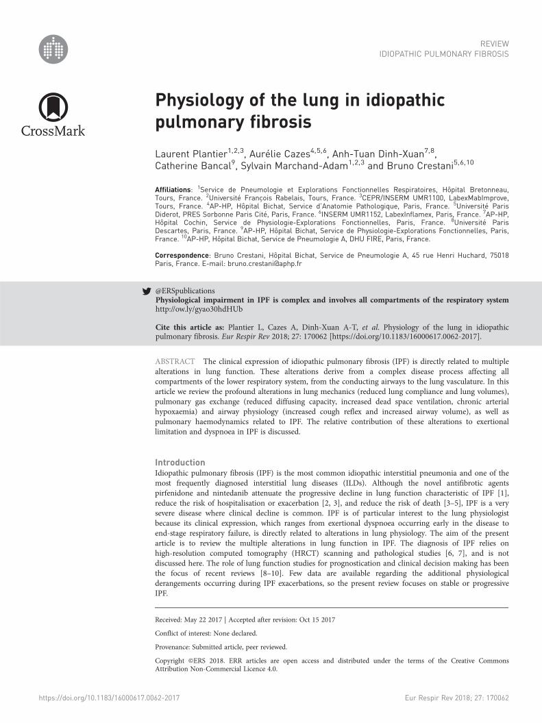
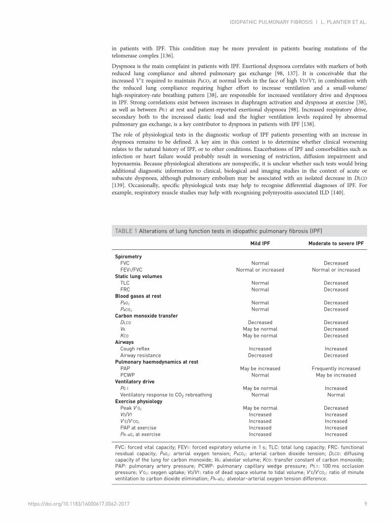
IPF is associated with multiple pathological alterations involving most compartments of the lowerrespiratory system (figure 1). Lung fibrosis is defined by the replacement of the normal, compliant lungextracellular matrix, which is rich in elastin, with an abnormal matrix that is rich in fibrillar collagen [20].Alveolar lesions conform to the usual interstitial pneumonia (UIP) pattern. The UIP pattern is defined by:1) spatial heterogeneity, as lesions alternate with areas of normal lung; 2) temporal heterogeneity, with theconcomitant presence of discrete lesions in lung tissue that appears otherwise normal (called fibroblasticfoci) and fibrotic areas composed mainly of dense acellular collagen; and 3) the presence of honeycomblesions, which are abnormal dilated airspaces with walls composed of fibrotic tissue, lined by anepithelium that shares characteristics with the airway epithelium [21, 22]. Tissular alterations are alsopresent in other parts of the respiratory system in IPF. Expression of Ki67, a marker of cell proliferation, isdetected in conducting airway epithelial cells [23, 24].
#
#
FIGURE 1 Pathological alterations in idiopathic pulmonary fibrosis (IPF). Lung biopsy of a patient with IPFshowing the usual interstitial pneumonia pattern (haematoxylin–eosin–saffron stain; ×10 magnification; scalebar=500 µm). Fibrotic lung with microscopic honeycomb change (#) and remodelled arteries (arrows) is visibleon the right, adjacent to preserved alveolar parenchyma on the left. Fibroblastic foci (arrowheads) are visiblein between. A bronchiole is visible in the upper left corner of the micrograph.
https://doi.org/10.1183/16000617.0062-2017 2
IDIOPATHIC PULMONARY FIBROSIS | L. PLANTIER ET AL.
Distinct vascular alterations are observed in the fibrotic regions and the more preserved regions of IPFlungs. In areas of dense fibrosis, sharp decreases in vessel density are observed [25, 26]. The walls ofpulmonary arteries show intimal fibrosis and medial hypertrophy, and are thickened [27, 28]. Pulmonaryveins and venules present loose and intimal fibrosis with reduced calibre [28]. In the histologicallypreserved areas, occlusion of pulmonary venules is frequent (65% of patients) and is associated withalveolar capillary multiplication [26] and/or muscularisation of arterioles, whereas arterial lesions are rare[28]. Capillary density is increased in regions bordering fibroblastic foci and honeycomb cysts [25].
Alterations in the mechanical properties of the lungReduction in lung complianceLung compliance describes the ability of the lung to expand and is key to describing the lung from amechanical point of view. It is defined as the change in lung volumes divided by the change intranspulmonary pressure. IPF results in profound reductions in lung compliance. This reduction in lungcompliance is driven both by reductions in the compliance of the lung extracellular matrix and byalterations in the pulmonary surfactant. In patients with IPF, lung surfactant shows alterations in its lipidprofile (reduced phosphatidyl glycerol, increased phosphatidylinositol and increased sphingomyelin levels,alterations in fatty acid composition) [29, 30], leading to severely impaired surface activity compared tocontrols [29]. The association of IPF with mutations in genes associated with surfactant metabolism [15,16] or with mutations in the telomerase complex [14, 17] driving accelerated epithelial cell senescencesuggests that surfactant alterations may contribute to the progress of IPF.
Lung compliance is typically measured under quasi-static conditions during lung deflation, when flow isinterrupted at several points during slow exhalation from total lung capacity (TLC). Transpulmonarypressure is defined as the difference between mouth pressure and oesophageal pressure in the absence offlow, under the assumption that mouth pressure approximates alveolar pressure and that oesophagealpressure approximates pleural pressure. Lung compliance measurements thus require placement of anoesophageal pressure probe. This invasive measurement is not routinely done in the clinic. Normal staticlung compliance is 328±102 mL·cmH2O
−1 and normal dynamic compliance is 285±105 mL·cmH2O−1 in
Swedish men, with little impact of age [31].
Reductions in lung compliance occur early in IPF. In one series, static lung compliance was reduced in allbut one out of 25 patients with IPF [32]. Among 31 IPF patients with a mean vital capacity (VC) of79±17% predicted values, static lung compliance was constantly and strikingly reduced (44±6% pred) [33].In another series of 14 IPF patients, none had normal static lung compliance [34]. Anecdotally, lungcompliance was markedly reduced in a patient with biopsy-proven IPF but a normal chest HRCT scan[35]. Altogether, these data suggest that measurements of lung compliance may be helpful for the earlydiagnosis of IPF.
Reductions in lung compliance may be tightly correlated with the degree of lung fibrosis. Among 23patients with biopsy-proven IPF, static lung compliance correlated with VC and TLC, but not with thediffusing capacity of the lung for carbon monoxide (DLCO) [36]. Importantly, although no correlation wasobserved between standard physiological studies (VC, TLC, DLCO) and pathological severity, static lungcompliance was strongly correlated with the degree of fibrosis assessed by scoring of lung biopsies. Suchan association between lung compliance and the extent of fibrosis was not replicated in another study [34].Reduction in lung compliance appears to progress with disease. In seven patients with end-stage IPFrequiring mechanical ventilation, dynamic lung compliance was considerably reduced(19±2.4 mL·cmH2O
−1) [37]. Reductions in dynamic compliance occur to the same extent as reductions instatic compliance in subjects with ILD [38]. The forced oscillation technique allows noninvasiveapproximation of the dynamic compliance of the respiratory system in the absence of airway obstructionand may be of interest in IPF [39]. However, in an earlier study [40], no correlation was observed in fivepatients between lung compliance and either respiratory system resistance or reactance.
It remains to be defined how reductions in lung compliance relate to clinical features such as dyspnoea.Such an association is highly likely, considering that lung compliance is a major determinant of the loadof the respiratory muscles and thus of the work of breathing [41]. The distribution of lesions isheterogeneous in IPF. It is therefore expected that the compliance of the lung is uneven among lungregions, as was shown in a sheep model of lung fibrosis [42], and consequently that convective ventilationpredominantly occurs in the less affected regions of the lungs. In support of this hypothesis, thedistribution of radiolabelled aerosols predominates in the upper regions of the lungs in IPF, whereaslesions predominate in the basal regions [43]. The distribution of ventilation to the less affected regions isan obstacle to the development of inhaled therapeutics for IPF.
https://doi.org/10.1183/16000617.0062-2017 3
IDIOPATHIC PULMONARY FIBROSIS | L. PLANTIER ET AL.
Reduction of lung volumesA restrictive ventilatory defect, defined by a reduction in static (TLC) and/or operating (VC) lung volumes,is typical in patients with IPF as in other ILDs [9]. Reduction of lung compliance is key to restrictionbecause both chest wall compliance [37] and respiratory muscle strength, as assessed by measurements oftransdiaphragmatic pressure [44] and maximal inspiratory pressure at the mouth [45], are mostlypreserved.
Restriction is often absent at the time of diagnosis. In 96 patients with biopsy-confirmed IPF, forced vitalcapacity (FVC) ranged from 26% to 112% pred, while TLC ranged from 42% to 125% pred [46]. In recentclinical trials, mean FVC was close to 80% pred, consistent with half of patients having normal operatingvolumes [47]. These elements indicate poor sensitivity of lung volume measurements for the diagnosis ofIPF. Although restriction of operating lung volumes is consistently associated with an increased risk ofdeath [9], it correlates weakly with dyspnoea or an altered quality of life in IPF [48], consistent with otherphysiological alterations also playing key roles in clinical expression of the disease.
It is not known whether the lack of restriction in some patients reflects the natural history of IPF, orillustrates a limitation of population-based reference values. For instance, IPF subjects who had better thanaverage lung function when healthy may present with apparently normal lung volumes before diseasereaches a severe stage. The confounding effect of smoking could explain the preservation of static lungvolumes in a fraction of patients, due to the effects of comorbid pulmonary emphysema on lungcompliance [49]. Patients with the combined pulmonary fibrosis and emphysema (CPFE) syndrome havehigher residual volume and TLC than patients with IPF [50].
Alterations in pulmonary gas exchangeIPF is associated with multiple alterations in the lung vasculature. In concert with alterations of thealveolar–capillary membrane, these lesions impair both gas diffusion and ventilation/perfusion (V′/Q′)relationships in the lung, leading to reduced diffusing capacity of the lung, increased dead spaceventilation, and increases in the alveolar–arterial oxygen tension difference (PA–aO2) and chronic arterialhypoxaemia.
Reduced diffusing capacity of the lungLung diffusing capacity is almost always reduced in patients with IPF. The DLCO was reduced in 98% ofIPF patients at the time of initial evaluation, although 27% of these patients had normal TLC and 56%had normal FVC [51]. Reduction of DLCO results from parenchymal and vascular lesions, as described bythe Roughton–Forster model where gas diffusion across the alveolar barrier depends on membraneconductance (DmCO) and vascular conductance, the latter being mostly dependent on the pulmonarycapillary volume [41].
DLCO is usually measured by a single-breath test where the subject inhales a gas mix comprising aninsoluble gas such as helium (He) or methane (CH4) along with carbon monoxide. The volume where gasexchange occurs (alveolar volume (VA)) and the transfer constant of carbon monoxide (KCO) arecalculated based on the reduction of He/CH4 and carbon monoxide concentrations in exhaled breath.DLCO is calculated by multiplying VA and KCO [52]. KCO can also be referred to as DLCO/VA, although thisterm is misleading as it implies that DLCO is the primary measurement from which DLCO/VA is thencalculated, when the opposite is actually correct. Overall, DLCO reflects the general gas-exchangingfunction of the whole lungs, while KCO reflects gas exchange per unit of lung volume. Reference values forDLCO, VA and KCO were obtained in healthy subjects at full lung inflation [53]. When the lungs areinflated below TLC (i.e. low VA), DLCO slightly decreases while KCO increases [54]. The increase in KCO atlow lung volume in normal individuals is due to the incomplete expansion (unfolding) of alveolar wallsresulting in increased mass of gas-exchanging tissue per unit of volume.
Both VA and KCO are reduced to varying degrees in IPF. Of note, KCO is in the normal range in up to30% of patients with IPF [55], particularly in patients with moderately altered DLCO [56]. It is importantnot to misinterpret this finding as being indicative of a preservation of gas exchange units, as it can besurmised that full lung inflation may not be attainable in IPF where subpleural fibrosis impairs lunginflation. In normal subjects, KCO increases at low lung volumes, so predicted values are inadequate tointerpret KCO in patients with restrictive disease [57]. In addition, the spatial heterogeneity of lesions inIPF may influence KCO as relatively preserved areas of the lung are preferentially ventilated [43]. Ouropinion is that a normal KCO value in IPF patients does not indicate that pulmonary gas exchange isnormal. DLCO correlates more strongly than KCO with exertional increases in PA–aO2 in IPF [58]. DLCO
and KCO both strongly correlate with the extent of disease as determined by scoring of computedtomography scans [59]. In support of the importance of DLCO measurements to the clinical appraisal ofIPF, DLCO is highly correlated both with dyspnoea [60] and survival [61].
https://doi.org/10.1183/16000617.0062-2017 4
IDIOPATHIC PULMONARY FIBROSIS | L. PLANTIER ET AL.
It is unclear whether alterations in the alveolar–capillary membrane or the lung vasculature are thepredominant mechanism of DLCO reductions in IPF. KCO is inversely correlated both with oxygendiffusion limitation and with alveolar ventilation (V′A)/Q′ mismatch, as shown by the multiple inert gaselimination technique (MIGET) [62]. Simultaneous carbon monoxide and nitric oxide diffusionmeasurements can help to dissect to what extent alterations in the alveolar–capillary membrane or thelung vasculature contribute to reductions in DLCO. Nitric oxide reacts almost immediately withhaemoglobin, so the diffusing capacity of the lung for nitric oxide (DLNO) is mostly independent ofvascular conductance and is equal to the conductance of the alveolar–capillary membrane to nitric oxide(DmNO). Because DmNO/DmCO is fixed, both DmCO and vascular conductance can be calculated with theDLCO/DLNO technique [63]. Such studies show severe and similar decreases in both membraneconductance and lung capillary volume in IPF patients [64], indicating that alterations in the alveolarmembrane and the lung vasculature both contribute to the impairment of gas diffusion in IPF [65].However, it is worth noting that in one study, the capillary volume was in the normal range for half of 30patients with IPF [66]. It is unclear whether discrepancies between these studies reflect differences inpatient selection or are due to differences in data acquisition, as different equipment and procedures wereused for carbon monoxide and nitric oxide measurements. Interestingly, in one study, the DLNO correlatedbetter than DLCO with the extent of fibrotic lesions as assessed by HRCT [66]. Recent approaches based onhyperpolarised 129Xe magnetic resonance imaging suggest that both impaired membrane conductance andtransfer to red blood cells participate in the reduced pulmonary gas exchange in IPF [67].
Dead space ventilationPatients with IPF have increased physiological dead space ventilation (increased ratio of dead space volumeto tidal volume (VD/VT)) at rest and at exercise [62, 68]. This feature results from both increasedanatomical dead space due to the increased volume of conducting airways [69], and from regionalincreases in V′/Q′ ratios, i.e. alveolar dead space. V′/Q′ lung scans demonstrate that fibrotic lesions, andhoneycomb lesions in particular, are very poorly perfused although they still receive some ventilation [70].
Interestingly, severe dead space ventilation may be a peculiar feature of IPF in comparison with otherILDs, as patients with IPF fail to reduce VD/VT at exercise [62], in contrast with patients with asbestosis[71]. VD/VT at exercise is strongly correlated with DLCO in IPF [55]. It is not known whether direct orindirect measures of VD/VT at rest and exercise provide additional information in comparison with restingmeasurements of gas diffusion in the lung, although experience acquired in the context of pulmonaryhypertension (PH) [72] or heart failure [73] suggests this may be so.
Chronic arterial hypoxaemiaAlterations in the mechanical properties of the lungs, abnormalities of the lung vasculature and diffusionimpairment lead to early-onset exertional chronic arterial hypoxaemia and later-onset resting chronicarterial hypoxaemia in IPF. Alveolar hypoventilation (hypercapnia) while awake is not common in IPF andis considered a feature of end-stage disease [74], when respiratory muscles fail in the face of a highlyincreased mechanical load (strongly reduced lung compliance). Alveolar hypoventilation is frequent duringsleep in IPF [75]. An increase in PA–aO2 is the main mechanism driving hypoxaemia in IPF [62]. PA–aO2,which is calculated from arterial oxygen tension (PaO2) and arterial carbon dioxide tension (PaCO2) usingthe ideal alveolar gas equation [41], can be increased because of reduced V′/Q′ ratios, right-to-leftshunting, or impairment of oxygen diffusion per se (referred to in the past as “alveolar–capillary block”).In a series of 15 patients, MIGET demonstrated that V′/Q′ mismatch and diffusion impairmentcontributed to chronic arterial hypoxaemia in IPF [62]. In that study, 2% and 4% of cardiac outputperfused areas with absent (shunting) or altered (low V′/Q′) ventilation, respectively, while breathing roomair at rest, suggesting that right-to-left shunting was in the physiological range in these patients. MIGETallows the calculation of a predicted PaO2 value based on the observed V′A/Q′ mismatch, under theassumption that diffusion limitation does not occur. In IPF, the observed PaO2 was lower than thepredicted value, allowing the attribution of 19% of PA–aO2 to diffusion limitation [62] at rest. At exercise,V′/Q′ and shunt accounted for 60% of PA–aO2 and diffusion limitation for 40% [62]. These data areconsistent with a more recent MIGET study [76]. It is not known whether exaggerated decreases in centralvenous oxygen tension contribute to hypoxaemia in IPF at rest. At submaximal exercise, the oxygentension of mixed venous blood was 29 mmHg in IPF patients [62], similar to healthy subjects [77].
Although V′/Q′ mismatch and diffusion limitation are the main contributors to increased PA–aO2 in IPF,anatomical right-to-left shunting may contribute in a fraction of patients. In a study of 15 IPF patientsbreathing 100% oxygen, mean PaO2 and PaCO2 were 481 mmHg and 38 mmHg [62], which translate to ashunt fraction of 12% according to the shunt equation. At variance with earlier reports [62, 78], brainimaging following intravenous injection of 99mTc-labelled albumin aggregates demonstrated right-to-leftshunting in two out of 22 patients with IPF [79]. It was not reported whether contrast echocardiography
https://doi.org/10.1183/16000617.0062-2017 5
IDIOPATHIC PULMONARY FIBROSIS | L. PLANTIER ET AL.
confirmed the existence of anatomical shunting, and it is unclear whether shunting resulted from the IPFdisease process or was incidental in these patients. Identifying the few patients with anatomical shuntingmay be clinically important. It is reported that closure of the abnormal communication partially correctedchronic arterial hypoxaemia in a patient with IPF [80]. However, the benefit of shunt closure should beprecisely evaluated because shunt could be life-preserving if the patient had concomitant PH.
Alterations in the structure and function of the conducting airwaysIPF is understood to primarily involve the alveolar regions. Several lines of evidence, however, suggest thatIPF also affects the airways. IPF lungs show evidence of airway epithelial cell proliferation [23] anddifferentiation [69], along with increased numbers of bronchioles in the distal regions [24]. In line withthese observations, alterations in the function of conducting airways have been observed.
Elevation of nerve growth factor levels in induced sputum, which preferentially samples proximal airways,raises the hypothesis that the proximal airways may be involved in IPF [81]. Patients with IPF have anincreased cough reflex to inhaled capsaicin. The inhalation of substance P induces cough in some IPFpatients, which does not occur in normal subjects [59]. These data suggest functional upregulation ofairway sensory neurons in IPF. Cough, however, may not be related to alterations in conducting airwaysonly, as direct stimulation of the chest wall suffices to induce cough in IPF patients [82].
Multiple data suggest reduced resistance of the conducting airways in IPF. Among 55 IPF patients with amean age of 71 years, the mean ratio of the forced expiratory volume in 1 s (FEV1) to FVC (FEV1/FVC)was 0.83 [56], which is higher than expected (0.74 for males, 0.75 for females according to EuropeanRespiratory Society reference equations) [83]. The ratio of the forced expiratory flow at 25–75% of FVC(FEF25–75%) to FVC (FEF25–75%/FVC) correlates positively with HRCT indices of IPF [39], suggesting thatairway dilation occurs as part of the disease process. The increase in FEV1/FVC and FEF25–75%/FVC isconsistent with data obtained with aerosol-derived morphometry, which show increased airwaydimensions at all lung depths in IPF [84]. Recently, we measured the volume of conducting airways byvolumetric capnography in patients with IPF, other ILDs and healthy controls. Interestingly, airwayvolume was higher in IPF than in controls and non-IPF ILDs, suggesting that increased airway volumemay be somewhat IPF-specific [69]. Anecdotal evidence indicates reduced distensibility of the proximalairways in IPF, although it is not clear whether this is related to either reduced compliance of the airwaywall or to changes in airway transmural pressure due to increased lung recoil [85].
Alterations in pulmonary haemodynamicsVascular lesions lead to disproportionate increases in pulmonary vascular resistance (PVR) and PH in asubset of patients with IPF. Right heart catheterisation is the gold standard for the diagnosis of PH, whichis defined as a mean pulmonary artery pressure (mPAP) ⩾25 mmHg at rest [86].
PH associated with IPF may represent a specific phenotype of IPF. In a series of 488 IPF patients withminimal honeycomb changes (<5% of the lung) and mild-to-moderate restriction (mean FVC between67% and 69% pred), right-heart catheterisation showed PH without elevated capillary wedge pressure in14% of patients, PH with elevated wedge pressure in 5%, and isolated elevated wedge pressure in 4% [87].Patients with PH and normal capillary wedge pressure had lower DLCO and increased haemoglobindesaturation at exercise. Interestingly, PH does not seem to progress rapidly in these patients, as repeatcatheterisation at 48 weeks yielded levels quite similar to baseline [87].
The prevalence of PH is 46.1% in patients with severe IPF listed for transplantation [88]. Severe PHcorrelates with elevated capillary wedge pressure, suggesting the participation of left-sided ventriculardysfunction to PH in IPF patients [88]. The prevalence of PH appears to be lower in IPF compared toconnective tissue disease-associated ILD of similar severity [89]. PH is associated with increased mortalityin IPF. In a series of 135 patients, there was a 2.4 increase in the hazard ratio (HR) for death per10 mmHg increase in mPAP, while an increase in PVR of 1 Wood unit was associated with a 1.4 increasein the HR for mortality [90].
The precise nature of lesions driving PH in IPF is not well understood. There is no correlation betweenmPAP and operating lung volumes [87, 91]. Anatomical–functional correlation analysis in 26 explantedlungs showed that mPAP is significantly correlated with the extent of fibrosis in damaged regions, but nocorrelation exists between mPAP and the extent of venous lesions in less-damaged areas [28]. It isprobable that the rarefaction of vessels in fibrotic areas contributes to PH in IPF, as suggested by thepresence of major defects on V′/Q′ lung scans [92]. It is possible that the discrepancy between globalvascular rarefaction [93] and the higher density of capillary vessels in alveolar septa of IPF lungs [94]pertains to variation in the sites of biopsy.
https://doi.org/10.1183/16000617.0062-2017 6
IDIOPATHIC PULMONARY FIBROSIS | L. PLANTIER ET AL.
Impairment of pulmonary haemodynamics at rest is detected late in IPF, although early alterations may bedetected at exercise. Increases in mPAP are observed in IPF patients during exercise at 60% of themaximal workload, while PVR does not decrease as observed in normal subjects [62]. In favour of theearly onset of vascular damage in IPF, mPAP increased from 20 mmHg to 40 mmHg in seven patientswith mild-to-moderate IPF [76]. The increase in oxygen diffusion impairment at exercise, documented byMIGET, is consistent with decrease of the alveolar–capillary contact time subsequent to the increase incardiac output, most probably indicating reduced recruitment of the lung vasculature in IPF [62].
Measurements of systolic pulmonary artery pressure by echocardiography lack sensitivity and specificity forthe diagnosis of PH in IPF [75]. Other echocardiographic indices may be useful for the identification ofright ventricle dysfunction and PH. A right ventricle to left ventricle diameter ratio >1 is associated with a5.4 increase in the HR for mortality in IPF [90], while moderate-to-severe right ventricle dysfunction,identified by tricuspid annular plane systolic excursion <1.6 cm, is associated with a 7.5 increase, suggestingthat echocardiography may indeed have a role for the identification of IPF patients with clinicallysignificant pulmonary vascular impairment [90]. HRCT measurements of the diameter of the pulmonaryartery do not accurately indicate PH in IPF, possibly due to the confounding effect of reduced pleuralpressure causing the dilation of cavitary intrathoracic organs [76]. Noninvasive measurements of pulmonaryblood flow using rebreathing of sulfur hexafluoride may be an interesting tool for the exploration ofhaemodynamic limitation in IPF [77].
Central control of ventilationAlterations in lung mechanics and gas exchange drive persistent activation of the central command ofventilation in IPF. An elevated ventilatory drive, detected by a rise in the 100 ms occlusion pressure (P0.1),was reported in patients with ILD, at rest and under carbon dioxide rebreathing [44, 95]. Correlationsbetween P0.1 and both lung compliance and VC were observed in IPF, suggesting that the increasedventilatory drive reflects the mechanical load imposed by fibrosis [96, 97]. An association was alsoobserved between P0.1 and KCO in patients with ILD, suggesting that impaired gas exchange contributes toventilatory drive [98]. Diaphragm activation is increased in IPF in comparison with healthy subjects, bothduring carbon dioxide rebreathing [99] and at exercise [38], consistent with the preservation of thecommand of ventilation in this disease.
Impact of comorbidities on lung function in IPFBecause IPF typically affects older patients and smokers, multiple comorbidities can affect patients withIPF [100, 101]. In particular, chronic obstructive pulmonary disease (COPD) and pulmonary emphysemaare common. The prevalence of emphysema in patients with IPF, which defines the CPFE syndrome,ranges from 6% to 67% [100]. Emphysema is associated with higher FVC and TLC at diagnosis in patientswith IPF [102]. CPFE is associated with markedly lower DLCO, especially when emphysema is present inlung areas not affected by fibrosis [103]. Of note, the pattern of lung function impairment in CPFE isquite distinct from COPD. Some, but not all, patients with CPFE present with airway obstruction andhyperinflation [104]. Impulse oscillometry showed that the expiratory increase in the reactance of therespiratory system at low frequency (5 Hz), which indicates expiratory collapse of the distal airways, wasmuch lower in CPFE than in COPD [104], consistent with the lack of dynamic hyperinflation in CPFE[104]. It is not known whether lung compliance is affected in CPFE to the same extent as in IPF withoutemphysema. 9.1% of IPF patients show reversible airflow obstruction as indicated by a 200 mL and 12%increase in either FVC or FEV1 after inhalation of a bronchodilator, although it is not clear whether thisreflects comorbid asthma or COPD, or an intrinsic feature of IPF [105].
Heart disease is a common occurrence in patients with IPF. The prevalence of coronary heart disease hasbeen reported to range from 3.2% to 68% [100]. 9% of patients with mild to moderate IPF and leftventricle ejection fraction ⩾40% have increased pulmonary artery wedge pressure, indicating occult heartfailure [87]. In terms of lung function, heart failure can be associated with restriction, obstruction, reducedDLCO [106, 107] and PH [108]. Heart failure with preserved ejection fraction is associated with reducedDLCO [109]. Venous thromboembolism may also contribute to low DLCO in a fraction of IPF patients.Venous thrombosis is twice as frequent in IPF patients as in the control population [110], while theincidence of pulmonary embolism is 6.4-fold higher in IPF than in the general population.
Lung function indices as indicators of prognosis and outcomes in clinical trialsThe severity of lung function impairment at the time of diagnosis and the decline of lung function overtime are both tightly associated with survival in IPF. Impairment of operating lung volumes, static lungvolumes and carbon monoxide transfer are associated with worse prognosis in IPF, with the strongestassociations observed with FVC, TLC and DLCO, respectively [111]. In a large cohort (n=1156), FVC<80% pred and DLCO ⩽45% pred were associated with increased mortality [112]. Since a >5% absolute
https://doi.org/10.1183/16000617.0062-2017 7
IDIOPATHIC PULMONARY FIBROSIS | L. PLANTIER ET AL.
decline in FVC (% pred) and a ⩾15% decline in DLCO (% pred) over 6 months are also highly associatedwith mortality [112], follow-up investigations are critical to determine prognosis in patients with IPF. Theprognostic impact of lung function may be assessed using composite indices such as the CompositePhysiological Index, which combines FVC, FEV1 and DLCO [113], or the Gender Age Physiology score,which combines FVC and DLCO with gender and age [114].
The rate of decline in FVC was the most common primary outcome end-point in recent clinical trials inIPF, expressed either in millilitres per year [47] or as percentage of the predicted value [115–117]. It iscurrently debated whether change in FVC over time is the optimal outcome for clinical trials in IPF. Thepresence of emphysema affects the rate of FVC decline in IPF. Patients with more emphysema show lessdecrease in FVC, while DLCO does decline, raising the hypothesis that FVC may not be the best end-pointin patients with emphysema [118]. In a series of 32 patients with IPF and moderate to severe emphysema,a 10% decline in FVC also failed to predict mortality [119]. In addition, although decline in both FVC(10%) and DLCO (15%) do predict mortality in the subsequent year, neither predicts change in lungfunction in the subsequent year [120]. These limitations to the use of FVC decline have led to the use ofprogression-free survival, defined as time to all-cause death or a categorical decrease from baseline in FVC% pred, in a recent trial of LOXL2 antibodies [121].
How do physiological alterations integrate in IPF? Exercise limitation and dyspnoeaThe multiple alterations of lung physiology in IPF translate into profound alterations in exercise capacityand dyspnoea. Oxygen uptake, power, tidal volume and PaO2 are reduced at exercise in IPF patientscompared with healthy controls [38, 68], while PA–aO2 and VD/VT are increased.
It is not clear whether reduced lung compliance, haemodynamic dysfunction, hypoxaemia or increasedVD/VT are the prime determinants of dyspnoea and exertional limitation in IPF. Short-term therapeuticintervention studies addressed this question. Alleviation of the load of the respiratory muscles bynoninvasive ventilation during submaximal exercise leads to increases in endurance time and arterialhaemoglobin saturation, and reductions in breathlessness [122]. These data clearly indicate that alterationsin the mechanical properties of the respiratory system play an important role in exercise limitation in IPFpatients, and are consistent with both the reduction in operating lung volumes and increaseddiaphragmatic activity at exercise [38].
PH may play key roles in a subset of patients. When PH is present, it is associated with reduced oxygenpulse at exercise, consistent with haemodynamic limitation [123], with more severe arterial haemoglobindesaturation at exercise [87] and with increased VD/VT, as suggested by an increase in the ratio of minuteventilation (V′E) to carbon dioxide elimination (V′CO2) at the ventilatory threshold [124]. Likewise,correlations exist between the 6-min walk distance and both mPAP and PVR [125]. It is unclear whetherthese associations reflect a causative relationship between the alteration of pulmonary haemodynamics andexertional limitation in IPF patients without severe PH. In seven patients with IPF (mean FVC 60% pred,DLCO 52% pred), two of whom had mPAP slightly over 25 mmHg at rest, the inhalation of nitric oxideduring submaximal exercise reduced the increase in mPAP and reduced PVR, but did not increase cardiacoutput or PaO2 and did not reduce VD/VT [76]. However, a 12-week course of oral sildenafil, whichpotentiates the effect of endogenous nitric oxide, yielded clinically significant benefits in terms ofdyspnoea and quality of life in patients with advanced IPF [126].
Gas exchange abnormalities during exercise are highly prevalent in IPF. Patients with mild-to-moderateIPF and normal or near-normal resting PaO2 have a significant decline in arterial haemoglobin saturationafter a 6-min walk [127]. Multiple factors contribute to exertional arterial hypoxaemia in IPF, withalterations of both V′/Q′ ratios [128] and diffusion [129] playing key roles. Uncontrolled retrospective[130, 131] and prospective [132] studies show increased walk performance, better quality of life andreduced dyspnoea in IPF patients treated with supplemental oxygen. This effect was observed both inpatients with resting arterial hypoxaemia and in patients without resting arterial hypoxaemia [131, 132].Whether the effect of oxygen therapy is related to a placebo effect remains subject to debate. In acontrolled study, supplemental oxygen given at exercise in IPF patients with exertional arterial hypoxaemiabut without resting arterial hypoxaemia failed to improve the 6-min walk distance and dyspnoea incomparison with placebo (air), despite improvements in arterial haemoglobin saturation [133], whileanother study reported beneficial effects with regard to endurance time and dyspnoea [134]. Indirectevidence suggests key roles of dead space ventilation in the genesis of dyspnoea, although the lack of apossibility to experimentally amend the VD/VT ratio precludes definitive demonstration. In a series of 25IPF patients, the V′E/V′CO2 slope, which is tightly correlated with VD/VT, was the physiological parametermost strongly correlated with patient-reported exertional dyspnoea [135]. In addition to alterations inpulmonary anatomy and physiology, anaemia may occasionally contribute to gas exchange abnormalities
https://doi.org/10.1183/16000617.0062-2017 8
IDIOPATHIC PULMONARY FIBROSIS | L. PLANTIER ET AL.
in patients with IPF. This condition may be more prevalent in patients bearing mutations of thetelomerase complex [136].
Dyspnoea is the main complaint in patients with IPF. Exertional dyspnoea correlates with markers of bothreduced lung compliance and altered pulmonary gas exchange [98, 137]. It is conceivable that theincreased V′E required to maintain PaCO2 at normal levels in the face of high VD/VT, in combination withthe reduced lung compliance requiring higher effort to increase ventilation and a small-volume/high-respiratory-rate breathing pattern [38], are responsible for increased ventilatory drive and dyspnoeain IPF. Strong correlations exist between increases in diaphragm activation and dyspnoea at exercise [38],as well as between P0.1 at rest and patient-reported exertional dyspnoea [98]. Increased respiratory drive,secondary both to the increased elastic load and the higher ventilation levels required by abnormalpulmonary gas exchange, is a key contributor to dyspnoea in patients with IPF [138].
The role of physiological tests in the diagnostic workup of IPF patients presenting with an increase indyspnoea remains to be defined. A key aim in this context is to determine whether clinical worseningrelates to the natural history of IPF, or to other conditions. Exacerbations of IPF and comorbidities such asinfection or heart failure would probably result in worsening of restriction, diffusion impairment andhypoxaemia. Because physiological alterations are nonspecific, it is unclear whether such tests would bringadditional diagnostic information to clinical, biological and imaging studies in the context of acute orsubacute dyspnoea, although pulmonary embolism may be associated with an isolated decrease in DLCO
[139]. Occasionally, specific physiological tests may help to recognise differential diagnoses of IPF. Forexample, respiratory muscle studies may help with recognising polymyositis-associated ILD [140].
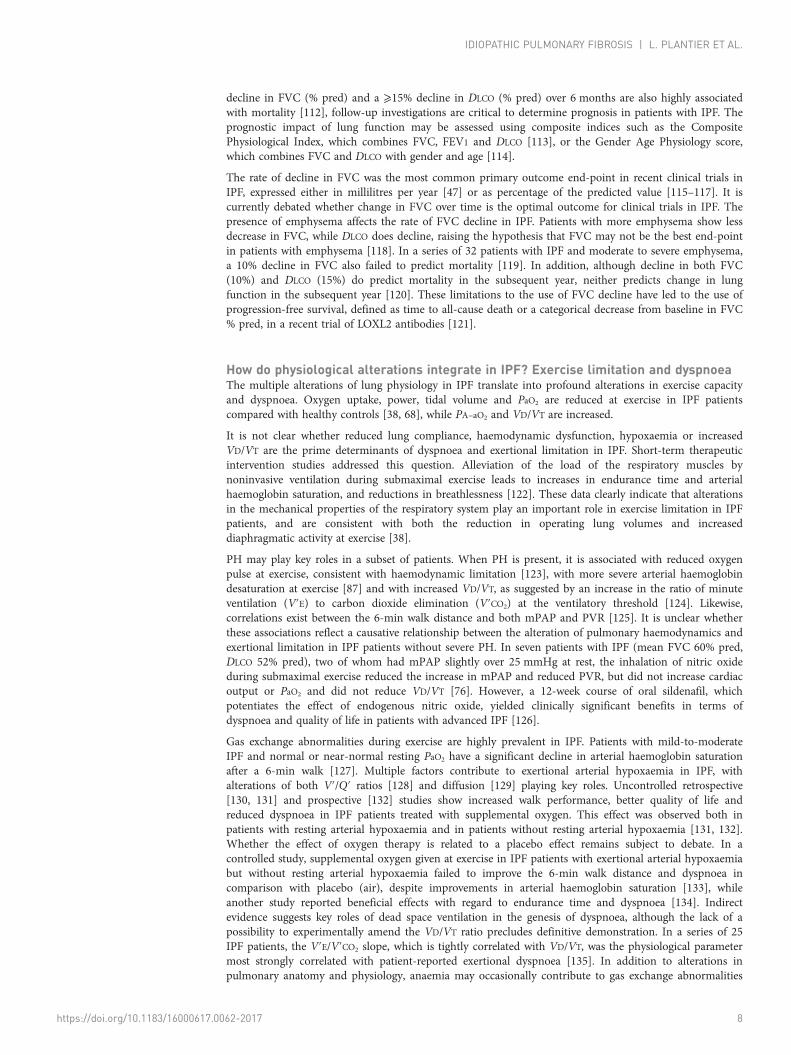
TABLE 1 Alterations of lung function tests in idiopathic pulmonary fibrosis (IPF)
Mild IPF Moderate to severe IPF
SpirometryFVC Normal DecreasedFEV1/FVC Normal or increased Normal or increased
Static lung volumesTLC Normal DecreasedFRC Normal Decreased
Blood gases at restPaO2 Normal DecreasedPaCO2 Normal Decreased
Carbon monoxide transferDLCO Decreased DecreasedVA May be normal DecreasedKCO May be normal Decreased
AirwaysCough reflex Increased IncreasedAirway resistance Decreased Decreased
Pulmonary haemodynamics at restPAP May be increased Frequently increasedPCWP Normal May be increased
Ventilatory driveP0.1 May be normal IncreasedVentilatory response to CO2 rebreathing Normal Normal
Exercise physiologyPeak V′O2 May be normal DecreasedVD/VT Increased IncreasedV′E/V′CO2 Increased IncreasedPAP at exercise Increased IncreasedPA–aO2 at exercise Increased Increased
FVC: forced vital capacity; FEV1: forced expiratory volume in 1 s; TLC: total lung capacity; FRC: functionalresidual capacity; PaO2: arterial oxygen tension; PaCO2: arterial carbon dioxide tension; DLCO: diffusingcapacity of the lung for carbon monoxide; VA: alveolar volume; KCO: transfer constant of carbon monoxide;PAP: pulmonary artery pressure; PCWP: pulmonary capillary wedge pressure; P0.1: 100 ms occlusionpressure; V′O2: oxygen uptake; VD/VT: ratio of dead space volume to tidal volume; V′E/V′CO2: ratio of minuteventilation to carbon dioxide elimination; PA–aO2: alveolar–arterial oxygen tension difference.
https://doi.org/10.1183/16000617.0062-2017 9
IDIOPATHIC PULMONARY FIBROSIS | L. PLANTIER ET AL.
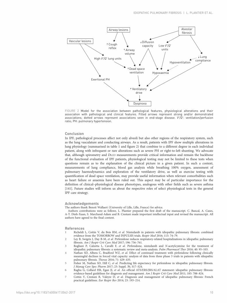
ConclusionIn IPF, pathological processes affect not only alveoli but also other regions of the respiratory system, suchas the lung vasculature and conducting airways. As a result, patients with IPF show multiple alterations inlung physiology (summarised in table 1 and figure 2) that combine to a different degree in each individualpatient, along with infrequent or rare alterations such as severe PH or right-to-left shunting. We advocatethat, although spirometry and DLCO measurements provide critical information and remain the backboneof the functional evaluation of IPF patients, physiological testing may not be limited to these tests whenquestions remain as to the explanation of the clinical picture in a given patient. In such a context,measurements of lung compliance, blood gas analysis while breathing 100% oxygen, assessment ofpulmonary haemodynamics and exploration of the ventilatory drive, as well as exercise testing withquantification of dead space ventilation, may provide useful information when relevant comorbidities suchas heart failure or anaemia have been ruled out. This aspect may be of particular importance for thedefinition of clinical–physiological disease phenotypes, analogous with other fields such as severe asthma[141]. Future studies will inform us about the respective roles of select physiological tests in the generalIPF care strategy.
AcknowledgementsThe authors thank Benoit Wallaert (University of Lille, Lille, France) for advice.Authors contributions were as follows. L. Plantier prepared the first draft of the manuscript. C. Bancal, A. Cazes,
A-T. Dinh-Xuan, S. Marchand-Adam and B. Crestani made important intellectual input and revised the manuscript. Allauthors have agreed to the final content.
References1 Richeldi L, Cottin V, du Bois RM, et al. Nintedanib in patients with idiopathic pulmonary fibrosis: combined
evidence from the TOMORROW and INPULSIS trials. Respir Med 2016; 113: 74–79.2 Ley B, Swigris J, Day B-M, et al. Pirfenidone reduces respiratory-related hospitalizations in idiopathic pulmonary
fibrosis. Am J Respir Crit Care Med 2017; 196: 756–761.3 Rogliani P, Calzetta L, Cavalli F, et al. Pirfenidone, nintedanib and N-acetylcysteine for the treatment of
idiopathic pulmonary fibrosis: a systematic review and meta-analysis. Pulm Pharmacol Ther 2016; 40: 95–103.4 Nathan SD, Albera C, Bradford WZ, et al. Effect of continued treatment with pirfenidone following clinically
meaningful declines in forced vital capacity: analysis of data from three phase 3 trials in patients with idiopathicpulmonary fibrosis. Thorax 2016; 71: 429–435.
5 Fisher M, Nathan SD, Hill C, et al. Predicting life expectancy for pirfenidone in idiopathic pulmonary fibrosis.J Manag Care Spec Pharm 2017; 23: Suppl. 3b, S17–S24.
6 Raghu G, Collard HR, Egan JJ, et al. An official ATS/ERS/JRS/ALAT statement: idiopathic pulmonary fibrosis:evidence-based guidelines for diagnosis and management. Am J Respir Crit Care Med 2011; 183: 788–824.
7 Cottin V, Crestani B, Valeyre D, et al. Diagnosis and management of idiopathic pulmonary fibrosis: Frenchpractical guidelines. Eur Respir Rev 2014; 23: 193–214.
Airway lesions
Coughreflex
Airwayvolume
Diffusioncapacity
Lungcompliance
Hypoxaemia
Hypoventilation
Low V'/Q'units
High V'/Q' lung units
Exertional PH
Dead spaceventilation
Ventilatorydrive
Alveolarfibrosis
Vascular lesions
Dyspnoea
FIGURE 2 Model for the association between pathological features, physiological alterations and theirassociation with pathological and clinical features. Filled arrows represent strong and/or demonstratedassociations; dotted arrows represent associations seen in end-stage disease. V′/Q′: ventilation/perfusionratio; PH: pulmonary hypertension.
https://doi.org/10.1183/16000617.0062-2017 10
IDIOPATHIC PULMONARY FIBROSIS | L. PLANTIER ET AL.
8 Egan JJ, Martinez FJ, Wells AU, et al. Lung function estimates in idiopathic pulmonary fibrosis: the potential fora simple classification. Thorax 2005; 60: 270–273.
9 Martinez FJ, Flaherty K. Pulmonary function testing in idiopathic interstitial pneumonias. Proc Am Thorac Soc2006; 3: 315–321.
10 Nathan SD, Shlobin OA, Weir N, et al. Long-term course and prognosis of idiopathic pulmonary fibrosis in thenew millennium. Chest 2011; 140: 221–229.
11 Kottmann RM, Kulkarni AA, Smolnycki KA, et al. Lactic acid is elevated in idiopathic pulmonary fibrosis andinduces myofibroblast differentiation via pH-dependent activation of transforming growth factor-β. Am J RespirCrit Care Med 2012; 186: 740–751.
12 Bagnato G, Harari S. Cellular interactions in the pathogenesis of interstitial lung diseases. Eur Respir Rev 2015;24: 102–114.
13 Zhou Y, Huang X, Hecker L, et al. Inhibition of mechanosensitive signaling in myofibroblasts amelioratesexperimental pulmonary fibrosis. J Clin Invest 2013; 123: 1096–1108.
14 Spagnolo P, Cottin V. Genetics of idiopathic pulmonary fibrosis: from mechanistic pathways to personalisedmedicine. J Med Genet 2017; 54: 93–99.
15 Borie R, Kannengiesser C, Crestani B. Familial forms of nonspecific interstitial pneumonia/idiopathic pulmonaryfibrosis: clinical course and genetic background. Curr Opin Pulm Med 2012; 18: 455–461.
16 Campo I, Zorzetto M, Mariani F, et al. A large kindred of pulmonary fibrosis associated with a novel ABCA3gene variant. Respir Res 2014; 15: 43.
17 Borie R, Tabèze L, Thabut G, et al. Prevalence and characteristics of TERT and TERC mutations in suspectedgenetic pulmonary fibrosis. Eur Respir J 2016; 48: 1721–1731.
18 Seibold MA, Wise AL, Speer MC, et al. A common MUC5B promoter polymorphism and pulmonary fibrosis.N Engl J Med 2011; 364: 1503–1512.
19 Borie R, Crestani B, Dieude P, et al. The MUC5B variant is associated with idiopathic pulmonary fibrosis but notwith systemic sclerosis interstitial lung disease in the European Caucasian population. PLoS One 2013; 8: e70621.
20 Thannickal VJ, Henke CA, Horowitz JC, et al. Matrix biology of idiopathic pulmonary fibrosis: a workshopreport of the national heart, lung, and blood institute. Am J Pathol 2014; 184: 1643–1651.
21 Plantier L, Crestani B, Wert SE, et al. Ectopic respiratory epithelial cell differentiation in bronchiolised distalairspaces in idiopathic pulmonary fibrosis. Thorax 2011; 66: 651–657.
22 Seibold MA, Smith RW, Urbanek C, et al. The idiopathic pulmonary fibrosis honeycomb cyst contains amucocilary pseudostratified epithelium. PLoS One 2013; 8: e58658.
23 Vuorinen K, Ohlmeier S, Leppäranta O, et al. Peroxiredoxin II expression and its association with oxidativestress and cell proliferation in human idiopathic pulmonary fibrosis. J Histochem Cytochem 2008; 56: 951–959.
24 Chilosi M, Poletti V, Murer B, et al. Abnormal re-epithelialization and lung remodeling in idiopathic pulmonaryfibrosis: the role of deltaN-p63. Lab Invest 2002; 82: 1335–1345.
25 Hanumegowda C, Farkas L, Kolb M. Angiogenesis in pulmonary fibrosis: too much or not enough? Chest 2012;142: 200–207.
26 Ebina M, Shimizukawa M, Shibata N, et al. Heterogeneous increase in CD34-positive alveolar capillaries inidiopathic pulmonary fibrosis. Am J Respir Crit Care Med 2004; 169: 1203–1208.
27 Nathan SD, Noble PW, Tuder RM. Idiopathic pulmonary fibrosis and pulmonary hypertension: connecting thedots. Am J Respir Crit Care Med 2007; 175: 875–880.
28 Colombat M, Mal H, Groussard O, et al. Pulmonary vascular lesions in end-stage idiopathic pulmonary fibrosis:histopathologic study on lung explant specimens and correlations with pulmonary hemodynamics. Hum Pathol2007; 38: 60–65.
29 Günther A, Schmidt R, Nix F, et al. Surfactant abnormalities in idiopathic pulmonary fibrosis, hypersensitivitypneumonitis and sarcoidosis. Eur Respir J 1999; 14: 565–573.
30 Schmidt R, Meier U, Markart P, et al. Altered fatty acid composition of lung surfactant phospholipids ininterstitial lung disease. Am J Physiol Lung Cell Mol Physiol 2002; 283: L1079–L1085.
31 Galetke W, Feier C, Muth T, et al. Reference values for dynamic and static pulmonary compliance in men. RespirMed 2007; 101: 1783–1789.
32 Radwan L, Zielonka TM, Maszczyk Z, et al. Zaburzenia czynnosciowe u chorych na srodmiazszowe choroby plucbez cech restrykcji [Functional disturbances in patients with interstitial lung diseases without signs of restriction].Pneumonol Alergol Pol 1999; 67: 180–188.
33 Zielonka TM, Demkow U, Radzikowska E, et al. Angiogenic activity of sera from interstitial lung disease patientsin relation to pulmonary function. Eur J Med Res 2010; 15: Suppl. 2, 229–234.
34 Sansores RH, Ramirez-Venegas A, Pérez-Padilla R, et al. Correlation between pulmonary fibrosis and the lungpressure–volume curve. Lung 1996; 174: 315–323.
35 Orens JB, Kazerooni EA, Martinez FJ, et al. The sensitivity of high-resolution CT in detecting idiopathicpulmonary fibrosis proved by open lung biopsy. A prospective study. Chest 1995; 108: 109–115.
36 Fulmer JD, Roberts WC, von Gal ER, et al. Morphologic-physiologic correlates of the severity of fibrosis anddegree of cellularity in idiopathic pulmonary fibrosis. J Clin Invest 1979; 63: 665–676.
37 Nava S, Rubini F. Lung and chest wall mechanics in ventilated patients with end stage idiopathic pulmonaryfibrosis. Thorax 1999; 54: 390–395.
38 Faisal A, Alghamdi BJ, Ciavaglia CE, et al. Common mechanisms of dyspnea in chronic interstitial andobstructive lung disorders. Am J Respir Crit Care Med 2016; 193: 299–309.
39 Lopes AJ, Capone D, Mogami R, et al. Correlacao dos achados tomograficos com parametros de funcaopulmonar na fibrose pulmonar idiopatica em nao fumantes [Correlation of tomographic findings withpulmonary function parameters in nonsmoking patients with idiopathic pulmonary fibrosis]. J Bras Pneumol2007; 33: 671–678.
40 van Noord JA, Clément J, Cauberghs M, et al. Total respiratory resistance and reactance in patients with diffuseinterstitial lung disease. Eur Respir J 1989; 2: 846–852.
41 Crystal R, West J, Weibel E. The Lung: Scientific Foundations. 2nd Edn. Philadelphia, Lippincott Williams &Wilkins, 1997.
https://doi.org/10.1183/16000617.0062-2017 11
IDIOPATHIC PULMONARY FIBROSIS | L. PLANTIER ET AL.

42 Organ L, Bacci B, Koumoundouros E, et al. Structural and functional correlations in a large animal model ofbleomycin-induced pulmonary fibrosis. BMC Pulm Med 2015; 15: 81.
43 Kanazawa M, Suzuki Y, Ishizaka A, et al. [Assessment of pulmonary aerosol deposition and epithelialpermeability in 99mTc-DTPA inhalation scintigram]. Nihon Kyōbu Shikkan Gakkai Zasshi 1993; 31: 593–600.
44 DiMarco AF, Kelsen SG, Cherniack NS, et al. Occlusion pressure and breathing pattern in patients withinterstitial lung disease. Am Rev Respir Dis 1983; 127: 425–430.
45 Jastrzebski D, Kozielski J, Zebrowska A. Rehabilitacja oddechowa chorych z idiopatycznym srodmiazszowymwloknieniem pluc za pomoca programu z cwiczeniami miesni wdechowych [Pulmonary rehabilitation in patientswith idiopathic pulmonary fibrosis with inspiratory muscle training]. Pneumonol Alergol Pol 2008; 76: 131–141.
46 Cherniack RM, Colby TV, Flint A, et al. Correlation of structure and function in idiopathic pulmonary fibrosis.Am J Respir Crit Care Med 1995; 151: 1180–1188.
47 Richeldi L, du Bois RM, Raghu G, et al. Efficacy and safety of nintedanib in idiopathic pulmonary fibrosis.N Engl J Med 2014; 370: 2071–2082.
48 du Bois RM, Weycker D, Albera C, et al. Ascertainment of individual risk of mortality for patients withidiopathic pulmonary fibrosis. Am J Respir Crit Care Med 2011; 184: 459–466.
49 Doherty MJ, Pearson MG, O’Grady EA, et al. Cryptogenic fibrosing alveolitis with preserved lung volumes.Thorax 1997; 52: 998–1002.
50 Mura M, Zompatori M, Pacilli AMG, et al. The presence of emphysema further impairs physiologic function inpatients with idiopathic pulmonary fibrosis. Respir Care 2006; 51: 257–265.
51 Cortes-Telles A, Forkert L, O’Donnell DE, et al. Idiopathic pulmonary fibrosis: new insights on functionalcharacteristics at diagnosis. Can Respir J 2014; 21: e55–e60.
52 Rosenberg E. The 1995 update of recommendations for a standard technique for measuring the single-breathcarbon monoxide diffusing capacity (transfer factor). Am J Respir Crit Care Med 1996; 154: 827–828.
53 Macintyre N, Crapo RO, Viegi G, et al. Standardisation of the single-breath determination of carbon monoxideuptake in the lung. Eur Respir J 2005; 26: 720–735.
54 Johnson DC. Importance of adjusting carbon monoxide diffusing capacity (DLCO) and carbon monoxide transfercoefficient (KCO) for alveolar volume. Respir Med 2000; 94: 28–37.
55 Wallaert B, Wemeau-Stervinou L, Salleron J, et al. Do we need exercise tests to detect gas exchange impairmentin fibrotic idiopathic interstitial pneumonias? Pulm Med 2012; 2012: 657180.
56 Pastre J, Plantier L, Planes C, et al. Different KCO and VA combinations exist for the same DLCO value in patientswith diffuse parenchymal lung diseases. BMC Pulm Med 2015; 15: 100.
57 Frans A, Nemery B, Veriter C, et al. Effect of alveolar volume on the interpretation of single breath DLCO. RespirMed 1997; 91: 263–273.
58 Agustí C, Xaubet A, Agustí AG, et al. Clinical and functional assessment of patients with idiopathic pulmonaryfibrosis: results of a 3 year follow-up. Eur Respir J 1994; 7: 643–650.
59 Wells AU, King AD, Rubens MB, et al. Lone cryptogenic fibrosing alveolitis: a functional-morphologiccorrelation based on extent of disease on thin-section computed tomography. Am J Respir Crit Care Med 1997;155: 1367–1375.
60 Swigris JJ, Han M, Vij R, et al. The UCSD shortness of breath questionnaire has longitudinal construct validityin idiopathic pulmonary fibrosis. Respir Med 2012; 106: 1447–1455.
61 Hamada K, Nagai S, Tanaka S, et al. Significance of pulmonary arterial pressure and diffusion capacity of thelung as prognosticator in patients with idiopathic pulmonary fibrosis. Chest 2007; 131: 650–656.
62 Agustí AG, Roca J, Gea J, et al. Mechanisms of gas-exchange impairment in idiopathic pulmonary fibrosis. AmRev Respir Dis 1991; 143: 219–225.
63 Guenard H, Varene N, Vaida P. Determination of lung capillary blood volume and membrane diffusing capacityin man by the measurements of NO and CO transfer. Respir Physiol 1987; 70: 113–120.
64 Martinot JB, Guénard H, Dinh-Xuan AT, et al. Nitrogen monoxide and carbon monoxide transfer interpretation:state of the art. Clin Physiol Funct Imaging 2017; 37: 357–365.
65 Wémeau-Stervinou L, Perez T, Murphy C, et al. Lung capillary blood volume and membrane diffusion inidiopathic interstitial pneumonia. Respir Med 2012; 106: 564–570.
66 Barisione G, Brusasco C, Garlaschi A, et al. Lung diffusing capacity for nitric oxide as a marker of fibroticchanges in idiopathic interstitial pneumonias. J Appl Physiol 2016; 120: 1029–1038.
67 Wang JM, Robertson SH, Wang Z, et al. Using hyperpolarized 129Xe MRI to quantify regional gas transfer inidiopathic pulmonary fibrosis. Thorax 2018; 73: 21–28.
68 Miki K, Maekura R, Hiraga T, et al. Acidosis and raised norepinephrine levels are associated with exercisedyspnoea in idiopathic pulmonary fibrosis. Respirology 2009; 14: 1020–1026.
69 Plantier L, Debray MP, Estellat C, et al. Increased volume of conducting airways in idiopathic pulmonary fibrosisis independent of disease severity: a volumetric capnography study. J Breath Res 2016; 10: 016005.
70 Strickland NH, Hughes JM, Hart DA, et al. Cause of regional ventilation–perfusion mismatching in patients withidiopathic pulmonary fibrosis: a combined CT and scintigraphic study. AJR Am J Roentgenol 1993; 161: 719–725.
71 Agusti AG, Roca J, Rodriguez-Roisin R, et al. Different patterns of gas exchange response to exercise in asbestosisand idiopathic pulmonary fibrosis. Eur Respir J 1988; 1: 510–516.
72 Paolillo S, Farina S, Bussotti M, et al. Exercise testing in the clinical management of patients affected bypulmonary arterial hypertension. Eur J Prev Cardiol 2012; 19: 960–971.
73 Poggio R, Arazi HC, Giorgi M, et al. Prediction of severe cardiovascular events by VE/VCO2 slope versus peak VO2
in systolic heart failure: a meta-analysis of the published literature. Am Heart J 2010; 160: 1004–1014.74 Bennett D, Fossi A, Bargagli E, et al. Mortality on the waiting list for lung transplantation in patients with
idiopathic pulmonary fibrosis: a single-centre experience. Lung 2015; 193: 677–681.75 Milioli G, Bosi M, Poletti V, et al. Sleep and respiratory sleep disorders in idiopathic pulmonary fibrosis. Sleep
Med Rev 2016; 26: 57–63.76 Blanco I, Ribas J, Xaubet A, et al. Effects of inhaled nitric oxide at rest and during exercise in idiopathic
pulmonary fibrosis. J Appl Physiol 2011; 110: 638–645.77 Stringer WW, Hansen JE, Wasserman K. Cardiac output estimated noninvasively from oxygen uptake during
exercise. J Appl Physiol 1997; 82: 908–912.
https://doi.org/10.1183/16000617.0062-2017 12
IDIOPATHIC PULMONARY FIBROSIS | L. PLANTIER ET AL.
78 Miller WC, Heard JG, Unger KM, et al. Anatomical lung shunting in pulmonary fibrosis. Thorax 1986; 41:208–209.
79 Graves MW, Kiratli PO, Mozley D, et al. Scintigraphic diagnosis of a right to left shunt in end-stage lung disease.Respir Med 2003; 97: 549–554.
80 Nguyen S, Leroy S, Bautin N, et al. Fibrose pulmonaire idiopathique et shunt droit-gauche par foramen ovalepermeable: amelioration clinique et gazometrique apres fermeture percutanee [Idiopathic pulmonary fibrosis andright-to left shunt by patent foramen ovale]. Rev Mal Respir 2007; 24: 631–634.
81 Hope-Gill BDM, Hilldrup S, Davies C, et al. A study of the cough reflex in idiopathic pulmonary fibrosis. Am JRespir Crit Care Med 2003; 168: 995–1002.
82 Jones RM, Hilldrup S, Hope-Gill BD, et al. Mechanical induction of cough in idiopathic pulmonary fibrosis.Cough 2011; 7: 2.
83 Quanjer PH, Tammeling GJ, Cotes JE, et al. Lung volumes and forced ventilatory flows. Report Working Party.Standardization of Lung Function Tests. European Community for Steel and Coal. Official Statement of theEuropean Respiratory Society. Eur Respir J 1993; 6: Suppl. 16, 5–40.
84 Brand P, Kohlhäufl M, Meyer T, et al. Aerosol-derived airway morphometry and aerosol bolus dispersion inpatients with lung fibrosis and lung emphysema. Chest 1999; 116: 543–548.
85 Baier H, Zarzecki S, Wanner A. Influence of lung inflation on the cross-sectional area of central airways innormals and in patients with lung disease. Respiration 1981; 41: 145–154.
86 Caminati A, Cassandro R, Harari S. Pulmonary hypertension in chronic interstitial lung diseases. Eur Respir Rev2013; 22: 292–301.
87 Raghu G, Nathan SD, Behr J, et al. Pulmonary hypertension in idiopathic pulmonary fibrosis withmild-to-moderate restriction. Eur Respir J 2015; 46: 1370–1377.
88 Shorr AF, Wainright JL, Cors CS, et al. Pulmonary hypertension in patients with pulmonary fibrosis awaitinglung transplant. Eur Respir J 2007; 30: 715–721.
89 Todd NW, Lavania S, Park MH, et al. Variable prevalence of pulmonary hypertension in patients with advancedinterstitial pneumonia. J Heart Lung Transplant 2010; 29: 188–194.
90 Rivera-Lebron BN, Forfia PR, Kreider M, et al. Echocardiographic and hemodynamic predictors of mortality inidiopathic pulmonary fibrosis. Chest 2013; 144: 564–570.
91 Nathan SD, Shlobin OA, Ahmad S, et al. Pulmonary hypertension and pulmonary function testing in idiopathicpulmonary fibrosis. Chest 2007; 131: 657–663.
92 Bourke SJ, Hawkins T, Keavey PM, et al. Ventilation perfusion radionuclide imaging in cryptogenic fibrosingalveolitis. Nucl Med Commun 1993; 14: 454–464.
93 Renzoni EA, Walsh DA, Salmon M, et al. Interstitial vascularity in fibrosing alveolitis. Am J Respir Crit CareMed 2003; 167: 438–443.
94 Kim KH, Maldonado F, Ryu JH, et al. Iron deposition and increased alveolar septal capillary density innonfibrotic lung tissue are associated with pulmonary hypertension in idiopathic pulmonary fibrosis. Respir Res2010; 11: 37.
95 Van Meerhaeghe A, Scano G, Sergysels R, et al. Respiratory drive and ventilatory pattern during exercise ininterstitial lung disease. Bull Eur Physiopathol Respir 1981; 17: 15–26.
96 Launois S, Clergue F, Medrano G, et al. Controle de la respiration dans les fibroses pulmonaires. Effets de l’O2 etdu CO2 [The control of respiration in pulmonary fibrosis. The effect of O2 and CO2]. Rev Mal Respir 1991; 8:67–73.
97 Renzi G, Milic-Emili J, Grassino AE. The pattern of breathing in diffuse lung fibrosis. Bull Eur PhysiopatholRespir 1982; 18: 461–472.
98 Londner C, Al Dandachi G, Plantier L, et al. Cross-sectional assessment of the relationships between dyspneadomains and lung function in diffuse parenchymal lung disease. Respiraton 2014; 87: 105–112.
99 Gorini M, Spinelli A, Ginanni R, et al. Neural respiratory drive and neuromuscular coupling during CO2
rebreathing in patients with chronic interstitial lung disease. Chest 1989; 96: 824–830.100 Raghu G, Amatto VC, Behr J, et al. Comorbidities in idiopathic pulmonary fibrosis patients: a systematic
literature review. Eur Respir J 2015; 46: 1113–1130.101 Oldham JM, Collard HR. Comorbid conditions in idiopathic pulmonary fibrosis: recognition and management.
Front Med 2017; 4: 123.102 Bodlet A, Maury G, Jamart J, et al. Influence of radiological emphysema on lung function test in idiopathic
pulmonary fibrosis. Respir Med 2013; 107: 1781–1788.103 Jacob J, Bartholmai BJ, Rajagopalan S, et al. Functional and prognostic effects when emphysema complicates
idiopathic pulmonary fibrosis. Eur Respir J 2017; 50: 1700379.104 Kitaguchi Y, Fujimoto K, Hanaoka M, et al. Pulmonary function impairment in patients with combined
pulmonary fibrosis and emphysema with and without airflow obstruction. Int J Chron Obstruct Pulmon Dis 2014;9: 805–811.
105 Assayag D, Vittinghoff E, Ryerson CJ, et al. The effect of bronchodilators on forced vital capacity measurementin patients with idiopathic pulmonary fibrosis. Respir Med 2015; 109: 1058–1062.
106 Agostoni P, Bussotti M, Cattadori G, et al. Gas diffusion and alveolar-capillary unit in chronic heart failure. EurHeart J 2006; 27: 2538–2543.
107 Melenovsky V, Andersen MJ, Andress K, et al. Lung congestion in chronic heart failure: haemodynamic, clinical,and prognostic implications. Eur J Heart Fail 2015; 17: 1161–1171.
108 Abraham WT, Adamson PB, Bourge RC, et al. Wireless pulmonary artery haemodynamic monitoring in chronicheart failure: a randomised controlled trial. Lancet 2011; 377: 658–666.
109 Olson TP, Johnson BD, Borlaug BA. Impaired pulmonary diffusion in heart failure with preserved ejectionfraction. JACC Heart Fail 2016; 4: 490–498.
110 Hubbard RB, Smith C, Le Jeune I, et al. The association between idiopathic pulmonary fibrosis and vasculardisease: a population-based study. Am J Respir Crit Care Med 2008; 178: 1257–1261.
111 Ley B, Collard HR, King TE. Clinical course and prediction of survival in idiopathic pulmonary fibrosis. Am JRespir Crit Care Med 2011; 183: 431–440.
https://doi.org/10.1183/16000617.0062-2017 13
IDIOPATHIC PULMONARY FIBROSIS | L. PLANTIER ET AL.
112 du Bois RM, Weycker D, Albera C, et al. Forced vital capacity in patients with idiopathic pulmonary fibrosis: testproperties and minimal clinically important difference. Am J Respir Crit Care Med 2011; 184: 1382–1389.
113 Wells AU, Desai SR, Rubens MB, et al. Idiopathic pulmonary fibrosis: a composite physiologic index derivedfrom disease extent observed by computed tomography. Am J Respir Crit Care Med 2003; 167: 962–969.
114 Ley B, Ryerson CJ, Vittinghoff E, et al. A multidimensional index and staging system for idiopathic pulmonaryfibrosis. Ann Intern Med 2012; 156: 684–691.
115 King TE, Bradford WZ, Castro-Bernardini S, et al. A phase 3 trial of pirfenidone in patients with idiopathicpulmonary fibrosis. N Engl J Med 2014; 370: 2083–2092.
116 Parker JM, Glaspole IN, Lancaster LH, et al. A phase 2 randomized controlled study of tralokinumab in subjectswith idiopathic pulmonary fibrosis. Am J Respir Crit Care Med 2018; 197: 94–103.
117 Raghu G, Martinez FJ, Brown KK, et al. CC-chemokine ligand 2 inhibition in idiopathic pulmonary fibrosis: aphase 2 trial of carlumab. Eur Respir J 2015; 46: 1740–1750.
118 Cottin V, Hansell DM, Sverzellati N, et al. Effect of emphysema extent on serial lung function in patients withidiopathic pulmonary fibrosis. Am J Respir Crit Care Med 2017; 196: 1162–1171.
119 Schmidt SL, Nambiar AM, Tayob N, et al. Pulmonary function measures predict mortality differently in IPFversus combined pulmonary fibrosis and emphysema. Eur Respir J 2011; 38: 176–183.
120 Schmidt SL, Tayob N, Han MK, et al. Predicting pulmonary fibrosis disease course from past trends inpulmonary function. Chest 2014; 145: 579–585.
121 Raghu G, Brown KK, Collard HR, et al. Efficacy of simtuzumab versus placebo in patients with idiopathicpulmonary fibrosis: a randomised, double-blind, controlled, phase 2 trial. Lancet Respir Med 2017; 5: 22–32.
122 Moderno EV, Yamaguti WPS, Schettino GPP, et al. Effects of proportional assisted ventilation on exerciseperformance in idiopathic pulmonary fibrosis patients. Respir Med 2010; 104: 134–141.
123 Boutou AK, Pitsiou GG, Trigonis I, et al. Exercise capacity in idiopathic pulmonary fibrosis: the effect ofpulmonary hypertension. Respirology 2011; 16: 451–458.
124 van der Plas MN, van Kan C, Blumenthal J, et al. Pulmonary vascular limitation to exercise and survival inidiopathic pulmonary fibrosis. Respirology 2014; 19: 269–275.
125 Minai OA, Santacruz JF, Alster JM, et al. Impact of pulmonary hemodynamics on 6-min walk test in idiopathicpulmonary fibrosis. Respir Med 2012; 106: 1613–1621.
126 Idiopathic Pulmonary Fibrosis Clinical Research Network, Zisman DA, Schwarz M, et al. A controlled trial ofsildenafil in advanced idiopathic pulmonary fibrosis. N Engl J Med 2010; 363: 620–628.
127 Nishiyama O, Taniguchi H, Kondoh Y, et al. Dyspnoea at 6-min walk test in idiopathic pulmonary fibrosis:comparison with COPD. Respir Med 2007; 101: 833–838.
128 Hempleman SC, Hughes JM. Estimating exercise DLO2 and diffusion limitation in patients with interstitialfibrosis. Respir Physiol 1991; 83: 167–178.
129 Hughes JM, Lockwood DN, Jones HA, et al. DLCO/Q and diffusion limitation at rest and on exercise in patientswith interstitial fibrosis. Respir Physiol 1991; 83: 155–166.
130 Visca D, Montgomery A, de Lauretis A, et al. Ambulatory oxygen in interstitial lung disease. Eur Respir J 2011;38: 987–990.
131 Frank RC, Hicks S, Duck AM, et al. Ambulatory oxygen in idiopathic pulmonary fibrosis: of what benefit? EurRespir J 2012; 40: 269–270.
132 Visca D, Mori L, Tsipouri V, et al. AmbOx: a randomised controlled, crossover trial evaluating the effect ofambulatory oxygen on health status in patients with fibrotic interstitial lung disease. Am J Respir Crit Care Med2017; 195: A7603.
133 Nishiyama O, Miyajima H, Fukai Y, et al. Effect of ambulatory oxygen on exertional dyspnea in IPF patientswithout resting hypoxemia. Respir Med 2013; 107: 1241–1246.
134 Dowman LM, McDonald CF, Bozinovski S, et al. Greater endurance capacity and improved dyspnoea with acuteoxygen supplementation in idiopathic pulmonary fibrosis patients without resting hypoxaemia. Respirology 2017;22: 957–964.
135 Manali ED, Lyberopoulos P, Triantafillidou C, et al. MRC Chronic Dyspnea Scale: relationships withcardiopulmonary exercise testing and 6-minute walk test in idiopathic pulmonary fibrosis patients: a prospectivestudy. BMC Pulm Med 2010; 10: 32.
136 Armanios MY, Chen JJ, Cogan JD, et al. Telomerase mutations in families with idiopathic pulmonary fibrosis.N Engl J Med 2007; 356: 1317–1326.
137 Swigris JJ, Streiner DL, Brown KK, et al. Assessing exertional dyspnea in patients with idiopathic pulmonaryfibrosis. Respir Med 2014; 108: 181–188.
138 Triantafillidou C, Manali E, Lyberopoulos P, et al. The role of cardiopulmonary exercise test in IPF prognosis.Pulm Med 2013; 2013: 514817.
139 Prost JF, Desfonds P, Genevray B, et al. Evaluation de la gravite des embolies pulmonaires. Interet de la mesurede la capacite de transfert du monoxyde de carbone a l’etat stable [Evaluation of the severity of pulmonaryembolism. Value of the measurement of stable carbon monoxide transfer capacity]. Presse Med 1984; 13:1193–1197.
140 Schiavi EA, Roncoroni AJ, Puy RJ. Isolated bilateral diaphragmatic paresis with interstitial lung disease. Anunusual presentation of dermatomyositis. Am Rev Respir Dis 1984; 129: 337–339.
141 Sears MR. Predicting asthma outcomes. J Allergy Clin Immunol 2015; 136: 829–836.
https://doi.org/10.1183/16000617.0062-2017 14
IDIOPATHIC PULMONARY FIBROSIS | L. PLANTIER ET AL.