Embed Size (px)
Citation preview

Physiology of visual analyzer
The eyes mediate sightThe eyes mediate sight
FunctionFunction Sensory organ for sight Sensory organ for sight Detects light and converts it into neural Detects light and converts it into neural
responses that the brain interpretsresponses that the brain interprets
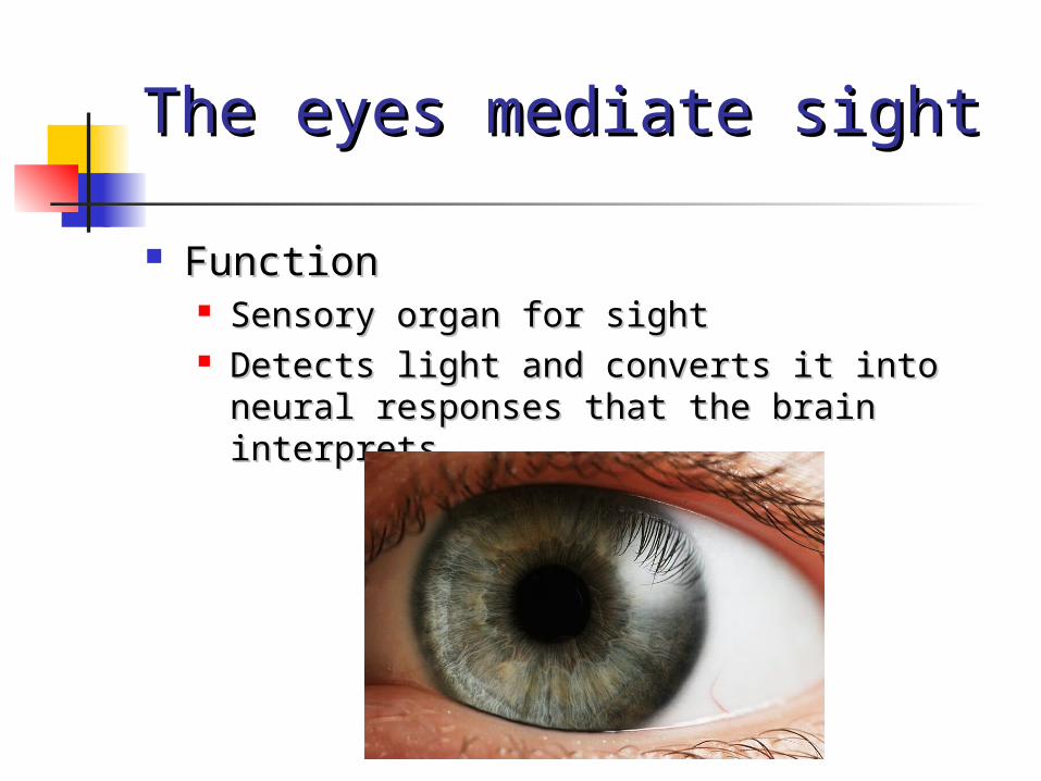
EYEExternal Eye Structures
The eyes are complex sense organs that have evolved from primitive light-sensitive spots on the surface of invertebrates.
Within its protective casing, each eye has a layer of receptors, a lens system that focuses light on these receptors, and a system of nerves that conducts impulses from the receptors to the brain.
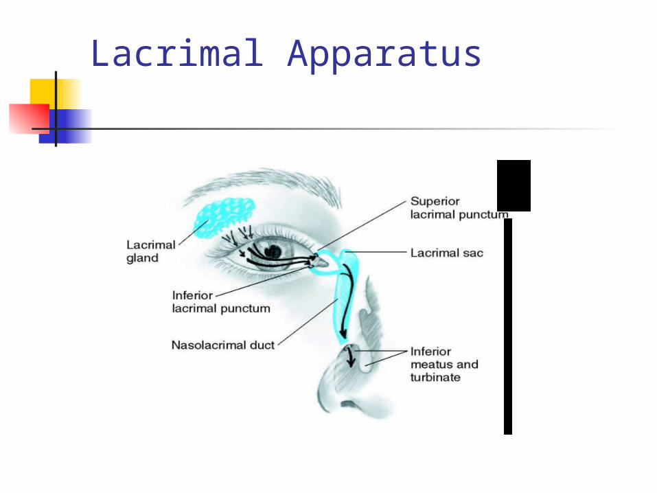
Lacrimal Apparatus
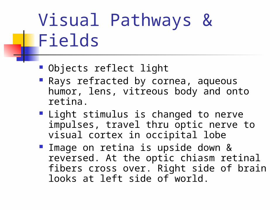
Visual Pathways & Fields Objects reflect light Rays refracted by cornea, aqueous humor,
lens, vitreous body and onto retina. Light stimulus is changed to nerve
impulses, travel thru optic nerve to visual cortex in occipital lobe
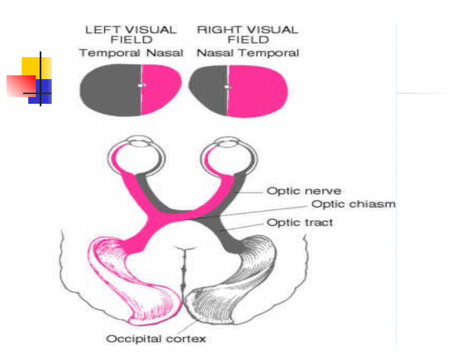
Image on retina is upside down & reversed. At the optic chiasm retinal fibers cross over. Right side of brain looks at left side of world.
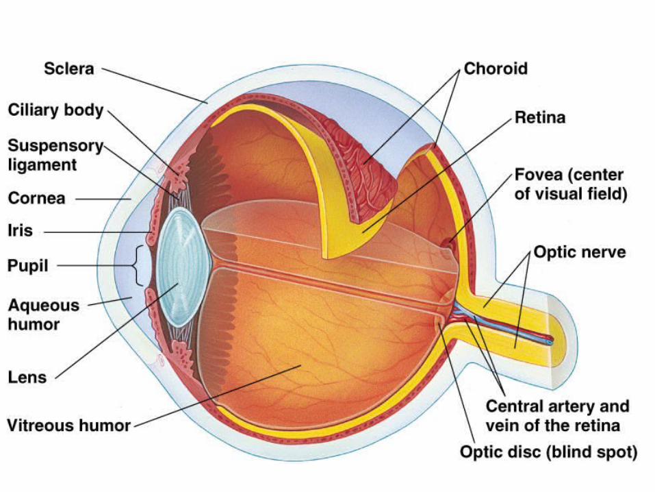
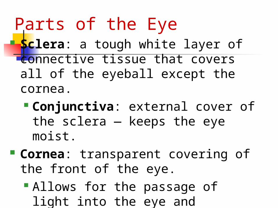
Sclera: a tough white layer of connective tissue that covers all of the eyeball except the cornea. Conjunctiva: external cover of the
sclera — keeps the eye moist. Cornea: transparent covering of the
front of the eye. Allows for the passage of light into
the eye and functions as a fixed lens.
Parts of the Eye
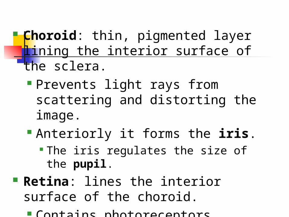
Choroid: thin, pigmented layer lining the interior surface of the sclera. Prevents light rays from scattering
and distorting the image. Anteriorly it forms the iris.
The iris regulates the size of the pupil. Retina: lines the interior surface of
the choroid. Contains photoreceptors.
Except at the optic disk (where the optic nerve attaches).
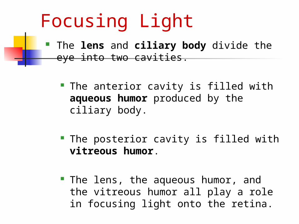
Focusing Light The lens and ciliary body divide the eye
into two cavities.
The anterior cavity is filled with aqueous humor produced by the ciliary body.
The posterior cavity is filled with vitreous humor.
The lens, the aqueous humor, and the vitreous humor all play a role in focusing light onto the retina.
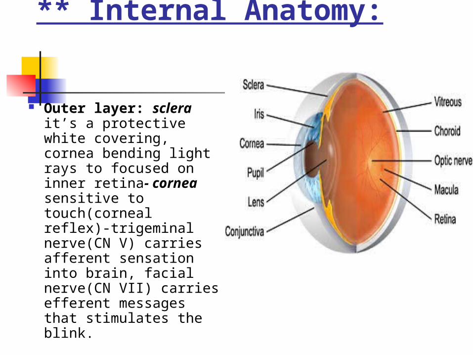
** Internal Anatomy:
Outer layer: sclera it’s a protective white covering, cornea bending light rays to focused on inner retina- cornea sensitive to touch(corneal reflex)-trigeminal nerve(CN V) carries afferent sensation into brain, facial nerve(CN VII) carries efferent messages that stimulates the blink.
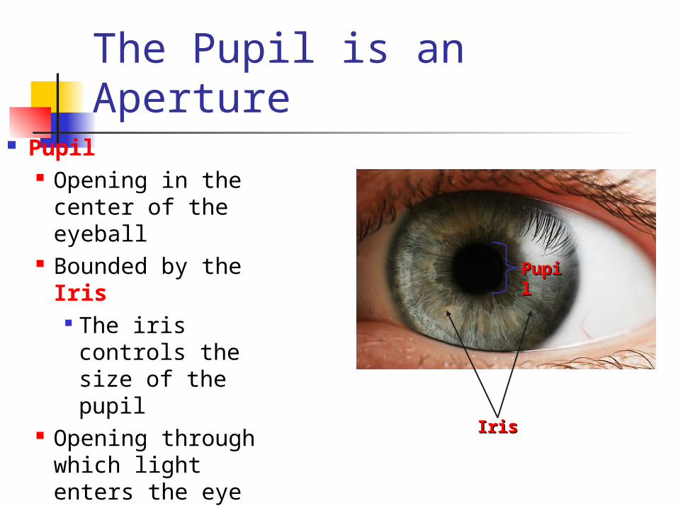
The Pupil is an Aperture Pupil
Opening in the center of the eyeball
Bounded by the Iris The iris controls
the size of the pupil
Opening through which light enters the eye
PupiPupill
IrisIris
Iris and Pupil
Iris = flat, round, regular, even color bilaterally.
Pupils = PERRLA Resting size norm = 3-5mm 5% population have pupils of 2 diff.
Sizes called Anisocoria
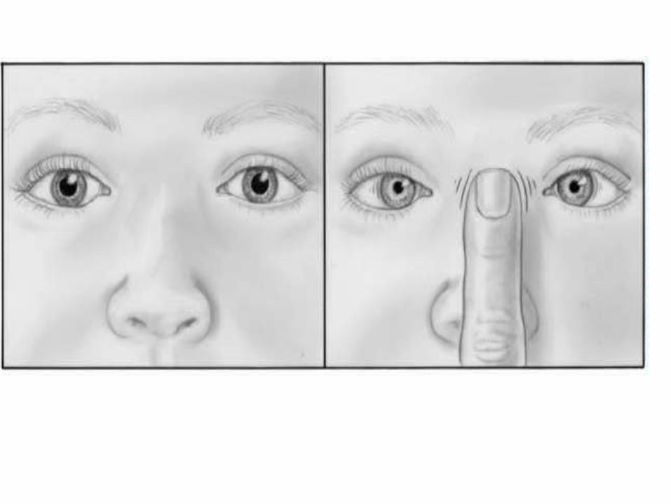
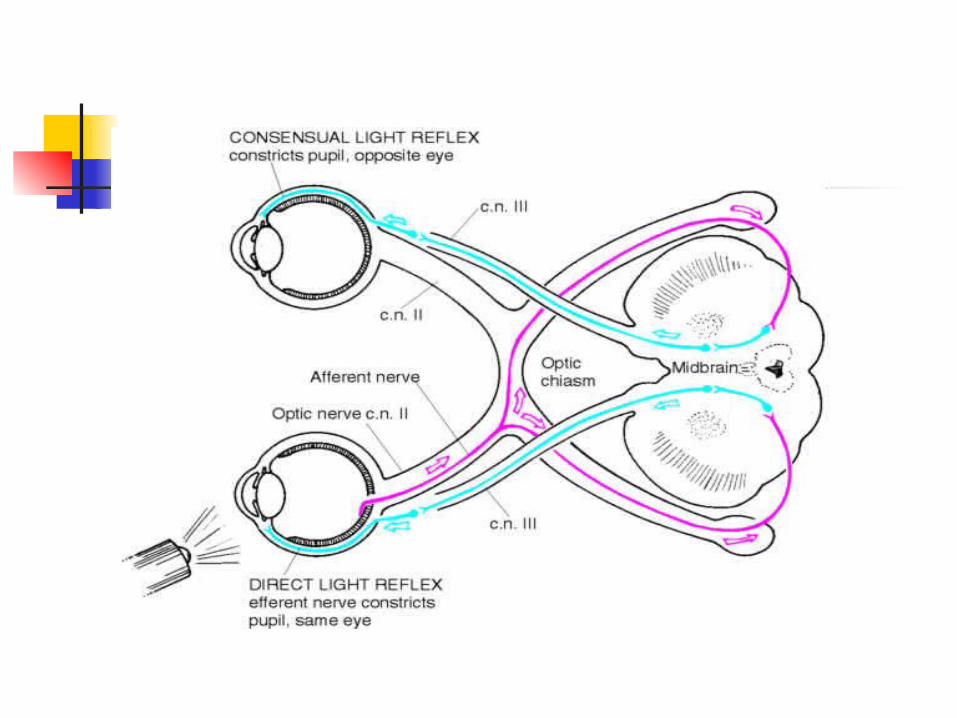
** Visual Reflexes:
# papillary light reflex: is a normal constriction of pupil when light shines on retina( a direct reflex & a consensual reflex).
Mechanism: light →retina → optic Nerve(II)__ efferent or sensory → midbrain → CN III (oculomotor) _ afferent(motor) → constriction of iris muscles for both eyes.
Direct reflex → same eye _Consensual reflex → opposite eye.
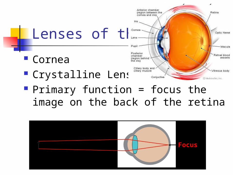
Lenses of the Eye
Cornea Crystalline Lens Primary function = focus the image
on the back of the retina
Focus
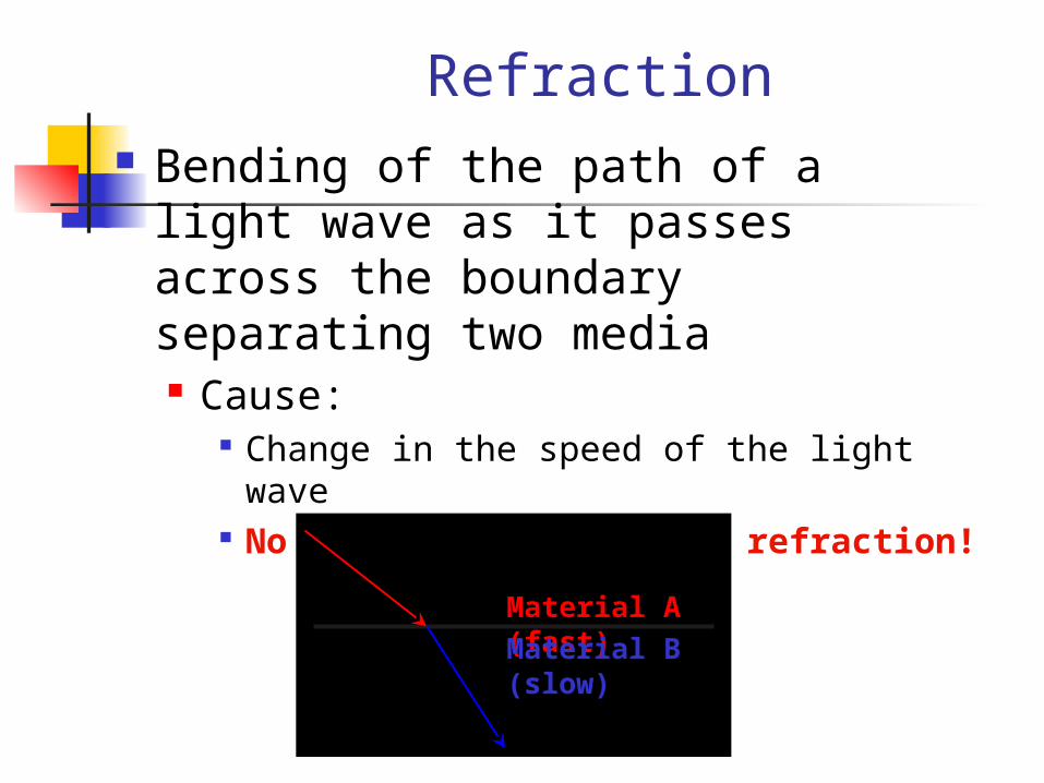
Refraction Bending of the path of a light wave
as it passes across the boundary separating two media Cause:
Change in the speed of the light wave No change in speed = no refraction!
Material A (fast) Material B (slow)
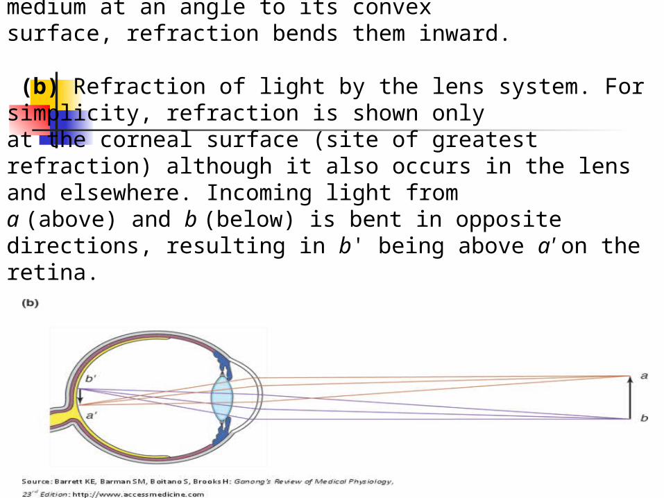
Focusing point sources of light. (a) When diverging light rays enter a dense medium at an angle to its convexsurface, refraction bends them inward.
(b) Refraction of light by the lens system. For simplicity, refraction is shown onlyat the corneal surface (site of greatest refraction) although it also occurs in the lens and elsewhere. Incoming light froma (above) and b (below) is bent in opposite directions, resulting in b' being above a' on the retina.
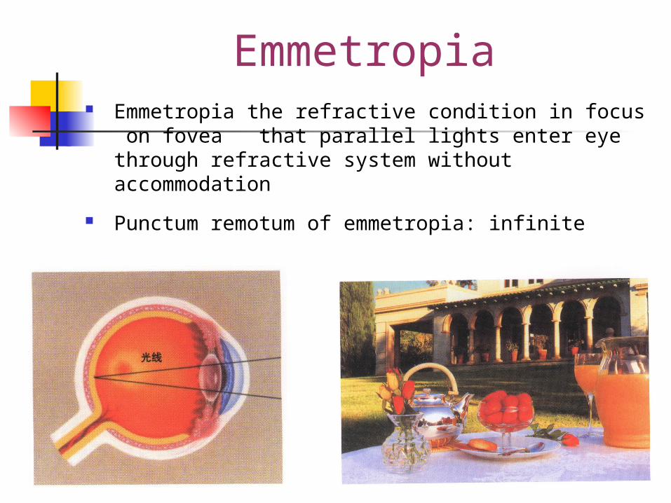
Emmetropia Emmetropia the refractive condition in focus on
fovea that parallel lights enter eye through refractive system without accommodation
Punctum remotum of emmetropia: infinite
Accomodation
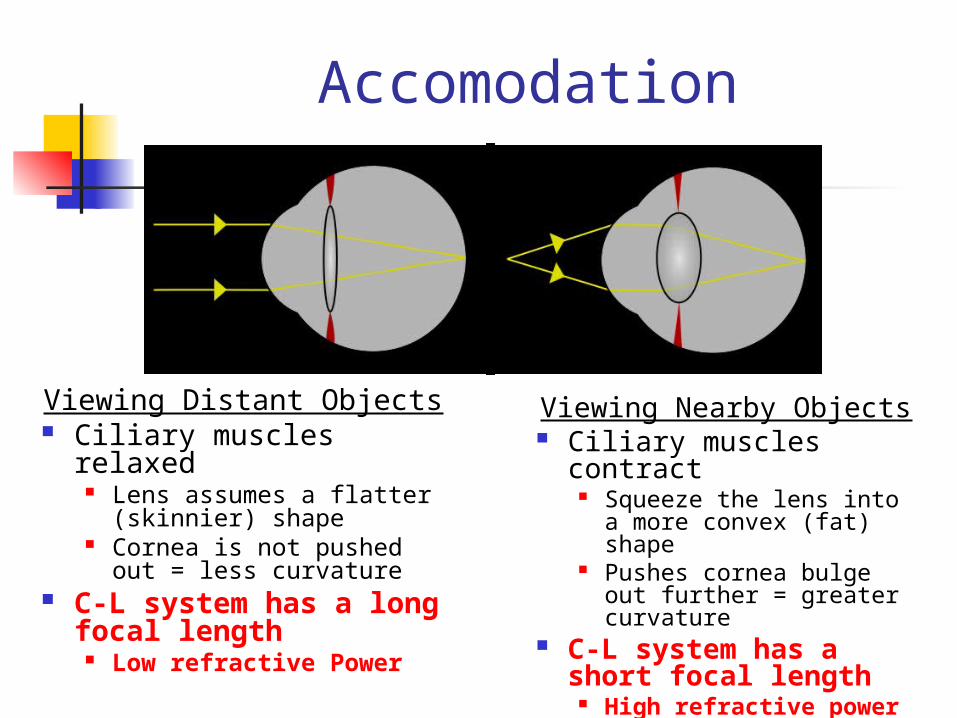
Viewing Nearby Objects Ciliary muscles contract
Squeeze the lens into a more convex (fat) shape
Pushes cornea bulge out further = greater curvature
C-L system has a short focal length
High refractive power
Viewing Distant Objects Ciliary muscles relaxed
Lens assumes a flatter (skinnier) shape
Cornea is not pushed out = less curvature
C-L system has a long focal length
Low refractive Power
Accommodation
Accommodation
the capability that eyes change refractive
condition in order to acquire clear
near sight
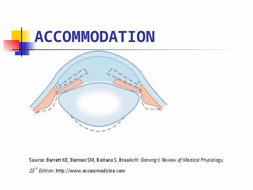
ACCOMMODATION
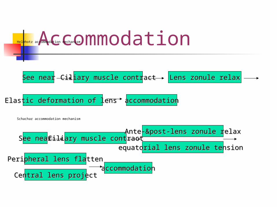
AccommodationHelmhotz accommodation mechanism
Schachar accommodation mechanism
Ciliary muscle contractCiliary muscle contract Lens zonule relaxLens zonule relax
accommodationaccommodationElastic deformation of lens Elastic deformation of lens
See nearSee near
Ciliary muscle contractCiliary muscle contractAnte-&post-lens zonule relaxAnte-&post-lens zonule relax
accommodationaccommodationPeripheral lens Peripheral lens flattenflatten
See nearSee nearequatorial equatorial lens zonule tension lens zonule tension
Central lens Central lens projectproject
Accommodation Accommodation = diopter for far diopter for
near
Range of accommodation distance of far point — distance of near point
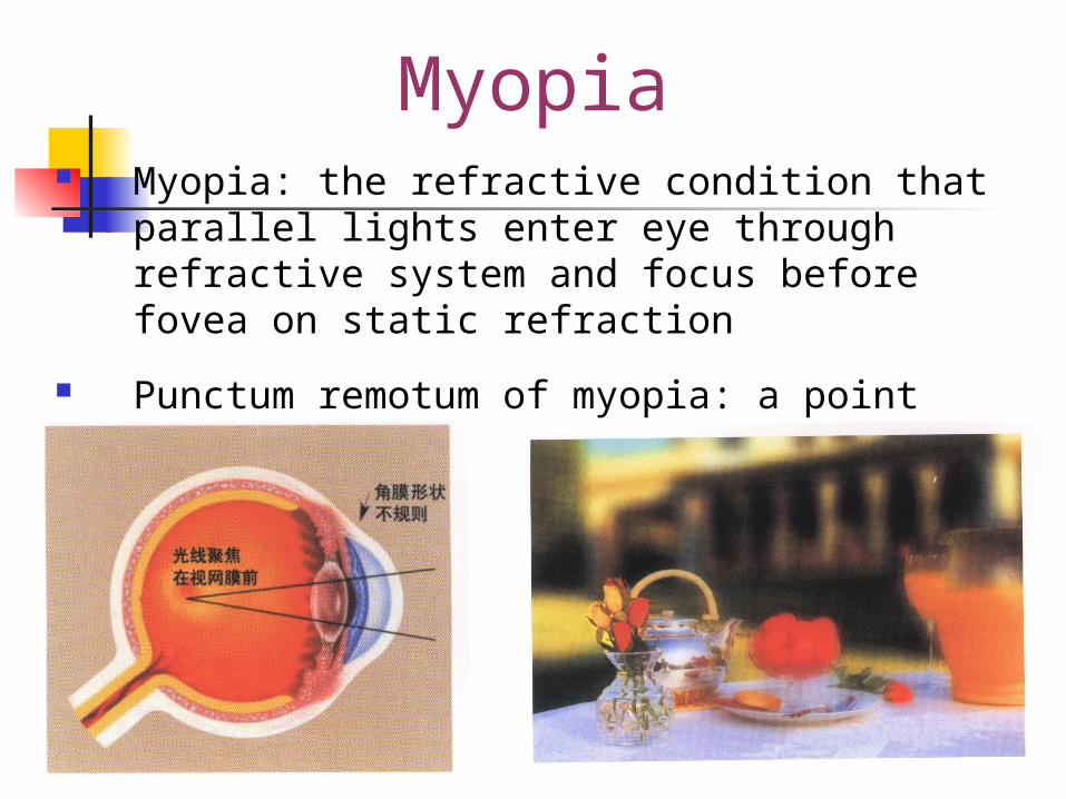
Myopia Myopia: the refractive condition that parallel lights enter
eye through refractive system and focus before fovea on static refraction
Punctum remotum of myopia: a point before eye
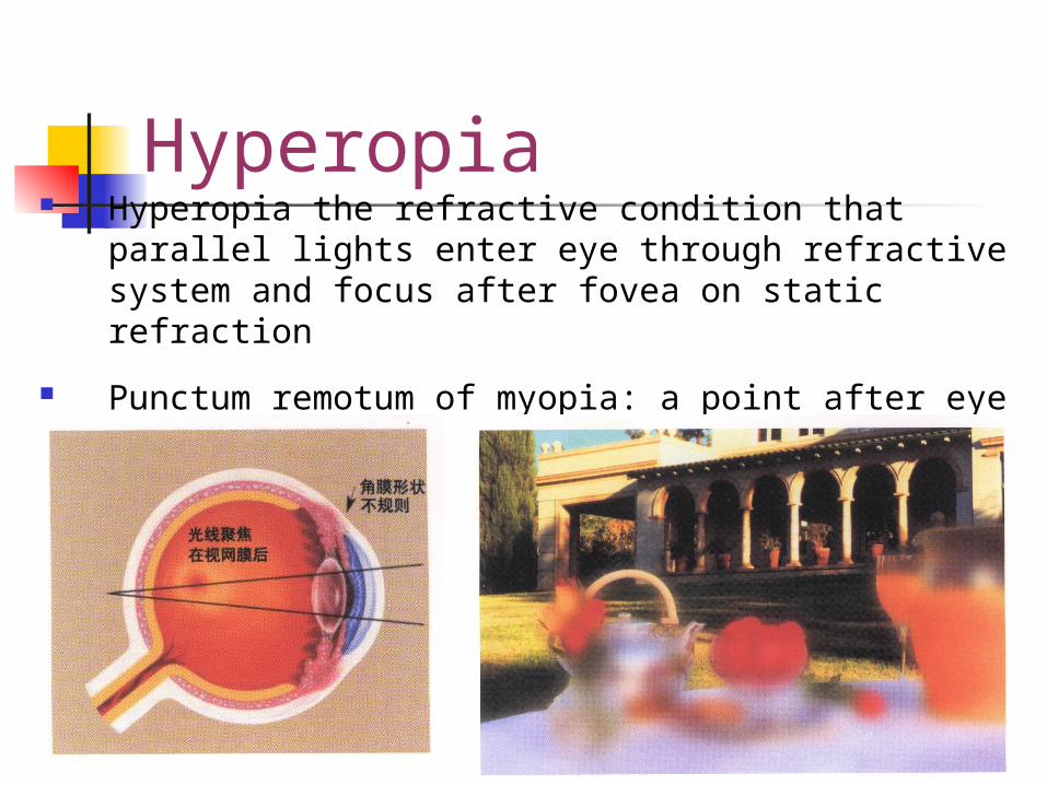
Hyperopia Hyperopia the refractive condition that parallel lights enter
eye through refractive system and focus after fovea on static refraction
Punctum remotum of myopia: a point after eye
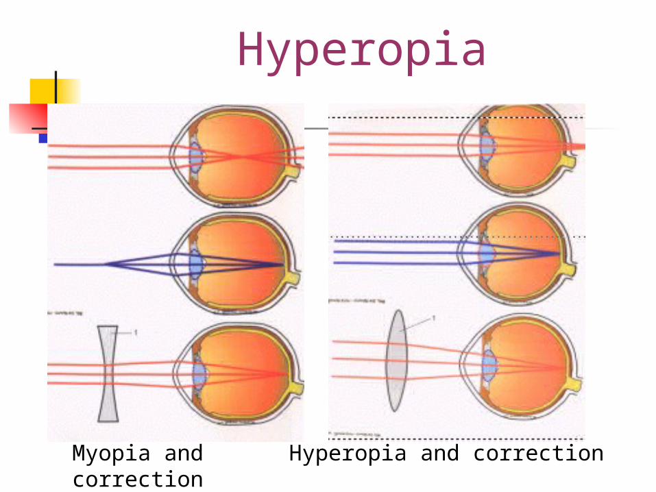
Myopia and correction Hyperopia and correction
Hyperopia
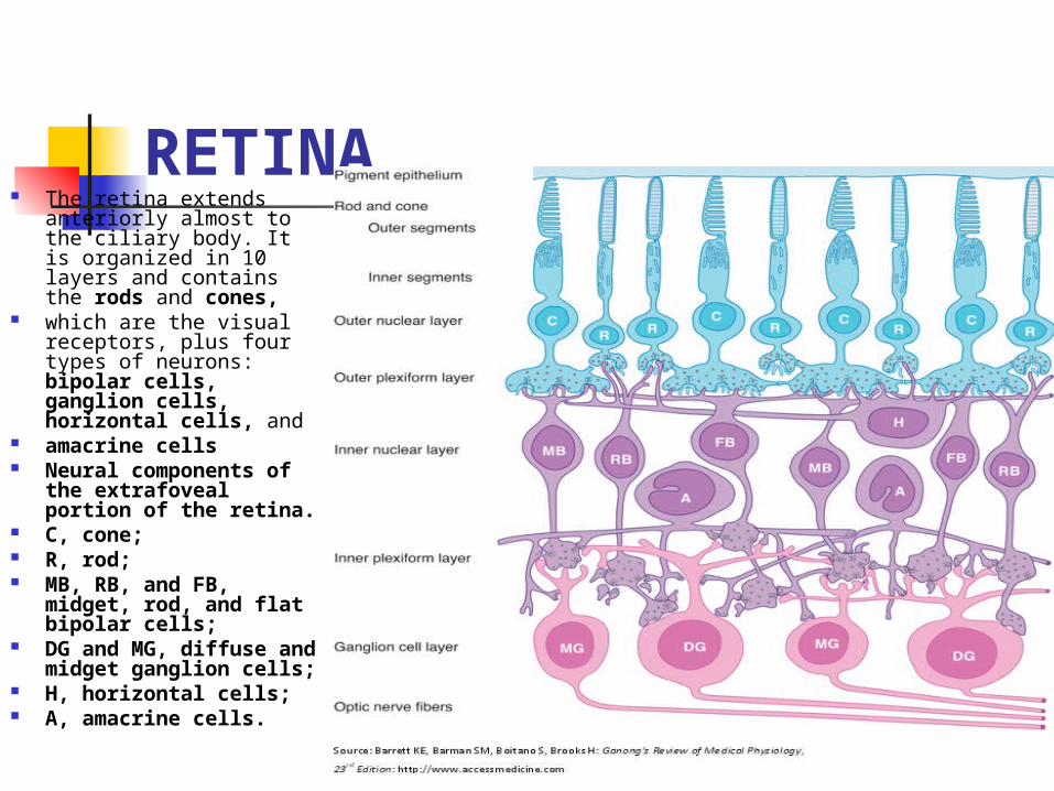
RETINA The retina extends anteriorly
almost to the ciliary body. It is organized in 10 layers and contains the rods and cones,
which are the visual receptors, plus four types of neurons: bipolar cells, ganglion cells, horizontal cells, and
amacrine cells Neural components of the
extrafoveal portion of the retina.
C, cone; R, rod; MB, RB, and FB, midget,
rod, and flat bipolar cells; DG and MG, diffuse and
midget ganglion cells; H, horizontal cells; A, amacrine cells.
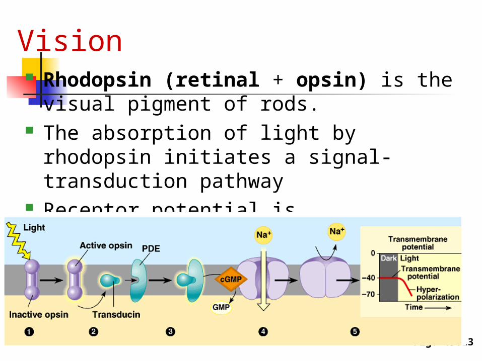
Vision Rhodopsin (retinal + opsin) is the
visual pigment of rods. The absorption of light by rhodopsin
initiates a signal-transduction pathway Receptor potential is hyperpolization .
Fig. 49.13
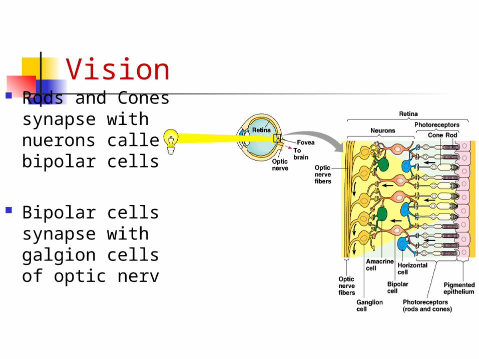
Vision Rods and
Cones synapse with nuerons called bipolar cells
Bipolar cells synapse with galgion cells of optic nerve
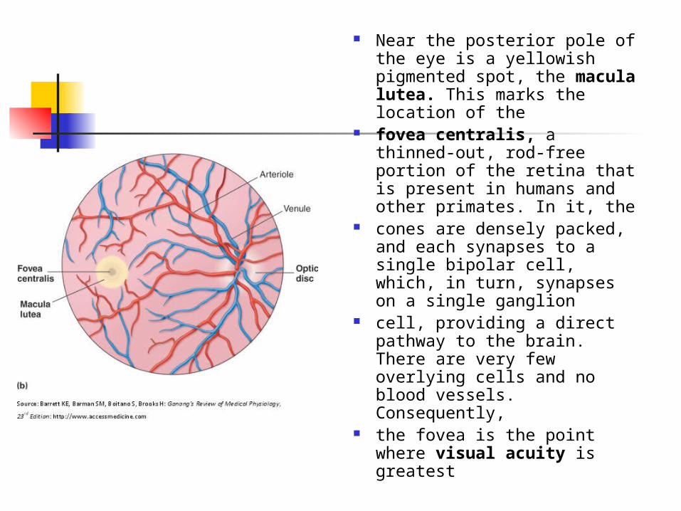
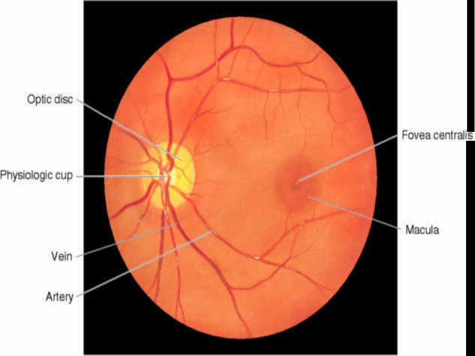
Near the posterior pole of the eye is a yellowish pigmented spot, the macula lutea. This marks the location of the
fovea centralis, a thinned-out, rod-free portion of the retina that is present in humans and other primates. In it, the
cones are densely packed, and each synapses to a single bipolar cell, which, in turn, synapses on a single ganglion
cell, providing a direct pathway to the brain. There are very few overlying cells and no blood vessels. Consequently,
the fovea is the point where visual acuity is greatest
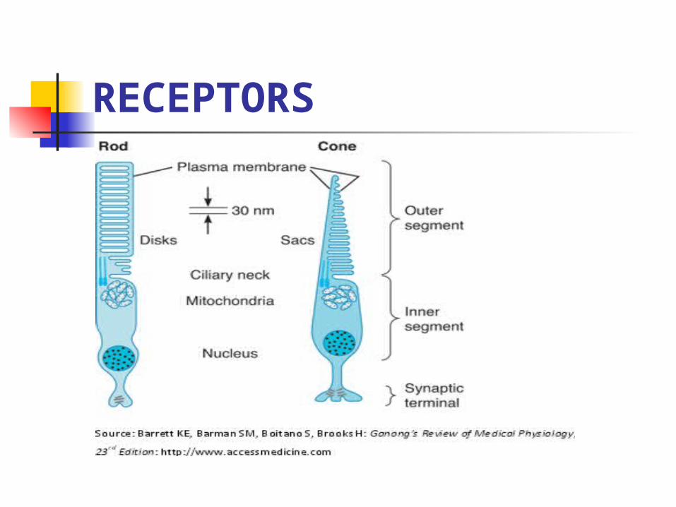
RECEPTORS
Visual Acuity Visual acuity is the degree to which
the details and contours of objects are perceived, and it is usually defined in
terms of the shortest distance by which two lines can be separated and still be perceived as two lines. Clinically,
visual acuity is often determined by the use of the familiar Snellen letter charts viewed at a distance of 20 ft (6m).