Embed Size (px)
Citation preview
Pharmacognosy Journal | October 2011 | Vol 3 | Issue 26 61
O r I g I n a l a r t I c l eP H c O g J .
Address for correspondence:E-mail: [email protected]
DOI: 10.5530/pj.2011.26.11
is almost bare between March and May and new leaves appear during April-July. The flowers appear along with the leaves, in dry areas the flowers however appear till October.[10]
The various chemical constituents isolated from the plant are fistucacidin, leucocyanidin, leucopelargonidin, hexacosanol, lupeol, Physcion, Rhein glycoside, Kaempferol, Chrysophanol, Sennoside A, Sennoside B, Quercetin, Epicatechin, Procyanidin B2, Stigmasterol and β-sitosterol.[11-13] Owing to their varied bioactivities exhibited by Cassia fistula, efforts have been made from time to time to generate libraries of these isolated compounds and screen them for potential biological activities.
MATERIALS AND METHODS
Plant Material
The plant material (Leaves) of Cassia fistula was collected from Mathura, Uttar Pradesh, India in the month of January 2008. It was identified by NISCAIR, New Delhi, Ref. no. NISCAIR/RHMD/Consult/2008-09/1145/177. Leaves of Cassia fistula were dried in shade and reduced to coarse powder for extraction, isolation and characterization of chemical constituents.
Phytochemical Investigation of Methanolic Extract of Cassia fistula Leaves
Manisha A. Nagpal1* Navneet Nagpal2, Sandeep Rahar1, Gagan Shah1, Gaurav Swami3, Reni Kapoor4
1Department of Pharmaceutical Chemistry, B.I.S. College of Pharmacy, Gagra, Moga, India, 2Department of Pharmaceutics, Khalsa College of Pharmacy, Amritsar, India, 3Department of Pharmaceutics, C.T. Institute of Technology, Jalandhar, India. 4Department of Pharmacognosy, Akal
College of Pharmacy, Masstuana Sahib, Sangrur, India
a B S t r a c t
Introduction: recently there has been a shift in universal trend from synthetic to herbal medicine, which we can say’ return to nature’. Cassia fistula or golden shower plant has plenty of medicinal values. this paper represent the isolation of two new chemical entities, present the leaves of cassia fistula and their medicinal aspects. Methods: leaves of Cassia fistula was used as a plant material. extraction was done with methanol followed by isolation procedure was done using HPTLC. Confirmation of compounds was done using UV, FTIR, NMR and MASS spectroscopy. In vitro antibacterial screening was carried out by disc diffusion method. Results: air dried powdered leaves of Cassia fistula were extracted using methanol. From this extract two compounds were isolated in their pure form using Column chromatography and HPTLC. Identification of these two isolated compounds was confirmed by physico-chemical data, spectral interpretation and elemental analysis. Conclusions: This is the first report of the isolation of the 2 new compounds from cassia fistula leaves extracted in methanol extract. these compounds are seen to exhibit moderate antimicrobial property.
Key words: Antimicrobial activity, HPTLC, MASS, NMR,
INTRODUCTION
The frequency of life-threatening infections caused by pathogenic microorganisms has increased worldwide and is becoming an important cause of morbidity and mortality in immune compromised patients in developing countries.[1] In recent years, attempts have been made to investigate the indigenous drugs against infectious diseases. This may help to develop safer antimicrobial drugs.[2]
Cassia fistula (family: Fabaceae), is commonly known as Amaltaash phal,[3-4] has been reported to possess laxative, Antioxidant, Hepatoprotective and Antiviral activity etc.[5-6] Cassia fistula has traditionally been used to treat leprosy, tuberculosis, syphilis, rheumatism, skin disease.[7] The Ayurvedic pharmacopoeia of India indicated the fruit pulp for constipation, colic, chlorosis and urinary disorders.[8] The fruit of Cassia fistula is used to treat diabetes.[9] Cassia fistula plant is rarely ever wholly leafless, but in some localities it
Nagpal, et al.: Phytochemical Investigation of Methanolic Extract of Cassia fistula Leaves
62 Pharmacognosy Journal | October 2011 | Vol 3 | Issue 26
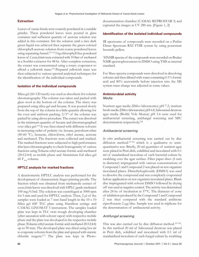
documentation chamber (CAMAG REPROSTAR 3) and captured the images at UV 280 nm. [Figure 1, 2]
Identification of the isolated individual compounds
IR spectrums of compounds were recorded on a Perkin Elmer Spectrum RXI FTIR system by using potassium bromide pellets.
1HNMR spectra of the compounds were recorded on Bruker NMR spectrophotometer in DMSO using TMS as internal standard.
For Mass spectra compounds were dissolved in dissolving solvents and then diluted with water containing 0.1% formic acid and 80% acetonitrile before injection into the MS system mass charge was adjusted at some values.
Antimicrobial activityMedia
Nutrient agar media (Difco laboratories) pH 7.2, nutrient broth media (Difco laboratories) pH 6.8, Sabouraud dextrose agar media (Biolife Vole Monza) pH 5.6 were used for antibacterial screening, antifungal screening and MIC determination respectively.[18]
Antibacterial screening
In vitro antibacterial screening was carried out by disc diffusion method.[19-22] which is a qualitative to semi-quantitative test. Briefly, 20 ml quantities of nutrient agar were plated in Petri dish, solidified and inoculated with 0.1 ml of standardized inoculum of each bacterial culture by swabbing over the agar surface. Filter paper discs (6 mm in diameter) impregnated with various concentrations of Compound 1 and Compound 2 was placed on test organism inoculated plates. Dimethylsuphoxide (DMSO) was used to dissolve the compound and was completely evaporated before application on test organism inoculated plates. Blank disc impregnated with solvent DMSO followed by drying off was used as negative control. The activity was determined after 24 hr. of incubation at 37°C. The diameter of zone of inhibition produced by the Compound 1 and Compound 2 was then compared with the standard antibiotic ciprofloxacin 2 μg/disc. Sample was used in triplicate for the determination of antibacterial activity.
Antifungal screening
This was also carried out by disc diffusion method [23-26]. In this method 20 ml of Sabouraud dextrose was plated in Petri dish, solidified and inoculated with 0.1 ml of standardized inoculum of each fungal culture by swabbing
Extraction
Leaves of cassia fistula were coarsely powdered in a suitable grinder. These powdered leaves were poured in glass container and sufficient quantity of acetone solution was added in this container. Stir the solution until a nice dark green liquid was achieved then separate the green colored chlorophyll-acetone solution from coarse powdered leaves using separating funnel.[14-15] 5 kg chlorophyll free powdered leaves of Cassia fistula were extracted with 10 liter of methanol in a Soxhlet extractor for 48 hr. After complete extraction, the extract was concentrated using a rotary evaporator to afford a yellowish mass.[15] Prepared yellowish mass was then subjected to various spectral analytical techniques for the identification of the individual compounds.
Isolation of the individual compounds
Silica gel (60-120 mesh) was used as absorbent for column chromatography. The column was taken and packed with glass wool at the bottom of the column. The slurry was prepared using silica gel and hexane. It was poured slowly from the top of the column in a little quantity allowing for the even and uniform packing. 2/3rd of the column was packed by using above procedure. The extract was dissolved in the minimum quantity of hexane and chromatographed over silica gel.[16] It was then eluted with different solvents in increasing order of polarity viz. hexane, petroleum ether (40-60 °C), benzene, chloroform, ethyl acetate, acetone and methanol. The fractions were collected and marked. The marked fractions were subjected to high performance thin layer chromatography to check homogeneity of various fractions using Toluene-ethyl acetate-formic acid-methanol (20:12:4:4) as mobile phase and Aluminium foil silica gel 60 F254 column.
HPTLC analysis for marked fractions
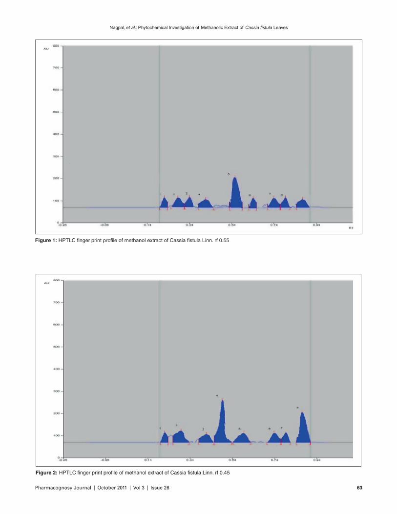
A densitometric HPTLC analysis was performed for the development of characteristic finger printing profile. The fraction which was obtained from methanolic extract of cassia fistula leaves was dissolved with HPLC grade methanol 100 mg/0.5ml. The solution was centrifuged at 3000 rpm for 5 min and used for HPTLC analysis. Then, 2 μl of the samples were loaded as 7 mm band length in the 10 x 10 Silica gel 60F TLC plate using Hamilton syringe and CAMAG LINOMAT 5 instrument. The samples loaded plate was kept in TLC twin trough developing chamber (after saturation with solvent vapor) with respective mobile phase and the plate was developed in the respective mobile phase (Toluene-ethyl acetate-formic acid-methanol 20:12:4:4) up to 90 mm. The developed plate was dried using hot air to evaporate solvents from the plate and sprayed with stannic chloride reagent.[17] The plate was kept in Photo-
Pharmacognosy Journal | October 2011 | Vol 3 | Issue 26 63
Nagpal, et al.: Phytochemical Investigation of Methanolic Extract of Cassia fistula Leaves
Figure 2: HPTLC finger print profile of methanol extract of Cassia fistula Linn. rf 0.45
Figure 1: HPTLC finger print profile of methanol extract of Cassia fistula Linn. rf 0.55
Nagpal, et al.: Phytochemical Investigation of Methanolic Extract of Cassia fistula Leaves
64 Pharmacognosy Journal | October 2011 | Vol 3 | Issue 26
[M-CH3]+, 219(19)[M-OCH3]
+, 191(29)[M-C3H7O]+, 161(28), 151(12), 124(16), 101(10), 59(20);
Elemental Analysis: Calculated: C 62.93, H 5.64; Found: C 62.90, H 5.60%
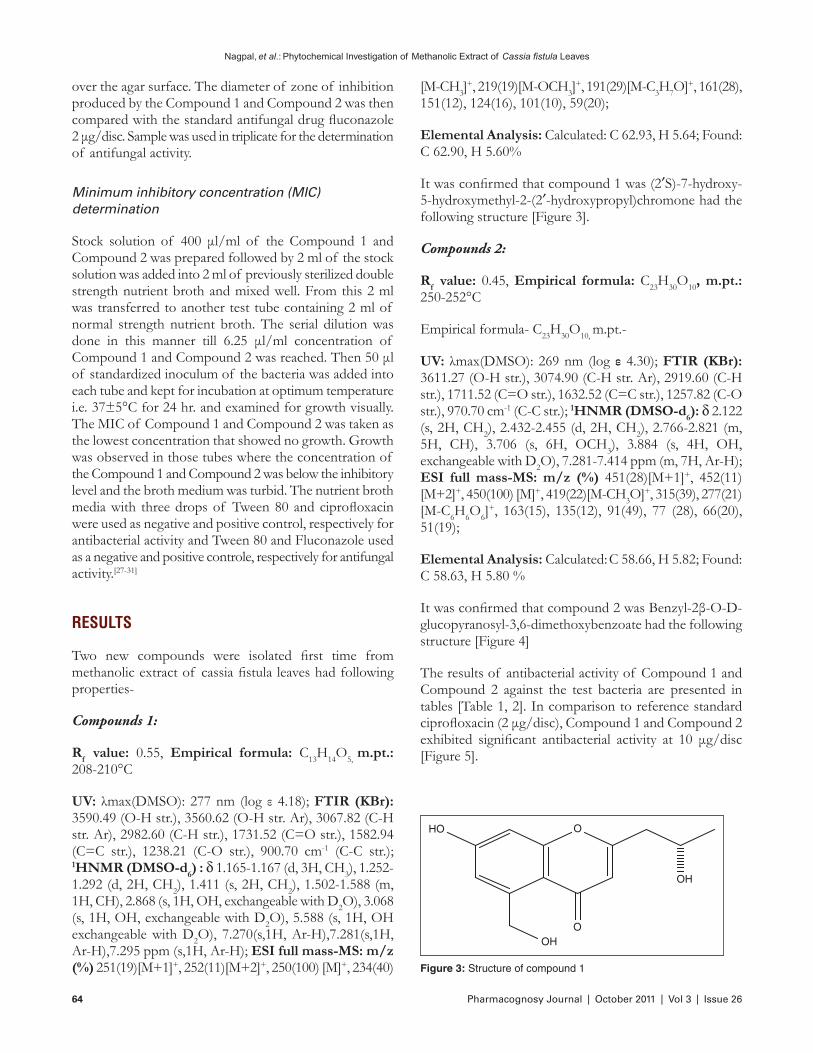
It was confirmed that compound 1 was (2′S)-7-hydroxy-5-hydroxymethyl-2-(2′-hydroxypropyl)chromone had the following structure [Figure 3].
Compounds 2:
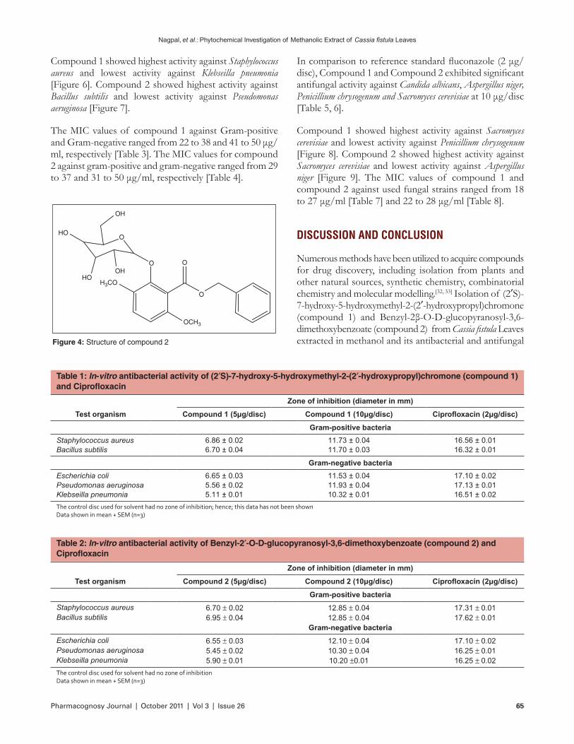
Rf value: 0.45, Empirical formula: C23H30O10, m.pt.: 250-252°C
Empirical formula- C23H30O10, m.pt.-
UV: λmax(DMSO): 269 nm (log ε 4.30); FTIR (KBr): 3611.27 (O-H str.), 3074.90 (C-H str. Ar), 2919.60 (C-H str.), 1711.52 (C=O str.), 1632.52 (C=C str.), 1257.82 (C-O str.), 970.70 cm-1 (C-C str.); 1HNMR (DMSO-d6): d 2.122 (s, 2H, CH2), 2.432-2.455 (d, 2H, CH2), 2.766-2.821 (m, 5H, CH), 3.706 (s, 6H, OCH3), 3.884 (s, 4H, OH, exchangeable with D2O), 7.281-7.414 ppm (m, 7H, Ar-H); ESI full mass-MS: m/z (%) 451(28)[M+1]+, 452(11)[M+2]+, 450(100) [M]+, 419(22)[M-CH3O]+, 315(39), 277(21)[M-C6H6O6]
+, 163(15), 135(12), 91(49), 77 (28), 66(20), 51(19);
Elemental Analysis: Calculated: C 58.66, H 5.82; Found: C 58.63, H 5.80 %
It was confirmed that compound 2 was Benzyl-2β-O-D-glucopyranosyl-3,6-dimethoxybenzoate had the following structure [Figure 4]
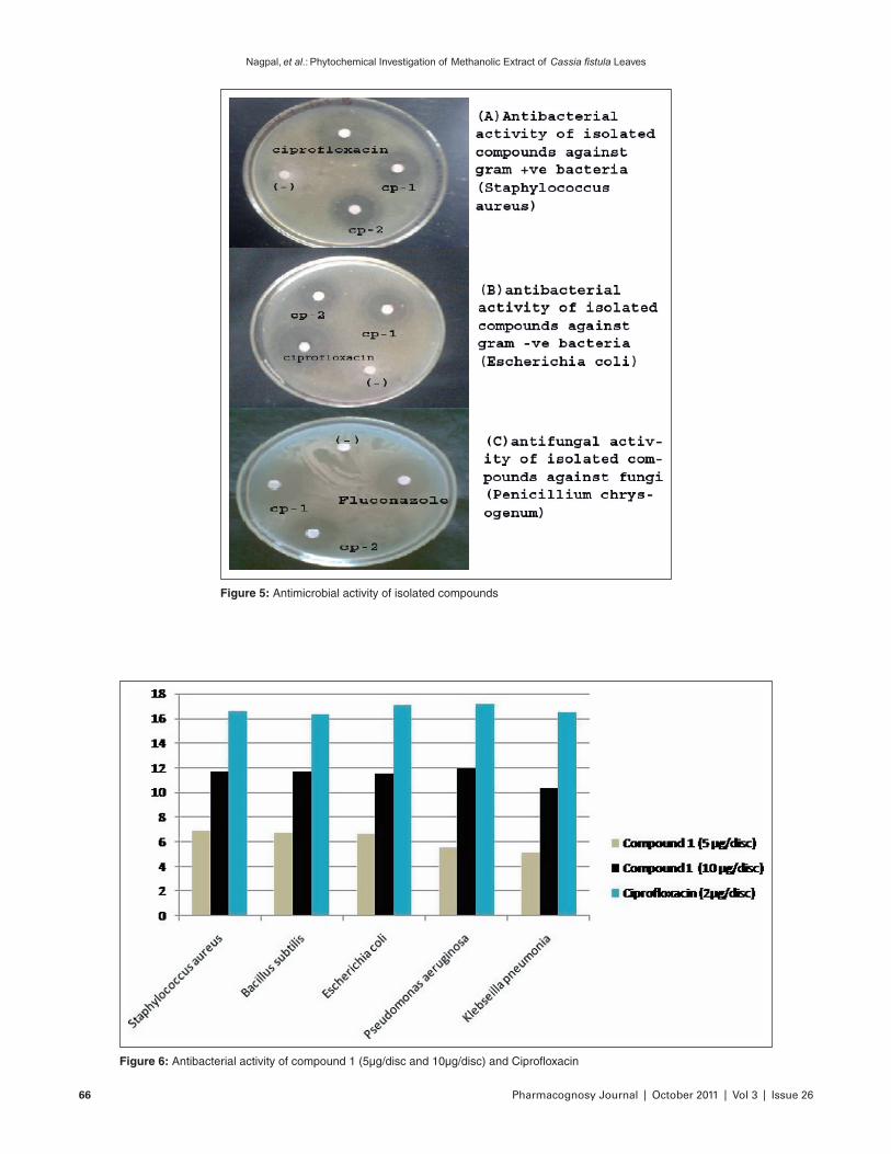
The results of antibacterial activity of Compound 1 and Compound 2 against the test bacteria are presented in tables [Table 1, 2]. In comparison to reference standard ciprofloxacin (2 μg/disc), Compound 1 and Compound 2 exhibited significant antibacterial activity at 10 μg/disc [Figure 5].
over the agar surface. The diameter of zone of inhibition produced by the Compound 1 and Compound 2 was then compared with the standard antifungal drug fluconazole 2 μg/disc. Sample was used in triplicate for the determination of antifungal activity.
Minimum inhibitory concentration (MIC) determination
Stock solution of 400 μl/ml of the Compound 1 and Compound 2 was prepared followed by 2 ml of the stock solution was added into 2 ml of previously sterilized double strength nutrient broth and mixed well. From this 2 ml was transferred to another test tube containing 2 ml of normal strength nutrient broth. The serial dilution was done in this manner till 6.25 μl/ml concentration of Compound 1 and Compound 2 was reached. Then 50 μl of standardized inoculum of the bacteria was added into each tube and kept for incubation at optimum temperature i.e. 37±5°C for 24 hr. and examined for growth visually. The MIC of Compound 1 and Compound 2 was taken as the lowest concentration that showed no growth. Growth was observed in those tubes where the concentration of the Compound 1 and Compound 2 was below the inhibitory level and the broth medium was turbid. The nutrient broth media with three drops of Tween 80 and ciprofloxacin were used as negative and positive control, respectively for antibacterial activity and Tween 80 and Fluconazole used as a negative and positive controle, respectively for antifungal activity.[27-31]
RESULTS
Two new compounds were isolated first time from methanolic extract of cassia fistula leaves had following properties-
Compounds 1:
Rf value: 0.55, Empirical formula: C13H14O5, m.pt.: 208 -210°C
UV: λmax(DMSO): 277 nm (log ε 4.18); FTIR (KBr): 3590.49 (O-H str.), 3560.62 (O-H str. Ar), 3067.82 (C-H str. Ar), 2982.60 (C-H str.), 1731.52 (C=O str.), 1582.94 (C=C str.), 1238.21 (C-O str.), 900.70 cm-1 (C-C str.); 1HNMR (DMSO-d6) : d 1.165-1.167 (d, 3H, CH3), 1.252-1.292 (d, 2H, CH2), 1.411 (s, 2H, CH2), 1.502-1.588 (m, 1H, CH), 2.868 (s, 1H, OH, exchangeable with D2O), 3.068 (s, 1H, OH, exchangeable with D2O), 5.588 (s, 1H, OH exchangeable with D2O), 7.270(s,1H, Ar-H),7.281(s,1H, Ar-H),7.295 ppm (s,1H, Ar-H); ESI full mass-MS: m/z (%) 251(19)[M+1]+, 252(11)[M+2]+, 250(100) [M]+, 234(40)
OHO
OHO
OH
Figure 3: Structure of compound 1
Pharmacognosy Journal | October 2011 | Vol 3 | Issue 26 65
Nagpal, et al.: Phytochemical Investigation of Methanolic Extract of Cassia fistula Leaves
Table 1: In-vitro antibacterial activity of (2′S)-7-hydroxy-5-hydroxymethyl-2-(2′-hydroxypropyl)chromone (compound 1) and Ciprofloxacin
Test organism
Zone of inhibition (diameter in mm)
Compound 1 (5μg/disc) Compound 1 (10μg/disc) Ciprofloxacin (2μg/disc)
Gram-positive bacteria
Staphylococcus aureus 6.86 ± 0.02 11.73 ± 0.04 16.56 ± 0.01Bacillus subtilis 6.70 ± 0.04 11.70 ± 0.03 16.32 ± 0.01
Gram-negative bacteria
Escherichia coli 6.65 ± 0.03 11.53 ± 0.04 17.10 ± 0.02Pseudomonas aeruginosa 5.56 ± 0.02 11.93 ± 0.04 17.13 ± 0.01Klebseilla pneumonia 5.11 ± 0.01 10.32 ± 0.01 16.51 ± 0.02
The control disc used for solvent had no zone of inhibition; hence; this data has not been shown Data shown in mean + SEM (n=3)
Table 2: In-vitro antibacterial activity of Benzyl-2′-O-D-glucopyranosyl-3,6-dimethoxybenzoate (compound 2) and Ciprofloxacin
Test organism
Zone of inhibition (diameter in mm)
Compound 2 (5μg/disc) Compound 2 (10μg/disc) Ciprofloxacin (2μg/disc)
Gram-positive bacteria
Staphylococcus aureus 6.70 ± 0.02 12.85 ± 0.04 17.31 ± 0.01Bacillus subtilis 6.95 ± 0.04 12.85 ± 0.04 17.62 ± 0.01
Gram-negative bacteria
Escherichia coli 6.55 ± 0.03 12.10 ± 0.04 17.10 ± 0.02Pseudomonas aeruginosa 5.45 ± 0.02 10.30 ± 0.04 16.25 ± 0.01Klebseilla pneumonia 5.90 ± 0.01 10.20 ±0.01 16.25 ± 0.02
The control disc used for solvent had no zone of inhibition Data shown in mean + SEM (n=3)
OCH3
O
H3CO
O O
O
OH
HO
HOOH
Figure 4: Structure of compound 2
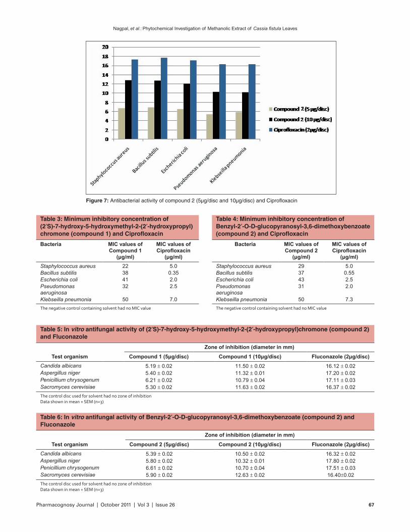
Compound 1 showed highest activity against Staphylococcus aureus and lowest activity against Klebseilla pneumonia [Figure 6]. Compound 2 showed highest activity against Bacillus subtilis and lowest activity against Pseudomonas aeruginosa [Figure 7].
The MIC values of compound 1 against Gram-positive and Gram-negative ranged from 22 to 38 and 41 to 50 μg/ml, respectively [Table 3]. The MIC values for compound 2 against gram-positive and gram-negative ranged from 29 to 37 and 31 to 50 μg/ml, respectively [Table 4].
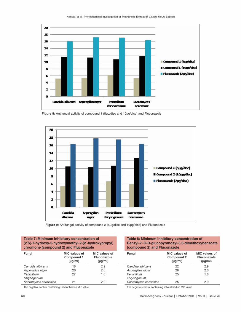
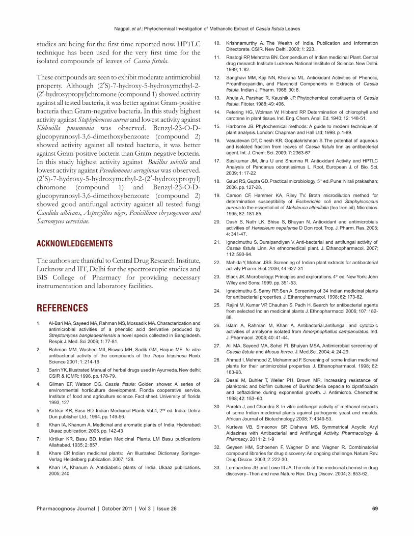
In comparison to reference standard fluconazole (2 μg/disc), Compound 1 and Compound 2 exhibited significant antifungal activity against Candida albicans, Aspergillus niger, Penicillium chrysogenum and Sacromyces cerevisiae at 10 μg/disc [Table 5, 6].
Compound 1 showed highest activity against Sacromyces cerevisiae and lowest activity against Penicillium chrysogenum [Figure 8]. Compound 2 showed highest activity against Sacromyces cerevisiae and lowest activity against Aspergillus niger [Figure 9]. The MIC values of compound 1 and compound 2 against used fungal strains ranged from 18 to 27 μg/ml [Table 7] and 22 to 28 μg/ml [Table 8].
DISCUSSION AND CONCLUSION
Numerous methods have been utilized to acquire compounds for drug discovery, including isolation from plants and other natural sources, synthetic chemistry, combinatorial chemistry and molecular modelling.[32, 33] Isolation of (2′S)-7-hydroxy-5-hydroxymethyl-2-(2′-hydroxypropyl)chromone (compound 1) and Benzyl-2β-O-D-glucopyranosyl-3,6-dimethoxybenzoate (compound 2) from Cassia fistula Leaves extracted in methanol and its antibacterial and antifungal
Nagpal, et al.: Phytochemical Investigation of Methanolic Extract of Cassia fistula Leaves
66 Pharmacognosy Journal | October 2011 | Vol 3 | Issue 26
Figure 5: Antimicrobial activity of isolated compounds
Figure 6: Antibacterial activity of compound 1 (5μg/disc and 10μg/disc) and Ciprofloxacin
Pharmacognosy Journal | October 2011 | Vol 3 | Issue 26 67
Nagpal, et al.: Phytochemical Investigation of Methanolic Extract of Cassia fistula Leaves
Figure 7: Antibacterial activity of compound 2 (5μg/disc and 10μg/disc) and Ciprofloxacin
Table 3: Minimum inhibitory concentration of (2′S)-7-hydroxy-5-hydroxymethyl-2-(2′-hydroxypropyl)chromone (compound 1) and Ciprofloxacin
Bacteria MIC values of Compound 1
(μg/ml)
MIC values of Ciprofloxacin
(μg/ml)
Staphylococcus aureus 22 5.0Bacillus subtilis 38 0.35Escherichia coli 41 2.0Pseudomonas aeruginosa
32 2.5
Klebseilla pneumonia 50 7.0
The negative control containing solvent had no MIC value
Table 4: Minimum inhibitory concentration of Benzyl-2′-O-D-glucopyranosyl-3,6-dimethoxybenzoate (compound 2) and Ciprofloxacin
Bacteria MIC values of Compound 2
(μg/ml)
MIC values of Ciprofloxacin
(μg/ml)
Staphylococcus aureus 29 5.0Bacillus subtilis 37 0.55Escherichia coli 43 2.5Pseudomonas aeruginosa
31 2.0
Klebseilla pneumonia 50 7.3
The negative control containing solvent had no MIC value
Table 5: In vitro antifungal activity of (2′S)-7-hydroxy-5-hydroxymethyl-2-(2′-hydroxypropyl)chromone (compound 2) and Fluconazole
Test organism
Zone of inhibition (diameter in mm)
Compound 1 (5μg/disc) Compound 1 (10μg/disc) Fluconazole (2μg/disc)
Candida albicans 5.19 ± 0.02 11.50 ± 0.02 16.12 ± 0.02Aspergillus niger 5.40 ± 0.02 11.32 ± 0.01 17.20 ± 0.02Penicillium chrysogenum 6.21 ± 0.02 10.79 ± 0.04 17.11 ± 0.03Sacromyces cerevisiae 5.30 ± 0.02 11.63 ± 0.02 16.37 ± 0.02
The control disc used for solvent had no zone of inhibition Data shown in mean + SEM (n=3)
Table 6: In vitro antifungal activity of Benzyl-2′-O-D-glucopyranosyl-3,6-dimethoxybenzoate (compound 2) and Fluconazole
Test organism
Zone of inhibition (diameter in mm)
Compound 2 (5μg/disc) Compound 2 (10μg/disc) Fluconazole (2μg/disc)
Candida albicans 5.39 ± 0.02 10.50 ± 0.02 16.32 ± 0.02Aspergillus niger 5.80 ± 0.02 10.32 ± 0.01 17.80 ± 0.02Penicillium chrysogenum 6.61 ± 0.02 10.70 ± 0.04 17.51 ± 0.03Sacromyces cerevisiae 5.90 ± 0.02 12.63 ± 0.02 16.40±0.02
The control disc used for solvent had no zone of inhibition Data shown in mean + SEM (n=3)
Nagpal, et al.: Phytochemical Investigation of Methanolic Extract of Cassia fistula Leaves
68 Pharmacognosy Journal | October 2011 | Vol 3 | Issue 26
Figure 8: Antifungal activity of compound 1 (5μg/disc and 10μg/disc) and Fluconazole
Figure 9: Antifungal activity of compound 2 (5μg/disc and 10μg/disc) and Fluconazole
Table 7: Minimum inhibitory concentration of (2′S)-7-hydroxy-5-hydroxymethyl-2-(2′-hydroxypropyl)chromone (compound 2) and Fluconazole
Fungi MIC values of Compound 1
(μg/ml)
MIC values of Fluconazole
(μg/ml)
Candida albicans 18 2.9Aspergillus niger 26 2.0Penicillium chrysogenum
27 1.6
Sacromyces cerevisiae 21 2.9
The negative control containing solvent had no MIC value
Table 8: Minimum inhibitory concentration of Benzyl-2′-O-D-glucopyranosyl-3,6-dimethoxybenzoate (compound 2) and Fluconazole
Fungi MIC values of Compound 2
(μg/ml)
MIC values of Fluconazole
(μg/ml)
Candida albicans 22 2.9Aspergillus niger 28 2.0Penicillium chrysogenum
25 1.6
Sacromyces cerevisiae 25 2.9
The negative control containing solvent had no MIC value
Pharmacognosy Journal | October 2011 | Vol 3 | Issue 26 69
Nagpal, et al.: Phytochemical Investigation of Methanolic Extract of Cassia fistula Leaves
10. Krishnamurthy A. The Wealth of India. Publication and Information Directorate. CSIR. New Delhi. 2000; 1: 223.
11. Rastogi RP, Mehrotra BN. Compendium of Indian medicinal Plant. Central drug research Institute Lucknow. National Institute of Science. New Delhi. 1999; 1: 82.
12. Sanghavi MM, Kaji NN, Khorana ML. Antioxidant Activities of Phenolic, Proanthocyanidin, and Flavonoid Components in Extracts of Cassia fistula. Indian J. Pharm. 1968; 30: 8.
13. Ahuja A, Parshad R, Kaushik JP. Phytochemical constituents of Cassia fistula. Fitoter. 1988; 49: 496.
14. Petering HG, Wolman W, Hibbard RP. Determination of chlorophyll and carotene in plant tissue. Ind. Eng. Chem. Anal. Ed. 1940; 12: 148-51.
15. Harborne JB. Phytochemical methods: A guide to modern technique of plant analysis. London: Chapman and Hall Ltd; 1998. p. 1-89.
16. Vasudevan DT, Dinesh KR, Gopalakrishnan S. The potential of aqueous and isolated fraction from leaves of Cassia fistula linn as antibacterial agent. Int. J. Chem. Sci. 2009; 7: 2363-67
17. Sasikumar JM, Jinu U and Shamna R. Antioxidant Activity and HPTLC Analysis of Pandanus odoratissimus L. Root, European J. of Bio. Sci. 2009; 1: 17-22
18. Gaud RS, Gupta GD. Practical microbiology: 5th ed. Pune: Nirali prakashan; 2006. pp. 127-28.
19. Carson CF, Hammer KA, Riley TV. Broth microdilution method for determination susceptibility of Escherichia coli and Staphylococcus aureus to the essential oil of Melaleuca altenifolia (tea tree oil). Microbios. 1995; 82: 181-85.
20. Dash S, Nath LK, Bhise S, Bhuyan N. Antioxidant and antimicrobials activities of Heracleum nepalense D Don root. Trop. J. Pharm. Res. 2005; 4: 341-47.
21. Ignacimuthu S, Duraipandiyan V. Anti-bacterial and antifungal activity of Cassia fistula Linn. An ethnomedical plant. J. Ethanopharmacol. 2007; 112: 590-94.
22. Mahida Y, Mohan JSS. Screening of Indian plant extracts for antibacterial activity Pharm. Biol. 2006; 44: 627-31
23. Black JK. Microbiology: Principles and explorations. 4th ed. New York: John Wiley and Sons; 1999. pp. 351-53.
24. Ignacimuthu S, Samy RP, Sen A. Screening of 34 Indian medicinal plants for antibacterial properties. J. Ethanopharmacol. 1998; 62: 173-82.
25. Rajini M, Kumar VP, Chauhan S, Padh H. Search for antibacterial agents from selected Indian medicinal plants J. Ethnopharmacol 2006; 107: 182-88.
26. Islam A, Rahman M, Khan A. Antibacterial,antifungal and cytotoxic activities of amblyone isolated from Amorphophallus campanulatus. Ind. J. Pharmacol. 2008; 40: 41-44.
27. Ali MA, Sayeed MA, Sohel FI, Bhuiyan MSA. Antimicrobial screening of Cassia fistula and Mesua ferrea. J. Med.Sci. 2004; 4: 24-29.
28. Ahmad I, Mehmood Z, Mohammad F. Screening of some Indian medicinal plants for their antimicrobial properties J. Ethanopharmacol. 1998; 62: 183-93.
29. Desai M, Buhler T, Weller PH, Brown MR. Increasing resistance of planktonic and biofilm cultures of Burkholderia cepacia to ciprafloxacin and ceftazidime during exponential growth. J. Antimicrob. Chemother. 1998; 42: 153–60.
30. Parekh J, and Chandra S. In vitro antifungal activity of methanol extracts of some Indian medicinal plants against pathogenic yeast and moulds. African Journal of Biotechnology. 2008; 7: 4349-53.
31. Kurteva VB, Simeonov SP, Disheva MS. Symmetrical Acyclic Aryl Aldazines with Antibacterial and Antifungal Activity. Pharmacology & Pharmacy. 2011; 2: 1-9
32. Geysen HM, Schoenen F, Wagner D and Wagner R. Combinatorial compound libraries for drug discovery: An ongoing challenge. Nature Rev. Drug Discov. 2003; 2: 222-30.
33. Lombardino JG and Lowe III JA. The role of the medicinal chemist in drug discovery–Then and now. Nature Rev. Drug Discov. 2004; 3: 853-62.
studies are being for the first time reported now. HPTLC technique has been used for the very first time for the isolated compounds of leaves of Cassia fistula.
These compounds are seen to exhibit moderate antimicrobial property. Although (2′S)-7-hydroxy-5-hydroxymethyl-2-(2′-hydroxypropyl)chromone (compound 1) showed activity against all tested bacteria, it was better against Gram-positive bacteria than Gram-negative bacteria. In this study highest activity against Staphylococcus aureus and lowest activity against Klebseilla pneumonia was observed. Benzyl-2β-O-D-glucopyranosyl-3,6-dimethoxybenzoate (compound 2) showed activity against all tested bacteria, it was better against Gram-positive bacteria than Gram-negative bacteria. In this study highest activity against Bacillus subtilis and lowest activity against Pseudomonas aeruginosa was observed. (2′S)-7-hydroxy-5-hydroxymethyl-2-(2′-hydroxypropyl)chromone (compound 1) and Benzyl-2β-O-D-glucopyranosyl-3,6-dimethoxybenzoate (compound 2) showed good antifungal activity against all tested fungi Candida albicans, Aspergillus niger, Penicillium chrysogenum and Sacromyces cerevisiae.
ACKNOWLEDGEMENTS
The authors are thankful to Central Drug Research Institute, Lucknow and IIT, Delhi for the spectroscopic studies and BIS College of Pharmacy for providing necessary instrumentation and laboratory facilities.
REFERENCES1. Al-Bari MA, Sayeed MA, Rahman MS, Mossadik MA. Characterization and
antimicrobial activities of a phenolic acid derivative produced by Streptomyces bangladeshiensis a novel specis collected in Bangladesh. Respir. J. Med. Sci 2006; 1: 77-81.
2. Rahman MM, Washed MII, Biswas MH, Sadik GM, Haque ME. In vitro antibacterial activity of the compounds of the Trapa bispinosa Roxb. Science 2001; 1: 214-16
3. Sarin YK. Illustrated Manual of herbal drugs used in Ayurveda. New delhi: CSIR & ICMR; 1996. pp. 178-79.
4. Gilman EF, Watson DG. Cassia fistula: Golden shower. A series of environmental horticulture development. Florida cooperative service. Institute of food and agriculture science. Fact sheet. University of florida 1993, 127
5. Kirtikar KR, Basu BD. Indian Medicinal Plants.Vol.4, 2nd ed. India: Dehra Dun publisher Ltd.; 1994. pp. 149-56.
6. Khan IA, Khanum A. Medicinal and aromatic plants of India. Hyderabad: Ukaaz publication; 2005. pp. 142-43
7. Kirtikar KR, Basu BD. Indian Medicinal Plants. LM Basu publications Allahabad. 1935; 2: 857.
8. Khare CP. Indian medicinal plants: An Illustrated Dictionary. Springer-Verlag Heidelberg publication. 2007; 128.
9. Khan IA, Khanum A. Antidiabetic plants of India. Ukaaz publications. 2005; 240.