Embed Size (px)
Citation preview
PHYTOCHEMICAL, PHARMACOLOGICAL ACTIVITY
AND MOLECULAR DOCKING INVESTIGATIONS OF
Tetracera indica (Christm. & Panz.) Merr., Averrhoa
bilimbi Linn. AND Gymnanthemum glaberrimum
(Welw. ex O.Hoffm.) H. Rob.
BY
ALHASSAN MUHAMMAD ALHASSAN
A thesis submitted in fulfillment of the requirement for the
degree of Doctor of Philosophy in Pharmaceutical Sciences
(Pharmaceutical Chemistry)
Kulliyyah of Pharmacy
International Islamic University Malaysia
APRIL 2018
ii
ABSTRACT
Tetracera indica and Averrhoa bilimbi are used in traditional treatment of diabetes
mellitus while Gymnanthemum glaberrimum is used as a remedy for skin cancer. The
objectives of the present study were to investigate the pharmacological activity of
leaves of T. indica, G. glaberrimum and A. bilimbi, and their phytochemical
constituents. Ethanol extract from the leaves of T. indica was subjected to α-
glucosidase inhibitory activity evaluation and isolation of bioactive compounds
followed by molecular docking. Methanol extract of G. glaberrium leaves was
subjected to cytotoxic investigations on A375, HT-29 and MCF7 cancer cell lines,
followed by isolation of bioactive constituents and molecular docking. Purified
compounds were characterized using spectroscopic analysis. Furthermore, the
antioxidant activity of methanol extract of A. bilimbi leaves and its fractions was
investigated and the active compounds were identified by LC-MS-QTOF analysis.
Nine compounds were isolated from T. indica leaves which include; lupeol, betulinic
acid, wogonin, norwogonin, quercetin, tectochrysin, kaempferol and two new
sulphated flavones viz. 5,7-dihydroxyflavone-O-8-sulphate and 5-hydroxy-8-
methoxyflavone-O-7-sulphate. Quercetin, kaempferol and 5,7-dihydroxyflavone-O-
8-sulphate showed significant (P<0.05) α-glucosidase inhibitory activity with IC50
values of 61.86 ± 2.4, 68.46 ± 3.5 and 133.57 ± 5.2 μM respectively, compared to
acarbose (IC50 419.42 ± 20.29 µM). Molecular docking result showed favourable
mode of interactions of these flavonoids in the active site of α-glucosidase.
Furthermore, four compounds viz. nonacosanoic acid, lupeol, 5-methylcoumarin-4-β-
glucoside and 4-hydroxy-5-methylcoumarin were isolated from G. glaberrimum.
Lupeol displayed significant (P<0.05) antiproliferative effect against MCF-7 cell
lines with IC50 of 34.15 ± 2.32 µg/mL. Lupeol and 5-methylcoumarin-4-β-glucoside
displayed moderate cytotoxic effect on A375 cells with IC50 of 59.18 ± 2.70 and
91.84 ± 6.78 µg/mL. 4-Hydroxy-5-methylcoumarin, and lupeol displayed significant
(P<0.01) cytotoxic effect on HT-29 cells with IC50 of 4.22 ± 0.13, 15.24 ± 1.15
µg/mL respectively, which is comparable with 5-fluorouracil (IC50 8.00 ± 0.78
µg/mL). CDK2 receptor and CA IX and XII were identified through molecular
docking as potential target for these compounds. The n-butanol fraction of A. bilimbi
extract displayed significant (P<0.05) DPPH radical scavenging effect with IC50
(4.14 ± 0.21 μg/mL). It also exhibited significant (P<0.05) XO inhibitory effect with
IC50 of 64.84 ± 3.93 μg/mL. Afzelechin 3-O-alpha-L-rhamnopyranoside and
cucumerin A were identified through LC-MS-QTOF as possible metabolites
responsible for the antioxidant activity of the n-butanol fraction.
iii
خلاصة البحثABSTRACT IN ARABIC
أما نبات جمنازيمم جلابيريمم شعبيا لعلاج داء السكر نايستخدمسيرا انديكا و ايفوريا بلمبي نباتات تيترا لتلك النباتات الحالية هي دراسة النشاط الدوائيكعلاج لسرطان الجلد. أهداف الدراسة يتسخدم
تيتراسيرا انديكا في تثبيط انزايم مستخلص الإيثانول من أوراقتم فحص فاعلية . ومكوناتهن الكيميائية الديثانول من أوراق مستخلص في حين تم التحقق من سمية. الالفا غليجوزيز وتم عزل الدركبات الفعالة
، تليها عزل الدكونات MCF7وA375 ،HT-29خطوط الخلايا السرطاني ستخدامجمنازيمم جلابيريمم باتم وصف الدركبات النقية باستخدام التحليل الطيفي. تم دراسة النشاط الدضاد النشطة بيولوجيا.
وتم تحديد الدركبات النشطة بواسطة تحليل مشتقاتهاو ايفوريا بلمبي للأكسدة لدستخلص الديثانول من أوراقLC-MS-QTOF. وحمضتشمل لوبيول والتيتيتراسيرا انديكا تم عزل تسعة مركبات من أوراق
، كيمبيفرول بالاضافة الى اثنين من ، كيرسيتين ، تيكتوكيرسين ، نوروجونين ،وجونين البوتيلينيك ثوكسي ميهيدروكسي -5 و ثماني الكبريت-ديهيدروكسي فلافون-7،9وهما مركبات الفلافون الكبريتيية
ثماني الكبريت-ديهيدروكسي فلافون-5،5أظهر كيرسيتين و كيمبيفرول و .سباعي الكبريت-فلافون
± 855.79و 5.7± 68.:8و 4.6± 8:.88من IC50 مع قيم (P <0.05) الالفا غلجوزيدز نشاط تثبيط
7.4 Mμ على التوالي ، مقارنة مع.( اكاربوزIC50 419.42± Mμ20.29 .) طريقة مواتية نتائجالأظهرتوعلاوة على ذلك ، تم عزل أربعة الالفا غليجوزيز للتفاعلات بين هذه الفلافونيدات في الدوقع النشط لـ
بيتا -6ميثيل كومارين -7 كسانويك، لوبيول ،ونا حمض نوهم من نبات جمنازيمم جلابيريمم مركبات ضد خطوط (P <0.05) معتبرتأثير ل لوبيو ميثيل كومارين. أظهر -7هيدروكسي -6جلوكوزايد و
ميثيل -7و كذلك أظهر كل من لوبيول و ميكروغرام/مل. 2.52± 51.45من IC50 مع MCF-7 الخلية± 14.51و 2.52± 51.45من IC50 مع A375 خلاياضد تأثير متوسط بيتا جلوكوزايد -6كومارين
ميثيل -7هيدروكسي -6بالإضافة إلى ذلك فقد أظهر كل من لوبيول و ميكروجرام / مل. 8.55 4.45± 45.21، 2.45± 1.22من IC50 مع HT-29 على سام للخلايا (P <0.05)تأثير كومارين
IC50 8.00) . ميكروجرام / مل) فلورويوراسيل-7ميكروغرام / مل على التوالي ، والتي يمكن مقارنتها مع
من خلال الالتحام الجزيئي كهدف XII و CA IX و CDK2 تتم تحديد مستقبلاكذلك 0.78 ± DPPH (P <0.05) جذري ايفوريا بلمبي تأثير من مستخلصبيوتانول -nمحتمل لذذه الدركبات. أظهر
IC50) ولكن أقل من (P <0.05) . كما أظهر تأثيراً كبيراً(ميكروجرام / مل 1.41 ±2.24) IC50 مع
64.84 ± 3.93 μg / mL) XO . كيومارين تم تحديدأخيراA من الفا رامنو بيرانوزايد -3افزيليسين و بيوتانول.-nلدركب ممكنة مسؤولة عن النشاط الدضاد للأكسدة نواتجك LC-MS-QTOF خلال
iv
APPROVAL PAGE
The thesis of Alhassan Muhammad Alhassan has been approved by the following:
_____________________________
Qamar Uddin Ahmed
Supervisor
____________________________
Alfi Khatib
Co-Supervisor
_____________________________
Siti Zaiton Mat So’ad
Internal Examiner
_____________________________
Jamia Azdina Jamal
External Examiner
_____________________________
Zainul Amiruddin Zakaria
External Examiner
_____________________________
Siti Aesah @ Naznin Muhammad
Chairman
v
DECLARATION
I hereby declare that this thesis is the result of my own investigations, except where
otherwise stated. I also declare that it has not been previously or concurrently
submitted as a whole for any other degrees at IIUM or other institutions.
Alhassan Muhammad Alhassan
Signature .................................................. Date .........................................
vi
COPYRIGHT PAGE
INTERNATIONAL ISLAMIC UNIVERSITY MALAYSIA
DECLARATION OF COPYRIGHT AND AFFIRMATION OF
FAIR USE OF UNPUBLISHED RESEARCH
PHYTOCHEMICAL, PHARMACOLOGICAL ACTIVITY AND
MOLECULAR DOCKING INVESTIGATIONS OF Tetracera
indica (Christm. & Panz.) Merr., Averrhoa bilimbi Linn. AND
Gymnanthemum glaberrimum (Welw. ex O.Hoffm.) H. Rob.
I declare that the copyright holders of this thesis are jointly owned by Alhassan
Muhammad Alhassan and IIUM.
Copyright © 2018 Alhassan Muhammad Alhassan and International Islamic University Malaysia.
All rights reserved.
No part of this unpublished research may be reproduced, stored in a retrieval
system, or transmitted, in any form or by any means, electronic, mechanical,
photocopying, recording or otherwise without prior written permission of the
copyright holder except as provided below
1. Any material contained in or derived from this unpublished research
may be used by others in their writing with due acknowledgement.
2. IIUM or its library will have the right to make and transmit copies
(print or electronic) for institutional and academic purposes.
3. The IIUM library will have the right to make, store in a retrieved
system and supply copies of this unpublished research if requested by
other universities and research libraries.
By signing this form, I acknowledged that I have read and understand the IIUM
Intellectual Property Right and Commercialization policy.
Affirmed by Alhassan Muhammad Alhassan
……..…………………….. ………………………..
Signature Date
vii
This thesis is dedicated to my father, Late Qadi Muhammad Alhassan who
encouraged me to undertake this PhD and provided ample resources towards its
accomplishment, but, as Allah so wished he did not survive to witness its completion.
May Allah (SWT) have mercy on him and grant him Al-jannatul firdaus.
viii
ACKNOWLEDGEMENTS
In the name of Allah, the Most Gracious, the Most Merciful. All praises and
adoration are due to Allah alone (SWT) by whose mercy this work was
accomplished. May His peace and blessings be upon Prophet Muhammad (SAW),
his household, companions and all those that follow them with sincerity and piety.
I wish to express my sincere gratitude to my supervisor Assoc. Prof. Dr.
Qamar Uddin Ahmed for his guidance, continuous support, encouragement and
leadership, and for that, I will be forever grateful.
I am grateful to members of my supervisory committee and all those who
provided their time, effort and support for this project.
I wish to express my profound appreciation to my dear mother; Hajiya
Zainab, step mother; Hajiya Hadiza, wife; Hauwa, children; Khadija and Fatima,
brothers; Aliyu, Aminu, Lukman and sisters; Hajiya Hadiza and Hajiya Rakiya. Your
love, patience and support have been very wonderful and were vital to the successful
achievement of this goal.
I also wish to express my gratitude to Usmanu Danfodiyo University Sokoto,
Nigeria, for giving me the fellowship opportunity to undertake this study under the
Tertiary Education Fund (Tetfund), 2013/2014 Intervention.
Finally, I am extremely indebted to the Ministry of Higher Education
(MOHE), Malaysia and the Research Management Center, IIUM for providing
financial support through Fundamental Grant Research Scheme (FRGS 13-089-
0330), and Research Initiative Grant Schemes (RIGS 16-294-0458), respectively to
accomplish this work.
ix
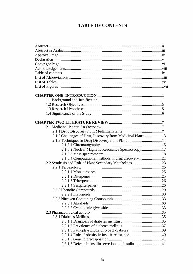
TABLE OF CONTENTS
Abstract .................................................................................................................... ii Abstract in Arabic .................................................................................................... iii Approval Page .......................................................................................................... iv
Declaration ............................................................................................................... v Copyright Page ......................................................................................................... vi Acknowledgements .................................................................................................. viii Table of contents ...................................................................................................... ix List of Abbreviations ............................................................................................... xiii
List of Tables ........................................................................................................... xv List of Figures .......................................................................................................... xvii
CHAPTER ONE INTRODUCTION .................................................................. 1 1.1 Background and Justification ................................................................. 1 1.2 Research Objectives................................................................................ 5 1.3 Research Hypotheses .............................................................................. 5
1.4 Significance of the Study ........................................................................ 6
CHAPTER TWO LITERATURE REVIEW ...................................................... 7 2.1 Medicinal Plants: An Overview.............................................................. 7
2.1.1 Drug Discovery from Medicinal Plants ........................................ 7
2.1.2 Challenges of Drug Discovery from Medicinal Plants ................. 13
2.1.3 Techniques in Drug Discovery from Plant ................................... 14 2.1.3.1 Chromatography ............................................................... 15 2.1.3.2 Nuclear Magnetic Resonance Spectroscopy ..................... 17
2.1.3.3 Mass spectrometry ............................................................ 18 2.1.3.4 Computational methods in drug discovery ....................... 21
2.2 Synthesis and Role of Plant Secondary Metabolites .............................. 23 2.2.1 Terpenoids ..................................................................................... 25
2.2.1.1 Monoterpenes ................................................................... 25 2.2.1.2 Diterpenes ......................................................................... 25 2.2.1.3 Triterpenes ........................................................................ 26
2.2.1.4 Sesquiterpenes .................................................................. 26
2.2.2 Phenolic Compounds .................................................................... 29 2.2.2.1 Flavonoids ........................................................................ 30
2.2.3 Nitrogen Containing Compounds ................................................. 33
2.2.3.1 Alkaloids ........................................................................... 33 2.2.3.2 Cyanogenic glycosides ..................................................... 33
2.3 Pharmacological activity ........................................................................ 35 2.3.1 Diabetes Mellitus .......................................................................... 35
2.3.1.1 Diagnosis of diabetes mellitus .......................................... 35
2.3.1.2 Prevalence of diabetes mellitus ........................................ 37 2.3.1.3 Pathophysiology of type 2 diabetes .................................. 39 2.3.1.4 Role of obesity in insulin resistance ................................. 40 2.3.1.5 Genetic predisposition ...................................................... 41
2.3.1.6 Defects in insulin secretion and insulin action ................. 41
x
2.3.1.7 Oral hypoglycemic agents ................................................ 42
2.3.2 Cancer: The Role of Plant Based Natural Products ...................... 44 2.4 Dilleniacea Family .................................................................................. 45
2.4.1 Genus Tetracera ............................................................................ 46 2.4.2 Tetracera indica (Christm. & Panz.) Merr. .................................. 46
2.5 Asteraceae Family .................................................................................. 48 2.5.1 Genus Gymnanthemum ................................................................. 49 2.5.2 Gymnanthemum glaberrimum (Welw. ex O.Hoffm) H. Rob ....... 50
2.6 Oxalidaceae Family ................................................................................ 51 2.6.1 Genus Averrhoa ............................................................................ 52 2.6.2 Averrhoa bilimbi Linn ................................................................... 52
CHAPTER THREE PHYTOCHEMICAL INVESTIGATION AND
ALPHA-GLUCOSIDASE INHIBITORY ACTIVITY OF Tetracera indica
Christm. & Panz.) Merr. LEAVES ...................................................................... 55 3.1 Introduction............................................................................................. 55 3.2 Materials and Methods ........................................................................... 56
3.2.1 Chemicals ...................................................................................... 56 3.2.2 Characterization and Spectroscopic Analysis ............................... 56
3.2.3 Phytochemical Tests ..................................................................... 57 3.2.4 Isolation and Purification of compounds from the Leaves of T.
indica ............................................................................................ 58 3.2.4.1 Plant collection and identification .................................... 58 3.2.4.2 Preparation of ethanol extract of T. indica leaves ............ 58
3.2.4.3 Chromatographic fractionation of crude ethanolic extract 59 3.2.4.4 Chloroform fraction .......................................................... 60
3.2.4.5 EtOAc fraction .................................................................. 61 3.2.4.6 Methanol fraction ............................................................. 62
3.2.5 Alpha-glucosidase Inhibition Assay ............................................. 63 3.2.5.1 Statistical analysis............................................................. 63
3.2.6 Molecular Docking Studies ........................................................... 64
3.2.6.1 Software and programs ..................................................... 64
3.2.6.2 Receptor and ligand preparation ....................................... 64 3.2.6.3 Molecular docking experiment ......................................... 65
3.3 Results and Discussion ........................................................................... 65 3.3.1 Isolation and Characterization of Bioactive Compounds of
Tetracera indica ........................................................................... 65 3.3.1.1 MAQ-1 (5,7-dihydroxy-8-methoxyflavone) (wogonin) ... 65 3.3.1.2 MAQ-2 (3,5,7,4ʹ-tetrahydroxyflavonol) (kaempferol) ..... 68
3.3.1.3 MAQ-3 (3,5,7,3′,4′-pentahydroxyflavonol) (quercetin) ... 71 3.3.1.4 MAQ-4 (5-hydroxyl-7-methoxyflavone) ......................... 75 3.3.1.5 MAQ-5 (5,7,8-trihydroxyflavone) (norwogonin) ............. 77 3.3.1.6 MAQ-6 (lupeol) ................................................................ 80 3.3.1.7 MAQ-7 Betulinic acid ...................................................... 83
3.3.1.8 MAQ-8 (5,7-dihydroxyflavone-O-8-sulphate) ................. 86 3.3.1.9 MAQ-9 (5-hydroxy-8-methoxyflavone-O-sulpate) .......... 90
3.3.2 Alpha-glucosidase Inhibitory Activity Evaluation ....................... 94 3.4 Molecular Docking ................................................................................. 96
3.5 Conclusion .............................................................................................. 104
xi
CHAPTER FOUR PHYTOCHEMICAL AND ANTIPROLIFERATIVE
INVESTIGATIONS OF Gymnanthemum glaberrimum (Welw. ex O.
Hoffm.) H. Rob ....................................................................................................... 110 4.1 Introduction............................................................................................. 110 4.2 Materials and Methods ........................................................................... 111
4.2.1 Collection and Identification of G. glaberrimum Leaves ............ 111 4.2.2 Preparation of Methanol Extract of G. glaberrimum Leaves ....... 111 4.2.3 Isolation and Purification of Bioactive Constituents .................... 112
4.2.3.1 Methanol fraction ............................................................. 112 4.2.3.2 Hydrolysis of VMQ-3 ....................................................... 113 4.2.3.3 Chloroform fraction .......................................................... 113 4.2.3.4 Hexane fraction ................................................................. 113
4.2.4 Characterization and Spectroscopic Analysis ............................... 114 4.2.5 In vitro Cytotoxic Evaluation ........................................................ 115
4.2.5.1 Cell lines and reagents ...................................................... 115
4.2.5.2 Cell culture techniques ..................................................... 115 4.2.5.3 Thawing cells .................................................................... 116 4.2.5.4 Sub culturing cells ............................................................ 116 4.2.5.5 Measuring cell viability using Trypan blue exclusion assay
method (TBEA) ............................................................................ 116 4.2.5.6 Seeding of cells for treatment ........................................... 117
4.2.5.7 Determination of 50 % inhibitory concentration (IC50) using
MTS (3-(4,5-dimethylthiazol-2-yl)-5-(3-carboxymethoxyphenyl)-2-
(4-sulfophenyl)-2H-tetrazolium) cell proliferation assay ............. 118
4.2.6 Statistical Analysis ........................................................................ 119 4.2.7 Molecular Docking ....................................................................... 119
4.3 Results and Discussion ........................................................................... 120 4.3.1 Isolation and Characterization ...................................................... 120
4.3.1.1 VMQ-1 (Nonacosanoic acid) ............................................ 121 4.3.1.2 VMQ-2 (lupeol) ................................................................ 123 4.3.1.3 VMQ-3 (5-Methylcoumarin-4-β-glucoside)..................... 126
4.3.1.4 VMQ-4 (4-hydroxy-5-methylcoumarin) .......................... 130
4.3.2 Cytotoxicity Studies ...................................................................... 132 4.3.3 Molecular Docking ....................................................................... 136
4.4 Conclusion .............................................................................................. 149
CHAPTER FIVE ANTIOXIDANT EVALUATON OF Averrhoa bilimbi
LEAVES AND IDENTIFICATION OF BIOACTIVE CONSTITUENTS ...... 151 THROUGH LC-MS ............................................................................................... 151
5.1 Introduction............................................................................................. 151 5.2 Materials and Methods ........................................................................... 152
5.2.1 Materials........................................................................................ 152 5.2.2 Plant Collection and Processing .................................................... 152 5.2.3 Extraction and Fractionation ......................................................... 153
5.2.4 Preliminary Phytochemical Screening .......................................... 153 5.2.5 LC-MS QTOF Analysis of Bioactive Constituents of n-
Butanol Fraction ........................................................................... 155 5.2.6 DPPH Free Radical Scavenging Activity ..................................... 155
5.2.7 Xanthine Oxidase Inhibitory Activity Assay ................................ 156
xii
5.2.8 Molecular Docking ....................................................................... 157
5.2.9 Statistical Analysis ........................................................................ 157 5.3 Results and Discussions .......................................................................... 158
5.3.1 Antioxidant Capacities .................................................................. 158 5.3.2 DPPH Radical Scavenging Activity ............................................. 159 5.3.3 Xanthine Oxidase Inhibitory Activity ........................................... 160 5.3.4 LC-MS QTOF Analysis ................................................................ 162 5.3.5 Identification of Compounds through MS/MS Fragmentation
Analysis ........................................................................................ 164 5.3.6 Molecular Docking Studies ........................................................... 171
5.4 Conclusion .............................................................................................. 174
CHAPTER SIX CONCLUSION AND RECOMMENDATION ....................... 176 6.1 Conclusion .............................................................................................. 176 6.2 Recommendations................................................................................... 177
REFERENCES ...................................................................................................... 179
Appendix 1 Spectra of MAQ-1 ............................................................................. 196 Appendix 2 Spectra of MAQ-2 ............................................................................. 203 Appendix 3 Spectra of MAQ-3 ............................................................................. 210
Appendix 4 Spectra of MAQ-4 ............................................................................. 217 Appendix 5 Spectra of MAQ-5 ............................................................................. 223
Appendix 6 Spectra of MAQ-6 ............................................................................. 230
Appendix 7 Spectra of MAQ-7 ............................................................................. 237
Appendix 8 Spectra of MAQ-8 ............................................................................. 244 Appendix 9 Spectra of MAQ-9 ............................................................................. 251
Appendix 10 Spectra of VMQ-1 ........................................................................... 259 Appendix 11 Spectra of VMQ-2 ........................................................................... 264 Appendix 12 Spectra of VMQ-3 ........................................................................... 271
Appendix 13 Spectra of VMQ-4 ........................................................................... 278 Appendix 14 PUBLICATIONS ............................................................................ 284
xiii
LIST OF ABBREVIATIONS
Ac Acetone
A375 cells Human malignant melanoma cells 13
C-NMR 13
Carbon nuclear magnetic resonance
C Carbon
CC Column chromatography
CDCl3 Deuterated chloroform
COSY Homonuclear Correlation Spectroscopy
CHCl3 Chloroform
2D Two dimensional
dd Doublet of the doublet
dL Deci litre
DCM Dichloromethane
DMSO Dimethyl sulphoxide
DPPH 2, 2-diphenyl-1-1-picrylhydrazyl radical
EtOH Ethanol
EtOAc Ethyl acetate
FeCl3 Ferric chloride
Fig. Figure
FTIR Fourier transform infrared spectroscopy
GC Gas chromatography 1H-NMR Proton nuclear magnetic resonance
HCl Hydrochloric acid
HPLC High performance liquid chromatography
HSQC Heteronuclear single quantum correlation
HT-29 cells Human caucasian colon adenocarcininoma cells
HMBC Heteronuclear multiple bond correlation
Hz Hertz
H2SO4 Sulphuric acid
IDDM Insulin dependent diabetes mellitus
I2 Iodine
J Coupling constant
Kg Kilogram
m/z Mass-to-charge ratio
MeOD Deuterated methanol
MeOH Methanol
MCF-7 cells Human breast adenocarcinoma cells
MS Mass Spectrometry
m Multiplet
Mg Milligram
NMR Nuclear magnetic resonance spectroscopy
NOESY Nuclear overhauser enhancement spectroscopy
NIDDM Non-insulin dependent diabetes mellitus
OGTT Oral glucose tolerance test
prep. Preparative
ppm Parts per million
xiv
s Singlet
t Triplet
T:E:F Toluene: Ethyl formate: Formic acid
TLC Thin layer chromatography
UV Ultraviolet
2D Two dimensional
xv
LIST OF TABLES
Table No. Page No.
2.1 Prevalence of diabetes and estimated diabetic people’s numbers
by region among adults aged 20–79 years for the years 2015
and 2040
38
3.1 1H and
13C-NMR spectral values for MAQ-1 68
3.2 1H and
13C-NMR spectral values for MAQ-2 71
3.3 1H and
13C-NMR spectral values for MAQ-3 74
3.4 1H and
13C-NMR spectral values for MAQ-4 77
3.5 1H and
13C-NMR spectral values for MAQ-5 79
3.6 1H and
13C-NMR spectral values for MAQ-6 82
3.7 1H and
13C-NMR spectral values for MAQ-7 85
3.8 1H and
13C-NMR spectral values for MAQ-8 89
3.9 1H and
13C-NMR spectral values for MAQ-9 93
3.10 IC50 for extracts and fractions of Tetracera indica 94
3.11 Alpha-glucosidase inhibitory activity of isolated compounds of
Tetracera indica
96
3.12 Docking scores of α-glucosidase inhibitors 97
3.13 Physical properties of the isolated compounds 105
4.1 1H and
13C-NMR spectral values for VMQ-1 122
4.2 1H and
13C-NMR spectral values for VMQ-2 125
4.3 1H and
13C-NMR spectral values for VMQ-3 129
4.4 1H and
13C-NMR spectral values for VMQ-4 131
4.5 The IC50 values for the cytotoxic activity of crude methanolic
extract of Gymnanthemum glaberrimum
132
4.6 IC50 values for 72 hours treatment with Gymnanthemum
glaberrimum compounds on different cell lines
134
xvi
4.7 Docking result for VMQ-2 on CDK2 137
4.8 Docking score of compounds on carbonic anhydrase XII 143
4.9 Docking score of compounds on carbonic anhydrase IX 143
4.10 Physical properties of the isolated compounds 150
5.1 Phytochemical screening for chemical class identification of
Averrhoa bilimbi
159
5.2 Free radical scavenging activity of crude methanolic extract of
Averrhoa bilimbi and fractions
160
5.3 IC50 for XO inhibitory activity of crude methanolic extract and
fractions
162
5.4 Results of LC-MS Q-TOF analysis of n-butanol fraction 163
5.5 Docking scores of cucumerin A, afzelechin 3-O-alpha-L-
rhamnopyranoside and acarbose on α-glucosidase
171
xvii
LIST OF FIGURES
Figure No. Page No.
2.1 Schematic representations of a typical medicinal
plant drug discovery process 8
2.2a Structures of some clinically useful drugs
developed through medicinal plant drug discovery
process
11
2.2b Structures of some clinically useful drugs
developed through medicinal plant drug discovery
process
12
2.3 Generic schemes for isolation of natural products 17
2.4 Basic components in a mass spectrometer 19
2.5 Mevalonic acid pathways showing the synthesis of
steroidal terpene-lanosterol
27
2.6 Structures of monoterpenes 28
2.7 Structures of some diterpenoids based compounds 28
2.8 Chemical structures of triterpenoids 29
2.9 Structure of main classes of flavonoids 31
2.10 Biosynthetic pathway for flavonoids 32
2.11 Different types of cyanogenic glycosides 34
2.12 Prevalence of diabetes mellitus in adults aged 18
years and above between 2006 and 2015
39
2.13 Photograph image of Tetracera indica leaves 48
2.14 Photograph image of Gymnanthemum glaberrimum 51
3.1 Structure of MAQ-1 67
3.2 Structure of MAQ-2 70
3.3 Structure of MAQ-3 73
3.4 Structure of MAQ-4 76
xviii
3.5 Structure of MAQ-5 79
3.6 Structure of MAQ-6 81
3.7 Structure of MAQ-7 84
3.8a HMBC of MAQ-8 88
3.8b Structure of MAQ-8 89
3.9 Structure of MAQ-9 92
3.10 HMBC of MAQ-9 92
3.11 Alpha-glucosidase inhibitory activity of crude
extracts and fractions of Tetracera. indica leaves
100
3.12 Surface structure of yeast α-glucosidase (PDB ID:
3A4A) with maltose posed in the catalytic pocket.
100
3.13 Binding interactions of MAQ-1 with active site
residues of yeast α-glucosidase
101
3.14 Binding interactions MAQ-5 with active site
residues of yeast α-glucosidase
101
3.15 Binding interactions of MAQ-2 with active site
residues of yeast α-glucosidase
102
3.16 Binding interactions of MAQ-3 with active site
residues of yeast α-glucosidase
102
3.17 Binding interactions of MAQ-8 with active site
residues of yeast α-glucosidase
103
3.18 Binding interactions of acarbose with active site
residues of yeast α-glucosidase
103
3.19 Surface structure of yeast α-glucosidase (PDB ID:
3A4A) with acarbose posed in the catalytic pocket
104
3.20 Flow chart for the preparation of ethanol extract
and fractions from Tetracera indica leaves
106
3.21 Flow for isolation of compounds from chloroform
fraction of Tetracera indica leaves extract
107
3.22 Flow chart for isolation of compounds from ethyl
acetate fraction of Tetracera indica leaves extract
108
3.23 Flow chart for isolation of compounds from
xix
methanol fraction of Tetracera indica leaves
extract 109
4.1 Structure of VMQ-1 122
4.2 Structure of VMQ-2 124
4.3 Structure of VMQ-3 128
4.4 Structure of VMQ-4 131
4.5 Effect of methanolic leaves extract of
Gymnanthemum glaberrimum on cancer cells
viability as determined by MTS assay
133
4.6 Effect of Gymnanthemum glaberrimum compounds
on the viability of A375 cell lines
136
4.7 Effects of Gymnanthemum glaberrimum
compounds on the viability of HT-29 cell lines
136
4.8 Active site residues of CDK2 138
4.9 Binding interactions of active site residues of
CDK2 (PDB ID: 2DUV) with VMQ-2
140
4.10 Binding interactions of active site residues of
CDK2 with 2-(3,4-dihydroxyphenyl)-8-(1,1-
dioxidoisothiazolidin-2-yl)-3-hydroxy-6-methyl-4h-
chromen-4-one (co-crystallized)
140
4.11 Surface structure of CDK2 showing the binding
pose of VMQ-2
141
4.12 Active site residues of CAIX (PDB ID 3IAI) 142
4.13 Binding interactions of active site residues of CAIX
with VMQ-3
144
4.14 Binding interactions of active site residues of CAIX
with VMQ-4
144
4.15 Binding interactions of active site residues of CAIX
with 5-acetamido-1,3,4-thiadiazole-2-sulfonamide
(co-crystallized ligand)
145
4.16 Binding interactions of active site residues of
CAXII (PDB ID 4HT2) with VMQ-3
146
4.17 Binding interactions of active site residues of
CAXII with VMQ-4
147
xx
4.18 Binding interactions of active site residues of
CAXII (PDB ID 4HT2) with 4-[(4,6-
dimethylpyrimidin-2-yl)thio]-2,3,5,6-
tetrafluorobenzene sulphonamide (co-crystallized
ligand
147
4.19 Flow chart for isolation of bioactive compounds of
Gymnanthemum glaberrimum leaves extract
150
5.1 Structure of 5,7,4′-trihydroxy-6-(1-ethyl-4-
hydroxyphenyl)flavone-8-glucoside (Cucumerin A)
164
5.2 Structure of Afzelechin 3-O-alpha-L-
rhamnopyranoside
164
5.3 MS/MS spectrum of 5,7,4′-trihydroxy-6-(1-ethyl-4-
hydroxyphenyl)flavone-8-glucoside (Cucumerin A)
167
5.4 MS/MS spectrum of Afzelechin 3-O-alpha-L-
rhamnopyranoside
168
5.5 Fragmentation pattern of 5,7,4′-trihydroxy-6-(1-
ethyl-4-hydroxyphenyl)flavone-8-glucoside
(cucumerin A)
169
5.6 Fragmentation pattern of Afzelechin 3-O-alpha-L-
rhamnopyranoside
170
5.7 Binding interactions of 5,7,4′-trihydroxy-6-(1-
ethyl-4-hydroxyphenyl)-flavone-8-glucoside
(cucumerin A) with active site residues of yeast α-
glucosidase (PDB ID 3A4A).
173
5.8 Binding interactions of afzelechin 3-O-alpha-L-
rhamnopyranoside with active site residues of yeast
α-glucosidase (PDB ID 3A4A)
174
1
CHAPTER ONE
INTRODUCTION
1.1 BACKGROUND AND JUSTIFICATION
Diabetes mellitus and cancer are two major diseases with huge impact on health
worldwide. They contribute significant percentage to the overall number of deaths
associated with non-communicable diseases (WHO, 2016).
Diabetes mellitus is a complex metabolic disorder characterized by increased
blood glucose level resulting from insulin inadequacy or defect in insulin action or
both. It affects both the young and the old, and it occurs in all regions of the world
(Jaacks et al., 2016; Zimmet, 2017). Most cases of diabetes mellitus are grouped
under two main categories namely; type-1 and type-2 diabetes. Type-1 diabetes is
also known as insulin-dependent diabetes mellitus (IDDM), in which the pancreatic
β-cells do not produce insulin at all, mostly befalls in children and young adults. It
accounts for 5-10% of diabetes (Maahs et al., 2010). Type-2 diabetes is also known
as non-insulin dependent diabetes mellitus, in which the pancreatic β-cells fail to
produce sufficient amount of insulin or the body lacks the ability to properly utilize
insulin. It is considered the most common form of the disease accounting for 90-95%
of total cases of diabetes worldwide (Maahs et al., 2010). Diabetes mellitus has
become a major public health problem across the world and is considered a key
contributor to the global burden of diseases and disabilities. Its prevalence has been
increasing rapidly due to obesity and sedentary life styles (Malik et al., 2013). It is
considered as one of the largest epidemics in human history (Zimmet, 2017).
According to the International Diabetic Federation (IDF, 2015), about 415 million
2
people are affected by the disease worldwide and the number is set to further
increase beyond 600 million by 2040. Type-2 diabetes mellitus is one of the major
diseases affecting Malaysians. Its incidence and prevalence is growing steadily. The
prevalence of type-2 diabetes among adults in Malaysia (aged ≥ 18 years) has
increased from 8.3% in 1996 to 11.6% in 2006 and subsequently to 15.2% in 2011
(Wan Nazaimoon et al., 2013). The Malaysian National Health Morbidity Survey
conducted recently in 2015, recorded an overall diabetes prevalence of 17.5% (aged
≥ 18 years) (NHMS, 2015). Although tremendous progress has been made in the area
of oral hypoglycemic agents, there are still unmet clinical needs with respect to the
prevention of type-2 diabetes in high risk individuals, control of blood glucose levels
and risk of developing long term complications (Bennett et al., 2014). The major
drawbacks of the conventional agents used in the treatment of type-2 diabetes
mellitus are frequent decrease in efficacy overtime leading to inadequate glycemic
control as well as drug related adverse effects (Bennett et al., 2014). Hence, there is
an urgent need for more researches to discover newer and more effective drugs to
overcome diabetes.
Cancer is another important disease that constitutes a major public health
problem. Cancer is a life threatening and debilitating incurable malady characterized
by the abnormal rapid proliferation of cells that invade and destroy other tissues. It
accounts for high number of morbidity and mortality associated with non-
communicable diseases worldwide. It is the second leading cause of death after
cardiovascular disorders. The total number of people living with cancer is growing
steadily due to increase in population as well as exposure to risk factors such as
smoking, poor diet, physical inactivity, reproductive changes and environmental
pollution (Torre et al., 2015). Recent report has shown that, there were
3
approximately 14 million new cases and 8.2 million cancer related deaths in 2012,
and more than 60% of the world’s total new annual cases occur in Africa, Asia and
Central and South America (Torre et al., 2015). These regions account of 70% of the
world’s cancer deaths. There are different types of cancers depending on the tissue or
organ affected. The most common among them include cancer of the brain, lungs,
breast, colorectal, skin, cervix, bladder and prostate. Today, cancer is one of the
leading causes of mortality in Malaysia. Recent report from Malaysian cancer
registry shows that, a total number of 103,507 new cancer cases were diagnosed
during 2007 to 2011. About 45.2% of cases were reported in males while females
have 54.8%. The five most common cancers among Malaysian males were cancers
of the colorectum, lung, nasopharynx, lymphoma and prostate while cancers of the
breast, colorectal, cervix uterus, ovary and lung are the most common among females
(Azizah et al., 2016).
Plants used in folklore medicine represent an important source of biologically
active compounds that could help to meet our current need for better anti-diabetic
and anticancer drugs. According to the World Health Organization (WHO), large
percentage of people living in Africa and Asia use the traditional or complementary
alternative medicine to help meet some of their main healthcare needs. People in
Europe, Australia, and United States have been increasingly embracing the use of
traditional herbal medications to complement orthodox medicine (Qu et al., 2014;
Rounds et al., 2017). Cumulative evidence from research with medicinal plants has
revealed that their bioactivity is not due to a single chemical entity but often results
from synergistic effect of multiple constituents, which implies that the secondary
metabolites in a plant extract that are derived from their diverse chemistry, can act on
4
different and multiple targets involved in the pathogenic process, to augment overall
therapeutic efficacy (Yang et al., 2014).
In spite of the great development in synthetic and combinatorial chemistry
techniques, natural products remain an important source of molecules for discovery
of new drug entities. For example, detailed investigation of new drugs approved by
the US Food and Drug Administration (FDA) between 1981 and 2010 has shown that
34% of those drugs that were based on small molecules were either natural products
or their direct derivatives including; hypolipidemics, anticancer drugs and immune
suppressants (Newman and Cragg, 2012). From around the 1940s to the end of 2014,
of all the 175 small molecules approved for cancer therapy, 85 (49%) are either
natural products or their semi-synthetic derivatives (Newman and Cragg, 2016). The
above analysis emphasizes the importance of natural product as source of valuable
drugs for treatment of diseases and enhancement of general well-being. The
discovery of drug from a medicinal plant often starts from the knowledge of its
application in folk medicine, which is then subjected to pharmacological
investigation to ascertain the authenticity of the claim. The natural product is
required as a pure and characterized compound for further pharmacological
investigation. This is achieved through bioactivity guided isolation and
characterization using spectroscopic techniques.
In view of the foregoing, this research was focussed on the phytochemical and
pharmacological investigations of Tetracera indica, Averrhoa bilimbi and
Gymnanthemum glaberrimum leaves extracts which are claimed to be useful in the
traditional treatment of diabetes mellitus and cancer but their phytoconstituents have
not been well studied.