Embed Size (px)
Citation preview
Kobe J. Med. Sci., Vol. 52, No. 6, pp. 181-194, 2006
Phone: 81-78-611-1821 Fax: 81-78-643-4361 E-mail: [email protected] 181
PKC Pathway and ERK/MAPK Pathway Are Required for Induction of Cyclin D1 and p21Waf1 during 12-o-tetradecanoylphorbol 13-acetate-induced
Differentiation of Myeloleukemia Cells
ERIKO MATSUMOTO1.2*, MICHIYO HATANAKA1, MIYAKO BOHGAKI1, and SAKAN MAEDA2
1Department of Medical Technology, Kobe-Tokiwa College, Kobe, Japan 2Department of Molecular Pathology, Kobe University Graduate School of Medicine
Received 8 March 2006/ Accepted 30 June 2006
Key words: Leukemia cell, differentiation, signal transduction, cyclin D1, p21Waf1
Treatment of human promyelocytic leukemia cell HL60 with 12-o-tetradecanoylphorbol 13-acetate (TPA) induces growth arrest, differentiation towards the monocyte/macrophage lineage, and expression of cell cycle-regulating genes cyclin D1 and p21Waf1. First, we demonstrated that p21Waf1 expression was increased by TPA in other leukemia cell lines also, including THP-1, U937, and KG-1, which differentiate into monocytes/macrophages by TPA. Secondly, we demonstrated the signal transduction pathways of cyclin D1 and p21Waf1 expressions in TPA-treated HL60 cells. Induction of cyclin D1 expression in TPA-treated HL60 cells was inhibited with protein kinase C (PKC) inhibitor bisindolylmaleimide I and mitogen activated protein kinase kinase (MEK) inhibitor PD98059. Induction of p21Waf1 expression in TPA-treated HL60 cells was inhibited with PKC inhibitor bisindolylmaleimide I and Gö6976, MEK inhibitor PD98059, and p38 mitogen-actibated protein kinase (MAPK) inhibitor SB202190. Thus, cyclin D1 and p21Waf1 expressions are considered to be induced via PKC and extracellular signal-regulated kinase/mitogen-activated protein kinase (MAPK/ERK) pathways in TPA-treated HL60 cells. The upregulation of p21Waf1
seems to play a critical role in TPA-induced cell differentiation by suppressing cyclin dependent kinase activity , while the upregulation of cyclin D1 seems to be compensated by p21Waf1.
Cell cycle is a complex and rather complicated process regulated by many genes. Cyclin
D1 is one of the cell-cycle regulating proteins, and is considered to bind to cdk (cyclin dependent kinase) 4 and cdk6 and activate these kinases. The activation promote cell cycle progression from G1 to S phases by phosphorylating Rb protein, non-phosphorylated form of which is known as a tumor suppressor. p21Waf1 is a cdk inhibitor that suppresses Rb protein phosphorylation and inhibits cell division via binding to a cdk-cyclin complex to inhibit its kinase activity.
Tumor promoter TPA (12-o-tetradecanoylphorbol 13-acetate) is a protein kinase C (PKC) activator that promotes cell division in various cells. However, TPA has inhibitory effects on the growth of leukemia cell lines, including HL60, and on several cancer cell lines such as MCF-7 human breast cancer cells. TPA induces growth arrest and differentiation into monocytes/macrophages in leukemia cells such as HL60, ML-1, U937, THP-1, and KG-1, while into megakaryocytes in Dami, K562, CMK, UT-7, and MEG-01s.
E. MATSUMOTO et al.
182
The differentiated HL60 cells exhibit altered expression of several cell-cycle related genes, such as increased levels of cyclin D1, p15, p16, and p21Waf1 (2,6,7,17,19,26,37,38,39), and decreased levels of cyclin A, cyclin B, cyclin E, p18, p19, cdk2, cdk4, cdc2, cdc25, and wee1 (6,15,16,17,26). Among them, overexpression of cyclin D1, observed when the cells are growth-arrested by TPA, is an intriguing phenomenon inconsistent with its original function, cell cycle progression.
Induction of p21Waf1 by DNA damage is known to be dependent on tumor suppressor protein p53 (13), but induction of p21Waf1 during differentiation does not require p53 (24,38). Concerning the signal transduction pathway of p53-independent p21Waf1 expression during the TPA-induced differentiation of HL60 cells, there are some earlier studies using inhibitors. Some report the involvement of the PKC pathway (27,29), and others report the involvement of extracellular signal-regulated kinases (ERKs) (10) in p21Waf1 expression by TPA treatment.
There are some reviews concerning the signal transduction pathways of cyclin D1 expression induced by growth factors (3,9,28,31). In fibroblasts and others, signals of growth factors and integrins are considered to induce cyclin D1 expression via multiple pathways, including ERK and phosphatidylinositol 3-kinase (PI3K), and promote cell cycle progression. Also, involvement of the PKC and MAPK pathways in the signal transduction has been reported in cells such as mouse embryonic fibroblast C3H 10T1/2, in which TPA stimulation induces cyclin D1 expression and promotes proliferation (14,18,36). However, the mechanisms of cyclin D1 expression in cells that stop proliferation and differentiate by TPA are poorly understood. Some report the involvement of the ERK/MAPK pathway in the induction of cyclin D1 expression in K562 cells that differentiate into megakaryocytes by TPA treatment (22). Others report the involvement of the PI3-K pathway, not the MAPK pathway, in the induction of cyclin D1 expression in TPA-treated MCF-7 human breast cancer cells that stop proliferating by TPA (11,12). But there is no finding about the signal transduction pathway of cyclin D1 expression in leukemia cells such as HL60 that stop proliferation and differentiate into monocytes/macrophages by TPA treatment.
In this study, we firstly examined whether the induction of cyclin D1 and p21Waf1 expressions, two genes with opposite functions, during TPA-induced differentiation in HL60 cells is a common phenomenon observed in other leukemia cells. Expressions of cyclin D1 and p21Waf1 during TPA-induced differentiation were analyzed using THP-1, U937, and KG-1 leukemia cells that differentiate into monocytes/macrophages by TPA treatment. Secondly, to reveal the signal transduction pathways of cyclin D1 and p21Waf1 expressions in TPA-treated HL60 cells, the effects of protein synthesis inhibitor cycloheximide (CHX) and signal transduction pathway inhibitors, including PKC, MEK, and p38, on the cyclin D1 and p21Waf1 gene expressions were examined.
MATERIALS AND METHODS
Cell culture. Human myeloleukemia cell lines, HL60 (JCRB0085) and U937 (JCRB9021), were obtained from Health Science Research Resources Bank, and THP-1(RCB1189) and KG-1(RCB1166) from RIKEN BioResource Center. All of them differentiate into monocytes/macrophages by TPA treatment. Cells were maintained in RPMI 1640 medium(GIBCO) supplemented with 10 % fetal bovine serum (ICN Biomedicals) at 37°C in a humidified atmosphere consisting of 5% CO2 .
12-o-tetradecanoylphorbol 13-acetate (Wako) dissolved in DMSO (Wako) at 1 μg/μL was added to 2 × 10⁵ cells/mL culture media at a final concentration of 30 ng/mL. Cycloheximide (Wako) dissolved in RPMI1640 and bisindolylmaleimide I, Gö6976,
CYCLIN D1 AND P21 WAF1 IN DIFFERENTIATED HL60
183
PD98059, and SB202190 (CALBIOCHEM) signal transduction inhibitors, each dissolved in DMSO, were added to the culture media at 15 or 30 minutes before TPA addition, respectively. In the experiment using signal transduction inhibitors, control cultures were treated with the solvent DMSO.
Non-specific esterase staining. Cells were stained by non-specific esterase assay kit(MUTO PURE CHEMICALS ) to evaluate monocyte/macrophage characteristics.
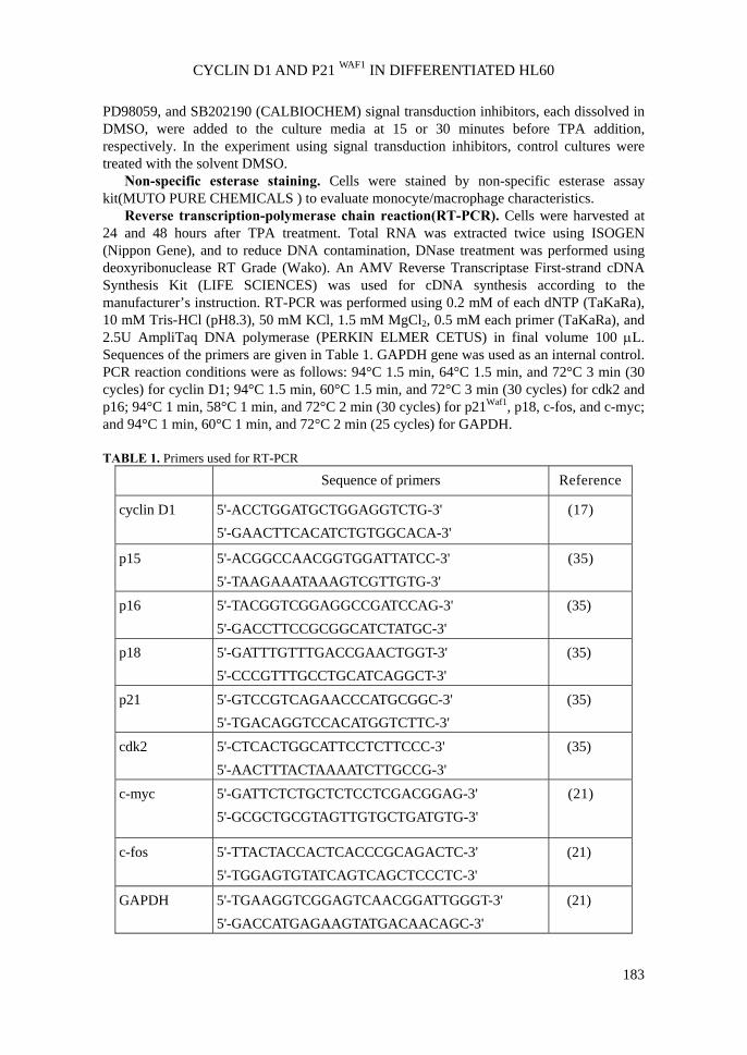
Reverse transcription-polymerase chain reaction(RT-PCR). Cells were harvested at 24 and 48 hours after TPA treatment. Total RNA was extracted twice using ISOGEN (Nippon Gene), and to reduce DNA contamination, DNase treatment was performed using deoxyribonuclease RT Grade (Wako). An AMV Reverse Transcriptase First-strand cDNA Synthesis Kit (LIFE SCIENCES) was used for cDNA synthesis according to the manufacturer’s instruction. RT-PCR was performed using 0.2 mM of each dNTP (TaKaRa), 10 mM Tris-HCl (pH8.3), 50 mM KCl, 1.5 mM MgCl2, 0.5 mM each primer (TaKaRa), and 2.5U AmpliTaq DNA polymerase (PERKIN ELMER CETUS) in final volume 100 μL. Sequences of the primers are given in Table 1. GAPDH gene was used as an internal control. PCR reaction conditions were as follows: 94°C 1.5 min, 64°C 1.5 min, and 72°C 3 min (30 cycles) for cyclin D1; 94°C 1.5 min, 60°C 1.5 min, and 72°C 3 min (30 cycles) for cdk2 and p16; 94°C 1 min, 58°C 1 min, and 72°C 2 min (30 cycles) for p21Waf1, p18, c-fos, and c-myc; and 94°C 1 min, 60°C 1 min, and 72°C 2 min (25 cycles) for GAPDH.
TABLE 1. Primers used for RT-PCR
Sequence of primers Reference
cyclin D1 5'-ACCTGGATGCTGGAGGTCTG-3' 5'-GAACTTCACATCTGTGGCACA-3'
(17)
p15 5'-ACGGCCAACGGTGGATTATCC-3' 5'-TAAGAAATAAAGTCGTTGTG-3'
(35)
p16 5'-TACGGTCGGAGGCCGATCCAG-3' 5'-GACCTTCCGCGGCATCTATGC-3'
(35)
p18 5'-GATTTGTTTGACCGAACTGGT-3' 5'-CCCGTTTGCCTGCATCAGGCT-3'
(35)
p21 5'-GTCCGTCAGAACCCATGCGGC-3' 5'-TGACAGGTCCACATGGTCTTC-3'
(35)
cdk2 5'-CTCACTGGCATTCCTCTTCCC-3' 5'-AACTTTACTAAAATCTTGCCG-3'
(35)
c-myc 5'-GATTCTCTGCTCTCCTCGACGGAG-3' 5'-GCGCTGCGTAGTTGTGCTGATGTG-3'
(21)
c-fos 5'-TTACTACCACTCACCCGCAGACTC-3' 5'-TGGAGTGTATCAGTCAGCTCCCTC-3'
(21)
GAPDH 5'-TGAAGGTCGGAGTCAACGGATTGGGT-3' 5'-GACCATGAGAAGTATGACAACAGC-3'
(21)
E. MATSUMOTO et al.
184
Western blot analysis. Western blot was performed to measure p38 activation in HL60. HL60 cells were lysed by lysis buffer containing 50 mM Tris-HCl(pH 7.5), 150mM NaCl, 10% glycerol, 1% Nonidet P-40, 1% SDS, 0.5% deoxycholate, 0.5mM PMSF, and 1 mM sodium orthovanadate overnight. The cell lysate was centrifuged at 10000r/min for 5 min and the supernatant was collected for Western blot analysis. Protein sample were subjected to 10/20% SDS-PAGE gel electrophoresis and then transferred onto a polyvinylidene fluoride (PVDF) membrane by electro-blotting. The membrane was incubated at 4°C overnight in TBS/Tween 20(50 mM Tris-HCl, pH7.5, 150mM NaCl, 0.05% Tween 20) with 5% nonfat milk. After blocking, the membrane was incubated for 1 hour at room temperature in TBS/Tween 20 containing an 1:200 dilution of mouse anti-phospho-p38 monoclonal antibody or rabbit anti-p38 polyclonal antibody(SANTA CRUZ BIOTECHNOLOGY,INC.). Then the membrane was incubated for 1 hour at room temperature in TBS/Tween 20 containing an 1:2000 dilution of anti-mouse IgG/HRP antibody or 1:10000 dilution of anti-rabbit IgG/HRP antibody(SANTA CRUZ BIOTECHNOLOGY,INC.). Specific binding of the antibody was visualized by the enhanced chemiluminescence(ECL) detection system(AMERSHAM BIOSCIENCES) following the manufacture’s instructions.
RESULTS
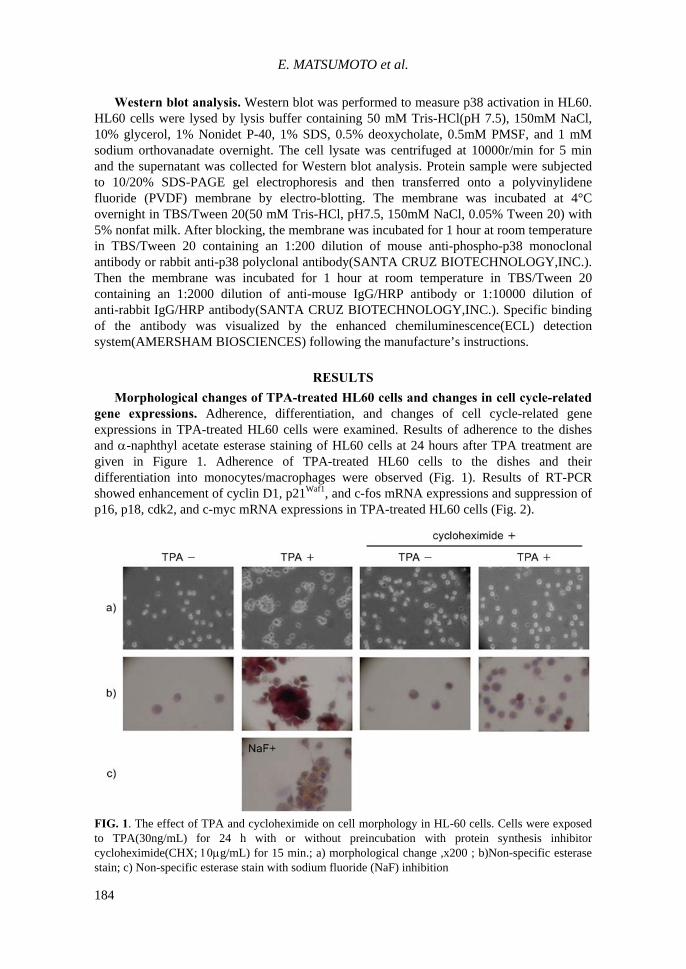
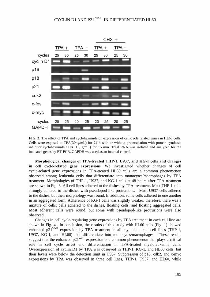
Morphological changes of TPA-treated HL60 cells and changes in cell cycle-related gene expressions. Adherence, differentiation, and changes of cell cycle-related gene expressions in TPA-treated HL60 cells were examined. Results of adherence to the dishes and α-naphthyl acetate esterase staining of HL60 cells at 24 hours after TPA treatment are given in Figure 1. Adherence of TPA-treated HL60 cells to the dishes and their differentiation into monocytes/macrophages were observed (Fig. 1). Results of RT-PCR showed enhancement of cyclin D1, p21Waf1, and c-fos mRNA expressions and suppression of p16, p18, cdk2, and c-myc mRNA expressions in TPA-treated HL60 cells (Fig. 2).
FIG. 1. The effect of TPA and cycloheximide on cell morphology in HL-60 cells. Cells were exposed to TPA(30ng/mL) for 24 h with or without preincubation with protein synthesis inhibitor cycloheximide(CHX; 10μg/mL) for 15 min.; a) morphological change ,x200 ; b)Non-specific esterase stain; c) Non-specific esterase stain with sodium fluoride (NaF) inhibition
CYCLIN D1 AND P21 WAF1 IN DIFFERENTIATED HL60
185
FIG. 2. The effect of TPA and cycloheximide on expression of cell-cycle related genes in HL60 cells. Cells were exposed to TPA(30ng/mL) for 24 h with or without preincubation with protein synthesis inhibitor cycloheximide(CHX; 10μg/mL) for 15 min. Total RNA was isolated and analyzed for the indicated genes by RT-PCR. GAPDH was used as an internal control.
Morphological changes of TPA-treated THP-1, U937, and KG-1 cells and changes
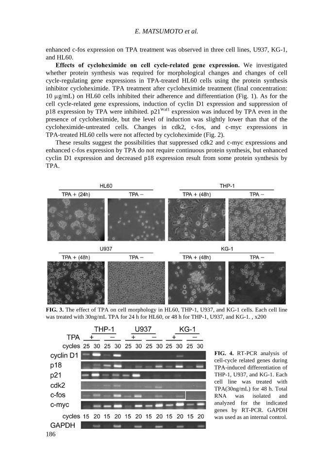
in cell cycle-related gene expressions. We investigated whether changes of cell cycle-related gene expressions in TPA-treated HL60 cells are a common phenomenon observed among leukemia cells that differentiate into monocytes/macrophages by TPA treatment. Morphologies of THP-1, U937, and KG-1 cells at 48 hours after TPA treatment are shown in Fig. 3. All cell lines adhered to the dishes by TPA treatment. Most THP-1 cells strongly adhered to the dishes with pseudopod-like protrusions. Most U937 cells adhered to the dishes, but their morphology was round. In addition, some cells adhered to one another in an aggregated form. Adherence of KG-1 cells was slightly weaker; therefore, there was a mixture of cells: cells adhered to the dishes, floating cells, and floating aggregated cells. Most adherent cells were round, but some with pseudopod-like protrusions were also observed.
Changes in cell cycle-regulating gene expressions by TPA treatment in each cell line are shown in Fig. 4 . In conclusion, the results of this study with HL60 cells (Fig. 1) showed enhanced p21Waf1 expression by TPA treatment in all myeloleukemia cell lines (THP-1, U937, KG-1, and HL60) that differentiate into monocytes/macrophages. These results suggest that the enhanced p21Waf1 expression is a common phenomenon that plays a critical role in cell cycle arrest and differentiation in TPA-treated myeloleukemia cells. Overexpression of cyclin D1 by TPA was observed in THP-1, KG-1, and HL60 cells, but their levels were below the detection limit in U937. Suppression of p18, cdk2, and c-myc expressions by TPA was observed in three cell lines, THP-1, U937, and HL60, while
E. MATSUMOTO et al.
186
enhanced c-fos expression on TPA treatment was observed in three cell lines, U937, KG-1, and HL60.
Effects of cycloheximide on cell cycle-related gene expression. We investigated whether protein synthesis was required for morphological changes and changes of cell cycle-regulating gene expressions in TPA-treated HL60 cells using the protein synthesis inhibitor cycloheximide. TPA treatment after cycloheximide treatment (final concentration: 10 μg/mL) on HL60 cells inhibited their adherence and differentiation (Fig. 1). As for the cell cycle-related gene expressions, induction of cyclin D1 expression and suppression of p18 expression by TPA were inhibited. p21Waf1 expression was induced by TPA even in the presence of cycloheximide, but the level of induction was slightly lower than that of the cycloheximide-untreated cells. Changes in cdk2, c-fos, and c-myc expressions in TPA-treated HL60 cells were not affected by cycloheximide (Fig. 2).
These results suggest the possibilities that suppressed cdk2 and c-myc expressions and enhanced c-fos expression by TPA do not require continuous protein synthesis, but enhanced cyclin D1 expression and decreased p18 expression result from some protein synthesis by TPA.
FIG. 3. The effect of TPA on cell morphology in HL60, THP-1, U937, and KG-1 cells. Each cell line was treated with 30ng/mL TPA for 24 h for HL60, or 48 h for THP-1, U937, and KG-1. , x200
FIG. 4. RT-PCR analysis of cell-cycle related genes during TPA-induced differentiation of THP-1, U937, and KG-1. Each cell line was treated with TPA(30ng/mL) for 48 h. Total RNA was isolated and analyzed for the indicated genes by RT-PCR. GAPDH was used as an internal control.
CYCLIN D1 AND P21 WAF1 IN DIFFERENTIATED HL60
187
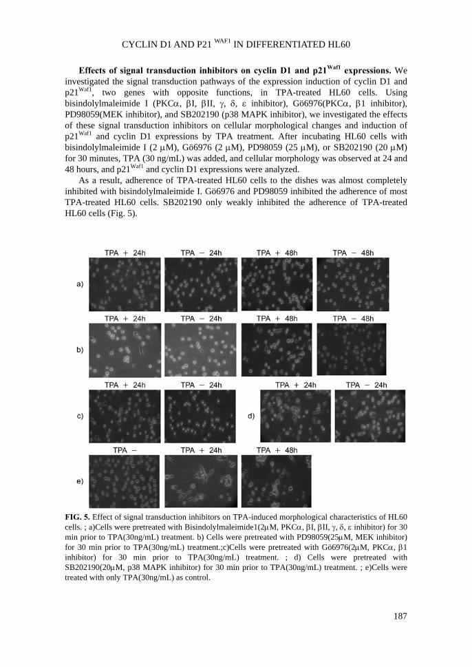
Effects of signal transduction inhibitors on cyclin D1 and p21Waf1 expressions. We investigated the signal transduction pathways of the expression induction of cyclin D1 and p21Waf1, two genes with opposite functions, in TPA-treated HL60 cells. Using bisindolylmaleimide I (PKCα, βI, βII, γ, δ, ε inhibitor), Gö6976(PKCα, β1 inhibitor), PD98059(MEK inhibitor), and SB202190 (p38 MAPK inhibitor), we investigated the effects of these signal transduction inhibitors on cellular morphological changes and induction of p21Waf1 and cyclin D1 expressions by TPA treatment. After incubating HL60 cells with bisindolylmaleimide I (2 μM), Gö6976 (2 μM), PD98059 (25 μM), or SB202190 (20 μM) for 30 minutes, TPA (30 ng/mL) was added, and cellular morphology was observed at 24 and 48 hours, and p21Waf1 and cyclin D1 expressions were analyzed.
As a result, adherence of TPA-treated HL60 cells to the dishes was almost completely inhibited with bisindolylmaleimide I. Gö6976 and PD98059 inhibited the adherence of most TPA-treated HL60 cells. SB202190 only weakly inhibited the adherence of TPA-treated HL60 cells (Fig. 5).
FIG. 5. Effect of signal transduction inhibitors on TPA-induced morphological characteristics of HL60 cells. ; a)Cells were pretreated with Bisindolylmaleimide1(2μM, PKCα, βI, βII, γ, δ, ε inhibitor) for 30 min prior to TPA(30ng/mL) treatment. b) Cells were pretreated with PD98059(25μM, MEK inhibitor) for 30 min prior to TPA(30ng/mL) treatment.;c)Cells were pretreated with Gö6976(2μM, PKCα, β1 inhibitor) for 30 min prior to TPA(30ng/mL) treatment. ; d) Cells were pretreated with SB202190(20μM, p38 MAPK inhibitor) for 30 min prior to TPA(30ng/mL) treatment. ; e)Cells were treated with only TPA(30ng/mL) as control.
E. MATSUMOTO et al.
188
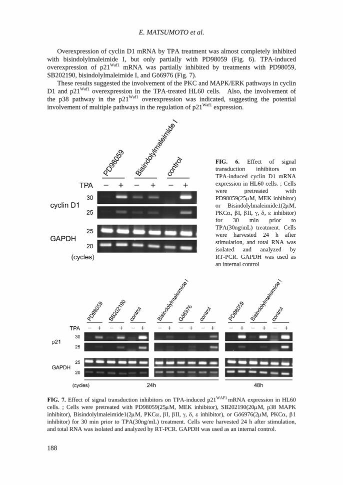
Overexpression of cyclin D1 mRNA by TPA treatment was almost completely inhibited with bisindolylmaleimide I, but only partially with PD98059 (Fig. 6). TPA-induced overexpression of p21Waf1 mRNA was partially inhibited by treatments with PD98059, SB202190, bisindolylmaleimide I, and Gö6976 (Fig. 7).
These results suggested the involvement of the PKC and MAPK/ERK pathways in cyclin D1 and p21Waf1 overexpression in the TPA-treated HL60 cells. Also, the involvement of the p38 pathway in the p21Waf1 overexpression was indicated, suggesting the potential involvement of multiple pathways in the regulation of p21Waf1 expression.
FIG. 6. Effect of signal transduction inhibitors on TPA-induced cyclin D1 mRNA expression in HL60 cells. ; Cells were pretreated with PD98059(25μM, MEK inhibitor) or Bisindolylmaleimide1(2μM, PKCα, βI, βII, γ, δ, ε inhibitor) for 30 min prior to TPA(30ng/mL) treatment. Cells were harvested 24 h after stimulation, and total RNA was isolated and analyzed by RT-PCR. GAPDH was used as an internal control
FIG. 7. Effect of signal transduction inhibitors on TPA-induced p21WAF1 mRNA expression in HL60 cells. ; Cells were pretreated with PD98059(25μM, MEK inhibitor), SB202190(20μM, p38 MAPK inhibitor), Bisindolylmaleimide1(2μM, PKCα, βI, βII, γ, δ, ε inhibitor), or Gö6976(2μM, PKCα, β1 inhibitor) for 30 min prior to TPA(30ng/mL) treatment. Cells were harvested 24 h after stimulation, and total RNA was isolated and analyzed by RT-PCR. GAPDH was used as an internal control.
CYCLIN D1 AND P21 WAF1 IN DIFFERENTIATED HL60
189
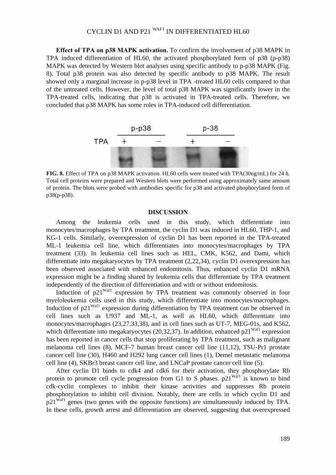
Effect of TPA on p38 MAPK activation. To confirm the involvement of p38 MAPK in TPA induced differentiation of HL60, the activated phosphorylated form of p38 (p-p38) MAPK was detected by Western blot analyses using specific antibody to p-p38 MAPK (Fig. 8). Total p38 protein was also detected by specific antibody to p38 MAPK. The result showed only a marginal increase in p-p38 level in TPA -treated HL60 cells compared to that of the untreated cells. However, the level of total p38 MAPK was significantly lower in the TPA-treated cells, indicating that p38 is activated in TPA-treated cells. Therefore, we concluded that p38 MAPK has some roles in TPA-induced cell differentiation.
FIG. 8. Effect of TPA on p38 MAPK activation. HL60 cells were treated with TPA(30ng/mL) for 24 h. Total cell proteins were prepared and Western blots were performed using approximately same amount of protein. The blots were probed with antibodies specific for p38 and activated phophorylated form of p38(p-p38).
DISCUSSION
Among the leukemia cells used in this study, which differentiate into monocytes/macrophages by TPA treatment, the cyclin D1 was induced in HL60, THP-1, and KG-1 cells. Similarly, overexpression of cyclin D1 has been reported in the TPA-treated ML-1 leukemia cell line, which differentiates into monocytes/macrophages by TPA treatment (33). In leukemia cell lines such as HEL, CMK, K562, and Dami, which differentiate into megakaryocytes by TPA treatment (2,22,34), cyclin D1 overexpression has been observed associated with enhanced endomitosis. Thus, enhanced cyclin D1 mRNA expression might be a finding shared by leukemia cells that differentiate by TPA treatment independently of the direction of differentiation and with or without endomitosis.
Induction of p21Waf1 expression by TPA treatment was commonly observed in four myeloleukemia cells used in this study, which differentiate into monocytes/macrophages. Induction of p21Waf1 expression during differentiation by TPA treatment can be observed in cell lines such as U937 and ML-1, as well as HL60, which differentiate into monocytes/macrophages (23,27,33,38), and in cell lines such as UT-7, MEG-01s, and K562, which differentiate into megakaryocytes (20,32,37). In addition, enhanced p21Waf1 expression has been reported in cancer cells that stop proliferating by TPA treatment, such as malignant melanoma cell lines (8), MCF-7 human breast cancer cell line (11,12), TSU-Pr1 prostate cancer cell line (30), H460 and H292 lung cancer cell lines (1), Demel metastatic melanoma cell line (4), SKBr3 breast cancer cell line, and LNCaP prostate cancer cell line (5).
After cyclin D1 binds to cdk4 and cdk6 for their activation, they phosphorylate Rb protein to promote cell cycle progression from G1 to S phases. p21Waf1 is known to bind cdk-cyclin complexes to inhibit their kinase activities and suppresses Rb protein phosphorylation to inhibit cell division. Notably, there are cells in which cyclin D1 and p21Waf1 genes (two genes with the opposite functions) are simultaneously induced by TPA. In these cells, growth arrest and differentiation are observed, suggesting that overexpressed
E. MATSUMOTO et al.
190
p21Waf1 might inactivate cyclin D1-cdk. Thus, p21Waf1 is considered to play a critical role in cell cycle arrest and differentiation by TPA treatment.
The cycloheximide treatment experiment suggested that continuous protein synthesis was not required for the TPA-induced suppression of cdk2 and c-myc expressions and induction of c-fos expression, but enhanced cyclin D1 expression and reduced p18 expression were phenomena that result from some protein synthesis by TPA treatment.
In our experiment, cycloheximide slightly inhibited p21Waf1 induction by TPA. However, according to the report by Jiang et al. (19), p21Waf1 was an immediate-early gene that did not require continuous protein synthesis in HL60 cells, since there was no difference in the p21Waf1 expression levels between the cycloheximide-treated HL60 cells and those treated simultaneously with cycloheximide and TPA. They analyzed p21Waf1 expression within 10 hours after simultaneously adding cycloheximide(10μg/mL) and TPA(3nM) , while we added TPA(30ng/mL) at 15 minutes after the cycloheximide(10μg/mL) treatment and analyzed p21Waf1 expression at 24 hours. Therefore, the differences of the results might have reflected the differences in the TPA and cycloheximide concentrations and the treatment time.
Few findings concerning the signal transduction pathway of cyclin D1 expression in TPA-treated cells have been reported. In K562 cells that differentiate into megakaryocytes, cyclin D1 is considered to be expressed via the ERK/MAPK pathway, since the induction of cyclin D1 expression was inhibited with PD98059 (22). Also, the PI3-K pathway, not the MAPK pathway, has been reported to be involved in the cyclin D1 expression in TPA-treated MCF-7 human breast cancer cells, since the induction of cyclin D1 expression is inhibited with LY294002 PI3-K inhibitor, but not with PD98059 (11,12). Our data suggests that the cyclin D1 expression in HL60 cells induced by TPA treatment was inhibited with bisindolylmaleimide I and PD98059; therefore, the cyclin D1 expression was considered to be induced via the PKC and MEK pathways. The differences between our results and those with MCF-7 cells (11,12) might suggest cell-type specificity in the mechanisms of the cyclin D1 expression by TPA treatment.
There are some reports concerning the induction mechanisms of p21Waf1 expression in cells that stop proliferating and differentiate by TPA treatment (1,4,5,8,11,19,20,30,32,33,37,38). In the
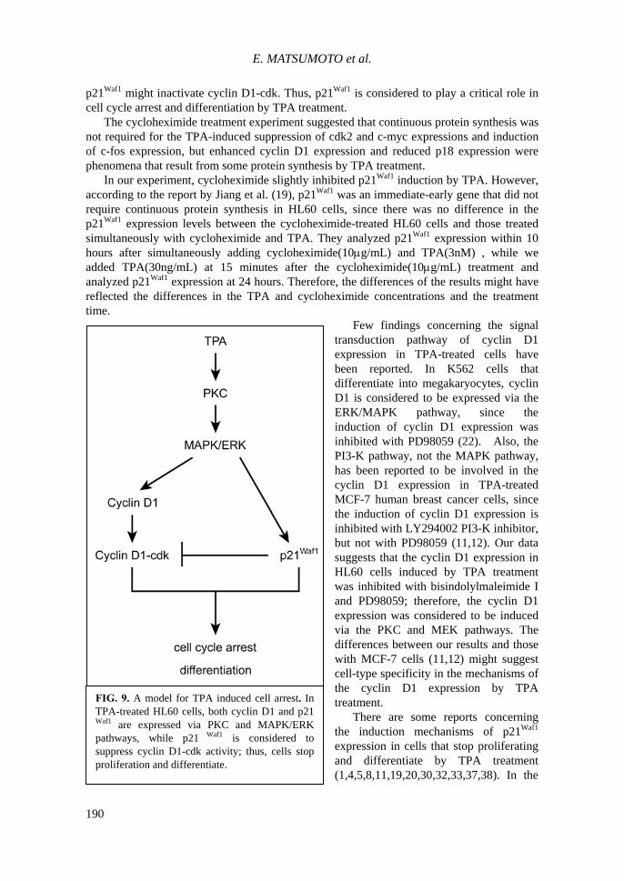
FIG. 9. A model for TPA induced cell arrest. InTPA-treated HL60 cells, both cyclin D1 and p21Waf1 are expressed via PKC and MAPK/ERKpathways, while p21 Waf1 is considered tosuppress cyclin D1-cdk activity; thus, cells stopproliferation and differentiate.
CYCLIN D1 AND P21 WAF1 IN DIFFERENTIATED HL60
191
experiments using HL60, ML-1, U937 leukemia cell lines and TSU-Pr1 prostate cancer cell line, TPA-induced p21Waf1 expression was inhibited with PKC inhibitors such as LY379196, Ro-31-8220, staurosporine, and GF109203X, suggesting the involvement of PKC in p21Waf1 expression (23,27,29,30,37). In addition, TPA-induced p21Waf1 expression was suppressed with PD98059 MEK inhibitor in the HL60 leukemia cell line, MCF-7 breast cancer cell line, H460 and H292 lung cancer cell lines, and TSU-Pr1 prostate cancer cell line, suggesting the involvement of the MAPK/ERK pathway (1,10,11,25,30).
In our experiment using HL60 cells, involvement of both PKC and MAPK/ERK pathways was confirmed in the signal inhibition experiment. It seems that the PKC→MAPK/ERK pathway is a signal transduction pathway shared by cells that stop proliferation due to p21Waf1 expression, and might have a critical role in cell cycle arrest and differentiation by TPA. Besides PKC and MAPK/ERK pathways, our study suggests the involvement of the p38-MAPK pathway, in the p21Waf1 expression in TPA-treated HL60 cell.
In conclusion, in TPA-treated HL60 cells, two genes, cyclin D1 that promotes cell cycle progress, and p21Waf1 that suppresses cell cycle progress, are simultaneously expressed during the differentiation induced by TPA. These genes are considered to be expressed via the same pathway, TPA→PKC→MAPK/ERK, which is known to be involved in various cellular functions such as cell division and differentiation. Of the two cell cycle regulators, p21Waf1 is considered to play a critical role that leads the cells to proliferation and differentiation (Fig.9).
ACKNOWLEDGMENTS
This study was supported by a Grant-in-Aid for research from Kobe-Tokiwa College.
REFERENCES 1. Agadir, A., Chen. G.-q., Bost, F., Li, Y., Mercola, D., Zhang, X.-k.. 1999. Differential
effect of retinoic acid on growth regulation by phorbol ester in human cancer cell lines. J Biol Chem. 274:29779-29785.
2. Akiyama, N., Sasaki, H., Katoh, O., Sato, T., Hirai, H., Yazaki, Y., Sugimura, T., and Terada, M. 1993. Increment of the cyclin D1 mRNA level in TPA-treated three human myeloid leukemia cell lines: HEL, CMK and HL-60 cells. Biochem Biophys Res Commun. 195:1041-1049.
3. Ammit, A.J., and Panettieri, R.A. Jr. 2001. Signal transduction in smooth muscle invited review: The circle of life: cell cycle regulation in airway smooth muscle. J Appl Physiol. 91:1431-1437.
4. Arita, Y., Buffolino, P., and Coppock, D.L. 1998. Regulation of the cell cycle at the G2/M boundary in metastatic melanoma cells by 12-o-tetradecanoyl phorbol-13-acetate(TPA) by blocking p34cdc2 kinase activity. Exp Cell Res. 242:381-390.
5. Blagosklonny, M.V., Prabhu, N.S., and El-Deiry, W.S. 1997. Defects in p21WAF1/CIP1, Rb, and c-myc signaling in phorbol ester-resistant cancer cells. Cancer Res. 57:320-325.
6. Bürger, C., Wick, M., and Müller, R. 1994. Lineage-specific regulation of cell cycle gene expression in differentiating myeloid cells. J Cell Sci. 107:2047-2054.
7. Cho, J.-W., Jeong, Y.-W., Kim, K.-S., OH, J.-Y., Park, J.-C., Lee, J.-C., Baek, W.-K., Suh, S.-I., and Suh, M.-H. 2001. p21WAF1 is associated with CDK2 and CDK4 protein during HL-60 cell differentiation by TPA treatment. Cell Prolif. 34:267-274.
E. MATSUMOTO et al.
192
8. Coppock, D.L., Buffolino, P., Kopman, C., and Nathanson, L. 1995. Inhibition of the melanoma cell cycle and regulation at the G1/S transition by 12-o-tetradecanoylphorbol-13-acetate(TPA) by modulation of CDK2 activity. Exp Cell Res . 221:92-102.
9. Danen, E.H.J., and Yamada, K.M. 2001. Fibronectin, integrins, and growth control. J Cell Physiol. 189:1-13.
10. Das, D., Pintucci, G., and Stern, A. 2000. MAPK-dependent expression of p21WAF and p27kip1 in PMA-induced differentiation of HL60 cells. FEBS Lett. 472:50-52.
11. Dufourny, B., Alblas, J., van Teeffelen, A.A.M., van Schaik, F.M.A., van der Burg, B., Steenbergh, P.H., and Sussenbach, J.S. 1997. Mitogenic signaling of insulin-like growth factor 1 in MCF-7 human breast cancer cells requires phosphatidylinositol 3-kinase and is independent of mitogen-activated protein kinase. J Biol Chem. 272:31163-31171.
12. Dufourny, B., van Teeffelen, H.A.A., Hamelers, I.H.L., Sussenbach, J.S., and Steenbergh, P.H. 2000. Stabilization of cyclin D1 mRNA via the phosphatidylinositol 3-kinase pathway in MCF-7 human breast cancer cells. J Endocrinol. 166:329-338.
13. el-Deiry, W.S., Tokino, T., Velculescu, V.E., Levy, D.B., Parsons, R., Trent, J.M., Lin, D., Mercer, W.E., Kinzler, K.W., and Vogelstein, B. 1993. WAF1, a potential mediator of p53 tumor suppression. Cell. 75:817-825.
14. Eto, I. 1998. Promotion-sensitive epidermal and mammary epithelial cells maintained in suspension over agarose. Cell Prolif. 31:71-92.
15. Hass, R., Gunji, H., Datta, R., Kharbanda, S., Hartmann, A., Weichselbaum, R., and Kufe, D. 1992. Differentiation and retrodifferentiation of human myeloid leukemia cells is associated with reversible induction of cell cycle-regulatory genes. Cancer Res. 52:1445-1450.
16. Horiguchi-Yamada, J., and Yamada, H. 1993. Differing responses of G2-related genes during differentiation of HL60 cells induced by TPA or DMSO. Mol Cell Biochem. 119:29-34.
17. Horiguchi-Yamada, J., Yamada, H., Nakada, S., Ochi, K., and Nemoto, T. 1994. Changes of G1 cyclins, cdk2, and cyclin A during the differentiation of HL60 cells induced by TPA. Mol Cell Biochem. 132:31-37.
18. Huang, T.-S., Duyster, J., and Wang, J.Y.J. 1995. Biological response to phorbol ester determined by alternative G1 pathways. Proc Natl Acad Sci USA. 92:4793-4797.
19. Jiang, H., Lin, J., Su, Z.-z., Collart, F.R., Huberman, E., and Fisher, P.B. 1994. Induction of differentiation in human promyelocytic HL-60 leukemia cells activates p21,WAF1/CIP1,expression in the absence of p53. Oncogene. 9:3397-3406.
20. Kikuchi, J., Furukawa, Y., Iwase, S., Terui, Y., Nakamura, M., Kitagawa, S., Kitagawa, M., Komatsu, N., and Miura, Y. 1997. Polyploidization and functional maturation are two distinct processes during megakaryocytic differentiation: involvement of cyclin-dependent kinase inhibitor p21 in polyploidization. Blood. 89:3980-3990.
21. Ko, S.-G., Lee, K.-S., Cho, K.-H., Kim, Y.-S., Bae, H.-S., and Moon, S.-K. 2000. Inhibition of human smooth muscle cell proliferation by gamigeonsim-tang through the transcriptional regulation of cell cycle-controlling genes. Am J Chin Med. 28(1):57-67.
22. Lee, C.H., Yun, H.J., Kang, H.S., and Kim, H.D. 1999. ERK/MAPK pathway is required for changes of cyclin D1 and B1 during phorbol 12-myristate 13-acetate-induced differentiation of K562 cells. IUBMB Life. 48:585-591.
23. Meinhardt, G., Roth, J., and Hass, R. 2000. Activation of protein kinase C relays
CYCLIN D1 AND P21 WAF1 IN DIFFERENTIATED HL60
193
distinct signaling pathways in the same cell type: differentiation and caspase-mediated apoptosis. Cell Death Differ. 7:795-803.
24. Michieli, P., Chedid, M., Lin, D., Pierce, J. H., Mercer, W.E., and Givol, D. 1994. Induction of WAF1/CIP1 by a p53-independent pathway. Cancer Res. 54:3391-3395.
25. Sato, H., Ogata, H., and De Luca, L.M. 2000. Annexin V inhibits the 12-o-tetradecanoylphorbol-13-acetate-induced activation of Ras/extracellular signal-regulated kinase(ERK) signaling pathway upstream of Shc in MCF-7 cells. Oncogene. 19:2904-2912.
26. Schwaller, J., Pabst, Th., Koeffler, H.P., Niklaus, G., Loetscher, P., Fey M.F., and Tobler, A. 1997. Expression and regulation of G1 cell-cycle inhibitors (p16INK4A, p15INK4B, p18INK4C, p19INK4D) in human acute myeloid leukemia and normal myeloid cells. Leukemia. 11:54-63.
27. Schwaller, J., Paters, U.R., Pabst, T., Niklaus, G., Macfarlane D.E., Fey M.F., and Tobler, A. 1997. Up-regulation of p21WAF1 expression in myeloid cells is activated by the protein kinase C pathway. Br J Cancer. 76:1554-1557.
28. Schwarts, M.A., and Assoian, R.K. 2001. Integrins and cell proliferation: regulation of cyclin-dependent kinase via cytoplasmic signaling pathways. J Cell Sci. 114:2553-2560.
29. Slosberg, E.D., Yao, Y., Xing, F., Ikui, A., Jirousek, M.R., and Weinstein, I.B. 2000. The protein kinase C β-specific inhibitor LY379196 blocks TPA-induced monocytic dfferentiation of HL60 cells. Mol Carcinog. 27:166-176.
30. Sugibayashi, R., Shimizu, T., Suzuki, T., Yamamoto, N., Hamada, H., and Takeda, K. 2001. Upregulation of p21WAF1/CIP1 leads to morphologic changes and esterase activity in TPA-mediated differentiation of human prostate cancer cell line TSU-Pr1. Oncogene. 20:1220-1228.
31. Takuwa, N., and Takuwa, Y. 2001. Regulation of cell cycle molecules by the Ras effector system. Mol Cell Endocrinol. 177:25-33.
32. Uchimaru, K., Taniguchi, T., Yoshikawa, M., Fujinuma, H., Fujita, T., and Motokura, T. 1998. Growth arrest associated with 12-o-tetradecanoylphorbol-13-acetate-induced hematopoietic differentiation with a defective retinoblastoma tumor suppressor-mediated pathway. Leuk Res. 22:413-420.
33. Ullmannová, V., Stöckbauer, P., Hradcová, M., Souček, J., and Haškovec, C. 2003. Relationship between cyclin D1 and p21Waf1/Cip1 during differentiation of human myeloid leukemia cell lines. Leuk Res. 27:1115-1123.
34. Wilhide, C.C., Van Dang, C., Dipersio, J., Kenedy, A.A., and Bray, P.F. 1995. Overexpression of cyclin D1 in the Dami megakaryocytic cell line causes growth arrest. Blood. 86:294-304.
35. Yamada, H., Ochi, K., Nakada, S., Takahara S., Nemoto, T., Sekikawa T., and Horiguchi-Yamada, J. 1995. Interferon modulates the messenger RNA of G1-controlling genes to suppress the G1-to S transition in Daudi cells. Mol Cell Biochem. 152:149-158.
36. Yan, S., and Wenner, C.E. 2001. Modulation of cyclin D1 and its signaling components by the phorbol ester TPA and the tyrosine phosphatase inhibitor vanadate. J Cell Physiol. 186:338-349.
37. Zeng, Y.-X., and El-Deiry, W. 1996. Regulation of p21WAF1/CIP1 expression by p53-independent pathways. Oncogene. 12:1557-1564.
38. Zhang, W., Grasso, L., McClain, C.D., Gambel, A.M., Cha, Y., Travali, S., Deisseroth, A.B., and Mercer, W.E. 1995. p53-independent induction of WAF1/CIP1
E. MATSUMOTO et al.
194
in human leukemia cells is correlated with growth arrest accompanying monocyte/macrophage differentiation. Cancer Res. 55:668-674.
39. Zhou, P., Yao, Y., Soh, J.-W., and Weinstein, I.B. 1999. Overexpression of p21Cip1 or p27Kip1 in the promyelocytic leukemia cell line HL60 accelerates its lineage-specific differentiation. Anticancer Res. 19:4935-4945.