Embed Size (px)
Citation preview

Plant Electrophysiology – Theory & Methods (ed. by Volkov)© Springer-Verlag Berlin Heidelberg 2006
12.1 Introduction
Plants possess most of the chemistry of the neuromotoric system in animals,i.e. neurotransmitter such as acetylcholine, cellular messengers like calmod-ulin, cellular motors, e.g. actin and myosin, voltage-gated ion channels andsensors for touch, light, gravity and temperature. Although this nerve-likecellular equipment has not reached the same great complexity as is the casein nerves, a simple neural network has been formed within the phloem,enabling it to communicate successfully over long distances. The reason whyplants have developed pathways for electrical signal transmission most prob-ably lies in the necessity to respond rapidly to environmental stress factors.Different environmental stimuli evoke specific responses in living cells whichhave the capacity to transmit a signal to the responding region. In contrast tochemical signals such as hormones, electrical signals are able to rapidly trans-mit information over long distances. Most of the plant action potentials stud-ied so far have a velocity in the range of 0.01–0.2 m s−1. However, in soybean,action potentials reached conduction rates of up to 30 m s−1, similar to thespeed of action potentials in nerves (Volkov et al. 2000).
As regards the origin of the neuronal system in plants, it appears unlikelythat it was adopted from animals. In our search for the common evolutionaryroots of action potentials in plants and animals, we need to look at unicellu-lar ancestors which do not need to transmit signals over long distances. Thefunction of electrical transmission has most probably evolved at a later evo-lutionary stage. The assumption is that in the course of evolution the devel-opment of plants and animals branched off into different directions. Sincecellular excitability was found to exist in primitive organisms, it is obviousthat both plants and animals inherited their basic neuronal capabilities fromtheir bacterial ancestors (Simons 1992). Szmelcman and Adler (1976)observed changes in membrane potential during bacterial chemotaxis. Eventhe sensitivity to mechanical touch is known to be an early evolutionaryachievement. Martinac et al. (1987) detected pressure-sensitive ion channels
12 Long-Distance Electrical Signaling andPhysiological Functions in Higher Plants
JÖRG FROMM
Fachgebiet Holzbiologie, TU München, Winzererstrasse 45, 80797 München, Germany (e-mail: [email protected])

in Escherichia coli, suggesting that these channels have an osmotic function.For the early evolution of action potentials, an osmotic function can also beassumed in unicellular alga such as Acetabularia (Mummert and Gradmann1976). A mechanosensitive ion channel was also found in the yeast plasmamembrane (Gustin et al. 1988), providing convincing evidence that plantsinherited mechanical sensitivity from bacterial ancestors in the course of mil-lions of years of evolution. The characean algae, which include Chara andNitella, are also known to be the ancestors of higher plants. Action potentialswere observed in the internodal cells of Nitella in 1898 by Hörmann, who usedextracellular electrodes long before they were observed in isolated nerve cellsby Adrian and Bronk (1928). Characean internodal cells respond to electricalstimulation in a manner similar to the contraction response displayed byskeletal muscles following electrical stimulation by nerve cells. In characeancells, electrical stimulation causes the cessation of protoplasmic streamingwhich is incited by the same interactions between actin and myosin that causecontraction in muscles (Hörmann 1898). In the course of evolution, onceplants had gained and settled on dry land, their excitability and neuronalcapability were used to develop numerous survival tactics. For instance, oneimportant step was the development of fast-moving stomatal guard cells inresponse to environmental changes, while another was the electrical commu-nication system which uses the phloem to transmit information over longdistances within the plant body (Fromm and Lautner 2005).
12.2 Perception of electrical signals
Electrical signals can be generated at any site of the symplastic continuum byenvironmental stimuli such as changes in temperature, touch or wounding.Recently, it was found that acid rain also induces action potentials (Shvetsovaet al. 2002), as well as irradiation at various wavelengths which induces actionpotentials in soybean with duration times and amplitudes of approximately 0.3ms and 60 mV, respectively (Volkov et al. 2004). Upon perception, electrical sig-nals can be propagated via plasmodesmata to other cells of the symplast (vanBel and Ehlers 2005). As a first step, the plasma membrane is being depolarized,a process known as formation of the receptor potential, e.g. by mechanical stim-ulation as observed in Chara (Kishimoto 1968). The receptor potential is anelectrical replica of the stimulus lasting for the period of time that the stimulusis present. An action potential is evoked when the stimulus is great enough todepolarize the membrane to below a certain threshold. Subsequently, the actionpotential characterized by a large transient depolarization allows the rapidtransmission of information via plasmodesmata (Fig. 12.1). An action potentialusually has an all-or-nothing and self-amplifying character, and it travelswith constant velocity and magnitude (Zawadzki et al. 1991). Electrical couplingvia plasmodesmata was demonstrated in a variety of species such as Nitella
270 Jörg Fromm

Long-Distance Electrical Signaling and Physiological Functions in Higher Plants 271
Fig. 12.1. Electrical signaling in higher plants. Stimulation by cold-shock or touch (star)induces calcium influx into a living cell, e.g. a mesophyll cell (MC, above). After the membranepotential is depolarized below a certain threshold level, an action potential is elicited by chlo-ride and potassium efflux. The signal is propagated over short distances through plasmodesmal(P) networks and, after it passed the few plasmodesmata between sieve element/companioncells (SE/CCs) and phloem parenchyma cells (PA), will enter the SE/CC-complex to be trans-mitted over long distances. Sieve pores (SP) with their large diameters present low-resistancecorridors for a rapid propagation of electrical signals along the SE plasma membrane. Such sig-nals can leave the phloem at any site via plasmodesmata (below) to affect certain physiologicalprocesses in the neighbouring tissue
(Spanswick and Costerton 1967), Elodea and Avena (Spanswick 1972) andLupinus (van Bel and van Rijen 1994), indicating that plasmodesmata are relaysin the signaling network between cells. However, long distances between

different organs can be bridged rapidly only via low resistance connections,which extend continuously throughout the whole plant. The sieve tube sys-tem seems to fulfill these conditions, because the structure of the sieve tubemembers is unique and appears to be suitable for the transmission of electri-cal signals due to the relatively large, unoccluded sieve plate pores, continu-ity of the plasma membrane and ER (Evert et al. 1973), as well as lack ofvacuoles. Moreover, the low degree of electrical coupling in lateral directioncaused by only few plasmodesmata at the interface between companion cellsand phloem parenchyma cells (Kempers et al. 1998) facilitates long distancesignaling. However, the plasmodesmata may open up, making it possible forlateral electrical signaling from neighboring cells to be transmitted to thesieve elements/companion cells (SE/CC, Fig. 12.1). In summary, signal trans-mission within the plant depends on the electrical conductance of plas-modesmata in lateral direction as well as on the high degree of electricalcoupling via the sieve pores in longitudinal direction.
12.3 Aphid technique as a tool for measuring electricalsignals in the phloem
Since the phloem is located inside the plant body several cell layers distantfrom the plant surface, experiments on electrical signaling via the phloem ofintact plants are difficult to perform. Microelectrode measurements in com-bination with dye solutions injected into the cell to be measured after obtain-ing electrophysiological results is a time-consuming technique because themeasured cell type can only be roughly estimated at the beginning and veryoften the microelectrode tip was not properly inserted in the phloem asrevealed by microscopic checks after the experiment. Microelectrodesbrought into contact with sieve tube exudates that appear at the cut end of anaphid stylet (Wright and Fisher 1981; Fromm and Eschrich 1988b), enabledus to monitor the membrane potential of sieve tubes and its changes afterplant stimulation (Fig. 12.2A, B). The successful use of aphid stylets to meas-ure electrical signals within the sieve tubes depends on their functioning asan effective salt bridge between the sieve tube cytoplasm and the microelec-trode. Sieve tube exudates typically contain high K+ concentrations; meas-urements on barley leaves gave values ranging from 50 to 110 mM (Frommand Eschrich 1989). The stylet’s food canal dimensions can be used to roughlycalculate its electrical resistance. Using an average area of 6 µm2 and assum-ing the canal to be filled with 100 mM KCl, its resistance would be about2.6 × 109 Ω (Wright and Fisher 1981). Although this value is about 3 timesgreater than the typical resistance of a glass microelectrode, it is still withinthe measuring capacities of the electrometer used (input impedance >1012 Ω).The stylets are embedded in hardened saliva, which insulates electrically. For
272 Jörg Fromm

instance in Mimosa pudica, a classic example for the conductance of rapidexcitation in higher plants, the microelectrode tip was brought into contactwith the stylet stump at the petiolus with its cut end sealed into saline solutionto which the Ag/AgCl reference electrode was connected. After successfulconnection of stylet and mirocelectrode tip a resting potential of −160 mV
Long-Distance Electrical Signaling and Physiological Functions in Higher Plants 273
Fig. 12.2. Transmission of action potentials (AP) and variation potentials (VP) in sieve tubes ofMimosa. A Front-view of Rhopalosiphum padi sucking at the base of a petiolus with its styletinserted into a sieve element (× 32). B After the aphid separated from its stylet by a laser pulse,the stylet stump exuded sieve tube sap to which the tip of a microelectrode was attached (× 400).Cooling the apical end of the petilous evoked an action potential (AP) while flaming triggered avariation potential (VP) transmitted basipetally within the sieve tubes. C Microautoradiographyof the petiolus. 14C-labeled photoassimilates from the leaves accumulated in the phloem (P)which is surrounded by a sclerenchyma sheath (Scl) in order to restrict electrical signaling tothe phloem. Bar, 150 µm. D Microautoradiograph of a cross section of the primary pulvinus atthe base of the petiolus. Labeled photoassimilates are restricted to the phloem strands (P). Sincesclerenchyma tissue is absent, electrical signals can be transmitted laterally from the phloem vialiving collenchyma cells (C) to the motor cells (M) which cause the leaf movements by eitherlosing or gaining turgor. X xylem; Pa parenchyma. Bar, 30 µm
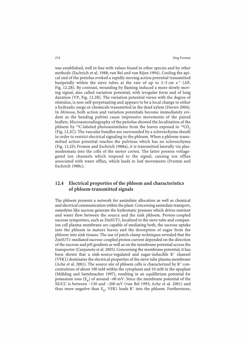
was established, well in line with values found in other species and by othermethods (Eschrich et al. 1988; van Bel and van Rijen 1994). Cooling the api-cal end of the petiolus evoked a rapidly moving action potential transmittedbasipetally within the sieve tubes at the rate of up to 3–5 cm s−1 (AP,Fig. 12.2B). By contrast, wounding by flaming induced a more slowly mov-ing signal, also called variation potential, with irregular form and of longduration (VP, Fig. 12.2B). The variation potential varies with the degree ofstimulus, is non-self-perpetuating and appears to be a local change to eithera hydraulic surge or chemicals transmitted in the dead xylem (Davies 2004).In Mimosa, both action and variation potentials become immediately evi-dent as the bending pulvini cause impressive movements of the pairedleaflets. Microautoradiography of the petiolus showed the localization of thephloem by 14C-labeled photoassimilates from the leaves exposed to 14CO2(Fig. 12.2C). The vascular bundles are surrounded by a sclerenchyma sheathin order to restrict electrical signaling to the phloem. When a phloem-trans-mitted action potential reaches the pulvinus which has no sclerenchyma(Fig. 12.2D; Fromm and Eschrich 1988a), it is transmitted laterally via plas-modesmata into the cells of the motor cortex. The latter possess voltage-gated ion channels which respond to the signal, causing ion effluxassociated with water efflux, which leads to leaf movements (Fromm andEschrich 1988c).
12.4 Electrical properties of the phloem and characteristics of phloem-transmitted signals
The phloem presents a network for assimilate allocation as well as chemicaland electrical communication within the plant. Concerning assimilate transport,osmolytes like sucrose generate the hydrostatic pressure which drives nutrientand water flow between the source and the sink phloem. Proton-coupledsucrose symporters, such as ZmSUT1, localized to the sieve tube and compan-ion cell plasma membrane are capable of mediating both, the sucrose uptakeinto the phloem in mature leaves and the desorption of sugar from thephloem into sink tissues. The use of patch-clamp techniques revealed that theZmSUT1-mediated sucrose-coupled proton current depended on the directionof the sucrose and pH gradient as well as on the membrane potential across thetransporter (Carpaneto et al. 2005). Concerning the membrane potential, it hasbeen shown that a sink-source-regulated and sugar-inducible K+ channel(VFK1) dominates the electrical properties of the sieve tube plasma membrane(Ache et al. 2001). The source site of phloem cells is characterized by K+ con-centrations of about 100 mM within the cytoplasm and 10 mM in the apoplast(Mühling and Sattelmacher 1997), resulting in an equilibrium potential forpotassium ions (EK) of around −60 mV. Since the membrane potential of theSE/CC is between −130 and −200 mV (van Bel 1993; Ache et al. 2001) andthus more negative than EK, VFK1 loads K+ into the phloem. Furthermore,
274 Jörg Fromm

K+ channels of the AKT2/3 family have been identified as photosynthate-induced phloem channels. From studies of an AKT2/3 loss-of-function mutant,it was shown that this mutant exhibited reduced K+ dependence of the phloempotential and that AKT2/3 regulates sucrose/H+ symporters via the membranepotential (Deeken et al. 2002). Furthermore, there is an electrogenic componentof the sieve tube membrane potential, the magnitude of which is substantiallygreater than that predicted for EK (Wright and Fisher 1981). With regard to cal-cium, (DHP)-type Ca2+ channels were localized in the phloem of leaf veins fromNicotiana and Pistia by immunolabeling techniques (Volk and Franceschi2000), indicating that Ca2+ channels are also abundant in sieve elements. Mostlikely, these channels are involved in the generation of electrical signals, mak-ing them the subject of further studies.
The ion transport processes which create the conditions necessary for thegeneration of an action potential were investigated intensively in members ofthe green algal family Characaea (Tazawa et al. 1987). One of the ion trans-port mechanisms responsible for depolarization is based on chloride theefflux of which increases upon membrane stimulation (Gaffey and Mullins1958; Oda 1976). Another ion involved in plasma membrane excitation is cal-cium where studies showed that both the peak of the action potential (Hope1961) and the inward current (Findlay 1961, 1962) are dependent on the cal-cium concentration outside the cell. Some workers suggest that both chlorideand calcium are involved in the formation of an individual action potential(Beilby and Coster 1979; Lunevsky et al. 1983). In addition to these ions, itwas found that potassium efflux from the cell increases upon stimulation ofthe membrane (Spyropoulos et al. 1961; Oda 1976).
These ion shifts during an action potential were confirmed in trees by amethod which uses inhibitors of ionic channels as well as energy-dispersive X-ray microanalysis (Fromm and Spanswick 1993). Results indicate that cal-cium influx as well as potassium and chloride efflux are involved in the gener-ation of action potentials. When action potentials were induced by electricalstimulation in willow, it became clear that the required stimulus depends onboth, its intensity and duration (Fromm and Spanswick 1993). An increase instimulus strength does not produce any change in the amplitude nor in theform of the action potential once it has been induced, showing that it con-forms to the all-or-nothing law. Concerning refractory periods, they werefound to be much longer in plants than in animal systems, with durationsbetween 50 s (Fromm and Bauer 1994) up to 5 h (Zawadzki et al. 1991).
12.5 Electrical signaling via the phloem and its effect onphloem transport
Strong evidence has accumulated that electrical transmission in sieve ele-ments also occurs in species that do not perform rapid leaf movements as,e.g. in Mimosa. In zucchini plants, electrical signal transmission via sieve
Long-Distance Electrical Signaling and Physiological Functions in Higher Plants 275
tubes between a growing fruit and the petiole of a mature leaf reached maxi-mum velocities of 10 cm s−1 (Eschrich et al. 1988). This is in the same velocityrange as the movement of the action potential in sieve tubes of Mimosapudica (Fromm and Eschrich 1988b). It is obvious that no chemical substanceis capable of moving so fast in the assimilate flow. By contrast, hydraulic sig-nals might transmit stimulations, but they would not be able to carry encodedplus- or minus-signals for hyperpolarization or depolarization, respectively,as shown in poplar sieve tubes (Lautner et al. 2005). It has not yet been shownwhether hydraulic signals occur in the turgescent sieve tube system. In thewounded tomato plant, the pathway for systemic electrical signal transmis-sion is also associated with the phloem (Rhodes et al. 1996), indicating that itregulates the induction of proteinase inhibitor activity in parts of the shootdistant from the wound (Wildon et al. 1992). As regards the function of thephloem, it has been shown that action potentials propagating in sieve tubesof Mimosa trigger phloem unloading in the pulvini (Fromm and Eschrich1990; Fromm 1991).
In maize leaves, both electrical stimulation as well as cold-stimulationinduce action potentials with amplitudes higher than 50 mV that are prop-agated basipetally in sieve tubes at speeds of 3–5 cm s−1 (Fromm and Bauer1994). Stimulation with ice water has been reported to induce action poten-tials in a number of plant species, including Biophytum (Sibaoka 1973) aswell as pumpkin and tomato (van Sambeek and Pickard 1976). The fact thatWoodley et al. (1976) observed that localized chilling temporarily stops orreduces translocation of 14C in sunflowers for 10–15 min and that thisreduction in translocation corresponds closely to electrical changes meas-ured along the stem gave rise to the idea of a possible relationship betweenaction potentials and the cold-shock-induced inhibition of phloem trans-port. In addition, Minchin and Thorpe (1983) showed that rapid tempera-ture drops of only 2.5 °C caused a brief abeyance of phloem transport inIpomea purpurea, Phaseolus vulgaris and Nymphoides geminata, a phe-nomenon not observed when the temperature was reduced at a slower rate.In maize leaves, rapid cold-shock treatments cause sieve elements to triggeraction potentials while phloem transport in distant leaf parts is stronglyreduced, as shown by autoradiography at a distance of over 15 cm from thesite of cold-stimulation (Fromm and Bauer 1994). When a maize leaf wasstimulated electrically (10 V) via surface electrodes action potentials wereinduced and phloem transport was interrupted at the site of stimulation.Evidence of a link between electrical signaling and the reduction of phloemtransport was found based on the decrease in symplastic K+ and Cl− con-centration. In Luffa cylindrica action potentials affected elongation growthof the stem, most likely by K+ and Cl− efflux which reduced cell turgor andcaused growth retardation (Shiina and Tazawa 1986). Since the concentra-tions of either ion are also reduced in the sieve element cytoplasm afterstimulation (Fromm and Bauer 1994), decreased cell turgor may have
276 Jörg Fromm

caused the reduction in phloem translocation since the latter requires theintracellular movement of water as a transport medium. However, thereduction in phloem translocation may have also been caused by a closureof sieve pores or a reduction of phloem loading because the latter dependson the membrane potential as well as on the K+ concentration in sieve tubes,both of which changed during stimulation. To obtain a better insight into theelectrical controlling points in the phloem transport system, further work isrequired.
12.6 Role of electrical signals in root-to-shootcommunication of water-stressed plants
Non-hydraulic signaling between roots and shoots of plants growing in dry-ing soil has evoked considerable interest in recent years. Since plants grow-ing in drying soil showed stomatal closure and leaf growth inhibition beforethe reductions in leaf turgor were measured, non-hydraulic signals fromroots may serve as a sensitive link between soil water changes and shootresponses (Davies and Zhang 1991). Therefore, stomata appear to be able toreceive information on the soil water status independent of the leaf waterpotential. Evidence that the nature of this information is chemical wasobtained by analyzing the xylem sap from unwatered plants, indicating theinvolvement of ion content, pH, amino acids and hormones (Schurr andGollan 1990). Since the velocity of a chemical substance in the phloem is rel-atively slow and typically proved to be 50–100 cm h−1 (Canny 1975), the openquestion was how is the leaf capable of responding rapidly to the changingwater status of the soil. Evidence of electrical root-to-shoot signaling wasobtained by both, extra- and intracellular potential measurements on 80 cmtall maize plants. They were subjected to a drying cycle of 5 days showing adecrease in CO2 uptake and transpiration rate while the electrical potentialdifference between two surface points showed a daily rhythm which seemedto be correlated with the soil water status (Fromm and Fei 1998). After soildrying the plants were watered and increases in CO2 and H2O exchange weredemonstrated to follow the arrival of an action potential in the leaves.Experiments with dye solution showed that the increase in gas exchangecould not be triggered by water ascent. In addition, the use of aphid stylets as“bioelectrodes” showed that sieve tubes served as a pathway for electrical sig-naling. The membrane potential of the sieve tubes responded rapidly uponwatering the dried plants as well as after inducing spontaneous water stressto the roots by polyethylene glycol (Fromm and Fei 1998). Results thereforesuggest that electrical root to shoot communication plays an essential role inthe co-ordination of processes between roots and leaves, especially via longpathways.
Long-Distance Electrical Signaling and Physiological Functions in Higher Plants 277

12.7 Role of electrical signaling during fertilization
Strong evidence also exists that electrical signals evoke specific responses ofthe ovary during the processes of pollination and fertilization. As regardspollination, two different kinds of electric potential changes were measuredin the style of flowers. First, Sinyukhin and Britikov (1967) recorded an actionpotential in the style of Lilium martagon and Incarvillea grandiflora a fewminutes after placing pollen on the stigma lobes. Furthermore, an actionpotential was detected after mere mechanical irritation of the Incarvillea lobe,causing closure of the stigma lobes without further transmission. In bothspecies the pollen-induced action potentials propagated towards the ovary tostimulate the oxygen consumption by 5–11%, 60–90 s after arrival of theaction potential. At this moment, most likely post-pollination effects begin,such as the induction of ovary enlargement and wilting of the corolla, whichoccur long before fertilization. Second, electrical potential changes weremeasured in the style of Lilium longiflorum flowers 5–6 h after pollination(Spanjers 1981). No signals were detectable when applying killed pollen orpollen of other species. In Hibiscus rosa-sinensis, different stimuli applied tothe stigma of flowers evoke specific electrical signals that propagate towardthe ovary at speeds of 1.3–3.5 cm s−1 (Fromm et al. 1995). To investigate thefirst reactions of the ovarian metabolism, various metabolites were analysed10 min after stimulating the stigma by pollen, wounding or cold-shock. Self-as well as cross-pollination hyperpolarized the resting potential of style cells50–100 s later, followed by a series of 10–15 action potentials. At 3–5 minafter pollination, the ovarian respiration rate increased transiently by 12%,with the levels of ATP, ADP and starch rising significantly (Fromm et al.1995). By contrast, cold-shock of the stigma caused a single action potential,whereas wounding generated a strong depolarization of the membranepotential with an irregular form and at a lower transmission rate. Eithertreatment caused a spontaneous decrease in the ovarian respiration rate, aswell as reduced metabolite concentrations in the ovary. Since there was noevidence that a chemical substance had been transported within 10 min overa distance of 8–10 cm from the stigma to the ovary, the metabolism musthave responded to the electrical signals (Fromm et al. 1995). In the light ofthese results, the question arises how does an electrical signal cause the bio-chemical response. Most likely the latter may be achieved through subcellu-lar changes of K+, Cl−, and Ca2+ ions which are responsible for the generationof action potentials. According to Davies (1987) local changes in ion con-centration can lead to modified activities of enzymes in the cell wall, theplasmalemma, and the cytoplasm. This kind of mechanism may also beinvolved in the fluctuation of the starch level of the ovary after stigma stim-ulation. The biochemical regulation of starch synthesis is centered almostexclusively on ADP-Glc-pyrophosphorylase (Preiss et al. 1985). The charac-teristics of this enzyme in ovaries will therefore be analyzed in future to gain
278 Jörg Fromm
a better understanding of the biochemical role of electrical signaling duringfertilization.
12.8 Long-distance electrical signaling in woody plants
In trees in particular, communication over long distances may be achievedthrough phloem-transmitted electrical signals. Bridging long distancesbetween different organs, these rapid signals possess the ability to coordinatephysiological activities. Due to environmental changes, different electricalsignals can be evoked in the symplast and transmitted to distant organs, withconcomitant specific effects on various physiological processes.
12.8.1 Membrane potential, electrical signals and growth of willow roots
Since willow roots were shown to respond to hormones with propagatingaction potentials (Fromm and Eschrich 1993), it was an important challengeto measure the magnitude of the current that flows during action potentials.With the use of the vibrating probe technique it was possible to quantify thecurrent, the sensitivity of the probe being in the range of µA cm−2, i.e. suffi-ciently sensitive to measure ion fluxes of pmol cm−2 s−1 (Fromm et al. 1997).Therefore, microelectrode recordings and vibrating probe measurementswere used in tandem to correlate changes in membrane potential withchanges in endogenous current. Transient depolarizations were elicited inroot cortex cells by spermine, while abscisic acid caused a transient hyperpo-larization. For the latter we assume that K+ leaves the cortex cells, similar tothe K+ efflux measured in guard cells (Mansfield et al. 1990). All changes inmembrane potential were accompanied by transient responses of the endoge-nous current. These responses suggested that first anions and then cationsleave the root during spermine-induced depolarizations. From the changes inthe endogenous current an apparent efflux of anions (presumably Cl−) andcations (presumably K+) of 200–700 pmol cm−2 per signal was calculated(Fromm et al. 1997). Furthermore, it was possible to demonstrate the effect ofthe growth regulators spermine and abscisic acid on root growth. The meangrowth rate of roots increased by up to 30% after application of spermine,while it almost came to a standstill after treatment with abscisic acid.
12.8.2 Electrical properties of wood-producing cells
In the course of the evolutionary process, plants found it necessary to developwood in order to increase their mechanical strength so as to be able to reachtree heights of 100 m and more. Extensive literature exists that addresseswood anatomy, chemistry and physical properties. However, we have only
Long-Distance Electrical Signaling and Physiological Functions in Higher Plants 279

just begun to form an understanding of the molecular and electrophysiolog-ical mechanisms of cambial activity and wood formation, a field now consid-ered a main research area in tree physiology. One of the main model treespecies for basic wood research is poplar. Because of its suitability for genetictransformation and its ease of vegetative propagation, poplar has become thecommonly used model tree species in Europe and the United States. To givea description of the electrophysiological processes in wood formation bio-physical and molecular techniques have been used to analyze K+ transportersof poplar. K+ transporters homologous to those of known function inArabidopsis phloem and xylem physiology were isolated from a poplar woodEST library and the expression profile of three distinct K+ channel types wasanalysed by quantitative RT-PCR (Langer et al. 2002). Thus, it was found thatthe P. tremula outward rectifying K+ channel (PTORK) and the P. tremulaK+ channel 2 (PTK2) correlated with the seasonal wood production. Both K+
channel genes are expressed in young poplar twigs, and while PTK2 was pre-dominantly found in the phloem fraction, PTORK was detected in bothphloem and xylem fractions. Following the heterologous expression inXenopus oocytes the biophysical properties of the different channels weredetermined. PTORK, upon membrane depolarization mediates potassiumrelease, while PTK2 is almost voltage-independent, carrying inward K+ flux athyperpolarized potential and K+ release upon depolarization (Langer et al.2002). In addition, in-vivo patch-clamp studies were performed on isolatedprotoplasts from PTORK and PTK2 expressing suspension cultures. Poplarbranches were therefore induced to build callus and the resulting meristem-atic tissues were used to generate suspension cultures. Protoplasts were iso-lated and the plasma membrane potassium conductances were comparedwith the electrical properties of Xenopus oocytes expressing PTORK andPTK2 individually. Concerning PTORK, it was shown that the properties ofthis channel are similar in both experimental systems and also to other plantdepolarization-activated K+ release channels (Gaymard et al. 1998; Ache et al.2000; Langer et al. 2002). In coincidence with the activity of the K+ channels aplasma membrane H+-ATPase, generating the necessary H+ gradient (proton-motive force) for the uptake of K+ into xylem cells, was localized in the poplarstem using specific antibodies (Arend et al. 2002, 2004). Since potassium isthe most abundant cation in plants, playing a central role in many aspects ofplant physiology, we conclude that K+ channels are involved in the regulationof K+-dependent wood formation. Since seasonal changes in cambial potas-sium content correlate strongly with the osmotic potential of the cambialzone (Wind et al. 2004), potassium may well play a key role in the regulationof wood formation due to its strong impact on osmoregulation in expandingcambial cells. On the other hand, since PTORK appears in the plasma mem-brane of sieve elements of the phloem as well as in xylem rays, this channelmay play a role in the generation of electrical signals within the poplarphloem and xylem. However, the potential role of xylem ray cells in radial
280 Jörg Fromm
transmission of electrical signals within the tree stem will have to be provedin the future.
12.8.3 Role of electrical signaling in the regulation of photosynthesis
Most of the work on functions for electrical signals in plants focused onresponses evoked by heating and evidence exists of their role in transcription,translation and respiration (Stankovic and Davies 1997; Davies 2004).Recently, evidence was found of a link between electrical signaling and pho-tosynthetic response in Mimosa (Koziolek et al. 2004). Flaming of a leaf pinnaevoked a variation potential that travels at a speed of 4–8 mm s−1 into theneighboring pinna of the leaf to transiently reduce the net CO2 uptake rate.Simultaneously, the PSII quantum yield of electron transport is reduced.Two-dimensional imaging analysis of the chlorophyll fluorescence signalshowed that the yield reduction spreads acropetally through the pinna andvia the veins through the leaflets. The results provide evidence of the role ofelectrical signals in the regulation of photosynthesis because the high speedof the signals rules out the involvement of a slow-moving chemical signal. Inaddition to the photosynthetic response, it was shown that wounding causeslateral chloroplast movement within 10 min after wounding in Elodeacanadensis (Gamalei et al. 1994). The time course of chloroplast movementcoincides with rapid changes in the membrane potential with low amplitudes(humming, 4–7 mV), recorded by microelectrodes impaled into the midrib ofthe attached leaf.
With regard to trees, hormone-induced action potentials in the roots wereshown to propagate throughout willow plants at velocities of 2–5 cm s−1 inorder to affect the gas exchange of the leaves (Fromm and Eschrich 1993). Togain a deeper understanding of the role of electrical signaling in the photo-synthesis of trees, poplar shoots were stimulated by flaming. In this species,depolarizing signals travel over long distances across the stem from heat-wounded leaves to adjacent leaves where the net CO2 uptake rate is tem-porarily depressed towards compensation (Lautner et al. 2005). Surprisingly,signals induced by cold-shock did not affect photosynthesis. In coincidencewith the results on Mimosa, electrical signaling also significantly reduced thequantum yield of electron transport through PSII in poplar. Cold-blocking ofthe stem proved that the electrical signal transmission via the phloembecomes disrupted, causing the leaf gas exchange to remain unaffected.Furthermore, calcium-deficient trees showed a marked contrast inasmuch asthe amplitude of the electrical signal was distinctly reduced, concomitantwith the absence of a significant response in leaf gas exchange upon flame-wounding (Lautner et al. 2005). Further research has to be done on theresponsiveness of the various types of molecules that are involved in electrontransport as well as on enzymes involved in the uptake of CO2 during electricalsignaling.
Long-Distance Electrical Signaling and Physiological Functions in Higher Plants 281

12.9 Conclusions
So far, we have seen glimpses of a complex electrical long-distance signalingsystem in plants and obtained evidence of the role played by electrical sig-nals in the daily processes of plant life. Obviously, plants have developed asimple neural network which responds to a variety of environmental stimuliwhich may be both, abiotic as well as biotic. Due to the impulses generatedby environmental changes action and variation potentials serve as informa-tion carriers. The primary step in signal perception may be the opening ofplasmalemmal calcium or chloride channels, leading to ion fluxes whichgenerate action or variation potentials. Astounding similarities existbetween action potentials in plants and animals. The generation of actionpotentials in plants follows the all-or-nothing law too (Shiina and Tazawa1986; Fromm and Spanswick 1993), and plant action potentials also showrefractory periods. Furthermore, the use of new methods provides opportuni-ties for detection of fast action potentials which reach speeds up to 40 m s−1
(Volkov and Mwesigwa 2000), i.e. similar to the velocities of action poten-tials in nerves. In higher plants, electrical signals are transmitted from cellto cell via plasmodesmata over short distances, while propagation overlong-distances along the plasma membrane of sieve tubes occurs throughthe successive opening and closure of ion channels (Fig. 12.1). Calcium,chloride and potassium channels are involved in the generation of actionpotentials and several ion channels postulated to be involved in electricaltransmission were identified in the phloem and xylem (Ache et al. 2001;Langer et al. 2002).
Concerning the physiological functions of electrical signals, numerousexamples exist. Apart from the role of action and variation potentials in car-nivorous plants and Mimosa, a concrete relationship between electrical stim-ulation and the increased production of proteinase inhibitors was found toexist in tomato (Stankovic and Davies 1997). Other work showed that actionpotentials regulate respiration (Dziubinska et al. 1989), phloem transport(Fromm and Bauer 1994), fertilization (Sinyukhin and Britikov 1967; Frommet al. 1995) and photosynthesis (Koziolek et al. 2004; Lautner et al. 2005). It isto be expected that future improvements in investigation methods will revealmore aspects of the signaling complexity and its physiological responses thatare as yet not fully understood.
References
Ache P, Becker D, Ivashikina N, Dietrich P, Roelfsema MRG, Hedrich R (2000) GORK, a delayedoutward rectifier expressed in guard cells of Arabidopsis thaliana, is a K+ selective, K+ sens-ing ion channel. FEBS Lett 486:93–98
Ache P, Becker D, Deeken R, Dreyer I, Weber H, Fromm J, Hedrich R (2001) VFK1, a Vicia fabaK+ channel involved in phloem unloading. Plant J 27:571–580
282 Jörg Fromm

Adrian ED, Bronk DW (1928) The discharge of impulses in motor nerve fibres. I. Impulses insingle fibres of the phrenic nerve. J Physiol 66:81–101
Arend M, Weisenseel MH, Brummer M, Osswald W, Fromm J (2002) Seasonal changes ofplasma membrane H+-ATPase and endogenous ion current during growth in poplar plants.Plant Physiol 129:1651–1663
Arend M, Monshausen G, Wind C, Weisenseel MH, Fromm J (2004) Effect of potassium defi-ciency on the plasma membrane H+-ATPase of the wood ray parenchyma in poplar. PlantCell Environ 27:1288–1296
Beilby MJ, Coster HGL (1979) The action potential in Chara corallina. II. Two activation-inac-tivation transients in voltage clamps of plasmalemma. Aust J Plant Physiol 6:329–335
Canny MJP (1975) Mass transfer. In: Zimmermann HM, Milburn JA (eds) Encyclopedia of plantphysiology. Springer, Berlin Heidelberg New York, pp 139–153
Carpaneto A, Geiger D, Bamberg E, Sauer N, Fromm J, Hedrich R (2005) Phloem-localized, pro-ton-coupled sucrose carrier ZmSUT1 mediates sucrose efflux under control of sucrose gra-dient and pmf. J Biol Chem 280:21437–21443
Davies E (1987) Action potentials as multifunctional signals in plants: a unifying hypothesis toexplain apparently disparate wound responses. Plant Cell Environ 10:623–631
Davies E (2004) New functions for electrical signals in plants. New Phytol 161:607–610Davies WJ, Zhang J (1991) Root signals and the regulation of growth and development of plants
in drying soil. Annu Rev Plant Physiol Plant Mol Biol 42:55–76Deeken R, Geiger D, Fromm J, Koroleva O, Ache P, Langenfeld-Heyser R, Sauer N, May ST,
Hedrich R (2002) Loss of the AKT2/3 potassium channel affects sugar loading into thephloem of Arabidopsis. Planta 216:334–344
Dziubinska H, Trebacz K, Zawadzki T (1989) The effect of excitation on the rate of respirationin the liverwort Conocephalum conicum. Physiol Plant 75:417–423
Eschrich W, Fromm J, Evert RF (1988) Transmission of electric signals in sieve tubes of zucchiniplants. Bot Acta 101:327–331
Evert RF, Eschrich W, Eichhorn SE (1973) P-protein distribution in mature sieve elements ofCucurbita maxima. Planta 109:193–210
Findlay GP (1961) Voltage-clamp experiments with Nitella. Nature 191:812–814Findlay GP (1962) Calcium ions and the action potential in Nitella. Aust J Biol Sci 15:69–82Fromm J (1991) Control of phloem unloading by action potentials in Mimosa. Physiol Plant
83:529–533Fromm J, Bauer T (1994) Action potentials in maize sieve tubes change phloem translocation.
J Exp Bot 45:463–469Fromm J, Eschrich W (1988a) Transport processes in stimulated and non-stimulated leaves of
Mimosa pudica. I. The movement of 14C-labelled photoassimilates. Trees 2:7–17Fromm J, Eschrich W (1988b) Transport processes in stimulated and non-stimulated leaves of
Mimosa pudica. II. Energesis and transmission of seismic stimulations. Trees 2:18–24Fromm J, Eschrich W (1988c) Transport processes in stimulated and non-stimulated leaves of
Mimosa pudica. III. Displacement of ions during seismonastic leaf movements. Trees2:65–72
Fromm J, Eschrich W (1989) Correlation of ionic movements with phloem unloading and load-ing in barley leaves. Plant Physiol Biochem 27:577–585
Fromm J, Eschrich W (1990) Seismonastic movements in Mimosa. In: Satter RL, Gorton HL,Vogelmann TC (eds) The pulvinus: motor organ for leaf movement. Am Soc Plant Physiol,Rockville, pp 25–43
Fromm J, Eschrich W (1993) Electric signals released from roots of willow (Salix viminalis L.)change transpiration and photosynthesis. J Plant Physiol 141:673–680
Fromm J, Fei H (1998) Electrical signaling and gas exchange in maize plants of drying soil. PlantSci 132:203–213
Fromm J, Lautner S (2005) Characteristics and functions of phloem-transmitted electrical sig-nals in higher plants. In: Baluska F, Mancuso S, Volkmann D (eds) Communication inplants—neuronal aspects of plant life. Springer, Berlin Heidelberg New York
Long-Distance Electrical Signaling and Physiological Functions in Higher Plants 283

Fromm J, Spanswick R (1993) Characteristics of action potentials in willow (Salix viminalis L.).J Exp Bot 44:1119–1125
Fromm J, Hajirezaei M, Wilke I (1995) The biochemical response of electrical signaling in thereproductive system of Hibiscus plants. Plant Physiol 109:375–384
Fromm J, Meyer AJ, Weisenseel MH (1997) Growth, membrane potential and endogenous ioncurrents of willow (Salix viminalis) roots are all affected by abscisic acid and spermine.Physiol Plant 99:529–537
Gaffey CT, Mullins LJ (1958) Ion fluxes during the action potential in Chara. J Physiol144:505–524
Gamalei YV, Fromm J, Krabel D, Eschrich W (1994) Chloroplast movement as response towounding in Elodea canadensis. J Plant Physiol 144:518–524
Gaymard F, Pilot G, Lacombe B, Bouchez D, Bruneau D, Boucherez J, Michaux-Ferriere N,Thibaud JB, Sentenac H (1998) Identification and disruption of a plant shaker-like outwardchannel involved in K+ release into the xylem sap. Cell 94:647–655
Gustin MC, Zhou XL, Martinac B, Kung C (1988) A mechanosensitive ion channel in the yeastplasma membrane. Science 242:762–765
Hörmann G (1898) Studien über die Protoplasmaströmung bei den Characaean. Fischer, JenaHope AB (1961) Ionic relations of cells of Chara corallina. V. The action potential. Aust J Biol
Sci 14:312–322Kempers R, Ammerlaan A, van Bel AJE (1998) Symplasmic constriction and ultrastructural fea-
tures of the sieve element/companion cell complex in the transport phloem of apoplasmi-cally and symplasmically phloem-loading species. Plant Physiol 116:271–278
Kishimoto U (1968) Response of Chara internodes to mechanical stimulation. Ann Rep BiolWorks, Fac Sci, Osaka Univ 16:61–66
Koziolek C, Grams TEE, Schreiber U, Matyssek R, Fromm J (2004) Transient knockout of pho-tosynthesis mediated by electrical signals. New Phytol 161:715–722
Langer K, Ache P, Geiger D, Stinzing A, Arend M, Wind C, Regan S, Fromm J, Hedrich R (2002)Poplar potassium transporters capable of controlling K+ homeostasis and K+ dependentxylogenesis. Plant J 32:997–1009
Lautner S, Grams TE, Matyssek R, Fromm J (2005) Characteristics of electrical signals in poplarand responses in photosynthesis. Plant Physiol 138:2200–2209
Lunevsky VZ, Zherelova OM, Vostrikov IY, Berestovsky GN (1983) Excitation of Characeae cellmembranes as a result of activation of calcium and chloride channels. J Membrane Biol72:43–58
Mansfield TA, Hetherington AM, Atkinson CJ (1990) Some current aspects of stomatal physi-ology. Annu Rev Plant Physiol Plant Mol Biol 41:55–75
Martinac B, Buechner M, Delcour AH, Adler J, Kung C (1987) Pressure-sensitive ion channel inEscherichia coli. Proc Natl Acad Sci USA 84:2297–2301
Minchin PEH, Thorpe MR (1983) A rate of cooling response in phloem translocation. J Exp Bot34:529–536
Mühling KH, Sattelmacher B (1997) Determination of apoplastic K+ in intact leaves by ratioimaging of PBFI fluorescence. J Exp Bot 48:1609–1614
Mummert E, Gradmann D (1976) Voltage dependent potassium fluxes and the significance ofaction potentials in Acetabularia. Biochim Biophys Acta 443:443–450
Oda K (1976) Simultaneous recording of potassium and chloride effluxes during an actionpotential in Chara corallina. Plant Cell Physiol 17:1085–1088
Preiss J, Robinson N, Spilatro S, McNamara K (1985) Starch synthesis and its regulation. In:Heath R, Preiss J (eds) Regulation of carbon partitioning in photosynthetic tissue. Am SocPlant Physiol, Rockville, pp 1–26
Rhodes J, Thain JF, Wildon DC (1996) The pathway for systemic electrical signal transductionin the wounded tomato plant. Planta 200:50–57
Schurr U, Gollan T (1990) Composition of xylem sap of plants experiencing root water stress: adescriptive study. In: Davies WJ, Jeffcoat B (eds) Importance of root to shoot communica-tion in the response to environmental stress. Br Soc Plant Growth Regul, Bristol, pp 201–214
284 Jörg Fromm

Shiina T, Tazawa M (1986) Action potential in Luffa cylindrica and its effects on elongationgrowth. Plant Cell Physiol 27:1081–1089
Shvetsova T, Mwesigwa J, Labady A, Kelly S, Thomas D, Lewis K, Volkov AG (2002) Soybeanelectrophysiology: effects of acid rain. Plant Sci 162:723–731
Sibaoka T (1973) Transmission of action potentials in Biophytum. Bot Mag 86:51–61Simons P (1992) The action plant. Movement and nervous behaviour in plants. Blackwell,
OxfordSinyukhin AM, Britikov EA (1967) Action potentials in the reproductive system of plants.
Nature 215:1278–1280Spanjers AW (1981) Bioelectric potential changes in the style of Lilium longiflorum Thunb. after
self- and cross-pollination of the stigma. Planta 153:1–5Spanswick RM (1972) Electrical coupling between cells of higher plants: a direct demonstration
of intercellular communication. Planta 102:215–227Spanswick RM, Costerton JWF (1967) Plasmodesmata in Nitella translucens: structure and elec-
trical resistance. J Cell Sci 2:451–464Spyropoulos CS, Tasaki I, Hayward G (1961) Fractination of tracer effluxes during action poten-
tial. Science 133:2064–2065Stankovic B, Davies E (1997) Intercellular communication in plants: electrical stimulation of
proteinase inhibitor gene expression in tomato. Planta 202:402–406Szmelcman S, Adler J (1976) Change in membrane potential during bacterial chemotaxis. Proc
Natl Acad Sci USA 73:4387–4391Tazawa M, Shimmen T, Mimura T (1987) Membrane control in the Characeae. Annu Rev Plant
Physiol 38:95–117Van Bel AJE (1993) The transport phloem. Specifics of its functioning. Prog Bot 54:134–150Van Bel AJE, Ehlers K (2005) Electrical signalling via plasmodesmata. In: Oparka KJ (ed)
Plasmodesmata. Blackwell, OxfordVan Bel AJE, van Rijen HVM (1994) Microelectrode-recorded development of symplasmic
autonomy of the sieve element/companion cell complex in the stem phloem of Lupinusluteus. Planta 192:165–175
Van Sambeek JW, Pickard BG (1976) Mediation of rapid electrical, metabolic, transpirationaland photosynthetic changes by factors released from wounds. I. Variation potentials andputative action potentials in intact plants. Can J Bot 54:2642–2650
Volk G, Franceschi VR (2000) Localization of a calcium-channel-like protein in the sieve elementplasma membrane. Aust J Plant Physiol 27:779–786
Volkov AG, Mwesigwa J (2000) Interfacial electrical phenomena in green plants: action poten-tials. In: Volkov AG (ed) Liquid interfaces in chemical, biological, and pharmaceuticalapplications. Dekker, New York, pp 649–681
Volkov AG, Collins DJ, Mwesigwa J (2000) Plant electrophysiology: pentachlorophenol inducesfast action potentials in soybean. Plant Sci 153:185–190
Volkov AG, Dunkley TC, Morgan SA, Ruff D II, Boyce YL, Labady AJ (2004) Bioelectrochemicalsignaling in green plants induced by photosensory systems. Bioelectrochem 63:91–94
Wildon DC, Thain JF, Minchin PEH, Gubb IR, Reilly AJ, Skipper YD, Doherty HM, Odonnell PJ,Bowles DJ (1992) Electrical signaling and systemic proteinase-inhibitor induction in thewounded plant. Nature 360:62–65
Wind C, Arend M, Fromm J (2004) Potassium-dependent cambial growth in poplar. Plant Biol6:30–37
Woodley SJ, Fensom DS, Thompson RG (1976) Biopotentials along the stem of Helianthus inassociation with short-term translocation of 14C and chilling. Can J Bot 54:1246–1256
Wright JP, Fisher DB (1981) Measurement of the sieve tube membrane potential. Plant Physiol67:845–848
Zawadzki T, Davies E, Dziubinska H, Trebacz K (1991) Characteristics of action potentials inHelianthus annuus. Physiol Plant 83:601–604
Long-Distance Electrical Signaling and Physiological Functions in Higher Plants 285