Embed Size (px)
Citation preview

REVIEW
Plasma cell leukemia: consensus statement on diagnosticrequirements, response criteria and treatment recommendationsby the International Myeloma Working GroupC Fernandez de Larrea1, RA Kyle2, BGM Durie3, H Ludwig4, S Usmani5, DH Vesole6, R Hajek7, JF San Miguel8, O Sezer9, P Sonneveld10,SK Kumar2, A Mahindra11, R Comenzo12, A Palumbo13, A Mazumber14, KC Anderson15, PG Richardson15, AZ Badros16, J Caers17,M Cavo18, X LeLeu19, MA Dimopoulos20, CS Chim21, R Schots22, A Noeul23, D Fantl24, U-H Mellqvist25, O Landgren26, A Chanan-Khan27,P Moreau28, R Fonseca29, G Merlini30, JJ Lahuerta31, J Blade1, RZ Orlowski32 and JJ Shah32 on behalf of the International MyelomaWorking Group33
Plasma cell leukemia (PCL) is a rare and aggressive variant of myeloma characterized by the presence of circulating plasma cells. It isclassified as either primary PCL occurring at diagnosis or as secondary PCL in patients with relapsed/refractory myeloma. PrimaryPCL is a distinct clinic-pathological entity with different cytogenetic and molecular findings. The clinical course is aggressive withshort remissions and survival duration. The diagnosis is based upon the percentage (X20%) and absolute number (X2� 109/l)of plasma cells in the peripheral blood. It is proposed that the thresholds for diagnosis be re-examined and consensusrecommendations are made for diagnosis, as well as, response and progression criteria. Induction therapy needs to begin promptlyand have high clinical activity leading to rapid disease control in an effort to minimize the risk of early death. Intensivechemotherapy regimens and bortezomib-based regimens are recommended followed by high-dose therapy with autologous stemcell transplantation if feasible. Allogeneic transplantation can be considered in younger patients. Prospective multicenter studiesare required to provide revised definitions and better understanding of the pathogenesis of PCL.
Leukemia advance online publication, 4 January 2013; doi:10.1038/leu.2012.336
Keywords: plasma cell leukemia; cytogenetics; bortezomib; transplantation; myeloma; prognosis
INTRODUCTIONMore than a century ago, the first case of plasma cell leukemia(PCL) was recognized by Gluzinski and Reichenstein.1 Thisuncommon form of clonal plasma cell dyscrasia is the mostaggressive variant of the human monoclonal gammopathies andit has been defined by the presence of more than 20% plasmacells in peripheral blood and an absolute plasma cell count greaterthan 2� 109/l.2,3 The incidence of PCL ranges between 2 and 4%of patients with multiple myeloma (MM).4–7 PCL is classified as
primary when it presents ‘de novo’ in patients with no evidence ofprevious MM and as secondary when it is observed as a leukemictransformation of relapsed or refractory disease in patientswith previously recognized MM.8 Of them, 60–70% of PCLare primary, and the remaining 30–40% are secondary.9 Morerecent data suggest that there is an increasing incidence ofsecondary PCL (sPCL), now accounting for about 50% of thecases.7 The aim of this article is to provide a consensus on thediagnostic criteria for PCL, response criteria and treatment
1Amyloidosis and Myeloma Unit, Department of Hematology, IDIBAPS, Hospital Clinic de Barcelona, Barcelona, Spain; 2Department of Hematology, Mayo Clinic, Rochester, MN,USA; 3Southwest Oncology Group, International Myeloma Foundation, Cedars-Sinai Outpatient Cancer Center at the Samuel Oschin Comprehensive Cancer Institute, Los Angeles,CA, USA; 4Department of Medicine, Center for Oncology and Hematology, Wilhelminenspital, Vienna, Austria; 5Myeloma Institute for Research & Therapy, University of Arkansasfor Medical Sciences, Little Rock, AR, USA; 6John Theurer Cancer Center at Hackensack University Medical Center, Hackensack, NJ, USA; 7Department of Haemato-Oncology,University Hospital Ostrava and School of Medicina University, Ostrava, Czech Republic; 8Servicio de Hematologia, Hospital Universitario de Salamanca, CIC, IBMCC (USAL-CSIC),Salamanca, Spain; 9University of Hamburg, University Medical Center, Hamburg, Germany; 10Department of Hematology, Erasmus University Medical Center, Rotterdam, TheNetherlands; 11Massachusetts General Hospital Cancer Center, Boston, MA, USA; 12Tufts Medical Center, Boston, MA, USA; 13Myeloma Unit, Division of Hematology, University ofTorino, Torino, Italy; 14NYU Comprehensive Cancer Center, New York, NY, USA; 15Dana-Farber Cancer Institute, Medical Oncology, Boston, MA, USA; 16Department of BMT,Hematology/Oncology, University of Maryland, Baltimore Campus, Baltimore, MD, USA; 17Centre Hospitalier Universitaire de Liege, Department of Hematology, Liege, Belgium;18Seragnoli Institute of Haematology, Bologna School of Medicine, S. Orsola’s University Hospital, Bologna, Italy; 19Department of Hematology, Hospital Claude Huriez, Lille,France; 20Department of Clinical Therapeutics, University of Athens, School of Medicine Athens, Greece; 21Division of Haematology & Medical Oncology, Queen Mary Hospital,Hong Kong; 22Department of Clinical Hematology, University Ziekenhuis Brussels, Brussels, Belgium; 23Hospital Ruiz y Paez, Bolivar, Venezuela; 24Hospital Italiano De Buenos Aires,Caba, Argentina; 25Department of Hematology, Sahlgrenska University Hospital, Gothenburg, Sweden; 26Metabolism Branch, National Cancer Institute, NIH, Bethesda, MD, USA;27Division of Hematology-Oncology, Mayo Clinic, Jacksonville, FL, USA; 28Department of Clinical Hematology, University Hospital, Nantes, France; 29Division of Hematology-Oncology, Mayo Clinic Scottsdale, Scottsdale, AZ, USA; 30Department of Biochemistry, University of Pavia, Pavia, Italy; 31Hospital Universitario 12 de Octubre, Department ofHematology, Madrid, Spain and 32Department of Lymphoma/Myeloma, Division of Cancer Medicine, University of Texas MD Anderson Cancer Center, Houston, TX, USA.Correspondence: Dr C Fernandez de Larrea, Department of Hematology, Hospital Clınic de Barcelona, Villarroel, 170, 08036 Barcelona, Spain.E-mail: [email protected] Dr JJ Shah, Department of Lymphoma/Myeloma, Division of Cancer Medicine, University of Texas, M D Anderson Cancer Center, 1515 Holcomber Blvd, Houston, 77030 TX, USA.E-mail: [email protected] Appendix for the complete list of all authors part of International Myeloma Working Group.Received 31 October 2012; accepted 8 November 2012; accepted article preview online 21 November 2012
Leukemia (2013), 1–12& 2013 Macmillan Publishers Limited All rights reserved 0887-6924/13
www.nature.com/leu

recommendations for primary PCL (pPCL) based upon a criticalreview of: 1) presenting features, 2) biological aspects includingcellular adhesion mechanisms, molecular genetics andbone marrow milieu factors, 3) response criteria, and finally,4) current treatment approaches, including hematopoietic stemcell transplantation.
PRESENTING CLINICAL FEATURESBecause of the relative low incidence and prevalence of this entity,most clinical data come from isolated case reports and smallretrospective studies.9,10 No prospective series have beenpublished and only seven reports including more than 20patients have been identified.3–7,11–13 The main clinical andlaboratory features, response to therapy and survival of patientswith pPCL reported in these articles are summarized in Table 1.The median age ranged between 52 and 65 years, about 10 yearsyounger than the median age of 65–70 years observed in thegeneral myeloma population14 and in sPCL.7 However, in anepidemiology study including 291 patients diagnosed between1973 and 2004, the median age was 67 years.6 Although the dataare limited, it appears that, as for MM, PCL is more common inAfrican Americans than in Caucasians.15 pPCL has a moreaggressive clinical presentation than MM, including a highertumor burden. Patients may present with symptoms due toprofound anemia, hypercalcemia or bleeding diathesis owing tothrombocytopenia. On physical examination, patients may exhibita higher prevalence of organomegaly with involvement of theliver, spleen, lymph nodes, pulmonary findings associated withpleural effusions, neurological deficits due to central nervoussystem involvement, pallor, petichae and palpable extramedullarysoft-tissue plasmacytomas (Figures 1 and 2). In contrast, thepresence of lytic bone lesions is lower than that observed in MM.7
Fewer IgA cases than for MM patients were found in some studies,and there was an unexpectedly low proportion of patients withIgG-type M-protein3 in one series. In contrast, the proportion of
patients with light-chain disease ranges from 26–44%, whereas ingeneral myeloma series the proportion of patients with BenceJones myeloma is only 15%.14 Bone marrow examination willoften demonstrate extensive bone marrow plasma cell infiltration,with anaplastic or plasmablastic morphology (Figure 3), resultingin a reduced bone marrow reserve, with a greater incidence ofanemia and thrombocytopenia as well as fewer normal plasmacells. Also, reflecting this aggressive clinical presentation, a higherproportion of patients with pPCL have significant leukocytosis,elevated lactate dehydrogenase (LDH) and b2-microglobulinserum levels. In fact, patients with MM usually show normal ormoderately increased LDH serum levels, with a significant LDHelevation only observed in patients with high tumor load (43%).16
Physicians must be aware of a potential tumor lysis syndrome,given the high tumor burden and elevated proliferative index.Thus, serum uric acid, calcium, phosphorous and serum creatininelevels must be monitored. Importantly, similar to acute leukemias,
Table 1. Main clinical and laboratory features of seven retrospective series of patients with primary plasma cell leukemia
Noel andKyle3
Dimopouloset al.4
Garcıa-Sanzet al.5
Tiedemannet al.7
Colovicet al.11
Peijinget al.12
Paganoet al.13
Number of patients 25 27 26 41 30 22 73Median age (years) 53 57 65 54.5 60 49.5 NASex, M/F 15/10 NA 12/14 24/17 22/8 14/8 43/30Lytic bone lesions (%) 44 NA 48 35 60 44.4 64
Extramedullary involvement (%)Liver 52 32 0 32 56 44.4 23Spleen 44 18 0 18 53 33.3 18Lymph nodes 12 6 11 6 3 NA 4Other NA NA NA NA NA NA 14
M-protein type (%)IgG 12.5 52 54 28 53 54.5 30IgA 25 15 4 13 23 9.1 8IgD 6 0 8 2 3 0 3Light chain 44 28 31 41 20 27.3 30Nonsecretory 12.5 7 4 8 0 9.1 18
Hemoglobin o10g/dl (%) 450 82 54 450 100 450 48Platetelet count o100� 109/l(%)
450 67 48 450 100 450 NA
High b2-microglobulin (%) NA 91 65 50 64 50 100High LDH (%) NA 63 48 50 37 NA 52Response to treatment (%) 47 37 38 NA NA 45.5 55Median survival (months) 6.8 12 8 11.1 4.5 14 12.6
Abbreviations: LDH, lactate dehydrogenase; NA, not available.
Figure 1. Abdominal tomography showing a focal lesion (40mm),highlighted by the arrow, suggestive of metastasic infiltration of theliver in a patient with PCL.
PCL consensusC Fernandez de Larrea et al
2
Leukemia (2013), 1 – 12 & 2013 Macmillan Publishers Limited

the progression of disease is very rapid (weeks). Rare findings,such as hemophagocytic syndrome,17 hyperammonaemia18 orexpression of solid tumor markers (CA125 and CA15.3)19 have alsobeen reported.
pPCL is a distinct clinic-pathologic entity from MM because itspresenting features and its natural history including response tochemotherapy and poorer prognosis.9 Reinforcing this fact, thePCL pattern always reappears at the time of relapse, whereas sPCLoccurs only in 1–2% of advanced and refractory MM patients whoevolve into a leukemic phase with an aggressive clinical picture. Incontrast, the constellation of adverse biological prognostic factorsin patients with advanced and refractory myeloma leading to asPCL is a multistep process. Thus, pPCL and sPCL are two distinctclinical and biological entities that only share the features ofplasma cells circulating in the peripheral blood and an ominousclinical course.
ConsensuspPCL is observed in younger patients than MM, with a increasedincidence of light-chain only (Bence Jones) type. The clinicalpicture is characterized by an aggressive clinical presentation withhigh tumor burden, high proliferative index (that is, S-phase DNA),rapid clinical course, leukocytosis, extramedullary involvement,marked bone marrow infiltration by immature plasma cells andhigh LDH serum levels. Finally, the presentation of relapsed pPCLroutinely mimics the initial clinical picture.
DIAGNOSTIC CRITERIAThe original diagnostic criteria of PCL were established in 1974 byNoel and Kyle,3 requiring both more than 20% circulating plasmacells and an absolute count greater than 2� 109/l plasma cells inperipheral blood. These criteria provide a framework to define this
disease entity along with the associated universal poor clinicaloutcome. These criteria have not been evaluated prospectively todetermine if a need for any modification is required.
What degree of circulating plasmacytosis should be used fordefining PCL?The control mechanisms by which plasma cells initially remainpredominantly confined to the bone marrow, only rarely enteringthe blood stream, are poorly understood. In fact, a low proportionof plasma cells can be detected in peripheral blood in patientswithin the entire spectrum of plasma cell dyscrasias, includingnewly diagnosed MM, smoldering MM and, exceptionally, inMGUS (monoclonal gammopathy of undetermined signifi-cance).20,21 It is also important to recognize that the presence ofcirculating plasma cells is not always indicative of PCL, as thepresence of a significant number of polyclonal peripheral blood
Figure 2. Positron emission tomography/computer tomographyscan of a patient with pPCL showing focal bone lesion withincreased uptake of fluorodeoxyglucose in vertebrae, ribs and pelvis.
Figure 3. Conventional morphology in PCL cases shows bonemarrow infiltration (a), with circulating plasma cells (b) and frequentextramedullary involvement, as hepatic infiltration (c).
PCL consensusC Fernandez de Larrea et al
3
& 2013 Macmillan Publishers Limited Leukemia (2013), 1 – 12

plasma cells can be transiently observed in non-malignantconditions, such as severe sepsis, infectious mononucleosis and,particularly, serum sickness.22,23 In this light, peripheral blood flowcytometry is important to demonstrate clonality of the plasmacells, and exclude other lymphoproliferative diseases, includinglow-grade B-cell or lymphoplasmacytic lymphoma.
Do we need both an absolute value of circulating plasma cells anda percentage of WBC?The criteria developed by Kyle2, requiring both more than 20%circulating plasma cells and an absolute count greater than2� 109/l plasma cells in peripheral blood seems too restrictive andthe degree of peripheral plasmacytosis merits to be reconsidered.
In many series, only one of these two criteria was consideredsufficient for the diagnosis of PCL.7,24–26 Patients with significanttreatment exposure and poor bone marrow reserve have baselineleukopenia and may not meet absolute criteria but may meetpercentage criteria. Probably only one of these criteria should besufficient for the diagnosis of this entity. The definition of PCL wasnot discussed in the recent International Myeloma WorkshopConsensus,27 though it is well accepted that the presence of PCLconstitutes an unfavorable prognostic factor and that it is a subsetof high-risk myeloma28 with an especially poor outcome.
Should we standardize methods to detect circulating plasma cellsin peripheral blood?The correct and timely diagnosis of PCL is dependent upon theability of the pathologist to screen and recognize plasma cells inthe peripheral blood smear. Hematologists and pathologistsshould be aware of the clinical relevance of a careful morpholo-gical examination of peripheral blood smears to exclude thepresence of circulating plasma cells.
With all the considerations above, the diagnostic criteria for PCLshould be revisited. The current definition, even when only one ofthe two criteria is required, may underestimate its real frequency.In any given patient, the presence of few circulating plasma cellsdemonstrated by conventional morphology is still a marker for ahighly proliferative and aggressive process. Patients with an ‘early’PCL can rapidly develop full-fledged PCL in the absence oftreatment. In this regard, the current proposal for prospectivestudies is to investigate if lower values (such as 5% or moreplasma cells in peripheral blood and/or an absolute peripheralblood plasma cell count X0.5� 109/l) have the same prognosticimpact as historical criteria. Additional criteria to detect an earlyPCL process, which would allow earlier intervention and thereforechange the natural history of the disease, are warranted, forexample incorporating flow cytometry to detect clonal plasmacells and DNA content analysis, cytogenetics and, ideally, novelmolecular markers.
ConsensusCareful examination of peripheral blood by conventional micro-scopy should be done in all patients with MM who present with aclinical scenario suspicious of PCL, such as leukocytosis and anelevated LDH. If there are more than 20% circulating plasma cellsand/or an absolute count greater than 2� 109/l plasma cells, thediagnosis of PCL should be established according to the presentcriteria. However, lower peripheral blood plasma cell counts (thatis, X5% peripheral blood plasma cells and/or an absolute numberX0.5� 109/l) should be recorded to revisit the diagnostic criteriaof PCL and prospectively analyze the biology and the clinicalcourse of these patients. Additional methods including flowcytometry to detect early PCL should be a high priority andwarrant further studies, encouraging prospective multicenterefforts in newly diagnosed patients.
BIOLOGY OF PCLImmunophenotypeWhile the main plasma cell markers (CD38 and CD138) are equallyexpressed in MM and PCL samples, the multiparametric flowcytometry shows a different pattern in PCL when compared withplasma cells from MM. In this regard, a higher expression of CD20antigen5 and lower CD9, CD117, CD56 and HLA-DR is observed.CD28 is more frequently expressed in sPCL.29 This is consistentwith the fact that the acquisition of CD28 antigen on plasma cellscorrelates with increased plasma cell proliferation and diseaseprogression.29 The increased CD27 expression in PCL has beenlinked to activation of an antiapoptotic pathway.30,31 Furthermore,it has recently been shown that CD27 overexpression can lead tothe activation of the nuclear factor kB, resulting in antiapoptoticenhancement.30 This may have therapeutic implications sincenuclear factor kB, which has a crucial role in the survival ofmalignant plasma cells, is inhibited by bortezomib and othernewer proteasome inhibitors. In addition, CD23 has been reportedto be associated with the presence of t(11;14).32 When comparedwith the studies performed on MM, the immunophenotypicinformation at diagnosis as well as on the minimal residual diseasefollow-up in PCL is really limited.
Mechanisms of extramedullary spreadPlasma cell dyscrasias are characterized by a proliferation ofplasma cells with a strong dependence on the bone marrowmicroenvironment.33 The bone marrow microenvironment has akey role in the pathogenesis of MM by triggering signalingcascades, which mediate myeloma cell proliferation, migrationand survival, with all of these contributing to myeloma growthand to the homing of malignant plasma cells within the bonemarrow. Disruption of these mechanisms could be crucial for theunique biology of PCL. A number of adhesion molecules havebeen involved in the egression of plasma cells to the peripheralblood stream. The lack of CD56 antigen,5,29 a neural cell adhesionmolecule, which is important in anchoring plasma cells to thebone marrow stroma and likely impairs their circulation toperipheral blood as well as their migration to extramedullarysites, is a frequent finding as in myeloma with t(14;16). In addition,it can result in a weaker myeloma cell interaction and increasedsecretion of metalloproteinase-9 (MMP-9). Downregulation ofCD106 and activated CD2934 and decreased expression of thesurface molecules HLA-1 and CD40 in PCL versus MGUS cells35 arealso in this sense. A higher expression of CD54 on plasma cells ascompared with adhesion molecules CD11a, CD18 and CD11b36
has been also demonstrated. Acquisition of this last molecule alsofacilitates egression of plasma cells through the capillary wall andleads to tumor dissemination. The high expression of VLA-4 in PCL,a requisite for invasiveness of leukemic cells because of thecontact with its ligand in capillary vessel wall, would increaseextravasation of leukemic cells from the blood into extravascularspace.35,37 Low expression of chemokine receptors CCR1, CCR2and CXCR4 has been observed in patients with active plasma celldisease as compared with those with inactive disease.38 In thisregard, recent findings indicate that thalidomide exposureinduces downregulation of CXCR4 and its ligand SDF-1alpha,which are involved in the BM homing of myeloma cells.39
However, although the CXCR4 inhibitor AMD 3100 disrupts theinteraction of myeloma cells with the BM microenvironmentresulting in an increased number of circulating myeloma cells inmice,40 it seems that AMD 3100 does not induce either an increasein tumor progression or an engraftment at extramedullary sites inthe AMD 3100-treated mice compared with control mice40 or onthe development of PCL. Cytokines are also involved in PCLproliferation, particularly interleukin-6 (IL-6).41 pPCL and sPCLhave spontaneous cell growth in culture, with increased growthwhen stimulated with exogenous IL-6.41 Autocrine IL-6 production
PCL consensusC Fernandez de Larrea et al
4
Leukemia (2013), 1 – 12 & 2013 Macmillan Publishers Limited

triggered by interferon-alpha has been postulated,42 based on apatient who developed PCL picture triggered by this treatment,enforcing the particular potential cytokine network in thepathogenesis of this entity. The same phenomenon has alsobeen described with IL-3 that upregulates IL-6 receptors.43 Finally,association with viral infections has been hypothesized, withcontradictory results.44–46
ConsensusPlasma cells from patients with PCL overlap in antigenicexpression with those of patients with MM. However, CD20(higher), CD56 (lower), CD117 (lower) and HLA-DR (lower) may beuseful for both discrimination of PCL from MM and for follow-upstudies. Further investigation of the pathogenetic role of surfacecell molecules resulting in extramedullary spread in this entity isclearly warranted.
Fluorescence in situ hybridization (FISH) and cytogeneticsThe molecular basis of PCL is poorly understood. Cytogeneticstudies show that plasma cells in pPCL have a number of geneticabnormalities. More than 80% of patients with PCL havehypodiploid or diploid cells, which is associated with poorprognosis, whereas about 60% of patients with MM displayhyperdiploidy, a favorable finding.5 Chromosomal abnormalities inPCL are summarized in Table 2. Results of these studies are veryheterogeneous, basically based on retrospective studies andunsorted samples.
Chromosome 13 deletion and monosomy are the most frequentfeatures.5,13 Alterations such as monosomy 7, rarely seen in MM,has been observed in PCL.47,48 Deletion of 17p13.1, causing allelicloss of TP53, has been detected in almost 50% of pPCL and in 75%of secondary forms in one report. This deletion wascomplemented by coding mutations in 24% of patients withPCL.7 The frequencies of IgH (14q32) translocations by FISHanalysis are common in both types of PCL with 87% and 82% inprimary and secondary forms, respectively. Thus, in a Mayo Clinicstudy the frequency of t(11;14) by FISH or by informativekaryotype in pPCL was 71%. Importantly, the IgH translocationin PCL involved chromosome 11 and cyclin D1 expression.7
Conversely, no cases of t(4;14) or t(14;16) were observed in pPCL.7
Interestingly, p53 loss due to mutation or deletion was observedin 56% of patients with pPCL and in 83% of patients with thesecondary form.7 Translocation t(11;14) is a favorable prognosticfactor in M; however, its high prevalence in PCL suggests that thistranslocation when associated with high-risk cytogenetics, such asloss of p53, confers a different prognosis.49,50 Of course, PCL maysimply be a completely different disease than MM, with differentrelevant high-risk factors. In the French and British experience,51,52
PCL also had significant differences when compared with MM:a higher incidence of t(11;14), t(14;16) and monosomy 13, withsimilar incidence of t(4;14).
Abnormalities in chromosome 1 are also frequent in PCL,particularly 1q21 amplification (involving CKS1B overexpression)53
and del(1p21).54 The first finding was confirmed in all pPCLpatients in a Spanish series by comparative genomic hybridization,as well as losses on 13q, chromosome 16, 2q and 6p.55
Certain genes, such as cMYC,56 are overexpressed by a complexmechanisms, such as cMYC,56 in spite of the fact that only 15% ofpPCL have a cMYC translocation.52,57 Mutations in N-Ras and K-Rasshow a similar frequency at diagnosis in PCL and in MM.58
Epigenetic changes, such as p16 inactivation, have alsobeen described in pPCL59,60 or global DNA hypomethylationof repetitive genomic sequences.61 Gene-specific DNAhypermethylation as either tumor suppressors, cell–cell signalingor as cell adhesion molecules in PCL versus MM cells, mayallow the clone to become independent of the bone marrowmicroenvironment.62 Interestingly, within the same cytogeneticgroup (that is, t(4;14) or t(11;14)) PCL samples were morehypermethylated in progression-related genes than thecorresponding MM cells.62 A relative high incidence of PTENdeletion, which results in Akt activation has been observed in PCLand it has been suggested that PTEN loss can be involved in thetransition from MM to PCL.
Gene-expression profile and whole-genome sequencingUsmani et al.63 recently described the experience in PCL with totaltherapy (TT) programs.The clinical outcomes were similar to thoseachieved with less intense therapy with an overall survival (OS) ofonly 18 months. Importantly, the GEP was completed in 16/27patients, and surprisingly only 44% of patients with pPCL had ahigh-risk signature defined by the GEP-70 model and 31% by theGEP-80 model.63 Importantly, in the GEP analysis, there was a tightclustering within the pPCL cohort as opposed to non-pPCLsuggesting distinct molecular and genomic features in thesegroups. CD14 (cell-membrane LPS receptor), TNF receptor-associated factor 2 and chemokine C-C motif ligand wereamong 203 genes differentially expressed in pPCL hypothesizingmyeloid differentiation of plasma cells during leukemicdevelopment.63
On the other hand, Egan et al.64 recently described whole-genome sequencing in serial samples from a single patientthrough different points in the natural history, includingdevelopment of sPCL. This methodology with whole-genomesequencing may provide unique insights into potentialmechanisms of PCL development.
ConsensusCytogenetics and FISH studies on bone marrow are mandatory inall patients with suspected PCL. On cytogenetics, the karyotype isfrequently complex and demonstrates hypodiploidy. With FISHanalysis, careful attention should be paid to the most frequentlyreported alterations: (t(11;14) as well as to chromosome 1 and 17abnormalities, particularly 1qþ and del17p. Additional molecular
Table 2. Cytogenetics data available in plasma cell leukemia series
Cytogenetics abnormalities(%)
Garcıa-Sanzet al.5
Dimopouloset al.4
Tiedemannet al.7
Paganoet al.13
Avet-Loiseauet al.51
Chiecchioet al.52
Hypodiploidy 41 60 12.2 47 41.6Hyperdiplody 0 4.9 8.8 33.3Complex karyotype 92 54.5 34.2 58.8 66.7del(13q14) or monosomy 84 50 85 19 68 58del(17p13) 50 7.3 11.8 25t(11;14) 71 19.5 33 42t(4;14) 0 0 12 8.3t(14;16) 0 0 16 25
PCL consensusC Fernandez de Larrea et al
5
& 2013 Macmillan Publishers Limited Leukemia (2013), 1 – 12

research aimed at understanding the development of pPCL andtransformation of MM into sPCL is needed.
RESPONSE TO THERAPY AND SURVIVALThe survival of patients with pPCL is short. In seven series,historically median survival, without novel therapies, has rangedfrom 6.8 to 12.6 months.3–7,11–13 Furthermore, the median survivalof 231 patients from a recently published epidemiology study wasonly 4 months.6 Of note, the survival rate at 5 years from diagnosisis less than 10% in all series. The best survival data, incorporatinghematopoietic stem cell transplantation, reported a mediansurvival longer than 3 years.65 Unfortunately, the significantimprovement in survival observed in MM in the past decade hasnot been seen in PCL.6
These discouraging survival results in pPCL are due to two facts:1) its aggressive presentation with severe complications leading toearly death within the first months from diagnosis, and 2) the lackof effective therapy to achieve sustained responses. Early mortalityis still of concern and reflects the aggressiveness of the disease. Inthe French cohort, 11 of 40 patients died within the first monthafter diagnosis.51 Unfortunately, data from transplantationregistries or clinical trials have a systematic bias to excludepatients not fulfilling the entry criteria and/or experiencing earlydeath. sPCL is usually a terminal event with a median OS of only1 month.3
Criteria of responseThere are no specific response criteria for PCL. Thus, the generalMM response criteria have been applied without distinctiveconsiderations. the leukemic nature of the disease as well as the
relative higher percentage of light-chain only (Bence Jones) andoligosecretory forms, the importance of a precise plasma cellevaluation in blood and bone marrow by morphology and flowcytometry as well as the measurement of the serum free-lightchain should be considered. Thus, the evaluation of response inpPCL should combine acute leukemia66 and MM requirements67,68
(Table 3). The impact of a rapid clearance in peripheral and/orbone marrow malignant plasma cells has not been evaluated. It isreasonable to suggest that in PCL, the disappearance of peripheralblood plasma cells and a bone marrow plasma cell count o5%should be required to qualify for complete response (CR) afterhematological recovery. Complete clearance by conventionalmorphology in bone marrow for complete remission is required(Table 3), and flow cytometry should be necessary to define‘stringent’ CR. In addition, the high frequency of extramedullaryinvolvement justifies evaluation of the patients by imagingtechniques such as magnetic resonance imaging and, particularly,fluorodeoxyglucose positron emission tomography/computertomography.
Relapse from CR is defined as the reappearance of M-protein inpatients in CR, extramedullary disease, reappearance of peripheralblood plasma cells at any level or increase in bone marrow plasmacells more than 10%. In contrast to MM, immediate therapy shouldbe initiated when any evidence of relapse is documented.
ConsensusImprovement in PCL outcomes need to be focus on 1) reducingearly mortality and 2) improving long-term disease control. In theabsence of specific response criteria for PCL, response to therapyhas been evaluated according to MM criteria. Given the primarilyleukemic nature of the disease and the frequency of oligo/
Table 3. Response criteria for plasma cell leukemia
Category Bone marrow criteriaa Peripheral bloodcriteriaa
Serological criteriab Other criteria
Stringentcompleteremission
Bone marrow plasma cells o5% andNo malignant plasma cell by flowcytometry
No plasma cells inperipheral bloodby flow cytometry
Negative serum and urineimmunofixationNormal serum FLC ratio
Absence ofextramedullarydisease
Completeremission
Bone marrow plasma cells o5% No plasma cells inperipheral blood
Negative serum and urineimmunofixationc
Absence ofextramedullarydisease.
Very goodpartialresponse
Bone marrow plasma cells o5% No plasma cells inperipheral blood
X90% reduction of serum M-protein and24-h urinary M-protein o100mg per24 hd
Absence ofextramedullarydisease
Partialresponse
Bone marrow plasma cells–5 to 25% Peripheral bloodplasma cellsfrom 1–5%
X50% reduction of serum M-protein andReduction in 24-h urinary M-protein byX90% and o200mg per 24 he
X50% reduction inthe size ofextramedullarydisease
Stabledisease
Not meeting the criteria of either partial response or progressive disease
Progressivedisease
425% increase in plasma cells in abone marrow aspirate or absoluteincrease X10%
45% absoluteincrease inperipheral bloodplasma cells
425% increase in the level of the serummonoclonal paraprotein with an absoluteincrease X5g/l425% increase in the 24-h urinary lightchain excretion with an absolute increaseX200mg/24h
HypercalcemiaDefinite increase inlytic bone lesionsDefinite increase inthe size or number ofextramedullarydisease
Relapsefromcompleteremission
More than 10% increase in bonemarrow plasma cells
Reappearance ofperipheral bloodplasma cells at anylevel
Reappearance of original M-protein inserum and/or urine immunofixation
Any extramedullarydisease
Abbreviation: FLC, free light chain. aIt is recommended that at least 200 leukocytes on blood smears and 500 nucleated cells on marrow smears be counted. bItshould be maintained for a minimum of 6 weeks. In case of discrepancy or undetectable serological parameter, the patient must be classified according tobone marrow criteria. cIf the serum and urine M-protein are unmeasurable, a normal serum kappa/lambda FLC ratio is also required. dIf the serum and urineM-protein are unmeasurable, a X90% decrease in the difference between involved and uninvolved FLC levels is required instead of the M-protein. eIf theserum and urine M-protein are unmeasurable, a X50% decrease in the difference between involved and uninvolved FLC levels is required instead of theM-protein.
PCL consensusC Fernandez de Larrea et al
6
Leukemia (2013), 1 – 12 & 2013 Macmillan Publishers Limited

nonsecretory forms, the evaluation of response should combineacute leukemia and MM criteria. Measurement of immunopheno-typic residual disease is needed when there is no evidence ofplasma cell infiltration with routine morphological evaluation.Finally, a careful evaluation of extramedullary disease at diagnosisand at response evaluation is required for all PCL patients.
THERAPEUTIC OPTIONSConventional regimensThe results of treatment with combinations of alkylating agents,mainly melphalan, and glucocorticoids, are unsatisfactory. Despitean overall response rate ranging from 23 to 67%, the median OShas been less than 1 year in all the reported series.3–5,11–13 In onestudy, the failure to achieve 50% clearance of blood plasma cellswithin 10 days after treatment initiation was a predictor of noresponse.4 The addition of more agents, such as VAD (vincristine,doxorubicin and dexamethasone) or the VCMP regimen(vincristine, carmustin, melphalan and prednisone) alternatingwith VBAP (vincristine, carmustin, doxorubicin and prednisone)modestly improved the results in terms of response rate andOS.4,5,7
Investigators have also attempted to improve outcomes withstandard regimens such as HyperCVAD (hyper-fractionatedcyclophosphamide, vincristine, doxorrubicin, dexamethasone).69
Novel drugs: thalidomide/lenalidomideAnecdotal reports in small series showed a potential role ofthalidomide in PCL,70,71 but without confirmation thus far.72
Severe cardiac and pulmonary toxicities have been described.73,74
Lenalidomide, a more potent immunomodulatory drug resultedin only transient responses.75–77 Combination of lenalidomidewith melphalan and glucocorticoids has also been used,78
achieving a transient PR in one case.Musto et al.79 have presented the results of a prospective
multicenter phase II trial of lenalidomide/dexamethasone in firstline in 23 patients with pPCL. Patients received lenalidomide25 mg for days 1–21 and weekly dexamethasone 40 mg in a 28-day cycle as primary induction therapy for four cycles and ifeligible for ASCT (autologous stem cell transplantation) couldproceed to ASCT or continue long-term primary therapy. Theinitial overall response rate was 60% and, with a median follow-upof 15 months, the OS and progression-free survival (PFS) were65.2% and 52.1%, respectively.
Novel therapies: bortezomibThe proteosome inhibitor bortezomib has shown clinical activity inboth pPCL and sPCL.80,81 An Italian group reported the results of aretrospective analysis on 12 evaluable patients with pPCL at relapseand sPCL treated with a bortezomib-based combination.82 Responserate was 92%, including two CRs. Responses did not appear to beinfluenced by previous therapy, including ASCT. The median PFS andOS after bortezomib were 8 and 12 months, respectively. The samegroup described a similar high response in untreated pPCL treated attime of diagnosis with bortezomib and various combinations (VD,n¼ 3; VTD, n¼ 2; PAD, n¼ 6; MPV, n¼ 1). There was a high overallresponse rate (79%), including 28% CR and 83% of patients werealive if the response was consolidated with stem cell transplantation,but the follow-up is still very short.83
Another single-institution experience has described a series of25 patients (13 pPCL; 12 sPCL) with a high response rate of 16out of 18 patients treated with a bortezomib-based regimen.Importantly, the OS of patients exposed to bortezomib was 28months compared with 4 months in those who did not receivebortezomib at induction.84
The efficacy of the combination of bortezomib with dexa-methasone and melphalan85 or doxorubicin82,86 has also been
reported in selected smaller case reports and series. Bortezomiband dexamethasone has been shown to be useful in threepatients with pPCL, t(4;14) and CD27 expression.87
High-dose therapy/stem cell transplantationConsidering the poor prognosis of this form of clonal plasma celldyscrasia, intensification with high-dose therapy followed byautologous stem cell rescue should be offered, provided that ageand clinical condition do not preclude this approach. In the MayoClinic series, patients who received ASCT had a longer median OSwhen compared with those who received chemotherapy alone (34versus 11 months),7 although at least part of this survival benefit isobviously due to a selection bias in favor of the transplant group.
The largest study in the transplantation setting is the retro-spective report by the European Group for Blood and MarrowTransplantation,88 reporting data on 272 patients with pPCL. Atthe time of conditioning for transplantation, a higher proportion ofpatients with PCL than MM were in CR (25.5% versus 11.9%). Also,patients with PCL achieved a higher CR rate at 100 days after ASCT(41.2% versus 28.2%), but a selection bias cannot be excluded.
This response pattern is consistent with the clinical behavior ofhigh-risk myeloma, which tends to have higher initial responserates to induction therapy and ASCT;89 however, they also haveshorter response duration with rapid relapse. The median PFS was14.3 months in PCL and 27.4 months in MM. This is translated intosignificantly shorter OS in the PCL group (median of 25.7 versus62.2 months), irrespective of the degree of response achieved,reflecting a more aggressive minimal residual disease in patientswith PCL. In addition, the fact that a significant proportion ofpatients with PCL potentially eligible for ASCT could die ordevelop progression within the first few months after diagnosis,51
precluding the high-dose procedure, represents an important biasin favor of transplant results. In summary, despite the relativegood response achieved in selected patients with PCL whorespond to initial treatment and receive intensification with ASCT,it usually does not translate into a prolonged survival. Therefore,other therapeutic approaches should be explored, such as the useof the new drugs in induction, consolidation or maintenance or asubsequent reduced-intensity allogeneic transplant.81
The Center for International Blood & Marrow TransplantResearch (CIBMTR) reported a PFS and OS at 3 years of 34% and62%, respectively, in 97 patients with pPCL who underwent ASCT.This experience supports the role of ASCT in transplant eligiblepatients and offers an opportunity for relatively prolongedremission. These results for the first time demonstrated survivalbeyond 3 years in a proportion of selected patients.65
Tandem ASCT could result in an improved depth and durationof remission. In this regard, in TT programs, timing of onset andeventual rate of CR were virtually identical for patients with orwithout PCL. However, median OS (1.8 years) and PFS (0.8 years)were inferior to those of the non-PCL group as a whole. Significantadvances in clinical outcomes were observed among non-PCLpatients with the transitions from TT1 to TT2, to TT3, but suchadvances were not observed in PCL patients.63 Other strategies toconsolidate after first ASCT, other than second ASCT, includingcombination regimens of VTD, RD or VRD may also have a role andneed further evaluation. The addition of aggressive long-termmaintenance therapy with lenalidomide or novel lenalidomide-based combinations provides yet another potential strategy toimprove duration of remission. The significant PFS benefitreported by CALBG 10014 and IFM with maintenancelenalidomide suggest that maintenance lenalidomide therapycould be an attractive possibility to be investigated in PCL.90,91
Importantly, relapses may occur very early after ASCT andtherefore it is worth considering early initiation of maintenancetherapy, in the first 30–60 days, as soon as a stable engraftment isdocumented.
PCL consensusC Fernandez de Larrea et al
7
& 2013 Macmillan Publishers Limited Leukemia (2013), 1 – 12
Allogeneic stem cell transplantationA retrospective report of 147 patients with pPCL from the CIBMTRexperience showed that 19 of the 50 patients (39%) whounderwent allogeneic transplantation were alive at 3 years.65
Few of these patients had received novel agents (thalidomide,lenalidomide or bortezomib) as part of their induction regimen.Progressive disease accounted for 22% of the deaths in theallogeneic transplant group, compared with 85% of the deaths inthe ASCT group.
The European Group for Blood and Marrow Transplantationrecently described their experience with 85 patients who under-went allogeneic transplantation in comparison with 411 patientswho underwent autologous SCT for PCL. PFS curves with themyeloablative and RIC allo-SCT possibly crossed the ASCT curvebetween 2 and 4 years, but with similar OS at 5 years. As seen inprevious experiences with allo-SCT described by the CIBMTR, therewas a high early mortality; however, there was also a clear plateauin survival at 20%.92
Careful selection of patients undergoing either myeloablative orreduced-intensity conditioning (mainly by age), and incorporatingnew drugs in induction and consolidation/maintenance couldpotentially further exploit the alloreactive immunotherapeuticeffect.
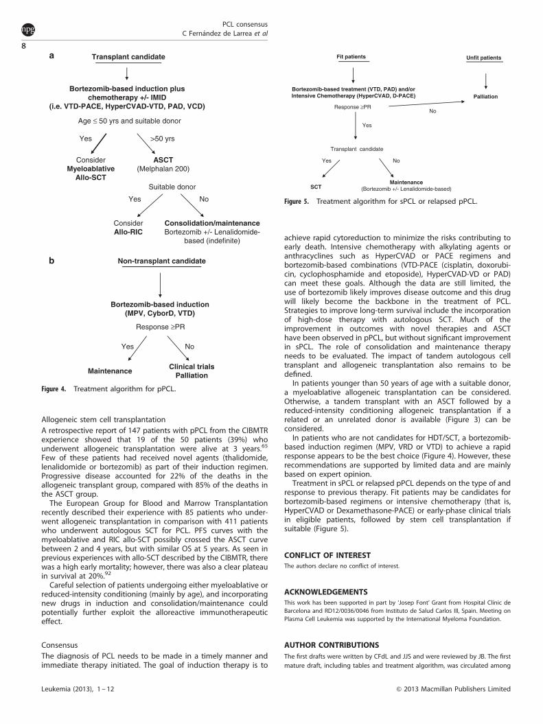
ConsensusThe diagnosis of PCL needs to be made in a timely manner andimmediate therapy initiated. The goal of induction therapy is to
achieve rapid cytoreduction to minimize the risks contributing toearly death. Intensive chemotherapy with alkylating agents oranthracyclines such as HyperCVAD or PACE regimens andbortezomib-based combinations (VTD-PACE (cisplatin, doxorubi-cin, cyclophosphamide and etoposide), HyperCVAD-VD or PAD)can meet these goals. Although the data are still limited, theuse of bortezomib likely improves disease outcome and this drugwill likely become the backbone in the treatment of PCL.Strategies to improve long-term survival include the incorporationof high-dose therapy with autologous SCT. Much of theimprovement in outcomes with novel therapies and ASCThave been observed in pPCL, but without significant improvementin sPCL. The role of consolidation and maintenance therapyneeds to be evaluated. The impact of tandem autologous celltransplant and allogeneic transplantation also remains to bedefined.
In patients younger than 50 years of age with a suitable donor,a myeloablative allogeneic transplantation can be considered.Otherwise, a tandem transplant with an ASCT followed by areduced-intensity conditioning allogeneic transplantation if arelated or an unrelated donor is available (Figure 3) can beconsidered.
In patients who are not candidates for HDT/SCT, a bortezomib-based induction regimen (MPV, VRD or VTD) to achieve a rapidresponse appears to be the best choice (Figure 4). However, theserecommendations are supported by limited data and are mainlybased on expert opinion.
Treatment in sPCL or relapsed pPCL depends on the type of andresponse to previous therapy. Fit patients may be candidates forbortezomib-based regimens or intensive chemotherapy (that is,HyperCVAD or Dexamethasone-PACE) or early-phase clinical trialsin eligible patients, followed by stem cell transplantation ifsuitable (Figure 5).
CONFLICT OF INTERESTThe authors declare no conflict of interest.
ACKNOWLEDGEMENTSThis work has been supported in part by ‘Josep Font’ Grant from Hospital Clınic deBarcelona and RD12/0036/0046 from Instituto de Salud Carlos III, Spain. Meeting onPlasma Cell Leukemia was supported by the International Myeloma Foundation.
AUTHOR CONTRIBUTIONSThe first drafts were written by CFdL and JJS and were reviewed by JB. The firstmature draft, including tables and treatment algorithm, was circulated among
Transplant candidate
Age ≤ 50 yrs and suitable donor
Yes >50 yrs
Bortezomib-based induction pluschemotherapy +/- IMID
(i.e. VTD-PACE, HyperCVAD-VTD, PAD, VCD)
ConsiderMyeloablative
Allo-SCT
ASCT(Melphalan 200)
Suitable donor
Yes No
ConsiderAllo-RIC
Consolidation/maintenanceBortezomib +/- Lenalidomide-
based (indefinite)
Non-transplant candidate
Bortezomib-based induction(MPV, CyborD, VTD)
Response ≥PR
Yes No
MaintenanceClinical trials
Palliation
Figure 4. Treatment algorithm for pPCL.
Fit patients Unfit patients
Response ≥PR
Bortezomib-based treatment (VTD, PAD) and/orIntensive Chemotherapy (HyperCVAD, D-PACE)
SCT
Palliation
No
Maintenance(Bortezomib +/- Lenalidomide-based)
Transplant candidate
Yes
NoYes
Figure 5. Treatment algorithm for sPCL or relapsed pPCL.
PCL consensusC Fernandez de Larrea et al
8
Leukemia (2013), 1 – 12 & 2013 Macmillan Publishers Limited

the authors on May 2012 and was presented and deeply discussed, particularlythe response criteria and treatment approach, at the International MyelomaFoundation Summit Meeting on 12th and 13th June 2012 in Amsterdam, at thegeneral sessions and at the ‘Workshop 5’. The suggestions were incorporatedand the draft was circulated among all the members of the InternationalMyeloma Working Group, for further comments and suggestions which wereincorporated when possible. All the authors approved the final version of themanuscript.
APPENDIXINTERNATIONAL MYELOMA WORKING GROUP
1. N Abildgaard, Syddansk Universitet, Odense, Denmark2. R Abonour, Indiana University School of Medicine, Indianapo-
lis, Indiana, USA3. R Alexanian, MD Anderson, Houston, TX, USA4. M Alsina, H Lee Moffitt Cancer Center and Research Institute,
Tampa, FL, USA5. KC Anderson, DFCI, Boston, MA, USA6. M Attal, Purpan Hospital, Toulouse, France7. H Avet-Loiseau, Institute de Biologie, Nantes, France8. A Badros, University of Maryland, Baltimore, MD, USA9. D Baris, National Cancer Institute, Bethesda, MD, USA
10. B Barlogie, M.I.R.T. UAMS Little Rock, AR, USA11. R Bataille, Institute de Biologie, Nantes, France12. M Beksac, Ankara University, Ankara, Turkey13. A Belch, Cross Cancer Institute, Alberta, Canada14. D Ben-Yehuda, Hadassah University Hospital, Hadassah, Israel15. B Bensinger, Fred Hutchinson Cancer Center, Seattle, WA, USA16. PL Bergsagel, Mayo Clinic Scottsdale, Scottsdale, AZ, USA17. J Bird, Bristol Haematology and Oncology Center, Bristol, UK18. J Blade, Hospital Clınic de Barcelona, Barcelona, Spain19. M Boccadoro, University of Torino, Torino, Italy20. J Caers, Centre Hospitalier Universitaire de Liege, Liege, Belgium21. M Cavo, Universita di Bologna, Bologna, Italy22. A Chanan-Khan, Mayo Clinic, Jacksonville, FL, USA23. W Ming Chen, MM Research Center of Beijing, Beijing, China24. M Chesi, Mayo Clinic Scottsdale, Scottsdale, AZ, USA25. T Child, Leeds General Hospital, Leeds, UK26. J Chim, Department of Medicine, Queen Mary Hospital,
Hong Kong27. W-J Chng, National University Health System, Singapore28. R Comenzo, Tufts Medical School, Boston, MA, USA29. J Crowley, Cancer Research and Biostatistics, Seattle, WA, USA30. W Dalton, H. Lee Moffitt, Tampa, FL, USA31. F Davies, Royal Marsden Hospital, London, England32. J de la Rubia, Hospital Universitario La Fe, Valencia, Spain33. C de Souza, Univeridade de Campinas, Caminas, Brazil34. M Delforge, University Hospital Gasthuisberg, Leuven, Belgium35. M Dimopoulos, University of Athens School of Medicine,
Athens, Greece36. A Dispenzieri, Mayo Clinic, Rochester, MN, USA37. J Drach, University of Vienna, Vienna, Austria38. M Drake, Mayo Clinic Rochester, Rochester, MN, USA39. BGM Durie, Cedars-Sinai Samuel Oschin Cancer Center, Los
Angeles, CA, USA40. H Einsele, Universitatsklinik Wurzburg, Wurzburg, Germany41. T Facon, Centre Hospitalier Regional Universitaire de Lille,
Lille, France42. D Fantl, Socieded Argentina de Hematologıa, Buenos Aires,
Argentina43. J-P Fermand, Hopitaux de Paris, Paris, France44. C Fernandez de Larrea, Hospital Clınic de Barcelona,
Barcelona, Spain45. R Fonseca, Mayo Clinic Arizona, Scottsdale, AZ, USA46. G Gahrton, Karolinska Institute for Medicine, Huddinge,
Sweden
47. R Garcıa-Sanz, University Hospital of Salamanca, Salamanca,Spain
48. C Gasparetto, Duke University Medical Center, Durham,NC, USA
49. M Gertz, Mayo Clinic, Rochester, MN, USA50. I Ghobrial, Dana-Farber Cancer Institute, Boston, MA, USA51. J Gibson, Royal Prince Alfred Hospital, Sydney, Australia52. P Gimsing, University of Copenhagen, Copenhagen, Denmark53. S Giralt, Memorial Sloan-Kettering Cancer Center, New York,
NY, USA54. H Goldschmidt, University Hospital Heidelberg, Heidelberg,
Germany55. P Greipp, Mayo Clinic, Rochester, MN, USA56. R Hajek, University Hospital Ostrava and School of Medicine,
University Ostrava, Czech Republic57. I Hardan, Tel Aviv University, Tel Aviv, Israel58. P Hari, Medical College of Wisconsin, Milwaukee, WI, USA59. H Hata, Kumamoto University Hospital, Kumamoto, Japan60. Y Hattori, Keio University School of Medicine, Tokyo, Japan61. T Heffner, Emory University, Atlanta, GA, USA62. J Ho, Royal Prince Alfred Hospital, Sydney, Australia63. A Hoering, Cancer Research and Biostatistics, Seattle, WA, USA64. J Hou, Shanghai Chang Zheng Hospital, Shanghai, China65. V Hungria, Clinica San Germano, Sao Paolo, Brazil66. S Ida, Nagoya City University Medical School, Nagoya,
Japan67. P Jacobs, Constantiaberg Medi-Clinic, Plumstead, South Africa68. S Jagannath, Mt. Sinai Cancer Institute, New York, NY, USA69. H Johnsen, Aalborg Hospital Science and Innovation Center,
Aalborg, Denmark70. D Joshua, Royal Prince Alfred Hospital, Sydney, Australia71. A Jurczyszyn, The Myeloma Treatment Foundation, Poland72. J Kaufman, Emory Clinic, Atlanta, GA, USA73. M Kawano, Yamaguchi University, Ube, Japan74. E Kovacs, Cancer Immunology Research-Life, Birsfelden,
Switzerland75. A Krishnan, City of Hope, Duarte, CA, USA76. S Kristinsson, Karolinska University Hospital and Karolinska
Institutet, Stockholm, Sweden77. N Kroger, University Hospital Hamburg, Hamburg, Germany78. S Kumar, Department of Hematology, Mayo Clinic, MN, USA79. RA Kyle, Department of Laboratory Med. and Pathology, Mayo
Clinic, MN, USA80. C Kyriacou, Northwick Park Hospital, London, UK81. M Lacy, Mayo Clinic Rochester, Rochester, MN, USA82. J Jose Lahuerta, Grupo Espanol de Mieloma, Hospital
Universitario 12 de Octubre, Madrid, Spain83. O Landgren, National Cancer Institute, Bethesda, MD, USA84. J Laubach, Dana-Farber Cancer Institute, Boston, MA, USA85. G Laurent, Hopital Saint Antoine, Paris, France86. F Leal da Costa, Instituto Portugues De Oncologia, Lisbon,
Portugal87. J Hoon Lee, Gachon University Gil Hospital, Incheon, Korea88. M Leiba, Sheba Medical Center, Tel Hashomer, Israel89. X LeLeu, Hospital Huriez, CHRU Lille, France90. S Lentzsch, University of Pittsburgh, Pittsburgh, PA, USA91. H Lokhorst, University Medical CenterUtrecht, Utrecht, The
Netherlands92. S Lonial, Emory University Medical School, Atlanta, GA, USA93. H Ludwig, Wilhelminenspital Der Stat Wien, Vienna, Austria94. A Mahindra, Dana-Farber Cancer Institute, Massachusetts
General Hospital, Boston, MA, USA95. A Maiolino, Rua fonte da Saudade, Rio de Janeiro, Brazil96. M Mateos, University of Salamanca, Salamanca, Spain97. A Mazumder, NYU Comprehensive Cancer Center, New York,
NY, USA98. P McCarthy, Roswell Park Cancer Center, Buffalo, NY, USA99. J Mehta, Northwestern University, Chicago, IL, USA
PCL consensusC Fernandez de Larrea et al
9
& 2013 Macmillan Publishers Limited Leukemia (2013), 1 – 12
100. U-H Mellqvist, Sahlgrenska University Hospital, Gothenburg,Sweden
101. GP Merlini, University of Pavia, Pavia, Italy102. J Mikhael, Mayo Clinic Arizona, Scottsdale, AZ, USA103. P Moreau, University Hospital, Nantes, France104. G Morgan, Royal Marsden Hospital, London, England105. N Munshi, Diane Farber Cancer Institute, Boston, MA, USA106. H Nahi, Karolinska University Hospital, Stockholm, Sweden107. R Niesvizky, Weill Cornell Medical College, New York, NY, USA108. A Nouel, Hospital Ruız y Paez, Bolivar, Venezuela109. Y Novis, Hospital Sırio Libanes, Bela Vista, Brazil110. E Ocio, Salamanca, Spain111. R Orlowski, MD Anderson Cancer Center, Houston, TX, USA112. A Palumbo, Cathedra Ematologia, Torino, Italy113. S Pavlovsky, Fundaleu, Buenos Aires, Argentina114. L Pilarski, University of Alberta, Alberta, Canada115. R Powles, Leukemia & Myeloma, Wimbledon, England116. N Raje, Massachusetts General Hospital, Boston, MA, USA117. S Vincent Rajkumar, Mayo Clinic, Rochester, MN, USA118. D Reece, Princess Margaret Hospital, Toronto, Canada119. T Reiman, Saint John Regional Hospital, Saint John, New
Brunswick, Canada120. PG Richardson, Dana Farber Cancer Institute, Boston, MA, USA121. A Rodrıguez Morales, Banco Metropolitano de Sangre,
Caracas, Venezuela122. KR Romeril, Wellington Hospital, Wellington, New Zealand123. D Roodman, University of Pittsburgh School of Medicine,
Pittsburgh, PA, USA124. L Rosinol, Hospital Clinic, Barcelona, Spain125. S Russell, Mayo Clinic, Rochester, MN, USA126. JS Miguel, University of Salamanca, Salamanca, Spain127. R Schots, Universitair Ziekenhuis Brussel, Brussels, Belgium128. S Sevcikova, Masaryk University, Brno, Czech Republic129. O Sezer, Universitat Hamburg, Hamburg, Germany130. JJ Shah, MD Anderson Cancer Institute, Houston, TX, USA131. J Shaughnessy, M.I.R.T. UAMS, Little Rock, AR, USA132. K Shimizu, Nagoya City Midori General Hospital, Nagoya, Japan133. C Shustik, McGill University, Montreal, Canada134. D Siegel, Hackensack, Cancer Center, Hackensack, NJ, USA135. S Singhal, Northwestern University, Chicago, IL, USA136. P Sonneveld, Erasmus MC, Rotterdam, The Netherlands137. A Spencer, The Alfred Hospital, Melbourne, Australia138. E Stadtmauer, University of Pennsylvania, Philadelphia, PA, USA139. K Stewart, Mayo Clinic Arizona, Scottsdale, AZ, USA140. E Terpos, University of Athens School of Medicine, Athens,
Greece141. P Tosi, Italian Cooperative Group, Istituto di Ematologia
Seragnoli, Bologna, Italy142. G Tricot, Huntsman Cancer Institute, Salt Lake City, UT, USA143. I Turesson, SKANE University Hospital, Malmo, Sweden144. S Usmani, M.I.R.T UAMS, Little Rock, AR, USA145. B Van Camp, Vrije Universiteit Brussels, Brussels, Belgium146. B Van Ness, University of Minnesota, Minneapolis, MN, USA147. I Van Riet, Brussels Vrija University, Brussels, Belgium148. I Vande Broek, Vrije Universiteit Brussels, Brussels, Belgium149. K Vanderkerken, Vrije University Brussels VUB, Brussels, Belgium150. R Vescio, Cedars-Sinai Cancer Center, Los Angeles, CA, USA151. D Vesole, Hackensack Cancer Center, Hackensack, NJ, USA152. P Voorhees, University of North Carolina, Chapel Hill, NC, USA153. A Waage, University Hospital, Trondheim, Norway NSMG154. M Wang, MD Anderson, Houston, TX, USA155. D Weber, MD Anderson, Houston, TX, USA156. J Westin, Sahlgrenska University Hospital, Gothenburg, Sweden157. K Wheatley, University of Birmingham, Birmingham, UK158. E Zamagni, University of Bologna, Bologna, Italy159. J Zonder, Karmanos Cancer Institute, Detroit, MI, USA160. S Zweegman, VU University Medical Center, Amsterdam, The
Netherlands
REFERENCES1 Gluzinski A, Reichentein M. Myeloma und leucaemia lymphatica plasmocellularis.
Wien Klin Wochenschr 1906; 19: 336.2 Kyle RA, Maldonado JE, Bayrd ED. Plasma cell leukemia. Report on 17 cases. Arch
Intern Med 1974; 133: 813–818.3 Noel P, Kyle RA. Plasma cell leukemia: an evaluation of response to therapy. Am J
Med 1987; 83: 1062–1068.4 Dimopoulos MA, Palumbo A, Delasalle KB, Alexanian R. Primary plasma cell leu-
kaemia. Br J Haematol 1994; 88: 754–759.5 Garcıa-Sanz R, Orfao A, Gonzalez M, Tabernero MD, Blade J, Moro MJ et al. Primary
plasma cell leukemia: clinical, immunophenotypic, DNA ploidy, and cytogeneticcharacteristics. Blood 1999; 93: 1032–1037.
6 Ramsingh G, Mehan P, Luo J, Vij R, Morgensztern D. Primary plasma cell leukemia:a surveillance, epidemiology, and end results database analysis between 1973and 2004. Cancer 2009; 115: 5734–5739.
7 Tiedemann RE, Gonzalez-Paz N, Kyle RA, Santana-Davila R, Price-Troska T, VanWier SA et al. Genetic aberrations and survival in plasma cell leukemia. Leukemia2008; 22: 1044–1052.
8 International Myeloma Working Group. Criteria for the classification ofmonoclonal gammopathies, multiple myeloma and related disorders: a reportof the International Myeloma Working Group. Br J Haemat 2003; 121:749–757.
9 Blade J, Kyle RA. Nonsecretory myeloma, immunoglobulin D myeloma, andplasma cell leukemia. Hematol Oncol Clin North Am 1999; 13: 1259–1272.
10 Costello R, Sainty D, Bouabdallah R, Fermand JP, Delmer A, Divine M et al. Primaryplasma cell leukaemia: a report of 18 cases. Leuk Res 2001; 25: 103–107.
11 Colovic M, Jankovic G, Suvajdzic N, Milic N, Dordevic V, Jankovic S. Thirty patientswith primary plasma cell leukemia: a single center experience. Med Oncol 2008;25: 154–160.
12 Peijing Q, Yan X, Yafei W, Dehui Z, Zengjun L, Junyuan Q et al. A retrospectiveanalysis of thirty-one cases of plasma cell leukemia from a single center in China.Acta Haematol 2009; 121: 47–51.
13 Pagano L, Valentini CG, De Stefano V, Venditti A, Visani G, Petrucci MT et al.Primary plasma cell leukemia: a retrospective multicenter study of 73 patients.Ann Oncol 2011; 22: 1628–1635.
14 Kyle RA. Multiple myeloma: review of 869 cases. Mayo Clin Proc 1975; 50: 29–40.15 Yamamoto JF, Goodman MT. Patterns of leukemia incidence in the United States
by subtype and demographic characteristics, 1997-2002. Cancer Causes Control2008; 19: 379–390.
16 Dimopoulos MA, Barlogie B, Smith TL, Alexanian R. High serum lactate dehy-drogenase level as a marker for drug resistance and short survival in multiplemyeloma. Ann Intern Med 1991; 115: 931–935.
17 Butterworth Jr CE, Frommeyer Jr W, Riser WH. Erythrophagocytosis in a case ofplasma cell leukemia. Blood 1953; 8: 519–523.
18 Minauchi K, Fujie T, Matsubara N, Kasahara H, Ogura Y, Tamura M et al. Primaryplasma cell leukemia (IgD-lambda) with hyperammonemia. Nihon Naika GakkaiZasshi 2004; 93: 139–141.
19 Fernandez de Larrea C, Cibeira MT, Vallansot R, Colomo L, Blade J. Increasedserum tumor markers (CA125 and CA15.3) in primary plasma cell leukemia: a casereport and review of the literature. Clin Lymphoma Myeloma 2008; 8: 312–314.
20 Kumar S, Rajkumar SV, Kyle RA, Lacy MQ, Dispenzieri A, Fonseca R et al. Prognosticvalue of circulating plasma cells in monoclonal gammopathy of undeterminedsignificance. J Clin Oncol 2005; 23: 5668–5674.
21 Nowakowski GS, Witzig TE, Dingli D, Tracz MJ, Gertz MA, Lacy MQ et al. Circulatingplasma cells detected by flow cytometry as a predictor of survival in 302 patientswith newly diagnosed multiple myeloma. Blood 2005; 106: 2276–2279.
22 Shtalrid M, Shvidel L, Vorst E. Polyclonal reactive peripheral blood plasmacytosismimicking plasma cell leukemia in a patient with Staphylococcal sepsis. LeukLymphoma 2003; 44: 379–380.
23 Touzeau C, Pellat-Deceunynck C, Gastinne T, Accard F, Jego G, Avet-Loiseau Het al. Reactive plasmacytoses can mimick plasma cell leukemia: therapeutical
implications. Leuk Lymphoma 2007; 48: 207–208.24 Toma VA, Retief FP, Potgieter GM, Anderson JD. Plasma cell leukaemia. Diagnostic
problems in our experience with 11 cases. Acta Haematol 1980; 63: 136–145.25 Woodruff RK, Malpas JS, Paxton AM, Lister TA. Plasma cell leukemia (PCL): A report
on 15 patients. Blood 1978; 52: 839–845.26 Kosmo MA, Gale RP. Plasma cell leukemia. Semin Hematol 1987; 24: 202–208.27 Dimopoulos M, Kyle R, Fermand JP, Rajkumar SV, San Miguel J, Chanan-Khan A et al.
Consensus recommendations for standard investigative workup: report of the Inter-national Myeloma Workshop Consensus Panel 3. Blood 2011; 117: 4701–4705.
28 Munshi NC, Anderson KC, Bergsagel PL, Shaughnessy J, Palumbo A, Durie B et al.Consensus recommendations for risk stratification in multiple myeloma:report of the International Myeloma Workshop Consensus Panel 2. Blood 2011;117: 4696–4700.
PCL consensusC Fernandez de Larrea et al
10
Leukemia (2013), 1 – 12 & 2013 Macmillan Publishers Limited

29 Pellat-Deceunynck C, Barille S, Jego G, Puthier D, Robillard N, Pineau D et al. Theabsence of CD56 (NCAM) on malignant plasma cells is a hallmark of plasma cellleukemia and of a special subset of multiple myeloma. Leukemia 1998; 12:1977–1982.
30 Guikema JE, Hovenga S, Vellenga E, Conradie JJ, Abdulahad WH, Bekkema R et al.CD27 is heterogeneously expressed in multiple myeloma: low CD27 expression inpatients with high-risk disease. Br J Haematol 2003; 121: 36–43.
31 Guikema JE, Vellenga E, Abdulahad WH, Hovenga S, Bos NA. CD27-triggering onprimary plasma cell leukaemia cells has anti-apoptotic effects involving mitogenactivated protein kinases. Br J Haematol 2004; 124: 299–308.
32 Walters M, Olteanu H, Van Tuinen P, Kroft SH. CD23 expression in plasma cellmyeloma is specific for abnormalities of chromosome 11, and is associated withprimary plasma cell leukaemia in this cytogenetic sub-group. Br J Haematol 2010;149: 292–293.
33 Mitsiades CS, McMillin DW, Klippel S, Hideshima T, Chauhan D, Richardson PGet al. The role of bone marrow microenvironment in the pathophysiology of
myeloma and its significance in the development of more effective therapies.Hematol Oncol Clin North Am 2007; 21: 1007–1034.
34 Luque R, Garcıa-Trujillo JA, Camara C, Moreno A, Eiras P, Roy G et al. CD106 andactivated-CD29 are expressed on myelomatous bone marrow plasma cells andtheir downregulation is associated with tumour progression. Br J Haematol 2002;119: 70–78.
35 Perez-Andres M, Almeida J, Martın-Ayuso M, Moro MJ, Martın-Nunez G, Galende Jet al. Clonal plasma cells from monoclonal gammopathy of undetermined sig-nificance, multiple myeloma and plasma cell leukemia show different expressionprofiles of molecules involved in the interaction with the immunological bonemarrow microenvironment. Leukemia 2005; 19: 449–455.
36 Kraj M, Kopec-Szlezak J, Pog"od R, Kruk B. Flow cytometric immunopheno-typic characteristics of 36 cases of plasma cell leukemia. Leuk Res 2011; 35:169–176.
37 Vande Broek I, Vanderkerken K, Van Camp B, Van Riet. Extravasation and homingmechanisms in multiple myeloma. Clin Exp Metastasis 2008; 25: 325–334.
38 Vande Broek I, Leleu X, Schots R, Facon T, Vanderkerken K, Van Camp B et al.Clinical significance of chemokine receptor (CCR1, CCR2 and CXCR4) expression inhuman myeloma cells: the association with disease activity and survival. Hae-matologica 2006; 91: 200–206.
39 Oliveira AM, Maria DA, Metzger M, Linardi C, Giorgi RR, Moura F et al. Thalidomidetreatment down-regulates SDF-1alpha and CXCR4 expression in multiple mye-loma patients. Leuk Res 2009; 33: 970–973.
40 Azab AK, Runnels JM, Pitsillides C, Moreau AS, Azab F, Leleu X et al. CXCR4inhibitor AMD3100 disrupts the interaction of multiple myeloma cells with thebone marrow microenvironment and enhances their sensitivity to therapy. Blood2009; 113: 4341–4351.
41 Zhang XG, Bataille R, Widjenes J, Klein B. Interleukin-6 dependence of advancedmalignant plasma cell dyscrasias. Cancer 1992; 69: 1373–1376.
42 Blade J, Lopez-Guillermo A, Tassies D, Montserrat E, Rozman C. Development ofaggressive plasma cell leukaemia under interferon-alpha therapy. Br J Haematol1991; 79: 523–525.
43 Kobayashi M, Tanaka J, Imamura M, Maeda S, Iwasaki H, Tanaka M et al. Up-regulation of IL-6-receptors by IL-3 on a plasma cell leukaemia cell line whichproliferates dependently on both IL-3 and IL-6. Br J Haematol 1993; 83: 535–538.
44 Heuberger L, Costello RT, Petit N, Fripiat F, Gastaut JA. First case of plasma-cellleukaemia co-existing with human immunodeficiency virus infection. Leukemia1998; 12: 103–104.
45 Duprez R, Lacoste V, Hermouet S, Troussard X, Valensi F, Merle-Beral H et al.Plasma-cell leukemia and human herpesvirus 8 infection. Leukemia 2004; 18:1903–1904.
46 Hermouet S, Corre I, Gassin M, Bigot-Corbel E, Sutton CA, Casey JW. Hepatitis Cvirus, human herpesvirus 8, and the development of plasma-cell leukemia. N EnglJ Med 2003; 348: 178–179.
47 Azar GM, Gogineni SK, Hyde P, Verma RS. Highly complex chromosomalabnormalities in plasma cell leukemia as detected by FISH technique. Leukemia1997; 11: 772–774.
48 Taniwaki M, Nishida K, Ueda Y, Takashima T. Non-random chromosomal rear-rangements and their implications in clinical features and outcome of multiplemyeloma and plasma cell leukemia. Leuk Lymphoma 1996; 21: 25–30.
49 Fonseca R, Blood EA, Oken MM, Kyle RA, Dewald GW, Bailey RJ et al. Myeloma andthe t(11;14)(q13;q32); evidence for a biologically defined unique subset ofpatients. Blood 2002; 99: 3735–3741.
50 Fonseca R, Hoyer JD, Aguayo P, Jalal SM, Ahmann GJ, Rajkumar SV et al. Clinicalsignificance of the translocation (11;14)(q13;q32) in multiple myeloma. LeukLymphoma 1999; 35: 599–605.
51 Avet-Loiseau H, Daviet A, Brigaudeau C, Callet-Bauchu E, Terre C, Lafage-Pochi-taloff M et al. Cytogenetic, interphase, and multicolor fluorescence in situhybridization analyses in primary plasma cell leukemia: a study of 40 patients
at diagnosis, on behalf of the Intergroupe Francophone du Myelomeand the Groupe Francais de Cytogenetique Hematologique. Blood 2001; 97:822–825.
52 Chiecchio L, Dagrada GP, White HE, Towsend MR, Protheroe RK, Cheung KL et al.Frequent upregulation of MYC in plasma cell leukemia. Genes ChromosomesCancer 2009; 48: 624–636.
53 Chang H, Yeung J, Xu W, Ning Y, Patterson B. Significant increase of CKS1Bamplification from monoclonal gammopathy of undetermined significance tomultiple myeloma and plasma cell leukaemia as demonstrated by interphasefluorescence in situ hybridisation. Br J Haematol 2006; 134: 613–615.
54 Chang H, Qi X, Yeung J, Reece D, Xu W, Patterson B. Genetic aberrations includingchromosome 1 abnormalities and clinical features of plasma cell leukemia. LeukRes 2009; 33: 259–262.
55 Gutierrez NC, Hernandez JM, Garcıa JL, Canizo MC, Gonzalez M, Hernandez J et al.Differences in genetic changes between multiple myeloma and plasma cell leu-kemia demonstrated by comparative genomic hybridization. Leukemia 2001; 15:840–845.
56 Avet-Loiseau H, Gerson F, Magrangeas F, Minvielle S, Harousseau JL, Bataille R.Rearrangements of the c-myc oncogene are present in 15% of primary humanmultiple myeloma tumors. Blood 2001; 98: 3082–3086.
57 Sumegi J, Hedberg T, Bjorkholm M. Amplification of the c-myc oncogene inhuman plasma-cell leukemia. Int J Cancer 1985; 36: 367–371.
58 Bezieau S, Devilder MC, Avet-Loiseau H, Mellerin MP, Puthier D, Pennarun E et al.High incidence of N and K-Ras activating mutations in multiple myeloma andprimary plasma cell leukemia at diagnosis. Hum Mutat 2001; 18: 212–224.
59 Urashima M, Teoh G, Ogata A, Chauhan D, Treon SP, Sugimoto Y et al. Char-acterization of p16(INK4A) expression in multiple myeloma and plasma cell leu-kemia. Clin Cancer Res 1997; 3: 2173–2179.
60 Mateos MV, Garcia-Sanz R, Lopez-Perez R, Balanzategui A, Gonzalez MI,Fernandez-Calvo J et al. p16/INK4a gene inactivation by hypermethylation isassociated with aggressive variants of monoclonal gammopathies. HematolJ 2001; 2: 146–149.
61 Bollati V, Fabris S, Pegoraro V, Ronchetti D, Mosca L, Deliliers GL et al. Differentialrepetitive DNA methylation in multiple myeloma molecular subgroups. Carcino-genesis 2009; 30: 1330–1335.
62 Walker BA, Wardell CP, Boyd KD, Smith EM, Nyegaard M, Petrucci MT et al.Hypermethylation is a key feature of the transition of multiple myelomato plasma cell leukemia. Blood (ASH Annual Meeting Abstract) 2010; 116Abstract 535.
63 Usmani SZ, Nair B, Qu P, Hansen E, Zhang Q, Petty N et al. Primary plasma cellleukemia: clinical and laboratory presentation, gene-expression profiling, andclinical outcome with total therapy protocols. Leukemia 2012; 26: 2398–2405.
64 Egan JB, Shi CX, Tembe W, Christoforides A, Kurdoglu A, Sinari S et al. Wholegenome sequencing of multiple myeloma from diagnosis to plasma cell leukemiareveals genomic initiating events, evolution and clonal tides. Blood 2012; 120:1060–1066.
65 Mahindra A, Kalaycio ME, Vela-Ojeda J, Vesole DH, Zhang MJ, Li P et al. Hema-topoietic cell transplantation for primary plasma cell leukemia: results from theCenter for International Blood and Marrow Transplant Research. Leukemia 2012;26: 1091–1097.
66 Dohner H, Estey EH, Amadori S, Appelbaum FR, Buchner T, Burnett AK et al.Diagnosis and management of acute myeloid leukemia in adults: recommenda-tions from an international expert panel, on behalf of the European LeukemiaNet.Blood 2010; 115: 453–474.
67 Blade J, Samson D, Reece D, Apperley J, Bjorkstrand B, Gahrton G et al. Criteria forevaluating disease response and progression in patients with multiple myelomatreated by high-dose therapy and haemopoietic stem cell transplantation.Myeloma Subcommittee of the EBMT. European Group for Blood and MarrowTransplant. Br J Haematol 1998; 102: 1115–1123.
68 Durie BG, Harousseau JL, Miguel JS, Blade J, Barlogie B, Anderson K et al. Inter-national uniform response criteria for multiple myeloma. Leukemia 2006; 20:1467–1473.
69 Saccaro S, Fonseca R, Veillon DM, Cotelingam J, Nordberg ML, Bredeson C et al.Primary plasma cell leukemia: report of 17 new cases treated with autologous orallogeneic stem-cell transplantation and review of the literature. Am J Hematol2005; 78: 288–294.
70 Johnston RE, Abdalla SH. Thalidomide in low doses is effective for the treatmentof resistant or relapsed multiple myeloma and for plasma cell leukaemia. LeukLymphoma 2002; 43: 351–354.
71 Bauduer F. Efficacy of thalidomide in the treatment of VAD-refractory plasma cellleukaemia appearing after autologous stem cell transplantation for multiplemyeloma. Br J Haematol 2002; 117: 996–997.
72 Petrucci MT, Martini V, Levi A, Gallucci C, Palumbo G, Del Bianco P et al. Thali-domide does not modify the prognosis of plasma cell leukemia patients:experience of a single center. Leuk Lymphoma 2007; 48: 180–182.
PCL consensusC Fernandez de Larrea et al
11
& 2013 Macmillan Publishers Limited Leukemia (2013), 1 – 12

73 Ballanti S, Mastrodicasa E, Bolli N, Lotti F, Capolsini I, Berchicci L et al. Sustainedventricular tachycardia in a thalidomide-treated patient with primary plasma-cellleukemia. Nat Clin Pract Oncol 2007; 4: 722–725.
74 Pretz J, Medeiros BC. Thalidomide-induced pneumonitis in a patient with plasmacell leukemia: no recurrence with subsequent lenalidomide therapy. Am JHematol 2009; 84: 698–699.
75 Benson Jr DM, Smith MK. Effectiveness of lenalidomide (Revlimid) for the treat-ment of plasma cell leukemia. Leuk Lymphoma 2007; 48: 1423–1425.
76 Musto P, Pietrantuono G, Guariglia R, Villani O, Martorelli MC, D’Auria F et al.Salvage therapy with lenalidomide and dexamethasone in relapsed primaryplasma cell leukemia. Leuk Res 2008; 32: 1637–1638.
77 Olivieri A, Attolico I, Cimminiello M, Discepoli G, Cifarelli RA. Lenalidomide caninduce graft versus leukemia effect in primary plasma cell leukemia: a case report.Leuk Res 2009; 33: e191–e193.
78 Guglielmelli T, Merlini R, Giugliano E, Saglio G. Lenalidomide, melphalan, andprednisone association is an effective salvage therapy in relapsed plasma cellleukaemia. J Oncol 2009; 2009: 867380.
79 Musto P, D’Auria F, Petrucci MT, Levi A, Cascavilla N, Falcone A et al. Final resultsof a phase ii study evaluating lenalidomide in combination with low dose dex-amethasone as first line therapy for primary plasma cell leukemia. Blood (ASHAnnual Meeting Abstracts) 2011; 118 Abstract 2925.
80 Esparis-Ogando A, Alegre A, Aguado B, Mateo G, Gutierrez N, Blade J et al.Bortezomib is an efficient agent in plasma cell leukemias. Int J Cancer 2005; 114:665–667.
81 van de Donk NW, Lokhorst HM, Anderson KC, Richardson PG. How I treat plasmacell leukemia. Blood 2012; 120: 2376–2389.
82 Musto P, Rossini F, Gay F, Pitini V, Guglielmelli T, D’Arena G et al. Efficacy andsafety of bortezomib in patients with plasma cell leukemia. Cancer 2007; 109:2285–2290.
83 D’Arena G, Valentini CG, Pietrantuono G, Guariglia R, Martorelli MC, Mansueto G etal. Frontline chemotherapy with bortezomib-containing combinations improvesresponse rate and survival in primary plasma cell leukemia: a retrospective studyfrom GIMEMA Multiple Myeloma Working Party. Ann Oncol 2012; 23: 1499–1502.
84 Lebovic D, Zhang L, Alsina M, Nishihori T, Shain KH, Sullivan D et al. Clinicaloutcomes of patients with plasma cell leukemia in the era of novel therapies andhematopoietic stem cell transplantation strategies: a single-institution experience.Clin Lymphoma Myeloma Leuk 2011; 11: 507–511.
85 Libby E, Candelaria-Quintana D, Moualla H, Abdul-Jaleel M, Rabinowitz I.Durable complete remission of primary plasma cell leukemia with the bortezomibplus melphalan and prednisone (VMP) regimen. Am J Hematol 2010; 85:733–734.
86 Al-Nawakil C, Tamburini J, Bardet V, Chapuis N, Bourry E, Roux C et al. Bortezomib,doxorubicin and dexamethasone association is an effective option for plasma cellleukemia induction therapy. Leuk Lymphoma 2008; 49: 2012–2014.
87 Katodritou E, Verrou E, Gastari V, Hadjiaggelidou C, Terpos E, Zervas K. Responseof primary plasma cell leukemia to the combination of bortezomib and dex-amethasone: do specific cytogenetic and immunophenotypic characteristicsinfluence treatment outcome? Leuk Res 2008; 32: 1153–1156.
88 Drake MB, Iacobelli S, van Biezen A, Morris C, Apperley JF, Niederwieser D et al.Primary plasma cell leukemia and autologous stem cell transplantation. Haema-tologica 2010; 95: 804–809.
89 Rosinol L, Oriol A, Teruel AI, Hernandez D, Lopez-Jimenez J, de la Rubia J et al.Superiority of bortezomib, thalidomide, and dexamethasone (VTD) as inductionpretransplantation therapy in multiple myeloma: a randomized phase 3PETHEMA/GEM study. Blood 2012; 120: 1589–1596.
90 Attal M, Lauwers-Cances V, Marit G, Caillot D, Moreau P, Facon T et al. Lenalido-mide maintenance after stem-cell transplantation for multiple myeloma. N Engl JMed 2012; 366: 1782–1791.
91 McCarthy PL, Owzar K, Hofmeister CC, Hurd DD, Hassoun H, Richardson PG et al.Lenalidomide after stem-cell transplantation for multiple myeloma. N Engl J Med2012; 366: 1770–1781.
92 Morris C, Iacobelli S, Gahrton G, Garderet Laurent, Drake Mary, Anja van Biezen etal. Has allogeneic transplantation a role in the management of plasma cell leu-kaemia? A study on behalf of the myeloma subcomittee of the Chronic Leukae-mia Working Party of the EBMT. Blood (ASH Annual Meeting Abstracts) 2011; 118Abstract 2008.
PCL consensusC Fernandez de Larrea et al
12
Leukemia (2013), 1 – 12 & 2013 Macmillan Publishers Limited