Embed Size (px)
Citation preview
Plasmid Isolation
RET Summer 2007
Overall Picture
Plasmid IsolationRemove plasmid pBS 60.6 from DH E. coli
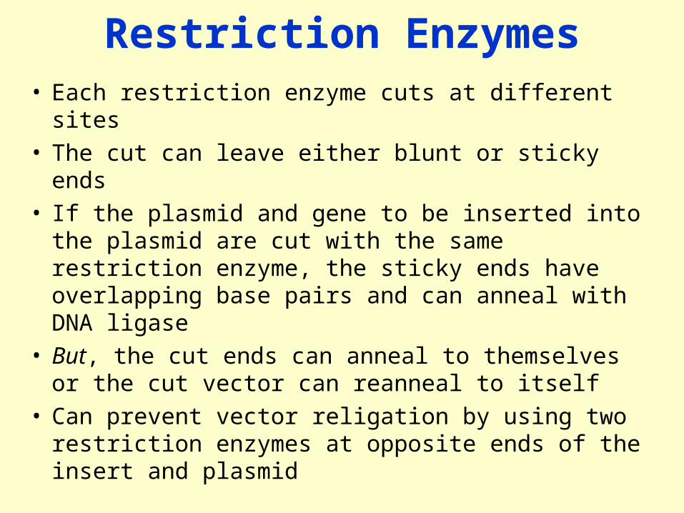
Restriction Enzymes• Each restriction enzyme cuts at different sites • The cut can leave either blunt or sticky ends• If the plasmid and gene to be inserted into the plasmid
are cut with the same restriction enzyme, the sticky ends have overlapping base pairs and can anneal with DNA ligase
• But, the cut ends can anneal to themselves or the cut vector can reanneal to itself
• Can prevent vector religation by using two restriction enzymes at opposite ends of the insert and plasmid
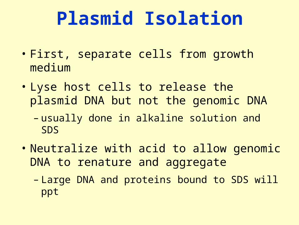
Plasmid Isolation
• First, separate cells from growth medium
• Lyse host cells to release the plasmid DNA but not the genomic DNA
– usually done in alkaline solution and SDS
• Neutralize with acid to allow genomic DNA to renature and aggregate
– Large DNA and proteins bound to SDS will ppt
Isolating Recombinant Plasmids
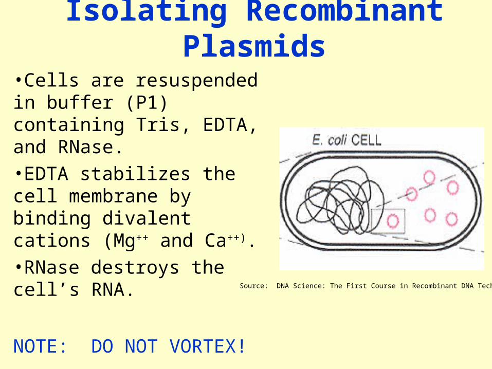
•Cells are resuspended in buffer (P1) containing Tris, EDTA, and RNase. •EDTA stabilizes the cell membrane by binding divalent cations (Mg++ and Ca++).•RNase destroys the cell’s RNA.
NOTE: DO NOT VORTEX! Source: DNA Science: The First Course in Recombinant DNA Technology
Isolating Recombinant Plasmids
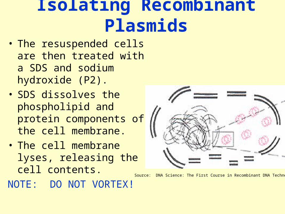
• The resuspended cells are then treated with a SDS and sodium hydroxide (P2).
• SDS dissolves the phospholipid and protein components of the cell membrane.
• The cell membrane lyses, releasing the cell contents.
NOTE: DO NOT VORTEX! Source: DNA Science: The First Course in Recombinant DNA Technology

Isolating Recombinant Plasmids
• Sodium hydroxide denatures both plasmid and chromosomal DNA into single strands.
• Chromosomal DNA separates completely into individual strands.
• The single-stranded plasmid loops remain linked together like interlocked rings.
Source: DNA Science: The First Course in Recombinant DNA Technology
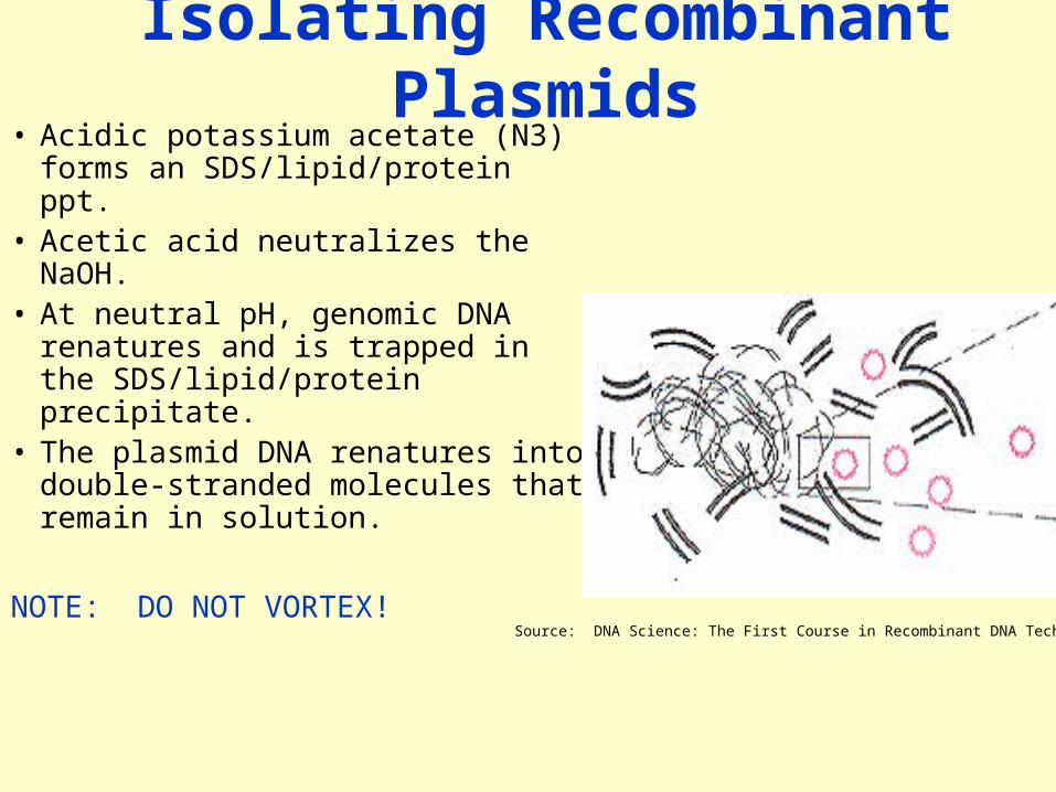
Isolating Recombinant Plasmids• Acidic potassium acetate (N3)
forms an SDS/lipid/protein ppt.• Acetic acid neutralizes the
NaOH. • At neutral pH, genomic DNA
renatures and is trapped in the SDS/lipid/protein precipitate.
• The plasmid DNA renatures into double-stranded molecules that remain in solution.
NOTE: DO NOT VORTEX!Source: DNA Science: The First Course in Recombinant DNA Technology
Isolating Recombinant Plasmids
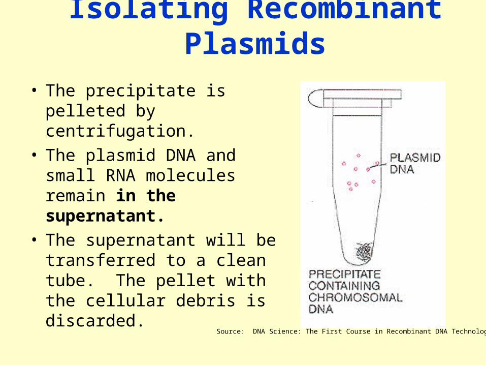
• The precipitate is pelleted by centrifugation.
• The plasmid DNA and small RNA molecules remain in the supernatant.
• The supernatant will be transferred to a clean tube. The pellet with the cellular debris is discarded.
Source: DNA Science: The First Course in Recombinant DNA Technology

Isolating Recominant Plasmids• Load supernatant with
plasmids onto spin columns.
• The pellet is washed with EtOH (PE), causing the plasmid DNA to ppt.
• The plasmid DNA binds the resin in the spin column under high salt conditions.
• Finally, the purified plasmid DNA is eluted in water.
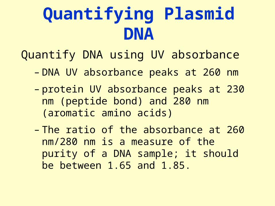
Quantifying Plasmid DNA
Quantify DNA using UV absorbance
– DNA UV absorbance peaks at 260 nm
– protein UV absorbance peaks at 230 nm (peptide bond) and 280 nm (aromatic amino acids)
– The ratio of the absorbance at 260 nm/280 nm is a measure of the purity of a DNA sample; it should be between 1.65 and 1.85.

Analyzing Recombinant Plasmids
• Purified plasmid DNA has now been isolated from transformed bacterial cells.
• After quantifying DNA yields, restriction analysis is performed on the purified plasmid DNA.
Ampicillin resist ancegene
puc plasmid vector
Origin of replicat ion
Lac Z gene
B
B
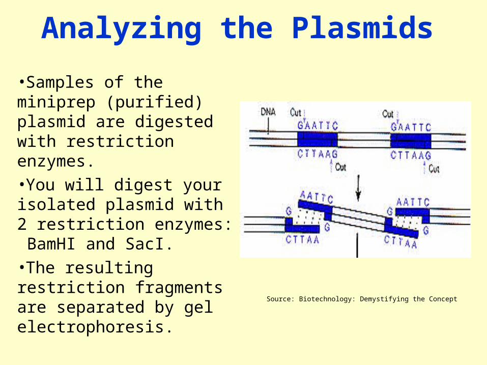
Analyzing the Plasmids
•Samples of the miniprep (purified) plasmid are digested with restriction enzymes. •You will digest your isolated plasmid with 2 restriction enzymes: BamHI and SacI. •The resulting restriction fragments are separated by gel electrophoresis.
Source: Biotechnology: Demystifying the Concept
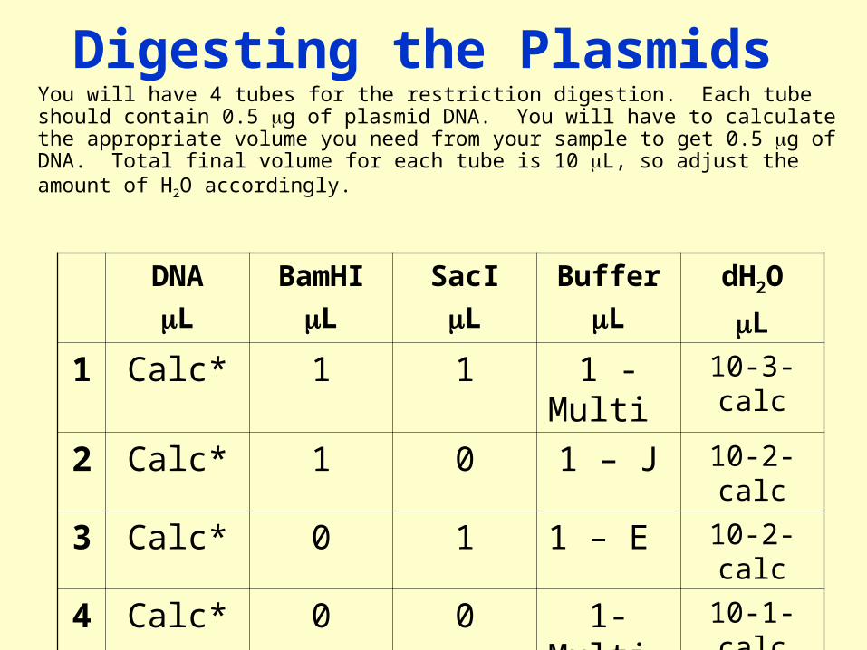
Digesting the PlasmidsYou will have 4 tubes for the restriction digestion. Each tube should contain 0.5 g of plasmid DNA. You will have to calculate the appropriate volume you need from your sample to get 0.5 g of DNA. Total final volume for each tube is 10 L, so adjust the amount of H2O accordingly.
DNA
L
BamHI
L
SacI
L
Buffer
L
dH2O
L
1 Calc* 1 1 1 - Multi 10-3-calc
2 Calc* 1 0 1 – J 10-2-calc
3 Calc* 0 1 1 – E 10-2-calc
4 Calc* 0 0 1- Multi 10-1-calc

Analyzing Recombinant Plasmids
•After digestion, the restriction fragments are separated by gel electrophoresis. •SyberSafe fluorescently labels the DNA, which can then be visualized under UV light. •The banding pattern from the restriction fragments provide a genetic “fingerprint” of the plasmid and gene insert.


![Isolation of Bacterial Plasmid DNA [Compatibility Mode]](https://img.pdfslide.net/doc/110x75/577cd39e1a28ab9e789743b1/isolation-of-bacterial-plasmid-dna-compatibility-mode.jpg)