Embed Size (px)
Citation preview

Kidney International, Vol. 48 (1995), pp. 146—154
CLINICAL INVESTIGATION
Platelet-derived growth factor A-chain expression in developingand mature human kidneys and in Wilms' tumor
CHARLES E. ALPERS, KELLY L. HUDKINS, MARINA FERGUSON, RICHARD J. JOHNSON,and JOE C. RUTLEDGE
Departments of Pathology, Medicine, and Laboratoiy Medicine, University of Washington School of Medicine and Childrens' Hospital and MedicalCenter, Seattle, Washington, USA
Platelet-derived growth factor A-chain expression in developing andmature human kidneys and in Wilms' tumor. Regulated expression ofPDGF A-chain may be important in kidney development. We employedtwo polyclonal antisera to detect expression of PDGF A-chain in fetal andnormal adult kidneys by immunohistochemistry. Specificity of the antiserawas demonstrated by Western blots of fetal and adult kidneys, demon-strating monospecific bands at 10 to 15 kD, and by absorption studies withPDGF-A peptide. PDGF A-chain is uniformly expressed by visceralglomerular epithelial cells and the epithelial cells of the distal nephron,including collecting ducts and contiguous urothelium lining the renalpelvis, in both fetal and adult kidneys. Fetal kidneys also demonstrateexpression of PDGF A-chain at the earliest stages of vesicle formationfrom the metanephric blastema; this expression is then only intermittentlydetectable in developing glomeruli until differentiation of visceral epithe-hal cells occurs. Fetal and mature arterial smooth muscle cells, and someneointimal smooth muscle cells in sclerotic arteries in adult kidneys alsoexpress PDGF A-chain. In situ hybridization with a riboprobe made fromPDGF A-chain cDNA showed close correlation of mRNA expression withprotein immunohistochemistry. PDGF A-chain expression was also iden-tified in epithelial elements of 5/6 Wilms' tumors studied. These are thefirst studies to localize PDGF A-chain expression in human kidney andsuggest sites of activity for PDGF A-chain in development, neoplasia, andin the renal arterial sclerosis of aging.
Platelet-derived growth factor (PDGF) exists as a dimer com-posed of two homologous but distinct peptides termed PDGF Aand B chains. These peptides have distinctive functional qualitiesand are encoded by genes located on different chromosomes [1,21. The functional PDGF molecule can exist in each of the threepossible isoforms: PDGF AA, AB, BB. The role of PDGF B-chainhas been extensively studied in glomerular cells in vitro [3—6], inexperimental models of glomerulonephritis [reviewed in 6—8],and in human renal development and disease [9—12]. The role ofPDGF A-chain in renal disease is not as well known, due largelyto the lack of useful reagents to evaluate expression of thismolecule in normal and diseased states. We have previouslyshown by Northern blotting techniques that PDGF A-chain geneexpression is up-regulated in rat glomeruli during the proliferativephase of mesangial proliferative glomerulonephritis induced by
Received for publication December 8, 1994and in revised form February 2, 1995Accepted for publication February 2, 1995
© 1995 by the International Society of Nephrology
administration of anti-Thy-i antibody [13]. Others have shownthat PDGF A-chain gene transcription can be up-regulated incultured mesangial cells exposed to phorbol ester, exogenousPDGF BB homodimer, and a number of peptide mitogens[14—16]. It is not known if other cell types within the rodentglomerulus express PDGF A-chain either constitutively or inresponse to specific stimuli.
In this study, we report additional characterization of twocommercially available antibodies to PDGF A-chain and the useof these antibodies to define the expression of PDGF A-chain inhuman renal development and mature renal tissue by immunocy-tochemical methods. We further report the localization of corre-sponding mRNA production for PDGF A-chain by mature renaltissue using in situ hybridization techniques. These studies dem-onstrate PDGF A-chain expression in the visceral epithelial cellsof differentiating fetal human glomeruli, and the persistent ex-pression of this molecule by these cells in the mature glomerulus.PDGF A-chain is widely expressed by urothelial epithelium of theurinary tract both in developing and mature kidneys. This studyalso demonstrates the expression of PDGF A-chain by medialsmooth muscle cells of the arterial vasculature in developing andmature human kidney, and its expression by a sub-population ofintimal smooth muscle cells in the intimal proliferative sclerosingprocess characteristic of arterial vessels in aging kidneys. Finally,we also demonstrate PDGF A-chain expression in epithelialelements of Wilms' tumors reminiscent of the patterns of expres-sion identified in normal kidney development.
Methods
Source of tissue
Human fetal kidneys (N = 12), estimated gestational ageranging from 54 to 122 days, were obtained fresh from tissueexamined after therapeutic abortions. Tissues were fixed in methylCarnoy's (methacarn) solution (60% methanol, 30% chloroform,10% acetic acid) and processed and embedded in paraffin accord-ing to conventional techniques.
Normal human kidney (N = 21) was obtained fresh fromuninvolved portions of kidneys surgically resected for localizedrenal cell carcinoma, or from cadaver donor kidneys unable to beutilized for transplantation. Portions of these tissues were fixed ineither methacarn or 10% neutral buffered formalin and processedas above.
146
Alpers et al: PDGF A-chain expression in kidneys 147
Tissue from Wilms' tumors (N = 6) was obtained fresh aftersurgical excision and portions fixed in both methacarn and 10%neutral buffered formalin. Tissue remaining after all sectionsneeded for medical diagnosis had been obtained was utilized forthis study. None of the tumors were obtained from patients withfamilial occurrence and none were known to occur in concert withWilms' tumor associated syndromes.
Antibodies
PDGF A-chain. Anti-PDGF A (Santa Cruz Biotechnology mc,Santa Cruz, CA, USA) is an affinity-purified rabbit polyclonalantibody raised against a 30 amino acid peptide corresponding tothe amino terminus of the human PDGF A-chain. The antibodyspecifically recognizes human PDGF A-chain under reducing andnon-reducing conditions, and is non-reactive with either reducedor unreduced PDGF B-chain or unreduced human PDGF-AB.The second antisera utilized is a rabbit polyclonal protein Apurified anti-human PDGF-AA (Upstate Biotechnology Inc.,Lake Placid, NY, USA) which specifically recognizes PDGF-AAhomodimer and PDGF-AB heterodimer. The immunogen for thisantisera was purified human recombinant PDGF-AA homodimer[17], and this antisera neutralizes PDGF-AA and PDGF-AB in aPDGF stimulation of 3T3 cells bioassay.
Smooth muscle cell markers. Murine monoclonal antibody 1A4(Dako Corp.) has been characterized by tissue immunohistochem-istly and Western blotting [18], and has been previously demon-strated to recognize smooth muscle a-actin in methyl Carnoy'sfixed tissues [19, 20].
Immunohistochemistry
Immunohistochemistry was performed on methyl Carnoy'sfixed, paraffin embedded tissues following a standard avidin-biotincomplex (ABC) method, as previously described [20, 21].
Briefly, sections were deparaffinized in xylene and rehydrated ingraded ethanols. Endogenous peroxidase was blocked by incuba-tion in 3% hydrogen peroxide and non-specific binding wasblocked by incubation in 10% normal goat serum. The sectionswere then incubated overnight with the anti-PDGF-A or anti-PDGF-AA antisera in a humid chamber at 4°C. Following washesin PBS, the sections were sequentially incubated with biotinylatedgoat-anti-rabbit antisera (Vector Laboratories, Burlingame, CA,USA), the ABC-Elite avidin reagent (Vector Laboratories) andfinally 3,3'-diaminobenzidine (with nickel chloride enhancement)as the chromogen. The sections were counterstained with methylgreen, dehydrated and coverslipped.
Double labeling immunocytochemisttyMethyl Carnoy's fixed, paraffin embedded tissues were sec-
tioned and mounted on aminopropylmethoxysilane (APTS)coated slides. After deparaffinization and rehydration, the slideswere sequentially incubated with the anti-a-smooth muscle actinantibody; goat anti-mouse IgG-gold (Amersham, ArlingtonHeights, IL, USA) diluted in PBS plus 1% BSA and 0.1% gelatinfor one hour at room temperature. Sections were washed, and thegold was visualized with an IntenSE M silver enhancement kit(Amersham). The sections were then incubated sequentially with:(1) rabbit-anti-PDGF A-chain diluted in PBS plus 1% BSAovernight at 4°C; (2) biotinylated goat-anti-rabbit IgG (VectorLaboratories); and (3) avidin-biotin-alkaline phosphatase com-plex (Vector Laboratories). The alkaline phosphatase was devel-
oped with a red substrate kit (Vector Laboratories) and the slideswere counterstained with methyl green. Negative controls in-cluded substituting absorbed anti-PDGF A-chain antisera (seebelow), and substituting normal mouse IgG for the anti-a-smoothmuscle actin.
Antibody absoiptionMicrotiter ELISA plates were coated with PDGF-A protein
and PDGF-A control peptide (both from Santa Cruz Biotechnol-ogy, Inc.) CA) diluted in 50 m carbonate buffer, pH 9.0, atconcentrations ranging from 1 xg/ml to 100 Wml. After over-night incubation at 4°C, the antigen solution was removed and theplates were blocked with PBS containing 1% BSA and 0.02%sodium azide for two hours at room temperature. The plates werethen washed with PBS and allowed to air dry. Rabbit anti-PDGF-A (Santa Cruz Biotechnology, Inc.) or rabbit anti-PDGF-AA (UBI) diluted 1/50 in PBS plus 2% BSA was addedand the plates were incubated overnight at 4°C. The supernatantwas removed from the wells and used as the absorbed primaryantibody in a standard ABC immunohistochemistry procedureand was also used as a control in the Western blotting experimentsdetailed below. Positive controls (that is, repetition of the absorp-tion procedure without antigen-specific absorption of the anti-sera) were done by using carbonate buffer only to coat themicrotiter wells. This control antisera was used in proceduresidentical to those of the primary unabsorbed and absorbedantisera, and gave results similar to that of the original unmanipu-lated antisera (data not shown).
Western blotting
Homogenates of normal adult and fetal kidneys were electro-phoresed on a 15% SDS polyacrylamide gel and then blotted ontonitrocellulose membranes. The blots were blocked with 5% BSAin PBS for one hour at 37°C and then incubated with anti-PDGF-A (Santa Cruz Biotechnology, Inc.) or anti-PDGF-AA(UBI) diluted 1/250 in 10 mrvi PBS containing 0.1% BSA and 10m sodium azide (PBS-BSA) for two hours at room temperature.After washing, the blots were incubated with a 1/100 dilution ofgold conjugated goat-anti-rabbit (Amersham Corp.) for twohours. The gold signal was then enhanced by incubation inIntenSE BL (Amersham Corp.). In control experiments, reducedPDGF-AA protein (Santa Cruz Biotechnology, Inc.) and recom-binant human PDGF-AA (UBI) and recombinant humanPDGF-BB (UBI) were used to demonstrate specificity of theantisera. As a negative control, the primary antibody was replacedby normal rabbit IgG at an equivalent dilution.
Molecular probeA 1280 bp human PDGF A-chain cDNA [22] was subcloned
into SP64 vector. This probe was a gift of Dr. J.N. Wilcox, EmoryUniversity. The construct was linearized and transcribed into anantisense riboprobe using reagents obtained from Promega Biotec(Madison, WI, USA), except 35S-UTP, which was obtained fromNew England Nuclear (Boston, MA, USA). The transcriptionreaction mixture contained 1 xg of PDGF A cDNA (either senseor antisense), 250 xCi 35S-UTP (1,100 to 1,300 Ci/mmol), 500 jxMeach of ATP, CTP, and GTP, 40 U RNasin, 10 mr'i dithiothreitol,40 mM Tris, and 10 U of either SP6 or T7 polymerase. After 75minutes at 37°C, the DNA was digested by adding 1 U DNase(Promega) and incubation at 37°C for an additional 15 minutes.

148 Alpers et al: PDGF A-chain expression in kidneys
Free nucleotides were then separated using a Sephadex G-50column. Specific activity of the probes ranged from 5 to 30 x i07cpm/mg. Probes were used immediately.
In situ hybridization
Adult kidney tissue which had been fixed in 10% formalin andembedded in paraffin was deparaffinized following standard pro-tocol. The sections were washed with 0.5>< standard saline citrate(SSC) (IX SSC = 150 mrvt NaCl, 15 m Na citrate, pH 7.0) anddigested with proteinase K (1 xg/mL) (Sigma) in RNase A(Promega) buffer for 40 minutes at 37°C. Several 0.5x SSCwashes were followed by prehybridization for two hours in 50 .tlof prehybridization buffer (0.3 M NaCl, 20 mrvt Tris pH 8.0, 5 mMEDTA, 1>< Denhardt's solution, 10% dextran sulfate, 10 mvtDTT). The hybridizations were started by adding 500,000 cpm of35S-labeled riboprobe in 50 of prehybridization buffer andallowed to proceed overnight at 50°C. After hybridization, sec-tions were washed with 0.5X SSC, treated with RNase A (20itgIm1, 30 mm room temperature), washed in 2X SSC (2 X 2 mm),followed by three high stringency washes in 0.1X SSC/Tween 20(Sigma) at 37°C, followed by several 2x SSC washes. After thetissue was air dried, it was dipped in NTB2 nuclear emulsion(Kodak) and exposed in the dark at 4°C for four weeks. Afterdeveloping, the sections were counterstained with hematoxylinand eosin, dehydrated, mounted and viewed.
Results
ImmunohistochemistiyTwo developmental stage-specific patterns of PDGF A-chain
expression in developing nephrons were identified. PDGF A-chain could be first identified at the earliest stage of conversion ofmetanephric blastema into an epithelial vesicle, when this differ-entiating epithelium is still in close contact with the inductivedifferentiating stimulus, the ureteric bud (Fig. 1). There is noexpression of PDGF A-chain by the adjacent blastema, uretericbud, or interstitial tissue. As glomerular development proceedsthrough subsequent comma and S-shape stages, as they have beencharacterized by Saxén, Ekblom, and others [23—25], PDGFA-chain expression often becomes undetectable, although focal,intermittent expression by the epithelial cells comprising theseprimitive glomeruli is present. This variability was encounterednot only between different kidney samples, but between glomer-ular structures of apparently identical morphologic developmentwithin the same tissue sections (Fig. 1B). In the last stages ofglomerular development, PDGF A-chain is uniformly expressedby the visceral epithelial cells overlying glomerular capillaries(Fig. 1 C, D). The staining pattern is cytoplasmic, and appears tobe of uniform intensity for glomeruli at all levels of maturationwithin developing kidneys (that is, superficial glomeruli as well as
those near the cortico-medullary junction) once visceral epithelialcell differentiation has occurred. A minority of these differenti-ated glomeruli also show diffuse cytoplasmic expression of PDGFA-chain by parietal epithelial cells. Expression of PDGF A-chainby mesangial cells or endothelial cells is not detectable. Extraglo-merular structures exhibiting widespread expression of PDGFA-chain included the urothelial lining of the ureter and renalpelvis, collecting duct epithelium in contiguity to the urothelium,and the smooth muscle cell layers of the developing renal arterialvasculature (Fig. 1 A, E).
The normal adult kidneys demonstrated persistence of patternsof PDGF A-chain expression exhibited late in renal development.Specifically, there is widespread expression by glomerular visceralepithelial cells and some parietal epithelial cells, but not otherglomerular structures (Fig. 2A), widespread expression by collect-ing ducts and the urothelium lining the lower urinary tract (Fig.2B), and diffuse expression by medial smooth muscle cells of therenal arterial tree (Fig. 2 A, C, D). In addition, many of the renalarteries sampled exhibited variable, most often mild to moderate,degrees of intimal sclerosis characteristic of aging kidneys. Themajority of neointimal smooth muscle cells (shown by labelingwith the a-smooth muscle actin antibody) present in these scle-rosing arteries also exhibited prominent expression of PDGFA-chain (Fig. 2 C, D). PDGF A-chain expression by medialsmooth muscle cells in these arteries was indistinguishable fromthat seen in arteries without prominent neointimal sclerosis.
The Wilms' tumor specimens exhibited the varied histopatho-logic features characteristic of these neoplasms, including blast-emal, epithelial, and mesenchymal elements. All but one of thesetumors exhibited focal expression of PDGF A-chain. PDGFA-chain expression was confined to more differentiated epithelialelements within these tumors, whereas neoplastic blastemal andmesenchymal structures had no detectable expression (Fig. 2E).Epithelial elements expressing PDGF A-chain often, but notinvariably, assumed a tubular configuration; rarely, the PDGFA-chain expression in such structures appeared to be confined tothe luminal surface, implying a degree of cell polarity. As expectedfrom studies of normal kidneys, smooth muscle cells and/orpericytes comprising the non-neoplastic, infiltrating tumor vascu-lature also expressed PDGF A-chain.
No significant differences in staining patterns were identifiedbetween either of the two primary antisera utilized, although astronger signal was obtained with the antisera raised against the30 amino acid terminus of PDGF A-chain monomer. No specificcellular staining was seen with substitution of control rabbit IgGor PBS for the primary antibody. Staining patterns were uniformin both adult and fetal kidneys except for the variability indetectable expression of PDGF A-chain by early differentiating
Fig. 1. Expression of PDGFA-chain in developing kidneys. A. Low power photomicrograph of 59 day gestational age kidney showing expression of PDGFA-chain in developing glomerular structures, muscular arteries (arrows) and urothelium lining the renal pelvis (arrowheads). There is no expression inthe rim of metanephric blastema (b) at the outer cortex of the kidney, or in interstitial cells. B. Same kidney as A. PDGF A-chain is expressed byepithelia of early differentiating structures (S and comma shaped/folded nephrons) but not by adjacent undifferentiated blastema. C. Differentiated fetalglomeruli uniformly demonstrate PDGF A-chain expression by visceral epithelial cells and irregularly by parietal epithelial cells lining Bowman'scapsules. D. High magnification of a fetal glomerulus demonstrating PDGF A-chain expression in visceral epithelial cells. E. PDGF A-chain expressionby smooth muscle cells of arteries (arrows) in a fetal kidney. F. Low power view of PDGF A-chain expression in fetal kidney of 81 days gestational age,demonstrating patterns described in A-E. G. Adjacent tissue section to that shown in F reacted with absorbed antisera. There is complete abolition ofstaining, providing strong evidence of antibody specificity.

4%4i- 4w.
a 4
c-I_a St -a s.— t1•
- -s-ti
Alpers et a!: PDGF A-chain expression in kidneys 149

150 Alpers et al: PDGF A-chain expression in kidneys
vesicular and glomerular structures as indicated above. Replica-tion of the immunohistochemical procedures with the absorbedantisera removed the specific staining of arterial smooth musclecells, urothelium, and glomerular epithelial cells (Fig. 1 F, G).
In situ hybridization
In situ hybridization studies of fetal kidneys could not beperformed due to degradation of the mRNA transcripts in thesetissues. Studies of the adult kidneys showed a pattern of PDGFA-chain mRNA production corresponding to the patterns ofprotein expression described above. Hybridization signal wasstrongest overlying the smooth muscle cells comprising the mediaof arteries (Fig. 2F). Discrete signals were also identified in someneointimal smooth muscle cells, in some glomerular visceralepithelial cells and, rarely, parietal epithelial cells (Fig. 2G).While the distribution of mRNA expression revealed no discrep-ancies with the immunolabeling results (that is, no cell typesappeared to produce PDGF A-chain mRNA that did not expressidentifiable PDGF A-chain protein), there was much less unifor-mity of mRNA production by positive cell types within eachindividual tissue section when assessed by our hybridizationtechniques as compared to the immunolabeling results. This maynot represent a meaningful biological difference as hybridizationincubation times were relatively short and other technical details(such as time of fixation after surgical removal of the specimen)for which hybridization studies are more sensitive than proteinimmunocytochemistry could not always be rigorously controlled.All positive hybridization results were obtained with use of the"antisense" strand probe; concomitant procedures utilizing the"sense" strand probe were uniformly negative (data not shown).
Western blotting
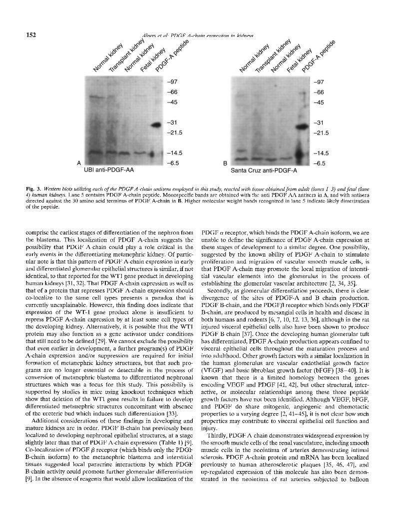
Both the anti-PDGF-A and anti-PDGF-AA antisera demon-strated discrete bands corresponding to proteins of 10 to 15 kDwhich correspond to the size of the PDGF A-chain monomer asindicated by simultaneous blotting of a control preparation ofpurified PDGF-AA (Fig. 3). Identical bands were found in bothfetal and mature kidneys. In separate blotting experiments, bothantisera recognized reduced and unreduced recombinant humanPDGF-AA protein, with discrete bands at 14 to 18 kD and 30 to32 kD, respectively. The antisera did not recognize reduced orunreduced PDGF-BB (data not shown). In separate blottingexperiments, the identification of the 10 to 15 kD band could beabolished by the use of the antiserum pre-absorbed with PDGF-Apeptide (data not shown).
Discussion
The potential significance of PDGF A-chain in renal develop-ment is suggested by studies identifying a Wilms' tumor gene andits protein product WT1 [26]. One of the few known activities ofthe WT1 protein is that it has been shown to bind and repress thetranscription activating sequence of the PDGF A-chain gene[27—29]. Mutations in this gene, which lead to the development ofWilms' tumor, might then be expected to lead to increasedexpression of PDGF A-chain, possibly in the Wilms' tumor itself,but also possibly within the non-tumorous renal parenchyma. Theeffects of such expression are not predictable, given how little isknown of the function or sites of expression of PDGF A-chain andits receptor in either kidney development or normal physiologicactivity.
This study demonstrates expression of PDGF A-chain inWilms' tumors, albeit in a pattern restricted to tumor elementsdemonstrating epithelial and most often tubular differentiation.Those portions of Wilms' tumors exhibiting features more likeundifferentiated blastema or primitive mesenchyme (in distinctionfrom the non-neoplastic mesenchymal structures comprising theinfiltrating tumor vasculature) were never found to express PDGFA-chain. Wilms' tumors typically demonstrate composite histo-logic features comprising blastema and mesenchymal elements aswell as more differentiated epithelial elements [30]; the signifi-cance of PDGF A-chain expression by only one of these elementsis of unknown clinical or biologic significance. However, thisfinding in Wilms' tumors does correspond to the pattern of PDGFA-chain expression by vesicular structures at the earliest stage ofepithelial differentiation from the blastema and by tubular struc-tures of the distal nephron in developing metanephric kidneys;tumor expression of PDGF A-chain may then be both a functionand potential histologic marker of tumor differentiation. Thedifferentiating elements of these tumors in this instance appear tomaintain fidelity of growth factor expression with normal renaldevelopment.
Our studies also demonstrate expression of PDGF A-chain inthe developing metanephric kidney in humans (Table 1). Theyshow expression that is predominantly localized to differentiatingvisceral epithelial cells, collecting duct structures and the contig-uous urothelial lining of lower urinary tract structures such asrenal pelvis and ureter, and the smooth muscle cells of the arterialvasculature, These relatively differentiated structures bear noobvious relation to the metanephric blastema from which Wilms'tumors arise. However, as noted above, PDGF A-chain is alsoexpressed in a restricted manner to vesicular structures which
Fig. 2. A. Adult normal human glomerulus showing persistent expression of PDGF A-chain in visceral epithelial cells, parietal epithelial cells, and thesmooth muscle cells of a hilar arteriole. B. Adult normal human kidney shows persistent, widespread expression of PDGF A-chain by urothelium liningthe renal pelvis (arrow) as well as by the contiguous epithelium of the renal collecting ducts. C. Arcuate artery from adult human kidney with intimalsclerosis. PDGF A-chain is expressed by smooth muscle cells comprising both the vessel media and intima (i). Several smaller arteries also demonstratePDGF A-chain expression by medial smooth muscle cells. D. Double immunolabeling of an intrarenal artery from an adult human kidney alsodemonstrating intimal (i) sclerosis. PDGF A-chain expression by smooth muscle cells is identified by red alkaline phosphatase label, while the smoothmuscle cell identity of intimal and medial cells is confirmed by co-localization of a black immunogold label for a-smooth muscle actin. E. Wilms' tumor.There is focal expression of PDGF A-chain by portions of the tumor exhibiting epithelial differentiation, while less well differentiated (blastemal)elements do not express detectable levels of this peptide. F. In situ hybridization of adult human kidney, with PDGF A-chain mRNA expressiondemonstrable in smooth muscle cells of renal arterial structures, in accordance with immunohistochemical results. Adventitia and adjacent matrix cellsshow no expression. G. Same kidney as F, with glomerulus showing PDGF A-chain mRNA expression which can generally be localized to visceralepithelial cells at periphery (arrows) and central portions of glomerular tuft.

I i±$
'
a
ttt01-'---. _t
'C'—.
$4
• 14
2 i:-
0.
j 'S
•
"'¼,
E5,-
.9.
,-'i"L '-'-:
/
G
Alpers et al: PDGF A-chain expression in kidneys 151
4,
—97
—66
—45
—31
—21.5
—14.5
—6.5UBI anti-PIQF-M
BSanta Cruz anti-PDGF-A
152 Alpers et aL PDGF A-chain expression in kidneys
Fig. 3. Western blots utilizing each of the PDGFA-chain antisera employed in this study, reacted with tissue obtained from adult (lanes 1—3) and fetal (lane4) human kidneys. Lane 5 contains PDGF A-chain peptide. Monospecific bands are obtained with the anti-PDGF-AA antisera in A, and with antiseradirected against the 30 amino acid terminus of PDGF A-chain in B. Higher molecular weight bands recognized in lane S indicate likely dimerizationof the peptide.
comprise the earliest stages of differentiation of the nephron fromthe blastema. This localization of PDGF A-chain suggests thepossibility that PDGF A-chain could play a role critical in theearly events in the differentiating metanephric kidney. Of partic-ular note is that this pattern of PDGF A-chain expression in earlyand differentiated glomerular epithelial structures is similar, if notidentical, to that reported for the WT1 gene product in developinghuman kidneys [31, 32]. That PDGF A-chain expression as well asthat of a protein that represses PDGF A-chain expression shouldco-localize to the same cell types presents a paradox that iscurrently unexplainable. However, this finding does indicate thatexpression of the WT-1 gene product alone is insufficient torepress PDGF A-chain expression by at least some cell types ofthe developing kidney. Alternatively, it is possible that the WT1protein may also function as a gene activator under conditionsthat still need to be defined [29]. We cannot exclude the possibilitythat even earlier in development, a further program(s) of PDGFA-chain expression and/or suppression are required for initialformation of metanephric kidney structures, but that such pro-grams are no longer essential or detectable in the process ofconversion of metanephric blastema to differentiated nephronalstructures which was a focus for this study. This possibility issupported by studies in mice using knockout techniques whichshow that deletion of the WT1 gene results in failure to developdifferentiated metanephric structures concomitant with absenceof the ureteric bud which induces such differentiation [33].
Additional considerations of these findings in developing andmature kidneys are in order. PDGF B-chain has previously beenlocalized to developing nephronal epithelial structures, at a stageslightly later than that of PDGF A-chain expression (Table 1) [9].Co-localization of PDGF /3 receptor (which binds only the PDGFB-chain isoform) to the metanephric blastema and interstitialtissues suggested local paracrine interactions by which PDGFB-chain activity could promote further glomerular differentiation[9]. In the absence of reagents that would allow localization of the
PDGF a receptor, which binds the PDGF A-chain isoform, we areunable to define the significance of PDGF A-chain expression atthese stages of development to a similar degree. One possibility,suggested by the known ability of PDGF A-chain to stimulateproliferation and migration of vascular smooth muscle cells, isthat PDGF A-chain may promote the local migration of intersti-tial vascular elements into the glomerulus in the process ofestablishing the glomerular vascular architecture [2, 34, 35].
Secondly, as glomerular differentiation proceeds, there is cleardivergence of the sites of PDGF-A and B chain production.PDGF B-chain, and the PDGF13 receptor which binds only PDGFB-chain, are produced by mesangial cells in health and disease inboth humans and rodents [6, 7, 10, 12, 13, 36], although in the ratinjured visceral epithelial cells also have been shown to producePDGF B-chain [37]. Once the developing human glomerular tufthas differentiated, PDGF A-chain production appears confined tovisceral epithelial cells throughout the maturation process andinto adulthood. Other growth factors with a similar localization inthe human glomerulus are vascular endothelial growth factor(VEGF) and basic fibroblast growth factor (bFGF) [38—40]. It isknown that there is a limited homology between the genesencoding VEGF and PDGF [41, 42], but other structural, inter-active, or molecular relationships among these three peptidegrowth factors have not been identified. Although VEGF, bFGF,and PDGF do share mitogenic, angiogenic and chemotacticproperties to a varying degree [2, 41—45], it is not clear how suchproperties may contribute to visceral epithelial cell function andinjury.
Thirdly, PDGF A-chain demonstrates widespread expression bythe smooth muscle cells of the renal vasculature, including smoothmuscle cells in the neointima of arteries demonstrating intimalsclerosis. PDGF A-chain protein and mRNA has been localizedpreviously to human atherosclerotic plaques [35, 46, 47], andup-regulated expression of this molecule has also been demon-strated in the neointima of rat arteries subjected to balloon

Alpers et a!: PDGF A-chain expression in kidneys 153
Table 1. Phenotypic characterization of stages of glomerulogenesis inhuman fetal kidney
Earlyglomerular
differentiationMetanephric
blastema Vesicle(comma,S-stage)
Differentiatedglomerulus
• PDGF B-chain — +1— — Mesangium• PDGFRI3 + — Mesangium• PDGF A-chain — focal + Visceral epithelial
cell• WT1 — focal + Visceral epithelial
cell
Expression of members of the PDGF system and the potentiallyregulatory protein WT1 during human glomerulogenesis. Data Ofl PDGFB-chain and PDGFRI3 are from [9]; data on WT1 expression are from[31, 32].
catheter injury [34]. Based on the sequence of pathologic eventsoccurring in experimental balloon catheter injury and on studiesof vascular smooth muscle cells in vitro, evidence has accumulatedthat PDGF A-chain may function as a smooth muscle cell mitogenand promoter of vascular smooth muscle cell migration. In thisstudy, the arterial localization of PDGF A-chain provides the firstdirect demonstration of the potential of PDGF A-chain toparticipate in human renal vascular injury. The identification ofPDGF A-chain in neointimal smooth muscle cells in particularsuggests the potential for both paracrine and autocrine activityinvolving these mesenchymal cells within the vessel neointima, aswell as the potential to stimulate the neighboring smooth musclecells of the arterial vessel media to replicate or migrate and hencepromote further neointimal expansion. It has even been suggestedthat PDGF A-chain may have non-mitogenic trophic functionsthat are otherwise unspecified but nonetheless important in themaintenance of arterial structures [47]. The apparently constitu-tive expression of PDGF A-chain mRNA and protein by medialsmooth muscle cells in both developing and mature arteries isevidence for such a trophic function in renal arteries. Sincevirtually nothing is known of the processes which cause the renalarterial sclerosis that is a hallmark of renal aging, nor of thosecausing sclerosis as a consequence of more discreet forms ofvascular injury, we are unable to more specifically identify thespecific functional roles of PDGF A-chain in these processes asyet. However, in situ identification of expression of molecules atboth the transcriptional and protein levels that could mediate suchprocesses is an initial approach towards understanding the patho-genetic basis of human renovascular disease. Some evidence insupport of a role for PDGF A-chain in such processes is providedin our recent study of allograft kidneys demonstrating widespreadexpression of PDGF A-chain by the neointimal cells of arteriesdemonstrating chronic vascular rejection in these kidneys [48].
In summary, our studies indicate regulated expression of PDGFA-chain in developing kidney glomerular epithelial cells, tubules,and blood vessels and persistence of these patterns into adult-hood. However, they do not identify the role of PDGF A-chain inhuman kidney development or answer the question of whether thePDGF A-chain expression observed is essential for normal kidneymaturation and function. The critical role for PDGF B-chain inthis process has recently been demonstrated in the PDGF B-chainknockout mouse which revealed that PDGF B-chain is required
for normal development of the glomerular mesangium [49]; theimportance of PDGF A-chain may be determined by a similarexperimental approach. Knowledge about the sites of activity forPDGF A-chain within the kidney awaits better localization of itsreceptor. It has been reported recently that in the adult kidneyPDGFa receptor co-localizes in a distribution similar to PDGF 13receptor [12]; however, none of this was illustrated and we remaincautious in our understanding of the distribution of PDGF areceptor. Some indication of a role for PDGF A-chain in thepathogenesis of renal vascular disease is already provided bydemonstration of its expression in atherosclerosis [35, 47], renalarteriosclerosis, and renal allograft rejection [48]. Finally, we haveidentified a pattern of glomerular expression of PDGF A-chainsimilar to that of the Wilms' tumor gene product and furtheridentified PDGF A-chain expression in a portion of Wilms'tumors, adding to the evidence associating these molecules in theprocesses of renal development and neoplasia.
Acknowledgments
This work was supported by grants HL42270, HL47151, DK43422 anda George O'Brien Kidney Research Center grant DK47659 from theNational Institutes of Health. The authors thank Drs. Christer Betscholtzand Cy Wilcox for provision of the PDGF A-chain probe, the CentralLaboratory for Human Embryology at the University of Washington(supported by grant HD00836 from the National Institutes of Health) forassistance in providing fetal tissue, and Sheri Storey for secretarialassistance.
Reprint requests to Dr. Charles E. Alpers, Department of Pathology, RC-72,University of Washington Medical Center, Seattle, Washington 98195, USA.
References
1. Ross R, RAINES EW, BOWEN-POPE DF: The biology of PDGF. Cell46:155—169, 1986
2. RAINES EW, Ross R: Platelet-derived growth factor in vivo, inCytokines (vol 5), edited by WESTERMARK B, SORG C, Basel, Karger,1993, pp 74—1 14
3. SILVER P1, JAFFER FE, ABBOUD HE: Platelet-derived growth factorsynthesis in mesangial cells: Induction by multiple peptide mitogens.Proc NatlAcad Sci USA 86:1056—1060, 1988
4. SHULTZ PJ, DICORLETO PE, SILVER BJ, ABBOUD HE: Mesangial cellsexpress PDGF mRNAs and proliferate in response to PDGF. Am JPhysiol 255(Renal Fluid Electrol Physiol. 24):F674-F684, 1988
5. FLOEGE J, TOPLEY N, Hoa J, BARRETT TB, RESCH K: Mitogeniceffect of platelet-derived growth factor in human glomerular mesang-ial cells: Modulation and/or suppression by inflammatory cytokines.Clin Exp Immunol 86:334—341, 1991
6. ABBOUD HE: Growth factors in glomerulonephritis. Kidney mt 43:252—267, 1993
7. JOHNSON RJ, FLOEGE J, COUSER WG, ALPERS CE: Role of platelet-derived growth factor in glomerular disease. JASIV 4:119—128, 1993
8. JOHNSON RI: The glomerular response to injury: Progression orresolution? Kidney mt 45:1769—1782, 1994
9. ALPERS CE, SEIFERT RA, HUDKINS KL, JOHNSON RJ, BOWEN-POPEDF: Developmental patterns of PDGF B-chain, PDGF-receptor, anda actin expression in human glomerulogenesis. Kidney mt 42:390—399,1992
10. GESUALDO L, PINZANI M, FLORIANO JJ, HASSAN MO, NAGY NU,SCHENA FP, EMANCIPATOR SN, ABBOUD HE: Platelet-derived growthfactor expression in mesangial proliferative glomerulonephritis. LabInvest 64:160—167, 1991
11. NAKAJIMA M, HEWITSON TD, MATHEWS DC, KINCAID-SMITH P:Platelet-derived growth factor mesangial deposits in mesangial IgAglomerulonephritis. Nephrol Dial Transplant 6:11—16, 1991
12. GESUALDO L, Dr PAOLO S, MILANI S, PINZANI M, GRAPPONE C,RANIERI B, PANNARALE G, Sd-lENA FP: Expression of platelet-derivedgrowth factor receptors in normal and diseased human kidney: An

154 Alpers et a!: PDGF A-chain expression in kidneys
immunohistochemistry and in Situ hybridization study. J Clin Invest94:50—58, 1994
13. IIDA H, SEIFERT R, ALPERS CE, GRONWALD RGK, PHILLIPS PE,PRITZL P, GORDON K, GOWN AM, Ross R, BOWEN-POPE DF,JOHNSON RJ: Platelet-derived growth factor (PDGF) and PDGFreceptor are induced in mesangial proliferative nephritis in the rat: Aneffect mediated by platelets and complement. Proc NatlAcad Sci USA88:6560—6564, 1991
14. BHANDARI B, ABBOUD HE: Platelet derived growth factor-A chaingene expression in cultured mesangial cells: Regulation by phorbolester at the level of mRNA abundance, transcription and mRNAstability. Mol Cell Endocrinol 91:185—191, 1993
15. BHANDARI B, GRANDALIANO G, ABBOUD HE: Platelet-derived growthfactor (PDGF) BB homodimer regulates PDGF A- and PDGFB-chain gene transcription in human mesangial cells. Biochem J297:385—388, 1994
16. SILVER BJ, JAFFER FE, ABBOUD HE: Platelet-derived growth factor(PDGF) synthesis in mesangial cells: Induction by multiple peptidemitogens. Proc Nati Acad Sci USA 86:1056—1060, 1989
17. HART CE, BAILEY M, CURTIS DA, OSBORN S, RAINES E, Ross R,FORSTROM JW: Purification of PDGF-AB and PDGF-BB from humanplatelet extracts and identification of all three PDGF dimers in humanplatelets. Biochem 29:166—172, 1990
18. SKALLI 0, ROPRAX P, TRZECIAK A, BENXONANA G, GILLESSEN D,GABBIANI G: A monoclonal antibody against a-smooth muscle actin:A new probe for smooth muscle differentiation. J Cell Biol 103:2787—2796, 1986
19. JOHNSON RJ, IIDA H, ALPERS CE, MAJESKY MW, SCHWARTZ SM,PRITZL P, GORDON K, GOWN AM: Expression of smooth muscle cellphenotype by rat mesangial cells in immune complex nephritis:a-smooth muscle actin is a marker of mesangial cell proliferation. JClin Invest 87:847—858, 1991
20. O'BRIEN KD, ALLEN MD, MCDONALD TO, CHAIT A, HARLAN JM,FISHBEIN D, MCCARTY J, FERGUSON M, HUDKINS K, BENJAMIN CD,LOBB R, ALPERS CE: Vascular cell adhesion molecule-i is expressedin human coronary atherosclerotic plaques: Implications for the modeof progression of advanced coronary atherosclerosis. J Clin Invest92:945—951, 1993
21. GOWN AM, VOGEL AM: Monoclonal antibodies to intermediatefilaments proteins of human cells. II. Distribution of filament proteinsin normal human tissues. Am JPathol 114:309—321, 1984
22. BETSHOLTZ C, JoHNssoN A, HELDIN C-H, WESTERMARK B, LIND P.URDEA MS, EDDY R, SHows TB, PHILPOTr K, MELLOR AL, Kr.torrTJ, SCOTr J: eDNA sequence and chromosomal localization of humanplatelet-derived growth factor A-chain and its expression in tumourcell lines. Nature 320:695—699, 1986
23. SAXEN L: Organogenesis of the Kidney. Cambridge, Cambridge Uni-versity Press, 1987
24. EKBLOM P: Developmentally regulated conversion of mesenchyme toepithelium. FASEB J 3:2141—2150, 1989
25. ABRAHAMSOM DR: Glomerulogenesis in the developing kidney. SeminNephrol 11:375—389, 1991
26. RAUSCHER FJ III: The WTI Wilms tumor gene product: A develop-mentally regulated transcriptional factor in the kidney that functionsas a tumor suppressor. FASEB J 7:896—903, 1993
27. GASHLER AL, BONTHRON DT, MADDEN SL, RAUSCHER FJ III, COL-LINS T, SUKHATME VP: Human platelet-derived growth factor A chainis transcriptionally repressed by the Wilms Tumor Suppressor WT1.Proc Nat! Acad Sci USA 89:10984—10988, 1992
28. WANG ZY, MADDEN SL, DEUEL TF, RAUSCHER FJ III: The Wilms'tumor gene product, WTI, represses transcription of the platelet-derived growth factor A-chain gene. J Biol Chem 267:21999—22002,1992
29. WANG ZY, QIU 00, DEUEL TF: The Wilms' tumor gene product
WT1 activates or suppresses transcription through separate functionaldomains. J Biol Chem 268:9172—9175, 1993
30. BECKWITH JB, PALMER NF: Histopathology and prognosis of Wilmstumors: Results from the First National Wilms' tumor study. Cancer41:1937—1948, 1978
31. PRITCHARD-JONES K, FLEMING 5, DAVIDSON D, BICKMORE W, POR-TEUS D, GOSDEN C, BARD J, BUCKLER A, PELLETIER J, HOUSMAN D,VAI'4 HEYNINGER V, HASTIE N: The candidate Wilms' tumour gene isinvolved in genitourinary development. Nature 346:194—197, 1990
32. MUNDLOS 5, PELLETIER J, DARVEAU A, BACHMANN M, WINTERPACHTA, ZABEL B: Nuclear localization of the protein encoded by theWilms' tumor gene WT1 in embryonic and adult tissues. Development119:1329—1341, 1993
33. KREIDBERG JA, SARIOLA H, LORING JM, MAEDA M, PELLETIER J,HOUSMAN D, JAENISCH R: WT-1 is required for early kidney devel-opment. Cell 74:679—691, 1993
34. MAJESKY MW, REIDY MA, BOWEN-POPE DF, HART CE, WILCOX JN,SCHWARTZ SM: PDGF ligand and receptor gene expression duringrepair of arterial injury. J Cell Biol 111:2149—2158, 1990
35. REKHTER MD, GORDON D: Does platelet-derived growth factor-Achain stimulate proliferation of arterial mesenchymal cells in humanatherosclerotic plaques? Circ Res 75:410—417, 1994
36. ALPERS CE, SEIFERT RA, HUDKINS KL, JOHNSON RJ, BOWEN-POPEDF: PDGF-receptor localizes to mesangial, parietal epithelial, andinterstitial cells in human and primate kidneys. Kidney mt 43:286—294,1993
37. FLOEGE J, JOHNSON RJ, ALPERS CE, FATEMI-NAINIE S, RICHARDSONCA, GORDON K, COUSER WG: Visceral glomerular epithelial cells canproliferate in vivo and synthesize PDGF B-chain. Am J Pathol142:637—650, 1993
38. BROWN LF, BER5E B, TOGNAZZI K, MANSEAU EJ, VAN DE WATER L,SENGER DR, DVORAK HF, ROSEN S: Vascular permeability factormRNA and protein expression in human kidney. Kidney mt 42:1457—1461, 1992
39. ALPERS CE, JOHNSON RJ: Growth factors and the glomerulus: Rela-tions between development and injury. Adv Nephrol 24:33—52, 1995
40. ALPERS CE, SCHELLING ME, HUDKINS KL, REIDY MA, LINDNER V:Localization of basic fibroblast growth factor (bFGF) and its receptor(FGFR1. fig) in fetal, mature, and transplanted human kidneys(abstract) Lab Invest 70:156A, 1994
41. LEUNG DW, CACHIANES G, KUANG W-J, GOEDDEL DV, FERRARA N:Vascular endothelial growth factor is a secreted angiogenic mitogen.Science 246:1306—1309, 1989
42. KECK PJ, HAUSER SD, KRIVI G, SANZO K, WARREN T, FEDER J,CONNOLLY DT: Vascular permeability factor, an endothelial cellmitogen related to PDGF. Science 246:1309—1312, 1989
43. RISAU W, DREXLER H, MIRONOV V, SMITS A, SIEGBAHN A, FUNA K,HELDIN C-H: Platelet-derived growth factor is angiogenic in vivo.Growth Factors 7:261—266, 1992
44. KLAGSBRUN M, EDELMAN ER: Biological and biochemical propertiesof fibroblast growth factors. Arteriosclerosis 9:269—278, 1989
45. JUDAH F, SHING Y: Angiogenesis. J Biol Chem 267:10931—10934, 199246. WILCOX JN, SMITH KM, WILLIAMS LT, SCHWARTZ SM, GORDON D:
Platelet-derived growth factor mRNA detection in human atheroscle-rotic plaques by in situ hybridization. J Clin Invest 82:1134—1 143, 1988
47. BARRETIT TB, BENDITIT EP: Platelet-derived growth factor gene ex-pression in human atherosclerotic plaques and normal artery wall.Proc Nat! Acad Sci USA 85:2810—2814, 1988
48. ALPERS CE, HUDKINS K: Platelet-derived growth factor (PDGF)A-chain expression in normal and rejecting renal arteries (abstract).Lab Invest 72:157A, 1995
49. LEVEEN P, PEKNY M, GEBRE-MEDHIN 5, SWOLIN B, LARSSON E,BETSHOLTZ C: Mice deficient for PDGF B show renal, cardiovascular,and hematological abnormalities. Gene Develop 8:1875—1887, 1994