Embed Size (px)
Citation preview
Guideline No: 0/C/14:9040-01:00 Guideline: Wound Assessment and Management
This document reflects what is currently regarded as safe practice. However, as in any clinical situation, there may be factors which cannot be covered by a single set of guidelines. This document does not replace the need for the application of clinical judgement to each individual presentation. Approved by: SCHN Policy, Procedure and Guideline Committee Date Effective: 1st June 2014 Review Period: 3 years
Team Leader: Clinical Nurse Consultants Area/Dept: Stomal Therapy (CHW) and Surgical and Wound Care (SCH)
Date of Publishing: 6 June 2014 3:23 PM Date of Printing: Page 1 of 17 K:\CHW P&P\ePolicy\May 14\Wound Assessment Mgt.docx This Guideline may be varied, withdrawn or replaced at any time.
WOUND ASSESSMENT AND MANAGEMENT
PRACTICE GUIDELINE ©
DOCUMENT SUMMARY/KEY POINTS
• This guideline is intended to serve as a guide for clinical staff in wound management.
• Maintaining skin integrity in hospitalised patients is a fundamental and critical goal of nursing practice.
• A thorough assessment of a wound is critical in determining how it should be managed.
• An ongoing process of assessment, clinical decision making, planning, intervention and education will minimise complications, promote healing and facilitate optimal wound healing.
• Consistent and high quality wound management and documentation is essential to enable treatment outcomes to be determined.
• Optimal healing is promoted by collaboration between all clinical staff involved in wound management.
• All patients with wounds will have their wounds appropriately assessed by nursing staff with timely referrals to treating medical team and /or Wound CNC where appropriate.
Guideline No: 0/C/14:9040-01:00 Guideline: Wound Assessment and Management
This document reflects what is currently regarded as safe practice. However, as in any clinical situation, there may be factors which cannot be covered by a single set of guidelines. This document does not replace the need for the application of clinical judgement to each individual presentation. Approved by: SCHN Policy, Procedure and Guideline Committee Date Effective: 1st June 2014 Review Period: 3 years
Team Leader: Clinical Nurse Consultants Area/Dept: Stomal Therapy (CHW) and Surgical and Wound Care (SCH)
Date of Publishing: 6 June 2014 3:23 PM Date of Printing: Page 2 of 17 K:\CHW P&P\ePolicy\May 14\Wound Assessment Mgt.docx This Guideline may be varied, withdrawn or replaced at any time.
CHANGE SUMMARY
• Respective SCH and CHW Wound care practice guidelines have been replaced by this SCHN wound care guideline. Content from both documents has formed the basis for this SCHN guideline.
• No new wound management practices have been introduced.
READ ACKNOWLEDGEMENT
• Clinical nurses, nurse managers and other clinical staff (as appropriate), with direct responsibilities to patients requiring wound care, must read and acknowledge they understand the contents of this guideline.
Related information:
• SCHN:
o Hand Hygiene Policy
• At CHW:
o Pressure Area Care – Prevention and Management Practice Guideline
o Burns Management Practice Guideline
• At SCH:
o Pressure Ulcer Prevention Guideline
o Treatment of Burn Injuries Guideline
Guideline No: 0/C/14:9040-01:00 Guideline: Wound Assessment and Management
Date of Publishing: 6 June 2014 3:23 PM Date of Printing: Page 3 of 17 K:\CHW P&P\ePolicy\May 14\Wound Assessment Mgt.docx This Guideline may be varied, withdrawn or replaced at any time.
TABLE OF CONTENTS 1 Introduction .................................................................................................................. 4 1.1 Stages of wound healing ............................................................................................... 4
Inflammatory or Reaction Phase ......................................................................................... 4 Proliferation Phase .............................................................................................................. 4 Maturation or Reconstruction Phase ................................................................................... 4
1.2 Wound Repair Mechanisms .......................................................................................... 4 1.3 General Principles of Wound Management ................................................................... 5 1.4 Pain Assessment ........................................................................................................... 5 2 Wound Assessment .................................................................................................... 6
Wound Bed Characteristics ................................................................................................. 6 2.1 Assessment Documentation .......................................................................................... 6 2.2 Infected Wounds ............................................................................................................ 7 3 Wound Management ................................................................................................... 8 3.2 Appropriate Dressing Selection ..................................................................................... 8 3.3 Wound dressing procedure ........................................................................................... 9 3.4 Wound management plan: Evaluate at each dressing change ................................... 10 4 Specific Procedures .................................................................................................. 10 4.1 Cardiac Wound Management ...................................................................................... 10 4.2 Chest Drain Site or Stab Wounds ................................................................................ 10 4.3 Removal of Sutures or Staples/Clips ........................................................................... 10 4.4 Surgical Drains – Management, Shortening and removing ......................................... 11
Nursing Management ........................................................................................................ 12 Shortening or Removal of a Drain ..................................................................................... 12
4.5 Negative Pressure Wound Therapy (NPWT) .............................................................. 13 Considerations ................................................................................................................... 13 Discharging patients from hospital with NPWT .................................................................. 14 Completion of NPWT ......................................................................................................... 14
5 References ................................................................................................................. 14 Appendix 1: Wound Management Chart ............................................................................ 16 Appendix 2: Wound Dressings .......................................................................................... 17
Guideline No: 0/C/14:9040-01:00 Guideline: Wound Assessment and Management
Date of Publishing: 6 June 2014 3:23 PM Date of Printing: Page 4 of 17 K:\CHW P&P\ePolicy\May 14\Wound Assessment Mgt.docx This Guideline may be varied, withdrawn or replaced at any time.
1 Introduction
Effective wound management requires an understanding of the processes of tissue repair and knowledge of the properties of the dressings available.1
1.1 Stages of wound healing It is critical to remember that wound healing is not linear and often wounds can progress or regress through the phases depending upon intrinsic and extrinsic forces at work within the patient.2
Inflammatory or Reaction Phase
The period of acute inflammatory response occurs. It starts immediately following injury or breach in skin integrity, and lasts approximately 0-3 days. Haemostasis through a process of vasoconstriction or spasm of the arterioles or capillaries in or around the wound leads to a platelet plug. This then binds with fibrin fibres to produce a clot. Local ischemia occurs as a result of the platelet clot and this causes a release of histamine which in turn causes a vasodilation of surrounding tissues. As more blood flows to the area it produces erythema, swelling, heat and discomfort e.g. a throbbing sensation. Phagocytosis then occurs. Neutrophils and leucocytes which are the body’s early natural defence against microbial invasion are released in huge numbers and they kill invading bacteria. Macrophages digest the dead bacteria and debris in an attempt to clean the wound, they also stimulate fibroblastic cells to make collagen to assist with healing. Wound exudate is often copious during this phase as the exudate provides a support medium for enzymes, antibodies and the various cells necessary for wound cleaning.
Proliferation Phase
This phase commences approximately 2-3 days post the initial injury, and can last for up to 24 days. The proliferation phase comprises of neo-granulation, epithelialisation, and wound contraction. The primary cells involved in this phase are fibroblasts. During this phase fresh red blood cells cover the surface of the wound linking up with the existing capillary network. As the wound site fills with granulation tissue, the wound margins pull together, thereby decreasing the wounds surface area. The final stage of this phase keratinocytes migrate from the wound edges and this is known as epithelialisation.
Maturation or Reconstruction Phase
This phase lasts approximately 21-365 days. During this process, epithelial cells cease to divide and contraction continues, through contractile fibroblasts. The basic principles being the remodelling of the scar. Collagen fibres become reorganised, increasing the tensile strength of the wound. Scar tissue regains 80% of its tensile strength.3
1.2 Wound Repair Mechanisms • Primary intention: the wound edges are held together by artificial means, for example
steri-strips, sutures, tissue adhesive (clean surgical wounds). Many acute wounds such as surgical incisions are closed by primary intention. Such wounds have a lower risk of infection, involve little tissue loss and heal quickly with minimal scarring.
Guideline No: 0/C/14:9040-01:00 Guideline: Wound Assessment and Management
Date of Publishing: 6 June 2014 3:23 PM Date of Printing: Page 5 of 17 K:\CHW P&P\ePolicy\May 14\Wound Assessment Mgt.docx This Guideline may be varied, withdrawn or replaced at any time.
• Secondary intention: healing takes place slowly by granulation, contraction and re-epithelisation, and usually occurs in wounds when the wound edges cannot be apposed. Examples would be pressure injuries, wound breakdown/dehiscence or injury with tissue loss.
1.3 General Principles of Wound Management 1. Prevention of wound breakdown is the primary strategy in wound management.
2. Management of wounds: Consider the cause, presence of infection, wound characteristics, availability of dressings and cost: to factor into management plans.
3. Dressing selection will be made in consultation with the medical officer, wound care nurse consultant/specialist when indicated and in line with guidelines.
4. Effective wound cleansing is essential for good healing. The cleansing agent should not be toxic to viable tissue or increase wound inflammation. Ensure the wound is adequately cleansed and necrotic or foreign material is removed prior to dressing application.
5. Normal saline is used as a cleansing or irrigating agent unless there is a specific order from the medical officer, or if the wound appears to be clinically infected.
6. All wounds are susceptible to infection, depending on the nature of the wound and the capacity of the patient to resist infecting organisms. Prevention of wound infection is an important aspect of wound management.
7. If infection is suspected, a swab for microbiology, culture and stain (MC&S) should be obtained and the treating medical officer notified.
8. If swab result is positive for a notifiable organism, then the CNC Infection Control is alerted.
9. Standard PPE precautions are to be used when handling wounds/dressings/wound discharge or exudates. Used wound dressings are to be treated as contaminated waste and disposed of accordingly.
10. Assess the need for appropriate pain relief prior to dressing change, removal of sutures, drains or wound interventions.
1.4 Pain Assessment Pain and/or anxiety are characteristic features of many healing and non-healing wounds. Pain can be caused by nociceptive and neuropathic stimuli. Pain assessment tools should be used to assess the nature and severity of the pain. Routine systematic use of pain assessment should:
• Take place prior to, during and after any intervention • Incorporate verbal and non-verbal cues • Must be based on a scale to assist with continuity • Provide a method of measuring the success of analgesia • May involve a parent/carer.
Guideline No: 0/C/14:9040-01:00 Guideline: Wound Assessment and Management
Date of Publishing: 6 June 2014 3:23 PM Date of Printing: Page 6 of 17 K:\CHW P&P\ePolicy\May 14\Wound Assessment Mgt.docx This Guideline may be varied, withdrawn or replaced at any time.
2 Wound Assessment
When assessing the wound there are a number of characteristics that require consideration. Document the assessment in the patients’ medical records (at CHW) and at SCH, the Wound Assessment and Management Plan (Form S0056).
Wound Bed Characteristics
• Granulating: healthy red tissue which is deposited during the repair process, presents as pinkish/red coloured moist tissue and comprises of newly formed collagen, elastin and capillary networks. The tissue is well vascularised and bleeds easily.
• Epithelializing: process by which the wound surface is covered by new epithelium, this begins when the wound has filled with granulation tissue. The tissue is pink, almost white, and only occurs on top of healthy granulation tissue.
• Slough: the presence of devitalized yellowish tissue. Is formed by an accumulation of dead cells. Must not be confused with pus.
• Necrotic: wound containing dead tissue. It may appear hard dry and black. Dead connective tissue may appear grey. The presence of dead tissue in a wound prevents healing.
• Hypergranulating: granulation tissue grows above the wound margin. This occurs when the proliferative phase of healing is prolonged usually as a result of bacterial imbalance or irritant forces.
2.1 Assessment Documentation • Initial wound documentation should include:
o date wound sustained
o Wound classification (i.e. chronic pressure ulcer, acute traumatic wound)
o Wound location
o Practitioners/clinicians involved in wound management (CNC, Plastics, Orthopaedics or Infection Control)
• Wound appearance is to be documented in the medical record at the time of the initial assessment, at each dressing change and following any change in the treatment with rationale for such. At SCH, it is to be clearly documented in the patients Wound Assessment and Management Plan (Form S0056): o Dimensions and subsequent changes in size (length, width, depth) o Pressure injury staging. (note: stage does not change from initial assessment in
pressure injury wounds). o Characteristics of wound bed (red, granulation tissue, necrotic, yellow slough). o Exudate type and amount (serous, haemoserous, purulent). o Odour o Pain associated with wound or at dressing change.
Guideline No: 0/C/14:9040-01:00 Guideline: Wound Assessment and Management
Date of Publishing: 6 June 2014 3:23 PM Date of Printing: Page 7 of 17 K:\CHW P&P\ePolicy\May 14\Wound Assessment Mgt.docx This Guideline may be varied, withdrawn or replaced at any time.
2.2 Infected Wounds Wound infection may be defined as the presence of bacteria or other organisms, which lead to a host reaction. A host reaction can present with one or a combination of the following local and systemic clinical signs.
If any of the following clinical indicators are present medical review should be instigated and a Wound Swab for Microscopy & Culture (MC&S) should be considered.
Level of bacterial impairment Bacterial activity Clinical signs
Contamination Bacteria are on the wound surface. No division is occurring
No impairment to healing No obvious clinical signs of infection
Colonisation Bacteria are dividing No impairment to healing No obvious clinical signs of infection
Topical infection (Critical colonisation)
Bacteria are dividing and have invaded the wound surface There may be an increasing variety of bacteria present
Impairment to healing Clinical signs of infection may not be obvious or are subtle; dull wound tissue, absence of vibrant granulation tissue, slough, hypergranualtion, rolled or raised wound edges
Local infection Bacteria and / or their products have invaded the local tissue
Impairment to healing Usually obvious signs of infection localised to the wound environment; wound breakdown, increase in size, erythema, increased pain, purulent or discoloured exudate, malodour and increased temperature at wound site
Regional/ Spreading infection /Cellulitis
Bacteria and / or their products have invaded the surrounding tissue
Impairment to healing Usually obvious signs of infection. May have systemic signs; spreading erythema (more than 2cm from wound edge), induration of regional tissue, fever, oedema of regional tissue, malaise and/or general feeling of un-wellness
Sepsis Bacteria and / or their products have entered the blood stream and may have spread to distant sites or organs
Impairment to healing Usually obvious systemic clinical signs; patient acutely unwell, damage to organs may occur, high fever, lymphangitis and regional lymphadenopathy, organ compromise or failure and possibly circulatory shock (including hypotension, tachypnoea, tachycardia)
Guideline No: 0/C/14:9040-01:00 Guideline: Wound Assessment and Management
Date of Publishing: 6 June 2014 3:23 PM Date of Printing: Page 8 of 17 K:\CHW P&P\ePolicy\May 14\Wound Assessment Mgt.docx This Guideline may be varied, withdrawn or replaced at any time.
3 Wound Management
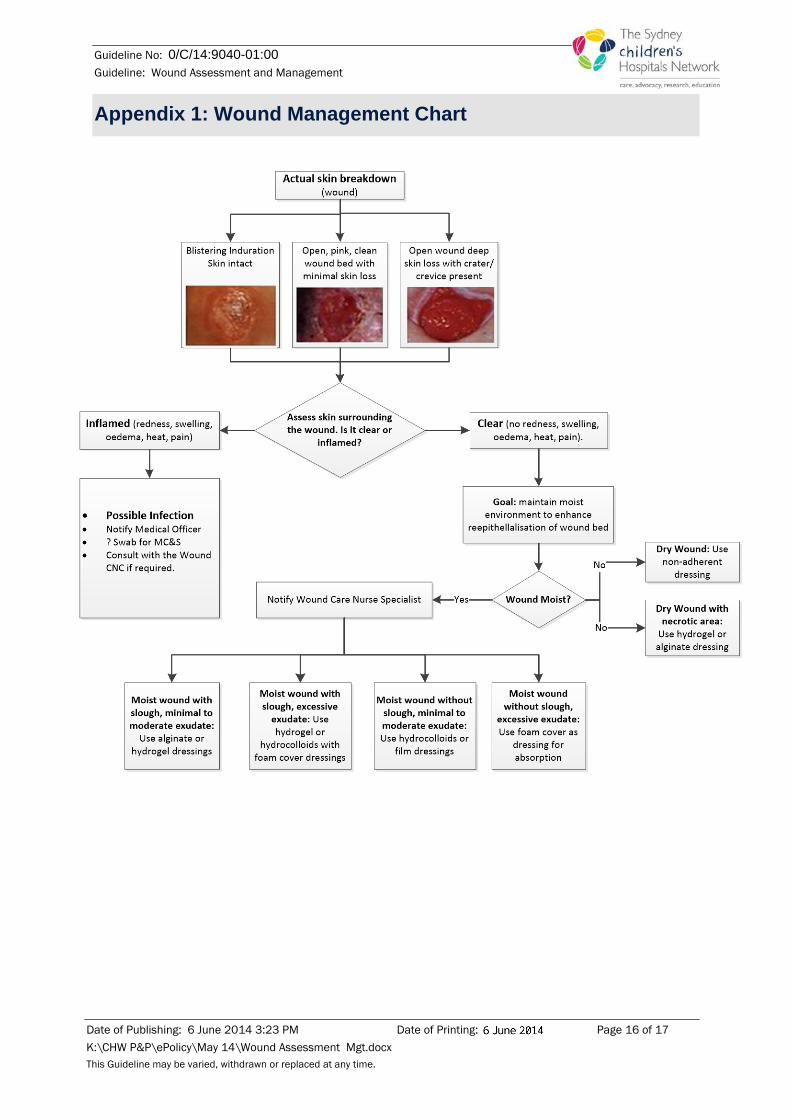
Also see Appendix 1
3.1 Wound Cleansing • Wound cleansing is the application of fluid to aid removal of exudate, debris, slough and
contaminants. Cleansing of the wound helps optimise the healing environment and decreases the potential for infection.
• The common indications for cleaning a wound are: o Reduce the risk of wound infection o Clear excessive exudate o Reduce or debride the presence of debris, eschar or slough
• The most commonly used cleansing agent is sterile 0.9% sodium chloride solution which is isotonic and not detrimental to wound tissue.
• Wound healing occurs at normal body core temperature so when the body surface temperature falls below this wound healing is delayed5. : It has been demonstrated that 37°C is considered optimal6. Using cold solutions combined with exposure on removing dressing reduces the temperature in the wound and it may take 3-4 hours to return to normal.
• The cleansing of wounds by irrigation has been shown to be more effective and causes less trauma than the swabbing method.7 Where possible the wound should be irrigated using a syringe or squeezing ampoules of warmed normal saline. There may be some instances where the patient can shower to cleanse their wound.
• Gauze swabs and cotton wool should be avoided or used with caution to prevent mechanical damage to new tissue and shedding fibres into the wound bed.
3.2 Appropriate Dressing Selection Assessment of the wound is a perquisite to the selection of an appropriate dressing. Dressing selection can be based on the phase of wound healing, location of the wound and amount of wound exudate. (See Appendix 1 and Appendix 2)
The principle reasons for applying a dressing can be summarised as followed:
• To optimise timely and cosmetically appropriate healing • Maintain a moist environment • Maintain a moist environment • Control or absorb excess exudate, and aid debridement of necrotic or slough tissue • To prevent or combat infection non adherent to the wound surface, not to shed fibres or
cause trauma to the wound or surrounding tissue on removal • Minimise interference with normal bodily function. • Prevent further injury to the site.
Guideline No: 0/C/14:9040-01:00 Guideline: Wound Assessment and Management
Date of Publishing: 6 June 2014 3:23 PM Date of Printing: Page 9 of 17 K:\CHW P&P\ePolicy\May 14\Wound Assessment Mgt.docx This Guideline may be varied, withdrawn or replaced at any time.
3.3 Wound dressing procedure Note: When performing a wound dressing hand hygiene and aseptic non-touch technique principles must be adhered to. “The aim of aseptic non touch technique is to prevent the transmission of micro-organisms to wounds or susceptible sites, to reduce the risk of infection.” 8 • Aseptic non touch technique refers to the identification of ‘key parts’ by not touching
them either directly or indirectly. This is the single most important step in achieving asepsis9.
• Key parts refer to the parts that if contaminated with micro-organisms increase the risk of infection.
• Aseptic non touch technique is achieved by using sterile equipment and ensuring that the sterile component of the product does not come into contact with a non-sterile surface.10
• Aseptic non touch technique includes performing hand hygiene at the following times: o prior to setting up for the procedure and o prior to application of non-sterile/sterile gloves11 and o At completion of procedure This is to protect the practitioner and patient from cross-contamination as per standard precautions.
Procedure
1. Ascertain treatment plan and dressing changes regime, as well as assessing the patients need for appropriate pain relief prior to dressing (e.g. pre-procedural analgesia or distraction technique).
2. Wash hands. 3. Clean the trolley with alcohol impregnated wipes. 4. Open up dressing pack, using standard precautions. 5. Open up appropriate dressings, cleaning solutions, gauze, scissors and gloves using
aseptic non- touch technique. 6. Using aseptic technique, remove old dressing and discard. If dressing does not lift
easily it may require soaking to avoid trauma to the wound. NB: Wounds are not routinely swabbed unless clinical evidence of infection is present or requested by the medical officer.
7. Wound cleaning should be performed in a way that minimises trauma to the wound bed. Irrigation is the preferred method, otherwise each wipe/swab should be used only once, start at the top of the wound and finish at the lower edge on linear wounds and from wound edges inwards on others.
8. Once the wound has been cleaned, change gloves. 9. Ensure the wound is dry before applying appropriate dressing. 10. Discard soiled materials and clean trolley. Soiled wound dressings are to be considered
contaminated waste and disposed of accordingly. 11. Wash hands. 12. Document the wound status/ assessment in the patients’ medical records.
Guideline No: 0/C/14:9040-01:00 Guideline: Wound Assessment and Management
Date of Publishing: 6 June 2014 3:23 PM Date of Printing: Page 10 of 17 K:\CHW P&P\ePolicy\May 14\Wound Assessment Mgt.docx This Guideline may be varied, withdrawn or replaced at any time.
3.4 Wound management plan: Evaluate at each dressing change The frequency of dressing changes will dictate an individualised wound management plan. The frequency of such dressing changes will be directed by the clinician co-ordinating the patients care, but must take into consideration the dressing properties and the stage of wound healing. The choice of dressing is determined by the individual needs of the patient and the wound and the type of dressing used may differ as the healing process progresses. When evaluating the wound management plan, consider:
• Is the dressing providing an environment that supports and is beneficial to the healing process?
• Is the wound progressing in a timely manner through the stages of wound healing?
• Are there any signs of clinical infection?
• Note any adverse reactions to dressings or tapes used.
If the wound is deteriorating or showing no expected signs of healing then re-assessment must be performed by an appropriately trained clinician and an alternative wound management plan prescribed.
4 Specific Procedures
4.1 Cardiac Wound Management • At CHW refer to Cardiac Patient: Post-operative Care on the Ward Practice Guideline
4.2 Chest Drain Site or Stab Wounds • At CHW refer to CHW Chest Drains Practice Guideline
• At SCH refer to Chest Drain management practice guideline.
4.3 Removal of Sutures or Staples/Clips All suture removal should be undertaken after instruction from the Surgeon or Resident Surgical Officer. Check postoperative order for specific instructions.
Equipment
• As for Wound Dressing Procedure plus
o Sterile scissors, sterile stitch cutter, staple or clip remover (as appropriate)
Procedure
• Follow as for Wound Dressing Procedure with the following additions:
o 2 nurses should be present (one to assist holding the patient) when the sutures or clips are removed.
o Alternate sutures or clips are usually removed first and the remainder removed the following day unless otherwise directed.
Guideline No: 0/C/14:9040-01:00 Guideline: Wound Assessment and Management
Date of Publishing: 6 June 2014 3:23 PM Date of Printing: Page 11 of 17 K:\CHW P&P\ePolicy\May 14\Wound Assessment Mgt.docx This Guideline may be varied, withdrawn or replaced at any time.
• Non-Continuous (interrupted) Sutures
o Lift the knot of the suture with forceps and slip scissors or a stitch cuter flat under the knot and cut. Pull the suture out and place on a gauze swab, count as removed – continue until complete. Re-dress the wound as requested by the Surgeon or Surgical Medical Officer.
• Continuous Sutures
o Cut suture loop across the middle of the wound. Remove the suture by pulling from the bottom then the top
o Cut at the knot, then cut at the third suture – to remove, pull gently; continue with cutting every third suture until complete. Re-dress the wound as requested by the Surgeon or Surgical Medical Officer.
• Staple/Clip Removal
o Slide the staple/clip remover under the clips then press the clip remover together. The staple/clip should bend in the middle and the sides should lift out and clear of the skin. If any difficulty arises, tilt the staple/clip to one side and lift the clip away from the skin, then repeat for the other side. Re-dress the wound as requested by the Surgeon or Surgical Medical Officer.
Documentation Document in the clinical record the number or type of sutures staples or clips removed. Document the wound appearance and current management plan.
4.4 Surgical Drains – Management, Shortening and removing Drains are a common feature of the postoperative management of surgical wounds. While they serve an important function, they are also associated with complications such as haemorrhage, tissue inflammation, infection and drain entrapment. Nurses should be familiar with the monitoring and management of surgical drains including the process involved in their removal.
A wide variety of drains are available for many different types of operations. They are used to drain a cavity where fluids or air are likely to collect following surgery or in cases where infection is present.
The type of drain used as well as the securing mechanism will be clearly documented on the MR18 or in the patients’ operation report (sutured, stapled or taped). The following describes commonly used drains, excluding ventricular and thoracic drains.
• Corrugated Portex and Yates Drains
o These drains contain alternate grooves to prevent sealing of tissue around the drain and allow the passage of fluid. Can be drained into a gauze pad or a stoma bag.
• Penrose
o This is a soft latex drain used mainly for small wounds. Fluid drains primarily along the outside surface of the drain.
Guideline No: 0/C/14:9040-01:00 Guideline: Wound Assessment and Management
Date of Publishing: 6 June 2014 3:23 PM Date of Printing: Page 12 of 17 K:\CHW P&P\ePolicy\May 14\Wound Assessment Mgt.docx This Guideline may be varied, withdrawn or replaced at any time.
• Portex Tubes
o Used mainly following surgery of the urinary tract such as nephrectomy.
• Haemovac and Surgivac
o Both are suction drains that are effective for short-term drainage. The attached container has a continuous vacuum, which exerts suction due to negative pressure.
• T-Tubes
o These are used following surgery to the biliary tract and liver.
Nursing Management
• On return to the ward post-operatively, clearly document location of drain, volume and colour of drainage. Ensure the type of drain is documented on the Operating Theatre record.
• Ensure to clearly document any changes in regards to volume and colour of discharge.
• Document all of the above in the Patients clinical records following every shift.
• Wound drainage should be measured and recorded on the fluid balance chart
Shortening or Removal of a Drain
The Surgeon (or Resident) will order for the drain to be shortened or removed. Generally, drains are removed once drainage has stopped or becomes less than about 20 – 25 mL/day. Drains can be shortened by withdrawing them gradually as instructed by the Clinician. Post drain removal, exit site dressings should be observed for excessive leakage.
Equipment as for wound dressing technique, plus:
o Sterile scissors
o +/– sterile safety pin
Procedure
1. Advise the patient and carer that there may be some discomfort when the drain is pulled out.
2. Prior to removal, consider the need for pain relief.
3. Loosen and clean wound as for wound dressing technique.
4. Cut or remove securing suture.
5. If shortening place a sterile towel distal to the patient and safety pin. Then, using sterile forceps hold the drain and gently withdraw to the length ordered by the Surgeon or Surgical Resident Officer.
6. If removing the drain continue to withdraw the drain until it is free of the skin. Check to ensure it appears intact.
7. Re-dress the wound as per wound dressing technique
Guideline No: 0/C/14:9040-01:00 Guideline: Wound Assessment and Management
Date of Publishing: 6 June 2014 3:23 PM Date of Printing: Page 13 of 17 K:\CHW P&P\ePolicy\May 14\Wound Assessment Mgt.docx This Guideline may be varied, withdrawn or replaced at any time.
4.5 Negative Pressure Wound Therapy (NPWT) The use of Negative Pressure Wound Therapy will be determined by the treating medical officer and/ or the Wound CNC.
• At SCH: Request for machine should be made via Wound CNC. Out of hours consignment unit can be obtained via After Hours Nurse Manager.
• At CHW: Request for machine hire must be signed off by the Ward NUM.
Negative Pressure Wound Therapy (NPWT) is the application of negative pressure (usually 75 to 125 mmHg) to a foam or gauze placed inside a wound. 12 The wound is sealed with an airtight adhesive film that prevents the entry of air from the external environment. It is used to promote wound healing by the removal of fluid and infectious materials from the wound, assist tissue granulation, decrease wound size, stimulate wound perfusion and support a moist wound healing environment
Topical Negative pressure has the following effects on a wound 13:
• Wound retraction by the negative pressure pulling on the wound margins
• Stimulation of granulation tissue
• Maintenance of a moist wound environment
• Wound cleansing through the removal of small tissue debris by suction
• Removal of wound exudate through a closed system
• Pressure related reduction of interstitial oedema with consequent improvement of local microcirculation
Patients with acute, traumatic, subacute and dehisced wounds, partial-thickness burns, skin flaps and skin grafts may benefit from using NPWT.14
Considerations
• There are different brands available of NPWT devices and the consumables used for each brand need to match the device chosen.
• Always follow the manufacturer’s guidelines for each device and wound type.
• All patients using NPWT need to have their wounds appropriately assessed.
• The wound needs to be surrounded with enough intact skin (approximately 2cm around the wound) in order for the drape to adhere well in order to maintain the seal (vacuum). Consideration must be given to protection of peri-wound skin e.g. use barrier wipes or hydrocolloid.
• NPWT dressing needs to be changed every 2-4 days in accordance with manufacturer’s guidelines (or more frequently in the case of infected wounds13) but can be left in-situ for a longer period when used on specific wound types and under the direction of the Medical Officer in charge.
• Documentation in the health care records needs to include the rate the NPWT machine is to be set at and whether the therapy is continuous or intermittent.
Guideline No: 0/C/14:9040-01:00 Guideline: Wound Assessment and Management
Date of Publishing: 6 June 2014 3:23 PM Date of Printing: Page 14 of 17 K:\CHW P&P\ePolicy\May 14\Wound Assessment Mgt.docx This Guideline may be varied, withdrawn or replaced at any time.
Discharging patients from hospital with NPWT
• Prior to discharging a patient into the community with NPWT the patient needs to be assessed with regards to compliance and suitability.
• Patients with NPWT may meet CAPAC criteria for transfer home under their care.
• All patients discharged into the community with NPWT need to have a follow-up appointment for review by the discharging team.
• The patient/carer needs to be educated in order for them to able to perform basic trouble-shooting with regards to the dressing and the machine.
• If the patient is in a private Health Fund the fund company should be contacted to seek confirmation that home NPWT is covered under policy. If they agree to fund home therapy a quote for up to 4 weeks rental and 4 weeks consumables needs to be obtained from the relevant NPWT company.
Completion of NPWT
At the completion of NPWT staff must:
• Clean equipment as per Infection Control Policy Directive e.g. with neutral detergent and if know multi-resistant organism colonisation as per facility protocol.
• Place equipment in plastic bag, label appropriately.
• Cancel rental agreement with the NPWT company.
• Transfer equipment to equipment collection point for collection, label appropriately.
5 References 1. Dealey C (2012) The Physiology of Wound Care (chapter1) in The Care of Wounds- A Guide for Nurses
4th edition. John Wiley and Sons Ltd. 2. Reinke JM, Sorq H (2012) Wound Repair and Regeneration. Eur Surg Res 49 (1); p35-43 3. Carville K (2012) Physiology of Wound Healing in Wound Care Manual 6th edition. Silver Chain Nursing
Association, WA. 4. Krasner D, Rodeheaver G, Sibbald G (Eds)(2007) Chronic Wound Care: A clinical source book for
healthcare professionals (4th ed) Malvern PA, HMP Communications 5. McGuinness W et al (2004) Influences of dressing changes on wound temperature. Journal of Wound
Care 13 (9); 383-5 6. Fletcher J (1997) Update: Wound Cleansing. Professional Nurse 12(11); 793-6 7. Trevelyan J (1996) Wound Cleansing Principles and Practice. Nursing Times 92 (16); 46-8
Guideline No: 0/C/14:9040-01:00 Guideline: Wound Assessment and Management
Date of Publishing: 6 June 2014 3:23 PM Date of Printing: Page 15 of 17 K:\CHW P&P\ePolicy\May 14\Wound Assessment Mgt.docx This Guideline may be varied, withdrawn or replaced at any time.
8. Preston RM. Aseptic Technique: Evidence-based Approach for Patient Safety. Br J Nurs. 2005 May 26 – June 8; 14(10): 540-2, 544-6.
9. Rowley S, Responsibility. A safe and efficient handling technique for IV therapy & other clinical procedures. United Bristol Healthcare Trust.
10. Hart S. Using an aseptic technique to reduce the risk of infection. Nursing Standard. 2007; 21(47):43-48. 11. Hemsworth S, Selwood K, van Saene R, Pizer B. Does the number of exogenous infections increase in
paediatric oncology patients when sterile surgical gloves are not worn for accessing Central Venous Access Devices? Euro J Oncol Nurs. 2007; 11:442 – 447.
12. Willy C (2006) The Theory and Practice of Vacuum Therapy. Scientific Basis, Indications for Use, Case Reports, Practical Advice. Lindqvist, Germany
13. Willy C & Schmidt R (2006) The Principle of Vacuum Therapy in C Willy (ed.) The Theory and Practice of Vacuum Therapy. Scientific Basis, Indications for Use, Case Reports, Practical Advice, Lindqvist, Germany.
14. V.A.C.Therapy Clinical Guidelines – A reference source for clinicians, 2-B-128 Rev. 7-0. KCI ,San Antonio, Texas.
Background Readings 15. Anderson DJ. (2011). Surgical Site Infections. Infectious Disease Clinics of North America. Vol 25(1). Pp
135-153 16. Bolton L. (2007). Operational Definition of Moist Wound Healing. Evidence-Based Report Card. Journal
of Wound, Ostomy and Continence Nursing. Vol 34(1): pp 23-29 17. Chand T. (2000). Paediatric Wounds: The need for special care. Pharmacy 2000. Issue no: 3: p.14. 18. Fleck CA. (2007).Managing wound pain: Today and in the future. Advances in Skin and Wound Care.
Vol 20 (3): pp 138-145. 19. Gaines RJ. Dunbar RP. (2008) The use of surgical drains in orthopedics. Orthopedics. Vol.31 (7).pp
702-705 20. Gray M. Doughty DB. (2001). Clean versus sterile technique when changing wound dressings. Journal of
WOCN. Vol 28 (3): pp 125 - 128. 21. Keast DH. Parslow N. Houghton P. Norton L. Frazer C. (2007). Best Practice Recommendation for the
Practice and Treatment of Pressure Ulcers: Update 2006. Advances in Skin and Wound Care. Vol 20(8): pp447-460.
22. Krasner DL. Shapshak D. Hopf HW. (2006). Managing Wound Pain. In Bryant RA. Nix DP (eds.) Acute and Chronic Wounds: Current Wound Management Concepts 3rd ed. St. Louis, MO. Mosby
23. Moore Z. Cowman S. (2007). Effective wound management: Identifying criteria for infection. Nursing Standard. Vol 21(24): pp68-76
24. McCord S. Stone MS (2006). Practical Guide to Pediatric Wound Care. Seminars in Plastic Surgery. Vol 20(3): pp 192-199
25. Parker L. (2000). Applying the principles of infection control to wound care. British Journal of Nursing. Vol 9 (7): pp 394-396.
26. Practices Advisory Committee. American Journal of Infection Control. Vol 27 (2): pp 97-136. 27. Rowley S. (2001). Aseptic non - touch technique. Nursing Times. Vol 97 (7): pp 6 - 8. 28. Seltzer J. McGrow K. Horsman A. Korniewicz D. (2002). Awareness of surgical site infections for
advanced practice nurses. AACN Clinical Issues: Advanced Practice in Acute Critical Care. Vol 13 (3): pp 398 – 401
29. Sharp CA. McLaws M. (2002). Wound dressings for surgical sites. (Protocol). In The Cochrane Library 30. Walker J. (2007). Patient preparation for safe removal of surgical drains. Nursing Standard.Vol.21 (49).
pp 39-41
Copyright notice and disclaimer:
The use of this document outside Sydney Children's Hospitals Network (SCHN), or its reproduction in whole or in part, is subject to acknowledgement that it is the property of SCHN. SCHN has done everything practicable to make this document accurate, up-to-date and in accordance with accepted legislation and standards at the date of publication. SCHN is not responsible for consequences arising from the use of this document outside SCHN. A current version of this document is only available electronically from the Hospitals. If this document is printed, it is only valid on the date of printing.
Guideline No: 0/C/14:9040-01:00 Guideline: Wound Assessment and Management
Date of Publishing: 6 June 2014 3:23 PM Date of Printing: Page 16 of 17 K:\CHW P&P\ePolicy\May 14\Wound Assessment Mgt.docx This Guideline may be varied, withdrawn or replaced at any time.
Appendix 1: Wound Management Chart
Guideline No: 0/C/14:9040-01:00 Guideline: Wound Assessment and Management
Date of Publishing: 6 June 2014 3:23 PM Date of Printing: Page 17 of 17 K:\CHW P&P\ePolicy\May 14\Wound Assessment Mgt.docx This Guideline may be varied, withdrawn or replaced at any time.
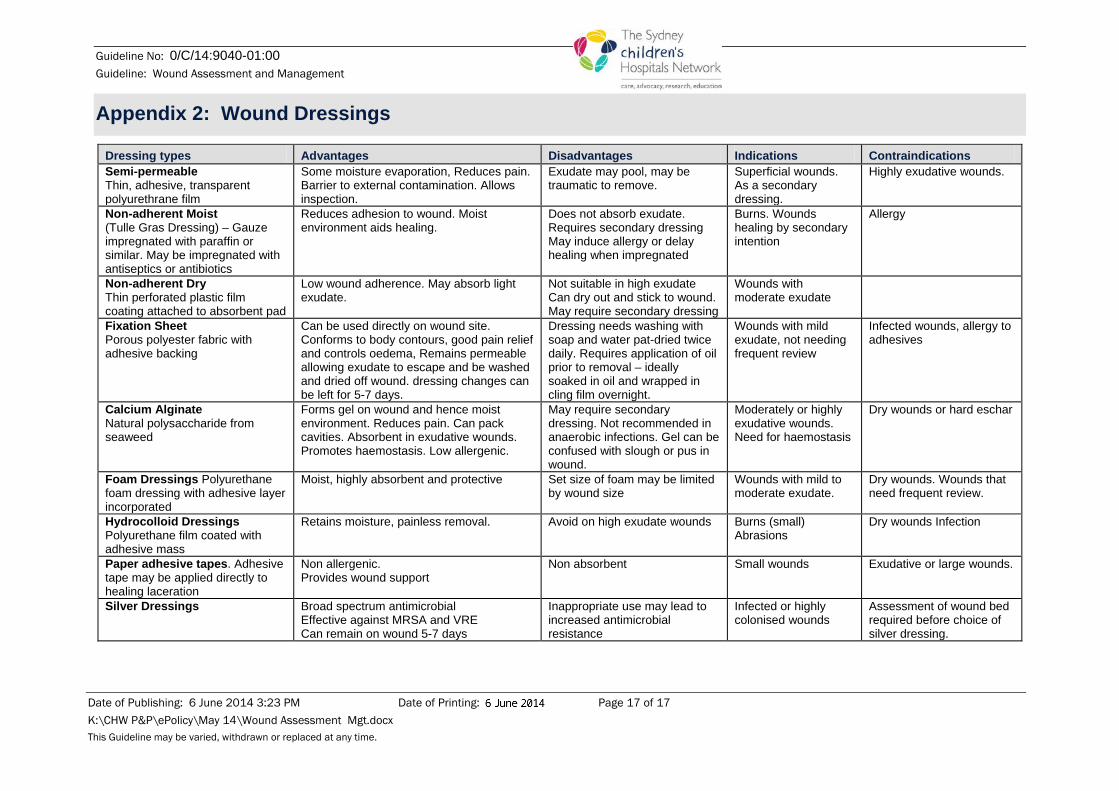
Appendix 2: Wound Dressings
Dressing types Advantages Disadvantages Indications Contraindications Semi-permeable Thin, adhesive, transparent polyurethrane film
Some moisture evaporation, Reduces pain. Barrier to external contamination. Allows inspection.
Exudate may pool, may be traumatic to remove.
Superficial wounds. As a secondary dressing.
Highly exudative wounds.
Non-adherent Moist (Tulle Gras Dressing) – Gauze impregnated with paraffin or similar. May be impregnated with antiseptics or antibiotics
Reduces adhesion to wound. Moist environment aids healing.
Does not absorb exudate. Requires secondary dressing May induce allergy or delay healing when impregnated
Burns. Wounds healing by secondary intention
Allergy
Non-adherent Dry Thin perforated plastic film coating attached to absorbent pad
Low wound adherence. May absorb light exudate.
Not suitable in high exudate Can dry out and stick to wound. May require secondary dressing
Wounds with moderate exudate
Fixation Sheet Porous polyester fabric with adhesive backing
Can be used directly on wound site. Conforms to body contours, good pain relief and controls oedema, Remains permeable allowing exudate to escape and be washed and dried off wound. dressing changes can be left for 5-7 days.
Dressing needs washing with soap and water pat-dried twice daily. Requires application of oil prior to removal – ideally soaked in oil and wrapped in cling film overnight.
Wounds with mild exudate, not needing frequent review
Infected wounds, allergy to adhesives
Calcium Alginate Natural polysaccharide from seaweed
Forms gel on wound and hence moist environment. Reduces pain. Can pack cavities. Absorbent in exudative wounds. Promotes haemostasis. Low allergenic.
May require secondary dressing. Not recommended in anaerobic infections. Gel can be confused with slough or pus in wound.
Moderately or highly exudative wounds. Need for haemostasis
Dry wounds or hard eschar
Foam Dressings Polyurethane foam dressing with adhesive layer incorporated
Moist, highly absorbent and protective Set size of foam may be limited by wound size
Wounds with mild to moderate exudate.
Dry wounds. Wounds that need frequent review.
Hydrocolloid Dressings Polyurethane film coated with adhesive mass
Retains moisture, painless removal. Avoid on high exudate wounds Burns (small) Abrasions
Dry wounds Infection
Paper adhesive tapes. Adhesive tape may be applied directly to healing laceration
Non allergenic. Provides wound support
Non absorbent Small wounds Exudative or large wounds.
Silver Dressings Broad spectrum antimicrobial Effective against MRSA and VRE Can remain on wound 5-7 days
Inappropriate use may lead to increased antimicrobial resistance
Infected or highly colonised wounds
Assessment of wound bed required before choice of silver dressing.

![[you type here] - Memori€¦ · [you type here] • [you type here] • [you type here] • [you type here] [you type here] • [you type here] ... Sell your persona on your solution!](https://img.pdfslide.net/doc/110x75/5ed940b56714ca7f47696bd1/you-type-here-you-type-here-a-you-type-here-a-you-type-here-a-you.jpg)

![christspringfield.orgchristspringfield.org/.../sites/53/...New-10-am-final-bulleti… · Web view[Type here][Type here][Type here] Fourth](https://img.pdfslide.net/doc/110x75/5f0484717e708231d40e5cdc/-web-view-type-heretype-heretype-here-fourth.jpg)


![CSD Paraprofessional Handbook - Home - · Web viewCSD Paraprofessional Handbook Page 13 of 13 [Type here][Type here][Type here] Chamberlain School District Paraprofessional Handbook](https://img.pdfslide.net/doc/110x75/5aa3aaf67f8b9ab4208e8de9/csd-paraprofessional-handbook-home-viewcsd-paraprofessional-handbook-page.jpg)
![School-Based Leadership Team (SBLT) Toolkit 2017-2018 · [Type here] [Type here] [Type here] School-Based Leadership Team (SBLT) Toolkit 2017-2018 mmsd.org/sblttoolkit](https://img.pdfslide.net/doc/110x75/5b360b767f8b9a6b548dee66/school-based-leadership-team-sblt-toolkit-2017-2018-type-here-type-here.jpg)


![[Type here] [Type here] [Type here]](https://img.pdfslide.net/doc/110x75/61ac36038ea0783b0a6313a4/type-here-type-here-type-here.jpg)


![[Type Client Here]](https://img.pdfslide.net/doc/110x75/62680d71277ff5675d1a0960/type-client-here.jpg)
![[ TYPE THESIS TITLE HERE ]](https://img.pdfslide.net/doc/110x75/588b11101a28ab090d8bc2fc/-type-thesis-title-here-.jpg)