Embed Size (px)
Citation preview
Please read this insert completely prior to using the product. For research use only. Not for use in diagnostic procedures.
www.ArborAssays.com
DetectX®
Sample Types Validated:
DualRead™ AssayExtended Standard Curve Range
PKA (PROTEIN KINASE A) Activity Kit
1 Plate Kit Catalog Number K027-H1
Species Independent
Cell Lysate, Tissue Extracts and Buffer Samples
WEB INSERT 161028
EXPECT ASSAY ARTISTRY
®
2
TABLE OF CONTENTS
Background 3
Assay Principle 4
Related Products 4
Supplied Components 5
Storage Instructions 5
Other Materials Required 6 Precautions 6
Sample Types 7
Sample Preparation 7
Reagent Preparation 8
Assay Protocol 9 Calculation of Results 10
Typical Data 11
Validation Data Sensitivity, Linearity, etc. 12
Inhibition Studies 13
Interferents and Cross Reactivity 14
Warranty & Contact Information 15 Plate Layout Sheet 16
WEB INSERT 161028
www.ArborAssays.com
®
3
BACKGROUNDPKA was discovered in the laboratory of Edwin G. Krebs in the 1960’s1. This important class of kinases, refered to as Arg-directed kinases or AGC-family kinases, includes cAMP-dependent protein kinase (PKA or cAPK), cGMP-dependent protein kinase (PKG), protein kinase C, Akt and RSK. These kinases share a substrate specificity characterized by Arg at position 3 relative to the phosphorylated serine or threonine2-4. The second messenger cyclic AMP (cAMP) activates PKA in mammalian cells and controls many cellular mechanisms such as gene transcription, ion transport, and protein phosphorylation2. Inactive PKA is a heterotetramer composed of a regulatory subunit (R) dimer and a catalytic subunit (C) dimer. In this inactive state, the pseudosubstrate sequences on the R subunits block the active sites on the C subunits. PKA shares substrate specificity with Akt (PKB) and PKC4. Substrates that present this consensus sequence and are phosphorylated by PKA are Bad (Ser155), CREB (Ser133), and GSK-3 (GSK-3α Ser21 and GSK-3ß Ser9) 5-7.
PKA has been implicated in numerous cellular processes, including modulation of other protein kinases, regulation of intracellular calcium concentration, and regulation of transcription8. Transcriptional responses to increased cAMP occur through activation of the cAMP response element–binding protein (CREB), cAMP response element modulator (CREM), and activating transcription factor 1 (ATF1)9. Each of these transcription factors contains a kinase-inducible domain containing a conserved site for phosphorylation by PKA.
1. Walsh, DA, Perkins, JP, Krebs, EG., “An adenosine 3’,5’-monophosphate-dependent protein kinase from rabbit skeletal muscle.”, J. Biol. Chem. 1968, 243:3763–3765.
2. Montminy, M. “Transcriptional regulation by cyclic AMP. “ Annu Rev Biochem., 1997, 66:807-822.
3. Pearson, RB. and Kemp, BE. “Protein kinase phosphorylation site sequences and consensus specificity motifs: Tabulations”., Methods Enzymol., 1991, 200:62-81.
4. Dell’Acqua, ML. and Scott, JD. “Protein kinase A anchoring.”, J. Biol. Chem., 1997, 272:12881-12884.
5. Tan, Y. et al. “BAD Ser-155 Phosphorylation Regulates BAD/Bcl-XL Interaction and Cell Survival”, J. Biol. Chem., 2000, 275:25865-25869.
6. Gonzalez, GA. and Montminy, MR. “Cyclic AMP stimulates somatostatin gene transcription by phosphorylation of CREB at serine 133.”, Cell, 1989, 59:675-680.
7. Fang, X. et al. “Phosphorylation and inactivation of glycogen synthase kinase 3 by protein kinase A”., Proc. Natl. Acad. Sci. USA 2000, 97:11960-11965.
8. Taskén, K, and Aandahl, EM., “Localized effects of cAMP mediated by distinct routes of protein kinase A.”, Physiol. Rev., 2004, 84:137–167.
9. Sands, WA, and Palmer, TM., “Regulating gene transcription in response to cAMP elevation.”, Cell. Signal. 2008, 20:460–466.
WEB INSERT 161028
EXPECT ASSAY ARTISTRY
®
4
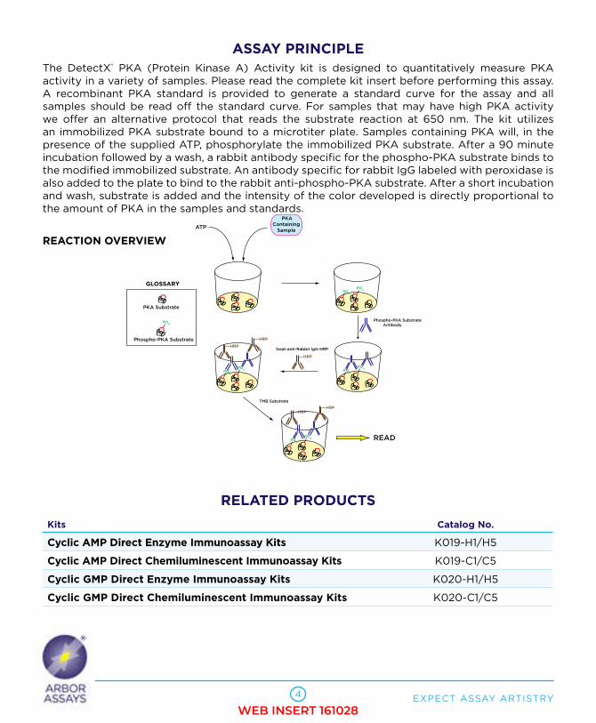
ASSAY PRINCIPLE The DetectX® PKA (Protein Kinase A) Activity kit is designed to quantitatively measure PKA activity in a variety of samples. Please read the complete kit insert before performing this assay. A recombinant PKA standard is provided to generate a standard curve for the assay and all samples should be read off the standard curve. For samples that may have high PKA activity we offer an alternative protocol that reads the substrate reaction at 650 nm. The kit utilizes an immobilized PKA substrate bound to a microtiter plate. Samples containing PKA will, in the presence of the supplied ATP, phosphorylate the immobilized PKA substrate. After a 90 minute incubation followed by a wash, a rabbit antibody specific for the phospho-PKA substrate binds to the modified immobilized substrate. An antibody specific for rabbit IgG labeled with peroxidase is also added to the plate to bind to the rabbit anti-phospho-PKA substrate. After a short incubation and wash, substrate is added and the intensity of the color developed is directly proportional to the amount of PKA in the samples and standards.
REACTION OVERVIEW
RELATED PRODUCTS
Kits Catalog No.
Cyclic AMP Direct Enzyme Immunoassay Kits K019-H1/H5
Cyclic AMP Direct Chemiluminescent Immunoassay Kits K019-C1/C5
Cyclic GMP Direct Enzyme Immunoassay Kits K020-H1/H5
Cyclic GMP Direct Chemiluminescent Immunoassay Kits K020-C1/C5
PKAContaining Sample
ATP
PKA Substrate
PO4
PO4PO
4
Phospho-PKA Substrate
PO4
PO4
Phospho-PKA Substrate Antibody
PO4
PO4
HRP
HRP
PO4
PO4
HRPHRP
TMB Substrate
READ
Goat-anti-Rabbit IgG-HRP
GLOSSARY
HRP
WEB INSERT 161028

www.ArborAssays.com
®
5
SUPPLIED COMPONENTSPKA Substrate 96 Well Plate Break-apart strip microtiter plate coated with PKA Substrate 1 Plate Catalog Number C107-1EA
PKA Standard 5,000 Units of recombinant fully active PKA in special stabilizing buffer. One unit is defined as the amount of PKA required to catalyze the transfer of 1 pmol of ATP phosphate to substrate in 1 minute at 30°C. PKA Standard must be stored at -20°C. 2 Vials Catalog Number C131-2EA
ATPATP lyophilized stored in a ziplock pouch with desiccant. 1 Vial Catalog Number X103-1EA
Phospho PKA Substrate AntibodyA solution of rabbit antibody specific for phospho-Substrate. 3 mL Catalog Number C104-3ML
Goat anti-Rabbit IgG HRP ConjugateA solution of goat antibody specific for rabbit IgG labeled with peroxidase. 3 mL Catalog Number C105-3ML
Kinase Reaction Buffer ConcentrateA 2X concentrate containing detergents and stabilizers. 60 mL Catalog Number X104-60ML
Cell Lysis BufferA Tris based buffer containing detergents. Cell Lysis Buffer must be stored at -20°C as it does not contain preservatives. 100 mL Catalog Number X050-100ML
Wash Buffer Concentrate A 20X concentrate that should be diluted with deionized or distilled water. 30 mL Catalog Number X007-30ML
TMB Substrate 11 mL Catalog Number X019-11ML
Stop Solution 1M solution of hydrochloric acid. CAUSTIC. 5 mL Catalog Number X020-5ML
Plate Sealer 2 Each Catalog Number X002-1EA
STORAGE INSTRUCTIONSThe unopened kit should be stored at -20°C until the expiration date of the kit. Once opened the kit can be stored at 4°C up to the expiration date on the kit label, except for the PKA Standard and Cell Lysis Buffer which must be stored at -20°C. The Cell Lysis Buffer has no preservative and must be kept frozen at -20°C. All components of this kit can be stored together at -20°C.
WEB INSERT 161028

EXPECT ASSAY ARTISTRY
®
6
OTHER MATERIALS REQUIREDDistilled or deionized water.
Glass test tubes.
Shaking plate incubator capable of maintaining 30°C.
Repeater pipet and disposable tips capable of dispensing 10, 25, 50 and 100 µL accurately.
The following Protease inhibitors MUST be added to all buffers that are used to measure PKA activity. See pages 7 & 8.
• Phenylmethanesulfonyl fluoride (PMSF), such as Sigma 78830 at 100 mM in ethanol.• A universal protease inhibitor cocktail (PIC) such as Sigma P1860 or Roche 05892970001.
In addition:
• A phosphatase inhibitor, such as Sodium Orthovanadate (See activation instruction opposite), or a phosphatase inhibitor cocktail, such as Sigma P5726, must added to the Cell Lysis buffer.
Colorimetric 96 well microplate reader capable of reading optical density at 450 and 650 nm.
Software for converting raw relative optical density readings from the plate reader and carrying out four parameter logistic curve (4PLC) fitting. Contact your plate reader manufacturer for details.
DualRead™ SystemThis kit uses our unique DualRead™ system. We include instructions for an alternative high standard which would typically generate ODs at 450 nm too high to be read on most plate readers. By reading the plate at 650 nm (where TMB optical density is about 3 fold lower) immediately before addition of the Stop Solution some samples outside the normal standard curve range can be read. See instructions on pages 8-10.
PRECAUTIONSAs with all such products, this kit should only be used by qualified personnel who have had laboratory safety instruction. The complete insert should be read and understood before attempting to use the product.
The coated plate needs to be stored desiccated. The silica gel pack included in the foil ziploc bag will keep the plate dry. The silica gel pack will turn from blue to pink if the ziploc has not been closed properly.
This kit utilizes a peroxidase-based readout system. Buffers, including other manufacturers Wash Buffers, containing sodium azide will inhibit color production from the enzyme. Make sure all buffers used for samples are azide free. Ensure that any plate washing system is rinsed well with deionized water prior to using the supplied Wash Buffer as prepared on Page 8.
The Stop Solution is acid. The solution should not come in contact with skin or eyes. Take appropriate precautions when handling this reagent.
WEB INSERT 161028

www.ArborAssays.com
®
7
SAMPLE TYPESThis assay has been validated for Jurkat cell lysates. Samples containing visible particulate should be centrifuged prior to using.
SAMPLE PREPARATION Cells must be lysed in the Activated Cell Lysis Buffer, after addition of protease inhibitors and either activated orthovanadate or a phosphatase inhibitor cocktail to the provided Cell Lysis Buffer (see below). All cells and the lysates made from them must be stored at ≤ -70°C and should be stored as aliquots for single use. Do not freeze-thaw samples. Do not store cells or lysates above -70°C.
The preparation of Activated Sodium Orthovanadate is as follows:
200 mM Activated Orthovanadate should be prepared by dissolving 1.84 g of sodium orthovanadate in 45 mL of water. Adjust the pH of the solution to 10 with 1M NaOH or HCl. At pH 10 the solution should be yellow. Boil the solution until it turns colorless (approximately 10 min). All of the orthovanadate should dissolve. Cool to room temperature and readjust the pH to 10. Repeat the boiling of the solution and pH readjustment until the solution is colorless and remains at pH 10. Adjust the final volume to 50 mL with water. Store the Activated Sodium Orthovanadate in aliquots and freeze at -20°C. Use an aliquot for preparing Activated Cell Lysis Buffer and discard.
Preparation of Activated Cell Lysis Buffer Prepare the Activated Cell Lysis Buffer by addition of 1 µL of PIC per mL of Cell Lysate Buffer. Add 1 mM PMSF and 10 mM Activated Orthovanadate. The resulting Activated Cell Lysis Buffer is a pH 8 Tris based buffer containing 1% NP-40 as a cell disruption agent. This assay may not be compatible with other cell lysis buffers containing high concentrations of SDS or other detergents and erroneous activity measurements may result.
Cell LysisAdd prepared Activated Cell Lysis Buffer to the cells (for Jurkat cells, we lysed at 100 million cells per mL). Incubate for 30 minutes on ice with occasional vortexing. Centrifuge at 10,000 rpm at 4°C for 10 minutes and carefully aspirate off the supernatant for analysis. Supernatants can be frozen at ≤ -70°C for later analysis.
The supernatants should be diluted at least 1:10 into prepared KINASE ASSAY BUFFER (see Page 8) prior to running in the assay. It is recommended that a control lysate be serially diluted in KINASE ASSAY BUFFER to determine the appropriate dilution to obtain a linear response. See Graph at right.
Samples diluted in KINASE ASSAY BUFFER can be frozen at ≤ -70°C for analysis later.
PKA Activity: Jurkat Cell Lysate Dilution
0
2
4
6
8
10
12
14
16
18
0 0.25 0.5 0.75 1 1.25 1.5 1.75 2
Jurkat Cell Lysate Diln (x 10^6)/mL
Obs. PKA Activity (U/mL)PK
A A
cti
vit
y (
U/m
L)
WEB INSERT 161028

EXPECT ASSAY ARTISTRY
®
8
REAGENT PREPARATIONAllow the kit reagents to come to room temperature for 30 minutes, except for the standard which must be kept on ice. Keep all samples on ice and ensure they have been diluted appropriately prior to running them in the kit.
KINASE ASSAY BUFFER CRITICAL STEP!Dilute Kinase Reaction Buffer Concentrate 1:2 by adding one part of the concentrate to one part of deionized water. Add 0.5 µL/mL of PIC and PMSF to 1 mM to make KINASE ASSAY BUFFER. Use within 8 hours.
ATPAllow the ziplock vial to warm completely to room temperature prior to opening. Remove the vial and add 1.2 mL of prepared KINASE ASSAY BUFFER to the ATP vial. Vortex to solubilize. Once diluted, store any unused ATP solution at -20°C for up to 3 months.
Wash Buffer Dilute Wash Buffer Concentrate 1:20 by adding one part of the concentrate to nineteen parts of deionized water. Once diluted this is stable at room temperature for 3 months.
Standard Preparation Spin down the contents of the PKA Standard vial in a microcentrifuge for 2 minutes at 14,000 rpm at 4°C. Keep all standards on ice during use.
Prepare an Intermediate Stock dilution by pipetting 1 mL of prepared KINASE ASSAY BUFFER into the PKA standard vial. Invert vial and vortex thoroughly to ensure complete mixing of contents. This Intermediate Stock will have an activity of 5,000 Units/mL. Intermediate Stock is single use only. Discard vial after preparing standards. Do not freeze/thaw.
Label tubes as #1 through #5. Pipet the standards using the Intermediate Stock according to the table below. The activity of PKA in tubes 1 through 5 will be 25, 20, 15, 10, and 5 Units/mL.
Alternative High Standard For samples that may exceed 25 U/mL prepare the 40 U/mL Alternative Standard by pipetting 8 µL of Intermediate Stock into 992 µL of prepared KINASE ASSAY BUFFER.
Keep all Standards and Intermediate Stock on Ice and use within 30 minutes of preparation.
Std 1 Std 2 Std 3 Std 4 Std 5 Alt. High Std.
KINASE ASSAY BUFFER (µL) 995 60 60 60 60 992
Addition Inter. Stock Std 1 Std 2 Std 3 Std 4 Inter. Stock
Vol. of Addition (µL) 5 240 180 120 60 8
Final Activity (U/mL) 25 20 15 10 5 40
WEB INSERT 161028
www.ArborAssays.com
®
9
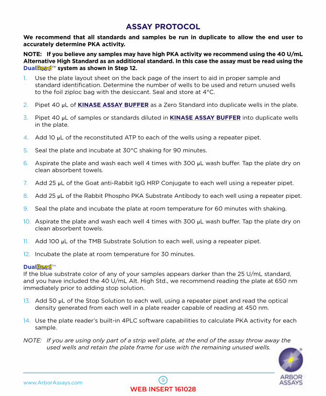
ASSAY PROTOCOLWe recommend that all standards and samples be run in duplicate to allow the end user to accurately determine PKA activity.
NOTE: If you believe any samples may have high PKA activity we recommend using the 40 U/mL Alternative High Standard as an additional standard. In this case the assay must be read using the DualRead™ system as shown in Step 12.
1. Use the plate layout sheet on the back page of the insert to aid in proper sample and standard identification. Determine the number of wells to be used and return unused wells to the foil ziploc bag with the desiccant. Seal and store at 4°C.
2. Pipet 40 µL of KINASE ASSAY BUFFER as a Zero Standard into duplicate wells in the plate.
3. Pipet 40 µL of samples or standards diluted in KINASE ASSAY BUFFER into duplicate wells in the plate.
4. Add 10 µL of the reconstituted ATP to each of the wells using a repeater pipet.
5. Seal the plate and incubate at 30°C shaking for 90 minutes.
6. Aspirate the plate and wash each well 4 times with 300 µL wash buffer. Tap the plate dry on clean absorbent towels.
7. Add 25 µL of the Goat anti-Rabbit IgG HRP Conjugate to each well using a repeater pipet.
8. Add 25 µL of the Rabbit Phospho PKA Substrate Antibody to each well using a repeater pipet.
9. Seal the plate and incubate the plate at room temperature for 60 minutes with shaking.
10. Aspirate the plate and wash each well 4 times with 300 µL wash buffer. Tap the plate dry on clean absorbent towels.
11. Add 100 µL of the TMB Substrate Solution to each well, using a repeater pipet.
12. Incubate the plate at room temperature for 30 minutes.
DualRead™ If the blue substrate color of any of your samples appears darker than the 25 U/mL standard, and you have included the 40 U/mL Alt. High Std., we recommend reading the plate at 650 nm immediately prior to adding stop solution.
13. Add 50 µL of the Stop Solution to each well, using a repeater pipet and read the optical density generated from each well in a plate reader capable of reading at 450 nm.
14. Use the plate reader’s built-in 4PLC software capabilities to calculate PKA activity for each sample.
NOTE: Ifyouareusingonlypartofastripwellplate,attheendoftheassaythrowawaythe usedwellsandretaintheplateframeforusewiththeremainingunusedwells.
WEB INSERT 161028

EXPECT ASSAY ARTISTRY
®
10
CALCULATION OF RESULTS Average the duplicate 450 nm (and optional 650 nm) OD readings for each standard and sample. Create a standard curve by reducing the data using the 4PLC fitting routine on the plate reader, after subtracting the mean ODs for the zero standard. The sample activity obtained should be multiplied by the dilution factor to obtain neat sample values.
Or use the online tool from MyAssays to calculate the data: www.myassays.com/arbor-assays-protein-kinase-a-activity-kit.assay
TYPICAL DATA (450 NM READ)
Sample Mean OD (450nm) Net OD (450nm)PKA Activity
(U/mL)
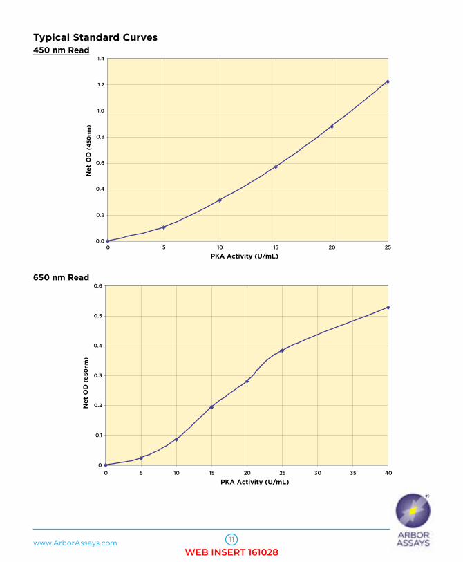
Standard 1 1.272 1.223 25
Standard 2 0.929 0.880 20
Standard 3 0.618 0.569 15
Standard 4 0.363 0.314 10
Standard 5 0.155 0.106 5
Zero 0.049 0.000 0
Sample 1 0.158 0.109 5.91
Sample 2 1.000 0.951 20.9
TYPICAL DATA (650 NM READ)
Sample Mean OD (650nm) Net OD (650nm)PKA Activity
(U/mL)
Alt. Std. 0.564 0.528 40
Standard 1 0.42 0.384 25
Standard 2 0.307 0.271 20
Standard 3 0.23 0.195 15
Standard 4 0.102 0.066 10
Standard 5 0.06 0.025 5
Zero 0.036 0 0
Sample 1 0.274 0.238 18.8
Sample 2 0.109 0.073 7.86
Always run your own standard curve for calculation of results. Do not use this data.
*The MyAssays logo is a registered trademark of MyAssays Ltd.
WEB INSERT 161028
www.ArborAssays.com
®
11
Typical Standard Curves450 nm Read
650 nm Read
0.0
0.2
0.4
0.6
0.8
1.0
1.2
1.4
0 5 10 15 20 25
PKA Activity (U/mL)
OD (450 nm)
Ne
t O
D (
45
0n
m)
0
0.1
0.2
0.3
0.4
0.5
0.6
0 5 10 15 20 25 30 35 40
PKA Activity (U/mL)
Net OD (650nm)
Ne
t O
D (
65
0n
m)
WEB INSERT 161028
EXPECT ASSAY ARTISTRY
®
12
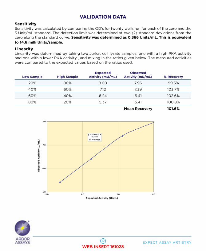
VALIDATION DATASensitivitySensitivity was calculated by comparing the OD’s for twenty wells run for each of the zero and the 5 Unit/mL standard. The detection limit was determined at two (2) standard deviations from the zero along the standard curve. Sensitivity was determined as 0.366 Units/mL. This is equivalent to 14.6 milli Units/sample.
LinearityLinearity was determined by taking two Jurkat cell lysate samples, one with a high PKA activity and one with a lower PKA activity , and mixing in the ratios given below. The measured activities were compared to the expected values based on the ratios used.
Low Sample High SampleExpected
Activity (mU/mL)Observed
Activity (mU/mL) % Recovery
20% 80% 8.00 7.96 99.5%
40% 60% 7.12 7.39 103.7%
60% 40% 6.24 6.41 102.6%
80% 20% 5.37 5.41 100.8%
Mean Recovery 101.6%
y = 0.9837x + 0.2156
R2 = 0.9858
5.0
6.0
7.0
8.0
5.0 6.0 7.0 8.0
Expected Activity (U/mL)
Observed Activity (U/mL)
Ob
serv
ed
Acti
vit
y (
U/m
L)
WEB INSERT 161028

www.ArborAssays.com
®
13
INHIBITION STUDIESStudies with recombinant PKA Approximately 30 Units/mL of human recombinant PKA was incubated with the reversible PKA inhibitor H 89 dihydrochloride from 0 to 10,000 nM in Assay Buffer for 30 minutes at room temperature prior to running in the assay. % Activity is expressed by comparison to the activity of the buffer control (28.02 U/mL). 4PLC data comparison determined the IC50% to be 19.1 nM.
Studies with Cell Lysates Aliquots of a Jurkat cell lysate containing approximately 40,000 cells were treated with the reversible PKA inhibitor H 89 dihydrochloride or Assay Buffer as the control and run in the assay.
30
40
50
60
70
80
90
100
0 10 100 1,000 10,000
H 89 Conc. (nM)
% Activity% A
cti
vit
y
0
2
4
6
8
10
12
14
16
18
+ H 89 PKA Inhibitor Control
Reaction Condition
PKA Activity (U/mL)
PK
A A
cti
vit
y (
U/m
L)
WEB INSERT 161028
EXPECT ASSAY ARTISTRY
®
14
INTERFERENTSA variety of solvents were tested as possible interfering substances in the assay. Ethanol at 0.5% in the well decreased the activity recorded by 12.7%, whereas 0.10% ethanol in the well decreased activity by 3.7%. DMSO at 0.5% in the well decreased activity by 2.8%. Methanol at 0.1% in the well increased activity by 3.1%. We expect solvent levels at 0.1% of well volume to have little or no effect on the measured activity. A solvent only control should be run by the end user when appropriate.
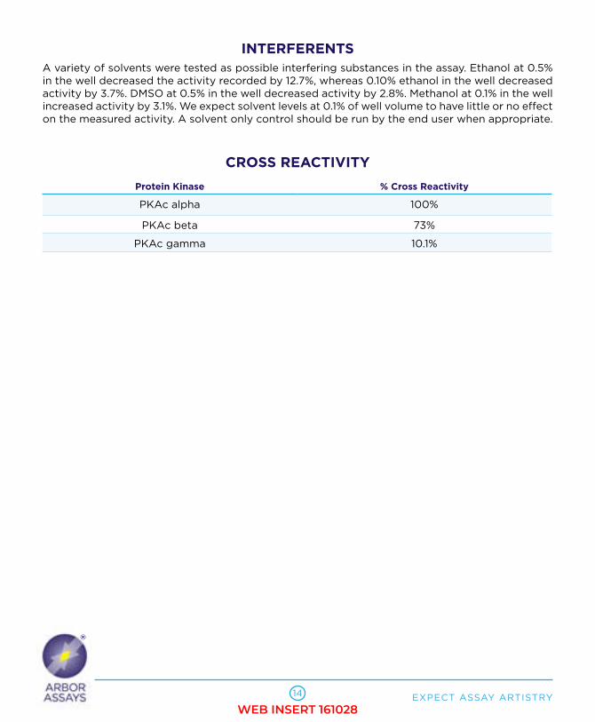
CROSS REACTIVITY
Protein Kinase % Cross Reactivity
PKAc alpha 100%
PKAc beta 73%
PKAc gamma 10.1%
WEB INSERT 161028

www.ArborAssays.com
®
15
LIMITED WARRANTYArbor Assays warrants that at the time of shipment this product is free from defects in materials and workmanship. This warranty is in lieu of any other warranty expressed or implied, including but not limited to, any implied warranty of merchantability or fitness for a particular purpose.
We must be notified of any breach of this warranty within 48 hours of receipt of the product. No claim shall be honored if we are not notified within this time period, or if the product has been stored in any way other than outlined in this publication. The sole and exclusive remedy of the customer for any liability based upon this warranty is limited to the replacement of the product, or refund of the invoice price of the goods.
CONTACT INFORMATIONFor details concerning this kit or to order any of our products please contact us: Arbor Assays
1514 Eisenhower Place Ann Arbor, Michigan 48108 USA
Phone: 734-677-1774
Fax: 734-677-6860
Web: www.ArborAssays.com
E Mail Addresses:
DetectX®,ThioStar®andtheArborAssayslogoareallregisteredtrademarks.
OFFICIAL SUPPLIER TO ISWEArbor Assays and the International Society of Wildlife Endocrinology (ISWE) signed an exclusive agreement for Arbor Assays to supply ISWE members with EIA kits for wildlife conservation research.
WEB INSERT 161028

Printed on Forest Stewardship Council certified paper
1 2
3 4
5 6
7
8 9
10 11 12
A B C D E F G H
16
©2011
K027-H1 161028
WEB INSERT 161028