Embed Size (px)
Citation preview

Plesiomonas shigelloides Revisited
J. Michael Janda,a Sharon L. Abbott,b* Christopher J. McIverc
Kern County Public Health Laboratory, Department of Public Health Services, Bakersfield, California, USAa; Microbial Diseases Laboratory, California Department of PublicHealth, Richmond, California, USAb; Microbiology Department (SEALS), St. George Hospital, Kogarah, and School of Medical Sciences, University of New South Wales,Sydney, NSW, Australiac
SUMMARY . . . . . . . . . . . . . . . . . . . . . . . . . . . . . . . . . . . . . . . . . . . . . . . . . . . . . . . . . . . . . . . . . . . . . . . . . . . . . . . . . . . . . . . . . . . . . . . . . . . . . . . . . . . . . . . . . . . . . . . . . . . . . . . . . . . . . . . . . . . . . . . . . .350INTRODUCTION . . . . . . . . . . . . . . . . . . . . . . . . . . . . . . . . . . . . . . . . . . . . . . . . . . . . . . . . . . . . . . . . . . . . . . . . . . . . . . . . . . . . . . . . . . . . . . . . . . . . . . . . . . . . . . . . . . . . . . . . . . . . . . . . . . . . . . . . . . . .350TAXONOMY . . . . . . . . . . . . . . . . . . . . . . . . . . . . . . . . . . . . . . . . . . . . . . . . . . . . . . . . . . . . . . . . . . . . . . . . . . . . . . . . . . . . . . . . . . . . . . . . . . . . . . . . . . . . . . . . . . . . . . . . . . . . . . . . . . . . . . . . . . . . . . . .351
Nomenclature . . . . . . . . . . . . . . . . . . . . . . . . . . . . . . . . . . . . . . . . . . . . . . . . . . . . . . . . . . . . . . . . . . . . . . . . . . . . . . . . . . . . . . . . . . . . . . . . . . . . . . . . . . . . . . . . . . . . . . . . . . . . . . . . . . . . . . . . . . .351Historical perspective . . . . . . . . . . . . . . . . . . . . . . . . . . . . . . . . . . . . . . . . . . . . . . . . . . . . . . . . . . . . . . . . . . . . . . . . . . . . . . . . . . . . . . . . . . . . . . . . . . . . . . . . . . . . . . . . . . . . . . . . . . . . . . . . . .351
Classification . . . . . . . . . . . . . . . . . . . . . . . . . . . . . . . . . . . . . . . . . . . . . . . . . . . . . . . . . . . . . . . . . . . . . . . . . . . . . . . . . . . . . . . . . . . . . . . . . . . . . . . . . . . . . . . . . . . . . . . . . . . . . . . . . . . . . . . . . . . . .351Phylogenetic investigations . . . . . . . . . . . . . . . . . . . . . . . . . . . . . . . . . . . . . . . . . . . . . . . . . . . . . . . . . . . . . . . . . . . . . . . . . . . . . . . . . . . . . . . . . . . . . . . . . . . . . . . . . . . . . . . . . . . . . . . . . . .351Reclassification of Plesiomonas into the family Enterobacteriaceae . . . . . . . . . . . . . . . . . . . . . . . . . . . . . . . . . . . . . . . . . . . . . . . . . . . . . . . . . . . . . . . . . . . . . . . . . . . . . . . . . . . .351Genomics . . . . . . . . . . . . . . . . . . . . . . . . . . . . . . . . . . . . . . . . . . . . . . . . . . . . . . . . . . . . . . . . . . . . . . . . . . . . . . . . . . . . . . . . . . . . . . . . . . . . . . . . . . . . . . . . . . . . . . . . . . . . . . . . . . . . . . . . . . . . . .351
Outstanding Issues . . . . . . . . . . . . . . . . . . . . . . . . . . . . . . . . . . . . . . . . . . . . . . . . . . . . . . . . . . . . . . . . . . . . . . . . . . . . . . . . . . . . . . . . . . . . . . . . . . . . . . . . . . . . . . . . . . . . . . . . . . . . . . . . . . . . . . .352ENVIRONMENTAL DISTRIBUTION AND ECOLOGIC ASSOCIATIONS. . . . . . . . . . . . . . . . . . . . . . . . . . . . . . . . . . . . . . . . . . . . . . . . . . . . . . . . . . . . . . . . . . . . . . . . . . . . . . . . . . . . . . .352
Aquatic Environments. . . . . . . . . . . . . . . . . . . . . . . . . . . . . . . . . . . . . . . . . . . . . . . . . . . . . . . . . . . . . . . . . . . . . . . . . . . . . . . . . . . . . . . . . . . . . . . . . . . . . . . . . . . . . . . . . . . . . . . . . . . . . . . . . . . .352Natural disasters . . . . . . . . . . . . . . . . . . . . . . . . . . . . . . . . . . . . . . . . . . . . . . . . . . . . . . . . . . . . . . . . . . . . . . . . . . . . . . . . . . . . . . . . . . . . . . . . . . . . . . . . . . . . . . . . . . . . . . . . . . . . . . . . . . . . . . .353
Invertebrate and Vertebrate Hosts . . . . . . . . . . . . . . . . . . . . . . . . . . . . . . . . . . . . . . . . . . . . . . . . . . . . . . . . . . . . . . . . . . . . . . . . . . . . . . . . . . . . . . . . . . . . . . . . . . . . . . . . . . . . . . . . . . . . . . .353Mollusks and crustaceans. . . . . . . . . . . . . . . . . . . . . . . . . . . . . . . . . . . . . . . . . . . . . . . . . . . . . . . . . . . . . . . . . . . . . . . . . . . . . . . . . . . . . . . . . . . . . . . . . . . . . . . . . . . . . . . . . . . . . . . . . . . . . .353Fish . . . . . . . . . . . . . . . . . . . . . . . . . . . . . . . . . . . . . . . . . . . . . . . . . . . . . . . . . . . . . . . . . . . . . . . . . . . . . . . . . . . . . . . . . . . . . . . . . . . . . . . . . . . . . . . . . . . . . . . . . . . . . . . . . . . . . . . . . . . . . . . . . . . .353
(i) Saltwater fish . . . . . . . . . . . . . . . . . . . . . . . . . . . . . . . . . . . . . . . . . . . . . . . . . . . . . . . . . . . . . . . . . . . . . . . . . . . . . . . . . . . . . . . . . . . . . . . . . . . . . . . . . . . . . . . . . . . . . . . . . . . . . . . . . . . . .353(ii) Freshwater fish . . . . . . . . . . . . . . . . . . . . . . . . . . . . . . . . . . . . . . . . . . . . . . . . . . . . . . . . . . . . . . . . . . . . . . . . . . . . . . . . . . . . . . . . . . . . . . . . . . . . . . . . . . . . . . . . . . . . . . . . . . . . . . . . . . .353(iii) Ornamental aquaria and aquariums. . . . . . . . . . . . . . . . . . . . . . . . . . . . . . . . . . . . . . . . . . . . . . . . . . . . . . . . . . . . . . . . . . . . . . . . . . . . . . . . . . . . . . . . . . . . . . . . . . . . . . . . . . . . .354
Marine mammals . . . . . . . . . . . . . . . . . . . . . . . . . . . . . . . . . . . . . . . . . . . . . . . . . . . . . . . . . . . . . . . . . . . . . . . . . . . . . . . . . . . . . . . . . . . . . . . . . . . . . . . . . . . . . . . . . . . . . . . . . . . . . . . . . . . . . .354Waterfowl. . . . . . . . . . . . . . . . . . . . . . . . . . . . . . . . . . . . . . . . . . . . . . . . . . . . . . . . . . . . . . . . . . . . . . . . . . . . . . . . . . . . . . . . . . . . . . . . . . . . . . . . . . . . . . . . . . . . . . . . . . . . . . . . . . . . . . . . . . . . . .354Other animals . . . . . . . . . . . . . . . . . . . . . . . . . . . . . . . . . . . . . . . . . . . . . . . . . . . . . . . . . . . . . . . . . . . . . . . . . . . . . . . . . . . . . . . . . . . . . . . . . . . . . . . . . . . . . . . . . . . . . . . . . . . . . . . . . . . . . . . . . .354
Animal Infections . . . . . . . . . . . . . . . . . . . . . . . . . . . . . . . . . . . . . . . . . . . . . . . . . . . . . . . . . . . . . . . . . . . . . . . . . . . . . . . . . . . . . . . . . . . . . . . . . . . . . . . . . . . . . . . . . . . . . . . . . . . . . . . . . . . . . . . . .354EPIDEMIOLOGY OF HUMAN INFECTIONS . . . . . . . . . . . . . . . . . . . . . . . . . . . . . . . . . . . . . . . . . . . . . . . . . . . . . . . . . . . . . . . . . . . . . . . . . . . . . . . . . . . . . . . . . . . . . . . . . . . . . . . . . . . . . . . . .354
Geographic Distribution and Seasonal Variation . . . . . . . . . . . . . . . . . . . . . . . . . . . . . . . . . . . . . . . . . . . . . . . . . . . . . . . . . . . . . . . . . . . . . . . . . . . . . . . . . . . . . . . . . . . . . . . . . . . . . . . . .354Case-Controlled Investigations . . . . . . . . . . . . . . . . . . . . . . . . . . . . . . . . . . . . . . . . . . . . . . . . . . . . . . . . . . . . . . . . . . . . . . . . . . . . . . . . . . . . . . . . . . . . . . . . . . . . . . . . . . . . . . . . . . . . . . . . . . .355Outbreaks . . . . . . . . . . . . . . . . . . . . . . . . . . . . . . . . . . . . . . . . . . . . . . . . . . . . . . . . . . . . . . . . . . . . . . . . . . . . . . . . . . . . . . . . . . . . . . . . . . . . . . . . . . . . . . . . . . . . . . . . . . . . . . . . . . . . . . . . . . . . . . . .355Risk Factors Associated with Plesiomonas Infections . . . . . . . . . . . . . . . . . . . . . . . . . . . . . . . . . . . . . . . . . . . . . . . . . . . . . . . . . . . . . . . . . . . . . . . . . . . . . . . . . . . . . . . . . . . . . . . . . . . . .356
Gastroenteritis . . . . . . . . . . . . . . . . . . . . . . . . . . . . . . . . . . . . . . . . . . . . . . . . . . . . . . . . . . . . . . . . . . . . . . . . . . . . . . . . . . . . . . . . . . . . . . . . . . . . . . . . . . . . . . . . . . . . . . . . . . . . . . . . . . . . . . . . .356Extraintestinal disease . . . . . . . . . . . . . . . . . . . . . . . . . . . . . . . . . . . . . . . . . . . . . . . . . . . . . . . . . . . . . . . . . . . . . . . . . . . . . . . . . . . . . . . . . . . . . . . . . . . . . . . . . . . . . . . . . . . . . . . . . . . . . . . . .357
Disease Transmission . . . . . . . . . . . . . . . . . . . . . . . . . . . . . . . . . . . . . . . . . . . . . . . . . . . . . . . . . . . . . . . . . . . . . . . . . . . . . . . . . . . . . . . . . . . . . . . . . . . . . . . . . . . . . . . . . . . . . . . . . . . . . . . . . . . . .357CLINICAL INFECTIONS AND ASSOCIATED DISEASE SYNDROMES . . . . . . . . . . . . . . . . . . . . . . . . . . . . . . . . . . . . . . . . . . . . . . . . . . . . . . . . . . . . . . . . . . . . . . . . . . . . . . . . . . . . . . . .358
Gastroenteritis and Diarrheal Disease-Related Syndromes . . . . . . . . . . . . . . . . . . . . . . . . . . . . . . . . . . . . . . . . . . . . . . . . . . . . . . . . . . . . . . . . . . . . . . . . . . . . . . . . . . . . . . . . . . . . . . .358Secretory enteritis . . . . . . . . . . . . . . . . . . . . . . . . . . . . . . . . . . . . . . . . . . . . . . . . . . . . . . . . . . . . . . . . . . . . . . . . . . . . . . . . . . . . . . . . . . . . . . . . . . . . . . . . . . . . . . . . . . . . . . . . . . . . . . . . . . . . .358Cholera-like diarrhea . . . . . . . . . . . . . . . . . . . . . . . . . . . . . . . . . . . . . . . . . . . . . . . . . . . . . . . . . . . . . . . . . . . . . . . . . . . . . . . . . . . . . . . . . . . . . . . . . . . . . . . . . . . . . . . . . . . . . . . . . . . . . . . . . . .359Dysentery . . . . . . . . . . . . . . . . . . . . . . . . . . . . . . . . . . . . . . . . . . . . . . . . . . . . . . . . . . . . . . . . . . . . . . . . . . . . . . . . . . . . . . . . . . . . . . . . . . . . . . . . . . . . . . . . . . . . . . . . . . . . . . . . . . . . . . . . . . . . . .359Chronic diarrhea . . . . . . . . . . . . . . . . . . . . . . . . . . . . . . . . . . . . . . . . . . . . . . . . . . . . . . . . . . . . . . . . . . . . . . . . . . . . . . . . . . . . . . . . . . . . . . . . . . . . . . . . . . . . . . . . . . . . . . . . . . . . . . . . . . . . . . .359Fatal gastrointestinal infections. . . . . . . . . . . . . . . . . . . . . . . . . . . . . . . . . . . . . . . . . . . . . . . . . . . . . . . . . . . . . . . . . . . . . . . . . . . . . . . . . . . . . . . . . . . . . . . . . . . . . . . . . . . . . . . . . . . . . . . .359Unresolved controversies . . . . . . . . . . . . . . . . . . . . . . . . . . . . . . . . . . . . . . . . . . . . . . . . . . . . . . . . . . . . . . . . . . . . . . . . . . . . . . . . . . . . . . . . . . . . . . . . . . . . . . . . . . . . . . . . . . . . . . . . . . . . . .359
Septicemia and CNS Disease . . . . . . . . . . . . . . . . . . . . . . . . . . . . . . . . . . . . . . . . . . . . . . . . . . . . . . . . . . . . . . . . . . . . . . . . . . . . . . . . . . . . . . . . . . . . . . . . . . . . . . . . . . . . . . . . . . . . . . . . . . . . .360Wound Infections . . . . . . . . . . . . . . . . . . . . . . . . . . . . . . . . . . . . . . . . . . . . . . . . . . . . . . . . . . . . . . . . . . . . . . . . . . . . . . . . . . . . . . . . . . . . . . . . . . . . . . . . . . . . . . . . . . . . . . . . . . . . . . . . . . . . . . . .360Ocular Disease. . . . . . . . . . . . . . . . . . . . . . . . . . . . . . . . . . . . . . . . . . . . . . . . . . . . . . . . . . . . . . . . . . . . . . . . . . . . . . . . . . . . . . . . . . . . . . . . . . . . . . . . . . . . . . . . . . . . . . . . . . . . . . . . . . . . . . . . . . . .361Miscellaneous Infections . . . . . . . . . . . . . . . . . . . . . . . . . . . . . . . . . . . . . . . . . . . . . . . . . . . . . . . . . . . . . . . . . . . . . . . . . . . . . . . . . . . . . . . . . . . . . . . . . . . . . . . . . . . . . . . . . . . . . . . . . . . . . . . . .361Treatment . . . . . . . . . . . . . . . . . . . . . . . . . . . . . . . . . . . . . . . . . . . . . . . . . . . . . . . . . . . . . . . . . . . . . . . . . . . . . . . . . . . . . . . . . . . . . . . . . . . . . . . . . . . . . . . . . . . . . . . . . . . . . . . . . . . . . . . . . . . . . . . .361
(continued)
Published 9 March 2016
Citation Janda JM, Abbott SL, McIver CJ. 2016. Plesiomonas shigelloides revisited.Clin Microbiol Rev 29:349–374. doi:10.1128/CMR.00103-15.
Address correspondence to J. Michael Janda, [email protected].
* Retired.
Copyright © 2016, American Society for Microbiology. All Rights Reserved.
crossmark
April 2016 Volume 29 Number 2 cmr.asm.org 349Clinical Microbiology Reviews
on Septem
ber 7, 2018 by guesthttp://cm
r.asm.org/
Dow
nloaded from
PATHOGENICITY. . . . . . . . . . . . . . . . . . . . . . . . . . . . . . . . . . . . . . . . . . . . . . . . . . . . . . . . . . . . . . . . . . . . . . . . . . . . . . . . . . . . . . . . . . . . . . . . . . . . . . . . . . . . . . . . . . . . . . . . . . . . . . . . . . . . . . . . . . . .361Lipopolysaccharide. . . . . . . . . . . . . . . . . . . . . . . . . . . . . . . . . . . . . . . . . . . . . . . . . . . . . . . . . . . . . . . . . . . . . . . . . . . . . . . . . . . . . . . . . . . . . . . . . . . . . . . . . . . . . . . . . . . . . . . . . . . . . . . . . . . . . . .361Enteropathogenicity . . . . . . . . . . . . . . . . . . . . . . . . . . . . . . . . . . . . . . . . . . . . . . . . . . . . . . . . . . . . . . . . . . . . . . . . . . . . . . . . . . . . . . . . . . . . . . . . . . . . . . . . . . . . . . . . . . . . . . . . . . . . . . . . . . . . .362Iron Acquisition. . . . . . . . . . . . . . . . . . . . . . . . . . . . . . . . . . . . . . . . . . . . . . . . . . . . . . . . . . . . . . . . . . . . . . . . . . . . . . . . . . . . . . . . . . . . . . . . . . . . . . . . . . . . . . . . . . . . . . . . . . . . . . . . . . . . . . . . . . .363Genetic Diversity . . . . . . . . . . . . . . . . . . . . . . . . . . . . . . . . . . . . . . . . . . . . . . . . . . . . . . . . . . . . . . . . . . . . . . . . . . . . . . . . . . . . . . . . . . . . . . . . . . . . . . . . . . . . . . . . . . . . . . . . . . . . . . . . . . . . . . . . .363
LABORATORY IDENTIFICATION . . . . . . . . . . . . . . . . . . . . . . . . . . . . . . . . . . . . . . . . . . . . . . . . . . . . . . . . . . . . . . . . . . . . . . . . . . . . . . . . . . . . . . . . . . . . . . . . . . . . . . . . . . . . . . . . . . . . . . . . . . .364Isolation and Identification . . . . . . . . . . . . . . . . . . . . . . . . . . . . . . . . . . . . . . . . . . . . . . . . . . . . . . . . . . . . . . . . . . . . . . . . . . . . . . . . . . . . . . . . . . . . . . . . . . . . . . . . . . . . . . . . . . . . . . . . . . . . . .364
Isolation. . . . . . . . . . . . . . . . . . . . . . . . . . . . . . . . . . . . . . . . . . . . . . . . . . . . . . . . . . . . . . . . . . . . . . . . . . . . . . . . . . . . . . . . . . . . . . . . . . . . . . . . . . . . . . . . . . . . . . . . . . . . . . . . . . . . . . . . . . . . . . . .364Identification. . . . . . . . . . . . . . . . . . . . . . . . . . . . . . . . . . . . . . . . . . . . . . . . . . . . . . . . . . . . . . . . . . . . . . . . . . . . . . . . . . . . . . . . . . . . . . . . . . . . . . . . . . . . . . . . . . . . . . . . . . . . . . . . . . . . . . . . . . .364
(i) Conventional and commercial systems . . . . . . . . . . . . . . . . . . . . . . . . . . . . . . . . . . . . . . . . . . . . . . . . . . . . . . . . . . . . . . . . . . . . . . . . . . . . . . . . . . . . . . . . . . . . . . . . . . . . . . . . . .365(ii) PCR amplification and gene sequencing . . . . . . . . . . . . . . . . . . . . . . . . . . . . . . . . . . . . . . . . . . . . . . . . . . . . . . . . . . . . . . . . . . . . . . . . . . . . . . . . . . . . . . . . . . . . . . . . . . . . . . . .365(iii) 16S rRNA and gyrB gene sequencing. . . . . . . . . . . . . . . . . . . . . . . . . . . . . . . . . . . . . . . . . . . . . . . . . . . . . . . . . . . . . . . . . . . . . . . . . . . . . . . . . . . . . . . . . . . . . . . . . . . . . . . . . . . .366(iv) Culture-independent diagnostic testing (CIDT) . . . . . . . . . . . . . . . . . . . . . . . . . . . . . . . . . . . . . . . . . . . . . . . . . . . . . . . . . . . . . . . . . . . . . . . . . . . . . . . . . . . . . . . . . . . . . . . . .366(v) MALDI-TOF MS. . . . . . . . . . . . . . . . . . . . . . . . . . . . . . . . . . . . . . . . . . . . . . . . . . . . . . . . . . . . . . . . . . . . . . . . . . . . . . . . . . . . . . . . . . . . . . . . . . . . . . . . . . . . . . . . . . . . . . . . . . . . . . . . . . .366
Serology . . . . . . . . . . . . . . . . . . . . . . . . . . . . . . . . . . . . . . . . . . . . . . . . . . . . . . . . . . . . . . . . . . . . . . . . . . . . . . . . . . . . . . . . . . . . . . . . . . . . . . . . . . . . . . . . . . . . . . . . . . . . . . . . . . . . . . . . . . . . . . . . . .366Antimicrobial Susceptibility Testing . . . . . . . . . . . . . . . . . . . . . . . . . . . . . . . . . . . . . . . . . . . . . . . . . . . . . . . . . . . . . . . . . . . . . . . . . . . . . . . . . . . . . . . . . . . . . . . . . . . . . . . . . . . . . . . . . . . . . .367
CONCLUSIONS . . . . . . . . . . . . . . . . . . . . . . . . . . . . . . . . . . . . . . . . . . . . . . . . . . . . . . . . . . . . . . . . . . . . . . . . . . . . . . . . . . . . . . . . . . . . . . . . . . . . . . . . . . . . . . . . . . . . . . . . . . . . . . . . . . . . . . . . . . . . .367ACKNOWLEDGMENTS. . . . . . . . . . . . . . . . . . . . . . . . . . . . . . . . . . . . . . . . . . . . . . . . . . . . . . . . . . . . . . . . . . . . . . . . . . . . . . . . . . . . . . . . . . . . . . . . . . . . . . . . . . . . . . . . . . . . . . . . . . . . . . . . . . . . . .368REFERENCES . . . . . . . . . . . . . . . . . . . . . . . . . . . . . . . . . . . . . . . . . . . . . . . . . . . . . . . . . . . . . . . . . . . . . . . . . . . . . . . . . . . . . . . . . . . . . . . . . . . . . . . . . . . . . . . . . . . . . . . . . . . . . . . . . . . . . . . . . . . . . . . .368AUTHOR BIOS . . . . . . . . . . . . . . . . . . . . . . . . . . . . . . . . . . . . . . . . . . . . . . . . . . . . . . . . . . . . . . . . . . . . . . . . . . . . . . . . . . . . . . . . . . . . . . . . . . . . . . . . . . . . . . . . . . . . . . . . . . . . . . . . . . . . . . . . . . . . . .374
SUMMARY
After many years in the family Vibrionaceae, the genus Plesiomo-nas, represented by a single species, P. shigelloides, currently re-sides in the family Enterobacteriaceae, although its most appropri-ate phylogenetic position may yet to be determined. Commonenvironmental reservoirs for plesiomonads include freshwaterecosystems and estuaries and inhabitants of these aquatic envi-rons. Long suspected as being an etiologic agent of bacterial gas-troenteritis, convincing evidence supporting this conclusion hasaccumulated over the past 2 decades in the form of a series offoodborne outbreaks solely or partially attributable to P. shigel-loides. The prevalence of P. shigelloides enteritis varies consider-ably, with higher rates reported from Southeast Asia and Africaand lower numbers from North America and Europe. Reasons forthese differences may include hygiene conditions, dietary habits,regional occupations, or other unknown factors. Other humanillnesses caused by P. shigelloides include septicemia and centralnervous system disease, eye infections, and a variety of miscella-neous ailments. For years, recognizable virulence factors poten-tially associated with P. shigelloides pathogenicity were lacking;however, several good candidates now have been reported, in-cluding a cytotoxic hemolysin, iron acquisition systems, and lipo-polysaccharide. While P. shigelloides is easy to identify biochemi-cally, it is often overlooked in stool samples due to its smallercolony size or relatively low prevalence in gastrointestinal sam-ples. However, one FDA-approved PCR-based culture-indepen-dent diagnostic test system to detect multiple enteropathogens(FilmArray) includes P. shigelloides on its panel. Plesiomonadsproduce �-lactamases but are typically susceptible to many first-line antimicrobial agents, including quinolones and carbapenems.
INTRODUCTION
One of the earliest names used in modern bacteriology to de-scribe a group of anaerogenic Gram-negative bacilli occa-
sionally found in association with cases of gastroenteritis was thegeneric term “paracolon.” The “paracolon group” of bacteria con-sisted of many different taxa that we now recognize today as dis-tinct species but in the 1940s were chiefly characterized by a lim-ited number of biochemical characteristics, including the ability
to ferment lactose. From such a potpourri of enteric bacteriasprang later genera and species, including the genus Providencia,Citrobacter freundii, and the Arizona subgroup of Salmonella (Sal-monella enterica subsp. arizonae) (1). Another member of thisparacolon collection was an unusual bacterium with “Shigella-like” antigenic characteristics that was sometimes isolated fromdiarrheal stools. This bacterium, originally dubbed “C27” or the“C27 paracolon” group was years later given the name that weknow it by today, Plesiomonas shigelloides (2, 3).
The existence of P. shigelloides has now been known for almost70 years, with its original discovery and description by Fergusonand Henderson in 1947 (2). Initially, this organism received littleattention due to its infrequent isolation from clinical samples andits singular association with sporadic episodes of enteritis. In fact,it wasn’t until almost 3 decades later that plesiomonads were doc-umented as human pathogens, with the first case description of P.shigelloides sepsis in a 62-year-old woman with sickle cell anemia(4). Over the next 2 decades, with an ongoing search for newcauses of bacterial gastroenteritis, Plesiomonas along with otherreputed pathogens, such as Aeromonas, came into the limelightand under scrutiny as potential etiologic agents of bacterial gas-troenteritis, with a number of authoritative reviews published onthe topic (5, 6, 7, 8). Despite these advances, many questions re-mained unanswered. Some of these questions included whether P.shigelloides was a bona fide enteropathogen, the lack of an associ-ation between this species and credible outbreaks of diarrheal dis-ease in healthy persons, an inability to identify potential patho-genic factors in this group, and taxonomy and classification issuesconcerning the correct nomenclature and phylogenetic positionof this nomenspecies.
Scientific and medical information on this pathogen has re-cently been published and has provided insights regarding manyof these and other unanswered questions. Furthermore, with theavailability of different molecular technologies, including DNAsequencing and gene amplification, many other advances havebeen made, including publication of the first full genome se-quence of a serogroup O1 strain of P. shigelloides (9). It seemsappropriate then to review where the clinical microbiology com-munity now stands in regard to this organism in light of past
Janda et al.
350 cmr.asm.org April 2016 Volume 29 Number 2Clinical Microbiology Reviews
on Septem
ber 7, 2018 by guesthttp://cm
r.asm.org/
Dow
nloaded from

viewpoints and misconceptions. This review will therefore revisitthe biology and microbiology of P. shigelloides in relationship tohuman disease, laboratory detection, and diagnosis, primarily fo-cusing on data generated since 2000, with previous seminal pub-lications included for a comprehensive overview.
TAXONOMY
Nomenclature
Historical perspective. Bacteria that originally were designatedmembers of the C27 group have a checkered history with regard toprevious taxonomic names and epithets prior to their current des-ignation as P. shigelloides. From their initial description by Fergu-son and Henderson (2), it was unclear whether they belongedwithin an existing genus or family or should be placed elsewhere.Bader (10) proposed the species designation “shigelloides” in 1954to reflect their possession of Shigella sonnei phase I antigen. C27strains were subsequently placed in a variety of families or generaover the next 2 decades, based upon selected phenotypic or sero-logic characteristics. These characteristics included possession ofcertain somatic antigens and anaerogenic fermentation of glucose(Enterobacteriaceae) or number and location of flagella and oxi-dase positivity (Pseudonomas). Other less frequently recognizedgenera proposed to include C27 strains were the genera Esche-richia, Fergusonia, Scatamonas, and Vibrio (11, 12, 13).
In 1961, Ewing and associates (12) suggested that these strainspossessed a number of characteristics in common with the genusAeromonas and included it in this group as a separate species.Thus, a number of early scientific publications can be found in theliterature concerning “Aeromonas shigelloides.” However, shortlyafter this proposal, Habs and Schubert in 1962 believed these or-ganisms should be transferred to a new genus they named Ple-siomonas by virtue of a substantially distinct G�C content (51mol%) from that of true aeromonads (57% to 60%), among othertraits (14). The Greek name “plesios,” which means neighbor, and“monas,” which means unit, implies a neighboring group to thegenus Aeromonas. The genus and species name Plesiomonasshigellloides gained wide acceptance as the preferred name forthese bacteria over the next decade, and only this designation wasincluded on the Approved List of Bacterial Names in 1980 (15).There appeared a fairly substantial amount of phenotypic dataindicating that plesiomonads shared some properties with mem-bers of both the Enterobacteriaceae and Vibrionaceae families. Vé-ron proposed that both genera (Aeromonas and Plesiomonas) beincluded in the family Vibrionaceae, based upon a number of com-mon properties shared by other genera (vibrios), including a cy-tochrome oxidase, fermentative metabolism, ecologic associa-tions, and disease presentations (16).
Classification
Phylogenetic investigations. Beginning in the mid-1980s, phylo-genetic data began to accumulate that suggested that the place-ment of P. shigelloides in the family Vibrionaceae was inappropri-ate. 5S rRNA sequence analysis of 31 type or reference strains ofmembers of the family Vibrionaceae indicated a closer ancestralrelationship between Plesiomonas and the family Enterobacteria-ceae than the Vibrionaceae (17). From strains studied, the authorsfound the closest relationship for plesiomonads in the entericgroup to be with Proteus mirabilis (18). The authors concludedfrom their studies that P. shigelloides should be transferred to the
tribe Proteeae (Proteus, Morganella, Providencia) within the familyEnterobacteriaceae.
Subsequent phylogenetic investigations regarding the poten-tial reclassification of plesiomonads tended to support earlierfindings based upon 5S rRNA gene sequence analysis. Martinez-Murcia and others (19) found the type strain of P. shigelloides(ATCC 14029T � NCIMB 9242T � CIP 63.5T) exhibited 93% to94.9% 16S rRNA sequence relatedness to two enteric species whilebeing only 90.7% related to Vibrio anguillarum. Still other studieslooking at the phylogenetic positions of various genera tradition-ally associated with the Vibrionaceae and employing differenttypes of analysis (e.g., unrooted trees, maximum likelihood) al-ways found P. shigelloides to genetically reside closer to entericbacteria than to true vibrios (20). Each of these studies, however,looked at only a limited number of species within the Enterobac-teriaceae in their analyses. Overall, plesiomonads display 93% to95% 16S homology to the family Enterobacteriaceae but only 91%relatedness to either the Vibrionaceae or Aeromonadaceae (13).
Reclassification of Plesiomonas into the family Enterobacteri-aceae. In the second edition of Bergey’s Manual of Systematic Bac-teriology, the genus Plesiomonas was officially transferred from thefamily Vibrionaceae to the family Enterobacteriaceae (13). Themain reasons for this transfer were the following. (i) Phylogenetic5S, 16S, and multilocus sequence typing (MLST) data indicatingthat this taxon is rooted within the enterobacteria and not with theVibrio and Photobacterium clades (17, 18, 19, 20, 21). (ii) P. shig-elloides possesses a heteropolymer antigen linked to the lipopoly-saccharide (LPS) termed the enterobacterial common antigen(22). This antigen is exclusively found in members of the Entero-bacteriaceae and not in vibrios, including Grimontia hollisae andPhotobacterium damselae (23). (iii) A number of cellular compo-nents, including polyamine composition and a lipid A structurecontaining a common 1,4=-bis-phosphorylated-�1,6=-linked glu-cosamine backbone with six amide-linked acyl-oxyacyl groups isidentical to that of Shigella sonnei (24, 25).
Salient features relative to the placement of plesiomonads inthe enterobacteria are listed in Table 1. Although some authorshave suggested P. shigelloides should be included in the tribe Pro-teeae and within the genus Proteus, that recommendation has notbeen generally accepted. Unlike genera within this grouping, Ple-siomonas lacks a number of tribe-defining traits associated withthese genera and absent in most other enterobacteria. These in-clude phenylalanine deaminase activity, production of a tyrosi-nase, pigmentation on D,L-tryptophan agar, urea hydrolysis, andswarming motility (1). Furthermore, neither the ecologic habitatsnor the spectrum of human diseases caused by plesiomonads mir-rors closely that of the Proteeae.
Genomics. Almost all present data strongly suggest that thegenus and species designation P. shigelloides is composed of a col-lection of homogeneous bacteria at the phenotypic as well as mo-lecular level. A population study of a diverse collection of 77 P.shigelloides strains from different geographic as well as environ-mental settings indicated a monophyletic clade nested within theEnterobacteriaceae (20, 21). Although extensive DNA-DNA hy-bridization (DDH) studies have not been formerly published,Fanning and coinvestigators (26) at the Centers for Disease Con-trol and Prevention (CDC) studied 18 P. shigelloides strains andfound them to be 87% and 81% related to the type strain at 60°Cand 75°C, respectively, with only 1.5% divergence. Furthermore,the species as a whole is “phenotypically tight,” with little variabil-
P. shigelloides
April 2016 Volume 29 Number 2 cmr.asm.org 351Clinical Microbiology Reviews
on Septem
ber 7, 2018 by guesthttp://cm
r.asm.org/
Dow
nloaded from

ity noted in carbohydrate fermentation patterns or other conven-tional biochemical characteristics (13, 27, 28, 29).
The complete genome of a single strain of P. shigelloides(strain 302-73, serogroup O1) was recently sequenced (9). Thegenome consists of a single circular chromosome of 3.9 Mbp with3,285 coding sequences. The GenBank accession number isAQQO01000000. A population study of plesiomonads indicatedhigh levels of nucleotide diversity (average of 1.49%) within fivehousekeeping genes sequenced (21). This finding suggests a veryhigh recombination rate similar to that found in transformabletaxa, such as Streptococcus pneumoniae and Neisseria meningitidis,and unlike low rates observed in other enteric bacteria, such asEscherichia coli and Yersinia. The exact ramifications of such a highrecombination rate, if verified, are presently unknown.
Outstanding Issues
Presently, P. shigellodes is one of a very few of the older establishedgenera in the family Enterobacteriaceae represented by a singlespecies, with the recent addition of new taxa to the genera Hafnia,Morganella, and Rahnella among others. While it is apparent thatPlesiomonas is not a true member of the Vibrionaceae, it is less clearwhether this taxon should permanently reside within the entericgroup. Most 5S and 16S rRNA phylogenetic investigations haveplaced plesiomonads at the periphery of the dendritic tree, withtheir nearest neighbors either the Proteeae (17, 18, 19, 30), Hafnia(20), or Photorhabdus (31). However, when other housekeepinggenes are looked at individually or as part of concatenated se-quences of five loci, P. shigelloides not only appears to be deeprooted within the Enterobacteriaceae but also aligned closer to theHafnia-Obseumbacterium group (32) or Edwardsiella tarda (21).
One 2003 study in fact found Plesiomonas branching from Vibriocholerae rather than E. coli in an analysis of gyrB gene sequences(33). These discrepancies may arise from the limited number ofenteric species analyzed in some studies, the number of loci se-quenced, or analytical methodologies employed. However, itpresently appears from most studies that plesiomonads assume aphylogenetic position at the periphery or margin of the family treerelative to core members (Table 1).
In addition to the controversial phylogenetic data, DDH studieshave found that P. shigelloides displays low-level relatedness to thetype strain of E. coli and nine other enteric strains (26). However,this same level of relatedness (8%) is also found with Aeromonasspecies and only slightly less with Vibrio cholerae (7%) and othervibrios (26). These data led Ruimy (20) to suggest that perhapsthis taxon should be placed in its own family, the Plesiomon-adaceae, a position with which others are in disagreement (21). Itseems logical, however, that until the exact phylogenetic positionof plesiomonads is established or confirmed by other independentgroups, it is difficult to determine precisely where it resides. Evenif it is found to reside on a peripheral branch of the Enterobacteri-aceae, it seems untenable at present to consider placing it in itsown family, given the single-species status of the genus and thelack of phylogenetic depth this situation would create.
For additional information on the taxonomy, nomenclature,and classification of plesiomonads, the reader is suggested to con-sult the reviews of Ewing, Hugh, and Johnson (12), Farmer, Ar-duino, and Hickman-Brenner (11), Janda (13), McNeely et al.(34), and Miller and Koburger (35).
ENVIRONMENTAL DISTRIBUTION AND ECOLOGICASSOCIATIONS
A number of Plesiomonas physiologic characteristics partially dic-tate the environmental distribution and ecology for this species.Plesiomonads are mesophiles with growth temperatures rangingbetween 8°C and 45°C (11, 13). The optimal temperature forgrowth for most strains occurs between 35°C and 39°C, with max-imal temperatures being 40°C to 45°C. One early study describeda psychrophilic strain of P. (Aeromonas) shigelloides (CDC882-69)with a minimal growth temperature of 0°C, but this seems to be anaberrant finding for the group as a whole (36). Few if any strainsroutinely grow below 10°C (35, 37), although a number of Ple-siomonas isolates were recovered from Lake Vettasjärvi in the Arc-tic region of Sweden, which is north of the Polar Circle (38). Therecorded water temperature there was 9°C with a pH of 6.5.
In addition to temperature characteristics, P. shigelloides pri-marily grows at pH ranges between 4.5 and 9 and can grow insalinities of 0% to 4% (11, 13, 35, 37). Salt concentrations exceed-ing 4% are problematic. Although one review (34) listed soil as acommon reservoir for P. shigelloides, the major habitat for thisspecies is water and inhabitants of such ecosystems, including fish,shellfish, and crustaceans, water fowl, marine mammals, amphib-ians, reptiles, and other vertebrates (28). Only soils associated withaquatic environs, such as sediments, are usually positive for P.shigelloides (35).
Aquatic Environments
Based upon the physiologic characteristics listed above, and inparticular salinity, it is predicted that the primary aquatic habitatsof P. shigelloides include freshwater sources (�0.5% NaCl; 0 to 5ppt) and brackish or estuary waters (0.05% to 3%; 5 to 25 ppt), as
TABLE 1 Common and distinguishing features between P. shigelloidesand core members of the family Enterobacteriaceae
Trait Characteristic
Presence/absence or valuefor:
Coremembersa Plesiomonas
Biochemical Oxidase � �Facultatively anaerobic � �Acid and gas from
D-glucose�b �
Acid from D-xylose � �Acid from m-inositol �b �Nitrate reductase � �
Cell associated ECAc � �Predominant
polyamines(spermidine,putrescine)
� �
Genomics G�C content (mol%) 48–59 5116S rRNA dendrograms
(position)Core or
centristPeripheral or
externalInterspecies relatedness
(DDH)d
30–50% 8%
a Core members of the Enterobacteriaceae include the genera Citrobacter, Escherichia,Klebsiella, Enterobacter, Salmonella, and Shigella.b Infrequent exceptions occur.c ECA, enterobacterial common antigen.d Observed at 60°C.
Janda et al.
352 cmr.asm.org April 2016 Volume 29 Number 2Clinical Microbiology Reviews
on Septem
ber 7, 2018 by guesthttp://cm
r.asm.org/
Dow
nloaded from

opposed to saline/brine offshore marine environments (�4%; 35ppt). This is exactly the case, and most studies documenting ple-siomonads in these ecosystems have reported positive findingsfrom streams, rivers, and lakes, in contrast to seawater. Unfortu-nately, definitive studies on the prevalence, distribution, and con-centration of plesiomonads in these aquatic systems are not avail-able, in contrast to studies for Aeromonas, for which surveys havebeen conducted by Hazen and others (39) of 147 natural aquatichabitats in the United States. How pH, temperature, turbidity,conductivity, and salinity affect P. shigelloides concentrations infreshwater sources is presently unknown.
Farmer et al. (11) described a limited number of studies wherePlesiomonas concentrations ranged from 10 CFU/100 ml in oneGerman river to 10,000 CFU/100 ml in a Florida estuary. Further-more, Miller and Koburger (35) listed several early studies whereplesiomonads were recovered from a variety of water sources.Over a quarter of a century ago, Arai et al. (40) analyzed 350 watersamples from the Tama River using a membrane filter techniqueand Salmonella-Shigella and deoxycholate-hydrogen sulfide-lac-tose agars and recovered P. shigelloides from 8.9% to 22.4% ofsampling sites as well as from 10.5% of river bed sludge samples.Plesiomonads were only recovered during the warmer months ofthe year in that study.
Few studies of the prevalence or concentration of Plesiomonasin freshwater samples have been published since 2000. A 2000Brazilian study surveyed the Cambé Stream and found 7.1% of 70sampling sites positive for P. shigelloides (41). Those authors usedboth MacConkey agar and a selective medium (inositol brilliantgreen-bile salts agar) to recover plesiomonads. Interestingly, onepositive site near the stream source suggested this organism couldbe isolated from nonpolluted waters. A subsequent multinationalstudy using a 23S rRNA probe recovered multiple strains of P.shigelloides from lake, river, and sewage waters in Slovakia andfrom river water in Sweden (42). An investigation of the NiluferStream in Bursa, Turkey, which employed membrane filtrationand Plesiomonas isolation agar found 30 of 36 samples (83%) pos-itive for plesiomonas, with geometric mean counts ranging from64 to 330/100 ml (43). Higher concentrations correlated with fecalpollution, where Escherichia coli geometric means varied from 104
to 107/100 ml. Other freshwater sites have also yielded P. shigel-loides, such as rearing waters for aquafarming systems for fish.One such study found 6.6% of such water samples positive forPlesiomonas (44).
Natural disasters. Plesiomonas has not yet been associated withpost-natural disaster infections, as has Aeromonas (45). However,indirect evidence suggests that plesiomonads could be involved inillnesses after major natural aquatic disasters. Kanungo and others(46) found P. shigelloides to be present in two hand pumps after atsunami hit southern India in 2004. A decomposing cadaver wasreported to harbor P. shigelloides in multiple internal organs aftera powerful typhoon (47). These preliminary results suggest thatthis species should be entertained as a possible human pathogensubsequent to major water-related natural disasters.
Invertebrate and Vertebrate Hosts
In an excellent review in 2000, Jagger (28) provided a detailed andcomprehensive overview of the isolation of P. shigelloides frommammals, marine mammals, fish, shellfish and crustaceans, rep-tiles and amphibian, and birds. Several points were noteworthyfrom this article. First, much of our current knowledge on the
association of plesiomonads with animals comes from a limitednumber of older publications, including one study from the Ant-werp Zoo (48). Thus, the isolation of plesiomonads from variousspecies is limited in many instances to a single citation from one offour or five older studies. Second, few studies have looked specif-ically at the frequency and concentration of this taxon in variousanimal species, a finding that parallels the aquatic ecosystemsmentioned above. What animal species Plesiomonas predominatesin under natural conditions is for the most part unknown. Finallythere is a paucity of information regarding P. shigelloides and zoo-notic disease associations, with the exception of certain piscinespecies. How often this species causes infection in nonhuman ver-tebrates or other animals is again not well appreciated.
Mollusks and crustaceans. Studies prior to 2000 occasionallyreported the presence of plesiomonads in a number of differentshellfish, including clams, oysters, shrimp, and crab (28, 35).However, surprisingly little additional information has accumu-lated over the past 15 years on this topic. One Australian studylooked at the concentration of a number of bacterial species, in-cluding Plesiomonas, in wild and cultured banana prawns, Penaeusmerguiensis (49). They found very low levels (�1 CFU/g [wetweight]) of plesiomonads in the gut of either group. A secondCalifornia-based investigation found 4% of surf and bay musselsin the dry season and 0% in the wet season yielded P. shigelloides(50).
Fish. Plesiomonads have been isolated from fish on multipleoccasions. Jagger (28) listed over 15 different species or groups offish from which P. shigelloides had been recovered. Virtually all ofthese isolations involved freshwater piscine species.
(i) Saltwater fish. Other than for herring (sardines), there havebeen few reports on the prevalence or distribution of Plesiomonasin saltwater species. Using conventional as well as PCR-basedmethodologies, Herrera et al. (51) sampled a number of pre-packed varieties of saltwater fish from two markets. They detectedP. shigelloides in 23% of marine fish lots, with grouper fillets beingthe most common fish yielding plesiomonads; one halibut samplewas also positive. Catadromous fish (such as Japanese eels) harborPlesiomonas as well (52). P. shigelloides represented 2.2% of 183bacterial isolates recovered from 621 farm-cultured sick eels in 26Korean eel farms over a 7-year period. Higher proportions (5.6%)of plesiomonads were recovered from 216 healthy eels in the samestudy. Associated water samples from fish-rearing activities rarelyyielded plesiomonads (0.8%).
(ii) Freshwater fish. A large number of freshwater species har-bor plesiomonads. In addition to those fish cited in a previousreview (28), recent investigations have isolated Plesiomonas fromrainbow trout (53, 54), carp (55), and tilapia (44, 56, 57). Studiesof the intestinal tracts of a number of freshwater fish suggest thatthe genus Plesiomonas is one of the most common species com-posing the bacterial microbiota of these vertebrates, in addition toFusobacterium and Aeromonas (58, 59). An Auburn Universitystudy pooled DNA samples from the intestinal contents of threecommercial freshwater species and subjected these samples to 16SrRNA gene pyrosequencing (58). Of over 58,000 bacterial se-quences generated and 311 operational taxonomic units identi-fied, Plesiomonas accounted for 7.64%, 2.84%, and 0.39% (relativeabundance) of the bacterial sequences present in largemouth bass(Micropterus salmoides), bluegill (Lepomis macrochirus), andchannel catfish (Ictalurus punctatus), respectively (58). Aquacul-ture-raised fish for commercial purposes appear to be strongly
P. shigelloides
April 2016 Volume 29 Number 2 cmr.asm.org 353Clinical Microbiology Reviews
on Septem
ber 7, 2018 by guesthttp://cm
r.asm.org/
Dow
nloaded from

associated with the presence of P. shigelloides in several studies.One survey found plesiomonad concentrations ranging from 1013
to 1016 CFU/g in the muscle or digestive contents of red hybridtilapia (56). In another, Plesiomonas was a common pathogen inthe gills and intestine of tilapia placed in earthen ponds in thePhilippines as well as in rearing waters (6.6% of isolates) and pondsediment (44).
(iii) Ornamental aquaria and aquariums. Aquaria appear to bea potentially rich source of plesiomonads, although very limiteddata currently exist linking this source to human infections (60).Ornamental fish from which P. shigelloides has previously beenisolated include firemouths, oscars, swordtails, barbs, gouramis,and platys (60). Using next-generation high-throughput ampli-con sequencing of 16S rRNA hypervariable regions, Smith andcoworkers (61) identified Plesiomonas in the water of two orna-mental aquarium fish, namely, goldfish and algae eaters. Otheraquarium fish found to contain plesiomonads include an Asianarowana and cichlids (62, 63).
Marine mammals. Plesiomonas is often found in associationwith aquatic mammals, including cetaceans, pinnipeds, and seaotters (64, 65, 66). Of the more than 35 species of dolphins knownto exist, recent publications have recovered plesiomonds frombottlenose dolphins (Tursiops truncates), La Plata dolphins (Pon-ttopria blainvillei), and costero or Guiana dolphins (Sotalia gui-anensis). P. shigelloides has been identified as the most common orone of the most common microbial species recovered from bot-tlenose dolphins (66, 67). Common sites of isolation from thismammal include anus (29%), blowholes (21.1%), and gastricfluid (11.4%) (68). Plesiomonas has also been recovered from thefeces of 5.3% of live and dead California sea otters (65) and themouth of one South American sea lion (65). Freshwater runoffand coastal urbanization may play roles in the frequency of isola-tion of this organism from coastal marine mammals.
Waterfowl. Aquatic birds that nest along shorelines, inlets, andcliffs or that prey off marine life for food or develop colonies onislands are often colonized with P. shigelloides. One of the mostcommon groups found to harbor plesiomonads are cormorants.Studies of the pharynx, cloaca, or feces of great or black cormo-rants have found positivity rates ranging from 47.8% to 74.4% forthese birds (69, 70). Plesiomonas has also been recovered from3.9% to 7.4% of ring-billed and herring gull feces on bathingbeaches located on southwestern Lake Michigan (71). Other avianspecies reported to yield P. shigelloides include whooper swan,black stork, goldeneyes, herons, and penguins (72, 73, 74).
Other animals. A number of reports listing the sources of Ple-siomonas strains have occasionally reported isolations from anumber of animal species. Detailed investigations of their relativefrequencies in such hosts have not been published. These taxainclude reptiles (alligator, lizard), cats, dogs, hares, red wolf, foxes,and a roach (42, 72, 74, 75, 76). Table 2 lists a compilation ofanimal species from which Plesiomonas has been recovered since2000.
Animal Infections
Little data exist at present that can clearly separate Plesiomonasinfections in animals into either enzootic or epizootic categories.Plesiomonads have been recovered from a wide array of animalsthat were healthy, diseased, or under autopsy conditions, yet thereis little pathological, microbiological, or clinical evidence to sup-port colonization versus infection status in these species. Rather,
current data suggest that plesiomonads can cause sporadic infec-tions in some animals and outbreak situations in others. Gál andothers (75) described a case of chronic inflammation and abscessformation in a young male lizard housed at the Budapest Zoo andBotanical Gardens. The lizard died after only being in captivity for1 month. P. shigelloides was isolated from its liver. P. shigelloideshas also been found to be associated with muscle erosive disease ingrass carps (55) and die-off in aquarium fish such as cichlids (63).
Plesiomonas shigelloides was previously linked to epizootic dis-ease involving the kidneys and livers of rainbow trout in Portugalin 1984 (77) and resulted in a high mortality rate. Rapidly risingwater temperature coupled with large amounts of organic matterwere postulated to have contributed to this outbreak. An outbreakof septicemia in King and African penguins at the Basel Zoo hasalso been traced to Plesiomonas (73). P. shigelloides was recoveredin pure culture from various internal organs (liver, kidney, smallintestine) in two of three fatally infected chicks. We have addition-ally worked on another outbreak of invasive disease in a colony ofrockhopper penguins located at a Midwestern zoo. It may be thatoutbreak disease in these instances is linked to stress brought onby one of a number of factors, such as overcrowding, oxygen lev-els, temperature, and climatic conditions, as well as food sources.Table 2 highlights species implicated in disease caused by ple-siomonads.
EPIDEMIOLOGY OF HUMAN INFECTIONS
Our knowledge regarding the association of P. shigelloides withhuman infections is, at present, primarily restricted to the diar-rheal disease state, as most epidemiologic data come from studiesor investigations conducted on general causes of bacterial gastro-enteritis or more specifically P. shigelloides. For extraintestinalmanifestations, which are much less common than enteritis,epidemiologic information is limited to a smaller number ofindividual case reports or series of systemic infections that in-clude a variety of maladies, such as bacteremia, peritonitis, andhepatobiliary disease.
Geographic Distribution and Seasonal Variation
P. shigelloides is a global pathogen and has a worldwide distribu-tion, with the possible exception of the polar caps. In addition tothose geographic locales previously identified as yielding ple-siomonads from clinical sources in several reviews (28, 35), recentstudies have documented cases or series of illnesses from Bangla-desh, China, Ecuador, Germany, Hong Kong, Nigeria, Romania,Taiwan, Senegal, South Africa, Taiwan, and Thailand. Infections
TABLE 2 Recent animal isolations of Plesiomonas
Category Specific organismsa
Invertebrates Mussels, prawnsFish Algae eaters, arowana (Asian), bass (largemouth),
bluegills, catfish (channel), carp (grass), cichlids,goldfish, grouper, halibut, eel (Japanese), roaches,tilapia, trout (rainbow)
Marine mammals Dolphins, sea lions (South American), sea otters (CA)Waterfowl Cormorants, goldeneyes, gulls, heron, penguins,
storks (black), swans (whooper)Other Alligators, cats, dogs, foxes, hares, lizards, wolvesa Isolations reported since 2000. Italics indicate species with multiple reports,suggesting that plesiomonads are common inhabitants. Boldface indicates speciesassociated with sporadic or outbreak disease.
Janda et al.
354 cmr.asm.org April 2016 Volume 29 Number 2Clinical Microbiology Reviews
on Septem
ber 7, 2018 by guesthttp://cm
r.asm.org/
Dow
nloaded from

have a seasonal aspect to them, as most reported cases of diseaseoccur during the warmer months of the year when freshwatertemperatures rise, allowing for increased proliferation of ple-siomonads via sewage contamination (78).
Some vertebrates, including domesticated pets such as healthycats (3.8% to 10.3%) and dogs (3.8%), appear to be often colo-nized with Plesiomonas and excrete the bacteria in their feces (28,40). However, this is not the case with regard to humans, as P.shigelloides is not considered to be part of the normal commensalflora of the human gastrointestinal tract. One 2010 study found avery high carriage rate of plesiomonads (11%) in healthy youngchildren between the ages of 3 months and 5 years (79). However,this study was conducted in a remote locale of western Thailandand is probably not reflective of many other geographic locations,including urbanized centers. Cumulative data from many differ-ent surveys over the past 40 years suggest that the asymptomaticcarrier rate for P. shigelloides in humans typically ranges from�0.1% to 0.3% and rarely exceeds 0.5% except under special cir-cumstances (8, 40).
There are practically no data available by means of conventionalmethodologies for the continental United States regarding eitherthe incidence or prevalence of P. shigelloides gastroenteritis in var-ious patient populations. Most indirect data suggest that the inci-dence of Plesiomonas-associated enteritis in the United States ismuch lower than 1%, although newer molecular methods devel-oped in the past few years could potentially alter these conclu-sions. In contrast, many international studies have found Ple-siomonas to be a common enteric pathogen in select patientpopulations. The frequency of plesiomonads recovered as entericpathogens in diarrheal stools in these settings ranges from 2% to�10% (79, 80, 81, 82, 83, 84, 85, 86), with the exception of oneretrospective study which found the prevalence of P. shigelloides ininpatients with gastroenteritis from 2001 to 2012 in southeastChina to be 0.009% (84). Most of these surveys have originatedfrom tropical or subtropical regions of the world where warmertemperatures may allow for the persistence of plesiomonads inhigher numbers in freshwater sources, leading to a greater infec-tivity rate. O’Ryan et al. (87) suggested that the higher incidence ofP. shigelloides diarrheal illness in developing countries may also berelated to substandard environmental sanitation conditions com-pared to those of industrialized nations. Support for this conceptcomes from army field training exercises in China (88), where thelack of personal hygiene and drinking raw water contributed to ahigh infectivity rate in the military (16.5%). Additionally, anotherSoutheast Asia report described a sizeable reduction in the fre-quency of stool pathogens among food handlers that occurredafter a continuing education program concerning food and per-sonal hygiene was introduced (89). However, unlike Vibrio para-haemolyticus, Salmonella, and Aeromonas, plesiomonad numbers
were not reduced in this exercise (17.1% before, 58.6% after) aftercompletion of the educational program.
Case-Controlled Investigations
Table 3 summarizes four case-controlled investigations con-ducted on Plesiomonas-associated diarrhea published since 2000.On the surface, three of these four studies suggest that Plesiomonasis more often found in association with the diarrheal disease staterather than colonization of healthy individuals. Furthermore, intwo of these studies no plesiomonads were recovered from thefeces of almost 1,000 asymptomatic persons (84, 85). Still, thesestudies are not without issues. An Ecuadorian study found thatwhen only single infections with P. shigelloides were considered,case prevalence rates dropped from 11.4% to �5%, which was lessthan the community prevalence rate (82). Those authors sug-gested that the pathogenicity of this organism, while apparentlylow, might be linked to concurrent infection with another entericpathogen(s). Other studies have also found high isolation rates ofPlesiomonas with other enteric pathogens (85). Similar to thesedata, a Thai study found plesiomonads in higher concentrationsin controls (11%) than in cases (10%) (79). It should be pointedout that in the latter study, in which children between the ages of3 months and 5 years were screened, a number of recognizedpathogens (Campylobacter, Salmonella, and several pathogenicEscherichia coli groups) were also not found to be significantlyassociated with symptomatic children. The overall environmentalburden of plesiomonads in this rural community may be high, andlocal immunity could impact such prevalence studies.
Outbreaks
One of the troubling aspects of Aeromonas gastroenteritis has beenthe inability to document a clear-cut association between out-breaks of diarrheal disease that are unquestionably epidemiolog-ically linked to it (90). The demonstration of such species-specificoutbreaks is a defining character or trait of recognized entero-pathogens. In the case of Plesiomonas, much more credible evi-dence of outbreaks is now available.
Table 4 summarizes 11 P. shigelloides-associated outbreaks ei-ther formally published or mentioned in the scientific literature(35, 91, 92, 93, 94, 95, 96, 97, 98). Of these, 11 outbreaks, includingat least 2 from Japan, had very strong microbiological as well asepidemiological evidence supporting the enteropathogenicity ofthis bacterium (91, 93). These data include descriptions of a pre-dominant strain isolated from multiple ill persons epidemiologi-cally linked by serogroup (both O17) or serotype to the samestrain recovered from the implicated food or water sources caus-ing the outbreak. In addition, the majority of P. shigelloides-asso-ciated outbreaks in Table 4 had the following characteristics: (i)plesiomonads were recovered as the predominant growth or in
TABLE 3 Case-controlled studies of Plesiomonas-associated gastroenteritis
Country Dates of survey Population
% (no. tested) in disease group
ReferernceIll Asymptomatic
Ecuador 2004–2008 All ages 11.8 (775) 7.2 (2,161) 82Thailand 2001–2002 Children (�5 yrs) 10 (236) 11 (236) 79Nigeria 2012–2013 All ages 7.2 (712) 0 (500) 85China 2010–2012 All ages 2.9 (3,536) 0 (478) 84
P. shigelloides
April 2016 Volume 29 Number 2 cmr.asm.org 355Clinical Microbiology Reviews
on Septem
ber 7, 2018 by guesthttp://cm
r.asm.org/
Dow
nloaded from
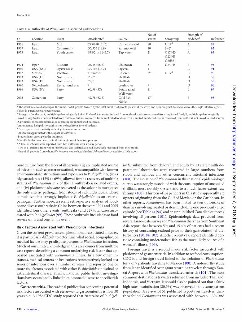
pure culture from the feces of ill persons, (ii) an implicated sourceof infection, such as water or seafood, was compatible with knownenvironmental distributions and exposures to P. shigelloides, (iii) ahigh attack rate (15% to 69%) allowed for the recovery of multiplestrains of Plesiomonas in 7 of the 11 outbreak-associated events,and (iv) plesiomonads were recovered as the sole or in most casesthe only enteric pathogen from stools of sick individuals. Thesecumulative data strongly implicate P. shigelloides as an entero-pathogen. Furthermore, a recent retrospective analysis of food-borne disease outbreaks in China between the years 1994 and 2005identified four other events (outbreaks) and 227 total cases asso-ciated with P. shigelloides (99). These outbreaks included two foodservice units and one family event.
Risk Factors Associated with Plesiomonas Infections
Given the current prevalence of plesiomonad-associated illnesses,it is particularly difficult to determine what social, geographic, ormedical factors may predispose persons to Plesiomonas infection.Much of our limited knowledge in this area comes from multiplecase reports describing a common underlying risk factor that ap-peared associated with Plesiomonas illness. In a few other in-stances, medical centers or institutions retrospectively looked at aseries of infections over a number of years and reported one ormore risk factors associated with either P. shigelloides intestinal orextraintestinal disease. Finally, national public health investiga-tions have occasionally linked plesiomonad disease to specific riskfactors.
Gastroenteritis. The cardinal publication concerning potentialrisk factors associated with Plesiomonas gastroenteritis is now 30years old. A 1986 CDC study reported that 28 strains of P. shigel-
loides submitted from children and adults by 13 state health de-partment laboratories were recovered in large numbers fromstools and without any other concurrent intestinal infections(100). The isolation of Plesiomonas in this national epidemiologicsurvey was strongly associated with the consumption of uncookedshellfish, most notably oysters and to a much lesser extent rawshrimp (100). Thirteen of 14 patients in this study apparently ateoysters originating from the Gulf of Mexico or the Caribbean. Inother reports, Plesiomonas has been linked to two outbreaks ofdiarrhea involving roasted oysters, including one previously citedepisode (see Table 4) (94) and an unpublished Canadian outbreakinvolving 18 persons (101). Epidemiologic data provided fromseveral large-scale surveys of Plesiomonas diarrhea from SoutheastAsia report that between 5% and 15.4% of patients had a recenthistory of consuming seafood prior to their gastrointestinal dis-turbances (80, 84, 102). Another recent case report identified por-ridge containing undercooked fish as the most likely source of awoman’s illness (103).
Foreign travel is a second major risk factor associated withplesiomonad gastroenteritis. In addition to seafood consumption,CDC found foreign travel linked to the isolation of Plesiomonasfor 7 of 9 patients traveling to Mexico (100). A noteworthy studyfrom Japan identified over 1,000 returning travelers through Kan-sai Airport with Plesiomonas-associated enteritis (104). The mostcommon destinations travelers returned from included Thailand,Indonesia, and Vietnam. It should also be pointed out that a fairlyhigh rate of coinfection (20.5%) was observed in this same patientpopulation. A review of 51 published reports on travelers’ diar-rhea found Plesiomonas was associated with between 1.3% and
TABLE 4 Outbreaks of Plesiomonas-associated gastroenteritis
Yr Location Event Attack ratea SourceNo. ofstrains Serogroup
Strength ofevidenceb Reference
1961 Japan Mill 275/870 (31.6) Cuttlefish salad 88c O:17d A 911965 Japan Community 53/355 (14.9) Salt mackerel 10 (�)e B 921973 Japan Youth center 878/2,141 (45.7) Tap water 21 O17:H2f A 93
O22:H3O8:H5
1974 Japan Bus tour 24/35 (68.5) Unknown 3 O24:H5 B 931980 USA (NC) Oyster roast 36/102 (35.2) Oysters 1 C 941982 Mexico Vacation Unknown Chicken 2f,g O:17d C 951983 USA (FL) Not provided 29/?h Shellfish D 351983 USA (FL) Not provided 29/? Shellfish D 351990 Netherlands Recreational area ? Freshwater 9 D 961996 USA (NY) Party 60/98 (57) Potato salad 11i B 97
Well water2003 Cameroon Party 49/78 (62.8) Cold fish 17j B 98
Ndoléa The attack rate was based upon the number of ill people divided by the total number of people present at the event and assuming that Plesiomonas was the single infective agent.Values in parentheses are percentages.b Strength of evidence: A, multiple epidemiologically linked P. shigelloides strains isolated from outbreak and also recovered from implicated food; B, multiple epidemiologicallylinked P. shigelloides strains isolated from outbreak but not recovered from implicated food source; C, limited number of strains recovered from outbreak not linked to food source;D, primarily anecdotal information regarding an unpublished outbreak.c An unnamed halophilic organism was isolated from 41% of patients.d Based upon cross-reactivity with Shigella sonnei antiserum.e All strains agglutinated with Shigella dysenteriae 7.f Predominant serotype in the outbreak.g Giardia lamblia was detected in the feces of one of these two persons.h A total of 29 cases were reported from two outbreaks over a 6-day period.i Two of 11 patients from whom Plesiomonas was isolated also had Salmonella recovered from their stools.j One of 17 patients from whom Plesiomonas was isolated also had Salmonella recovered from their stools.
Janda et al.
356 cmr.asm.org April 2016 Volume 29 Number 2Clinical Microbiology Reviews
on Septem
ber 7, 2018 by guesthttp://cm
r.asm.org/
Dow
nloaded from

5.41% of all episodes, depending upon geographic locale (105).The lowest frequencies were seen in cases originating from LatinAmerica and the Caribbean, while higher numbers were noted inSouth and Southeast Asia (105).
Persons in immunocompromised states, in particular thosewho are HIV positive, may be more prone to developing Plesiomo-nas diarrhea than healthy individuals (29). This is supported by arelatively higher prevalence rate (4.9% to 16.6%) of Plesiomonasgastroenteritis in HIV-positive or AIDS patients in comparison tofrequencies of diarrhea seen in unselected patient populations(106, 107). However, in one of these two studies Plesiomonas wasmore prevalent in non-AIDS patients (11.7%) than in those withAIDS (4.9%) (106). Some studies have found the highest fre-quency (33% to 35%) of plesiomonad enteritis in infants 19 to 31months of age (85) and in children under 2 years of age (80),suggesting a naive immunologic system as a potentially predispos-ing factor in young children for acquiring Plesiomonas, although acase-controlled study in a remote part of Thailand found P. shig-elloides in high percentages (10% to 11%) in both cases and con-trols (79).
In addition to these conditions, it is apparent that the con-sumption of untreated water or freshwater sources in nations withlow socioeconomic status or poor hygiene conditions puts anyoneat risk of acquiring Plesiomonas diarrhea (Table 4) (28, 35). Thesefacts parallel the seasonality of the disease and its association withtemperate and tropical/subtropical climates, where the prevalenceof the general disease appears much higher (28). Elevated aquatictemperatures in lakes and rivers may lead to the multiplication ofthis species (29) and higher surface concentrations.
Extraintestinal disease. Identification of potential risk factorsassociated with extraintestinal P. shigelloides infections is evenmore difficult, given the limited number of published cases incomparison to those associated with gastrointestinal disease. Themost common of these extraintestinal syndromes is bacteremia,where only 40 or so instances have been recorded in which Ple-siomonas was isolated as the sole or copathogen in a case of sepsis.Woo et al. (108) compared 7 cases of Plesiomonas occurring intheir institution over a 9-year period to 31 other cases reported inthe literature. They found that bacteremias in their medical centerwere significantly associated with advancing age (�75 years), un-derlying biliary tract disease, acute cholangitis, and polymicrobicsepsis, compared to previous cases in the literature (108). Sincemany of these illnesses were polymicrobic in nature, it is difficultto ascertain how many of these risk factors can be directly associ-ated with Plesiomonas.
Most reports involving Plesiomonas septicemia occur in per-sons suffering from one or more underlying medical illnessesleading to an immunocompromised state. In addition, secondarymedical sequelae may result as a direct consequence of these pri-mary medical conditions, which further increases the risk of inva-sive disease. Many cases of Plesiomonas sepsis are observed in in-dividuals with multiple risk factors that may either individually orcollectively be associated with plesiomonads. However, these riskfactors can also be less species specific and more a reflection ofGram-negative bacteremia.
The pattern of developing Plesiomonas sepsis parallels that ofgastroenteritis in the sense that most infections appear to resultfrom ingestion of seafood or contaminated water, particularly inpersons living in tropical, subtropical, or temperate regions (28,29, 35). Conditions proposed to be associated with plesiomonad
sepsis in addition to biliary disease include cancer, cirrhosis, HIV,and bloodborne dyscrasias, such as sickle cell anemia and thalas-semia (4, 34, 84, 108, 109, 110, 111, 112). The latter diseases cansometimes lead to other conditions sporadically associated with P.shigelloides bacteremia, including splenectomy (108, 111) andhemochromatosis (108, 113). Approximately a dozen cases ofperinatal bacteremia with central nervous system (CNS) involve-ment have been reported to date. In several cases the mother haddiarrhea immediately prior to delivery and in a couple of casesPlesiomonas was recovered from maternal feces. This suggests ver-tical transmission from mother to child during the birth process(114).
A number of rare Plesiomonas infections result from unappar-ent injuries or traumas associated with water environs. Theseevents include swimming in seawater or penetrating traumas tothe head connected to diving or submerged projectiles (115, 116,117).
Disease Transmission
The exact mode of disease transmission from natural reservoirs tohumans, eliciting a variety of illnesses, is still speculative. Jagger(28) proposed a dual-schematic scenario for the apparently highfrequency of Plesiomonas colonizing/infecting the guts of cats andfor acquisition of these same bacteria by humans from environ-mental sources. In the former category, natural aquatic habitatsand secondary reservoirs (amphibians, fish and shellfish) serve aspotential sources for the transmission of plesiomonads directly tocats or indirectly through birds living near aquatic ecosystemswho have fed on freshwater fish. Subsequent consumption of birdcarcasses or fresh fish by cats could also lead to colonization. In thecase of humans, the same two reservoirs (contaminated water, fishand shellfish) would serve as vehicles for ingestion of adulteratedfood or water containing plesiomonads. A cycle of reintroducingP. shigelloides into natural habitats could also occur through sew-age contamination (28).
Figure 1 represents a diagrammatic flow chart concerning thepotential acquisition and transmission of P. shigelloides to hu-mans. Best available evidence suggests that the main avenue oftransmission of plesiomonads from the environment to persons isvia freshwater sources. Higher rates observed for cases of gastro-enteritis in the Far East and other locales could be related to thegreater proliferation of this species in warmer climates or socio-economic factors or sanitary/hygiene conditions in developingcountries. A second mode of acquisition involves consumption offood sources intimately linked to aquatic habitats. Ironically, thesetwo sources together do not represent the majority of cases ofillness linked to this species in epidemiologic investigations. In aCDC study by Holmberg et al. (100), 21 of 31 (68%) patients hada history of seafood consumption or foreign travel immediatelypreceding their diarrheal episode. More recent studies have re-ported even lower percentages. A Hong Kong study of 167 cases ofPlesiomonas gastroenteritis found only 36% of individuals with ahistory of foreign travel or consumption of seafood or uncookedfood (102). Similar studies from Taiwan reported cumulative val-ues of 11% (80). The question mark in Fig. 1 suggests that theremay be one or more unrecognized sources of acquisition that havenot as yet been identified. Alternatively, since epidemiologic stud-ies typically do not include questionnaires regarding water con-sumption (except in the case of a recognized outbreak), it may wellbe that the majority of unidentified sources of infection are in fact
P. shigelloides
April 2016 Volume 29 Number 2 cmr.asm.org 357Clinical Microbiology Reviews
on Septem
ber 7, 2018 by guesthttp://cm
r.asm.org/
Dow
nloaded from

water related. Further environmental sampling studies on the fre-quency of plesiomonads in various aquatic environments in re-gions of the world where P. shigelloides disease is more commonare needed.
CLINICAL INFECTIONS AND ASSOCIATED DISEASESYNDROMES
P. shigelloides has been isolated on one or more occasions fromvirtually every common infectious disease syndrome or complica-tion that has been previously reported. In practical terms, how-ever, these infections or syndromes can be broken down into thefollowing groups or categories, namely: (i) gastrointestinal infec-tions or complications involving the small or large intestine, (ii)systemic disease manifested by frank septicemia with or withoutassociated CNS complications, such as meningitis, and (iii) a myr-iad of miscellaneous infections, including intraabdominal disease,soft tissue and wound infections, and ocular illnesses.
Gastroenteritis and Diarrheal Disease-Related Syndromes
Although once a fairly controversial topic, Plesiomonas is nowa-days considered an enteropathogen by most in the medical andscientific communities. While at one time it was rarely listed inleading publications on bacterial gastroenteritis as a pathogen, itnow can be found in many authoritative reviews or references onthe subject, along with more common agents such as Campylobac-ter, Salmonella, and Shigella (87, 118, 119, 120).
Plesiomonas enteritis can present in one of three forms: an acutesecretory gastroenteritis (most common), bloody or dysentericcolitis, or chronic or persistent diarrhea of �14 days duration(121).There have been very few published case reports on thistopic over the past 15 years (103). Most of our recent clinicalknowledge on Plesiomonas gastroenteritis stems from prospectiveor retrospective studies on this syndrome from regions of theworld where the prevalence of the disease is presumed to be much
higher, since limited data from industrialized nations suggest thatthe infection rate is quite low (122, 123).
Table 5 presents cumulative data from several large retrospec-tive or prospective investigations on P. shigelloides gastroenteritisin Southeast Asia and Africa. Plesiomonas gastroenteritis occurs inall age groups, with several studies documenting infections in in-fants as young as 22 days (80) to those �90 years of age (84, 102).Several reports indicate that the disease may be more common inchildren under 5 years of age (80, 83, 85, 102), suggesting thatimmunologic immaturity might be a predisposing factor to ac-quiring the illness. Plesiomonas has been found to be the third orfourth ranking cause of gastroenteritis in Nigeria (85) and China(84), respectively. Mixed infections are fairly frequent (16% to28%), with common coinfecting pathogens including Salmonella,Aeromonas, and V. parahaemolyticus (80, 84, 85, 102). The associ-ation of the latter two species with Plesiomonas suggests contam-inated water or seafood/fish as common vehicles of infection(80, 84).
Secretory enteritis. As previously noted, watery diarrhea is themost common clinical presentation of Plesiomonas gastroenteri-tis, with between 50% and 88% of all patients presenting with thistype of diarrhea (8, 80, 124). Prominent symptoms associatedwith plesiomonad enteritis include watery diarrhea and abdomi-nal pain (29, 85, 124) (Table 5). One study of adult patients withnonbloody P. shigelloides gastroenteritis seeking medical attentionat an emergency department had a mean duration of diarrhea of1.6 days with 10 bowel movements (bm) per day (125). Othersurveys or case reports documented evacuation rates of 5 to �10bm/day (102, 103, 124). Typical cases of untreated enteritis had amean duration of symptoms of 11 days in one CDC study (100),while another report placed the range of symptoms from 1 day to2 months (102). Duration of acute diarrhea in hospitalized pa-tients in this second study was 2.2 days (102).
Watery gastroenteritis is most often observed in healthy per-sons (29, 80, 103). Wong et al. (102) found that almost three-fourths of the 167 persons studied with P. shigelloides diarrheawere healthy, with the remaining 25% with one or more underly-ing condition, including diabetes, renal disease, cirrhosis, and co-lon cancer. Typically, the disease is mild and self-limiting (8, 29).Klontz et al. (124) found that gastroenteritis induced by P. shigel-loides was typically milder and less severe than that caused byeither non-O1 V. cholerae or V. parahaemolyticus (124). Less com-mon symptoms associated with a minority of cases include dehy-dration, hypokalemia, and peritonism (102).
FIG 1 Diagrammatic representation of the main known routes of transmis-sion and major disease manifestations associated with P. shigelloides infection.
TABLE 5 Gastrointestinal symptomatology associated with P.shigelloides diarrhea
Location
No.ofcases
Symptoms (%)a
ReferenceDiarrhea Blood FeverAbdominalpain Vomiting
HongKong
167b 99 25 51 72 38 102
Taiwan 111 96 45 51 45 41 80Bangladesh 253 93 5 6 6 74 124China 104 84 17 33 69 14 84Nigeria 51 88 18 35 67 14 85a Percentage of cases or isolates.b Cumulative data (for all age groups), including single and mixed infections with P.shigelloides.
Janda et al.
358 cmr.asm.org April 2016 Volume 29 Number 2Clinical Microbiology Reviews
on Septem
ber 7, 2018 by guesthttp://cm
r.asm.org/
Dow
nloaded from

Cholera-like diarrhea. Although not well-documented or de-scribed, P. shigelloides has been implicated on several occasions asa rare cause of diarrhea resembling cholera, one of which occurredin a woman returning from a trip to Kenya (126, 127). Subse-quently, a case from San Lazaro Hospital in the Philippines de-scribed a patient (age and sex unknown) with severe dehydrationof 5-h duration and 25 bm/day (128). The stool had a “rice water”consistency with mucus, and P. shigelloides was isolated. However,the stool of this patient also contained V. cholerae and Trichurisspp. In 2002, a 53-year-old man in Djibouti was hospitalized be-cause of severe dehydration secondary to a cholera-like illness of 1day duration (129). He had passed 8 liquid stools of a greenishconsistency each day. P. shigelloides was isolated in pure culturefrom his stool. Furthermore, the finding in some surveys that oneor more patients experienced diarrheal episodes consisting of 30bm/day suggests that this more severe form of secretory gastroen-teritis may have been overlooked in the case of Plesiomonas (8,102).
Dysentery. Dysentery or infective colitis is the second mostcommon form of Plesiomonas gastroenteritis. The syndrome ischaracterized by the macroscopic appearance of blood in stoolaccompanied by significant abdominal pain or tenderness andvomiting (Table 5). More severe disease in one study was associ-ated with fever in 13.2% of cases (102). Mucus in stools is found in17% to 19% of feces where P. shigelloides is the only pathogen (84,85). Many persons with these symptoms are hospitalized with thepresumptive diagnosis of bacillary dysentery.
The frequency of Plesiomonas acute dysentery in selected pa-tient populations has been reviewed by Pfeiffer and others (123). Ithas ranged from a reported high of 25.1% in a combined survey ofchildren and adults to 15.8% in children alone. Two studies con-ducted in the United States in the 1980s by the CDC (100) and theTexas Department of Health (130) placed the frequency of bloodydiarrhea at 53% and 22%, respectively. Unfortunately, there havenot been any individual case reports describing Plesiomonas dys-entery over the last 15 years. One of the best-described cases ofplesiomonad bloody diarrhea is that of a 42-year-old woman withpersistent dysentery and histopathology indicating pseudomem-branous colitis. P. shigelloides was recovered from her on two oc-casions in pure culture (131).
Chronic diarrhea. P. shigelloides is an infrequent but recog-nized cause of chronic diarrhea (132). Wong and others (102)found 5% of their patients with chronic Plesiomonas disease had
diarrhea of 2 weeks to 2 months duration. In a similar study fromTaiwan, chronic diarrhea was seen in 23% of children and 12% ofadults (80). Except for the lengthy duration of the illness, specificsymptomatology associated with persistent gastroenteritis ispoorly described.
Fatal gastrointestinal infections. While not well appreciated,Plesiomonas gastroenteritis has been linked to a negative outcomeon several instances, most of which involved invasive (bloody)diarrhea. A 19-year-old woman with bulimia nervosa who hadbeen previously thrown into a water fountain developed unrelent-ing severe bloody diarrhea with dehydration of 9 days durationprior to her death (133). P. shigelloides and Aeromonas veroniibiotype sobria was recovered from her stool. In 1989, Sinnott et al.(134) reported on a healthy 53-year-old woman who developedsevere watery diarrhea that subsequently became bloody a daylater after consuming sushi and raw oysters. After initial treatmentshe returned to the emergency room 24 h later hypotensive, andshe subsequently expired. P. shigelloides was recovered on twoseparate occasions from her stool. Wong and colleagues (102)described two fatal infections associated with plesiomonad diar-rhea. In one instance, an 82-year-old man with alcoholic cirrhosispresented with moderately severe diarrhea and mucus, dehydra-tion, and hypokalemia. He died 3 days later (cause of death notmentioned). In a second case, a 68-year-old male with diabetesmellitus and end-stage renal disease had diarrhea of 1 day dura-tion and turbid peritoneal fluid. Stool culture grew P. shigelloides,while the peritoneal fluid yielded Pseudomonas aeruginosa. Causeof death was multiorgan failure. In most of these cases, it wasdifficult to determine exactly to what extent Plesiomonas contrib-uted to the person’s eventual demise.
Unresolved controversies. The conundrum concerning the ev-idence supporting the role of Plesiomonas in gastrointestinal dis-ease is presented in Table 6 (5, 8, 100, 103, 131, 135). Since 2000,several case-controlled investigations have been published on therole of plesiomonads in diarrheal disease. However, of these fourstudies only two (84, 85) strongly supported a role for this speciesin gastroenteritis. This finding may not be as problematic as it firstappears (Table 3) (79, 82, 84, 85). Since almost all of these studieswere centered in developing nations located in tropical/subtropi-cal climates, they all appeared to have a higher incidence of thedisease. Thus, the opportunity for exposure and reexposure toplesiomonads from infancy to adulthood may be greater andthereby may be potentially protective to a certain segment of the
TABLE 6 Outstanding issues concerning Plesiomonas gastroenteritis
Type of study
Information from studies:
Before 2000 Current
Case reports �10 well-described case reports or series of case reports Only one case since 2000 (103)Case-controlled
investigationsOne study through 2000 (5) Four studies through 2000 (see Table 3); two with positive findings,
one with negative findings, one with equivocal findingsOutbreaks P. shigelloides not clearly shown to cause outbreak of
gastroenteritis (5)At least 11 outbreaks described, including 1 since 2000; several with
good epidemiologic data (see Table 4); several more anecdotallydescribed in the literature
Immune response Data from a CDC study not supportive (100); several casereports more supportive (8, 131)
No new data
Histopathology Good histopathology from a few selected case reports (131, 135) No new dataPathogenicity No well-described enteropathogenic mechanisms (8) Some now described but poorly characterized (see Pathogenicity
section)
P. shigelloides
April 2016 Volume 29 Number 2 cmr.asm.org 359Clinical Microbiology Reviews
on Septem
ber 7, 2018 by guesthttp://cm
r.asm.org/
Dow
nloaded from

population. This supposition is supported by the hypothesis thatP. shigelloides O17 is common in surface waters in developingcountries and may also have a protective effect against infectionwith Shigella sonnei by virtue of shared lipopolysaccharide anti-gens (136). Thus, there may be at least several subpopulations ofresidents in these locales who may be naive, partially protected, orprotected from diarrheal infections produced by this entero-pathogen, which has a relatively low pathogenic potential.
In addition to this evidence there are a sizeable number ofoutbreaks described where P. shigelloides is a sole or copathogen incomparison to Aeromonas, where not a single credible outbreakhas been attributed to this bacterium over the past 3 decades (90).What is needed are more case-controlled surveys and detailed casereports supported by clinical data, such as humoral immune re-sponses (acute, convalescent) and recovery of the enteropathogenfrom pathological biopsy material, not simply accounts reportingisolation.
Septicemia and CNS Disease
Septicemia with or without CNS involvement is the primary clin-ical syndrome associated with extraintestinal P. shigelloides infec-tion. Table 7 summarizes some retrospective reviews of cumula-tive data on cases of Plesiomonas sepsis reported in the Englishliterature (29, 108, 137). Based upon the review of Woo (108) andseveral published case reports since 2005, there are now over 40cases of P. shigelloides bacteremia in the literature (111, 114, 138,139). This is almost double the number reported by Lee (137) in1996.
Plesiomonas sepsis is a community-acquired male-dominateddisease almost invariably observed in persons with one or moreunderlying medical conditions leading to the immunocompro-mised state (Table 7). Common predisposing factors leading tosepsis in decreasing order of frequency include blood dyscrasias(thalassemia, sickle cell disease, leukemia), asplenia (functional oranatomic), and iron overload conditions, such as hemochroma-tosis, biliary tract disease, and cirrhosis (alcoholic, viral) (108, 111,112). Slightly under one-third of all bacteremias occur in neonateswith meningitis or meningoencephalitis (114). Almost all of thesecases of infection occur within the first 5 days of life and appear tohave been vertically transmitted (perinatal) rather than transpla-cental (114). Plesiomonas has only been recovered from maternalfeces on three occasions. The mortality rate in neonates exceeds50% (114).
About 90% of all cases of sepsis are monomicrobic. In almost
half of these cases, plesiomonads are recovered from a site otherthan blood. These specimen sources include CSF, abscess/wound,joint fluid, pleural fluid, stool, and urine (108). The study of Wooet al. (108), which described 7 cases of Plesiomonas bacteremia,differed appreciably from the remaining body of cases in that thesepatients were older (median, 82 years), had biliary tract disease,and infections were more often polymicrobic. Reasons for thesedifferences are not immediately apparent. While the source ofmost community-acquired infections in adults is unknown, con-taminated water, raw seafood, or other foods are most often sus-pected (137). A case of sepsis originated in a 13-year-old girl withsickle cell who consumed many crab legs at a crab buffet 4 daysprior to her admission (110). Another case of pleisomonad sepsisoccurred in a 51-year-old man with liver disease that received adonor liver from a teenage boy who drowned in a freshwater lake(138). Blood cultures drawn from the donor prior to transplanta-tion eventually yielded P. shigelloides. In another instance, a rap-idly fatal case of combined Plesiomonas and Clostridium perfrin-gens sepsis and meningitis occurred in a 71-year-old man who hadjust disembarked from an international flight (139). He had justreturned from a hunting trip in the Swiss Alps and expired 3 hafter initial presentation. Autopsy suggested death resulted fromseptic complications. Those authors suspected contaminatedmeat or water from his hunting trip as the source of infection.
Wound Infections
Unlike other aquatic pathogens, such as Aeromonas and She-wanella, there are virtually no published reports in the literaturedescribing isolation of P. shigelloides from open or penetratingwounds, nor an association of this species with soft tissue infection(90, 140). The reason for this anomaly is presently unclear, al-though the lack of protease expression by plesiomonads may be acontributory factor. Two cases of cellulitis of the leg and foot,respectively, have been reported where cultures grew plesiomon-ads from tissue or exudate material (141, 142). On both occasions,other pathogenic bacteria were also isolated, bringing into ques-tion what contribution this microbe may have played in the infec-tious process. A more definitive role for Plesiomonas in bacterialcellulitis was described by Gopal and Burns (143) 20 years later ina 59-year-old man who sustained a penetrating trauma to his lefthand with a knife while cleaning 20 or more freshwater fish. P.shigelloides was recovered not only from the surgical wound infec-tion but also in pure culture from the initial blood culture. Themost recent case of a wound infection caused by Plesiomonas in-volves a healthy 31-year-old diver who sustained a penetratingtrauma to his skull when he hit submerged rocks in a pond whilediving (116). He developed a frontal cutaneous abscess of the headas a result of the trauma, which required medical interventionincluding antimicrobial therapy.
These are currently no data available that suggest that Plesiomo-nas is ever an animal-related pathogen in the traditional sense ofother zoonotic pathogens, including those associated with aquatichabitats. There are also no known reports of plesiomonads beingassociated with traumatic human wound infections acquiredthrough penetrating injuries or bites induced by vertebrate or in-vertebrate species. In fact, Plesiomonas is not listed as a possibleaquatic zoonotic pathogen in the broadest sense (144). Limitedevidence suggests that the couple of cases of cellulitis reported aredue to penetrating traumas that come in contact with material(fish, rocks) associated with freshwater environs. No aggressive
TABLE 7 Characteristics of Plesiomonas septicemia reported in theliterature
Characteristic
Value reported in study
Lee et al.,1996 (137)
Stock, 2004(29)
Woo et al.,2005 (108)
No. of cases reported 21 24 38a
Mean age (yrs) 22.7 28.8 38.4Age range 1 day to 68
yrs2 days to 68
yrs1 day to 94
yrsMale:female ratio 2:1 2.7:1 1.8:1Immunocompromised (%) 81 88 82Mortality rate (%) 62 50 42Reference 137 29 108a Includes 31 cases in the literature and 7 new cases described by the authors.
Janda et al.
360 cmr.asm.org April 2016 Volume 29 Number 2Clinical Microbiology Reviews
on Septem
ber 7, 2018 by guesthttp://cm
r.asm.org/
Dow
nloaded from

wound infections, such as necrotizing fasciitis, have even beenlinked to the genus Plesiomonas.
Ocular Disease
There are surprisingly as many cases of Plesiomonas-associated eyeinfections in the literature as there are wound infections. Clinicalpresentations of eye disease range from endophthalmitis to kera-titis (117). In three instances, the precipitating event resulted froma penetrating injury to the eye caused by a fishhook (two in-stances) or a rock from a creek bed (117, 145). One ocular infec-tion in a neonate appeared to have been acquired transplacentally(146). In some cases, these devastating illnesses are solely causedby plesiomonads (117), while in other instances they are of a poly-microbic etiology involving other aquatic or marine bacteria, suchas Aeromonas, Shewanella, or Vibrio (145).
Miscellaneous Infections
A number of infrequent to extremely rare complications resultingfrom P. shigelloides infection have been summarized in one ormore reviews on the topic (27, 28, 29). These conditions includepolyarthritis/osteomyelitis, cholangitis, cholecystitis, epidid-ymoorchitis, peritonitis, pyosalpingitis, and pancreatic andsplenic abscesses. Two cases of peritonitis associated with contin-uous ambulatory peritoneal dialysis in 62- and 73-year-old Chi-nese women were reported in 2004 (147). The source and portal ofentry in both patients was unknown. In 2009, Schneider and as-sociates (148) described a case of P. shigelloides pneumonia in a76-year-old woman who had previously undergone a curative gas-trectomy. X-rays and a computed tomography scan indicated anodular lesion in the right upper lung and a cavernous lesion inthe right upper lobe, respectively. Bronchial lavage yielded 105
CFU/ml; an equivalent number of E. coli was also recovered. Thepatient was successfully treated with multiple antimicrobial agentsbut suffered severe postinfectious complications, including thetacoma.
Treatment
Most gastrointestinal infections caused by P. shigelloides are mildin nature and do not require antimicrobial intervention or othermedical treatments. One large study of Plesiomonas diarrheafound 85% of such illnesses to be self-limiting (102). However,antimicrobial therapy or other medical interventions may be war-ranted for some cases of moderate to severe diarrhea or to coun-teract physiologic or metabolic issues arising from such illnesses.DuPont (119) recommended that for severe cases of dysentery-like diarrhea caused by P. shigelloides should be treated like shig-ellosis, namely, with azithromycin or ceftriaxone for children andciprofloxacin or azithromycin for adults. Quinolone therapy forthe treatment of more severe cases of plesiomonad enteritis hasalso been suggested by other authors (102, 103). Some earlierstudies found that the duration of diarrhea (up to 10 days) couldbe appreciably shortened by intervention with tetracycline or tri-methoprim-sulfamethoxazole (100). In cases where dehydrationis clinically significant or the patient presents with cholera-likedisease, the administration of oral rehydration salts or intrave-nous fluids may be appropriate alone or in combination with anantibiotic such as a quinolone (103, 125, 129).
Guidelines regarding the appropriate treatment of extraintesti-nal infections caused by Plesiomonas, of which septicemia is themost common, are difficult to determine because of a multiplicity
of factors. While most illnesses are community acquired and mo-nomicrobic, they occur so rarely that plesiomonad sepsis is almostnever suspected as part of the initial diagnosis upon clinical pre-sentation. Furthermore, most patients with Plesiomonas sepsissuffer from other serious underlying diseases, including sickle celldisease (110), thalassemia (111, 112), or HIV infection (149). Theinterval in time between each of these case reports is significant(four cases in the last 15 years), making comparisons in treatmentregimens virtually impossible. Once P. shigelloides was identified,in each instance, antimicrobial regimens were switched to quino-lones (ciprofloxacin), carbapenems (imipenem), or combinationsthereof. A review of a dozen cases of neonatal plesiomonad sepsisaccompanied by meningitis or menigoencephalitis indicated thatall neonates, with one exception, received combination therapy (atwo- or three-drug regimen) with such combinations, including apenicillin derivative, an aminoglycoside (most often gentamicin),cephalosporin (cefotaxime), or carbapenem (114). Again, becauseof the length in years separating publication of these single casereports, it is difficult to draw any firm conclusions on the effec-tiveness of directed treatment on patient outcomes. In a series ofelderly persons with biliary disease and polymicrobic bacteremia,including P. shigelloides, four of five patients responded favorablyto one of several drug regimens administered, including cefu-roxime and cefotaxime (108).
PATHOGENICITY
While P. shigelloides has been associated with acute bacterial gas-trointestinal illness and rare extraintestinal infections (112, 114,150, 151), conflicting data on virulence factors and a lack of rele-vant animal models have stalled elucidation of the mechanismsinvolved in its pathogenicity. A variety of virulence factors havebeen associated with infections, including �-hemolysin (152), en-terotoxins (153), cholera-like toxins (154), and a cytotoxin LPScomplex (155). However, seminal work and the evolution of mo-lecular biology have advanced our understanding of this orga-nism’s pathogenicity, particularly with respect to the structure ofLPS, somatic antigenicity, cytotoxin activity, iron acquisition, andgenetic diversity.
Lipopolysaccharide
The barrier properties of the outer membrane of Gram-negativebacteria are attributed to the LPS structure, which is highly immu-nogenic. Also referred to as an “endotoxin,” cell-bound LPS isassociated with stimulation of severe pathological manifestationsof immuno-activation, such as septic shock. As in other membersof the Enterobacteriaceae family, the LPS in P. shigelloides is com-prised of three domains which have significant biological activityand involvement in host-bacterium interactions. It includes anendotoxic glycolipid (lipid A), an O-polysaccharide (O-PS) or O-specific antigen, and an intervening core oligosaccharide (core-OS) region.
To date, LPS structures have been studied for 11 strains of P.shigelloides (Table 8) (156, 157, 158, 159, 160, 161, 162, 163, 164,165, 166, 167, 168, 169, 170). The majority of LPS regions studiedin the Enterobacteriaceae that link the core to the lipid A moiety(KdoI) contain at least one residue of 3-deoxy-D-manno-oct-2-(ketodeoxyoctonic) acid (KdoI) (156, 159). In addition, they maypossess L-GLYCERO-D-MANNO-heptopyranose (L,D-Hep), theoligosaccharide L-�-D-Hep-(1¡3)-L-�-D-Hep-(1¡5)-[�-Kdo-(2¡4)]-�-Kdo (D,D-Hep), or do not have a heptose (156, 159,
P. shigelloides
April 2016 Volume 29 Number 2 cmr.asm.org 361Clinical Microbiology Reviews
on Septem
ber 7, 2018 by guesthttp://cm
r.asm.org/
Dow
nloaded from

171). Among the Enterobacteriaceae, P. shigelloides is included inthe Klebiella-Serratia-Proteus LPS group. This is based on the pos-session of the outer core disaccharide galactose A-glucosamine Nand the substitution of �-glucose-HEPI, which is absent in theother major enteric LPS group, the Escherichia-Salmonella-Shi-gella LPS group (159). However, peculiar to P. shigelloides is thepresence of �-galactose–HEPI as the substituted moiety instead of�-glucose–HEPI (157, 158, 159, 172).
As is found in other Enterobacteriaceae, the chromosomal genesof P. shigelloides that are involved in LPS core biosynthesis arefound in the waa gene cluster (formerly rfa) (172, 173). In a studyby Aquilini et al. (159), P. shigelloides 302-73 and CNCTC 113/92(serotypes O1 and O54, respectively) shared all the genes of thewaa cluster except for one common core biosynthetic gene(wapG), which was found at a different chromosome locationoutside the cluster. Further, these workers were also able to assigna proteomic function to all the P. shigelloides waa genes that wereidentified in these two strains to encode six new glycosyltrans-ferases (WapA, -B, -C, -D, -F, and -G). Thus, despite there being asingle species of Plesiomonas, strains studied have showed a differ-ence in LPS genetics as well as a high homologous recombinationlevel in housekeeping genes.
Enteropathogenicity
The enteropathogenicity of P. shigelloides continues to be nebu-lous, but it has long been suspected that manifestation of illness inthe host is multifactorial (174). For example, ingested plesiomon-ads do not always cause gastrointestinal disease, and these bacteriahave been isolated from the stools of healthy individuals (175).Global reports of isolation as enteric pathogens also differ signif-icantly. An assessment by Shah et al. (105) of 51 published studiesbetween 1973 and 2004 of the etiology of diarrhea in 57 groups oftravelers showed the isolation of P. shigelliodes to differ betweenregions. These findings contrasted with those of an earlier study byUeda et al. (176) of overseas travelers presenting with diarrhea atOsaka Airport Quarantine Station between January 1992 and Sep-tember 1994. Here, P. shigelloides was isolated in 1,127 of 1,882(59.9%) stool samples and was the most common bacterial patho-gen recovered in this period.
Such is also the diversity of virulence-associated properties in P.shigelloides. This was demonstrated by Cižnár (175) in a study of20 isolates from environmental sources, human cases of gastroen-teritis, and animal sources. Here, the isolates belonged to 13 dif-
ferent serovars; the majority of strains were both hydrophobic,indicating their ability to interact with a wide variety of surfacesincluding mammalian cells, and had a relatively high level of fla-gellar motility to facilitate attachment; all produced triacylglycerollipase, which is a virulence factor found in Staphylococcus aureus(177), Staphylococcus cohnii (178), and Pseudomonas (Burkhold-eria) cepacia (179). However, among these strains, there was sig-nificant variation in elastase, proteinase, histidine decarboxylase,and hemolysin activities. Furthermore, these workers were unableto detect signal molecules such as C4 to C8 unsubstituted N-acyl-homoserine lactones (AHLs), such as those evident in metabolicactivities of many other bacterial species (180).
Using a different phenotypic methodology, Salerno et al. (151)demonstrated greater diversity of putative virulence markers (in-cluding motility, hemolysin, and four exoenzymes) in 60 bio-chemically diverse strains of P. shigelloides that were isolated fromhuman (n � 25), animal (n � 18), and environmental (n � 17)specimens in different countries. All environmental and animalisolates were motile but, notably, 4 of the 25 human strains (16%)were nonmotile. Of the exoenzymes, DNase activity was preva-lent, particularly among the human strains; gelatinase activity wasdetected in none of the human strains and in only 22% and 35% ofanimal and environmental strains, respectively. All isolates werenegative for lipase and elastase activities (151). Most strains in thisstudy demonstrated hemolytic activity, although it was predomi-nantly found among the animal and environmental strains. Thiscontrasted with the findings of a study by Cižnár et al. (175), inwhich hemolysin activity was demonstrable in 7 of 30 (23.3%) ofthe isolates tested. Such variation is expected given that detectionof hemolysis is dependent on the method used, and it has beenshown previously to be influenced by the concentrations of ironand calcium, medium composition, level of oxygen, and the typeof erythrocytes used (151, 152, 175, 181, 182). Hemolysin activitymay be a useful maker for pathogenicity, as was implicated in astudy by Baratéla et al. (182), who found a correlation between thevacuolation of various cell lines with such cytotoxic activity infiltrates of P. shigelloides (Fig. 2). This correlation has also beenshown for Aeromonas hydrophila (183), Serratia marcescens (184),and Vibrio cholerae (185).
The diversity of the prevalence of virulence factors in recentstudies of P. shigelloides (above) and the technical difficulties as-
TABLE 8 Recent structural analytic studies of lipopolysaccharide instrains of Plesiomonas shigelloides
Somaticantigen Strain no.a Reference(s)
O1 302-73 157, 158, 159O13 CNCTC 80/89 160O17 PCM 2231, 7-63 161, 162O24 CNCTC 92/89 163O33 CNCTC 34/89 156O37 CNCTC 39/89 164O51 CNTC 110/92 162O54 CNTC 113/92 159, 165, 166, 167O74 CNCTC 144/92, AM36565,
22074, 12254168, 169, 170
a CNCTC, Czech National Collection of Type Cultures (Prague, Czech Republic).
FIG 2 Effect of P. shigelloides toxin filtrates on Y1 adrenal cells.
Janda et al.
362 cmr.asm.org April 2016 Volume 29 Number 2Clinical Microbiology Reviews
on Septem
ber 7, 2018 by guesthttp://cm
r.asm.org/
Dow
nloaded from

sociated with their detection continues to confound the associa-tion with gastrointestinal disease. Thus, the organism’s role as anenteric pathogen is suspected only when it dominates in a stoolculture of a symptomatic individual (176). Yet, the organism pos-sesses a large plasmid (�120 mDa) that has been shown to assist incell invasion (186). In addition, it is known to produce three formsof enterotoxin, including forms that have cholera-like (153, 154),thermo-stable (153, 187), and thermo-labile (182, 188) proper-ties. Furthermore, it has long been suspected of being adhesiveand invasive (8, 175).
Theodoropoulos et al. (189) was able to demonstrate for thefirst time the ability of P. shigelloides to adhere and enter mamma-lian cells in vitro. Using human colonic adenocarcinoma cells(Caco-2) as a model, these workers showed the organism’s abilityto invade the cytosol and escape from the cytoplasmic vacuoles.Following this work, Okawa et al. (155) isolated and characterizeda 40-kDa cytotoxic outer membrane protein (ComP) produced byP. shigelloides P-1 strain. This virulence factor was found to be aheat-stable complex comprised of three major LPS-binding pro-teins and an LPS moiety. However, in this study, only the formercomponent expressed cytotoxic or enterotoxigenic activity insuckling mouse studies. Using the same in vitro model of The-odoropoulos et al. (189), Tsugawa et al. (190) demonstrated bac-terial attachment and the induction of apoptosis. Further, theyhypothesized that these properties may evade an immune re-sponse conducive to metastatic infection and related to the pa-thology and clinical manifestations.
Seminal work on a number of other pathogenic organisms hasalso established the involvement of GroEL (a chaperone) or heatshock protein in cell attachment and invasion (191, 192, 193, 194,195, 196). Tsugawa et al. (197) also showed the importance of P.shigelloides GroEL by demonstrating induction of its expressionby contact with host cells (Caco-2 cells) and a subsequent in-creased cellular surface association of the protein. These workersspeculated that the involvement of GroEL in attachment of P.shigelloides to Caco-2 cells may be due to overexpression of a re-ceptor involved in attachment, such as they demonstrated by theupregulation of the gene for intercellular adhesion molecule-1(ICAM-1) by this chaperone or by binding with Caco-2 cell sur-face protein and forming a bridge with the organism. The latterproposal was not explored by that group. However, further mo-lecular studies by Tsugawa et al. (198) on the cytotoxin previouslycharacterized by Okawa et al. (155) showed that not only GroELbut also ComP is an important factor in the interaction of P.shigelloides and host cells. These workers showed that ComP is thepredominant virulence factor that causes apoptotic cell death inthe infected host cell. However, they observed that both a wild-type strain and a mutant strain with a comP deletion were notinsignificantly different in their abilities to internalize and multi-ply within Caco-2 cells. This observation suggests that apoptosisassociated with ComP is not involved in the internalization andgrowth within the host cells (198).
Iron Acquisition
Iron is an essential element for most bacteria, including P. shigel-loides (199, 200). However, in its oxidized form the element isinsoluble, and it is highly toxic for most macromolecules in itsreduced form. Consequently, bacteria have acquisition systemsthat involve iron and heme carrier proteins for iron sequestrationand assimilation from other sources (201). Thus, iron acquisition
systems have been shown to be involved in the pathogenicity of arange of bacterial pathogens as they acquire iron in host cells(199).
Siderophore-mediated iron assimilation systems are widely dis-tributed in bacteria (202) yet have not been found in P. shigelloides(153, 200). In a study by Daskaleros et al. (200), strains of P.shigelloides did not use siderophores such as enterobactin, aero-bactin, or vibriobactin for growth in medium low in iron. Simi-larly, Henderson et al. (203) found that P. shigelloides could notutilize siderophores of ferrichrome or schizokinen or be cross-fedby Vibrio parahaemolyticus strains that produced vibrioferrin orby an identified siderophore in Vibrio alginolyticus. Daskaleros etal. (200) further established that the growth of P. shigelloides wassupported by hemin and hemoglobin in iron-deficient media, butneither transferrin nor lactoferrin served as a source of iron. Thus,the acquisition of iron from these two compounds is thought to beassociated with the liberation of intracellular iron-containingcompounds by the lytic activity of hemolysins or cytotoxins.Daskaleros et al. (200) investigated the relationship between ac-quisition of iron and the production of hemolysins or cytotoxinsby using cloned DNA sequences of P. shigelloides that encodedhemolysin production and the ability to utilize heme or hemoglo-bin for expression in E. coli.They established that the genes forhemolysin production were not linked to those for heme ironutilization. Furthermore, they showed that production of P. shig-elloides hemolysin was derepressed under conditions of iron star-vation, suggesting the presence of a fur-like regulatory system forferric uptake that could influence the expression of iron transportsystem genes as well as other genes, as previously described for E.coli (203). Henderson et al. (203) characterized 10 genes in P.shigelloides that encode the heme iron utilization system, includ-ing HugA, the putative heme receptor in the outer membrane,TonB and ExbBD, which facilitate HugA movement of heme intothe periplasm, HugB, the putative periplasmic binding proteinwhich moves heme across the periplasm, HugC and HugD, theputative inner membrane permeases that allow movement ofheme into the cytoplasm, HugW and HugX, which may be re-quired for metabolism of iron and are not involved in transport,and HugZ, which is essential for binding and utilization of heme(203, 204, 205). Three of the genes, hugA, hugZ, and tonB, may becontrolled by iron, as they contain a FUR (ferric uptake repressor)box in their putative promoters (203, 206, 207). Interestingly, astudy by Villarreal et al. (205) showed that coexpression ofhuman hemoglobin genes and P. shigelloides heme transportgenes enhanced recombinant hemoglobin production in E. coliBL21(DE3) grown in medium containing heme. These findingshave implications for the use of recombinant hemoglobin as analternative to human blood for patients requiring transfusions(205).
Genetic Diversity
Salerno et al. (21) investigated the diversity of 77 P. shigelloidesstrains from different sources and countries via multilocus se-quencing typing based on five housekeeping genes (fusA, leuS,pyrG, recG, and rpoB). In the study, they found significant diver-sity, implicating a high propensity for homologous recombina-tion. This reduces resemblance between phylogenetic trees basedon single genes and compromises interaction between serotypesand the genomic background (21). Furthermore, the rate of re-combination observed in P. shigelloides strains studied by these
P. shigelloides
April 2016 Volume 29 Number 2 cmr.asm.org 363Clinical Microbiology Reviews
on Septem
ber 7, 2018 by guesthttp://cm
r.asm.org/
Dow
nloaded from
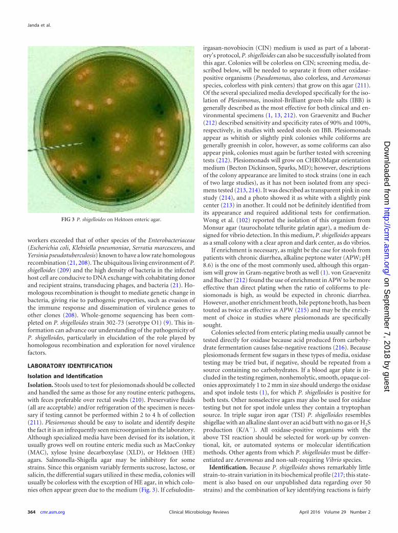
workers exceeded that of other species of the Enterobacteriaceae(Escherichia coli, Klebsiella pneumoniae, Serratia marcescens, andYersinia pseudotuberculosis) known to have a low rate homologousrecombination (21, 208). The ubiquitous living environment of P.shigelloides (209) and the high density of bacteria in the infectedhost cell are conducive to DNA exchange with cohabitating donorand recipient strains, transducing phages, and bacteria (21). Ho-mologous recombination is thought to mediate genetic change inbacteria, giving rise to pathogenic properties, such as evasion ofthe immune response and dissemination of virulence genes toother clones (208). Whole-genome sequencing has been com-pleted on P. shigelloides strain 302-73 (serotype O1) (9). This in-formation can advance our understanding of the pathogenicity ofP. shigelloides, particularly in elucidation of the role played byhomologous recombination and exploration for novel virulencefactors.
LABORATORY IDENTIFICATION
Isolation and Identification
Isolation. Stools used to test for plesiomonads should be collectedand handled the same as those for any routine enteric pathogens,with feces preferable over rectal swabs (210). Preservative fluids(all are acceptable) and/or refrigeration of the specimen is neces-sary if testing cannot be performed within 2 to 4 h of collection(211). Plesiomonas should be easy to isolate and identify despitethe fact it is an infrequently seen microorganism in the laboratory.Although specialized media have been devised for its isolation, itusually grows well on routine enteric media such as MacConkey(MAC), xylose lysine decarboxylase (XLD), or Hektoen (HE)agars. Salmonella-Shigella agar may be inhibitory for somestrains. Since this organism variably ferments sucrose, lactose, orsalicin, the differential sugars utilized in these media, colonies willusually be colorless with the exception of HE agar, in which colo-nies often appear green due to the medium (Fig. 3). If cefsulodin-
irgasan-novobiocin (CIN) medium is used as part of a laborat-ory’s protocol, P. shigelloides can also be successfully isolated fromthis agar. Colonies will be colorless on CIN; screening media, de-scribed below, will be needed to separate it from other oxidase-positive organisms (Pseudomonas, also colorless, and Aeromonasspecies, colorless with pink centers) that grow on this agar (211).Of the several specialized media developed specifically for the iso-lation of Plesiomonas, inositol-Brilliant green-bile salts (IBB) isgenerally described as the most effective for both clinical and en-vironmental specimens (1, 13, 212). von Graevenitz and Bucher(212) described sensitivity and specificity rates of 90% and 100%,respectively, in studies with seeded stools on IBB. Plesiomonadsappear as whitish or slightly pink colonies while coliforms aregenerally greenish in color, however, as some coliforms can alsoappear pink, colonies must again be further tested with screeningtests (212). Plesiomonads will grow on CHROMagar orientationmedium (Becton Dickinson, Sparks, MD); however, descriptionsof the colony appearance are limited to stock strains (one in eachof two large studies), as it has not been isolated from any speci-mens tested (213, 214). It was described as transparent pink in onestudy (214), and a photo showed it as white with a slightly pinkcenter (213) in another. It could not be definitely identified fromits appearance and required additional tests for confirmation.Wong et al. (102) reported the isolation of this organism fromMonsur agar (taurocholate tellurite gelatin agar), a medium de-signed for vibrio detection. In this medium, P. shigelloides appearsas a small colony with a clear apron and dark center, as do vibrios.
If enrichment is necessary, as might be the case for stools frompatients with chronic diarrhea, alkaline peptone water (APW; pH8.6) is the one of the most commonly used, although this organ-ism will grow in Gram-negative broth as well (1). von Graevenitzand Bucher (212) found the use of enrichment in APW to be moreeffective than direct plating when the ratio of coliforms to ple-siomonads is high, as would be expected in chronic diarrhea.However, another enrichment broth, bile peptone broth, has beentouted as twice as effective as APW (215) and may be the enrich-ment of choice in studies where plesiomonads are specificallysought.
Colonies selected from enteric plating media usually cannot betested directly for oxidase because acid produced from carbohy-drate fermentation causes false-negative reactions (216). Becauseplesiomonads ferment few sugars in these types of media, oxidasetesting may be tried but, if negative, should be repeated from asource containing no carbohydrates. If a blood agar plate is in-cluded in the testing regimen, nonhemolytic, smooth, opaque col-onies approximately 1 to 2 mm in size should undergo the oxidaseand spot indole tests (1), for which P. shigelloides is positive forboth tests. Other nonselective agars may also be used for oxidasetesting but not for spot indole unless they contain a tryptophansource. In triple sugar iron agar (TSI) P. shigelloides resemblesshigellae with an alkaline slant over an acid butt with no gas or H2Sproduction (K/A�). All oxidase-positive organisms with theabove TSI reaction should be selected for work-up by conven-tional, kit, or automated systems or molecular identificationmethods. Other agents from which P. shigelloides must be differ-entiated are Aeromonas and non-salt-requiring Vibrio species.
Identification. Because P. shigelloides shows remarkably littlestrain-to-strain variation in its biochemical profile (217; this state-ment is also based on our unpublished data regarding over 50strains) and the combination of key identifying reactions is fairly
FIG 3 P. shigelloides on Hektoen enteric agar.
Janda et al.
364 cmr.asm.org April 2016 Volume 29 Number 2Clinical Microbiology Reviews
on Septem
ber 7, 2018 by guesthttp://cm
r.asm.org/
Dow
nloaded from

unique, making it is easy to identify with conventional tests. TheTSI reaction coupled with positive oxidase, lysine, and ornithinedecarboxylase and arginine dihyrolase reactions and myo-inositolfermentation separate this organism from other Enterobacteria-ceae, Aeromonadaceae, and Vibrionaceae (13). It is also 100% pos-itive for indole, D-glucose, trehalose, and nitrate, 95% positive formaltose and 90% positive for ONPG (o-nitrophenyl-�-D-galacto-pyranoside) (217). Citrate, urea, Voges-Proskauer, and potassiumcyanide tests, gas production from any carbohydrate fermented,and utilization tests with most sugars are uniformly negative. Lac-tose and salicin reaction results are variable. P. shigelloides is sus-ceptible to O129 (2,4-diamino-6,7-diiopropyl-pteridine), withthe reaction most clearly defined on Mueller-Hinton agar (1). P.shigelloides does not require salt for growth but can grow in saltconcentrations up to 4% (13). While hemolysis is usually not pres-ent on plates with 5% sheep blood agar, �-hemolysin can be de-tected in greater than 90% of strains in contact-dependent or agaroverlay assays (152) or on Luria agar (182).
(i) Conventional and commercial systems. Very few compre-hensive studies evaluating manual or automated identificationsystems have included P. shigelloides, and even then usually only 1to 3 strains are used, so it is difficult to assess their ability to accu-rately identify this organism. Additionally, versions of automatedequipment or systems (with the exception of API strips [bioMéri-eux, Marcy l’Etoile, France]) that were evaluated in older publi-cations have been replaced with newer equipment versions ornext-generation platforms for which publications includingplesiomonads were not found by us. Three articles publishedsince 2000 evaluated 14 strains of P. shigelloides in various systems.In a 2001 publication, one strain was identified correctly by theVitek 2 system (bioMérieux, Marcy l’Etoile, France) and con-firmed by API (218). In 2002, O’Hara and Miller (219) tested 3strains, 2 of which were correctly identified by the MicroScan
Walk/Away 96 instrument with a Rapid Neg ID 3 card (producedat the time by DadeBehring, Inc., West Sacramento, CA), whilethe third isolate was incorrectly identified, giving three differentresponses. This was followed by a 2006 article where 10/10 strainsof P. shigelloides were correctly identified by the Phoenix 100 ID/AST NID card (Becton Dickinson, Sparks, MD) (220).
(ii) PCR amplification and gene sequencing. Molecular tech-niques, particularly those based on PCR, are increasingly beingutilized in public health and diagnostic laboratories because oftheir ability to rapidly detect microorganisms and circumventconventional culture methods. Such technologies aid in the publichealth imperative to detect P. shigelloides and other enteric patho-gens rapidly in foodborne outbreaks, in gastroenteritis prevalencestudies, and in studies of other extraintestinal infections in immu-nocompromised individuals.
A range of PCR assays targeting different genomic fragmentshave been developed for the detection of P. shigelloides (Table 9)(42, 51, 221, 222, 223, 224, 225). One of the earliest assays wasconfigured by González-Rey et al. (42) and targeted a specific vari-able region of the 23S rRNA gene previously identified by VanCamp et al. (226). This assay focused on the 5= half of this gene, forwhich the variability affords optimal discernment from closelyrelated genera, including Aeromonas, Vibrio, and Proteus. As it hasbeen shown to be highly specific, the assay has been used to detectP. shigelloides in aquatic settings, animals, and human infections(42). Other PCR assays targeting the 23S rRNA gene have beendescribed (222) and adapted to real-time PCR (221). However,Herrera et al. (51) found the use of the primers designed byGonzález-Rey et al. (42) to target 23S rRNA yielded a similar-sizedPCR product (284 bp) when they tested a verotoxigenic E. colistx2
� strain. When tested for the hugA gene, which encodes anouter membrane receptor required for heme iron utilization in P.shigelloides, a larger discerning product (435 bp) was amplified
TABLE 9 Primers and targets used to detect Plesiomonas shigelloides in environmental and clinical specimens by PCR and LAMP assays
Target gene Methoda Primer or probe sequence (5=–3=)PCR productsize (bp) Reference(s)
23S rRNA C Forward (PS23FW3), CTCCGAATACCGTAGAGTGCTATCC 284 42Reverse (PS23RV3), CTCCCCTAGCCCAATAACACCTAAA
23S rRNA RT Forward, AGCGCCTCGGACGAACACCTA 112 221Reverse, GTGTCTCCCGGATAGCACLCRed640, GGTAGAGCACTGTTAAGGCTAGGGGGTCATC-P
23S rRNA C Forward (PS-F), GCAGGTTGAAGGTTGGGTAA 628 222Reverse (PS-R), TTGAACAGGAACCCTTGGTC
gyrB SY Forward (237-F), TTCCAGTACGAGATCCTGGCTAA 68 223, 224Reverse (304-R), TGAATCGACACGCCAGAGTTC
hugA C Forward, GCGAGCGGGAAGGGAAGAACC 435 51Reverse, GTCGCCCCAAACGCTAACTCATCA
hugA LA F3, AACACGTTGCAGCCCATC 225B3, ACTTTACCGCCGAAGACAAGFIP, CGTTACGACGAAGCGTTCCGTGAAGTGAGTACCGGTGGTGTb
BIP, GTCAGCCAATCAGTCGCCGCAATATCGCCGGCTCCGAGc
LF- ACCGAGCATGGAAGAGATGTLB, GCGACAGGTGATCTTCGCTAC
hugA RT Forward, GGAATATCGGCCTGTACAT 116 225Reverse, TATGGCGGCGATATTTAProbe, FAM-CCCCAGACTTTGCTGCGACCATCGG-BHQ-1
a Abbreviations: C, conventional PCR; RT, real-time PCR using hybridization probes; SY, real-time PCR using SYBR green stain; LA, loop-mediated isothermal amplification;FAM, 6-carboxyfluorescein; BHQ-1, Black Hole quencher 1.b FIP, forward inner primer. The sequence for target recognition is underlined, and the sequence for loop formation is not underlined.c BIP, backward inner primer. The sequence for target recognition is underlined, and the sequence for loop formation is not underlined.
P. shigelloides
April 2016 Volume 29 Number 2 cmr.asm.org 365Clinical Microbiology Reviews
on Septem
ber 7, 2018 by guesthttp://cm
r.asm.org/
Dow
nloaded from

(51). Primers targeting the hugA gene were shown to have highspecificity for P. shigelloides when tested with a range of Gram-negative strains of different genera and were used by these workersto detect the organism in saltwater fish. Meng et al. (225) devel-oped a detection assay for P. shigelloides targeting hugA (Table 9)that is based on a loop-mediated isothermal amplification(LAMP) reaction mediated by Bst DNA polymerase (227). Whenused to test spiked human stool samples, the assay proved to bemore accurate, sensitive, and faster (in assay time) than quantita-tive PCR. Furthermore, the LAMP assay may facilitate rapid andreliable detection of P. shigelloides in basic clinical and field labo-ratories (225).
(iii) 16S rRNA and gyrB gene sequencing. 16S rRNA gene se-quencing is a common tool used for reference identification ofunusual or infrequent pathogenic microorganisms, as well as fordefining phylogenetic lineages (228). In the clinical microbiologylaboratory, it has rarely been used due to the ease of identificationof this microbial species via conventional tests or commercial pan-els or strips. Sequencing of this gene has also been used to confirmthe isolation of P. shigelloides in disease outbreaks in fish (63, 229).
However, sequences of the gyrB gene, which encodes theATPase domain of the DNA gyrase essential for DNA replication,has been sequenced in a wide range of microorganisms and hasbeen shown to be more useful than 16S rRNA for determiningevolutionary relationships. Furthermore, phylogenetic analysis ofgyrB gene sequences in P. shigelloides showed phylogenetic char-acteristics distinguishable from the closely related species of theEnterobacteriaceae (230) and the Aeromonadaceae (33, 230) fami-lies. This is an advance on 16S rRNA sequencing, where P. shigel-loides previously showed a closer affinity with species of the Enter-obacteriaceae than with the Aeromonadaceae (19). The gyrB genehas also been used as a target in a PCR assay to detect P. shigelloidesin investigations of foodborne outbreaks (223, 224).
(iv) Culture-independent diagnostic testing (CIDT). One ofthe more recent applications of PCR-based technology has beenthe development of FDA-approved commercial panels for the de-tection and diagnosis of syndromic diseases such as gastroenteri-tis, respiratory tract infections, and bacteremia. Several such pan-els exist for gastrointestinal illnesses that have received FDAapproval for the detection of enteric pathogens in stools (231,232). One such assay is the FilmArray gastrointestinal panel (Bio-fire Diagnostics, Salt Lake City, UT), which is a nested PCR mul-tiplex real-time PCR with melting point analysis targeting 23pathogens (14 bacterial, 5 viral, and 4 parasitic), including P. shig-elloides. Preliminary studies suggest that both the sensitivity andspecificity of the FilmArray assay for the detection of P. shigelloidesapproach 100%, although the prevalence rate for this pathogen intwo of these studies was exceedingly low (233, 234, 235). However,in a multicenter study, 1,556 specimens from four geographicallydistinct clinical sites across the United States were examined, andP. shigelloides was detected in 18 of 1,180 (1.5%) samples, as op-posed to only 3 isolates recovered by routine culture (234). Dis-cordant results were arbitrated by hugA PCR, which confirmed thetest results of FilmArray. Furthermore, 16 of the 18 (88.9%) de-tections were associated with coinfection (234). FilmArray ap-pears to be a more sensitive means of detecting uncommon orinfrequent enteric pathogens, compared to traditional culturemethods.
(v) MALDI-TOF MS. Matrix-assisted laser desorption ioniza-tion–time of flight mass spectrometry (MALDI-TOF MS) is a rel-
atively new technology for the clinical laboratory and has beentraditionally used in proteomic studies, including the elucidationof the structure of the LPS of P. shigelloides (157, 159, 163, 165,168). Its main interest to clinical microbiologists, however, is as aneasy, rapid, and high-throughput system to provide accurate mi-crobial identifications in a matter of minutes (236). Kolínská et al.(237) used this technology to identify unique biomarker ionsranging from 3 to 12 kDa in mass and species-specific spectraldatabase constructed from 74 isolates to create an accurate iden-tification system for P. shigelloides. Commercial MALDI-TOF MSsystems with their own databases have been marketed for thispurpose and include the Vitek MS system (bioMérieux) and Mi-croflex LT Biotyper (Bruker Daltonics, GmbH, Bremen, Ger-many). Neville et al. (238) tested two isolates of P. shigelloides intriplicate on the MicroFlex MALDI Biotyper 2.0, all of which werecorrectly identified. Five of the six runs which yielded correctionidentifications had scores of �2.0. Other studies have generatedsimilar results, although the number of strains/isolates tested todate on these commercial MS systems is very low (239, 240, 241).In 2014, Deng and others (242) tested 3 isolates of P. shigelloideson the Vitek MS system, which gave correct identifications foreach strain. Although the numbers are small, these results suggestthat both systems are highly accurate in the identification of thissingle-species genus.
Serology
An antigenic schema has been used to epidemiologically typestrains of P. shigelloides for over 20 years; it currently is comprisedof 102 somatic (O) and 51 flagellar (H) antigens (243). In additionto agglutinating in their homologous sera, many strains of P. shig-elloides cross-react in various shigella antisera and, conversely, anumber of Shigella spp. have been found to react in antisera madefrom strains of P. shigelloides (244). In the latter situation, strainsof S. dysenteriae 1 and S. flexneri serotypes 1b, 2a, 3a, and 6 agglu-tinated in serum made from a single strain of P. shigelloides, andthose authors identified the common group 1 antigen found onthese serotypes to be responsible (244). Of the 102 O types of P.shigelloides, O17 is most notable because it is identical to the O-antigen of Shigella sonnei and will agglutinate in S. sonnei antisera(29, 245). Lefebvre et al. (246) noted in a large study on commer-cially available sonnei antisera that one strain of P. shigelloidescross-reacted in 5 of 7 products tested. An interesting hypothesishas been advanced to account for the low incidence of S. sonneiinfections in developing countries, and it is based on the possiblenatural immunization of the population due to reoccurring expo-sure to Plesiomonas O17, which is prevalent in aqueous environ-ments (29). Shepherd et al. (245) sequenced the entire O-antigengene cluster in a strain of P. shigelloides O17 and compared it to theplasmid-borne O-gene cluster in S. sonnei and found they differedin only two genes, wzz and wbgZ. Further, in sequencing the chro-mosome S. sonnei O-antigen gene cluster, it was determined thatonly a remnant remains, indicating that it was lost by deletionfollowing acquisition of the Plesiomonas O-genes via an invasionplasmid. Based on the high level of similarity and the fact that theO-antigen of S. sonnei resides on a plasmid, it is suggested thatthere has been a recent (within the last 10,000 years) transfer of theO-gene cluster from P. shigelloides to E. coli (of which S. sonnei isactually a species), allowing this organism to adapt to a new niche,that of a diarrheal pathogen.
Janda et al.
366 cmr.asm.org April 2016 Volume 29 Number 2Clinical Microbiology Reviews
on Septem
ber 7, 2018 by guesthttp://cm
r.asm.org/
Dow
nloaded from

Antimicrobial Susceptibility Testing
The methodology and breakpoints described for the Enterobacte-riaceae in Clinical and Laboratory Standards Institute documentM100-S24 should be used for P. shigelloides susceptibility testing(247).
There have been a number of large studies (84, 102, 248, 249)testing a combined 392 human, environmental, and animal iso-lates. Where the serotype was included there was no associationbetween serotype and susceptibility pattern (249), nor was there adifference seen with the source of the isolate and susceptibility(248). It was difficult to compare results from these studies, as avariety of methods were used and the antimicrobial agents testeddiffered from study to study. However, Table 10 gives the percentresistance reported for a number of antimicrobial agents. Bravo etal. (249) reported that 22 of 54 strains (22%) were multiresistant,with the most common patterns being ampicillin/tetracycline/ka-namycin (n � 4; 7%).
Plesiomonads are reported to produce �-lactamases (248, 250,251). Avison et al. (250) found 10 of 20 human and environmentalstrains of P. shigelloides, all of which were resistant to ampicillinand carbenicillin, expressed chromosomally encoded noninduc-ible �-lactamases. The �-lactamases produced were diverse, hav-ing 5 different isoelectric focusing points and belonging to 2a, 2c,and 2d class enzymes. No correlation with the MIC and the pro-duction of �-lactamase was found, and the authors surmisedstrains without a �-lactamase that are resistant to penicillins pro-duce resistance by another mechanism. Since both environmentaland human strains contained �-lactamases and some strains with�-lactamases were isolated prior to the introduction of ampicillin
and carbenicillin, it appears that resistance has not arisen solelyfrom the selective pressure of treatment (228). Stock and Wiede-mann (248) maintain that P. shigelloides produces two patterns ofsusceptibility, depending on the inoculum used. Pattern 1 con-tains strains susceptible to aztreonam and cephalosporins when a1 104 CFU/ml inoculum is used, whereas at 1 107 CFU/mlthere is decreased susceptibility to these drugs. They feel this phe-nomenon is due to quorum sensing, where high cell density me-diates a change in gene expression. Data from Wiegand and Burak(251), however, showed that a strain without a detectable �-lacta-mase experienced the same inoculum effect as strains with a �-lac-tamase and that the MIC seen with higher inocula were not influ-enced by the presence of a �-lactamase inhibitor. These facts ledthe investigators to believe resistance was not due to inoculum-dependent regulation of �-lactamases but rather to the formationof filamentous forms ranging in size from 100 m to 2 mm. Thus,it is the presence of nonviable filamentous forms in the bottom ofthe wells in MIC tests that account for appearance of resistance. Itis not known if these filamentous forms occur in vivo during an-timicrobial therapy. Also, while Wiegand and Burak’s (251) dataindicate that plesiomonads are actually susceptible to cephalospo-rins, use of these antibiotics may lead to an antibiotic-inducedrelease of endotoxins, which may be detrimental to the patient.
CONCLUSIONS
Our knowledge of the biology, taxonomy, and pathogenicity ofP. shigelloides has increased significantly over the past 2 decades,although many questions remain unanswered. Some of thesequestions still concern the species correct taxonomic and phy-logenetic position, as well as the need for better, well-docu-mented case reports and series of infections. Other areas inneed of better data include risk factors associated with bothintestinal and systemic infections and molecular investigationsinto the genetic makeup of several strains with different bio-logic characteristics. In another decade, many of these ques-tions should be answered.
While P. shigelloides was once regarded as only a potential en-teropathogen, convincing data from most case-controlled inves-tigations and disease-associated outbreaks now support this con-clusion (Tables 3 and 4). Still unresolved are issues concerningpotential enteropathogenic mechanisms and why the prevalenceof P. shigelloides enteritis varies dramatically in relationship togeographic location. It may well be that sanitary conditions, di-etary choices, or environmental factors and stimuli play key rolesin determining the global incidence of this disease. However,many more case-controlled surveys of bacterial diarrhea need tobe undertaken, as well as environmental studies looking at otherpotential natural reservoirs for P. shigelloides, to definitively re-solve these issues.
While this bacterium is easy to identify biochemically in thelaboratory, it is often overlooked for a number of reasons, includ-ing a perceived low frequency and cultural characteristics, includ-ing screening reactions. Some studies have suggested that P. shig-elloides is often found in association with other enteric pathogens(82, 234), and this needs to be more fully investigated. The devel-opment of CIDT systems, such as the FilmArray gastrointestinalpanel that includes this enteropathogen, may help to resolve someof these issues in the near future.
TABLE 10 P. shigelloides susceptibility to a variety of antimicrobialagents
Antimicrobial agent % resistance Reference(s)
Ampicillin 72–92 84, 102, 248Piperacillin 8 248Amoxicillin-clavulanic acid 0–3 84, 248Cefoperazone-sulbactam 0 84Ampicillin-sulbactam 0 84, 248Piperacillin-tazobactam 0 84, 248Cefazolin 0–16 84, 248Cefuroxime 0–3 84, 248Ceftazidime 0–31 84, 248, 249Cefotaxime 0–1 84, 248Cefepime 2–8 84, 248Cefoxitin 0–6 84, 249Ceftriaxone 0 102, 248, 249Cefoperazone 0–22 248, 249Aztreonam 4–7 84, 248, 249Imipenem 0 84, 248Meropenem 0 84, 248Amikacin 1–46 84, 248, 249Gentamicin 0–37 84, 248Ciprofloxacin 0–9 84, 248Levofloxacin 0–3 84, 102Ofloxacin 0 102, 248Trimethoprim-sulfamethoxazole 2–47 84, 102, 248, 249Tetracycline 0–67 84, 102, 248, 249Chloramphenicol 0–5 102, 248Nitrofurantoin 0 248, 249Erythromycin 19–58 248, 249
P. shigelloides
April 2016 Volume 29 Number 2 cmr.asm.org 367Clinical Microbiology Reviews
on Septem
ber 7, 2018 by guesthttp://cm
r.asm.org/
Dow
nloaded from

ACKNOWLEDGMENTS
None of the authors has any conflict of interest to disclose or any financialinterest related to the submission of this review. No author received anyfinancial support from or has any interest in institutions or companiesmentioned in the manuscript.
REFERENCES1. Janda JM, Abbott SL. 1998. The enterobacteria. Lippincott-Raven, Phil-
adelphia, PA.2. Ferguson WW, Henderson ND. 1947. Description of strain C27: a
motile organism with major antigens of Shigella sonnei. J Bacteriol 54:179 –181.
3. Schmid EE, Velaudapilllai T, Niles GR. 1954. Study of paracolon or-ganisms with the major antigen of Shigella sonnei form I. J Bacteriol68:50 –52.
4. Ellner PD, McCarthy LR. 1974. Aeromonas shigelloides bacteremia: acase report. Am J Clin Pathol 59:216 –218.
5. Holmberg SD, Farmer JJ, III. 1984. Aeromonas hydrophila and Ple-siomonas shigelloides as causes of intestinal infections. Rev Infect Dis6:633– 639.
6. Sack DA, Chowdhury KA, Huq A, Kay BA, Sayeed S. 1988. Epidemi-ology of Aeromonas and Plesiomonas diarrhoea. J Diarrhoeal Dis Res6:107–112.
7. Khardori N, Fainstein V. 1988. Aeromonas and Plesiomonas as etiolog-ical agents. Annu Rev Microbiol 42:395– 419.
8. Brenden RA, Miller MA, Janda JM. 1988. Clinical disease spectrum andpathogenic factors associated with Plesiomonas shigelloides infections inhumans. Rev Infect Dis 10:303–316.
9. Piqué N, Aquilini E, Alioto T, Miana-Galbis D, Tomás JM. 2013. Genomesequence of Plesiomonas shigelloides strain 302-73 (serotype O1). GenomeAnnounc 1:e00404–13. http://dx.doi.org/10.1128/genomeA.00404-13.
10. Bader RE. 1954. Ueber die Herstllung eines agglutineerenden Serumsgegen die Rundform von Shigella sonnei mit einem Stamm der GattungPseudomonas. Ziet Hyg Infektionskr 140:450 – 456.
11. Farmer JJ, III, Arduino MJ, Hickman-Brenner FW. 2006. The generaAeromonas and Plesiomonas, p 564 –596. In Dworkin M, Falkow S,Rosenberg E, Schleifer K-H, Stackebrandt E (ed), The prokaryotes, ahandbook on the biology of bacteria. Proteobacteria: gamma subclass,3rd ed, vol 6. Springer, New York, NY.
12. Ewing WH, Hugh R, Johnson JG. 1961. Studies on the Aeromonasgroup. U.S. Department of Health and Human Services, Centers for Dis-ease Control, Atlanta, GA.
13. Janda JM. 2005. Genus XXVII. Plesiomonas, Habs and Schubert 1962,324AL, p 740 –744. In Brenner DJ, Krieg NR, Staley JT, Garrity GM (ed),Bergey’s manual of systematic bacteriology, 2nd ed, vol 2. Springer, NewYork, NY.
14. Habs H, Schubert RHW. 1962. Uber die biochemischen Merkmale unddie taxonomische Stellung von Pseudomonas shigelloides (Bader). Zen-tralbl Bakteriol I Abt Orig 186:316 –327.
15. Skerman VBD, McGowan V, Sneath PHA. 1980. Approved lists ofbacterial names. Int J Syst Bacteriol 30:225– 420.
16. Véron MM. 1965. La position taxonomiqe des Vibrio et de certainesbacteries comparables. C R Hebd Seances Acad Sci 261:5243–5246. (InFrench.)
17. MacDonell MT, Colwell RR. 1985. Phylogeny of the Vibrionaceae, andrecommendation for two new genera, Listonella and Shewanella. SystemAppl Microbiol 6:171–182.
18. MacDonell MT, Swartz DG, Ortiz-Conde BA, Last GA, Colwell RR.1986. Ribosomal RNA phylogenies for the vibrio-enteric group of eubac-teria. Microbiol Sci 3:172–178.
19. Martinez-Murcia AJ, Benlloch S, Collins MD. 1992. Phylogenetic in-terrelationships of members of the genera Aeromonas and Plesiomonas asdetermined by 16S ribosomal DNA sequencing: lack of congruence withresults of DNA-DNA hybridizations. Int J Syst Bacteriol 42:412– 421.
20. Ruimy R, Breitmayer V, Elbaze P, Lafay B, Boussemart O, GauthierM, Christen R. 1994. Phylogenetic analysis and assessment of the generaVibrio, Photobacterium, Aeromonas, and Plesiomonas deduced fromsmall-subunit rRNA sequences. Int J Syst Bacteriol 44:416 – 426.
21. Salerno A, Delétoile A, Lefevre M, Ciznar I, Krovacek K, Grimont P,Brisse S. 2007. Recombining population structure of Plesiomonas shig-elloides (Enterobacteriaceae) revealed by multilocus sequence typing. JBacteriol 189:7808 –7818. http://dx.doi.org/10.1128/JB.00796-07.
22. Whang HY, Heller ME, Neter E. 1972. Production by Aeromonas ofcommon enterobacterial antigen and its possible taxonomic significance.J Bacteriol 110:161–164.
23. Ramia S, Neter E, Brenner DJ. 1982. Production of enterobacterialcommon antigen as an aid to classification of newly identified species ofthe families Enterobacteriaceae and Vibrionaceae. Int J Syst Bacteriol 32:395–398.
24. Yamamoto S, Chowdhury MA, Kuroda M, Nakano T, Koumoto Y,Shinoda S. 1991. Further study on polyamine compositions in Vibrion-aceae. Can J Microbiol 37:148 –153.
25. Basu S, Tharanathan RN, Kontrohr T, Mayer H. 1985. Chemicalstructure of the lipid A component of Plesiomonas shigelloides and itstaxonomical significance. FEMS Microbiol Lett 28:7–10.
26. Fanning GR, Hickman-Brenner FW, Farmer JJ, III. 1985. DNA relat-edness and phenotypic analysis of the genus Aeromonas, p 319. Abstr85th Gen Meet Am Soc Microbiol. American Society for Microbiology,Washington, DC.
27. Janda JM, Abbott SL. 1999. Unusual food-borne pathogens. Listeriamonocytogenes, Aeromonas, Plesiomonas, and Edwardsiella species.Clin Lab Med 19:553–582.
28. Jagger TD. 2000. Plesiomonas shigelloides: a veterinary perspective. InfectDis Rev 2:199 –210.
29. Stock I. 2004. Plesiomonas shigelloides: an emerging pathogen with un-usual properties. Rev Med Microbiol 15:129 –139.
30. Priest FG, Barker M. 2010. Gram-negative bacteria associated withbrewery yeasts: reclassification of Obesumbacterium proteus biogroup 2as Shimwellia pseudoproteus gen. nov., sp. nov., and transfer of Esche-richia blattae to Shimwellia blattae comb. nov. Int J Syst Evol Microbiol60:828 – 832. http://dx.doi.org/10.1099/ijs.0.013458-0.
31. Spröer C, Mendrock U, Swiderski J, Lang E, Stackebrandt E. 1999. Thephylogenetic position of Serratia, Buttiauxella and some other genera ofthe family Enterobacteriaceae. Int J Syst Bacteriol 49:1433–1438.
32. Paradis S, Boissinot M, Paquette N, Bélanger SD, Martel EA, Boud-reau DK, Picard FJ, Ouellette M, Roy PH, Bergeron MG. 2005.Phylogeny of the Enterobacteriaceae based on genes encoding elongationfactor Tu and F-ATPase �-subunit. Int J Syst Evol Microbiol 55:2013–2025. http://dx.doi.org/10.1099/ijs.0.63239-0.
33. Yáñez MA, Catalán V, Apráiz D, Figueras MJ, Martínez-Murcia AJ.2003. Phylogenetic analysis of members of the genus Aeromonas based ongyrB gene sequences. Int J Syst Evol Microbiol 53:875– 883. http://dx.doi.org/10.1099/ijs.0.02443-0.
34. McNeeley D, Ivy P, Craft JC, Cohen I. 1984. Plesiomonas: biology of theorganism and diseases in children. Pediatr Infect Dis J 3:176 –181.
35. Miller ML, Koburger JA. 1985. Plesiomonas shigelloides: an opportunis-tic food and waterborne pathogen. J Food Prot 48:449 – 457.
36. Rouf MA, Rigney MM. 1971. Growth temperatures and temperaturecharacteristics of Aeromonas. Appl Microbiol 22:503–506.
37. European Food Safety Authority. 2012. Scientific opinion on the min-imum hygiene criteria to be applied to clean seawater and on publichealth risks and hygiene criteria for bottled seawater intended for domes-tic use. EFSA J 10:1– 85.
38. Gonzalez-Rey C, Svenson SB, Eriksson LM, Ciznar I, Krovacek K.2003. Unexpected finding of the “tropical” bacterial pathogen Plesiomo-nas shigelloides from lake water north of the Polar Circle. Polar Biol26:495– 499.
39. Hazen TC, Fliermans CB, Hirsch RP, Esch GW. 1978. Prevalence anddistribution of Aeromonas hydrophila in the United States. Appl EnvironMicrobiol 36:731–738.
40. Arai T, Ikejima N, Itoh T, Sakai S, Shimada T, Sakazaki R. 1980. Asurvey of Plesiomonas shigelloides from aquatic environments, domesticanimals, pets and humans. J Hyg (Lond) 84:203–211.
41. Gibotti A, Saridakis HO, Pelayo JS, Tagliari KC, Falcão DP. 2000.Prevalence and virulence properties of Vibrio cholera non-O1, Aeromo-nas spp. and Plesiomonas shigelloides isolated from Cambé Stream (Stateof Paraná, Brazil). J Appl Microbiol 89:70 –75. http://dx.doi.org/10.1046/j.1365-2672.2000.01077.x.
42. González-Rey C, Svenson SB, Bravo L, Rosinsky J, Ciznar I, KrovacekK. 2000. Specific detection of Plesiomonas shigelloides isolated fromaquatic environments, animals and human diarrhoeal cases by PCRbased on 23S rRNA gene. FEMS Immunol Med Microbiol 29:107–113.http://dx.doi.org/10.1111/j.1574-695X.2000.tb01512.x.
43. Bülger B. 2004. Occurrence of Plesiomonas shigelloides and relationship
Janda et al.
368 cmr.asm.org April 2016 Volume 29 Number 2Clinical Microbiology Reviews
on Septem
ber 7, 2018 by guesthttp://cm
r.asm.org/
Dow
nloaded from

with faecal pollution in Nilufer Stream, Bursa, Turkey. Turkish ElectronJ Biotechnol 2:22–29.
44. Pakingking R, Jr, Palma P, Usero R. 2015. Quantitative and qualitativeanalyses of the bacterial microbiota of tilapia (Oreochromis niloticus) cul-tured in earthen ponds in the Philippines. World J Microbiol Biotechnol31:265–275. http://dx.doi.org/10.1007/s11274-014-1758-1.
45. Presley SM, Rainwater TR, Austin GP, Platt SG, Zak JC, Cobb GP,Marsland EJ, Tian K, Zhang B, Anderson TA, Cox SB, Abel MT,Leftwich BD, Huddleston JR, Jeter R, Kendall RJ. 2006. Assessment ofpathogens and toxicants in New Orleans, LA following hurricaneKatrina. Environ Sci Technol 40:468 – 474.
46. Kanungo R, Shashikala Karunasagar I, Srinivasan S, Sheela D, Ven-katesh K, Anitha P. 2007. Contamination of community water sourcesby potentially pathogenic vibrios following sea water inundation. Com-mun Dis 39:229 –232.
47. Kakizaki E, Kozawa S, Matsuda H, Muraoka E, Uchiyama T, Sakai M,Yukawa N. 2010. Freshwater bacterioplankton cultured from liver, kid-ney and lungs of a decomposed cadaver retrieved from a sandy seashore:possibility of drowning in a river and then floating out to sea. Leg Med(Tokyo) 12:195–199. http://dx.doi.org/10.1016/j.legalmed.2010.03.008.
48. Bauwens L, De Meurichy W, Lemmens P, Vandepitte J. 1983. Isolationof Plesiomonas shigelloides and Edwardsiella species in the Antwerp zoo.Acta Pathol Antwerp 77:61–74.
49. Oxley APA, Shipton W, Owens L, McKay D. 2002. Bacterial flora fromthe gut of the wild and cultured banana prawn, Penaeus merguiensis. JAppl Microbiol 93:214 –223. http://dx.doi.org/10.1046/j.1365-2672.2002.01673.x.
50. Miller WA, Miller MA, Gardner IA, Atwill ER, Byrne BA, Jang S,Harris M, Ames J, Jessup D, Paradies D, Worcester K, Melli A, ConradPA. 2006. Salmonella spp., Vibrio spp., Clostridium perfringens, and Ple-siomonas shigelloides in marine and freshwater invertebrates from coastalCalifornia ecosystems. Microb Ecol 52:198 –206. http://dx.doi.org/10.1007/s00248-006-9080-6.
51. Herrera PC, Santos JA, Otero A, Garcia-López ML. 2006. Occurrenceof Plesiomonas shigelloides in displayed portions of saltwater fish deter-mined by a PCR assay based upon the hugA gene. Int J Food Microbiol108:233–238. http://dx.doi.org/10.1016/j.ijfoodmicro.2005.12.008.
52. Joh S-J, Ahn E-H, Lee H-J, Shin G-W, Kwon J-H, Park C-G. 2013.Bacterial pathogens and flora isolated from farm-cultured eels (Anguillajaponica) and their environmental waters in Korean eel farms. Vet Mi-crobiol 163:190 –195. http://dx.doi.org/10.1016/j.vetmic.2012.11.004.
53. Huber I, Spanggaard B, Appel KF, Rossen L, Nielsen T, Gram L.2004. Phylogenetic analysis and in situ identification of the intestinalmicrobial community of rainbow trout (Oncorhynchus mykiss, Wal-baum). J Appl Microbiol 96:117–132. http://dx.doi.org/10.1046/j.1365-2672.2003.02109.x.
54. Salgado-Miranda C, Palomares E, Jurado M, Marín A, Vega F, So-riano-Vargas E. 2010. Isolation and distribution of bacterial flora infarmed rainbow trout from Mexico. J Aquatic Anim Health 22:244 –247.http://dx.doi.org/10.1577/H09-004.1.
55. Hu Q, Lin Q, Shi C, Fu X, Li N, Liu L, Wu S. 2014. Isolation andidentification of a pathogenic Plesiomonas shigelloides from diseasedgrass carp. Wei Sheng Wu Xue Bao 54:229 –235. (In Chinese.)
56. Nadirah M, Ruhil HH, Jalal KCA, Najiah M. 2012. Occurrence ofPlesiomonas shigelloides in cultured red hybrid tilapia (Oreochromis ni-loticus) from tropical rivers, east coast Malaysia. Pak J Biol Sci 15:600 –603. http://dx.doi.org/10.3923/pjbs.2012.600.603.
57. Liu Z, Ke X, Lu M, Gao F, Cao J, Zhu H, Wang M. 2015. Identificationand pathological observation of a pathogenic Plesiomonas shigelloidesstrain isolated from cultured tiplapia (Orechromis niloticus). Wei ShengWu Xue Bao 55:96 –105. (In Chinese.)
58. Larsen AM, Mohammed HH, Arias CR. 2014. Characterization of thegut microbiota of three commercially valuable warm water fish species. JAppl Microbiol 116:1396 –1404. http://dx.doi.org/10.1111/jam.12475.
59. Duarte S, e Silva FC, Zauli DA, Nicoli JR, Araújo FG. 2014. Gram-negative intestinal indigenous microbiota from two Siluriform fishes in atropical reservoir. Braz J Microbiol 45:1283–1292. http://dx.doi.org/10.1590/S1517-83822014000400019.
60. Tippen PS, Meyer A, Blank EC, Donnell HD. 1989. Aquarium-associated Plesiomonas shigelloides infection—Missouri. MMWR MorbMortal Wkly Rep 38:617– 619.
61. Smith KF, Schmidt V, Rosen GE, Amaral-Zettler L. 2012. Microbial
diversity and potential pathogens in ornamental fish aquarium water.PLoS One 7:e39971. http://dx.doi.org/10.1371/journal.pone.0039971.
62. Jun JW, Kim JH, Choresca CH, Jr, Shin SP, Han JE, Jeong DS, ParkSC. 2011. Isolation and molecular detection of Plesiomonas shigelloidescontaining tetA gene from Asian arowana (Scleropages formosus) in aKorean aquarium. Afr J Microbiol Res 5:5019 –5021.
63. Nisha RG, Rajathi V, Manikandan R, Prabhu NM. 2014. Isolation ofPlesiomonas shigelloides from infected cichlid fishes using 16S rRNAcharacterization and its control with probiotic Pseudomonas sp. Acta SciVet 42:1–7.
64. Buck JD, Wells RS, Rhinehart HL, Hansen LJ. 2006. Aerobic micro-organisms associated with free-ranging bottlenose dolphins in coastalGulf of Mexico and Atlantic Ocean waters. J Wildl Dis 42:536 –544. http://dx.doi.org/10.7589/0090-3558-42.3.536.
65. Miller MA, Byrne BA, Jang SS, Dodd EM, Dorfmeier E, Harris MD,Ames J, Paradies D, Worcester K, Jessup DA, Miller WA. 2010. Entericbacterial pathogen detection in southern sea otters (Enhydra lutris nereis)is associated with coastal urbanization and freshwater runoff. Vet Res41:1 http://dx.doi.org/10.1051/vetres/2009049.
66. Pereira CS, Amorim SD, Santos AF, Siciliano S, Moreno IB, Ott PH,Rodrigues Ddos P. 2008. Plesiomonas shigelloides and Aeromonadaceaefamily pathogens from marine mammals of southern and southeasternBrazilian coast. Braz J Microbiol 39:749 –755. http://dx.doi.org/10.1590/S1517-83822008000400029.
67. Schaefer AM, Goldstein JD, Reif JS, Fair PA, Bossart GD. 2009.Antibiotic-resistant organisms cultured from Atlantic bottlenose dol-phins (Tursiops truncates) inhabiting estuarine waters of Charleston, SCand Indian River Lagoon, FL. Ecohealth 6:33– 41. http://dx.doi.org/10.1007/s10393-009-0221-5.
68. Morris PJ, Johnson WR, Pisani J, Bossart GD, Adams J, Reif JS, FairPA. 2011. Isolation of culturable microorganisms from free-ranging bot-tlenose dolphins (Tursiops truncates) from the southeastern UnitedStates. Vet Microbiol 148:440 – 447. http://dx.doi.org/10.1016/j.vetmic.2010.08.025.
69. Stenkat J, Krautwald-Junghanns M-E, Schmitz Ornés A, Eilers A,Schmidt V. 2014. Aerobic cloacal and pharyngeal bacterial flora in sixspecies of free-living birds. J Appl Microbiol 117:1564 –1571. http://dx.doi.org/10.1111/jam.12636.
70. Matsuyama R, Kuninaga N, Morimoto T, Shibano T, Sudo A, Sudo K,Asano M, Suzuki M, Asai T. 2015. Isolation and antimicrobial suscep-tibility of Plesiomonas shigelloides from great cormorants (Phalacrocoraxcarbo hanedae) in Gifu and Shiga prefectures, Japan. J Vet Med Sci 77:1179 –1181. http://dx.doi.org/10.1292/jvms.15-0014.
71. Kinzelman J, McLellan SL, Amick A, Preedit J, Scopel CO, Olapade O,Gradus S, Singh A, Sedmak G. 2008. Identification of human entericpathogens in gull feces at southwestern Lake Michigan bathing beaches.Can J Microbiol 54:1006 –1015. http://dx.doi.org/10.1139/W08-096.
72. Niskanen M, Salmela P. 2000. Plesiomonas shigelloides: a possible patho-gen in animals. Suomen Eläinlääkärilehti 106:451– 455.
73. Nimmervoll H, Wenker C, Robert N, Albini S. 2011. Septicaemiacaused by Edwardsiella tarda and Plesiomonas shigelloides in captive pen-guin chicks. Schweiz Arch Tierheilk 153:117–121. http://dx.doi.org/10.1024/0036-7281/a000165.
74. González-Rey C, Siitonen A, Pavlova A, Ciznar I, Svenson SB, Kro-vacek K. 2011. Molecular evidence of Plesiomonas shigelloides as a pos-sible zoonotic agent. Folia Microbial (Praha) 56:178 –184. http://dx.doi.org/10.1007/s12223-011-0032-2.
75. Gál J, Sós E, Medösi, Radványi LS, Mándoki M, Tóth T. 2006. Abscessformation caused by Plesiomonas shigelloides in the body cavity of thelizard Ameiva ameiva (Linnnaeus, 1758). Salamandra 42:53–56.
76. Johnston MA, Porter DE, Scott GI, Rhodes WE, Webster LF. 2010.Isolation of faecal coliform bacteria from the American alligator (Alliga-tor mississippiensis). J Appl Microbiol 108:965–973. http://dx.doi.org/10.1111/j.1365-2672.2009.04498.x.
77. Cruz JM, Saraiva A, Eiras JC, Branco R, Sousa JC. 1986. An outbreakof Plesiomonas shigelloides in farmed rainbow trout, Salmo gairdneriRichardson, in Portugal. Bull Eur Assoc Fish Pathol 6:20 –22.
78. Greenlees KJ, Machado J, Bell T, Sunlof SF. 1998. Food bornemicrobial pathogens of cultured aquatic species. Vet Clin North Am114:101–112.
79. Bodhidatta L, McDaniel P, Sornsakrin S, Srijan A, Serichantalergs O,Mason CJ. 2010. Case-control study of diarrheal disease etiology in a
P. shigelloides
April 2016 Volume 29 Number 2 cmr.asm.org 369Clinical Microbiology Reviews
on Septem
ber 7, 2018 by guesthttp://cm
r.asm.org/
Dow
nloaded from

remote rural area of western Thailand. Am J Trop Med Hyg 83:1106 –1109. http://dx.doi.org/10.4269/ajtmh.2010.10-0367.
80. Tseng H-K, Liu C-P, Li W-C, Lee C-M. 2002. Characteristics of Ple-siomonas shigelloides infections in Taiwan. J Microbiol Immunol Infect35:47–52.
81. Ahmed D, Hoque A, Elahi MSB, Endtz HP, Hossain MA. 2012.Bacterial aetiology of diarrhoeal diseases and antimicrobial resistance inDhaka, Bangladesh, 2005-2008. Epidemiol Infect 140:1678 –1684. http://dx.doi.org/10.1017/S0950268811002135.
82. Escobar JC, Bhavani D, Trueba G, Ponce Karina, Cevallos W,Eisenberg J. 2012. Plesiomonas shigelloides infection, Ecuador,2004-2008. Emerg Infect Dis 18:322–324. http://dx.doi.org/10.3201/eid1802.110562.
83. Sire J-M, Garin B, Chartier L, Fall NK, Tall A, Seck A, Weill F-X,Breurec S, Vray M. 2013. Community-acquired infectious diarrhoea inchildren under 5 years of age in Dakar, Senegal. Paediatr Int Child Health33:139 –144. http://dx.doi.org/10.1179/2046905512Y.0000000046.
84. Chen X, Chen Y, Yang Q, Kong H, Yu F, Han D, Zheng S, Cui D, LiL. 2013. Plesiomonas shigelloides infection in southeast China. PLoS One8:e77877. http://dx.doi.org/10.1371/journal.pone.0077877.
85. Nwokocha ARC, Onyemelukwe NF. 2014. Plesiomonas shigelloides di-arrhea in Enugu area of the southeastern Nigeria: incidence, clinical andepidemiological features. J Dent Med Sci 13:68 –73.
86. Qu M, Deng Y, Zhang X, Liu G, Huang Y, Lin C, Li J, Yan H, Li X,Jia L, Kan B, Huang F, Wang Q. 2012. Etiology of acute diarrhea due toenteropathogenic bacteria in Beijing, China. J Infect 65:214 –222. http://dx.doi.org/10.1016/j.jinf.2012.04.010.
87. O’Ryan M, Prado V, Pickering LK. 2005. A millennium update onpediatric diarrheal illness in the developing world. Semin Pediatr InfectDis 16:125–136. http://dx.doi.org/10.1053/j.spid.2005.12.008.
88. Bai Y, Dai Y-C, Li J-D, Nie J, Chen Q, Wang H, Rui Y-Y, Zhang Y-L,YuS-Y. 2004. Acute diarrhea during army field exercises in southernChina. World J Gastroenterol 10:127–131.
89. Danchaivijitr S, Riongrungruang Y, Kachintorn U, Techasathit V,Pakaworavuthi S, Kachintorn K. 2005. Prevalence and effectiveness ofan education program on intestinal pathogens of food handlers. J MedAssoc Thai 10:S31–S35.
90. Janda JM, Abbott SL. 2010. The genus Aeromonas: taxonomy, pathoge-nicity, and infection. Clin Microbiol Rev 23:35–73. http://dx.doi.org/10.1128/CMR.00039-09.
91. Ueda S, Yamazaki S, Hori M. 1963. The isolation of paracolon C27 andpathogenic halophile organisms from an outbreak of food poisoning. JpnJ Public Health 10:67–70. (In Japanese.)
92. Hori M, Hayashi K. Maeshima K, Kigawa M, Miyasato T, Yoneda Y,Hagihara Y. 1966. Food poisoning caused by Aeromonas shigelloides withan antigen common to Shigella dysenteriae 7. J Jpn Assoc Infect Dis 39:433– 441. (In Japanese.)
93. Tsukamoto T, Kinoshita Y, Shimada T, Sakazaki R. 1978. Two epidemicsof diarrhoeal disease possibly caused by Plesiomonas shigelloides. J Hyg(London) 80:275–280. http://dx.doi.org/10.1017/S0022172400053638.
94. Rutala WA, Sarubbi FA, Jr, Finch CS, MacCormack JN, SteinkrausGE. 1982. Oyster-associated outbreak of diarrhoeal disease possiblycaused by Plesiomonas shigelloides. Lancet i:739.
95. Newsom R, Gallois C. 1982. Diarrheal disease caused by Plesiomonasshigelloides. Clin Microbiol Newslett 4:158 –159. http://dx.doi.org/10.1016/S0196-4399(82)80049-4.
96. Medema G, Schets C. 1993. Occurrence of Plesiomonas shigelloides insurface water: relationship with faecal pollution and trophic state. Zen-tralbl Hyg 194:398 – 404.
97. Van Houten R, Farberman D, Norton J, Ellison J, Kielbauch J, MorrisT, Smith P. 1998. Plesiomonas shigelloides and Salmonella serotype Hart-ford infections associated with a contaminated water supply—Livingston County, New York, 1996. MMWR Morb Mortal Wkly Rep47:394 –396.
98. Wouafo M, Pouillot R, Kwetche PF, Tejiokem M-C, Kamgno J, Fonk-oua M-C. 2006. An acute foodborne outbreak due to Plesiomonas shig-elloides in Yamounde, Cameroon. Foodborne Pathog 3:209 –211. http://dx.doi.org/10.1089/fpd.2006.3.209.
99. Wang S, Duan H, Zhang W, Li J-W. 2007. Analysis of bacterialfoodborne outbreaks in China between 1994 and 2005. FEMS Immu-nol Med Microbiol 51:8 –13. http://dx.doi.org/10.1111/j.1574-695X.2007.00305.x.
100. Holmberg SD, Wachsmuth IK, Hickman-Brenner FW, Blake PA,
Farmer JJ, III. 1986. Plesiomonas enteric infections in the United States.Ann Intern Med 105:690 – 694. http://dx.doi.org/10.7326/0003-4819-105-5-690.
101. Potasman I, Paz A, Odeh M. 2002. Infectious outbreaks associated withbivalve shellfish consumption: a worldwide perspective. Clin Infect Dis35:921–928. http://dx.doi.org/10.1086/342330.
102. Wong TY, Tsui HY, So MK, Lai JY, Tse CWS, Ng TK. 2000. Plesiomo-nas shigelloides infection in Hong Kong: retrospective study of 167 labo-ratory-confirmed cases. Hong Kong Med J 6:375–380.
103. Sule AA. 2009. Severe Plesiomonas shigelloides gastroenteritis in a younghealthy patient. Crit Care Shock 12:120 –122.
104. Shigematsu M, Kaufmann ME, Charlett A, Niho Y, Pitt TL. 2000. Anepidemiological study of Plesiomonas shigellolides diarrhoea among Jap-anese travelers. Epidemiol Infect 12:523–530.
105. Shah N, DuPont H, Ramsey DJ. 2009. Global etiology of travelers’diarrhea: systematic review from 1973 to the present. Am J Trop MedHyg 80:609 – 614.
106. Suthienkul O, Aiumlaor P, Siripanichgon K, Eampokalap B, Likha-nonsakul S, Utrarachkij F, Rakus Y. 2001. Bacterial causes of AIDS-associated diarrhea in Thailand. Southeast Asian J Trop Med PublicHealth 32:158 –170.
107. Obi C, Bessong PO. 2002. Diarrhoeagenic bacterial pathogens in HIV-positive patients with diarrhoea in rural communities of Limpopo Prov-ince, South Africa. J Health Popul Nutr 20:230 –234.
108. Woo PCY, Lau SKP, Yuen K-Y. 2005. Biliary tract diseases as a riskfactor for Plesiomonas shigelloides bacteraemia: a nine-year experiencein a Hong Kong hospital and review of the literature. New Microbiol28:45–55.
109. Brann OS. 2001. Infectious complications of cirrhosis. Curr Gastroen-terol Rep 3:285–292. http://dx.doi.org/10.1007/s11894-001-0051-2.
110. Ampofo K, Graham P, Ratner A, Rajagopalan L, Della-Latta P, SaimanL. 2001. Plesiomonas shigelloides sepsis and splenic abscess in an adoles-cent with sickle cell disease. Pediatr Infect Dis J 20:1178 –1179. http://dx.doi.org/10.1097/00006454-200112000-00019.
111. Tzanetea R, Konstantopoulos K, Xanthaki A, Kalotychou V, Spilio-poulou C, Michalopoulos A, Rombos Y. 2002. Plesiomonas shigelloidessepsis in a thalassemia intermedia patient. Scand J Infect Dis 34:687– 689.http://dx.doi.org/10.1080/00365540210147877.
112. Auxiliadora-Martins M, Bellissimo-Rodrigues F, Viana JM, TeixeiraGCA, Nicolini EA, Cordeiro KSM, Colozza G, Martinez R, Martins-Filho OS, Basile-Filho A. 2010. Septic shock caused by Plesiomonasshigelloides in a patient with sickle beta-zero thalassemia. Heart Lung39:335–339. http://dx.doi.org/10.1016/j.hrtlng.2009.06.015.
113. Khan FA, Fisher MA, Khakoo RA. 2007. Association of hemochroma-totosis with infectious diseases: expanding spectrum. Int J Infect Dis11:482– 487. http://dx.doi.org/10.1016/j.ijid.2007.04.007.
114. Xia F-Q, Liu P-N, Zhou Y-H. 2015. Meningoencephalitis caused byPlesiomonas shigelloides in a Chinese neonate: case report and literaturereview. Ital J Pediatr 41:3. http://dx.doi.org/10.1186/s13052-014-0107-1.
115. Roth T, Hentsch C, Erard P, Tschantz P. 2002. Pyosalpinx: not alwaysa sexual transmitted disease? Pyosalpinx caused by Plesiomonas shigel-loides in an immunocompetent host. Clin Microbiol Infect 8:803– 805.
116. Herve V, Bigaillon C, Duhamel P, Petit MP, Soler C. 2007. Cutaneousabscess due to Plesiomonas shigelloides consecutive to a trauma in freshwater. Med Mal Infect 37:840. (In French.) http://dx.doi.org/10.1016/j.medmal.2007.03.003.
117. Klatte JM, Daswtjerdi MH, Clark K, Harrison CJ, Grigorian F, StahlED. 2012. Hyperacute infectious keratitis with Plesiomonas shigelloidesfollowing traumatic lamellar corneal laceration. Pediatr Infect Dis J 31:1200 –1201. http://dx.doi.org/10.1097/INF.0b013e318266b61f.
118. Gadewar S, Fasano A. 2005. Current concepts in the evaluation, diag-nosis and management of acute infectious diarrhea. Curr Opin Pharma-col 5:559 –565. http://dx.doi.org/10.1016/j.coph.2005.08.002.
119. DuPont HL. 2009. Bacterial diarrhea. N Engl J Med 361:1560 –1569.http://dx.doi.org/10.1056/NEJMcp0904162.
120. Schlenker C, Surawicz CM. 2009. Emerging infections of the gastroin-testinal tract. Best Pract Res Clin Gastroenterol 23:89 –99. http://dx.doi.org/10.1016/j.bpg.2008.11.014.
121. Kelly P. 2011. Diarrhoeal disease. Clin Med 11:488 – 491. http://dx.doi.org/10.7861/clinmedicine.11-5-488.
122. Tam CC, O’Brien SJ, Tompkins DS, Bolton FJ, Berry L, Dodds J,Choudhury D, Halstead F, Iturriza-Gómara M, Mather Kstring-name>, Rait G, Ridge A, Rodrigues LC, Wain J, Wood B, Gray JJ,
Janda et al.
370 cmr.asm.org April 2016 Volume 29 Number 2Clinical Microbiology Reviews
on Septem
ber 7, 2018 by guesthttp://cm
r.asm.org/
Dow
nloaded from
IID2 Study Executive Committee. 2012. Changes in causes of acutegastroenteritis in the United Kingdom over 15 years: microbiologic find-ings from 2 prospective, population-based studies of infectious intestinaldisease. Clin Infect Dis 54:1275–1286. http://dx.doi.org/10.1093/cid/cis028.
123. Pfeiffer ML, DuPont HL, Ochoa TJ. 2012. The patient presenting withacute dysentery: a systematic review. J Infect 64:374 –386. http://dx.doi.org/10.1016/j.jinf.2012.01.006.
124. Klontz EH, Faruque ASG, Das SK, Malek MA, Islam Z, Luby SP,Klontz KC. 2012. Clinical and epidemiologic features of diarrheal dis-ease due to Aeromonas hydrophila and Plesiomonas shigelloides infectionscompared with those due to Vibrio cholera non-O1 and Vibrio parahae-molyticus in Bangladesh. ISRN Microbiol 120:654819. http://dx.doi.org/10.5402/2012/654819.
125. Chan SSW, Ng KC, Lyon DJ, Cheung WL, Cheng AFB, Rainer TH.2003. Acute bacterial gastroenteritis: a study of adult patients with posi-tive stool cultures treated in the emergency department. Emerg Med J20:335–338. http://dx.doi.org/10.1136/emj.20.4.335.
126. Chatterjee BD, Neogy KN. 1972. Studies on Aeromonas and Plesiomonasspecies isolated from cases of choleraic diarrhoera. Indian J Med Res60:520 –524.
127. Jandl G, Linke K. 1976. Report about two cases of gastroenteritis causedby Plesiomonas shigelloides. Zentralbl Baketeriol Orig A 236:136 –140. (InGerman.)
128. Alcantara AK, Escamilla J, Ranoa CP, Cross JH. 1984. Plesiomonasshigelloides in 11 cases with diarrhea at San Lazaro Hospital. Phil J Mi-crobiol Infect Dis 13:5–9.
129. Chakour M, Koeck JL, Boulland P, Trueba F, Ruttimann M, TeyssouR. 2002. Diarrhee choleriforme severe due to a Plesiomonas shigelloideschez un expatrie a Djibouti. Med Trop (Mars) 62:205. (In French.)
130. Martin DL, Gustafson TL. 1985. Plesiomonas gastroenteritis in Texas.JAMA 254:2063. http://dx.doi.org/10.1001/jama.1985.03360150039013.
131. van Loon FPL, Rahim Z, Chowdhury KA, Kay BA, Rahman R. 1989.Case report of Plesiomonas shigelloides-associated persistent dysenteryand pseudomembranous colitis. J Clin Microbiol 27:1913–1915.
132. Kaiser L, Surawicz CM. 2012. Infectious causes of chronic diarrhoea.Best Pract Res Clin Gastroenterol 26:563–571. http://dx.doi.org/10.1016/j.bpg.2012.11.001.
133. Sawle GV, Das BC, Acland PR, Heath DA. 1986. Fatal infection withAeromonas sobria and Plesiomonas shigelloides. Br Med J 292:525–526.http://dx.doi.org/10.1136/bmj.292.6519.525.
134. Sinnott JT, Turnquest DG, Milam MW. 1989. Plesiomonas shigelloidesgastroenteritis. Clin Microbiol Newslett 11:103–104. http://dx.doi.org/10.1016/0196-4399(89)90051-2.
135. Penn RG, Giger DK, Knoop FC, Preheim LC. 1982. Plesiomonas shig-elloides overgrowth in the small intestine. J Clin Microbiol 15:869 – 872.
136. Sack DA, Hoque ATMS, Huq A, Etheridge M. 1994. Is protection againstshigellosis induced by natural infection with Plesiomonas shigelloides? Lan-cet 343:1413–1415. http://dx.doi.org/10.1016/S0140-6736(94)92531-3.
137. Lee ACW, Yuen KY, Ha SY, Chiu DCK, Lau YL. 1996. Plesiomonasshigelloides septicemia: case report and literature review. Pediatr Hema-tol Oncol 13:265–269. http://dx.doi.org/10.3109/08880019609030826.
138. Bonatti H, Sifri C, Sawyer RG. 2012. Successful liver transplantationfrom donor with Plesiomonas shigelloides sepsis after freshwater drown-ing: case report and review of literature on gram-negative bacterial aspi-ration during drowning and utilization of organs from bacteremic do-nors. Surg Infect 13:114 –120. http://dx.doi.org/10.1089/sur.2010.018.
139. Okon E, Bishburg E, Ugras S, Chan T, Wang H. 2013. Clostridiumperfringens meningitis, Plesiomonas shigelloides sepsis: a lethal combina-tion. Am J Case Rep 14:70 –72. http://dx.doi.org/10.12659/AJCR.883830.
140. Janda JM, Abbott SL. 2014. The genus Shewanella: from the brinydepths below to human pathogen. Crit Rev Microbiol 40:293–312. http://dx.doi.org/10.3109/1040841X.2012.726209.
141. von Graevenitz A, Mensch AH. 1968. The genus Aeromonas in humanbacteriology. Report of 30 cases and review of the literature. N Engl J Med278:245–249.
142. McCracken AW, Barkley R. 1972. Isolation of Aeromonas species fromclinical sources. J Clin Pathol 25:970 –975. http://dx.doi.org/10.1136/jcp.25.11.970.
143. Gopal V, Burns FE. 1991. Cellulitis and compartment syndrome due toPlesiomonas shigelloides: a case report. Mil Med 156:43.
144. Lowry T, Smith SA. 2007. Aquatic zoonoses associated with food, bait,
ornamental, and tropical fish. J Am Vet Med Assoc 231:876 – 880. http://dx.doi.org/10.2460/javma.231.6.876.
145. Butt AA, Figueroa J, Martin DH. 1997. Ocular infection caused by threeunusual marine organisms. Clin Infect Dis 24:740. http://dx.doi.org/10.1093/clind/24.4.740.
146. Marshman WE, Lyons CJ. 1998. Congenital endophthalmitis followingmaternal shellfish ingestion. Aust N Z J Ophthamol 26:161–163. http://dx.doi.org/10.1111/j.1442-9071.1998.tb01534.x.
147. Woo PCY, Lau SKP, Wong SSY, Yuen K-Y. 2004. Two cases of con-tinuous ambulatory peritoneal dialysis-associated peritonitis due to Ple-siomonas shigelloides. J Clin Microbiol 42:933–935. http://dx.doi.org/10.1128/JCM.42.2.933-935.2004.
148. Schneider F, Lang N, Reibke R, Michaely HJ, Hiddemann W, Oster-mann H. 2009. Plesiomonas shigelloides pneumonia. Med Mal Infect39:397– 400. http://dx.doi.org/10.1016/j.medmal.2008.11.010.
149. Young AZ, Neujahar D, Estok L. 2001. Epididymo-orchitis and bacte-remia caused by Plesiomonas shigelloides in an HIV-infected patient.AIDS Read 11:617– 619.
150. Ozdemir O, Sari S, Terzioglu S, Zenciroglu A. 2010. Plesiomonasshigelloides sepsis and meningoencephalitis in a surviving neonate. J Mi-crobiol Immunol Infect 43:344 –346. http://dx.doi.org/10.1016/S1684-1182(10)60053-9.
151. Salerno A, Cižnar I, Krovacek K, Conte M, Dumontet S, González-ReyC, Pasquale V. 2010. Phenotypic characterization and putative virulencefactors of human, animal and environmental isolates of Plesiomonas shig-elloides. Folia Microbiol (Praha) 55:641– 647. http://dx.doi.org/10.1007/s12223-010-0104-8.
152. Janda JM, Abbott SL. 1993. Expression of hemolytic activity by Ple-siomonas shigelloides. J Clin Microbiol 31:1206 –1208.
153. Abbott SL, Kokka RP, Janda JM. 1991. Laboratory investigations on thelow pathogenic potential of Plesiomonas shigelloides. J Clin Microbiol29:148 –153.
154. Gardner SE, Fowlston SE, George WL. 1987. In vitro production ofcholera toxin-like activity by Plesiomonas shigelloides. J Infect Dis 156:720 –722. http://dx.doi.org/10.1093/infdis/156.5.720.
155. Okawa Y, Ohtoma Y, Tsugawa H, Matsuda Y, Kobayashi H, Tsuka-moto T. 2004. Isolation and characterization of a cytotoxin produced byPlesiomonas shigelloides P-1 strain. FEMS Microbiol Lett 239:125–130.http://dx.doi.org/10.1016/j.femsle.2004.08.026.
156. Nestor G, Lukasiewicz J, Sandström C. 2014. Structural analysis ofthe core oligosaccharide and the O-specific polysaccharide from thePlesiomonas shigelloides O33:H3 (strain CNCTC 34/89) lipopolysac-charide. Eur J Org Chem 2014:1241–1252. http://dx.doi.org/10.1002/ejoc.201301399.
157. Pieretti G, Carillo S, Lindner B, Lanzetta R, Parrilli M, Jimenez N,Regué M, Tomás JM, Corsaro MM. 2010. The complete structure of thecore of the LPS from Plesiomonas shigelloides 302-73 and the indentifica-tion of its O-antigen biological repeating unit. Carbohydr Res 345:2523–2528. http://dx.doi.org/10.1016/j.carres.2010.09.007.
158. Aquilini E, Merino S, Tomás JM. 2013. The Plesiomonas shigelloideswbO1 gene cluster and the role of O1-antigen LPS in pathogenicity. Mi-crob Pathog 63:1–7. http://dx.doi.org/10.1016/j.micpath.2013.05.010.
159. Aquilini E, Merino S, Regué M, Tomás JM. 2014. Genomic andproteomic studies on Plesiomonas shigelloides lipopolysaccharide corebiosynthesis. J Bacteriol 196:556 –567. http://dx.doi.org/10.1128/JB.01100-13.
160. Kaszowska M, Jachymek W, Niedziela T, Koj S, Kenne L, Lugowski C.2013. The novel structure of the core oligosaccharide backbone of thelipopolysaccharide from Plesiomonas shigelloides strain CNCTC 80/89(serotype O13). Carbohydr Res 380:45–50. http://dx.doi.org/10.1016/j.carres.2013.07.005.
161. Kubler-Kielb J, Schneerson R, Mocca C, Vinogradov E. 2008. Theelucidation of the structure of the core part of the LPS from Plesiominasshigeloides serotype O17 expressing O-polysaccharide chain identical tothe Shigella sonnei O-chain. Carbohyd Res 343:3123–3127. http://dx.doi.org/10.1016/j.carres.2008.09.017.
162. Maciejewska A, Lukasiewicz J, Niedziela T, Szewczuk Z, Lugowski C.2009. Structural analysis of the O-specific polysaccharide isolated fromPlesiomonas shigelloides O51 lipopolysaccharide. Carbohyd Res 344:894 –900. http://dx.doi.org/10.1016/j.carres.2009.02.020.
163. Lundqvist L, Kaszowska CEM, Sandström C. 2015. NMR study of theO-specific polysaccharide and the core oligosaccharide from the lipo-polysaccharide produced by Plesiomonas shigelloides O24:H8 (strain
P. shigelloides
April 2016 Volume 29 Number 2 cmr.asm.org 371Clinical Microbiology Reviews
on Septem
ber 7, 2018 by guesthttp://cm
r.asm.org/
Dow
nloaded from

CNCTC 92/89). Molecules 20:5729 –5739. http://dx.doi.org/10.3390/molecules20045729.
164. Kasowska M, Jachymek W, Lukasiewicz J, Niedziela T, Kenne L,Lugowski C. 2013. The unique structure of complete lipopolysaccharideisolated from semi-rough Plesiomonas shigelloides O37 (strain CNCTC39/89) containing (2S)-O-(4-oxopentanoic acid)-�-D-Glcp (�-D-lenose). Carbohyd Res 378:98 –107. http://dx.doi.org/10.1016/j.carres.2013.04.015.
165. Czaja J, Jachymek W, Niedziela T, Lugowski C, Aldova E, Kenne L.2000. Structural studies of the O-specific polysaccharide from Plesiomo-nas shigelloides strain CNCTC 113/92. J Biol Chem 267:1672–1679. http://dx.doi.org/10.1046/j.1432-1327.2000.01161.x.
166. Niedziela T, Lukasiewicz J, Jachymek W, Dzieciatkowska M, LugowskiC, Kenne L. 2002. Core oligosaccharides of Plesiomonas shigelloidesO54:H2 (strain CNCTC 113/92). J Biol Chem 277:11653–11663. http://dx.doi.org/10.1074/jbc.M111885200.
167. Lukasiewicz J, Niedziela T, Jachymek W, Kenne L, Lugowski C. 2006.Structure of the lipid A-inner core region and biological activity of Ple-siomonas shigelloides O54 (strain CNCTC 113/92) lipopolysaccharide.Glycobiology 16:538 –550. http://dx.doi.org/10.1093/glycob/cwj094.
168. Säwén E, Ostervall J, Landersjö C, Edblad M, Weintraub A, Ansaru-zzaman M, Widmalm G. 2012. Structural studies of the O-antigenicpolysaccharide from Plesiomonas shigelloides strain AM36565. Carbo-hydr Res 348:99 –103. http://dx.doi.org/10.1016/j.carres.2011.10.021.
169. Das R, Mukhopadhyay B. 2013. Chemical synthesis of the tetrasaccha-ride repeating unit of the O-antigenic polysaccharide from Plesiomonasshigelloides strain AM36565. Carbohydr Res 376:1– 6. http://dx.doi.org/10.1016/j.carres.2013.04.033.
170. Linnerborg M, Widmalm G, Weintraub A, Albert MJ. 1995. Structuralelucidation of the O-antigen lipopolysaccharide from two strains of Ple-siomonas shigelloides that share a type-specific antigen with Shigella flex-neri 6, and the common group 1 antigen with Shigella flexneri spp. andShigella dysenteriae 1. Eur J Biochem 231:839 – 844. http://dx.doi.org/10.1111/j.1432-1033.1995.0839d.x.
171. Caroff M, Karibian D. 2003. Structure of bacterial lipopolysaccharides.Carbohyd Res 338:2431–2447. http://dx.doi.org/10.1016/j.carres.2003.07.010.
172. Izquierdo L, Abtiu N, Coderch N, Hita B, Merino S, Gavin R,Tomás JM, Regué M. 2002. The inner-core lipopolysaccharide bio-synthetic waaE gene: function and genetic distribution among someEnterobacteriaceae. Microbiology 148:3485–3496. http://dx.doi.org/10.1099/00221287-148-11-3485.
173. Raetz C, Whitfield C. 2002. Lipopolysaccharide endotoxins. Annu RevBiochem 71:635–700. http://dx.doi.org/10.1146/annurev.biochem.71.110601.135414.
174. Vitovec J, Aldova E, Vladik P, Krovacek K. 2001. Enteropathogenicityof Plesiomonas shigelloides and Aeromonas spp. in experimental mono-and coinfection with Cryptosporidium parvum in the intestine of neona-tal BALB/c mice. Comp Immunol Microbiol Infect Dis 24:39 –35. http://dx.doi.org/10.1016/S0147-9571(00)00012-6.
175. Ciznár I, Hostacka A, González-Rey C, Krovacek K. 2004. Potentialvirulence-associated properties of Plesiomonas shigelloides strains. FoliaMicrobiol (Praha) 49:543–548. http://dx.doi.org/10.1007/BF02931531.
176. Ueda Y, Suzuki N, Mori H, Miyagi K, Noda K, Hirose H, Takegaki Y,Hashimoto S, Oosumi Y, Miyata Y, Taguchi M, Ishibashi M, Honda T.1996. Bacteriological studies of traveller’s diarrhoea. Analysis of entero-pathogenic bacteria at Osaka Airport Quarantine Station from January1992 through September 3rd, 1994. Kansenshogaku Zasshi 70:29 – 41.
177. Rollof J, Braconier JH, Söderström C, Nilsson-Ehle P. 1988. Interfer-ence of Staphylococcus aureus lipase with human granulocyte function.Eur J Clin Microbiol Infect Dis 7:505–510. http://dx.doi.org/10.1007/BF01962601.
178. Waldon E, Sobis-Glinkowska M, Szewcyk EM. 2002. Evaluation ofselected features of Staphylococcus cohnii enabling colonization of hu-mans. Folia Microbiol (Praha) 47:565–572. http://dx.doi.org/10.1007/BF02818799.
179. Straus DC, Lonon MK, Hutson JC. 1992. Inhibition of rat aveolarmacrophage function by a Pseudomonas cepacia lipase. J Med Microbiol37:335–340. http://dx.doi.org/10.1099/00222615-37-5-335.
180. Fuqua C, Winans S, Greenberg EP. 1996. Census and concensus inbacterial ecosystems: the LuxR-LuxI family of quorum-sensing tran-scriptional regulators. Annu Rev Microbiol 50:727–751. http://dx.doi.org/10.1146/annurev.micro.50.1.727.
181. Santos JA, Gonzalez CJ, Lopez TM, Otero A, Garcia LML. 1999.Hemolytic and elastolytic activities influenced by iron in Plesiomonasshigelloides. J Food Prot 62:1475–1477.
182. Baratéla KC, Saridakis HO, Gaziri LCJ, Pelayo JS. 2001. Effects ofmedium composition, calcium, iron and oxygen on haemolysin produc-tion by Plesiomonas shigelloides isolated from water. J Appl Microbiol90:482– 487. http://dx.doi.org/10.1046/j.1365-2672.2001.01270.x.
183. Abrami L, Fivaz M, de Groot FG. 1998. A pore-forming toxin interactswith a GPI-anchored protein and causes vacuolation of the endoplasmicreticulum. J Cell Biol 140:525–540. http://dx.doi.org/10.1083/jcb.140.3.525.
184. Hertle R, Hilger M, Weingart-Kocher S, Walev I. 1999. Cytotoxicaction of Serratia marscesens haemolysin on human epithelial cells. InfectImmun 67:817– 825.
185. Coelho A, Andrade JRC, Vincente ACP, DiRita VJ. 2000. Cytotoxic cellvacuolating activity from Vibrio cholerae haemolysin. Infect Immun 68:1700 –1705. http://dx.doi.org/10.1128/IAI.68.3.1700-1705.2000.
186. Herrington DA, Tzipori S, Robins-Browne RM, Tall BD, Levine MM.1987. In vitro and in vivo pathogenicity of Plesiomonas shigelloides. InfectImmun 55:979 –985.
187. Matthews B, Douglas H, Guiney D. 1988. Production of a heat stableenterotoxin by Plesiomonas shigelloides. Microb Pathog 5:207–213. http://dx.doi.org/10.1016/0882-4010(88)90023-X.
188. Levin RE. 2008. Plesiomonas shigelloides: an aquatic foodborne patho-gen. A review of its characteristics, pathogenicity, ecology, and moleculardetection. Food Biotechnol 22:189 –202.
189. Theodoropoulos C, Wong TH, Obrien M, Stenzel D. 2001. Plesiomonasshigelloides enters polarized human intestinal Caco-2 cells in an in vitromodel system. Infect Immun 69:2260 –2269. http://dx.doi.org/10.1128/IAI.69.4.2260-2269.2001.
190. Tsugawa H, Ono T, Murakami H, Okawa Y. 2005. Invasive phenotypeand apoptosis induction of Plesiomonas shigelloides P-1 strain to Caco-2cells. J Appl Microbiol 99:1435–1443. http://dx.doi.org/10.1111/j.1365-2672.2005.02721.x.
191. Ensgraber M, Loos M. 1992. A 66-kilodalton heat shock protein ofSalmonella typhimurium is responsible for binding of the bacterium tointestinal mucus. Infect Immun 60:3072–3078.
192. Galdiero M, Delero GC, Marcatili A. 1997. Cytokine and adhesionmolecule expression in human monocytes and endothelial cells stimu-lated with bacterial heat shock proteins. Infect Immun 65:699 –707.
193. Frisk A, Ison CA, Lagergård T. 1998. GroEL heat shock protein ofHaemophilus ducreyi: association with cell surface and capacity to bind toeukaryotic cells. Infect Immun 66:1252–1257.
194. Garduño RA, Garduño E, Hoffman PS. 1998. Surface-associated Hsp60chaperonin of Legionella pneumophila mediates invasion in a HeLa cellmodel. Infect Immun 66:4602– 4610.
195. Goulhen F, Hafezi A, Uitto VJ, Hinode D, Nakamura R, Grenier D,Mayrand D. 1998. Subcellular localzaton and cytotoxc activity of theGroEL-lke protein solated from Actnobacllus actinomycecomtans. In-fect Immun 66:5307–5313.
196. Hennequin C, Collignon A, Karjalainen T. 2001. Analysis of expressionof GroEL (Hsp60) of Clostridium difficile in response to stress. MicrobPathog 31:255–260. http://dx.doi.org/10.1006/mpat.2001.0468.
197. Tsugawa H, Ito H, Ohshima M, Okawa Y. 2007. Cell adherence-promoted activity of Plesiomonas shigelloides GroEL. J Med Microbiol56:23–29. http://dx.doi.org/10.1099/jmm.0.46766-0.
198. Tsugawa H, Ogawa A, Takehara S, Kimura M, Okawa Y. 2008. Primarystructure and function of a cytotoxic outer-membrane protein (ComP)of Plesiomonas shigelloides. FEMS Microbiol Lett 281:10 –16. http://dx.doi.org/10.1111/j.1574-6968.2007.01041.x.
199. Payne SM, Neilands JB. 1988. Iron and virulence in the family Entero-bacteriaceae. Crit Rev Microbiol 16:81–111. http://dx.doi.org/10.3109/10408418809104468.
200. Daskaleros PA, Stoebner JA, Payne SM. 1991. Iron uptake in Plesiomo-nas shigelloides: cloning of the genes for the heme-iron uptake system.Infect Immun 59:2706 –2711.
201. Wandersman C, Delepelaire P. 2004. Bacterial iron sources: from sid-erophores to hemophores. Annu Rev Microbiol 58:611– 647. http://dx.doi.org/10.1146/annurev.micro.58.030603.123811.
202. Neilands JB. 1984. Siderophores of bacteria and fungi. Microbiol Sci1:9 –14.
203. Henderson DP, Wyckoff EE, Rashidi CE, Verlei H, Oldham AL. 2001.Characterization of the Plesiomonas shigelloides genes encoding the heme
Janda et al.
372 cmr.asm.org April 2016 Volume 29 Number 2Clinical Microbiology Reviews
on Septem
ber 7, 2018 by guesthttp://cm
r.asm.org/
Dow
nloaded from

iron utilization system. J Bacteriol 183:2715–2723. http://dx.doi.org/10.1128/JB.183.9.2715-2723.2001.
204. Oldham AL, Wood TA, Henderson DP. 2008. Plesiomonas shigelloideshugZ encodes an iron-regulated heme binding protein required for hemeiron utilization. Can J Microbiol 54:97–102. http://dx.doi.org/10.1139/W07-122.
205. Villarreal DM, Phillips CL, Kelley AM, Villarreal S, Villaloboz A,Hernandez P, Olson JS, Henderson DP. 2008. Enhancement of recom-binant hemoglobin production in Escherichia coli BL21(DE3) containingthe Plesiomonas shigelloides heme transport system. Appl Environ Micro-biol 74:5854 –5856. http://dx.doi.org/10.1128/AEM.01291-08.
206. de Lorenzo V, Wee S, Herrero M, Neilands JB. 1987. Operator se-quences of the aerobactin operon of plasmid ColV-K30 binding the ferricuptake regulation (fur) repressor. J Bacteriol 169:2624 –2630.
207. Baichoo N, Helmann JD. 2002. Recognition of DNA by Fur: a reinter-pretation of the Fur box consensus sequence. J Bacteriol 184:5826 –5832.http://dx.doi.org/10.1128/JB.184.21.5826-5832.2002.
208. Feil EJ, Spratt BG. 2001. Recombination and the population structure ofbacterial pathogens. Annu Rev Microbiol 55:561–590. http://dx.doi.org/10.1146/annurev.micro.55.1.561.
209. Krovacek K, Eriksson LM, González-Rey C, Rosinsky J, Ciznar I. 2000.Isolation, biochemical and serological characterization of Plesiomonas shig-elloides from freshwater in northern Europe. Comp Immunol MicrobiolInfect Dis 23:45–51. http://dx.doi.org/10.1016/S0147-9571(99)00058-2.
210. Humphries RM, Linscott AJ. 2015. Laboratory diagnosis of bacterialgastroenteritis. Clin Microbiol Rev 28:3–31. http://dx.doi.org/10.1128/CMR.00073-14.
211. Abbott SL. 2007. Klebsiella, Enterobacter, Citrobacter, Serratia, Plesiomo-nas, and other Enterobacteriaceae, p 698 –715. In Murray PR, Baron EJ,Jorgeson JH, Landry ML, Pfaller MA (ed), Manual of clinical microbiol-ogy, 9th ed. ASM Press, Washington, DC.
212. von Graevenitz A, Bucher C. 1983. Evaluation of differential and selec-tive media for isolation of Aeromonas and Plesiomonas spp. from humanfeces. J Clin Microbiol 17:16 –21.
213. Ohkusu K. 2000. Cost-effective and rapid presumptive identification ofgram-negative bacilli in routine urine, pus, and stool cultures: evaluationof the use of CHROMagar orientation medium in conjunction with sim-ple biochemical tests. J Clin Microbiol 38:4586 – 4592.
214. Merlino J, Siarakas S, Robertson GJ, Funnell GR, Gottlieb T, BradburyR. 1996. Evaluation of CHROMagar Orientation for differentiation andpresumptive identification of gram-negative bacilli and Enterococcus spe-cies. J Clin Microbiol 34:1788 –1793.
215. Rahim Z, Kay BA. 1988. Enrichment for Plesiomonas shigelloides fromstools. J Clin Microbiol 26:789 –790.
216. Kohlerschmidt DJ, Mingle LA, Dumas NB. 2015. Identification ofaerobic gram-negative bacteria, p 77–92. In Goldman E, Green LH (ed),Practical handbook of microbiology, 3rd ed. CRC Press, Boca Raton, FL.
217. Farmer JJ, Boatright KD, Janda JM. 2007. Enterobacteriaceae: introduc-tion and identification, p 649 – 669. In Murray PR, Baron EJ, Jorgeson JH,Landry ML, Pfaller MA (ed), Manual of clinical microbiology, 9th ed.ASM Press, Washington, DC.
218. Ling TKW, Tam PC, Liu ZK, Cheng AFB. 2001. Evaluation of VITEK2 rapid identification and susceptibility testing system against gram-negative clinical isolates. J Clin Microbiol 39:2964 –2966. http://dx.doi.org/10.1128/JCM.39.8.2964-2966.2001.
219. O’Hara CM, Miller JM. 2002. Ability of the MicroScan rapid gram-negative type 3 panel to identify nonenteric glucose-fermenting and non-fermenting gram-negative bacilli. J Clin Microbiol 40:3750 –3752. http://dx.doi.org/10.1128/JCM.40.10.3750-3752.2002.
220. O’Hara CM. 2006. Evaluation of the Phoenix 100 ID/AST system andNID panel for identification of Enterobacteriaceae, Vibrionaceae, andcommonly isolated nonenteric gram-negative bacill. J Clin Microbiol44:928 –933. http://dx.doi.org/10.1128/JCM.44.3.928-933.2006.
221. Loh JP, Yap EPH. 2001. Rapid and specific detection of Plesiomonasshigelloides directly from stool by LightCycler PCR, p 161–169. InReischl U, Wittwer C, Cockerill ER (ed), Rapid cycle real-time PCR,methods and applications, microbiology and food analysis. Springer-Verlag, Berlin, Germany.
222. Gu W, Levin RE. 2006. Factors affecting quantitative PCR assay ofPlesiomonas shigelloides. Food Technol 20:219 –230.
223. Fukushima H, Tsunomori Y. 2005. Study of real-time PCR assays forrapid detection of food-borne pathogens. Kansenshogaku Zasshi9:644 – 655.
224. Fukushima H, Katsube K, Tsunomori Y, Kishi R, Atsuta J, Akiba Y.2009. Comprehensive and rapid real-time PCR analysis of 21 foodborneoutbreaks. Int J Microbiol 2009:1–13. http://dx.doi.org/10.1155/2009/917623.
225. Meng S, Xu J, Xiong Y, Ye C. 2012. Rapid and sensitive detection ofPlesiomonas shigelloides by loop-mediated isothermal amplification ofthe hugA gene. PLoS One 7:e41978. http://dx.doi.org/10.1371/journal.pone.0041978.
226. Van Camp G, Chapelle S, De Wachter R. 1993. Amplification andsequencing of variable regions in bacterial 23S ribosomal RNA gene withconserved primer sequences. Curr Microbiol 27:147–151. http://dx.doi.org/10.1007/BF01576012.
227. Notomi T, Okayama H, Masubuchi H, Yonekawa T, Watanabe K,Amino N, Hase T. 2000. Loop-mediated isothermal amplification ofDNA. Nucleic Acids Res 28:e63. http://dx.doi.org/10.1093/nar/28.12.e63.
228. Janda JM, Abbott SL. 2007. 16S rRNA gene sequencing for bacterialidentification in the diagnostic laboratory: pluses, perils, pitfalls. J ClinMicrobiol 45:2761–2764. http://dx.doi.org/10.1128/JCM.01228-07.
229. Wang X, Xu L, Cao H, Wang J, Wang S. 2013. Identification and drugsensitivity of a Plesiomonas shigelloides isolated from diseased sturgeons.Wei Sheng Wu Xue Bao 53:723–729.
230. Dauga C. 2002. Evolution of the gyrB gene and the molecular phylogeny ofEnterobacteriaceae: a model molecule for molecular systematic studies. Int JSyst Evol Microbiol 52:531–547. http://dx.doi.org/10.1099/00207713-52-2-531.
231. Reddington K, Tuite N, Minogue E, Barry T. 2014. A current overviewof commercially available Nucleic acid diagnostics approaches to detectand identify human gastroenteritis pathogens. Biomol Detect Quantif1:3–7. http://dx.doi.org/10.1016/j.bdq.2014.07.001.
232. Janda JM, Abbott SL. 2014. Culture-independent diagnostic testing:have we opened Pandora’s box for good? Diagn Microbiol Infect Dis80:171–176. http://dx.doi.org/10.1016/j.diagmicrobio.2014.08.001.
233. Khare R, Espy MJ, Cebelinski E, Boxrud D, Sloan LM, CunninghamSA, Pritt BS, Patel R, Binnicker MJ. 2014. Comparative evaluation oftwo commercial multiplex panels for detection of gastrointestinal patho-gens by use of clinical stool specimens. J Clin Microbiol 52:3667–3673.http://dx.doi.org/10.1128/JCM.01637-14.
234. Buss SN, Leber A, Chaplin K, Fey PD, Bankowski MJ, Kones MK,Rogatcheva M, Kanack KJ, Bourzac KM. 2015. Multicenter evaluationof the BioFire FilmArray gastrointestinal panel for etiological diagnosisof infectious gastroenteritis. J Clin Microbiol 53:915–925. http://dx.doi.org/10.1128/JCM.02674-14.
235. Spina A, Kerr KG, Barbut F, Eigentier A, Zerva L, Tassios P, PopescuGA, Rafila A, Eerola E, Batista J, Maass M, Aschbacher R, Olsen KEP,Allerberger F. 2015. Spectrum of enteropathogens detected by the Fil-mArray GI panel in a multicenter study of community-acquired gastro-enteritis. Clin Microbiol Infect Dis 21:719 –728. http://dx.doi.org/10.1016/j.cmi.2015.04.007.
236. Croxatto A, Prod’hom G, Greub G. 2012. Applications of MALD-TOF mass spectrometry in clinical diagnostic microbiology. FEMSMicrobiol 36:380 – 407. http://dx.doi.org/10.1111/j.1574-6976.2011.00298.x.
237. Kolínská R, Drevínek M, Aldová E, Žemlicková H. 2010. Identificationof Plesiomonas spp.: serological and MALDI-TOF MS methods. FoliaMicrobiol (Praha) 55:669 – 672. http://dx.doi.org/10.1007/s12223-010-0109-3.
238. Neville SA, LeCordier A, Ziochos H, Chater MJ, Gosbell IB, MaleyMW, van Hal SJ. 2011. Utility of matrix-Assisted laser desorption ion-ization-time of flight mass spectrometry following introduction for rou-tine laboratory bacterial identification. J Clin Microbiol 49:2980 –2984.http://dx.doi.org/10.1128/JCM.00431-11.
239. Jamal W, Albert MJ, Rotimi VO. 2014. Real-time comparative evalua-tion of bioMerieux VITEK MS versus Bruker Microflex MS, two matrix-assisted laser desorption-ionization time-of-flight mass spectrometrysystems, for identification of clinically significant bacteria. BMC Micro-biol 14:289. http://dx.doi.org/10.1186/s12866-014-0289-0.
240. Martiny D, Burson L, Wybo I, Ait El Haj R, Dediste A, Vanderberg O.2012. Comparison of the Microflex LT and Vitek MS systems for routineidentification of bacteria by matrix-assisted laser desorption ionization-time of flight mass spectrometry. J Clin Microbiol 50:1313–1325. http://dx.doi.org/10.1128/JCM.05971-11.
241. Ford BA, Burnham C-DD. 2013. Optimization of routine identification
P. shigelloides
April 2016 Volume 29 Number 2 cmr.asm.org 373Clinical Microbiology Reviews
on Septem
ber 7, 2018 by guesthttp://cm
r.asm.org/
Dow
nloaded from

of clinically relevant gram-negative bacteria by use of matrix-assistedlaser desorption ionization-time of flight mass spectrometry and theBruker Biotyper. J Clin Microbiol 51:1412–1420. http://dx.doi.org/10.1128/JCM.01803-12.
242. Deng J, Fu L, Wang R, Yu N, Ding X, Jiang L, Fang Y, Jiang C, Lin L,Wang Y, Che X. 2014. Comparison of MALDI-TOF MS, gene sequenc-ing and the Vitek 2 for the identification of seventy-three clinical isolatesof enteropathogens. J Thorac Dis 6:539 –544. http://dx.doi.org/10.3978/j.issn.2072-1439.2014.02.20.
243. Aldova E. 2000. New serovars of Plesiomonas shigelloides: 1992-1998.Cent Eur J Public Health 8:150 –151.
244. Albert MJ, Ansaruzzaman M, Qadri F, Hossain A, Kibriya AKM,Haider K, Nahar S, Faruque SM, Alam AN. 1993. Characterisation ofPlesiomonas shigelloides strains that share type-specific antigen with Shi-gella flexneri 6 and common group 1 antigen with Shigella flexneri spp.and Shigella dysenteriae 1. J Med Microbiol 39:211–217. http://dx.doi.org/10.1099/00222615-39-3-211.
245. Shepherd JG, Wang L, Reeves PR. 2000. Comparison of O-antigen geneclusters of Escherichia coli (Shigella) sonnei and Plesiomonas shigelloidesO17: sonnei gained its current plasmid-borne O-antigen genes from P.
shigelloides in a recent event. Infect Immun 68:6056 – 6061. http://dx.doi.org/10.1128/IAI.68.10.6056-6061.2000.
246. Lefebvre J, Gosselin F, Ismail J, Lorange M, Lior H, Woodward D.1995. Evaluation of commercial antisera for Shigella serogrouping. J ClinMicrobiol 33:1997–2001.
247. Humphries RM, Schuetz AN. 2015. Antimicrobial susceptibility testingof bacteria that cause gastroenteritis. Clin Lab Med 35:313–331. http://dx.doi.org/10.1016/j.cll.2015.02.005.
248. Stock I, Wiedemann B. 2001. Natural antimicrobial susceptibilities ofPlesiomonas shigelloides strains. J Antimicrob Chemother 48:803– 811.http://dx.doi.org/10.1093/jac/48.6.803.
249. Bravo LF, Correa YM, Clausell JFI, Fernandez AA, Ramirez MA, NunezFF, Ledo YG, CruzYI. 2009. Caracterizacion de factores de virulencia ysusceptibilidad antimicrobiana en cepas de Plesiomonas shigelloides aisladasde pacientes con diarrea en Cuba. Rev Chil Infect 26:233–238.
250. Avison MB, Bennett PM, Walsh TR. 2000. �-Lactamase expression inPlesiomonas shigelloides. J Antimicrob Chemother 45:877– 880. http://dx.doi.org/10.1093/jac/45.6.877.
251. Wiegand I, Burak S. 2004. Effect of inoculum density on susceptibility ofPlesiomonas shigelloides to cephalosporins. J Antimicrob Chemother 54:418 – 423. http://dx.doi.org/10.1093/jac/dkh322.
J. Michael Janda received his undergraduatedegree from Loyola University (Westchester),his M.S. in Microbiology from California StateUniverisity—Los Angeles, and his Ph.D. in Mi-crobiology and Immunology from UCLA.From 1979 to 1986, he was first a postdocoralfellow (ABMM) at The Mt. Sinai Medical Cen-ter and then Assistant/Associate Professor ofClinical Microbiology there, where we was As-sistant Director of the Microbiology Laboratoryand Codirector of the ABMM program. From1986 to 2011, he was a Section Chief and then Branch Chief for the MicrobialDiseases Laboratory, California Department of Public Health. Subsequentpositions have included Public Health Laboratory Director for Los Angelesand Alameda Counties. Currently he is the Public Health Laboratory Direc-tor for Kern County. Over his career of more than 30 years, Dr. Janda hasfocused on the study of bacterial gastroenteritis, gram-negative bacteria,including the family Enterobacteriaceae, and aquatic pathogens, includingAeromonas, Plesiomonas, and Vibrio.
Sharon L. Abbott received her Bachelor of Artsdegree from California State University atChico. For over 35 years she worked in the Mi-crobial Diseases Laboratory Branch (MDL) ofthe California Department of Public Health,primarily in enteric bacteriology. In 1990, shebecame the Supervisor of the Enterics Unit foralmost 15 years. After retiring from the State shewas employed by the University of California atBerkeley as a Training Coordinator for a Post-doctoral Fellowship program at MDL. Her re-search focus has centererd on the identification of enteric bacteria, particu-larly Aeromonas species, and the development of in vitro pathogenicityassays.
Christopher J. McIver completed a Ph.D. in1993 at The University of Technology, Sydney(Australia). That work was a study of the phys-iology and epidemiology of clinically isolatedcysteine-requiring enterobacteria. He has alsocompleted postgraduate studies in publichealth and management. His research interestsare in diverse areas of clinical microbiology, in-cluding detection, epidemiology, physiology,and antibiotic resistance mechanisms of enter-obacteria. In 2007, he was awarded a WinstonChurchill Memorial Fellowship to study innovative molecular detectionmethods for enteric pathogens. This study was undertaken at the MicrobialDiseases Laboratory, California Department of Public Health, and the Cen-ters for Disease Control and Prevention, Atlanta, GA, USA. Currently, he isemployed as a Principal Hospital Scientist in the Microbiology Department,St. George Hospital, Sydney, where he is the Scientist-in-Charge of the mo-lecular detection section. He is also a Conjoint Associate Professor in theSchool of Medical Science, University of New South Wales, Sydney.
Janda et al.
374 cmr.asm.org April 2016 Volume 29 Number 2Clinical Microbiology Reviews
on Septem
ber 7, 2018 by guesthttp://cm
r.asm.org/
Dow
nloaded from