Embed Size (px)
Citation preview
1
PLEURAL EFFUSION PLEURAL EFFUSION
ANDAND
PNEUMOTHORAXPNEUMOTHORAXBy:By:
WIDIRAHARDJOWIDIRAHARDJO
Pulmonary Department, Faculty of Medicine, Pulmonary Department, Faculty of Medicine,
Sumatera Utara University/ Adam Sumatera Utara University/ Adam MalikMalik HospitalHospital
MedanMedan
20201111
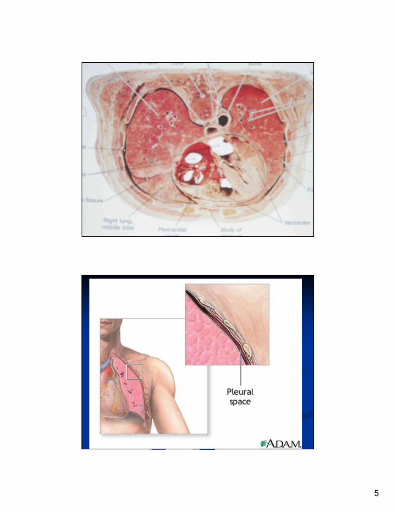
ANATOMY OF THE PLEURAANATOMY OF THE PLEURAI. I. Pleura is the serous membrane:Pleura is the serous membrane:
1. Visceral pleura: covers the lung parenchyma, until 1. Visceral pleura: covers the lung parenchyma, until
interlobarinterlobar fissures fissures
2. Parietal pleura: covers the 2. Parietal pleura: covers the mediastinummediastinum, ,
diaphragm and the rib cage. diaphragm and the rib cage.
The space between the two layers of pleura call as The space between the two layers of pleura call as
pleural space.pleural space.
II. Pleural space contain a film of fluid: pleural fluid, as II. Pleural space contain a film of fluid: pleural fluid, as
lubricant and allows the sliding between the two lubricant and allows the sliding between the two pleuraspleuras
during respiratory movements.during respiratory movements. No air in the pleural No air in the pleural
space and no communication between right and left space and no communication between right and left
pleural space.pleural space.
2
ANATOMY OF THE PLEURA ANATOMY OF THE PLEURA
(contd)(contd)
III. Histology: covered by a single layer of III. Histology: covered by a single layer of mesothelial cells. Within the pleura are blood mesothelial cells. Within the pleura are blood vessels, mainly capillaries, lymphatic lacunas vessels, mainly capillaries, lymphatic lacunas (only in the parietal pleura), and connective (only in the parietal pleura), and connective tissue.tissue.
Two important function of the connective Two important function of the connective tissue in the visceral pleura:tissue in the visceral pleura:
-- contributes to the elastic recoil of the lungcontributes to the elastic recoil of the lung
-- restricts the volume to which the lung can restricts the volume to which the lung can
be inflatedbe inflated
ANATOMY OF THE PLEURA ANATOMY OF THE PLEURA
(contd)(contd)
Elastic and collagen fibers are Elastic and collagen fibers are interdependent elements.interdependent elements.
The mesothelial cells are active cells, The mesothelial cells are active cells, sensitive and responsive to various stimuli sensitive and responsive to various stimuli and very fragile. They may be and very fragile. They may be transformed into macrophage.transformed into macrophage.
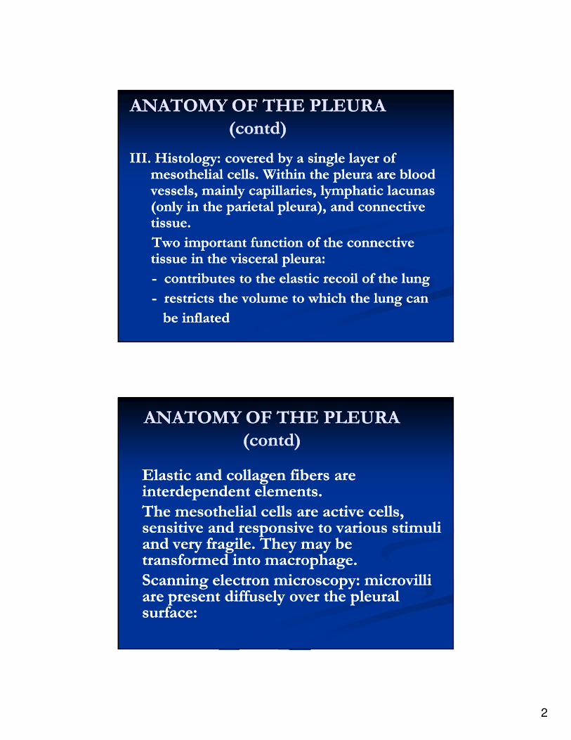
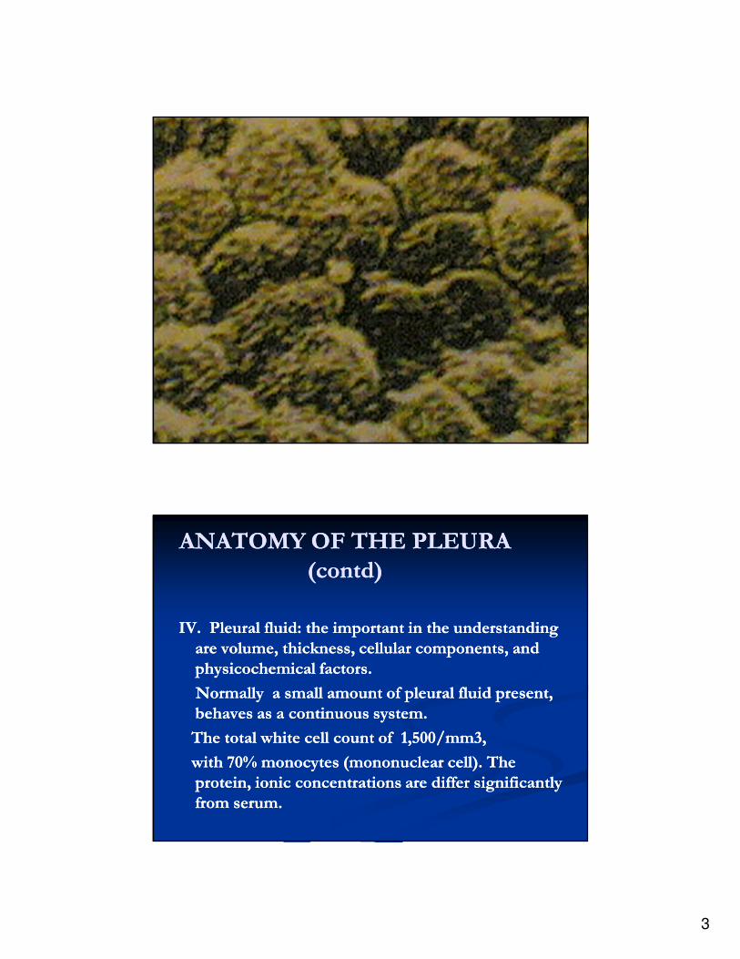
Scanning electron microscopy: microvilli Scanning electron microscopy: microvilli are present diffusely over the pleural are present diffusely over the pleural surface:surface:
3
ANATOMY OF THE PLEURA ANATOMY OF THE PLEURA
(contd)(contd)
IV. Pleural fluid: the important in the understanding IV. Pleural fluid: the important in the understanding
are volume, thickness, cellular components, and are volume, thickness, cellular components, and
physicochemical factors.physicochemical factors.
Normally a small amount of pleural fluid present, Normally a small amount of pleural fluid present,
behaves as a continuous system.behaves as a continuous system.
The total white cell count of 1,500/mm3,The total white cell count of 1,500/mm3,
with 70% with 70% monocytesmonocytes (mononuclear cell)(mononuclear cell). The . The
protein, ionic concentrations are differ significantly protein, ionic concentrations are differ significantly
from serum.from serum.
4
ANATOMY OF THE PLEURA ANATOMY OF THE PLEURA
(contd)(contd)
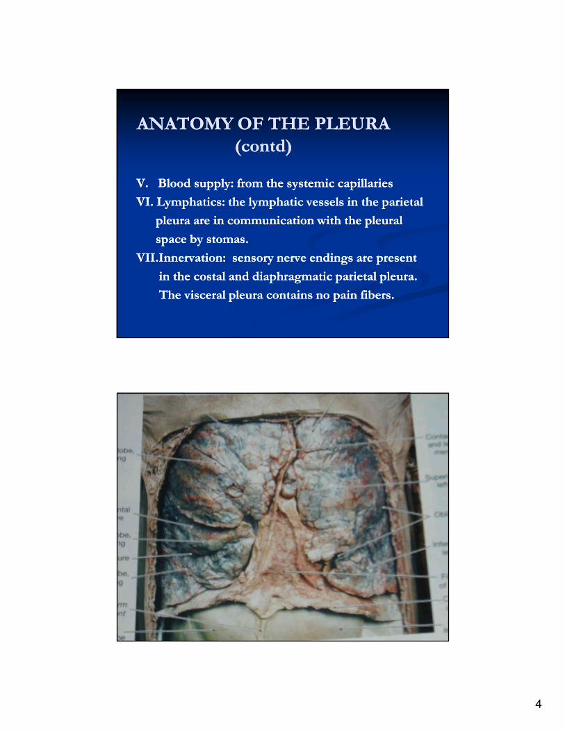
V. V. Blood supply: from the systemic capillaries Blood supply: from the systemic capillaries
VI. VI. LymphaticsLymphatics: the lymphatic vessels in the parietal : the lymphatic vessels in the parietal
pleura are in communication with the pleural pleura are in communication with the pleural
space by stomas.space by stomas.
VII.InnervationVII.Innervation: sensory nerve endings are present : sensory nerve endings are present
in the costal and diaphragmatic parietal pleura. in the costal and diaphragmatic parietal pleura.
The visceral pleura contains no pain fibers.The visceral pleura contains no pain fibers.
5
6
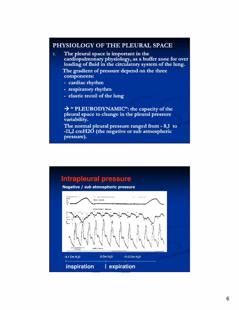
PHYSIOLOGY OF THE PLEURAL SPACEPHYSIOLOGY OF THE PLEURAL SPACE
I.I. The pleural space is important in the The pleural space is important in the cardiopulmonary physiology, as a buffer zone for over cardiopulmonary physiology, as a buffer zone for over loading of fluid in the circulatory system of the lung. loading of fluid in the circulatory system of the lung.
The gradient of pressure depend on the three The gradient of pressure depend on the three components:components:
-- cardiac rhythmcardiac rhythm
-- respiratory rhythmrespiratory rhythm
-- elastic recoil of the lungelastic recoil of the lung
�������� “ PLEURODYNAMIC”: the capacity of the “ PLEURODYNAMIC”: the capacity of the pleural space to change in the pleural pressure pleural space to change in the pleural pressure variability.variability.
The normal pleural pressure ranged from The normal pleural pressure ranged from -- 8,1 to 8,1 to --11,2 cmH2O (the negative or sub atmospheric 11,2 cmH2O (the negative or sub atmospheric pressure).pressure).
Intrapleural pressure
- 8,1 Cm H2O 0 Cm H2O
inspiration expiration
Negative / sub atmospheric pressure
-11,2 Cm H2O
7
PHYSIOLOGY OF THE PLEURAL SPACE PHYSIOLOGY OF THE PLEURAL SPACE
(contd)(contd)
�� The pleural pressure changes associated with many The pleural pressure changes associated with many pleural diseases. Commonly by the increasing of pleural diseases. Commonly by the increasing of pleural pressure.pleural pressure.
�� Pleural fluid formation from:Pleural fluid formation from:
-- pleural capillariespleural capillaries
-- interstitial spaces of the lunginterstitial spaces of the lung
-- intrathoracic lymphaticintrathoracic lymphatic
-- intrathoracic blood vesselsintrathoracic blood vessels
-- peritoneal cavityperitoneal cavity
PHYSIOLOGY OF THE PLEURAL SPACE PHYSIOLOGY OF THE PLEURAL SPACE
(contd)(contd)
�� Pleural fluid absorption: Pleural fluid absorption:
-- Lymphatic clearance: fluid clearance throughLymphatic clearance: fluid clearance through
the pleural lymphatics is though to explain the the pleural lymphatics is though to explain the
lack of fluid accumulation normally.lack of fluid accumulation normally.
Stomas in the parietal pleura, as an initialStomas in the parietal pleura, as an initial
drainage. There are no stomas in the visceral drainage. There are no stomas in the visceral
pleura.pleura.
-- Capillaries clearance: few for small molecules andCapillaries clearance: few for small molecules and
water across both pleural surfaces.water across both pleural surfaces.
8
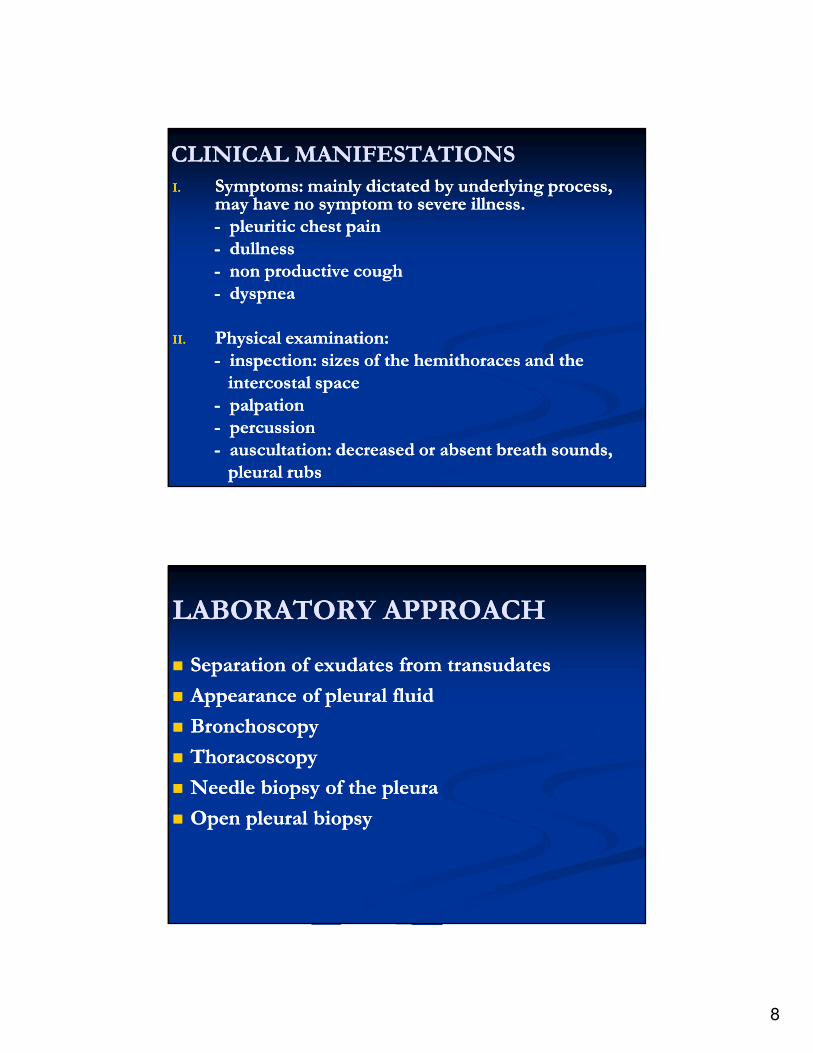
CLINICAL MANIFESTATIONSCLINICAL MANIFESTATIONS
I.I. Symptoms: mainly dictated by underlying process, Symptoms: mainly dictated by underlying process, may have no symptom to severe illness.may have no symptom to severe illness.
-- pleuritic chest painpleuritic chest pain
-- dullnessdullness
-- non productive coughnon productive cough
-- dyspneadyspnea
II.II. Physical examination: Physical examination:
-- inspection: sizes of the hemithoraces and the inspection: sizes of the hemithoraces and the
intercostal spaceintercostal space
-- palpationpalpation
-- percussionpercussion
-- auscultation: decreased or absent breath sounds,auscultation: decreased or absent breath sounds,
pleural rubspleural rubs
LABORATORY APPROACHLABORATORY APPROACH
�� Separation of exudates from transudates Separation of exudates from transudates
�� Appearance of pleural fluidAppearance of pleural fluid
�� BronchoscopyBronchoscopy
�� ThoracoscopyThoracoscopy
�� Needle biopsy of the pleuraNeedle biopsy of the pleura
�� Open pleural biopsyOpen pleural biopsy
9
PLEURAL DISEASESPLEURAL DISEASES
�� Pleural effusionPleural effusion
�� PneumothoraxPneumothorax
�� EmpyemaEmpyema
�� HydropneumothoraxHydropneumothorax
�� PyopneumothoraxPyopneumothorax
�� HemothoraxHemothorax
�� ChylothoraxChylothorax
�� MesotheliomaMesothelioma
�� EtcEtc
PLEURAL EFFUSIONPLEURAL EFFUSION
�� Definition: an accumulation of pleural fluid Definition: an accumulation of pleural fluid
in the pleural space.in the pleural space.
�� Pathogenesis:Pathogenesis:
= Increased pleural fluid formation= Increased pleural fluid formation
= Decreased pleural fluid absorption= Decreased pleural fluid absorption
= Both increased formation and decreased= Both increased formation and decreased
absorptionabsorption
10
PLEURAL EFFUSION (contd)PLEURAL EFFUSION (contd)
�� Increased pleural fluid formation:Increased pleural fluid formation:
-- increased interstitial fluid in the lungincreased interstitial fluid in the lung
-- increased intravascular pressure in pleuraincreased intravascular pressure in pleura
-- increased permeability of the capillaries inincreased permeability of the capillaries in
the pleurathe pleura
-- decreased pleural pressuredecreased pleural pressure
-- increased fluid in the peritoneal cavityincreased fluid in the peritoneal cavity
-- disruption of the thoracic ductdisruption of the thoracic duct
-- disruption of the blood vessel in the thorax disruption of the blood vessel in the thorax
PLEURAL EFFUSION (contd)PLEURAL EFFUSION (contd)
�� Decreased pleural fluid absorption:Decreased pleural fluid absorption:
-- obstruction of the lymphatics drainingobstruction of the lymphatics draining
-- elevation of systemic vascular pressureelevation of systemic vascular pressure
11
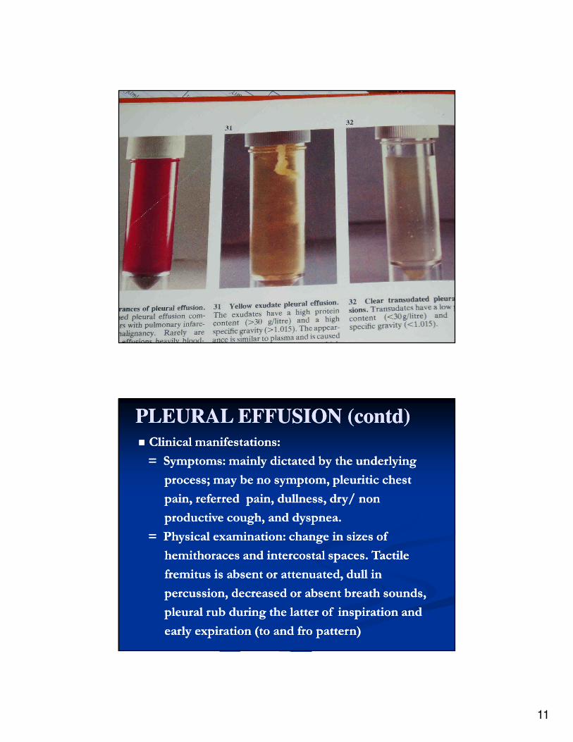
�� Clinical manifestations:Clinical manifestations:
= Symptoms: mainly dictated by the underlying = Symptoms: mainly dictated by the underlying
process; process; may be may be no symptom, no symptom, pleuriticpleuritic chest chest
pain, referred pain, dullness, dry/ non pain, referred pain, dullness, dry/ non
productive cough, and productive cough, and dyspneadyspnea..
= Physical examination: change in sizes of= Physical examination: change in sizes of
hemithoraceshemithoraces and and intercostalintercostal spaces. Tactilespaces. Tactile
fremitusfremitus is absent or attenuated, dull inis absent or attenuated, dull in
percussion, decreased or absent breath sounds,percussion, decreased or absent breath sounds,
pleural rub during the latter of inspiration andpleural rub during the latter of inspiration and
early expiration (to and fro pattern) early expiration (to and fro pattern)
PLEURAL EFFUSION (contd)PLEURAL EFFUSION (contd)
12
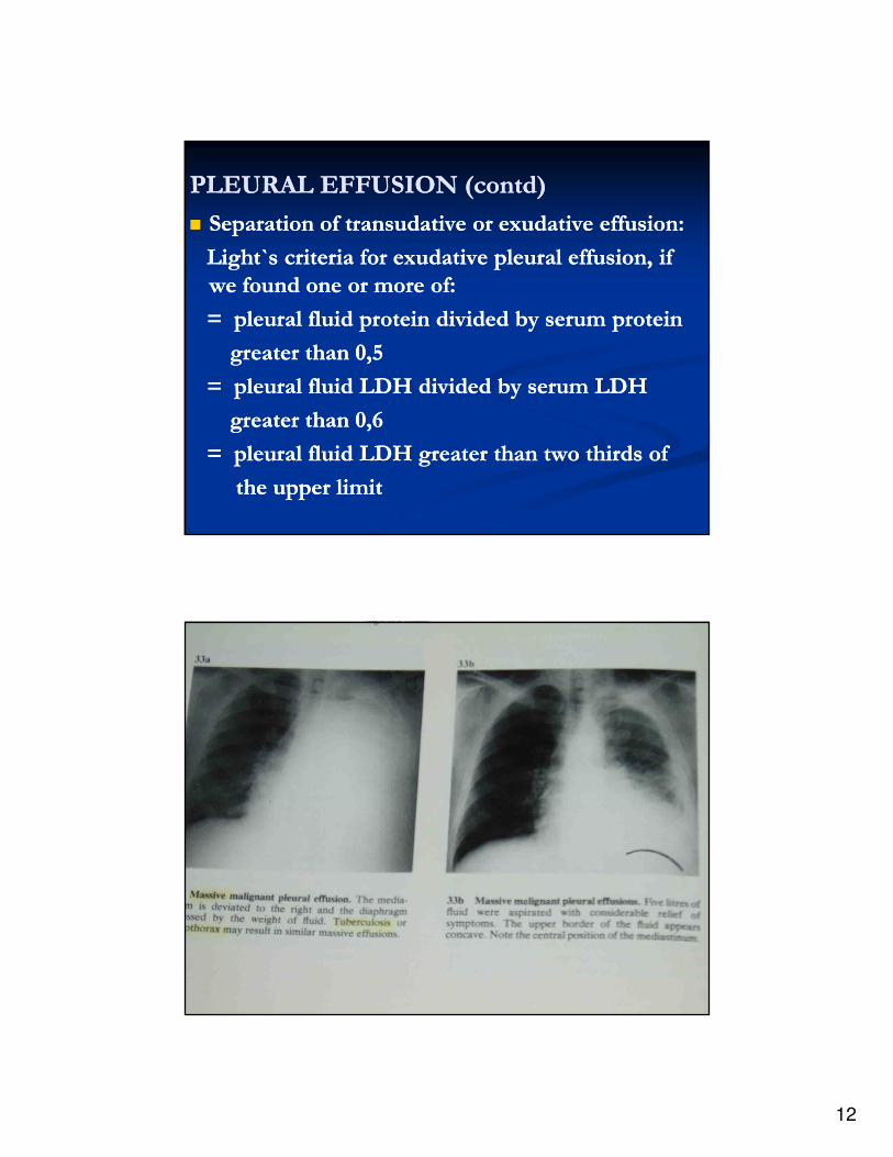
PLEURAL EFFUSION (contd)PLEURAL EFFUSION (contd)
�� Separation of Separation of transudativetransudative or or exudativeexudative effusion:effusion:
Light`s criteria for Light`s criteria for exudativeexudative pleural effusion, pleural effusion, if if
we found we found oneone or more or more of:of:
= pleural fluid protein divided by serum protein = pleural fluid protein divided by serum protein
greater than 0,5greater than 0,5
= pleural fluid LDH divided by serum LDH = pleural fluid LDH divided by serum LDH
greater than 0,6greater than 0,6
= pleural fluid LDH greater than two thirds of = pleural fluid LDH greater than two thirds of
the upper limitthe upper limit
13
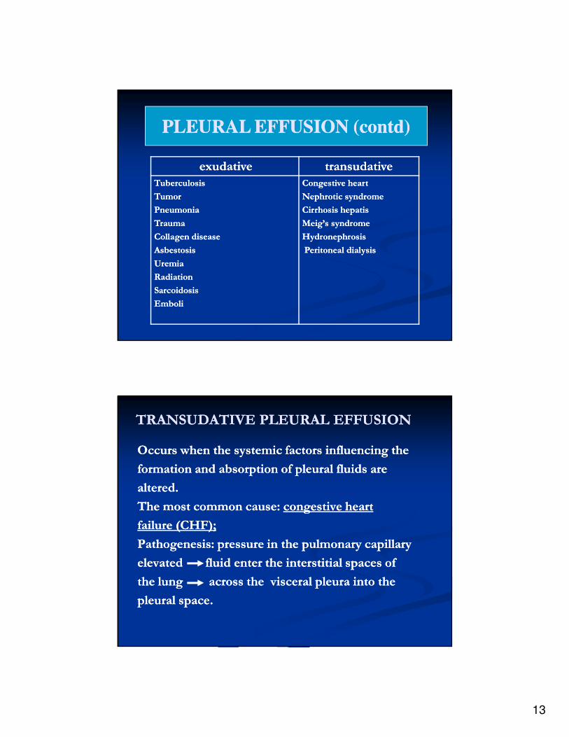
exudativeexudative transudativetransudative
TuberculosisTuberculosis
Tumor Tumor
Pneumonia Pneumonia
Trauma Trauma
Collagen disease Collagen disease
Asbestosis Asbestosis
Uremia Uremia
RadiationRadiation
Sarcoidosis Sarcoidosis
EmboliEmboli
Congestive heartCongestive heart
Nephrotic syndrome Nephrotic syndrome
Cirrhosis hepatis Cirrhosis hepatis
Meig’s syndrome Meig’s syndrome
Hydronephrosis Hydronephrosis
Peritoneal dialysis Peritoneal dialysis
PLEURAL EFFUSION (contd)PLEURAL EFFUSION (contd)
TRANSUDATIVE PLEURAL EFFUSIONTRANSUDATIVE PLEURAL EFFUSION
Occurs when the systemic factors influencing theOccurs when the systemic factors influencing the
formation and absorption of pleural fluids areformation and absorption of pleural fluids are
aalteredltered..
The most common cause: The most common cause: congestive heart congestive heart
failure (CHF);failure (CHF);
Pathogenesis: pressure in the pulmonary capillaryPathogenesis: pressure in the pulmonary capillary
eelevatedlevated fluid enter the interstitial spaces of fluid enter the interstitial spaces of
the lung across the visceral pleura into the the lung across the visceral pleura into the
pleural space.pleural space.
14
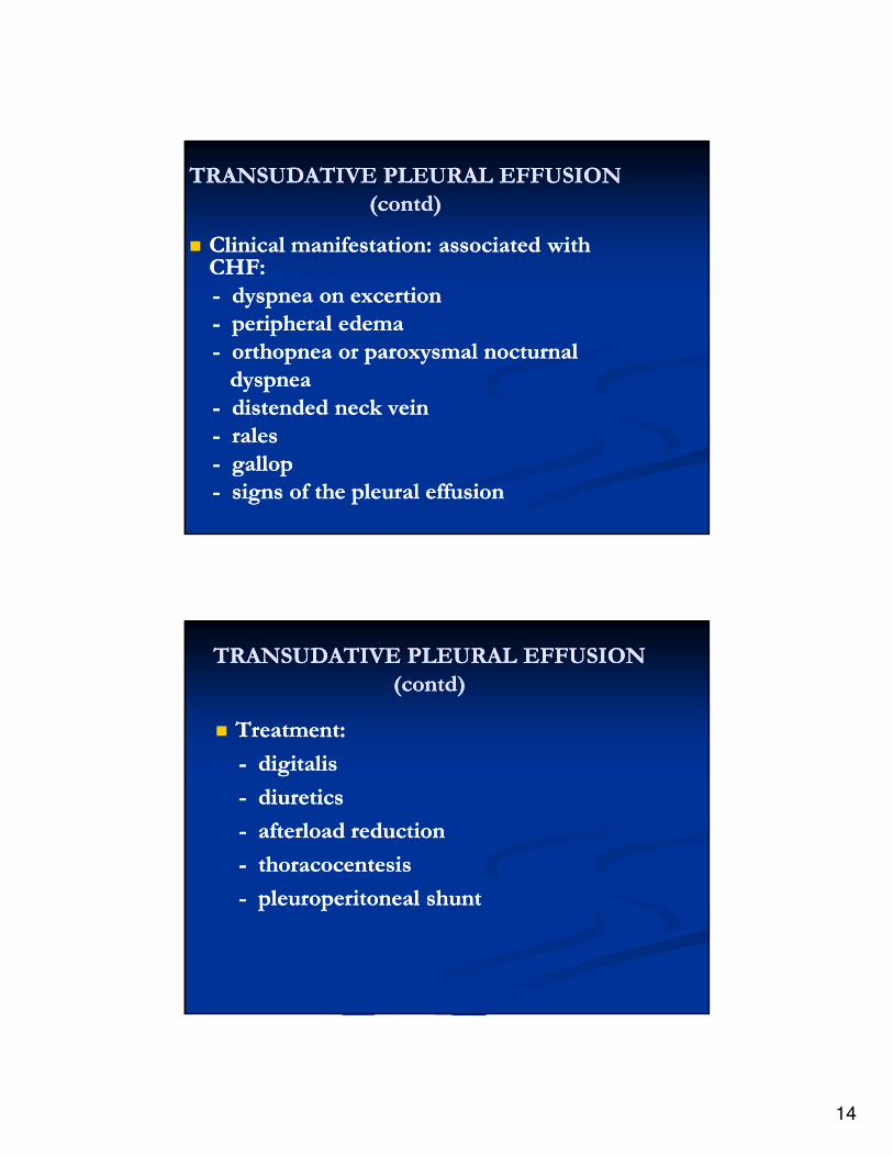
TRANSUDATIVE PLEURAL EFFUSION TRANSUDATIVE PLEURAL EFFUSION
(contd)(contd)
�� Clinical manifestation: associated with Clinical manifestation: associated with CHF:CHF:
-- dyspnea on excertiondyspnea on excertion
-- peripheral edemaperipheral edema
-- orthopnea or paroxysmal nocturnal orthopnea or paroxysmal nocturnal
dyspneadyspnea
-- distended neck veindistended neck vein
-- ralesrales
-- gallopgallop
-- signs of the pleural effusionsigns of the pleural effusion
TRANSUDATIVE PLEURAL EFFUSION TRANSUDATIVE PLEURAL EFFUSION
(contd)(contd)
�� Treatment:Treatment:
-- digitalisdigitalis
-- diureticsdiuretics
-- afterload reductionafterload reduction
-- thoracocentesisthoracocentesis
-- pleuroperitoneal shuntpleuroperitoneal shunt
15
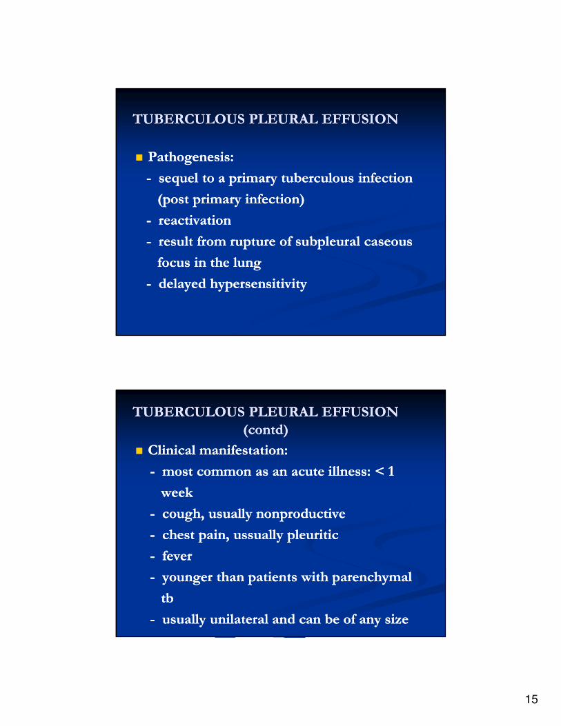
TUBERCULOUS PLEURAL EFFUSIONTUBERCULOUS PLEURAL EFFUSION
�� Pathogenesis:Pathogenesis:
-- sequel to a primary tuberculous infectionsequel to a primary tuberculous infection
(post primary infection)(post primary infection)
-- reactivationreactivation
-- result from rupture of subpleural caseous result from rupture of subpleural caseous
focus in the lungfocus in the lung
-- delayed hypersensitivitydelayed hypersensitivity
TUBERCULOUS PLEURAL EFFUSION TUBERCULOUS PLEURAL EFFUSION
(contd)(contd)
�� Clinical manifestation:Clinical manifestation:
-- most common as an acute illness: < 1most common as an acute illness: < 1
weekweek
-- cough, usually nonproductivecough, usually nonproductive
-- chest pain, chest pain, ussuallyussually pleuriticpleuritic
-- feverfever
-- younger than patients with younger than patients with parenchymalparenchymal
tbtb
-- usually unilateral and can be of any sizeusually unilateral and can be of any size
16
TUBERCULOUS PLEURAL EFFUSION TUBERCULOUS PLEURAL EFFUSION
(contd)(contd)
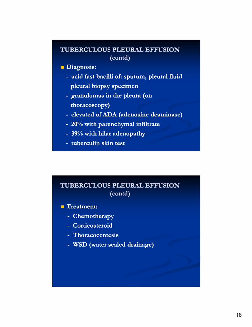
�� Diagnosis:Diagnosis:
-- acid fast bacilli of: sputum, pleural fluidacid fast bacilli of: sputum, pleural fluid
pleural biopsy specimenpleural biopsy specimen
-- granulomas in the pleura (on granulomas in the pleura (on
thoracoscopy)thoracoscopy)
-- elevated of ADA (adenosine deaminase) elevated of ADA (adenosine deaminase)
-- 20% with parenchymal infiltrate20% with parenchymal infiltrate
-- 39% with hilar adenopathy39% with hilar adenopathy
-- tuberculin skin testtuberculin skin test
TUBERCULOUS PLEURAL EFFUSION TUBERCULOUS PLEURAL EFFUSION
(contd)(contd)
�� Treatment:Treatment:
-- ChemotherapyChemotherapy
-- CorticosteroidCorticosteroid
-- ThoracocentesisThoracocentesis
-- WSDWSD (water sealed drainage)(water sealed drainage)
17
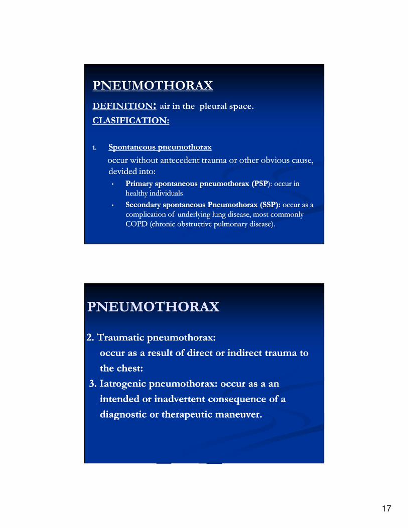
PNEUMOTHORAX
DEFINITION: air in the pleural space.
CLASIFICATION: CLASIFICATION:
1.1. Spontaneous Spontaneous pneumothoraxpneumothorax
occur without antecedent trauma or other obvious cause, occur without antecedent trauma or other obvious cause,
devideddevided into:into:
•• Primary spontaneous Primary spontaneous pneumothoraxpneumothorax (PSP(PSP): occur in ): occur in
healthy individualshealthy individuals
•• Secondary spontaneous Secondary spontaneous PneumothoraxPneumothorax (SSP):(SSP): occur as a occur as a
complication of underlying lung disease, most commonly complication of underlying lung disease, most commonly
COPD (chronic obstructive pulmonary disease).COPD (chronic obstructive pulmonary disease).
PNEUMOTHORAXPNEUMOTHORAX
2. Traumatic pneumothorax:2. Traumatic pneumothorax:
occur as a result of direct or indirect trauma tooccur as a result of direct or indirect trauma to
the chest:the chest:
3. Iatrogenic pneumothorax: occur as a an 3. Iatrogenic pneumothorax: occur as a an
intended or inadvertent consequence of aintended or inadvertent consequence of a
diagnostic or therapeutic maneuver. diagnostic or therapeutic maneuver.
18
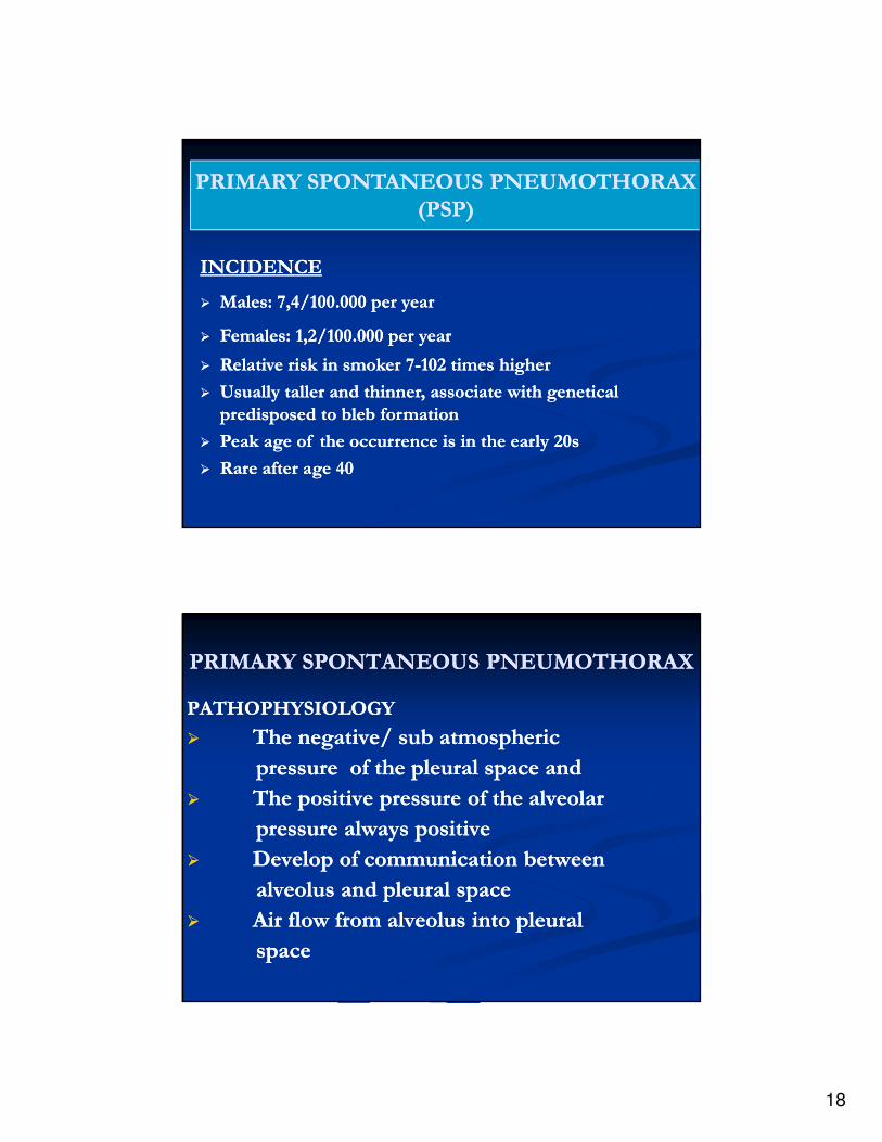
INCIDENCE INCIDENCE
�� Males: 7,4/100.000 per yearMales: 7,4/100.000 per year
�� Females: 1,2/100.000 per year Females: 1,2/100.000 per year
�� Relative risk in smoker 7Relative risk in smoker 7--102 times higher102 times higher
�� Usually taller and thinner, associate with genetical Usually taller and thinner, associate with genetical
predisposed to bleb formationpredisposed to bleb formation
�� Peak age of the occurrence is in the early 20sPeak age of the occurrence is in the early 20s
�� Rare after age 40Rare after age 40
PRIMARY SPONTANEOUS PNEUMOTHORAXPRIMARY SPONTANEOUS PNEUMOTHORAX
(PSP)(PSP)
PRIMARY SPONTANEOUS PNEUMOTHORAXPRIMARY SPONTANEOUS PNEUMOTHORAX
PATHOPHYSIOLOGYPATHOPHYSIOLOGY
�� The negative/ sub atmosphericThe negative/ sub atmospheric
pressure of the pleural space andpressure of the pleural space and
�� The positive pressure of the alveolar The positive pressure of the alveolar
pressure always positivepressure always positive
�� Develop of communication between Develop of communication between
alveolus and pleural spacealveolus and pleural space
�� Air flow from alveolus into pleuralAir flow from alveolus into pleural
spacespace
19
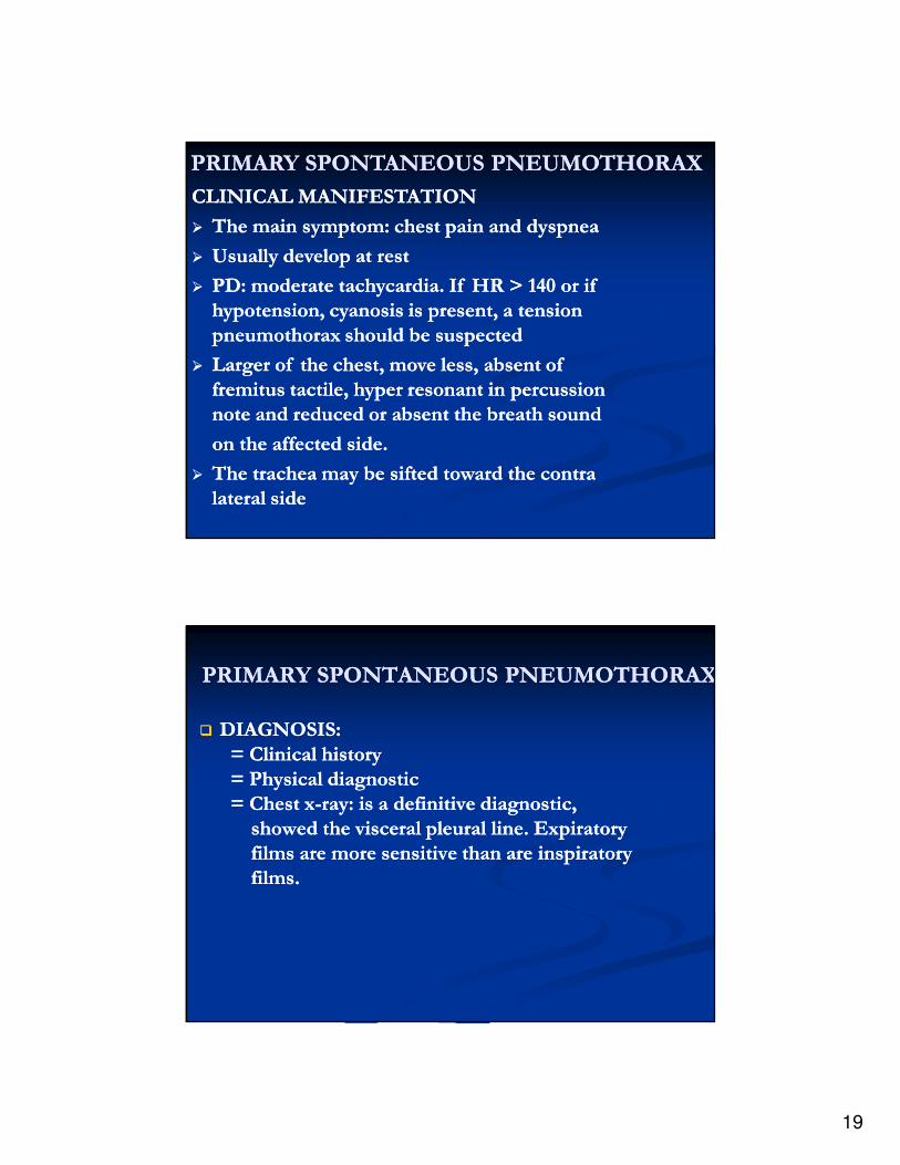
CLINICAL MANIFESTATIONCLINICAL MANIFESTATION
�� The main symptom: chest pain and dyspneaThe main symptom: chest pain and dyspnea
�� Usually develop at restUsually develop at rest
�� PD: moderate tachycardia. If HR > 140 or if PD: moderate tachycardia. If HR > 140 or if
hypotension, cyanosis is present, a tension hypotension, cyanosis is present, a tension
pneumothorax should be suspectedpneumothorax should be suspected
�� Larger of the chest, move less, absent of Larger of the chest, move less, absent of
fremitus tactile, hyper resonant in percussion fremitus tactile, hyper resonant in percussion
note and reduced or absent the breath soundnote and reduced or absent the breath sound
on the affected side.on the affected side.
�� The trachea may be sifted toward the contra The trachea may be sifted toward the contra
lateral sidelateral side
PRIMARY SPONTANEOUS PNEUMOTHORAXPRIMARY SPONTANEOUS PNEUMOTHORAX
PRIMARY SPONTANEOUS PNEUMOTHORAXPRIMARY SPONTANEOUS PNEUMOTHORAX
�� DIAGNOSIS:DIAGNOSIS:
= Clinical history = Clinical history
= Physical diagnostic= Physical diagnostic
= Chest x= Chest x--ray: is a definitive diagnostic, ray: is a definitive diagnostic,
showed the visceral pleural line. Expiratoryshowed the visceral pleural line. Expiratory
films are more sensitive than are inspiratoryfilms are more sensitive than are inspiratory
films.films.
20
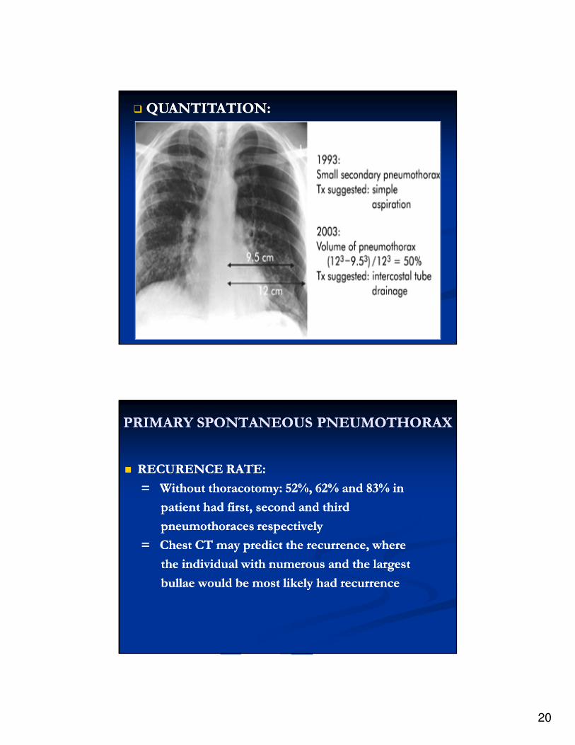
� QUANTITATION:QUANTITATION:
PRIMARY SPONTANEOUS PNEUMOTHORAXPRIMARY SPONTANEOUS PNEUMOTHORAX
�� RECURENCE RATE:RECURENCE RATE:
= Without thoracotomy: 52%, 62% and 83% in = Without thoracotomy: 52%, 62% and 83% in
patient had first, second and third patient had first, second and third
pneumothoraces respectivelypneumothoraces respectively
= Chest CT may predict the recurrence, where= Chest CT may predict the recurrence, where
the individual with numerous and the largestthe individual with numerous and the largest
bullae would be most likely had recurrencebullae would be most likely had recurrence

21
TREATMENTTREATMENT
A. ObservationA. Observation
= = Resorbed of the air in the pleural space about Resorbed of the air in the pleural space about
1,25% per day, if the communication between 1,25% per day, if the communication between
the alveoli and pleural space is eliminatethe alveoli and pleural space is eliminate
= Bed rest= Bed rest
B. Supplemental OxygenB. Supplemental Oxygen
= = Supplemental oxygen: increased the rate of air Supplemental oxygen: increased the rate of air
absorption until 6 timeabsorption until 6 time
= As a routine treatment for all type of= As a routine treatment for all type of
pneumothoraxpneumothorax
TREATMENTTREATMENTC. AspirationC. Aspiration
= As a initial treatment for psp > 15%= As a initial treatment for psp > 15%
= By G= By G--16 needle with internal 16 needle with internal
polyethylene catheter, inserted into polyethylene catheter, inserted into
anterior 2anterior 2ndnd ICS at mid clavicle line afterICS at mid clavicle line after
local anesthesia, a three way stopcocklocal anesthesia, a three way stopcock
and 60ml syringeand 60ml syringe
= 64% successful = 64% successful
= Tube thoracostomy for unexpanded lung= Tube thoracostomy for unexpanded lung
22
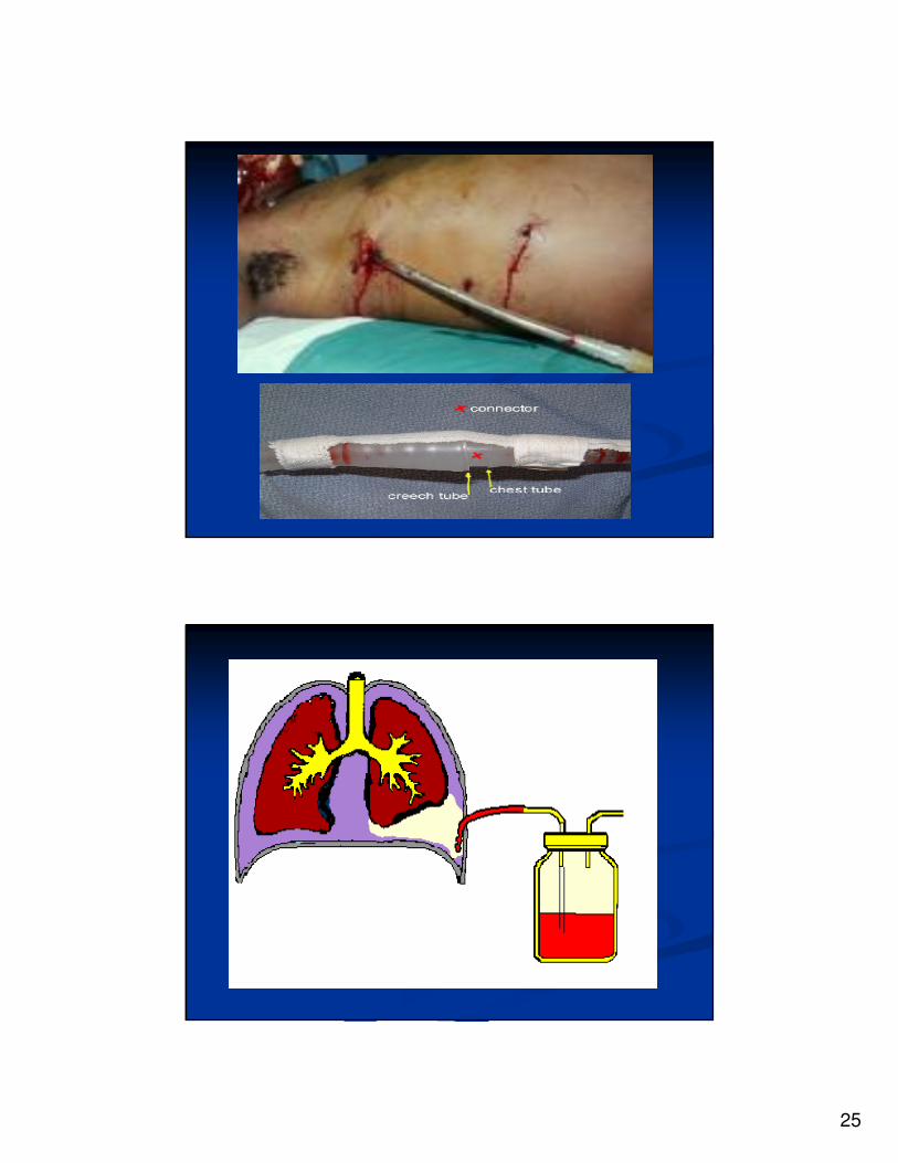
TREATMENTTREATMENTD. Tube D. Tube thoracostomythoracostomy
= Permits the air to be evacuated effectively= Permits the air to be evacuated effectively
and rapidlyand rapidly
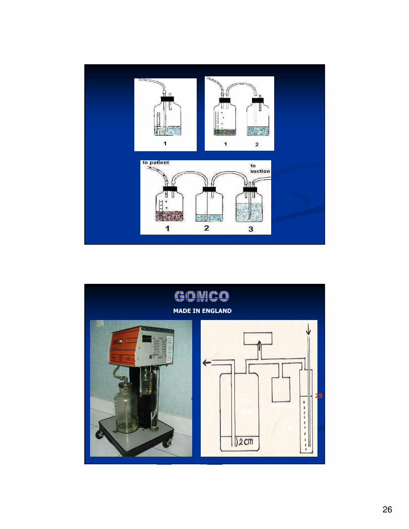
= Connected to underwater seal= Connected to underwater seal (WSD)(WSD), low, low
pressure continuous suction (up topressure continuous suction (up to
100cmH2O), or to a Heimlich valve100cmH2O), or to a Heimlich valve..
E. E. PleurodesisPleurodesis
= = InstilationInstilation of any of any sclerosingsclerosing agent to the pleuralagent to the pleural
space or by abrasion space or by abrasion of the pleuras of the pleuras to create to create
obliteration of theobliteration of the pleural spacepleural space..
TREATMENTTREATMENTF. ThoracoscopyF. Thoracoscopy
= Direct view to the entire thoracic cavity= Direct view to the entire thoracic cavity
= To treat the bullous disease responsible = To treat the bullous disease responsible
for the pneumothoraxfor the pneumothorax
= To create a pleurodesis= To create a pleurodesis
G. ThoracotomyG. Thoracotomy
= For patient who fail to previous= For patient who fail to previous
treatmenttreatment
23
SECONDARY SPONTANEOUS SECONDARY SPONTANEOUS
PNEUMOTHORAX (SSP)PNEUMOTHORAX (SSP)�� SSP are more serious than PSP, because SSP are more serious than PSP, because
decreased the lung function of patient with decreased the lung function of patient with already compromised lung functionalready compromised lung function..
�� INCIDENS: 6,3/100.000/year (US)INCIDENS: 6,3/100.000/year (US)
�� ETIOLOGIC FACTORS:ETIOLOGIC FACTORS:
= COPD= COPD
= TB= TB
= Asthma= Asthma
= Pneumonia= Pneumonia
= Lung cancer= Lung cancer
SECONDARY SPONTANEOUS SECONDARY SPONTANEOUS
PNEUMOTHORAXPNEUMOTHORAX�� CLINICAL MANIFESTATIONS: CLINICAL MANIFESTATIONS:
= More severe than PSP= More severe than PSP
= Mostly: = Mostly: dyspneadyspnea, chest pain, cyanosis,, chest pain, cyanosis,
and hypotensionand hypotension
= Mortality: 16%, is associated with= Mortality: 16%, is associated with
respiratory failurerespiratory failure
= Recurrent rate: 44%= Recurrent rate: 44%
= PD: similar to PSP, but less helpful,= PD: similar to PSP, but less helpful,
especially for patient with COPDespecially for patient with COPD
24
SECONDARY SPONTANEOUS SECONDARY SPONTANEOUS
PNEUMOTHORAXPNEUMOTHORAX
�� DIAGNOSIS: established by chest xDIAGNOSIS: established by chest x--ray, show of a ray, show of a
visceral pleural line. Must differentiation from visceral pleural line. Must differentiation from
large bulla, if any doubt, CT thorax may be done.large bulla, if any doubt, CT thorax may be done.
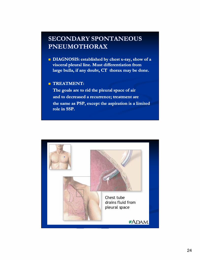
�� TREATMENT:TREATMENT:
The goals are to rid the pleural space of air The goals are to rid the pleural space of air
and to decreased a recurrence; treatment areand to decreased a recurrence; treatment are
the same as PSP, except the aspiration is a limited the same as PSP, except the aspiration is a limited
role in SSP.role in SSP.
25
26
MADE IN ENGLAND
PUMP
CC
WSC
PCC
SAFETY TUBE
25
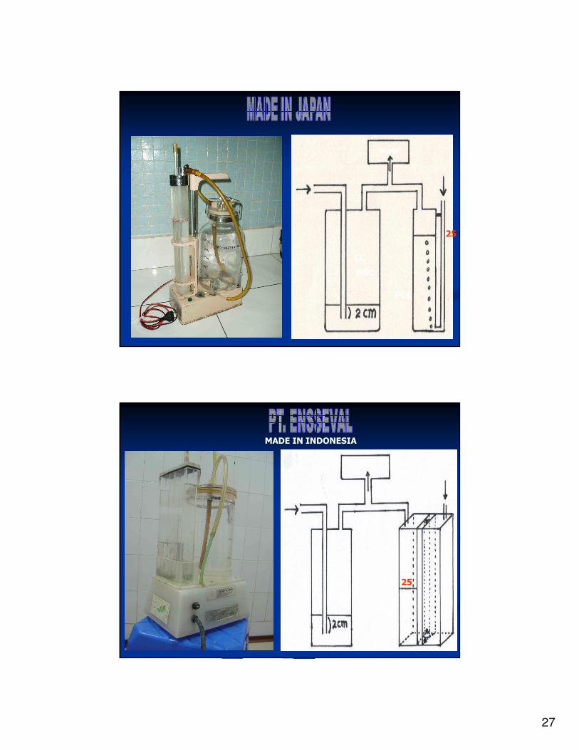
27
PUMP
CC
WSC
PCC
25
MADE IN INDONESIA
PUMP
CC
WSC
PCC
25
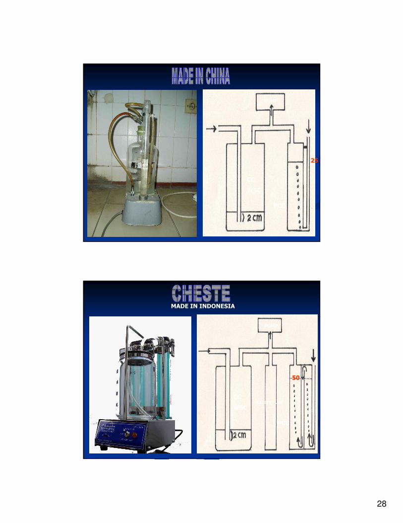
28
PUMP
CC
WSC
PCC
25
MADE IN INDONESIA
PUMP
CC
WSCSAFETY TUBE
50
PCC
29