Embed Size (px)
Citation preview

pleural Fluid Analysis

ll- pleural fluid analysisIt comprises of- pleural fluid appearance - Biochemical tests ( Protein, LDH).- Cytological tests ( malignant cells, differential
cell count)- Microbiological tests ( Gram stain , AFB and
culture).- PH analysis
• Appearance ; - Bloody ( could be due to trauma, malignancy.
PE and Mesothelioma ) - Turbid or milky ( could be due to chylothorax
or empyema) - putrid odour ( anaerobic empyema)- Food Particle ( oesophageal rupture)

• Differential cell count - Predominant Neutrophils ( could be due to
para pneumonic effusion or PE ).- Predominant Mononeuclear cell ( could be
due to TB or malignancy)- Predominant Lymphocyte, TB could be the
possible cause if > 80% of the cells are lymphocytes. Other possible causes are malignancy , RA, or post CABG.
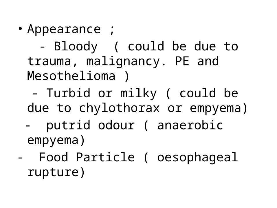
- Predominant Eosinophils, ( could be due to fungal and parasitic infection or PE and medications).

• pleural fluid PH and GlucoseThe pleural fluid PH should be appropriately
heparinised for processing, normal pleural PH is about 7.6. low pleural PH is often associate with a low pleural fluid glucose < 60mg/dl.
Causes of pleural PH and low glucose are;1- para pneumonic effusion2- RA3- malignant effusion4- TB effusion5- oesophageal rupture

• pleural fluid Triglyceride and Cholesterol , usually measured when there is chylothorax suspected,
Causes of chylothorax, ( malignancy such us lymphoma, Trauma, and following thoracotomy).

• pleural fluid amylase, abnormal if more than the upper limit of serum amylase,
Causes are1- oesophageal rupture or pleural malignancy
when salivary amylase will be raised.2- pancreatic disease

• Some times it is difficult to differentiate between exudative and transudative, in that case we consider Light’s criteria;
pleural fluid is exudative if one of the following criteria found,
- pleural fluid Protein/ serum protein >0.5- pleural fluid LDH / serum LDH > 0.6- pleural fluid LDH > 2/3 of the upper limit of
normal serum LDH.

lll –pleural biopsy especially in cases of TB or malignancy.
lV- Thoracoscopy and biopsy
V- Bronchoscopy , rarely used

• Management of pleural effusion1- treat underline causes ( Pneumonia, PE, LVF)2- therapeutic aspiration may be required to
palliate breathlessness, removing of > 1.5 L of pleural fluid in one episode is not advisable as this will increase the risk of re-expanding pulmonary oedema.
3- Chest drain and pleurodesis especially in patients with recurrent effusion or malignant effusion.
Empyema
• It describes presence of pus in the pleural space, the pus may be thin as serous fluid or so thick that is impossible to aspirate. The possible causative organism may or may not be isolated from the pus . the empyema may involve the whole pleural space or only part of it ( Loculated ) and it is almost invariably unilateral.

• Aetiology;• Is always secondary to infection in the
neighbouring structure with the lungs, the commonest causes are;
1- community acquired pneumonia CAP. 40% of bacteria pneumoia may complicate by pleural effusion and empayema develops in 15% of them.
2- TB 3- Infection of haemothorax4- rupture of sub- phrenic abscess through
diaphragm.

• Clinical picture- Systemic features like fever , rigor, sweating ,
malaise and weight loss.- Local features, mainly pleuretic chest pain,
SOB, cough and copious sputum if the empyema ruptures in to the bronchus.
- Clinical signs are those of pleural effusion.

• Investigations;- CXR, the radiological appearance is similar to those
with pleural effusion, when air present in addition to pus that is called pyopneumothorax.
- USS of chest cavity, shows the position of the fluid , the extend of plural thickness, and presence of loculations.
- CT chest , in addition to above, it will help to assess lung parenchyma.
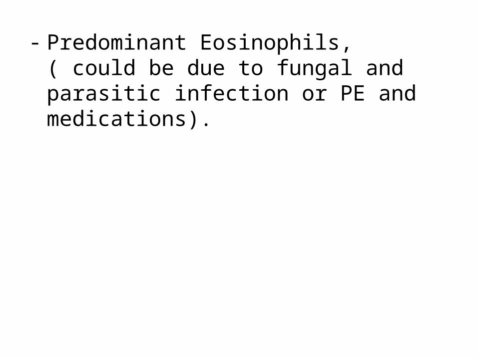
- Aspiration of pus will be diagnostic .

• Management of Empyema;Despite wide spread availability of Antibiotic ,
empyema continues to have significant morbidity and mortality even in developed countries.
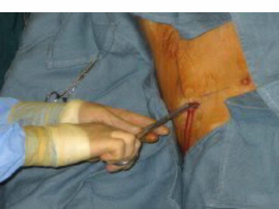
l- Treatment of non tuberculous empyema;All acutely ill patient should have intercostal
chest tube insertion under Uss guided and connect it to a water seal drain system.
If the aspiration is turbid or frank pus or if loculated effusion the chest tube should be put on suction ( 5-10 cm ) and flushed regularly with 20mls of normal saline.
If above failed, surgical intervention required either by cleaning the empyema cavity of pus and adhesions with insertion of wide bore tube to allow optimal drainage,
In addition to that all patients required combined antibiotic therapy for long period.
II- treatment of TB empyemaTB chemotherapy must be started immediately
and the pus should be drained frequently through inter costal chest drains.
Occasionally surgical intervention required.