Embed Size (px)
Citation preview
Thorax (1949), 4, 3 1.
PNEUMOCONIOSIS DUE TO GRAPHITE DUSTBY
S. ROODHOUSE GLOYNE, GEOFFREY MARSHALL, AND CLIFFORD HOYLEFrom the Brompton Hospital, the London Chest Hospital, and the Institute of Social Medicine, Oxford
Lassar Dunner (1945a) recorded the clinical andradiological findings in five men who had beenexposed to " pure graphite " dust. They had beenexposed for 17, 18, 20, 20, and 34 years respec-tively, and no other dusts were known to have beenimplicated. Three of the men had cough, sputum,and dyspnoea; the other two had no complaints.Radiographs showed reticulation with nodulation,and the question was raised in correspondence(Dunner, 1945b) whether there was a tissue reactionconsistent with pneumoconiosis. Dunner believedthat there was, and in later publications (Dunnerand Bagnall, 1946; Dunner, 1948) described aform with a " snowstorm " appearance whichbecame nodulated and in one case formed acavity. It is doubtful, however, if such a thingas " pure graphite " exists commercially (except formanufactured varieties like " kish ") because, asexplained by Dassanayake (1948), graphite is minedfrom vertical lodes in granite rock, and the minersdevelop similar lung changes to those found inmetalliferous miners.The two fatalities here recorded followed long
exposure to a mixed dust of graphite, free silica,various silicates, and possibly other substances usedin the manufacture of crucibles. The hazard wasnot strictly comparable with Dunner's cases, there-fore, but was more like that of coal-miners,judging from King and Nagelschmidt's (1945)analyses of dusts extracted from the dried lungsof South Wales coal-miners. There they foundboth free silica (quartz) and silicates (mica andkaolin) mixed with the coal. In our cases coal wasreplaced by graphite.
CASE REPORTSCASE I.-J. F., a man aged 59 years, had been
employed by the same firm for 37 years. He wasleader of a gang unloading raw material for makingcrucibles from barges at the company's wharf. Thiswork was done partly out of doors and partly in thefactory, where dust exhaustion plant was used. Thematerial unloaded consisted chiefly of graphite, butsand and probably other substances were handled fromtime to time, as the formula of the crucible "mix"
was complicated and included various silicates in addi-tion to graphite and sand. The graphite was receivedfrom Ceylon and Madagascar. Latterly the sand hadbeen delivered in bags.
This man first attended the Brompton Hospital in1939, when silicosis was diagnosed. He complainedof shortness of breath for one year, tightness in thechest, a productive cough with much thick sputum,lassitude on slight exertion, and some lower abdomi-nal pain. Apart from influenza in 1937 he had hadno previous illnesses. He weighed 140 lb. (64 kg.),was afebrile, not wasted, and apart from short distantbreath sounds he had no abnormal physical signs.The pulse and respiration rates were normal. Theblood pressure was 150/90 mm. Hg. The blood sedi-*mentation rate was 25 mm. (Westergren) in an hour,the vital capacity 2,400 ml., and sputum repeatedlyfailed to disclose any tubercle bacilli. A radiograph(Plate Ta) showed emphysema at both bases and nodu-lation in the upper and middle zones of both lungswith some massive fibrosis in the upper zone of theright lung. IV the next year he gained half a stonein weight but became increasingly short of breath.For the last eighteen months of his life he was
unfit for regular employment, and in December, 1942,his health rapidly deteriorated. He was admittedto St. James's L.C.C. Hospital, and died the sameday.Autopsy.-The body was thinly covered, and there
was no clubbing of the extremities. The upper respi-ratory and digestive tracts were healthy. Soft, recentlyformed adhesions obliterated the pleural sacsposteriorly and there was old-standing thickening ofthe visceral pleura over the right middle lobe of thelung. The inter-lobar fissures were closed by adhe-sions on this side. Both lungs were markedly pig-mented and emphysematous bullae were present atthe apices and bases. On section copious glisteningblack mucous secretion exuded from the cut ends ofthe bronchi. The bases of the lungs were congestedand there were some small white patches of broncho-pneumonia in these areas. Small black sphericalfibrotic nodules, varying in size up to a third of aninch (0.8 cm.) in diameter, were distributed irregu-larly throughout the lungs, and were especiallyprominent immediately beneath the pleura. In theupper and lower lobes on both sides these noduleshad become confluent to form rounded fibrotic masses1 to 3 in. in diameter. One of these masses in the
on 25 August 2018 by guest. P
rotected by copyright.http://thorax.bm
j.com/
Thorax: first published as 10.1136/thx.4.1.31 on 1 M
arch 1949. Dow
nloaded from

S. ROODHOUSE GLOYNE ANL$ OTHERS
upper part of the right lower lobe contained.a raggedcavity filled with lustrous black mucoid secretion.The mediastinal glands were enlarged, pigmented, andfibrotic. The heart was enlarged and the chambersof the right side dilated, but no valvular lesions werepresent and the coronary arteries were patent andhea:thy. The thoracic aorta showed extensiveatheroma. The liver exhibited signs of congestivefailure and the spleen contained several small infarcts.No significant lesions were noted in other abdominalorgans. The brain and meninges were healthy.
Histology.-The lung wa§ extensively infiltrated withpolygonal scale-like black particles with straight edgesand sharp angles, ranging in size from 20 to 30 ,u intheir longest diameter to small frag rTents of the dimen-sions of pathogenic bacteria. Many of the particleswere collected together in clumps, and the smallerones were frequently phagocyted. A few weresurrounded by golden-yellow crenated envelopes(" graphite bodies ") like the "anthracosis bodies"of coal-miners' lungs. On dark-ground iluminationthe par:icles retained their black appearance andcould only be detected by their bright luminous edges,which had a wavy outline not shown up by trans-mitted light. These were not refractile with crossedNicol prisms, but a few small refractile particles ofquartz (2-3 b') were seen.The lesions in the lung tissue were patchy. In some
parts there was relatively litt e dust, and the picturewas that of a terminal bronchopneumonia with poly-morphonuclear cell reaction. In others there werevarying degrees of graphite infiltra:ion with a colla-genous response. In the least affected of these areasthe lesion was confined to the respiratory bronchioles,which contained free particles of graphite, dust-!adenmacrophages, and cellular debris, and which weregenerally surrounded by peribronchial sheaths ofreticulin fihres and imprisoned graphite particles.Similar sheaths were seen around the vessels. As thelesion extended the neighbouring alveoli became in-vo:ved. They were filled with dust-laden macrophagesand a few free graphite particles. The capillaries inthe walls were obscured by cellular reaction aroundthem, and finally they became compressed and un-recognizable. lnf peripheral portions of the lung the",pleural drift" of dust particles characteristic ofpneumoconiosis was well marked, this accumulationof pigment resulting in collagenous thickening of thesubpleural layer. These perivascular and peribronchialareas of reticulin-staining fibres became confluent inmany places to forn patches of fibrosis with irregu'aroutlines in which all that remained to identify lungstructture was an occasional short strip of low cuboidalepitheliun from a small bronchus or a ring of wavyelastic tissue from an obli'erated vessel. Side by sidewith these patches were whor'ed nodules of silicosis.the fibres of which had in n-any cases become swollenand hyalinized; a few had the irregular margins withtendrils trailing out into lung tissue-as described byBelt and Ferris (1942) in coal-miners' lungs (" Medusahead " or "'mixed" nodules), but this was not a
conspicuous feature. No evidence of tuberculosi wasfound.CASE 2.-J. M. C., a man aged 55 years, had been
employed in the sa.ne factory as the previous patient.Before going to this factory he had been a stoker inthe Royal Navy between the ages of 18 and 23 years;and between the ages of 23 and 27 years had workedas a p asterer's labourer. He had then worked for27 years as a mill hand grinding various rawmaterials used in the manufacture of crucibles. Twoyears before he came under observation he had beengranted a certificate of total disability by the SilicosisBoard.When he first attended the Brompton Hospital, in
March, 1945, hiF symp.oms had already extended overthe last eight years. He complained of increasingshortness of breath on exertion and cough with a littleviscid sputum. The cough had been much worseduring the previous five months. Four years previ-ously he had had dry p'eurisy on the left side.He was an elderly man wi;h considerable dyspnoea
on exertion. There was no clubbing of either fingersor toes. The chest expansion was poor, but otherwisethere were no significant signs of disease in the lungs.His blood pressure was 110/65 mm. Hg. His weightwas 123 lb. (56 kg.). Five months previously he hadbeen 130 lb. (59 kg.), and ten years previously 151 lb.(69 kg.). His blood sedi.nentation rate was 19 mm.in one hour (Westergren), haemoglobin 100 per cent,leucocytes 12,000 per c.mm. of blood, with polymrorphs80 per cent, lymphocytes 12.8 per cent, and eosinophils3.6 per cent. Sputum was 1 to 2 oz. (30 to 60 ml.)daily, mucoid in character, and repeatedly failed toshow tubercle bacilli.A radiograph (Plate Ib) of the chest showed diffuse
nodulation with many massive shadows in both lungs.There was evident gross emphysema.A utopsy.-A post-mortem examination was trade
on- Aug. 21, 1947. The body was much wasted. Noclubbing of the extremities was noted. The upperdigestive and respiratory tracts were healthy. Recentadhesions were present at the bases of both pleural sacs,and there were patches of thickened visceral pleuraover both lungs with some scarring. Numerous smallgrey subpleural nodules of fibrosis were also noted.Both lungs were markedly pigmented and emphysema-tous. On section lustrous black mucinous secre-tion oozed fro n the cut ends of the bronchi. Distri-buted throughout both lungs were numerous small,rough, pigrrented fibrotic nodules with focal emphy-sema around them. In both upper lobes and in theright middle lobe some of these nodules had becomeconfluent to form large masses of fibrosis. and themass in the left upper lobe contained two irregularlyshaped cavities + to I in. (1.2-1.8 cm.) in diameterwith rough ragged wal's and filled with black lustrousmucinous -secretion. The mediastinal glands weremoderately enlarged, p-grented, and fibrotic. Theheart was dilated and the muscle pale and flabby, butthere was no valvular disease. The other organs werenormal.
32
on 25 August 2018 by guest. P
rotected by copyright.http://thorax.bm
j.com/
Thorax: first published as 10.1136/thx.4.1.31 on 1 M
arch 1949. Dow
nloaded from
PNEUMOCONIOSIS DUE TO GRAPHITE DUST
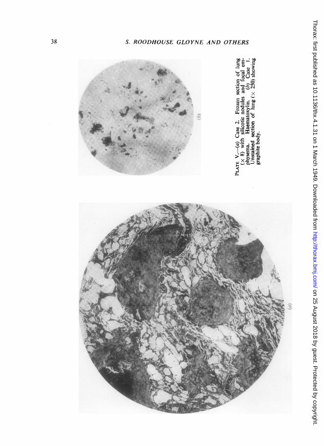
Histology.-As in the previous case the lung wasextensively infiltrated with graphite particles. In un-stained sections they showed as thin plates withgreenish edges, most of them being three- or four-sided figures varying from 10 to 20 ,u in size. Theywere not refractile with crossed Nicol prisms, but afew small refractile particles of quartz were noted.The histological pattern in the lung resembled that
of the previous case but indicated a more advancedstage of fibrosis. The infiltration with graphiteparticles was much more marked, and the confluentareas of perivascular and peribronchial fibrosis wereextensive. Conglomerate nodules of silicosis andmixed nodules were present. No active tuberculosiscould be found, but there were minute areas of calcifi-cation near the cavity in the lung and in one hilargland.
DISCUSSION
Graphite is widely distributed in nature. It isgenerally found with quartz (Milner, 1940) andusually as rounded plates or irregular dull blackgrains recognized chiefly by their appearance inreflected light. According to Strong (1945), itcontains from 50 to 97 per cent of carbon in itsnatural form. Dunner and Bagnell (1946) gavefigures for powered graphite purchased in the openmarket and for a fraction thereof prepared byblowing a current of air through it. The latteryielded the following: loss on ignition (mainlycarbon) 52.7, total silica 25.1, and soluble silica0.26 per cent.
Graphite has long been used industrially for themanufacture of pencils. It is also used extensivelyfor the production of plumbago crucibles, when itis mixed with fireclay; for the manufacture ofcrucible steel and alloys and for arc-light carbons;for polishing and coating iron articles to preventrusting; in an admixture with rubber as an acid-proof coating, and as a pigment and lubricant(Strong, 1945). It is therefore an important articlein commerce. A crystalline form of graphite," kish," is deposited in iron furnaces from molteniron on cooling. In all these various industrialprocesses there is a risk of exposure to the inhala-tion of the particles.The two men whose fatal illnesses are described
in this paper had both been exposed to dust inwhich there was a high proportion of graphitemixed with small amounts of free silica andvarious silicates. The resulting effects resemblethose found in the coal-miner's lung with graphitesubstituting for coal.
B
SUMMARY1. Two fatal cases are recorded of pneumo-
coniosis due to a mixed dust containing graphite,free silica, and certain silicates. They both hadmassive progressive fibrosis superimposed upondust reticulation.
2. At autopsy the lungs were blackened from thepresence' of graphite dust, which had also mixedwith bronchial mucus to form a characteristicslimy glistening black secretion oozing from thecut bronchi. Massive nodules of silicosis were
present with ragged cavities in them containingsimilar black secretion.
3. The histological pattern of the pulmonarylesion was that of a dust reticulation with super-imposed conglomerate nodules of silicosis andmixed nodules resembling those of the coal-miner'slung, as described by Cummins and Sladden(1930), Belt and Ferris (1942), Heppleston (1947),Gough (1947), and others.
4. " Graphite bodies " similar to the "anthra-cosis bodies" found in the lungs of coal-minerswere present in considerable numbers.
Acknowledgments are due to the Medical ResearchCouncil for a grant for this work; to the RadiologicalDepartment of the Brompton Hospital and the Patho-logical Department of the London Chest Hospital fortechnical assistance; and to Mr. D. Stevenson Clarkfor photomicroscopy.
REFERENCESBelt, T. H., and Ferris, A. A. (1942). "Chronic Pul-
monary Disease in South Wales Coalminers: Part I.Section C. M.R.C. Spec. Rep. Series No. 243.London: H.M. Stationery Office.
Cummins, S. L., and Sladden, A. F. (1930). J. Path.Bact., 33, 1095.
Dassanayake, W. L. P. (1948). Brit. J. industr. Med., 5,141.
Dunner, L. (1945a). Brit. J. Radiol., 18, 33.Dunner, L. (1945b). Brit. med. J., 2, 195.Dunner, L. (1948). Brit. J. Radio!., 21, 182.Dunner, L., and Bagnell, D. J. T. (1946). Brit. J. Radiol.,
19, 165.Gough, J. (1947). Occupat. Med., 4, 86.Heppleston, A. G. (1947). J. Path. Bact., 59, 453.King, E. J., and Nagelschmidt, G. (1945). "Chronic
Pulmonary Disease in South Wales Coalminers:III. Experimental Studies, Part B." M.R.C. Spec.Rep. Series No. 250. London: H.M. StationeryOffice.
Milner, H. R. (1940). "Sedimentary Petrography."Third edit. London : Thomas Morley and Co.
Strong, R. K. (1945). "Kingzett's Chemical Encyclo-paedia." Seventh edit. London: Bailli6re, Tindalland Cox.
33
on 25 August 2018 by guest. P
rotected by copyright.http://thorax.bm
j.com/
Thorax: first published as 10.1136/thx.4.1.31 on 1 M
arch 1949. Dow
nloaded from

0%I
40
(A
at
a)
0
0I-0
a
t-I
0%
c0
9C,i0
d
._
a"t
a-a.
I:11
F..:f
A:111,'* .1
Ab,
on 25 August 2018 by guest. P
rotected by copyright.http://thorax.bm
j.com/
Thorax: first published as 10.1136/thx.4.1.31 on 1 M
arch 1949. Dow
nloaded from

I
Cu
0
10
cA
Z,
8
co
0)
.l .
i ..
19
Ca-
Cu
,
C)
d,Cu
*q.0i)U,
0S
CusaU
h.:. ._,. b ,~~~~~~~~~~~~~~~~~~.40F:tb~~~~~~~~~~~~I
;;4.tX....-....-,I"liki'e ?.U,
on 25 August 2018 by guest. P
rotected by copyright.http://thorax.bm
j.com/
Thorax: first published as 10.1136/thx.4.1.31 on 1 M
arch 1949. Dow
nloaded from

.i
PLATE III.-(a) Case 2. Glistening black mucinous secretion from cavity.(b) Case 1. Section of lung (x 50) showing perivascular andperibronchial reticulin fibrosis. Foot stain.
*w:
-A
on 25 August 2018 by guest. P
rotected by copyright.http://thorax.bm
j.com/
Thorax: first published as 10.1136/thx.4.1.31 on 1 M
arch 1949. Dow
nloaded from

PNEUMOCONIOSIS DUE TO GRAPHITE DUST
x x*R)U~ct 4).
4)
s,c,"s(4
. . Co*0.04_
-Q U,6t
> 82 e
X 0Dc
0I
37
Ul-0 . 46
"001
f4?.W'.v 1-1
A _lz
ip 11.I
-6
on 25 August 2018 by guest. P
rotected by copyright.http://thorax.bm
j.com/
Thorax: first published as 10.1136/thx.4.1.31 on 1 M
arch 1949. Dow
nloaded from
38 S. ROODHOUSE GLOYNE AND OTHERS
C E-
0i'. 4_; _ '
to
rn X
*~~~~~~~~ U,_
b0..4g g * 8 Y Xlk r<lva
aa.2Mr>;.iE0X.D MX
on 25 August 2018 by guest. P
rotected by copyright.http://thorax.bm
j.com/
Thorax: first published as 10.1136/thx.4.1.31 on 1 M
arch 1949. Dow
nloaded from