Embed Size (px)
Citation preview
PneumoniaH2012 -Chapter 257
Infection of the pulmonary parenchyma
Proliferation of microbial pathogens at the alveolar level and the host's
response to those pathogens
Most common :aspiration from the oropharynx
Many inhaled as contaminated droplets
Rarely, hematogenous spread (tricuspid endocarditis)
Contiguous extension from an infected pleural or mediastinal space.

Mechanical factors
Hairs and turbinates of the nares
Branching architecture of the tracheobronchial tree(where mucociliary clearance and local antibacterial factors
either clear or kill)
Gag reflex and Cough
Normal flora of the oropharynx, components are remarkably constant
Macrophages are extremely efficient at clearing and killing pathogens.
Local proteins (e.g., surfactant proteins A and D) have intrinsic opsonizing properties
or antibacterial or antiviral activity
Once engulfed by the macrophage, the pathogens—even if they are not killed—are
eliminated via either the mucociliary elevator or the lymphatics and no longer
represent an infectious challenge
The host inflammatory response, rather than the
proliferation of microorganisms, triggers the clinical syndrome of
pneumonia
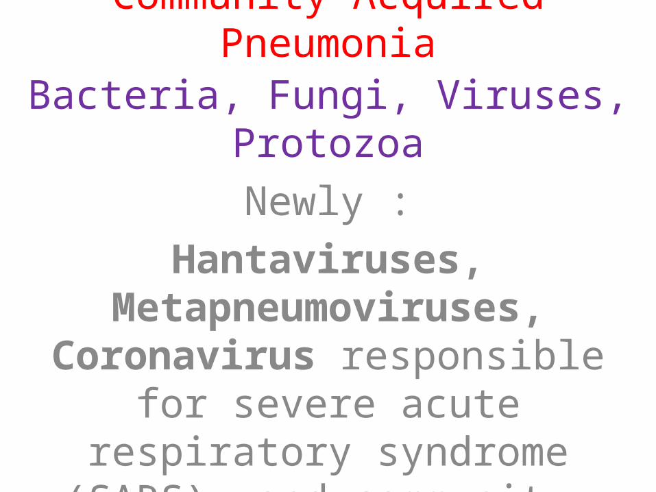
Community-Acquired Pneumonia
Bacteria, Fungi, Viruses, Protozoa
Newly :
Hantaviruses, Metapneumoviruses, Coronavirus responsible for severe acute respiratory syndrome (SARS), and community-acquired strains of
methicillin-resistant Staphylococcus aureus (MRSA)
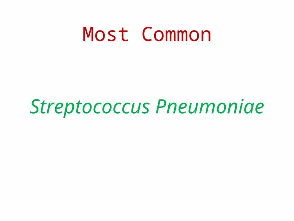
Most Common
Streptococcus Pneumoniae
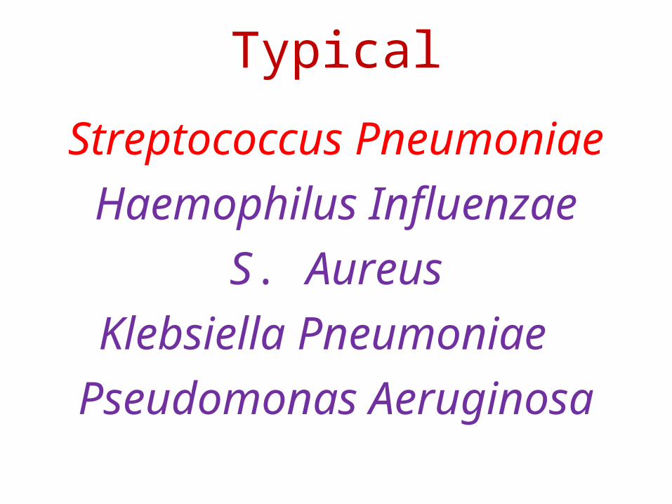
Typical
Streptococcus Pneumoniae
Haemophilus Influenzae
S. Aureus
Klebsiella Pneumoniae
Pseudomonas Aeruginosa
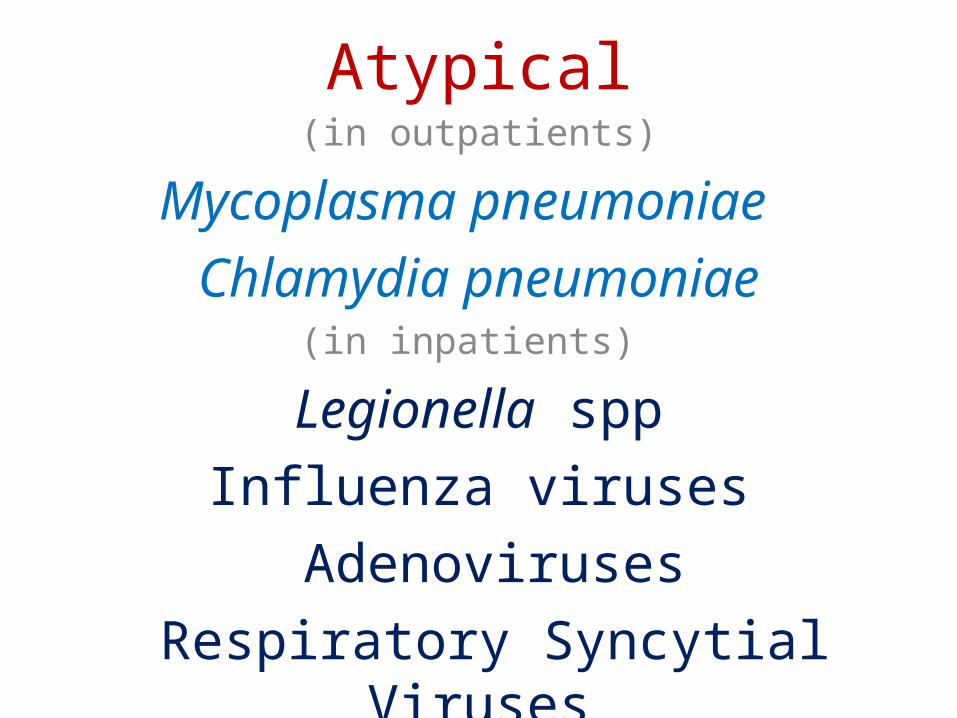
Atypical(in outpatients)
Mycoplasma pneumoniae
Chlamydia pneumoniae(in inpatients)
Legionella spp
Influenza viruses
Adenoviruses
Respiratory Syncytial Viruses

Atypical organisms
Cannot be cultured on standard media, nor can they be seen on
Gram's stain.
Intrinsically resistant to all β-lactam agents and must be treated with a
Macrolide, Fluoroquinolone, Tetracycline
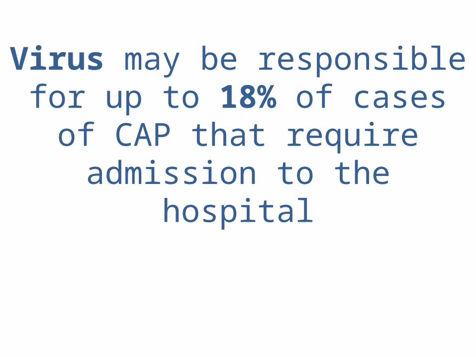
Virus may be responsible for up to 18% of cases of CAP that require
admission to the hospital
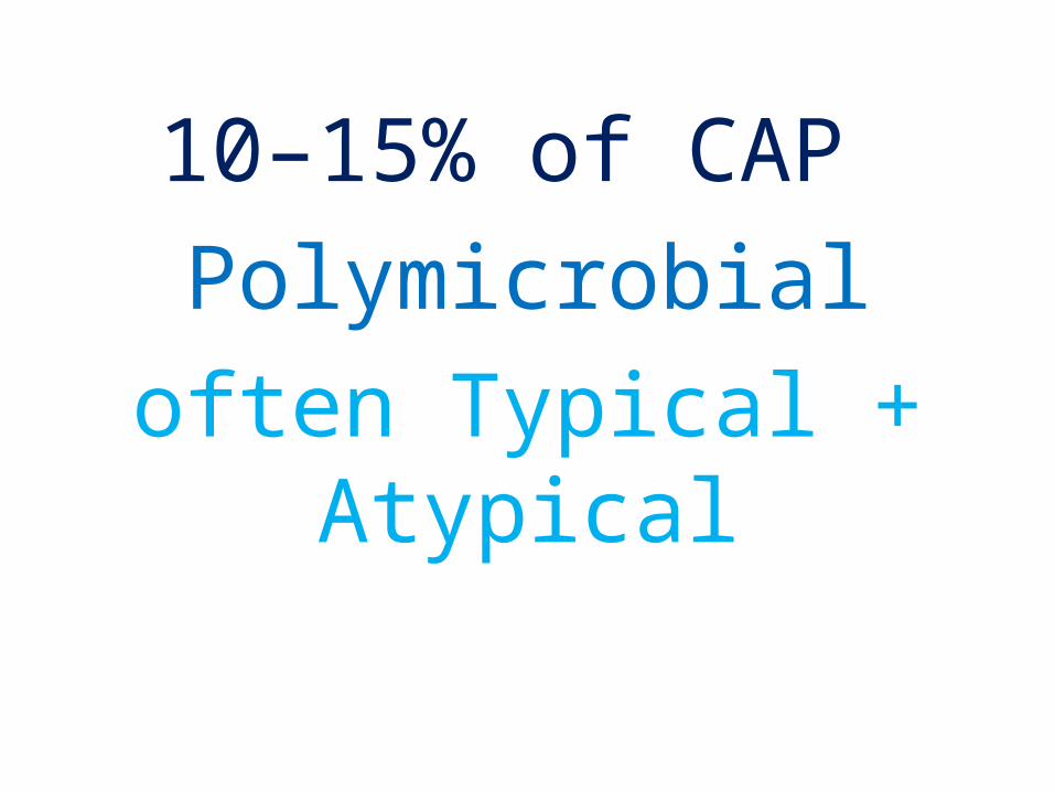
10–15% of CAP
Polymicrobial
often Typical + Atypical

ما ! که ده آن را ما الهیبه آن ! را
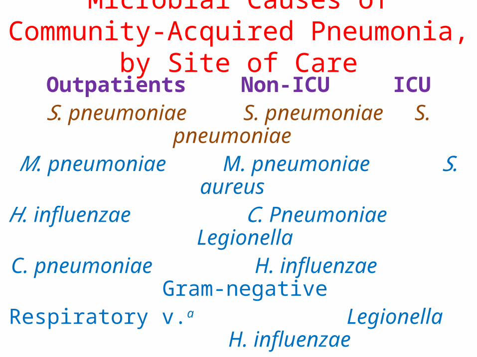
Microbial Causes of Community-Acquired Pneumonia, by Site of Care
Outpatients Non-ICU ICUS. pneumoniae S. pneumoniae S. pneumoniae M. pneumoniae M. pneumoniae S. aureus H. influenzae C. Pneumoniae Legionella
C. pneumoniae H. influenzae Gram-negativeRespiratory v.a Legionella H. influenzae
Respiratory virusesa
listed in descending order of frequency
aInfluenza A and B viruses, adenoviruses, respiratory syncytial viruses, parainfluenza viruses
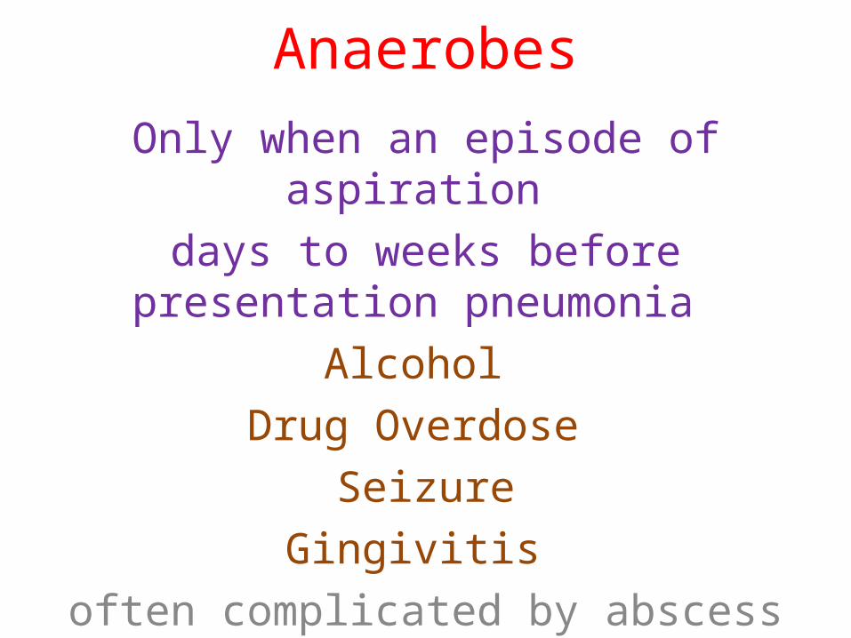
Anaerobes
Only when an episode of aspiration
days to weeks before presentation pneumonia
Alcohol
Drug Overdose
Seizure
Gingivitis
often complicated by abscess formation and significant empyemas or parapneumonic
effusions.
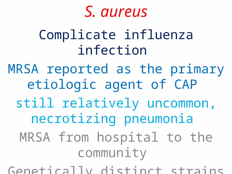
S. aureus
Complicate influenza infection
MRSA reported as the primary etiologic agent of CAP
still relatively uncommon, necrotizing pneumonia
MRSA from hospital to the community
Genetically distinct strains of MRSA in the community
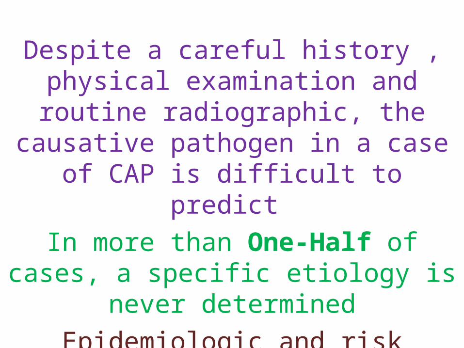
Despite a careful history , physical examination and routine radiographic, the
causative pathogen in a case of CAP is difficult to predict
In more than One-Half of cases, a specific etiology is never determined
Epidemiologic and risk factors may suggest the involvement of certain pathogens

Epidemiologic Factors Suggesting Possible Causes of Community-
Acquired Pneumonia
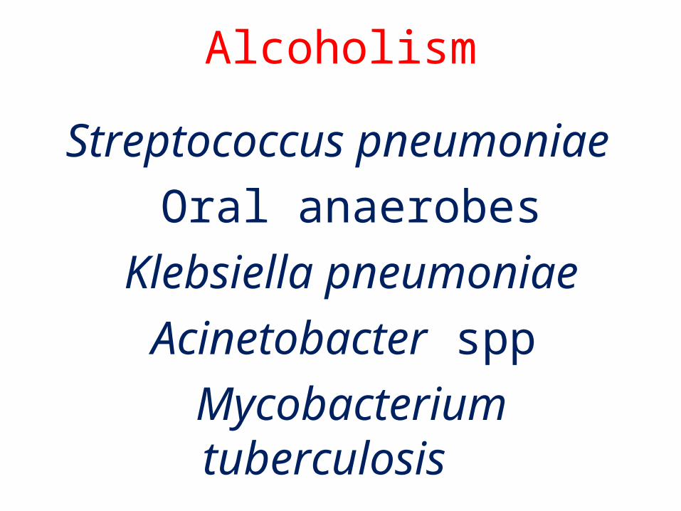
Alcoholism
Streptococcus pneumoniae
Oral anaerobes
Klebsiella pneumoniae
Acinetobacter spp
Mycobacterium tuberculosis
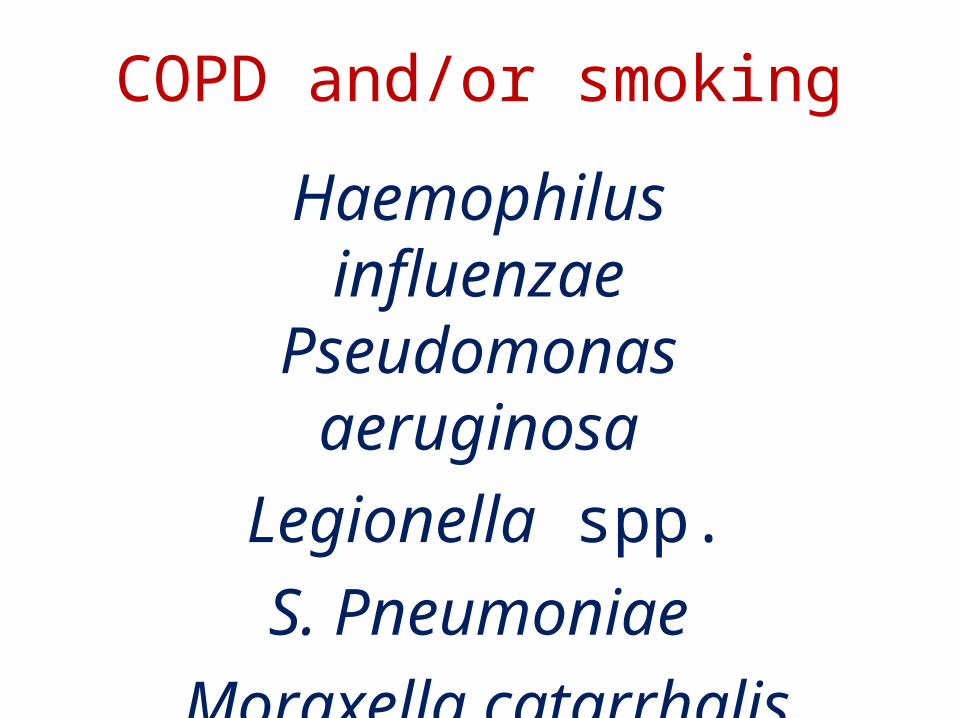
COPD and/or smoking
Haemophilus influenzae Pseudomonas
aeruginosa
Legionella spp.
S. Pneumoniae
Moraxella catarrhalis
Chlamydia pneumoniae
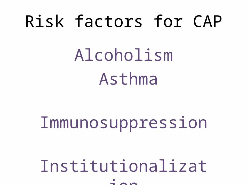
Risk factors for CAP
Alcoholism
Asthma
Immunosuppression
Institutionalization
≥70 years
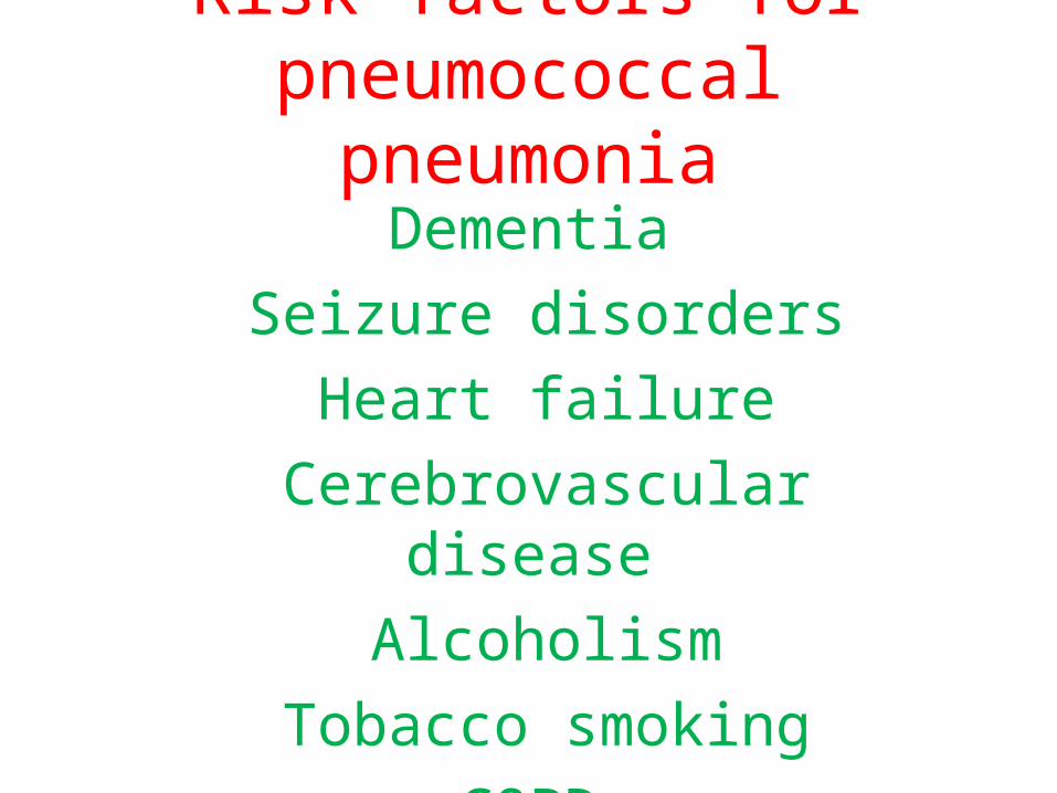
Risk factors for pneumococcal pneumonia
Dementia
Seizure disorders
Heart failure
Cerebrovascular disease
Alcoholism
Tobacco smoking
COPD
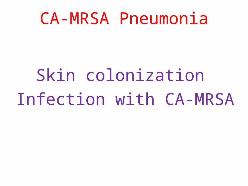
CA-MRSA Pneumonia
Skin colonization
Infection with CA-MRSA
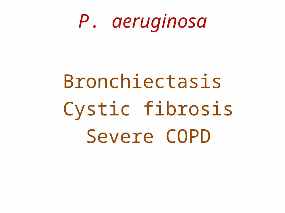
P. aeruginosa
Bronchiectasis
Cystic fibrosis
Severe COPD
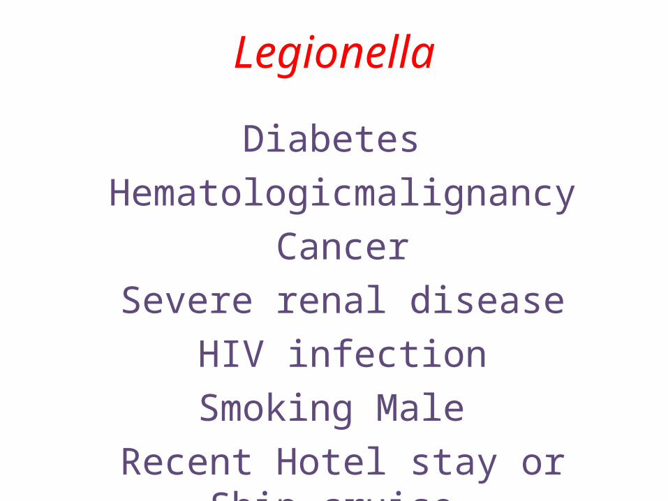
Legionella
Diabetes
Hematologicmalignancy
Cancer
Severe renal disease
HIV infection
Smoking Male
Recent Hotel stay or Ship cruise
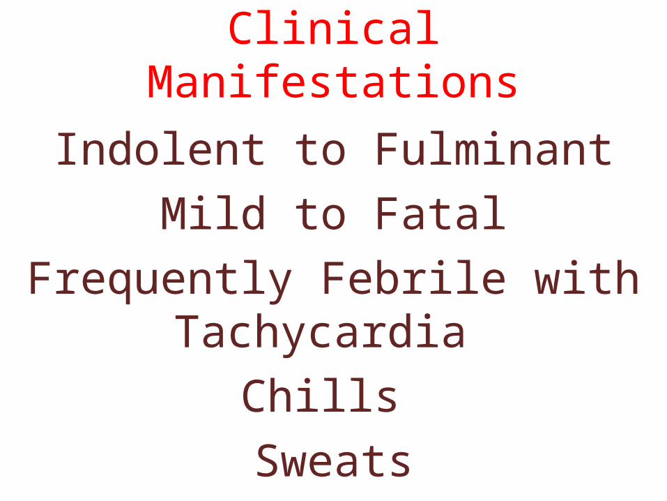
Clinical Manifestations
Indolent to Fulminant
Mild to Fatal
Frequently Febrile with Tachycardia
Chills
Sweats
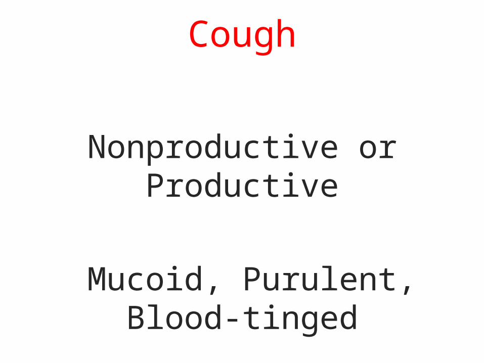
Cough
Nonproductive or Productive
Mucoid, Purulent, Blood-tinged

If the pleura is involved, : Pleuritic Chest Pain
20% : Gastrointestinal symptoms
Nausea, Vomiting, Diarrhea
Other symptoms may include
Fatigue, Headache, Myalgias, Arthralgias
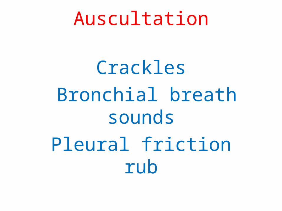
Auscultation
Crackles
Bronchial breath sounds
Pleural friction rub

Diagnosis
1:Is this pneumonia?
Clinical and Radiographic methods
2:what is the likely etiology?
aid of Laboratory techniques
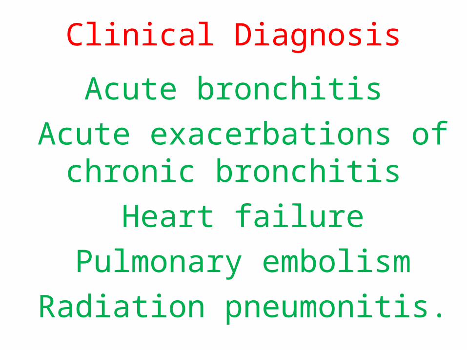
Clinical Diagnosis
Acute bronchitis
Acute exacerbations of chronic bronchitis
Heart failure
Pulmonary embolism
Radiation pneumonitis.
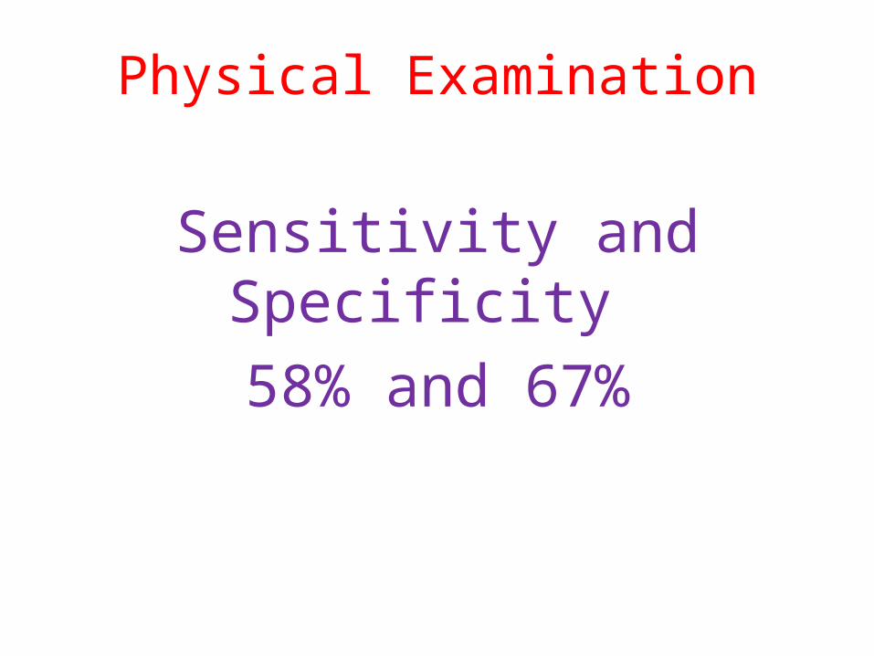
Physical Examination
Sensitivity and Specificity
58% and 67%
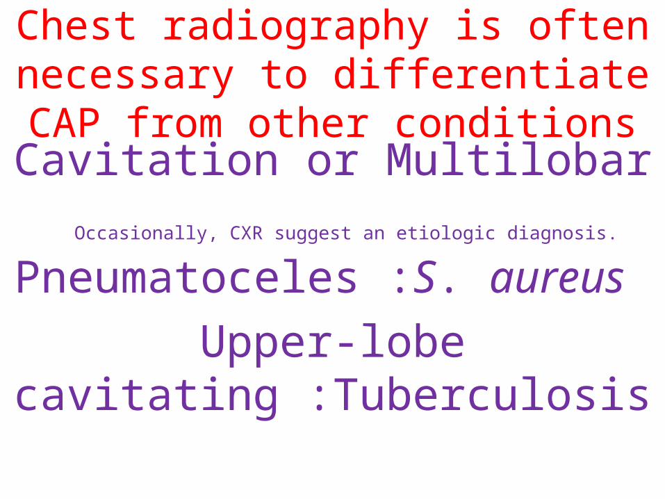
Chest radiography is often necessary to differentiate CAP from other
conditionsCavitation or Multilobar
Occasionally, CXR suggest an etiologic diagnosis.
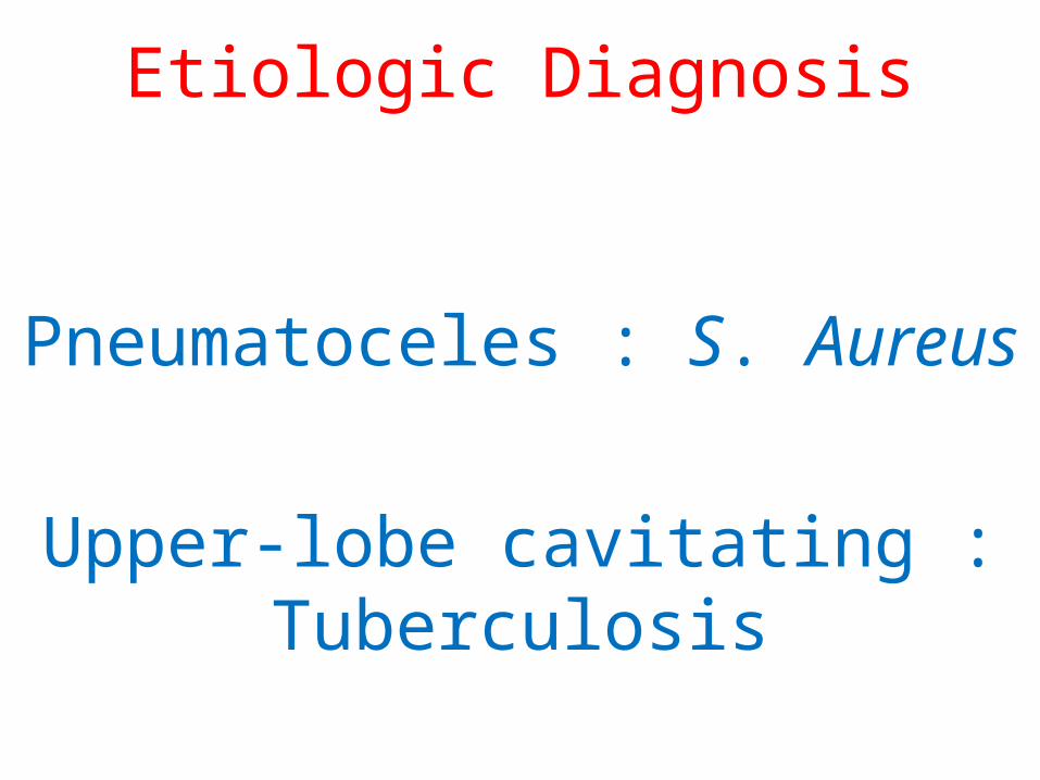
Pneumatoceles :S. aureus
Upper-lobe cavitating :Tuberculosis

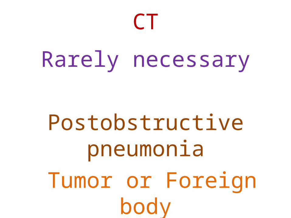
CT is rarely necessary
suspected postobstructive pneumonia caused by a Tumor or
Foreign body
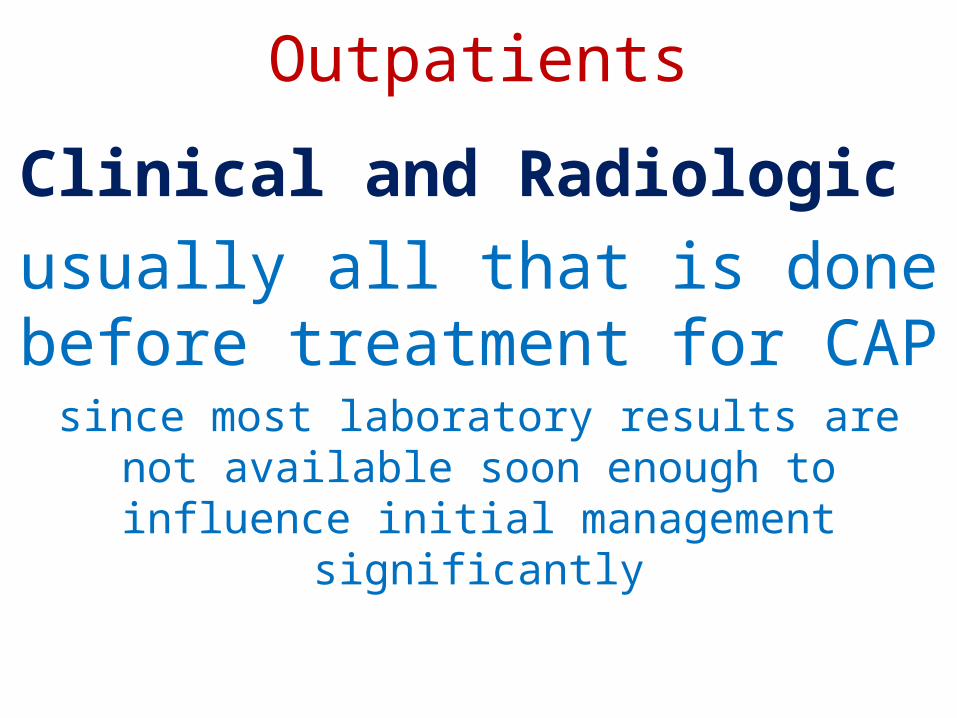
Outpatients
Clinical and Radiologic
usually all that is done before treatment for CAP
since most laboratory results are not available soon enough to influence initial management significantly
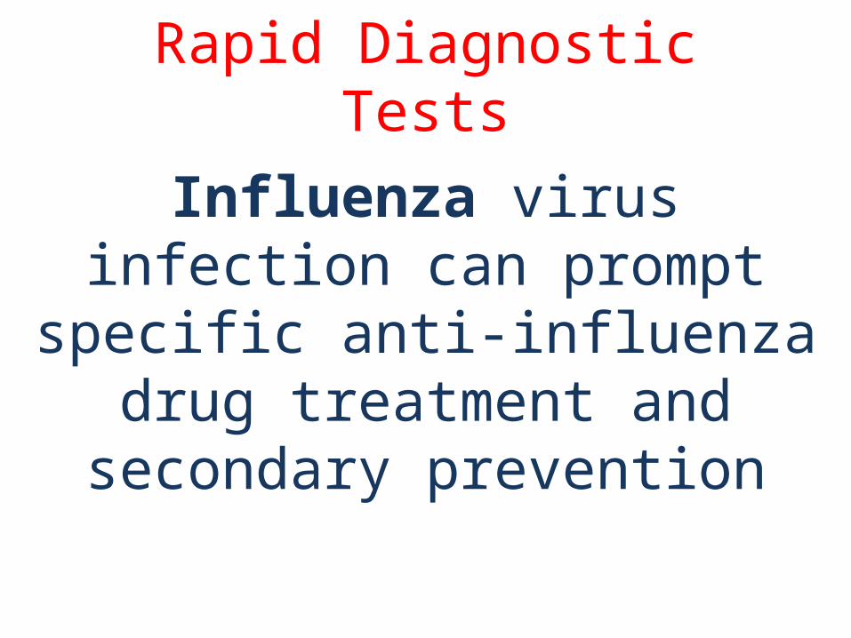
Rapid Diagnostic Tests
Influenza virus infection can prompt specific anti-influenza drug treatment and secondary prevention

Etiologic Diagnosis
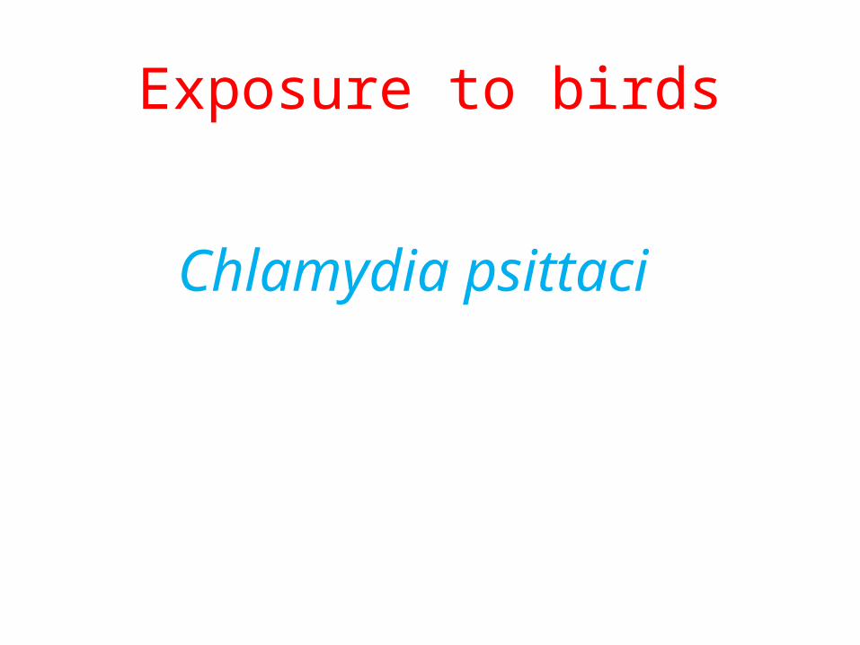
Exposure to birds
Chlamydia psittaci

Exposure to rabbits
Francisella tularensis

Local influenza activity
Influenza virus
S. pneumoniae
S. aureus
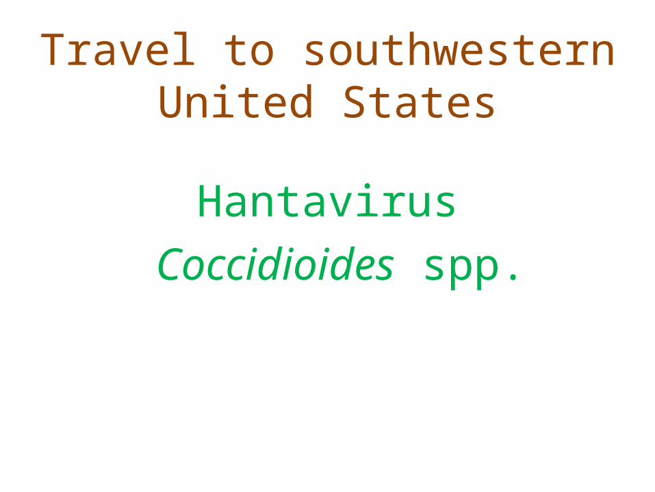
Travel to southwestern United States
Hantavirus
Coccidioides spp.

Dementia, stroke, decreased level of consciousness
Oral anaerobes
Gram-negative enteric bacteria
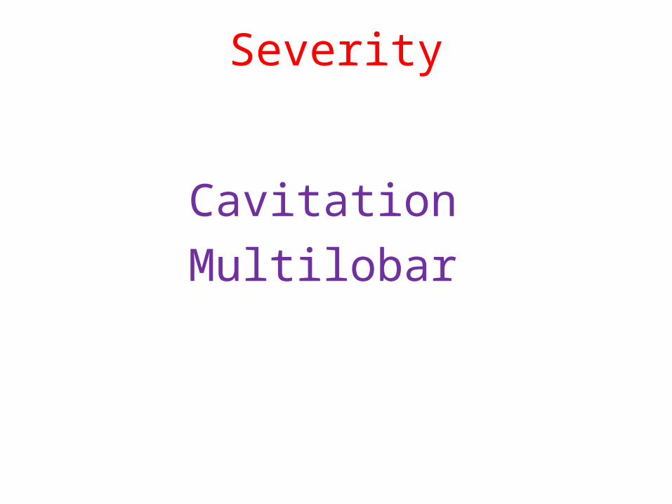
Severity
Cavitation
Multilobar
Etiologic Diagnosis
Pneumatoceles : S. Aureus
Upper-lobe cavitating : Tuberculosis
CT
Rarely necessary
Postobstructive pneumonia
Tumor or Foreign body

Etiologic Diagnosis
Except for the 2% of CAP patients who are admitted to the ICU
no data exist to show that treatment directed at a specific pathogen is statistically superior to empirical
therapyMycobacterium tuberculosis and influenza
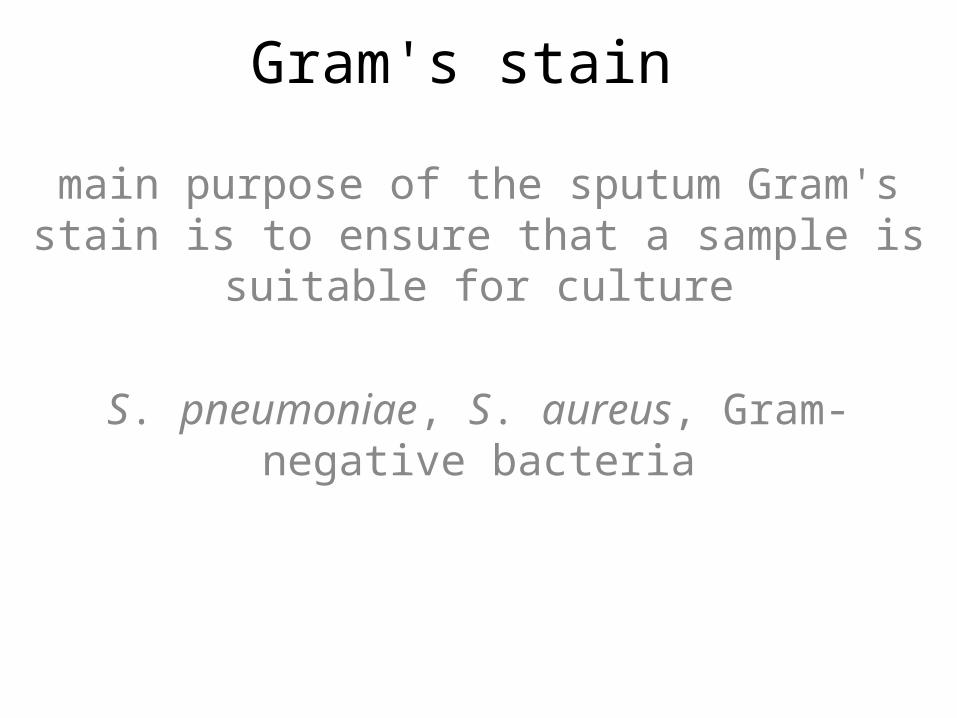
Gram's stain
main purpose of the sputum Gram's stain is to ensure that a sample is suitable for culture
S. pneumoniae, S. aureus, Gram-negative bacteria

Adequate for Culture
>25 Neutrophils
<10 Squamous epithelial cells
per low-power field
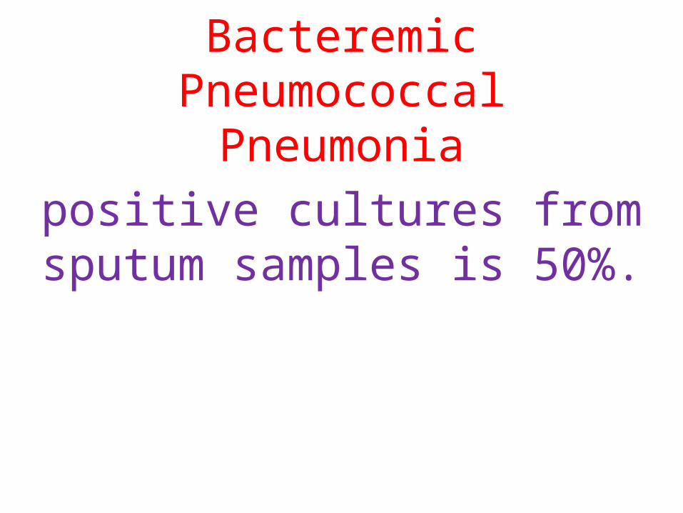
Bacteremic Pneumococcal Pneumonia
positive cultures from sputum samples is 50%.
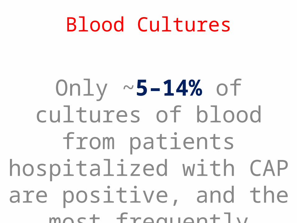
Blood Cultures
Only ~5–14% of cultures of blood from patients hospitalized with CAP are positive, and the
most frequently isolated pathogen is S. pneumoniae
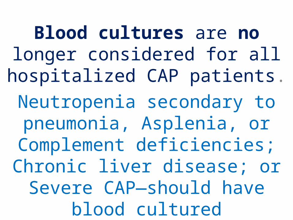
Blood cultures are no longer considered for all hospitalized CAP
patients.
Neutropenia secondary to pneumonia, Asplenia, or Complement deficiencies; Chronic liver disease; or Severe CAP—
should have blood cultured
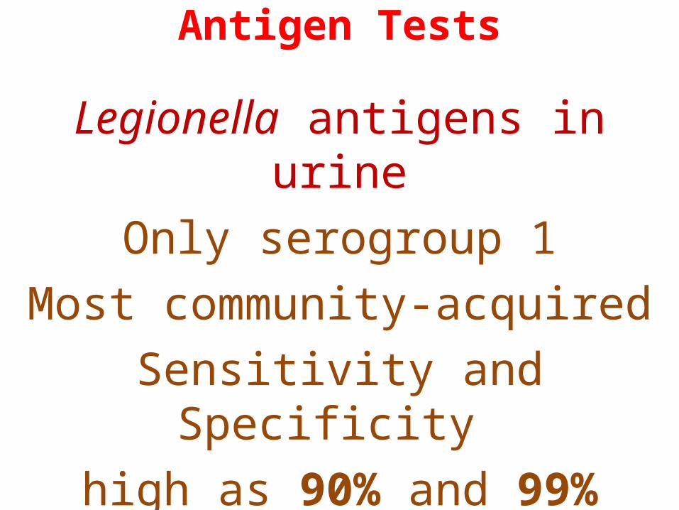
Antigen Tests
Legionella antigens in urineOnly serogroup 1
Most community-acquiredSensitivity and Specificity
high as 90% and 99%
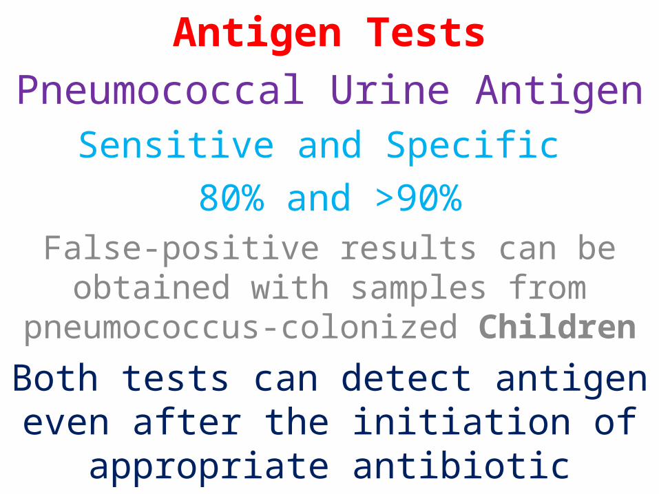
Antigen Tests
Pneumococcal Urine AntigenSensitive and Specific
80% and >90%False-positive results can be obtained with
samples from pneumococcus-colonized Children
Both tests can detect antigen even after the initiation of appropriate antibiotic therapy
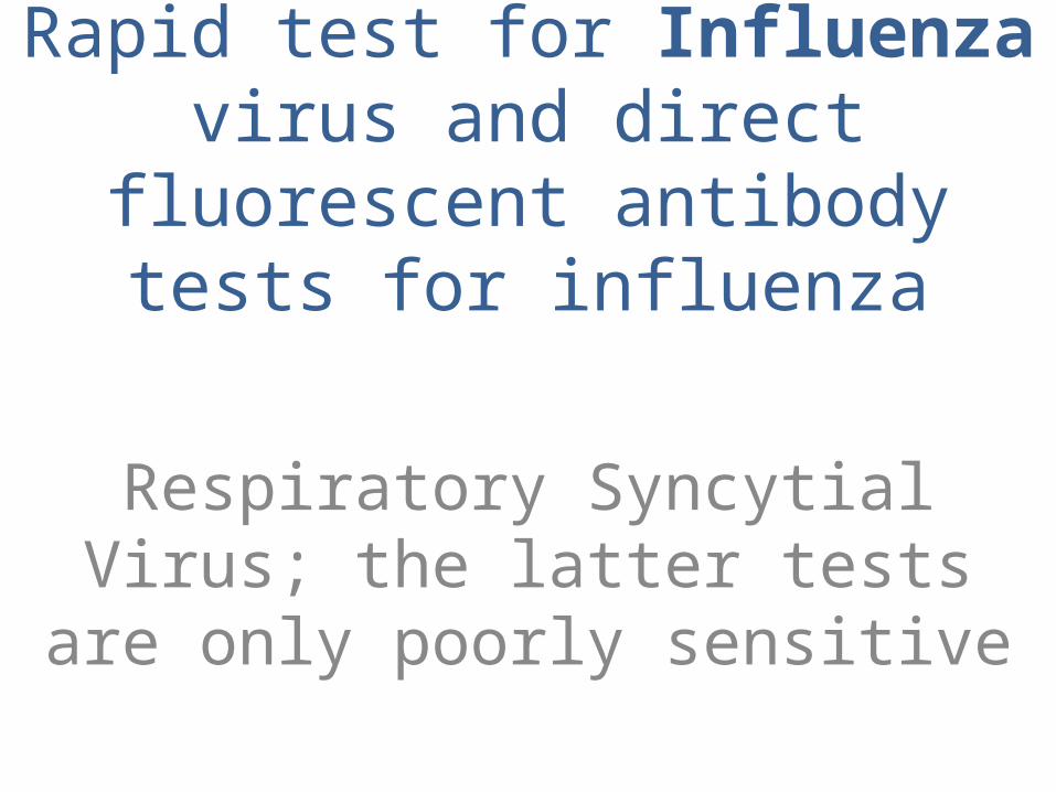
Rapid test for Influenza virus and direct fluorescent antibody tests for
influenza
Respiratory Syncytial Virus; the latter tests are only poorly sensitive
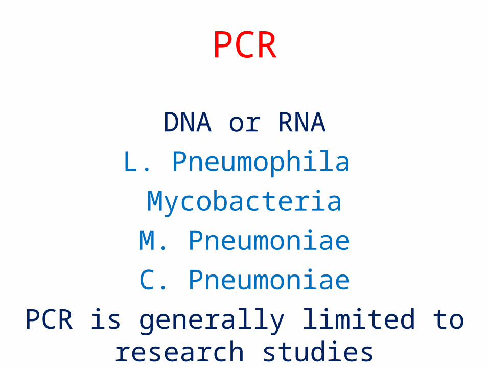
PCR
DNA or RNA
L. Pneumophila
Mycobacteria
M. Pneumoniae
C. Pneumoniae
PCR is generally limited to research studies
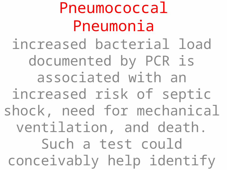
Pneumococcal Pneumonia
increased bacterial load documented by PCR is associated with an increased
risk of septic shock, need for mechanical ventilation, and death. Such a test could conceivably help identify patients suitable for ICU
admission.

Serology
A fourfold rise in specific IgM between acute- and convalescent-phase serum is
generally considered diagnostic
Coxiella burnetii
Recently, however, they have fallen out of favor because of the time required to
obtain a final result for the convalescent-phase sample.
Treatment: CAP
Certain patients clearly can be managed at Home, and others clearly require treatment in the
Hospital
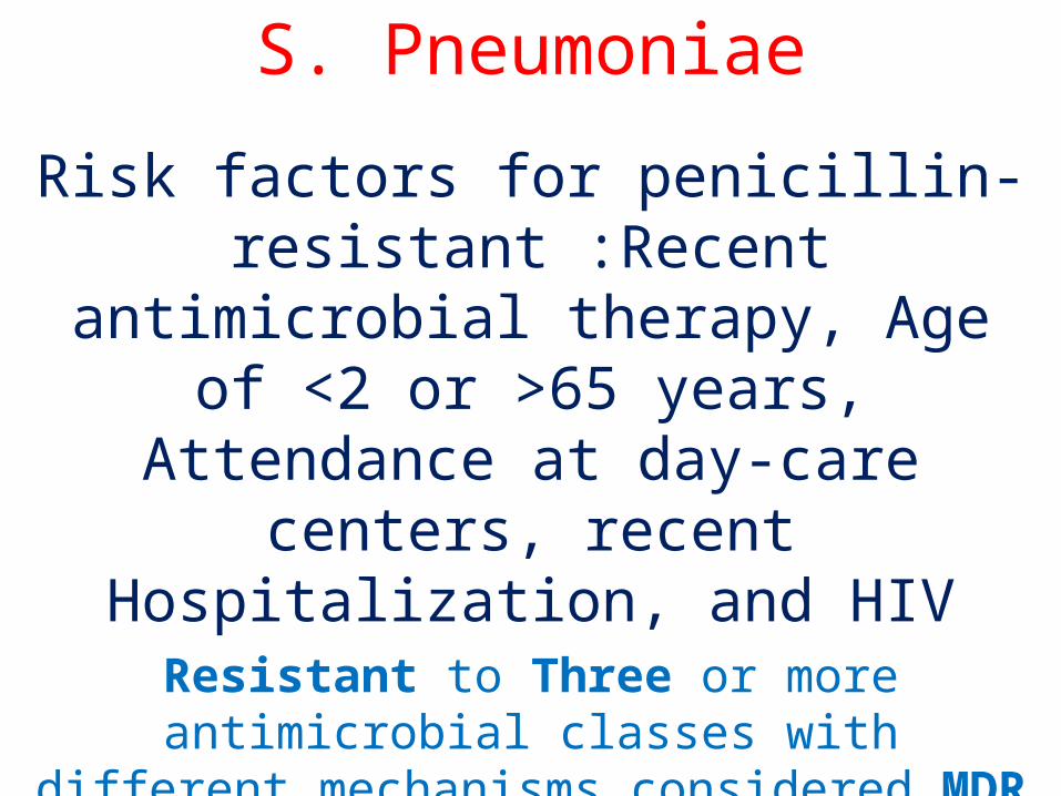
S. Pneumoniae
Risk factors for penicillin-resistant :Recent antimicrobial therapy, Age of <2 or >65 years, Attendance at
day-care centers, recent Hospitalization, and HIV
Resistant to Three or more antimicrobial classes with different mechanisms considered MDR
Penicillin ,Macrolides, Tetracyclines, Trimethoprim-sulfamethoxazole
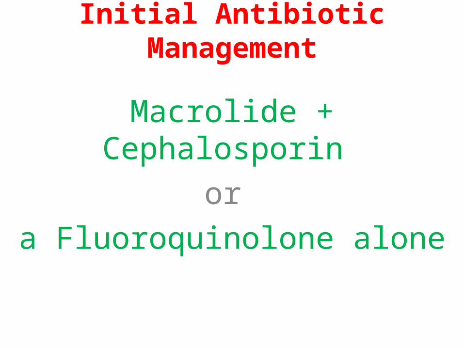
Initial Antibiotic Management
Macrolide + Cephalosporin
or
a Fluoroquinolone alone
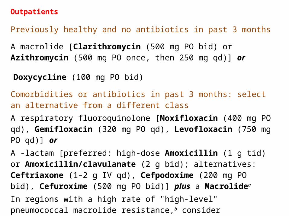
Outpatients
Previously healthy and no antibiotics in past 3 months
A macrolide [Clarithromycin (500 mg PO bid) or Azithromycin (500 mg PO once, then 250 mg qd)] or
Doxycycline (100 mg PO bid)
Comorbidities or antibiotics in past 3 months: select an alternative from a different class
A respiratory fluoroquinolone [Moxifloxacin (400 mg PO qd), Gemifloxacin (320 mg PO qd), Levofloxacin (750 mg PO qd)] or
A -lactam [preferred: high-dose Amoxicillin (1 g tid) or Amoxicillin/clavulanate (2 g bid); alternatives: Ceftriaxone (1–2 g IV qd), Cefpodoxime (200 mg PO bid), Cefuroxime (500 mg PO bid)] plus a Macrolidea
In regions with a high rate of "high-level" pneumococcal macrolide resistance,b consider alternatives listed above for patients with comorbidities.
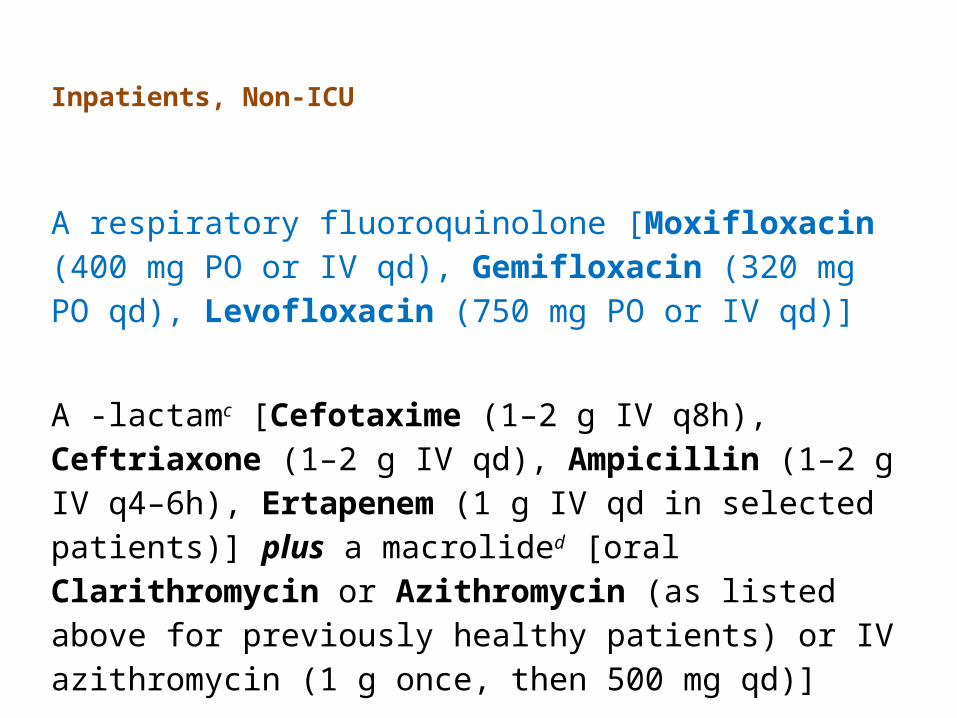
Inpatients, Non-ICU
A respiratory fluoroquinolone [Moxifloxacin (400 mg PO or IV qd), Gemifloxacin (320 mg PO qd), Levofloxacin (750 mg PO or IV qd)]
A -lactamc [Cefotaxime (1–2 g IV q8h), Ceftriaxone (1–2 g IV qd), Ampicillin (1–2 g IV q4–6h), Ertapenem (1 g IV qd in selected patients)] plus a macrolided [oral Clarithromycin or Azithromycin (as listed above for previously healthy patients) or IV azithromycin (1 g once, then 500 mg qd)]
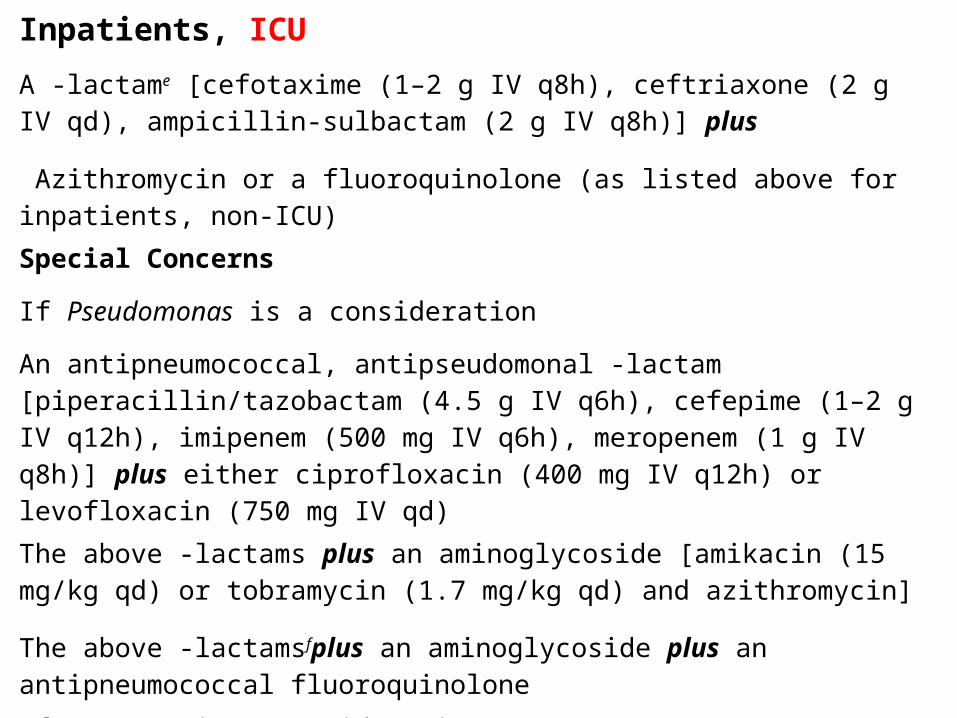
Inpatients, ICU
A -lactame [cefotaxime (1–2 g IV q8h), ceftriaxone (2 g IV qd), ampicillin-sulbactam (2 g IV q8h)] plus
Azithromycin or a fluoroquinolone (as listed above for inpatients, non-ICU)
Special Concerns
If Pseudomonas is a consideration
An antipneumococcal, antipseudomonal -lactam [piperacillin/tazobactam (4.5 g IV q6h), cefepime (1–2 g IV q12h), imipenem (500 mg IV q6h), meropenem (1 g IV q8h)] plus either ciprofloxacin (400 mg IV q12h) or levofloxacin (750 mg IV qd)
The above -lactams plus an aminoglycoside [amikacin (15 mg/kg qd) or tobramycin (1.7 mg/kg qd) and azithromycin]
The above -lactamsfplus an aminoglycoside plus an antipneumococcal fluoroquinolone
If CA-MRSA is a consideration
Add linezolid (600 mg IV q12h) or vancomycin (1 g IV q12h).
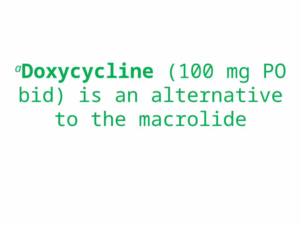
aDoxycycline (100 mg PO bid) is an alternative to the macrolide
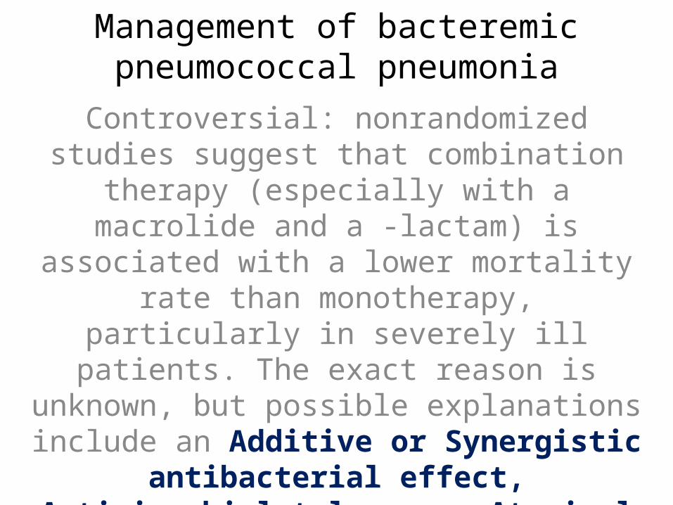
Management of bacteremic pneumococcal pneumonia
Controversial: nonrandomized studies suggest that combination therapy (especially with a macrolide and a -lactam) is associated with a lower mortality rate than monotherapy, particularly in severely ill
patients. The exact reason is unknown, but possible explanations include an Additive or Synergistic antibacterial effect, Antimicrobial tolerance,
Atypical co-infection, or the Immunomodulatory effects of the macrolides
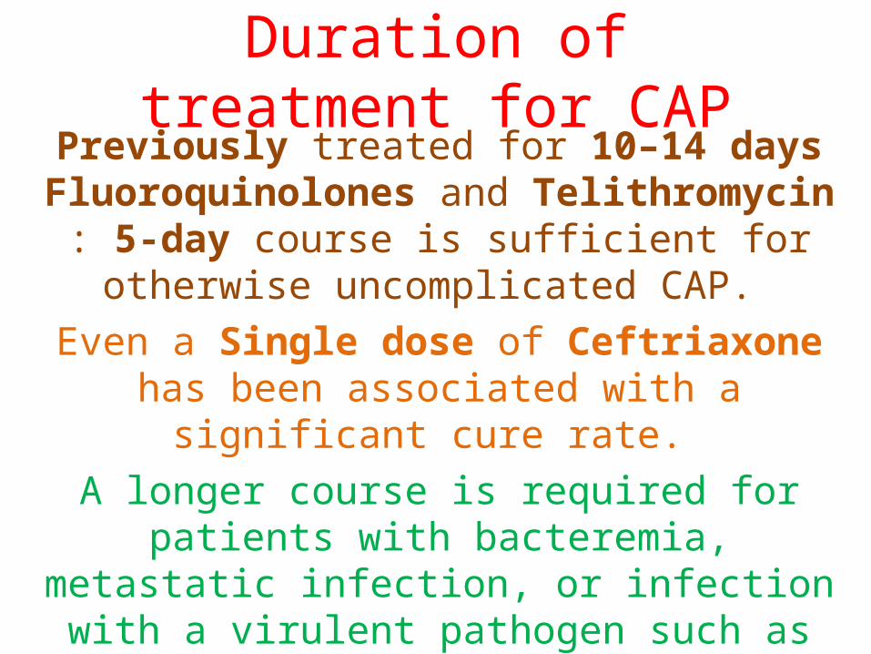
Duration of treatment for CAPPreviously treated for 10–14 days
Fluoroquinolones and Telithromycin : 5-day course is sufficient for otherwise uncomplicated
CAP.
Even a Single dose of Ceftriaxone has been associated with a significant cure rate.
A longer course is required for patients with bacteremia, metastatic infection, or infection with a
virulent pathogen such as P. aeruginosa or CA-MRSA.
General Considerations
Adequate hydration, oxygen therapy for hypoxemia, and assisted ventilation
severe CAP who remain Hypotensive despite fluid resuscitation may have
adrenal insufficiency and may respond to Glucocorticoid treatment
Immunomodulatory therapy in the form of drotrecogin alfa (activated) should be considered for CAP patients with persistent septic shock and APACHE II scores of
25, particularly if the infection is caused by S. pneumoniae
Failure to Improve
Slow to respond to therapy reevaluated at about day 3 (sooner if is worsening)
Pulmonary edema, Pulmonary embolism, Lung carcinoma, Radiation , Hypersensitivity pneumonitis,
, Connective tissue disease involving the lungs
Resistant to the drug selected, or a sequestered focus (e.g., a lung abscess or empyema)
Also possible that CAP is the correct diagnosis but that an unsuspected pathogen (e.g., CA-MRSA, M.
tuberculosis, or a fungus) is the cause
Complications
Respiratory failure, Shock and Multiorgan failure, Coagulopathy
Metastatic infection, Lung abscess, and complicated Pleural Effusion. Metastatic
infection (e.g., Brain abscess or Endocarditis)
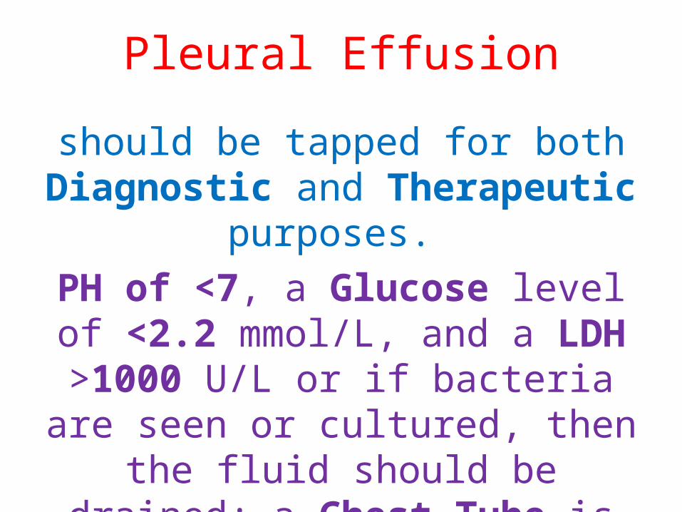
Pleural Effusion
should be tapped for both Diagnostic and Therapeutic purposes.
PH of <7, a Glucose level of <2.2 mmol/L, and a LDH >1000 U/L or if bacteria are seen or cultured, then the
fluid should be drained; a Chest Tube is usually required.
Follow-Up
Fever and Leukocytosis usually resolve 2–4 days
Physical findings may persist longer
CXR may require 4–12 weeks to clear
Age and Underlying lung disease
particularly for elderly patients follow-up radiograph can be done ~4–6 weeks later
relapse or recurrence :particularly in the same lung segment, underlying neoplasm must be considered.
Prognosis
Age, Comorbidities, and site of treatment (inpatient or outpatient)
Young well and usually recover fully after ~2 weeks
Older patients and those with comorbid conditions can take several weeks longer to recover fully
Mortality rate for the outpatient group is <1%
For patients requiring hospitalization, the overall mortality rate is estimated at 10%, with ~50% of
deaths directly attributable to pneumonia.
Prevention
Main preventive measure is Vaccination
Influenza and Pneumococcal
influenza outbreak, vaccinated immediately and given
chemoprophylaxis with either Oseltamivir or Zanamivir for 2 weeks—i.e., until vaccine-induced antibody
levels are sufficiently high
Nosocomial superinfections—both pulmonary and
extrapulmonary—are possible explanations for failure to
improve or worsening

تشکر با


![[XLS]ugandajournalistsresourcecentre.comugandajournalistsresourcecentre.com/wp-content/uploads/... · Web viewRICHARD MARTIN H2012 TWAHIRWA NEBAYOSI I3078 TWASE C0151 TWA-TWA MUTWALANTE](https://img.pdfslide.net/doc/110x75/5af9bfec7f8b9a19548d102a/xlsugandajourn-viewrichard-martin-h2012-twahirwa-nebayosi-i3078-twase-c0151-twa-twa.jpg)