Embed Size (px)
Citation preview
Pneumonia inNormal andImmunocompromisedChildren: An Overviewand Update
Hedieh K. Eslamy, MD, Beverley Newman, MD*KEYWORDS
� Pneumonia � Children � Imaging � Complicated pneumonia
Pneumonia is an infection of the lower respiratorytract, involving the lung parenchyma. The WorldHealth Organization estimates that there are 150.7million cases of pulmonary infection each year inchildren younger than 5 years, with as many as 20million cases severe enough to require hospitaladmission.1 In North America and Europe, theannual incidence of pneumonia in children youngerthan 5 years is estimated to be 34 to 40 cases per1000, and decreases to 7 cases per 1000 in adoles-cents 12 to 15 years of age.2,3 Themortality in pedi-atric patients caused by pneumonia in developedcountries is currently low (<1 per 1000 per year).3
However, pneumonia is still the number one causeof childhood mortality in developing countries.1,4
The overarching goal of this article is to reviewcause, current role of imaging, imaging techniques,and the spectrumof acute andchronic pneumoniasin children. Pneumonia in the neonate and immuno-compromised host is also discussed.
CAUSE OF PNEUMONIA
Infectious agents causing pneumonia in childreninclude viruses, bacteria, mycobacteria, myco-plasmas, fungi, protozoa, and helminths. Etiologicdiagnoses of pneumonia are not so easy to deter-mine or so accurate as is sometimes implied. Inaddition, proof of the cause of pneumonia is not
The authors have nothing to disclose.Department of Radiology, Lucile Packard Children’s HoWelch Road, Stanford, CA 94305, USA* Corresponding author.E-mail address: [email protected]
Radiol Clin N Am 49 (2011) 895–920doi:10.1016/j.rcl.2011.06.0070033-8389/11/$ – see front matter � 2011 Elsevier Inc. All
obtained in most cases. There is a great deal ofoverlap in the radiographic appearance of pneu-monias caused by different organisms. Imaging isusually poor at predicting the broad category (eg,bacterial vs viral) of infectious agent, let alone thespecific agent. Preexisting lung disease may notonly predispose to pulmonary infection but alsomodify the appearance of pulmonary consolida-tion. Furthermore, because the lungs can respondto a diverse disease processes in only a limitednumber of ways, it is common for the radiographicfeatures of both acute and chronic infectious pneu-monia to overlap considerably withmany noninfec-tious lung diseases. Such noninfectious lungdiseases are identified as pneumonia mimics inthis article.
Viral pneumonia is rare in the neonatal period,because of conferred maternal antibody protec-tion, whereas bacterial pneumonia is most fre-quently caused by pathogens acquired duringlabor and delivery, and is more prevalent in prema-ture babies. With decreasing maternal antibodylevels, viral pneumonia occurs at a peak between2 months to 2 years of age. Bacterial infectionsbecome relatively more common in older childrenfrom 2 years to 18 years of age.5 The lung responseto an infective antigen seems to be more age-specific than antigen-dependent (ie, bacteria vsviral). Therefore, lobar and alveolar lung opacities
spital, Stanford University School of Medicine, 725
rights reserved. radiologic.th
eclinics.com

Eslamy & Newman896
are more common in older children and are morefrequently caused by bacterial infections, whereasinterstitial opacities are seen in all age groups, andare relatively nonspecific as to the type of causativeorganism.6,7
ROLE OF IMAGING AND IMAGINGTECHNIQUES
The role of imaging, including chest radiographs,ultrasound (US) and computed tomography (CT),is to detect the presence of pneumonia, deter-mine its location and extent, exclude other tho-racic causes of respiratory symptoms, and showcomplications such as parapneumonic effusion/empyema and suppurative lung complications.5
Although magnetic resonance (MR) imaging isnot routinely used for evaluating pneumonia inchildren, it is a promising imaging modality partic-ularly for children with chronic lung conditions whorequire repeat imaging studies.Frontal and lateral chest radiographs are the
mainstay, and often the only, imaging needed inpediatric pulmonary infection. This imaging can besupplemented with other views such as lateral de-cubitus or other imaging modalities as the cir-cumstances warrant. Decubitus views are notuseful when an entire hemithorax is opacifiedbecause layering fluid cannot be identified withoutany adjacent air. The main use of US is to identify,quantify, and characterize a parapneumonic effu-sion/empyema, as well as provide image guidancefor drainage and identify residual collections aftertreatment.8,9 Operator availability and expertiseare important factors in making US a useful tool forevaluating pulmonary infection. Although intrapul-monary fluid-filled cavities andeven lungabscesseswithin consolidated lungcanbe identifiedonUS,CTprovides amore global view of the disease process.CT is often used to further evaluate: (1) suppurativelung complications and to differentiate these fromparapneumonic effusion/empyema; (2) patientswith recurrent or chronic pneumonia and concernfor an underlying lesion; and (3) immunocompro-mised children with noncontributory or confusingchest radiographs and clinical findings that couldbe secondary to lung infection.5
Close attention to CT technique is crucial forimaging evaluation of pneumonia in pediatricpatients. CT with low radiation dose techniqueshould be carefully performed in all cases. Eightyto 120 kVp with weight-based low milliampere-seconds coupled with radiation dose modulationtechniques is appropriate in most children for eval-uation of pneumonia. Multiple CT image acquisi-tions are usually not needed and the scan field ofview should be tailored to the area of interest
(especially if following a specific lesion seriallyover time) to further decrease the overall radiationdose.10 Occasionally, it may be useful to acquireadditional expiratory scans to assess air trapping,which is an early imaging finding associated withsmall airway disease. In this situation, often at least1 or both CT acquisitions can be obtained usinga high-resolution CT (HRCT) gap technique. Toobtain optimal CT imaging at peak inspiration andclose to expiratory residual volume, controlledventilation (CViCT) in infants and young children(�5 years old) or spirometer-controlled CT in olderchildren may be needed.11 Young children havelittle intrinsic tissue contrast. Therefore, intrave-nous contrast is almost always needed for CTimaging of infection especially if mediastinal delin-eation is required. The exception is when HRCT isused only for evaluating lung parenchymal andairway disease. Breath-holding is usually desirablebut can be adapted on a case-by-case basis de-pending on the needs of the study and the abilityof the child to cooperate. However, for the studyto be interpretable, gross patient motion shouldbe absent. Sedation or anesthesiamay be requiredin infants, young children, or children with intellec-tual disability. Delays between induction of anes-thesia and scanning need to be minimized toprevent the potential for lung atelectasis with anes-thesia. The anesthesiologist needs to pay closeattention to techniques for preventing atelectasisor recruiting lung before the CT imaging.12
Peltola and colleagues13 recently published theirexperience with MR imaging of lung infections inchildren using free-breathing T2-weighted, shorttau inversion recovery, and T1-weighted with fatsaturation precontrast and postcontrast sequ-ences. Their study showed that lung parenchymal,pleural, and lymphnode inflammatory abnormalitiescan be characterized by MR imaging in childrenwith lung infection. Therefore, MR imaging mightpotentially be used to further evaluate suspected,acute complications of pneumonia.13 Children withchronic lung conditions and recurrent infection,such as cystic fibrosis, who are often subjected tosubstantial radiation exposure from repeated CTstudies, would benefit the most from MR imagingevaluation of the lungs instead of CT. Although MRimaging may not provide as much detail comparedwith CT especially with early, small or subtlechanges (Fig. 1), there are promising indications ofa role for MR imaging in pulmonary infection.13–15
RADIOGRAPHIC CHANGES IN RESPIRATORYINFECTION
There are several different descriptions of basicpatterns of lung diseases on chest radiographs.
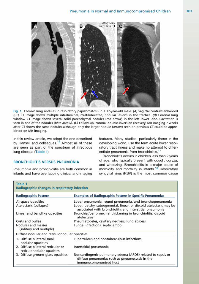
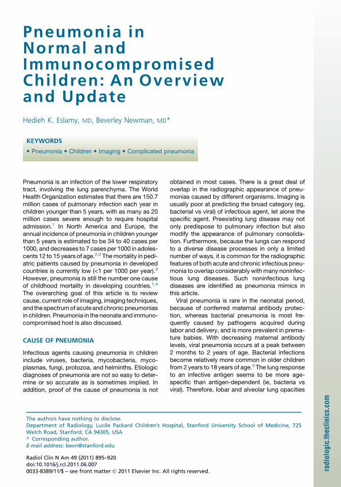
Fig. 1. Chronic lung nodules in respiratory papillomatosis in a 17-year-old male. (A) Sagittal contrast-enhanced(CE) CT image shows multiple intraluminal, multilobulated, nodular lesions in the trachea. (B) Coronal lungwindow CT image shows several solid parenchymal nodules (red arrow) in the left lower lobe. Cavitation isseen in one of the nodules (blue arrow). (C) Follow-up, coronal double-inversion recovery, MR imaging 7 weeksafter CT shows the same nodules although only the larger nodule (arrow) seen on previous CT could be appre-ciated on MR imaging.
Pneumonia in Normal and Immunocompromised Children 897
In this review article, we adopt the one describedby Hansell and colleagues.16 Almost all of theseare seen as part of the spectrum of infectiouslung disease (Table 1).
BRONCHIOLITIS VERSUS PNEUMONIA
Pneumonia and bronchiolitis are both common ininfants and have overlapping clinical and imaging
Table 1Radiographic changes in respiratory infection
Radiographic Pattern Examples of Rad
Airspace opacitiesAtelectasis (collapse)
Linear and bandlike opacities
Cysts and bullaeNodules and masses
(solitary and multiple)
Lobar pneumonLobar, patchy, suassociated wit
Bronchial/peribratelectasis
Pneumatoceles,Fungal infection
Diffuse nodular and reticulonodular opacities
1. Diffuse bilateral smallnodular opacities
2. Diffuse bilateral reticular orreticulonodular opacities
3. Diffuse ground-glass opacities
Tuberculous and
Interstitial pneu
Noncardiogenicdiffuse pneumimmunocomp
features. Many studies, particularly those in thedeveloping world, use the term acute lower respi-ratory tract illness and make no attempt to differ-entiate pneumonia from bronchiolitis.17
Bronchiolitis occurs in children less than 2 yearsof age, who typically present with cough, coryza,and wheezing. Bronchiolitis is a major cause ofmorbidity and mortality in infants.18 Respiratorysyncytial virus (RSV) is the most common cause
iographic Pattern in Specific Pneumonias
ia, round pneumonia, and bronchopneumoniabsegmental, linear, or discoid atelectasis may beh bronchiolitis and interstitial pneumoniaonchial thickening in bronchiolitis; discoid
cavitary necrosis, lung abscesss, septic emboli
nontuberculous infections
monia
pulmonary edema (ARDS) related to sepsis oronias such as pneumocystis in theromised host
Eslamy & Newman898
of bronchiolitis, followed by rhinovirus. Other lesscommon causes of bronchiolitis include parain-fluenza virus, human metapneumovirus, adeno-virus, influenza virus, coronaviruses, and humanbocavirus. Chest radiographs usually show hyper-inflation, perihilar opacities, peribronchial thick-ening, and patchy, often mobile, atelectasis(Fig. 2). Such imaging findings are related to diffuseairway inflammation and partial (air trapping) orcomplete (atelectasis) airway obstruction.19 Sim-ilar changes are seen in older children (>2 yearsof age) with bronchitis although the features ofdiffuse small airway obstruction are less commonin these older children with larger airways.
Box 1Causes of acute focal pneumonia in children
SPECTRUM OF PNEUMONIA
Pneumonia can be divided into several syndromesbased on clinical presentation, imaging appear-ance, underlying predisposition, and cause. Pneu-monia syndromes that are discussed in this articleinclude acute focal pneumonia, atypical pneu-monia, miliary or nodular pneumonia, progressiveor fulminant pneumonia, aspiration pneumonias,pulmonary infiltrates with eosinophilia (PIE), andchronic or recurrent pneumonia.20 Neonatal pneu-monia is briefly highlighted separately. Pneumoniain immunosuppressed individuals is included inthe general discussion of pneumonia syndromesand then specifically reviewed with regard to thedifferent infections associated with various typesof immunodeficiency. Acute and chronic compli-cations of pneumonia are also reviewed.
Fig. 2. RSV bronchiolitis in a 6- week-old boy. Frontalchest radiograph shows perihilar streaky opacities,peribronchial thickening, hyperinflation, and patchyatelectasis.
Acute Focal Pneumonia
Characteristics that are typical for acute focal pneu-monia include fever more than 38.8�C (102�F),a toxic appearance, and a focal opacity on chestradiographs. Pleuritic chest pain in lower-lobepneumonia is sometimes referred to the abdomenandmaybemistaken clinically for an acute abdom-inal condition. Acute focal pneumonia is most oftencaused by bacterial infection with streptococcuspneumonia. Other causes of acute focal pneu-monia are summarized in Box 1. The chest radio-graph of acute focal pneumonia usually showsadense, typicallymoreperipheral airspaceopacity,which may appear segmental, lobar, or spherical(Figs. 3 and 4).21–23 In a febrile childwith a sphericaldensity on a chest radiograph, the most likely diag-nosis is a round pneumonia but the possibility of anunderlying neoplasm may be considered. Roundpneumonias tend to be solitary, have well-definedborders, and are often located in theperihilar regionorposteriorly in the lungs.The radiographshouldbecarefully scrutinized for features of consolidationsuch as air bronchograms as opposed to those ofa mass such as vascular/airway displacement orbonyerosion.A secondviewsuchas a lateral radio-graph may be helpful because a round pneumonia
Usual
Streptococcus pneumonia
Uncommon
Bacteria: Hemophilus influenzae type B, non-typable H influenza, Staphylococcus aureus,group A streptococcus, Mycoplasma pneumo-niae, Chlamydia pneumoniae
Rare
Bacteria and mycobacteria: Francisella tularen-sis, Mycobacterium tuberculosis,Meningococcus,enteric bacteria
Viruses (usually lobular): RSV, parainfluenza,adenovirus, human metapneumo virus
Fungi: Histoplasma, other systemic fungi
Data from Fisher RG, Boyce TG. Pneumonia syndromes.In: Fisher RG, Boyce TG, editors. Moffet’s pediatricinfectious diseases: a problem-oriented approach.4th edition. Philadelphia: Lippincott Williams & Wil-kins; 2005. p. 174–221; and Brodzinski H, Ruddy RM.Review of new and newly discovered respiratory tractviruses in children. Pediatr Emerg Care 2009;25(5):352–60.
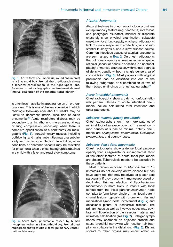
Fig. 3. Acute focal pneumonia (ie, round pneumonia)in a 3-year-old boy. Frontal chest radiograph showsa spherical consolidation in the right upper lobe.Follow-up chest radiograph after treatment showedinterval resolution of this spherical consolidation.
Pneumonia in Normal and Immunocompromised Children 899
is often less masslike in appearance on an orthog-onal view. This is one of the few scenarios in whichradiologic follow-up after about 2 weeks may beuseful to document interval resolution of acutepneumonia.22 Acute respiratory distress may besecondary to an intrathoracic mass causing airwayor lung compression, especially when there iscomplete opacification of a hemithorax on radio-graphs (Fig. 5). Intrapulmonary masses includingbothbenign andmalignant entitiesmaypresent clin-ically with acute superinfection. In addition, otherconditions or anatomic variants may be mistakenfor pneumonia when a chest radiograph is obtainedin a child with a fever and respiratory symptoms.
Fig. 4. Acute focal pneumonia caused by humanmetapneumovirus in a 3-month-old boy. Frontal chestradiograph shows multiple focal pulmonary consoli-dations bilaterally.
Atypical Pneumonia
Atypical features in pneumonia include prominentextrapulmonary features (eg, headache, sore throat,and pharyngeal exudates), minimal or disparatechest signs on physical examination, subacuteonset, nonfocal lung opacity on chest radiographs,lack of clinical response to antibiotics, lack of sub-stantial leukocytosis, and a slow disease course.Common infectious causes of atypical pneumoniaare summarized in Box 2. On chest radiographs,the pulmonary opacity is seen as either airspace,reticular (linear), or bandlike opacities in a nonfocal,patchy, ormottleddistribution,with variousdegreesof density, usually without a single dense area ofconsolidation (Fig. 6). Most patients with atypicalpneumonia can be classified into one of thefollowing subgroups or a combination of two ofthem based on findings on chest radiographs:20
Acute interstitial pneumoniaChest radiographs show a patchy, nonfocal retic-ular pattern. Causes of acute interstitial pneu-monia include self-limited viral infections andother pathogens.
Subacute minimal patchy pneumoniaChest radiographs show 1 or more patches ofminimal foci of airspace opacity. The most com-mon causes of subacute minimal patchy pneu-monia are Mycoplasma pneumoniae, Chlamydiapneumoniae, and adenoviruses.
Subacute dense focal pneumoniaChest radiographs show a dense focal airspaceopacity that is segmental or subsegmental. Mostof the other features of acute focal pneumoniaare absent. Tuberculosis needs to be excluded inthese patients.
Most children exposed to Mycobacterium tu-berculosis do not develop active disease but canhave latent foci that may reactivate at a later dateparticularly if they become immunosuppressed ordebilitated. Primary infection of Mycobacteriumtuberculosis is more likely in infants with localspread from the initial parenchymal/lymph nodecomplex to form larger single or multifocal paren-chymal lesions, typically with prominent hilar andmediastinal lymph node involvement (Fig. 7) andoccasional pleural or pericardial disease. Theprimary focus as well as involved nodes may cavi-tate with liquefaction of the caseous material andultimately calcification (see Fig. 7). Enlarged lymphnodes may encroach on adjacent bronchi andcause bronchial narrowing with resultant air trap-ping or collapse in the distal lung (Fig. 8). Distantspread to other organs may occur either via

Fig. 5. Opacification of left hemithorax and respiratory distress caused by ruptured paraspinal neuroblastoma ina 5-week-old-boy. (A) Frontal chest radiograph shows complete opacification of the left hemithorax with contra-lateral mediastinal shift, which was initially believed to represent pneumonia with large pleural effusion. (B)Axial contrast-enhanced CT shows a left paraspinal mass (blue arrow) with intraspinal extension associatedwith a large pleural effusion, compressive atelectasis of the left lung, and contralateral mediastinal shift. (C) AxialT2-weighted MR image shows the left paraspinal mass (blue arrows) and a complex left pleural effusion withmultiple loculations and fluid-debris levels (red arrows).
Eslamy & Newman900
lymphatics or hematogenously (including militarylung involvement).24,25
Infections with more than 1 organismmay causethe atypical pneumonia pattern, resulting inconfusing persistence of the illness or prominentfindings in another organ system. An example
Box 2Common infectious causes of atypicalpneumonia syndrome in children
Viruses: RSV (<5 years), adenoviruses, parain-fluenza viruses, influenza virus (in epidemics),cytomegalovirus (CMV), varicella zoster virus(immunosuppressed)
Bacteria: Chlamydia trachomatis (<4 months),Mycoplasma pneumoniae (>5 years), Chlamydiapneumoniae (>5 years), Bordatella pertussis
Data from Fisher RG, Boyce TG. Pneumonia syndromes.In: Fisher RG, Boyce TG, editors. Moffet’s pediatricinfectious diseases: a problem-oriented approach.4th edition. Philadelphia: Lippincott Williams & Wil-kins; 2005. p. 174–221.
of this situation is influenza infection with superim-posed typical or atypical pneumonia (see Fig. 6B).The more common mimics that simulate the app-earance of atypical pneumonia syndromes aresummarized in Box 3.
Miliary or Nodular Pneumonia
Miliary or nodular pneumonia is characterized bychest radiographic findings of multiple miliary orlarger nodular opacities. Miliary pneumonia inpediatric patients is seen most commonly in tuber-culous and fungal infections (Fig. 9). Nodularpneumonia (including reticular and reticulonodularpatterns) in pediatric patients is seen in septicemboli, viral pneumonia, lymphocytic interstitialpneumonia associated with Epstein-Barr virus(EBV) infection with underlying human immunode-ficiency virus (HIV) infection, and some fungal andbacterial infections (Box 4; Figs. 10 and 11).25,26
Septic pulmonary emboli usually occur secondaryto a focal Staphylococcus aureus infection(eg, right-sided bacterial endocarditis, septicthrombophlebitis, osteomyelitis, soft tissue
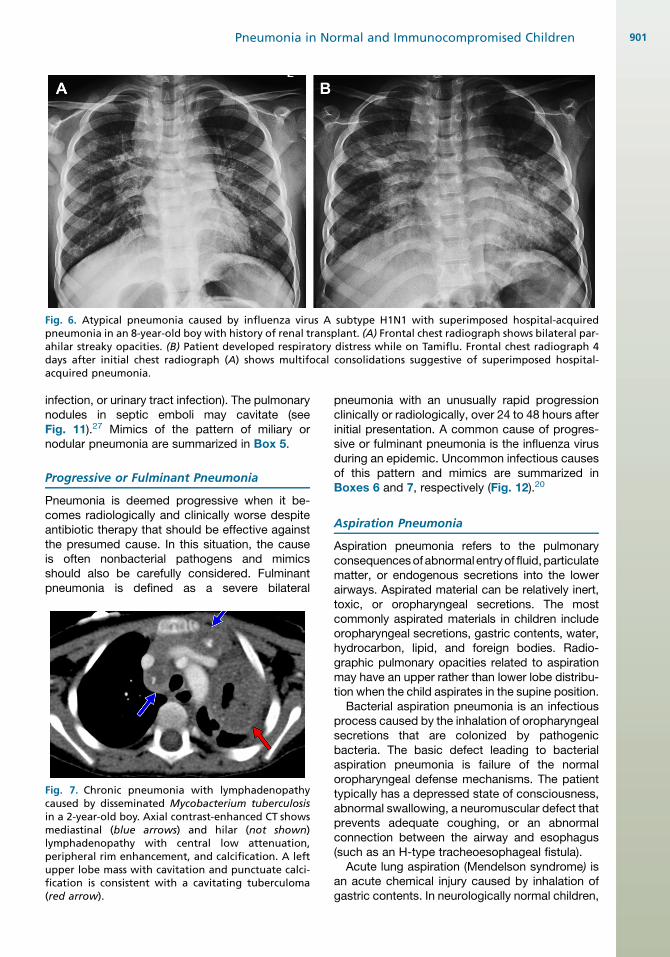
Fig. 6. Atypical pneumonia caused by influenza virus A subtype H1N1 with superimposed hospital-acquiredpneumonia in an 8-year-old boy with history of renal transplant. (A) Frontal chest radiograph shows bilateral par-ahilar streaky opacities. (B) Patient developed respiratory distress while on Tamiflu. Frontal chest radiograph 4days after initial chest radiograph (A) shows multifocal consolidations suggestive of superimposed hospital-acquired pneumonia.
Pneumonia in Normal and Immunocompromised Children 901
infection, or urinary tract infection). The pulmonarynodules in septic emboli may cavitate (seeFig. 11).27 Mimics of the pattern of miliary ornodular pneumonia are summarized in Box 5.
Progressive or Fulminant Pneumonia
Pneumonia is deemed progressive when it be-comes radiologically and clinically worse despiteantibiotic therapy that should be effective againstthe presumed cause. In this situation, the causeis often nonbacterial pathogens and mimicsshould also be carefully considered. Fulminantpneumonia is defined as a severe bilateral
Fig. 7. Chronic pneumonia with lymphadenopathycaused by disseminated Mycobacterium tuberculosisin a 2-year-old boy. Axial contrast-enhanced CT showsmediastinal (blue arrows) and hilar (not shown)lymphadenopathy with central low attenuation,peripheral rim enhancement, and calcification. A leftupper lobe mass with cavitation and punctuate calci-fication is consistent with a cavitating tuberculoma(red arrow).
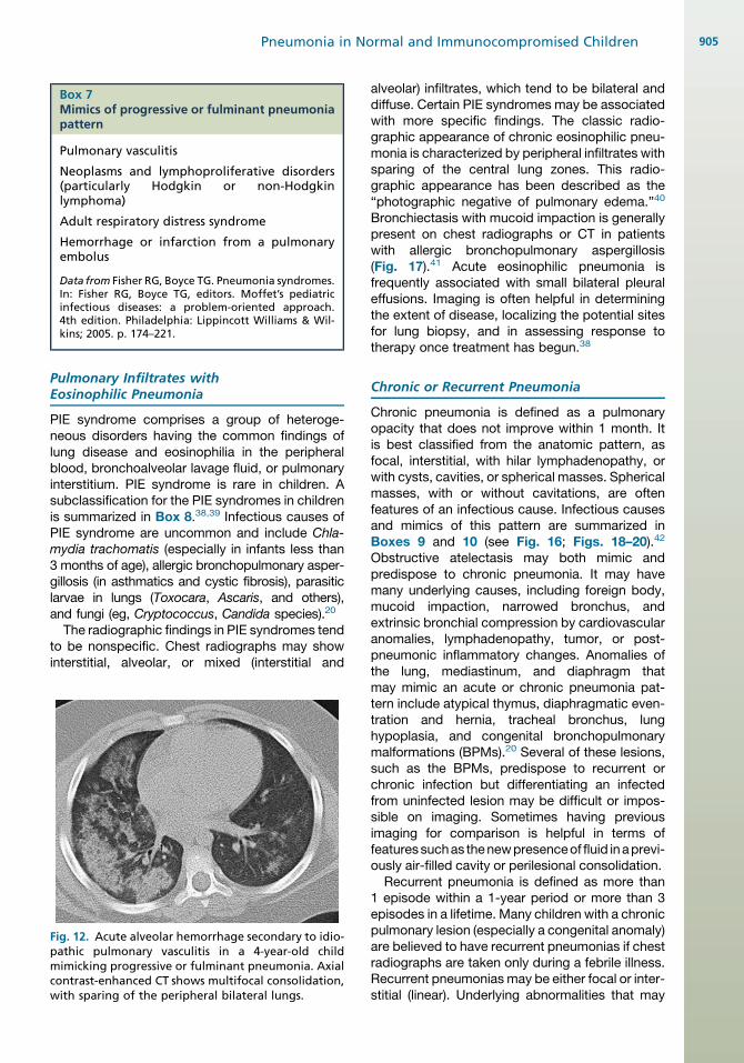
pneumonia with an unusually rapid progressionclinically or radiologically, over 24 to 48 hours afterinitial presentation. A common cause of progres-sive or fulminant pneumonia is the influenza virusduring an epidemic. Uncommon infectious causesof this pattern and mimics are summarized inBoxes 6 and 7, respectively (Fig. 12).20
Aspiration Pneumonia
Aspiration pneumonia refers to the pulmonaryconsequencesofabnormalentryof fluid,particulatematter, or endogenous secretions into the lowerairways. Aspirated material can be relatively inert,toxic, or oropharyngeal secretions. The mostcommonly aspirated materials in children includeoropharyngeal secretions, gastric contents, water,hydrocarbon, lipid, and foreign bodies. Radio-graphic pulmonary opacities related to aspirationmay have an upper rather than lower lobe distribu-tion when the child aspirates in the supine position.
Bacterial aspiration pneumonia is an infectiousprocess caused by the inhalation of oropharyngealsecretions that are colonized by pathogenicbacteria. The basic defect leading to bacterialaspiration pneumonia is failure of the normaloropharyngeal defense mechanisms. The patienttypically has a depressed state of consciousness,abnormal swallowing, a neuromuscular defect thatprevents adequate coughing, or an abnormalconnection between the airway and esophagus(such as an H-type tracheoesophageal fistula).
Acute lung aspiration (Mendelson syndrome) isan acute chemical injury caused by inhalation ofgastric contents. In neurologically normal children,
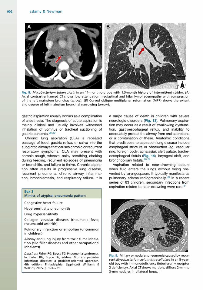
Fig. 8. Mycobacterium tuberculosis in an 11-month-old boy with 1.5-month history of intermittent stridor. (A)Axial contrast-enhanced CT shows low attenuation mediastinal and hilar lymphadenopathy with compressionof the left mainstem bronchus (arrow). (B) Curved oblique multiplanar reformation (MPR) shows the extentand degree of left mainstem bronchial narrowing (arrow).
Eslamy & Newman902
gastric aspiration usually occurs as a complicationof anesthesia. The diagnosis of acute aspiration ismainly clinical and usually involves witnessedinhalation of vomitus or tracheal suctioning ofgastric contents.28,29
Chronic lung aspiration (CLA) is repeatedpassage of food, gastric reflux, or saliva into thesubglottic airways that causes chronic or recurrentrespiratory symptoms. CLA may present withchronic cough, wheeze, noisy breathing, chokingduring feeding, recurrent episodes of pneumoniaor bronchitis, and failure to thrive. Chronic aspira-tion often results in progressive lung disease,recurrent pneumonia, chronic airway inflamma-tion, bronchiectasis, and respiratory failure. It is
Box 3Mimics of atypical pneumonia pattern
Congestive heart failure
Hypersensitivity pneumonitis
Drug hypersensitivity
Collagen vascular diseases (rheumatic fever,rheumatoid arthritis)
Pulmonary infarction or embolism (uncommonin children)
Airway and lung injury from toxic fume inhala-tion (silo filler diseases and other occupationalinhalants)
Data from Fisher RG, Boyce TG. Pneumonia syndromes.In: Fisher RG, Boyce TG, editors. Moffet’s pediatricinfectious diseases: a problem-oriented approach.4th edition. Philadelphia: Lippincott Williams &Wilkins; 2005. p. 174–221.
a major cause of death in children with severeneurologic disorders (Fig. 13). Pulmonary aspira-tion may occur as a result of swallowing dysfunc-tion, gastroesophageal reflux, and inability toadequately protect the airway from oral secretionsor a combination of these. Anatomic conditionsthat predispose to aspiration lung disease includeesophageal stricture or obstruction (eg, vascularring, foreign body, achalasia), cleft palate, trache-oesophageal fistula (Fig. 14), laryngeal cleft, andbronchobiliary fistula.28,29
Aspiration related to near-drowning occurswhen fluid enters the lungs without being pre-vented by laryngospasm. It typically manifests aspulmonary edema radiographically.30 In a recentseries of 83 children, secondary infections fromaspiration related to near-drowning were rare.31
Fig. 9. Miliary or nodular pneumonia caused by recur-rent Mycobacterium avium-intracellulare in an 8-year-old boy with immunodeficiency (interferon g receptor2 deficiency). Axial CT shows multiple, diffuse 2-mm to3-mm nodules in bilateral lungs.

Box 4Infectious causes of acute miliary and nodularpattern (including reticular and reticulonodularpatterns)
Miliary: tuberculosis, fungi (histoplasmosis,coccidioidomycosis, aspergillosis, candidiasis, mu-cormycosis, paracoccidiodomycosis, and blasto-mycosis); listeriosis and streptococci in newborns
Nodular: septic emboli, viral (CMV, varicella zos-ter, herpes simplex virus), psittacosis, Myco-plasma; nocardiosis; aspergillosis; Legionella,cryptococcosis
Reticulonodular: common respiratory viruses,lymphocytic interstitial pneumonitis (LIP)a, Cox-iella burnetti, leptospirosis
a Associated with EBV infection in children with HIVinfection
Data from Fisher RG, Boyce TG. Pneumoniasyndromes. In: Fisher RG, Boyce TG, editors. Moffet’spediatric infectious diseases: a problem-oriented ap-proach. 4th edition. Philadelphia: Lippincott Williams& Wilkins; 2005. p. 174–221.
Pneumonia in Normal and Immunocompromised Children 903
Hydrocarbon pneumonia is an acute, intensechemical pneumonitis after unintentional aspira-tion of volatile hydrocarbon compounds. Mostcases of hydrocarbon pneumonia occur in chil-dren. Chest radiographs typically show bilateral,scattered pulmonary densities with middle andlower zone predominance. Such densities maybecome confluent and progress to acute respira-tory distress syndrome (ARDS) and respiratoryfailure. They typically worsen over the first 72hours and then clear over the next few days.However, occasionally radiographic changesmay take weeks to months to be cleared. Obstruc-tive emphysema, pneumatoceles, subsegmental,or segmental atelectasis may also be seen.32
Fig. 10. Miliary or nodular pneumonia caused by lymphocHIV. (A) Frontal chest radiograph shows diffuse reticulonoeral 2-mm to 3-mm nodules (arrows).
Lipoid pneumonia is a rare form of pneumoniacaused by inhalation or aspiration of a fattysubstance. Oral administration of various oils isa common cultural practice, including mineral oil,olive oil, shark liver oil, cod liver oil, coconut oil,and ghee. Such oily materials can readily slideinto the airway even in normal infants and youngchildren without eliciting a cough reflex and arepoorly removed by cilia. Lipoid pneumonias aretypified by mild, subacute, or chronic clinical find-ings with accompanying marked radiographicchanges. Chest radiographs of children with lipoidpneumonia typically show bilateral parahilar ill-defined, airspace opacities. In a series of 7 pedi-atric patients, CT showed dense consolidationsurrounded by ground-glass opacity witha geographic lobular distribution.33 Within thedense consolidations, areas with relatively lowattenuation were identified in only 1 patient. There-fore, low-density consolidation in the posteriorlungs is an infrequent CT finding in the diagnosisof lipoid pneumonia in children (Fig. 15). Interlob-ular septal thickening in areas of ground-glassopacity (ie, crazy paving pattern) has also beendescribed in children with lipoid pneumonia.33
Lipoid pneumonias may be complicated by super-imposed infection especially with atypical myco-bacteria. Slow recovery usually takes place withcessation of the oil administration. There may beresidual scarring/fibrosis especially with animalrather than vegetable oils.34,35
Foreign body aspiration can also result in pneu-monia. Accidental aspiration of both organicand nonorganic foreign bodies is a cause of child-hood morbidity and mortality, requiring promptrecognition and early treatment to minimize thepotentially serious and sometimes fatal conse-quence. Eating is the most common circumstanceduring which it occurs, with small food items being
ytic interstitial pneumonia in a 19-month-old boy withdular pattern. (B) Axial chest CT shows multiple bilat-
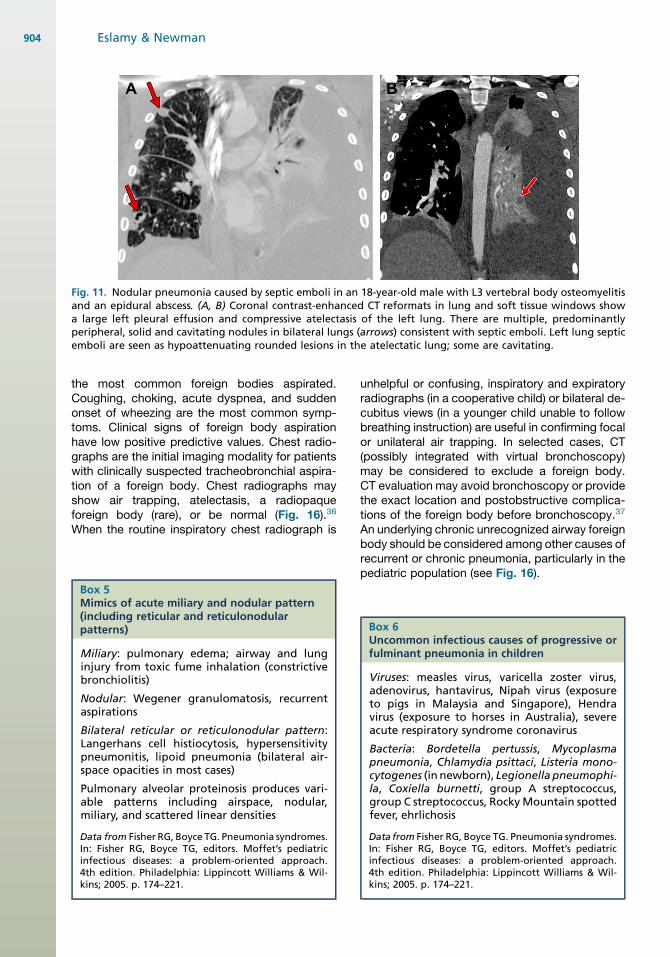
Fig. 11. Nodular pneumonia caused by septic emboli in an 18-year-old male with L3 vertebral body osteomyelitisand an epidural abscess. (A, B) Coronal contrast-enhanced CT reformats in lung and soft tissue windows showa large left pleural effusion and compressive atelectasis of the left lung. There are multiple, predominantlyperipheral, solid and cavitating nodules in bilateral lungs (arrows) consistent with septic emboli. Left lung septicemboli are seen as hypoattenuating rounded lesions in the atelectatic lung; some are cavitating.
Eslamy & Newman904
the most common foreign bodies aspirated.Coughing, choking, acute dyspnea, and suddenonset of wheezing are the most common symp-toms. Clinical signs of foreign body aspirationhave low positive predictive values. Chest radio-graphs are the initial imaging modality for patientswith clinically suspected tracheobronchial aspira-tion of a foreign body. Chest radiographs mayshow air trapping, atelectasis, a radiopaqueforeign body (rare), or be normal (Fig. 16).36
When the routine inspiratory chest radiograph is
Box 5Mimics of acute miliary and nodular pattern(including reticular and reticulonodularpatterns)
Miliary: pulmonary edema; airway and lunginjury from toxic fume inhalation (constrictivebronchiolitis)
Nodular: Wegener granulomatosis, recurrentaspirations
Bilateral reticular or reticulonodular pattern:Langerhans cell histiocytosis, hypersensitivitypneumonitis, lipoid pneumonia (bilateral air-space opacities in most cases)
Pulmonary alveolar proteinosis produces vari-able patterns including airspace, nodular,miliary, and scattered linear densities
Data from Fisher RG, Boyce TG. Pneumonia syndromes.In: Fisher RG, Boyce TG, editors. Moffet’s pediatricinfectious diseases: a problem-oriented approach.4th edition. Philadelphia: Lippincott Williams & Wil-kins; 2005. p. 174–221.
unhelpful or confusing, inspiratory and expiratoryradiographs (in a cooperative child) or bilateral de-cubitus views (in a younger child unable to followbreathing instruction) are useful in confirming focalor unilateral air trapping. In selected cases, CT(possibly integrated with virtual bronchoscopy)may be considered to exclude a foreign body.CT evaluation may avoid bronchoscopy or providethe exact location and postobstructive complica-tions of the foreign body before bronchoscopy.37
An underlying chronic unrecognized airway foreignbody should be considered among other causes ofrecurrent or chronic pneumonia, particularly in thepediatric population (see Fig. 16).
Box 6Uncommon infectious causes of progressive orfulminant pneumonia in children
Viruses: measles virus, varicella zoster virus,adenovirus, hantavirus, Nipah virus (exposureto pigs in Malaysia and Singapore), Hendravirus (exposure to horses in Australia), severeacute respiratory syndrome coronavirus
Bacteria: Bordetella pertussis, Mycoplasmapneumonia, Chlamydia psittaci, Listeria mono-cytogenes (in newborn), Legionella pneumophi-la, Coxiella burnetti, group A streptococcus,group C streptococcus, RockyMountain spottedfever, ehrlichosis
Data from Fisher RG, Boyce TG. Pneumonia syndromes.In: Fisher RG, Boyce TG, editors. Moffet’s pediatricinfectious diseases: a problem-oriented approach.4th edition. Philadelphia: Lippincott Williams & Wil-kins; 2005. p. 174–221.
Box 7Mimics of progressive or fulminant pneumoniapattern
Pulmonary vasculitis
Neoplasms and lymphoproliferative disorders(particularly Hodgkin or non-Hodgkinlymphoma)
Adult respiratory distress syndrome
Hemorrhage or infarction from a pulmonaryembolus
Data from Fisher RG, Boyce TG. Pneumonia syndromes.In: Fisher RG, Boyce TG, editors. Moffet’s pediatricinfectious diseases: a problem-oriented approach.4th edition. Philadelphia: Lippincott Williams & Wil-kins; 2005. p. 174–221.
Pneumonia in Normal and Immunocompromised Children 905
Pulmonary Infiltrates withEosinophilic Pneumonia
PIE syndrome comprises a group of heteroge-neous disorders having the common findings oflung disease and eosinophilia in the peripheralblood, bronchoalveolar lavage fluid, or pulmonaryinterstitium. PIE syndrome is rare in children. Asubclassification for the PIE syndromes in childrenis summarized in Box 8.38,39 Infectious causes ofPIE syndrome are uncommon and include Chla-mydia trachomatis (especially in infants less than3 months of age), allergic bronchopulmonary asper-gillosis (in asthmatics and cystic fibrosis), parasiticlarvae in lungs (Toxocara, Ascaris, and others),and fungi (eg, Cryptococcus, Candida species).20
The radiographic findings in PIE syndromes tendto be nonspecific. Chest radiographs may showinterstitial, alveolar, or mixed (interstitial and
Fig. 12. Acute alveolar hemorrhage secondary to idio-pathic pulmonary vasculitis in a 4-year-old childmimicking progressive or fulminant pneumonia. Axialcontrast-enhanced CT shows multifocal consolidation,with sparing of the peripheral bilateral lungs.
alveolar) infiltrates, which tend to be bilateral anddiffuse. Certain PIE syndromes may be associatedwith more specific findings. The classic radio-graphic appearance of chronic eosinophilic pneu-monia is characterized by peripheral infiltrates withsparing of the central lung zones. This radio-graphic appearance has been described as the“photographic negative of pulmonary edema.”40
Bronchiectasis with mucoid impaction is generallypresent on chest radiographs or CT in patientswith allergic bronchopulmonary aspergillosis(Fig. 17).41 Acute eosinophilic pneumonia isfrequently associated with small bilateral pleuraleffusions. Imaging is often helpful in determiningthe extent of disease, localizing the potential sitesfor lung biopsy, and in assessing response totherapy once treatment has begun.38
Chronic or Recurrent Pneumonia
Chronic pneumonia is defined as a pulmonaryopacity that does not improve within 1 month. Itis best classified from the anatomic pattern, asfocal, interstitial, with hilar lymphadenopathy, orwith cysts, cavities, or spherical masses. Sphericalmasses, with or without cavitations, are oftenfeatures of an infectious cause. Infectious causesand mimics of this pattern are summarized inBoxes 9 and 10 (see Fig. 16; Figs. 18–20).42
Obstructive atelectasis may both mimic andpredispose to chronic pneumonia. It may havemany underlying causes, including foreign body,mucoid impaction, narrowed bronchus, andextrinsic bronchial compression by cardiovascularanomalies, lymphadenopathy, tumor, or post-pneumonic inflammatory changes. Anomalies ofthe lung, mediastinum, and diaphragm thatmay mimic an acute or chronic pneumonia pat-tern include atypical thymus, diaphragmatic even-tration and hernia, tracheal bronchus, lunghypoplasia, and congenital bronchopulmonarymalformations (BPMs).20 Several of these lesions,such as the BPMs, predispose to recurrent orchronic infection but differentiating an infectedfrom uninfected lesion may be difficult or impos-sible on imaging. Sometimes having previousimaging for comparison is helpful in terms offeaturessuchas thenewpresenceof fluid inaprevi-ously air-filled cavity or perilesional consolidation.
Recurrent pneumonia is defined as more than1 episode within a 1-year period or more than 3episodes in a lifetime. Many children with a chronicpulmonary lesion (especially a congenital anomaly)are believed to have recurrent pneumonias if chestradiographs are taken only during a febrile illness.Recurrent pneumonias may be either focal or inter-stitial (linear). Underlying abnormalities that may
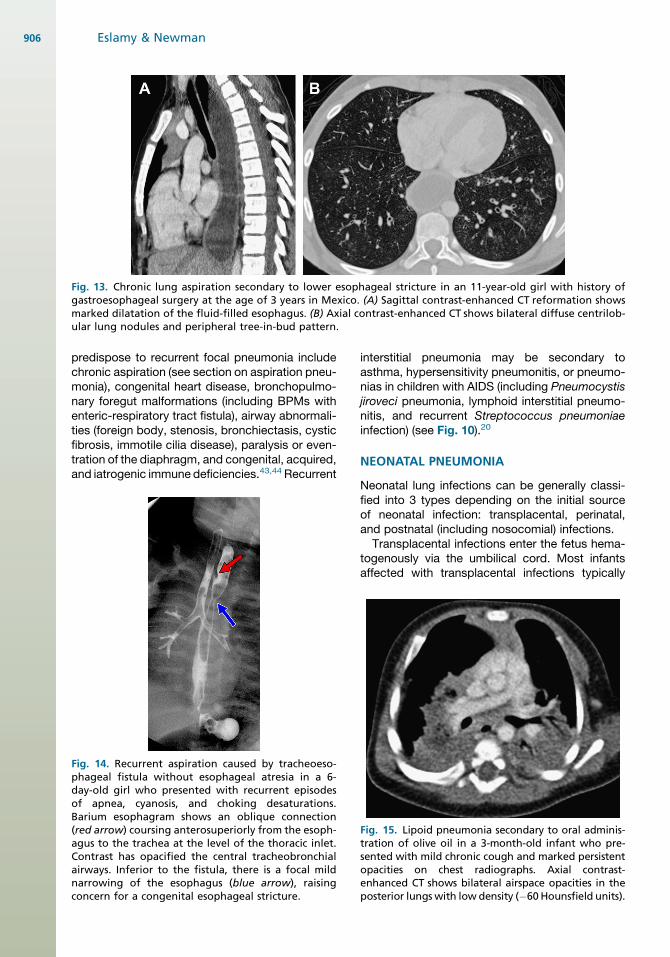
Fig. 13. Chronic lung aspiration secondary to lower esophageal stricture in an 11-year-old girl with history ofgastroesophageal surgery at the age of 3 years in Mexico. (A) Sagittal contrast-enhanced CT reformation showsmarked dilatation of the fluid-filled esophagus. (B) Axial contrast-enhanced CT shows bilateral diffuse centrilob-ular lung nodules and peripheral tree-in-bud pattern.
Eslamy & Newman906
predispose to recurrent focal pneumonia includechronic aspiration (see section on aspiration pneu-monia), congenital heart disease, bronchopulmo-nary foregut malformations (including BPMs withenteric-respiratory tract fistula), airway abnormali-ties (foreign body, stenosis, bronchiectasis, cysticfibrosis, immotile cilia disease), paralysis or even-tration of the diaphragm, and congenital, acquired,and iatrogenic immune deficiencies.43,44 Recurrent
Fig. 14. Recurrent aspiration caused by tracheoeso-phageal fistula without esophageal atresia in a 6-day-old girl who presented with recurrent episodesof apnea, cyanosis, and choking desaturations.Barium esophagram shows an oblique connection(red arrow) coursing anterosuperiorly from the esoph-agus to the trachea at the level of the thoracic inlet.Contrast has opacified the central tracheobronchialairways. Inferior to the fistula, there is a focal mildnarrowing of the esophagus (blue arrow), raisingconcern for a congenital esophageal stricture.
interstitial pneumonia may be secondary toasthma, hypersensitivity pneumonitis, or pneumo-nias in children with AIDS (including Pneumocystisjiroveci pneumonia, lymphoid interstitial pneumo-nitis, and recurrent Streptococcus pneumoniaeinfection) (see Fig. 10).20
NEONATAL PNEUMONIA
Neonatal lung infections can be generally classi-fied into 3 types depending on the initial sourceof neonatal infection: transplacental, perinatal,and postnatal (including nosocomial) infections.Transplacental infections enter the fetus hema-
togenously via the umbilical cord. Most infantsaffected with transplacental infections typically
Fig. 15. Lipoid pneumonia secondary to oral adminis-tration of olive oil in a 3-month-old infant who pre-sented with mild chronic cough and marked persistentopacities on chest radiographs. Axial contrast-enhanced CT shows bilateral airspace opacities in theposterior lungs with lowdensity (�60Hounsfield units).
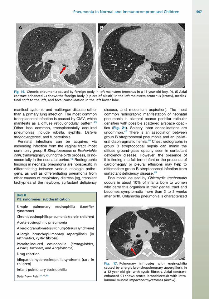
Fig. 16. Chronic pneumonia caused by foreign body in left mainstem bronchus in a 13-year-old boy. (A, B) Axialcontrast-enhanced CT shows the foreign body (a piece of plastic) in the left mainstem bronchus (arrow), medias-tinal shift to the left, and focal consolidation in the left lower lobe.
Pneumonia in Normal and Immunocompromised Children 907
manifest systemic and multiorgan disease ratherthan a primary lung infection. The most commontransplacental infection is caused by CMV, whichmanifests as a diffuse reticulonodular pattern.45
Other less common, transplacentally acquiredpneumonias include rubella, syphilis, Listeriamonocytogenes, and tuberculosis.
Perinatal infections can be acquired viaascending infection from the vaginal tract (mostcommonly group B Streptococcus or Escherichiacoli), transvaginally during the birth process, or no-socomially in the neonatal period.46 Radiographicfindings in neonatal pneumonia are nonspecific indifferentiating between various etiologic patho-gens, as well as differentiating pneumonia fromother causes of respiratory distress (eg, transienttachypnea of the newborn, surfactant deficiency
Box 8PIE syndromes: subclassification
Simple pulmonary eosinophilia (Loefflersyndrome)
Chronic eosinophilic pneumonia (rare in children)
Acute eosinophilic pneumonia
Allergic granulomatosis (Churg-Strauss syndrome)
Allergic bronchopulmonary aspergillosis (inasthmatics, cystic fibrosis)
Parasite-induced eosinophilia (Strongyloides,Ascaris, Toxocara, and Ancylostoma)
Drug reaction
Idiopathic hypereosinophilic syndrome (rare inchildren)
Infant pulmonary eosinophilia
Data from Refs.20,38,39
disease, and meconium aspiration). The mostcommon radiographic manifestation of neonatalpneumonia is bilateral coarse perihilar reticulardensities with possible scattered airspace opaci-ties (Fig. 21). Solitary lobar consolidations areuncommon.47 There is an association betweengroup B streptococcal pneumonia and an ipsilat-eral diaphragmatic hernia.48 Chest radiographs ingroup B streptococcal sepsis can mimic thediffuse ground-glass opacity seen in surfactantdeficiency disease. However, the presence ofthis finding in a full-term infant or the presence ofcardiomegaly or pleural effusions may help todifferentiate group B streptococcal infection fromsurfactant deficiency disease.49
Pneumonia caused by Chlamydia trachomatisoccurs in about 10% of infants born to womenwho carry this organism in their genital tract andbecomes symptomatic more than 2 to 3 weeksafter birth. Chlamydia pneumonia is characterized
Fig. 17. Pulmonary infiltrates with eosinophiliacaused by allergic bronchopulmonary aspergillosis ina 12-year-old girl with cystic fibrosis. Axial contrast-enhanced CT shows central bronchiectasis with intra-luminal mucoid impaction/mycetomas (arrow).
Box 9Infectious causes of chronic pneumonia syndrome
1. Chronic focal pulmonary disease
a. Untreated or undertreated acute pneumonia
i. Bacteria and Mycobacteria: Mycobacterium tuberculosis, nontuberculous mycobacteria (espe-cially in children with defective cell-mediated immunity)
ii. Systemic fungi: histoplasmosis, coccidioidomycosis, blastomycosis cryptococcosis, sporotrichosis
iii. Parasites: Paragonimiasis
2. Chronic interstitial pulmonary disease
a. Bacteria: Chlamydia trachomatis
b. Viruses: CMV, late-onset congenital rubella syndrome, LIP (in HIV infection)
c. Fungi: Pneumocystis jiroveci
3. Chronic pneumonia with hilar lymphadenopathy
a. Common
i. Bacteria and Mycobacteria: Mycobacterium tuberculosis, Mycoplasma pneumonia, Chlamydiapneumonia; Fungi: histoplasmosis
b. Uncommon
i. Bacteria: nontuberculous mycobacteria, Actinomyces, Francisella tularensis, Bacillus anthracis
ii. Fungi: coccidioidomycosis, blastomycosis
iii. Lung abscess
4. Chronic cavitary, cystic, or nodular pneumonias
a. Lung abscess, necrotizing bacterial pneumonia
i. Bacteria and Mycobacteria: tuberculosis, actinomycosis, nocardiosis; Nocardia and Rhodococ-cus equi in immunocompromised patients
ii. Fungi: Aspergillus, Pneumocystis jiroveci and other systemic fungi in immunocompromisedpatients
iii. Viruses: atypical measles, papillomatosis of lung
iv. Protozoa and helminths: paragonimiasis, Entamoeba histolytica, Ecchinococcus, Dirofilariaimmitis
Data from Fisher RG, Boyce TG. Pneumonia syndromes. In: Fisher RG, Boyce TG, editors. Moffet’s pediatric infectiousdiseases: a problem-oriented approach. 4th edition. Philadelphia: Lippincott Williams & Wilkins; 2005. p. 174–221; andKawanami T, Bowen A. Juvenile laryngeal papillomatosis with pulmonary parenchymal spread. Case report and reviewof the literature. Pediatr Radiol 1985;15(2):102–4.
Eslamy & Newman908
by hyperinflation and bilateral diffuse reticular peri-hilar densities that are disparate with relatively mildclinical symptoms.50 Concomitant conjunctivitis,which used to be a useful clue to the cause, is pre-vented by the routine instillation of antibacterial eyedrops at birth. Neonates with chlamydia pneu-monia frequently have accompanying eosinophilia.Bordetella pertussis has recently resurfaced to
produce epidemics of infection, probably relatedto waning community immunity. The clinicalpresentation of pertussis in the newborn maylack some features (characteristic whoopingcough and fever) typical of the disease in olderchildren. The clinical presentation of the most
severely affected newborns may be dominatedby marked respiratory distress, cyanosis, andapnea. Mortality caused by pertussis usuallyresults from secondary pneumonia, encephalop-athy, cardiac failure, or pulmonary hypertension.A suggested mechanism for pulmonary hyperten-sion that may develop in newborns with Bordetellapertussis infection is formation of leukocytethrombi in pulmonary venules secondary tohyperleukocytosis.51,52 The classic radiographicappearance in pertussis is the shaggy heart withdiffuse peribronchial cuffing related to airwayinflammation. However, chest radiographic find-ings such as hyperaeration, atelectasis, segmental

Box 10Mimics of chronic pneumonia pattern
1. Chronic focal pulmonary disease pattern
a. Malignancy (neuroblastoma)
b. Obstructive atelectasis
c. Foreign body, mucous plug, or endobron-chial tumor
d. Congenital anomalies of lung, thymus, ormediastinum
e. Vascular rings
f. Eventration of diaphragm
g. Inflammatory pseudotumor of lung
2. Chronic interstitial pulmonary disease pattern
a. Bronchopulmonary dysplasia
b. Congestive heart failure
c. Pulmonary sarcoidosis
d. Collagen vascular diseases
e. Idiopathic interstitial pneumonia
f. Pulmonary hemosiderosis
g. Constrictive bronchiolitis
h. Histiocytosis (including Langerhans cellhistiocytosis)
i. Pulmonary alveolar proteinosis
3. Chronic pneumonia pattern with hilarlymphadenopathy
a. Benign lymphoproliferative disorders andlymphoma
b. Other neoplasms
4. Chronic cavitary, cystic, or nodular pneu-monia pattern
a. Congenital anomalies
b. Malignancy (lymphoma and metastases)
c. Traumatic pneumatocele
d. Hyper-IgE syndrome
e. a1-antitrypsin deficiency
f. Langerhans cell histiocytosis
g. Pulmonary sarcoidosis
h. Bronchopulmonary dysplasia
i. Cystic bronchiectasis
j. Wegener granulomatosis
Data from Fisher RG, Boyce TG. Pneumonia syndromes.In: Fisher RG, Boyce TG, editors. Moffet’s pediatricinfectious diseases: a problem-oriented approach.4th edition. Philadelphia: Lippincott Williams & Wil-kins; 2005. p. 174–221.
Pneumonia in Normal and Immunocompromised Children 909
consolidation, and lymphadenopathy are usuallynonspecific.
Recurrent pulmonary infection, with bacterial,viral, or occasionally fungal pathogens, are frequentproblems in neonates undergoing prolonged hospi-talization and complex treatments, especially inpremature infants with chronic lung disease. Radio-graphic alterations caused by infection maybe subtle when superimposed on chronic lungchanges.47
PNEUMONIA IN IMMUNOCOMPROMISEDHOSTS
Pneumonia is a common disease in the immuno-compromised host. Immunocompromise maybe congenital (congenital immunodeficiencies),acquired (HIV/AIDS, malnutrition) or iatrogenic(during chemotherapy for cancer or after tissuetransplantation). Immunodeficient states can resultin: (1) humoral immunodeficiency (hypogammaglo-bulinemia, functional B-lymphocyte deficiencyaccompanying HIV infection); (2) cellular immuno-deficiency (severe malnutrition, late stages ofAIDS, some congenital immunodeficiencies suchas DiGeorge syndrome); and (3) neutrophildysfunction and neutropenia (chronic granulo-matous disease, pure neutropenia). Iatrogenicimmunodeficiencies may be a combination ofneutropenia or neutrophil dysfunction, innate ordrug-induced defective lymphocyte function, anddrug-induced breaks in the oral and intestinalmucosal barriers.53
The causes of pneumonia in the immunocom-promised host consist not only of the same agentsthat cause pneumonia in the normal host but alsoof several opportunistic agents depending onthe type and severity of immunodeficiency aswell as temporal pattern after chemotherapy ortransplant.
In an immunocompromised child with a noncon-tributory chest radiograph and clinical findings thatcould be attributed to a lung infection, chest CT isoften required for evaluation of a possible lunginfection. In this situation, there are 4 major advan-tages of chest CT over chest radiographs. First,the presence, pattern, and extent of the diseaseprocess are better visualized. Second, more than1 pattern of abnormality may be detected, sug-gesting dual pathologic entities. Third, invasivediagnostic procedures (eg, bronchoscopy or nee-dle aspiration) can be more precisely planned.Fourth, CT also allows for increased sensitivity inassessment of the response to treatment.54,55
Although the radiographic or CT appearancemight not be specific for a pathogen, knowledgeof the clinical setting in combination with the
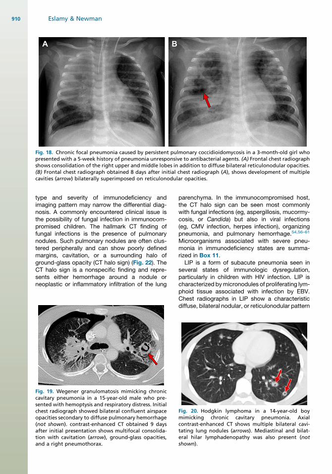
Fig. 18. Chronic focal pneumonia caused by persistent pulmonary coccidioidomycosis in a 3-month-old girl whopresented with a 5-week history of pneumonia unresponsive to antibacterial agents. (A) Frontal chest radiographshows consolidation of the right upper and middle lobes in addition to diffuse bilateral reticulonodular opacities.(B) Frontal chest radiograph obtained 8 days after initial chest radiograph (A), shows development of multiplecavities (arrow) bilaterally superimposed on reticulonodular opacities.
Eslamy & Newman910
type and severity of immunodeficiency andimaging pattern may narrow the differential diag-nosis. A commonly encountered clinical issue isthe possibility of fungal infection in immunocom-promised children. The hallmark CT finding offungal infections is the presence of pulmonarynodules. Such pulmonary nodules are often clus-tered peripherally and can show poorly definedmargins, cavitation, or a surrounding halo ofground-glass opacity (CT halo sign) (Fig. 22). TheCT halo sign is a nonspecific finding and repre-sents either hemorrhage around a nodule orneoplastic or inflammatory infiltration of the lung
Fig. 19. Wegener granulomatosis mimicking chroniccavitary pneumonia in a 15-year-old male who pre-sented with hemoptysis and respiratory distress. Initialchest radiograph showed bilateral confluent airspaceopacities secondary to diffuse pulmonary hemorrhage(not shown). contrast-enhanced CT obtained 9 daysafter initial presentation shows multifocal consolida-tion with cavitation (arrow), ground-glass opacities,and a right pneumothorax.
parenchyma. In the immunocompromised host,the CT halo sign can be seen most commonlywith fungal infections (eg, aspergillosis, mucormy-cosis, or Candida) but also in viral infections(eg, CMV infection, herpes infection), organizingpneumonia, and pulmonary hemorrhage.54,56–61
Microorganisms associated with severe pneu-monia in immunodeficiency states are summa-rized in Box 11.
LIP is a form of subacute pneumonia seen inseveral states of immunologic dysregulation,particularly in children with HIV infection. LIP ischaracterized bymicronodules of proliferating lym-phoid tissue associated with infection by EBV.Chest radiographs in LIP show a characteristicdiffuse, bilateral nodular, or reticulonodular pattern
Fig. 20. Hodgkin lymphoma in a 14-year-old boymimicking chronic cavitary pneumonia. Axialcontrast-enhanced CT shows multiple bilateral cavi-tating lung nodules (arrows). Mediastinal and bilat-eral hilar lymphadenopathy was also present (notshown).
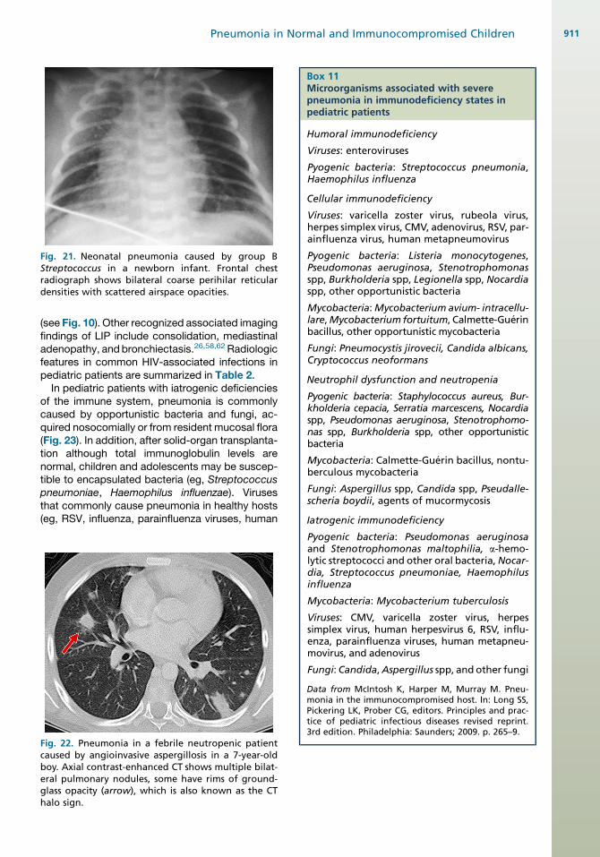
Fig. 21. Neonatal pneumonia caused by group BStreptococcus in a newborn infant. Frontal chestradiograph shows bilateral coarse perihilar reticulardensities with scattered airspace opacities.
Box 11Microorganisms associated with severepneumonia in immunodeficiency states inpediatric patients
Humoral immunodeficiency
Viruses: enteroviruses
Pyogenic bacteria: Streptococcus pneumonia,Haemophilus influenza
Cellular immunodeficiency
Viruses: varicella zoster virus, rubeola virus,herpes simplex virus, CMV, adenovirus, RSV, par-ainfluenza virus, human metapneumovirus
Pyogenic bacteria: Listeria monocytogenes,Pseudomonas aeruginosa, Stenotrophomonasspp, Burkholderia spp, Legionella spp, Nocardiaspp, other opportunistic bacteria
Mycobacteria:Mycobacterium avium- intracellu-lare,Mycobacterium fortuitum, Calmette-Guerinbacillus, other opportunistic mycobacteria
Fungi: Pneumocystis jirovecii, Candida albicans,Cryptococcus neoformans
Neutrophil dysfunction and neutropenia
Pyogenic bacteria: Staphylococcus aureus, Bur-kholderia cepacia, Serratia marcescens, Nocardiaspp, Pseudomonas aeruginosa, Stenotrophomo-nas spp, Burkholderia spp, other opportunisticbacteria
Mycobacteria: Calmette-Guerin bacillus, nontu-berculous mycobacteria
Fungi: Aspergillus spp, Candida spp, Pseudalle-scheria boydii, agents of mucormycosis
Iatrogenic immunodeficiency
Pneumonia in Normal and Immunocompromised Children 911
(see Fig. 10). Other recognized associated imagingfindings of LIP include consolidation, mediastinaladenopathy, andbronchiectasis.26,58,62 Radiologicfeatures in common HIV-associated infections inpediatric patients are summarized in Table 2.
In pediatric patients with iatrogenic deficienciesof the immune system, pneumonia is commonlycaused by opportunistic bacteria and fungi, ac-quired nosocomially or from resident mucosal flora(Fig. 23). In addition, after solid-organ transplanta-tion although total immunoglobulin levels arenormal, children and adolescents may be suscep-tible to encapsulated bacteria (eg, Streptococcuspneumoniae, Haemophilus influenzae). Virusesthat commonly cause pneumonia in healthy hosts(eg, RSV, influenza, parainfluenza viruses, human
Fig. 22. Pneumonia in a febrile neutropenic patientcaused by angioinvasive aspergillosis in a 7-year-oldboy. Axial contrast-enhanced CT shows multiple bilat-eral pulmonary nodules, some have rims of ground-glass opacity (arrow), which is also known as the CThalo sign.
Pyogenic bacteria: Pseudomonas aeruginosaand Stenotrophomonas maltophilia, a-hemo-lytic streptococci and other oral bacteria,Nocar-dia, Streptococcus pneumoniae, Haemophilusinfluenza
Mycobacteria: Mycobacterium tuberculosis
Viruses: CMV, varicella zoster virus, herpessimplex virus, human herpesvirus 6, RSV, influ-enza, parainfluenza viruses, human metapneu-movirus, and adenovirus
Fungi:Candida,Aspergillus spp, and other fungi
Data from McIntosh K, Harper M, Murray M. Pneu-monia in the immunocompromised host. In: Long SS,Pickering LK, Prober CG, editors. Principles and prac-tice of pediatric infectious diseases revised reprint.3rd edition. Philadelphia: Saunders; 2009. p. 265–9.
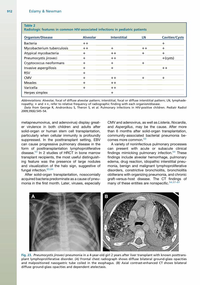
Table 2Radiologic features in common HIV-associated infections in pediatric patients
Organism/Disease Alveolar Interstitial LN Cavities/Cysts
Bacteria 11 1
Mycobacterium tuberculosis 11 1 11 1
Atypical mycobacteria 1 11 1 1
Pneumocystis jiroveci 1 11 1(cysts)
Cryptococcus neoformans 1 1 1
Invasive aspergillosis 1 1 11
RSV 1
CMV 1 11 1 1
Measles 1 11
Varicella 1 11
Herpes simplex 1
Abbreviations: Alveolar, focal of diffuse alveolar pattern; interstitial, focal or diffuse interstitial pattern; LN, lymphade-nopathy; 1 and 11, refer to relative frequency of radiographic finding with each organism/disease.
Data from George R, Andronikou S, Theron S, et al. Pulmonary infections in HIV-positive children. Pediatr Radiol2009;39(6):545–54.
Eslamy & Newman912
metapneumovirus, and adenovirus) display great-er virulence in both children and adults aftersolid-organ or human stem cell transplantation,particularly when cellular immunity is profoundlysuppressed. In the posttransplant setting, EBVcan cause progressive pulmonary disease in theform of posttransplantation lymphoproliferativedisease.53 In 2 studies of HRCT in bone marrowtransplant recipients, the most useful distinguish-ing feature was the presence of large nodulesand visualization of the halo sign, suggestive offungal infection.63,64
After solid-organ transplantation, nosocomiallyacquired bacteria predominate as a cause of pneu-monia in the first month. Later, viruses, especially
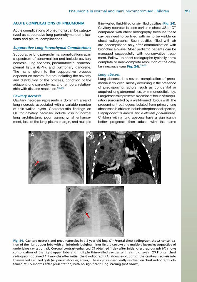
Fig. 23. Pneumocystis jiroveci pneumonia in a 4-year-old gplant lymphoproliferative disorder. (A) Frontal chest radiand malpositioned nasogastric tube coiled in the esophdiffuse ground-glass opacities and dependent atelectasis.
CMV and adenovirus, as well as Listeria, Nocardia,and Aspergillus, may be the cause. After morethan 6 months after solid-organ transplantation,community-associated bacterial pneumonia be-comes more common.53
A variety of noninfectious pulmonary processescan present with acute or subacute clinicalfindings mimicking pulmonary infection.54 Thesefindings include alveolar hemorrhage, pulmonaryedema, drug reaction, idiopathic interstitial pneu-monia, benign and malignant lymphoproliferativedisorders, constrictive bronchiolitis, bronchiolitisobliterans with organizing pneumonia, and chronicgraft-versus-host disease. The CT findings ofmany of these entities are nonspecific.54,57–61
irl 2 years after liver transplant with known posttrans-ograph shows diffuse bilateral ground-glass opacitiesagus. (B) Axial contrast-enhanced CT shows bilateral
Pneumonia in Normal and Immunocompromised Children 913
ACUTE COMPLICATIONS OF PNEUMONIA
Acute complications of pneumonia can be catego-rized as suppurative lung parenchymal complica-tions and pleural complications.
Suppurative Lung Parenchymal Complications
Suppurative lung parenchymal complications spana spectrum of abnormalities and include cavitarynecrosis, lung abscess, pneumatocele, broncho-pleural fistula (BPF), and pulmonary gangrene.The name given to the suppurative processdepends on several factors including the severityand distribution of the process, condition of theadjacent lung parenchyma, and temporal relation-ship with disease resolution.65,66
Cavitary necrosisCavitary necrosis represents a dominant area oflung necrosis associated with a variable numberof thin-walled cysts. Characteristic findings onCT for cavitary necrosis include loss of normallung architecture, poor parenchymal enhance-ment, loss of the lung-pleural margin, and multiple
Fig. 24. Cavitary necrosis and pneumatoceles in a 2-year-otion of the right upper lobe with an inferiorly bulging minunderlying cavitation. (B) Coronal contrast-enhanced CT oconsolidation of the right upper lobe and multiple thin-radiograph obtained 1.5 months after initial chest radiogthin-walled air-filled cysts (ie, pneumatoceles; arrow). Thestained at 3.5 months after presentation, with no significa
thin-walled fluid-filled or air-filled cavities (Fig. 24).Cavitary necrosis is seen earlier in chest US or CTcompared with chest radiography because thesecavities need to be filled with air to be visible onchest radiographs. Such cavities filled with airare accomplished only after communication withbronchial airways. Most pediatric patients can bemanaged successfully with conservative treat-ment. Follow-up chest radiographs typically showcomplete or near-complete resolution of the cavi-tary necrosis (see Fig. 24).65,66
Lung abscessLung abscess is a severe complication of pneu-monia in children, mostly occurring in the presenceof predisposing factors, such as congenital oracquired lung abnormalities, or immunodeficiency.Lungabscess representsadominant focusof suppu-ration surrounded by a well-formed fibrous wall. Thepredominant pathogens isolated from primary lungabscesses in children include streptococcal species,Staphylococcus aureus and Klebsiella pneumoniae.Children with a lung abscess have a significantlybetter prognosis than adults with the same
ld boy. (A) Frontal chest radiograph shows consolida-or fissure (arrow) and multiple lucencies suggestive ofbtained 1 day after initial chest radiograph (A) showswalled cavities with air-fluid levels. (C) Frontal chestraph (A) shows evolution of the cavitary necrosis intoe cysts subsequently resolved on chest radiographs ob-nt lung scarring (not shown).
Eslamy & Newman914
condition.67 Lung abscesses are uncommon inimmunocompetent children. On contrast-enhancedCT, a lung abscess appears as a fluid-filled or air-filled cavity with a thick definable, enhancing wall(Fig. 25).65,66,68 Although lung abscess in childrenhas been managed successfully for many yearswith prolonged courses of intravenous antibiotics,the evolution of interventional radiology has seenthe accelerated use of percutaneously placed pigtaildrainage catheters using US and CT guidance.67
PneumatocelePneumatocele is a term given to thin, smooth-walled air-filled cysts seen at imaging and mayrepresent a later or less severe stage of resolvingor healing lung necrosis (see Fig. 24). Pneumato-celes are most often caused by severe lung infec-tion from staphylococcal pneumonia. However,they may be seen with other bacterial infectionsincluding streptococcus pneumonia and afterhydrocarbon aspiration. On CT, thin-walled smallor large cysts containing air with or without fluidare identified. The wall of a pneumatocele doesnot enhance. The surrounding lung may be opaci-fied but does not typically show findings of lungnecrosis.65,66 Pneumatoceles usually resolvespontaneously over time although pneumatocelesmay be atypically persistent in children with hyper-IGE syndrome. Large pneumatoceles containingfluid can be a source of ongoing infection andmay occasionally require drainage.
Bronchopleural fistulaBPF is defined as a communication between thelung parenchyma or airways and the pleuralspace. Central BPFs (ie, main or lobar bronchicommunicating with the pleural cavity) most often
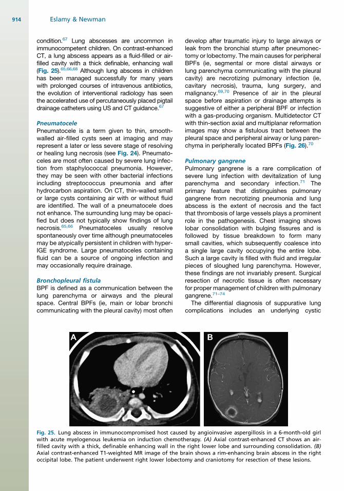
Fig. 25. Lung abscess in immunocompromised host causewith acute myelogenous leukemia on induction chemothfilled cavity with a thick, definable enhancing wall in theAxial contrast-enhanced T1-weighted MR image of the boccipital lobe. The patient underwent right lower lobecto
develop after traumatic injury to large airways orleak from the bronchial stump after pneumonec-tomy or lobectomy. Themain causes for peripheralBPFs (ie, segmental or more distal airways orlung parenchyma communicating with the pleuralcavity) are necrotizing pulmonary infection (ie,cavitary necrosis), trauma, lung surgery, andmalignancy.69,70 Presence of air in the pleuralspace before aspiration or drainage attempts issuggestive of either a peripheral BPF or infectionwith a gas-producing organism. Multidetector CTwith thin-section axial and multiplanar reformationimages may show a fistulous tract between thepleural space and peripheral airway or lung paren-chyma in peripherally located BPFs (Fig. 26).70
Pulmonary gangrenePulmonary gangrene is a rare complication ofsevere lung infection with devitalization of lungparenchyma and secondary infection.71 Theprimary feature that distinguishes pulmonarygangrene from necrotizing pneumonia and lungabscess is the extent of necrosis and the factthat thrombosis of large vessels plays a prominentrole in the pathogenesis. Chest imaging showslobar consolidation with bulging fissures and isfollowed by tissue breakdown to form manysmall cavities, which subsequently coalesce intoa single large cavity occupying the entire lobe.Such a large cavity is filled with fluid and irregularpieces of sloughed lung parenchyma. However,these findings are not invariably present. Surgicalresection of necrotic tissue is often necessaryfor proper management of children with pulmonarygangrene.71–74
The differential diagnosis of suppurative lungcomplications includes an underlying cystic
d by angioinvasive aspergillosis in a 6-month-old girlerapy. (A) Axial contrast-enhanced CT shows an air-right lower lobe and surrounding consolidation. (B)
rain shows a rim-enhancing brain abscess in the rightmy and craniotomy for resection of these lesions.
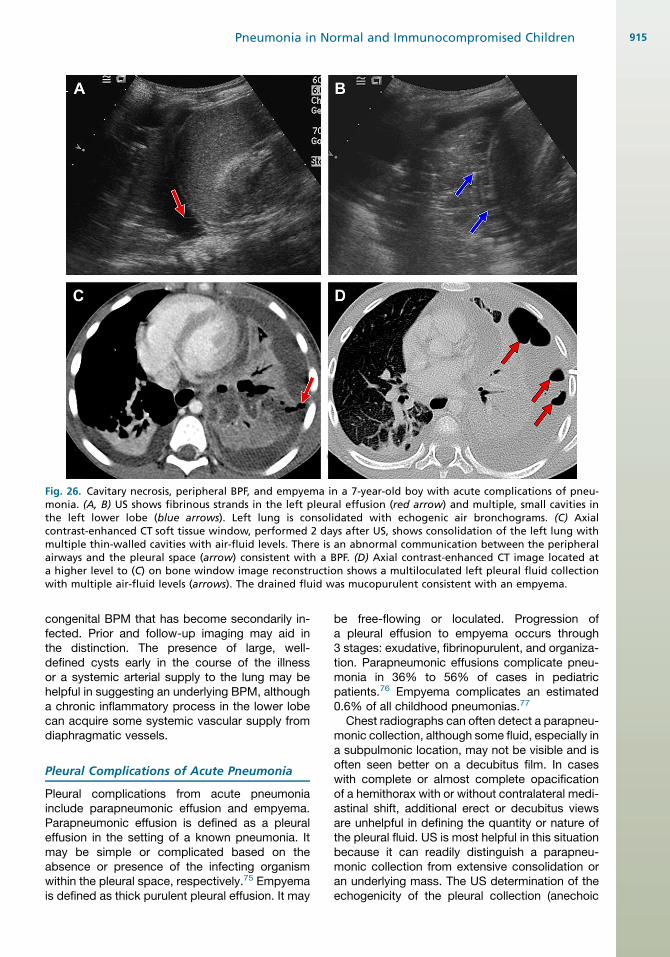
Fig. 26. Cavitary necrosis, peripheral BPF, and empyema in a 7-year-old boy with acute complications of pneu-monia. (A, B) US shows fibrinous strands in the left pleural effusion (red arrow) and multiple, small cavities inthe left lower lobe (blue arrows). Left lung is consolidated with echogenic air bronchograms. (C) Axialcontrast-enhanced CT soft tissue window, performed 2 days after US, shows consolidation of the left lung withmultiple thin-walled cavities with air-fluid levels. There is an abnormal communication between the peripheralairways and the pleural space (arrow) consistent with a BPF. (D) Axial contrast-enhanced CT image located ata higher level to (C) on bone window image reconstruction shows a multiloculated left pleural fluid collectionwith multiple air-fluid levels (arrows). The drained fluid was mucopurulent consistent with an empyema.
Pneumonia in Normal and Immunocompromised Children 915
congenital BPM that has become secondarily in-fected. Prior and follow-up imaging may aid inthe distinction. The presence of large, well-defined cysts early in the course of the illnessor a systemic arterial supply to the lung may behelpful in suggesting an underlying BPM, althougha chronic inflammatory process in the lower lobecan acquire some systemic vascular supply fromdiaphragmatic vessels.
Pleural Complications of Acute Pneumonia
Pleural complications from acute pneumoniainclude parapneumonic effusion and empyema.Parapneumonic effusion is defined as a pleuraleffusion in the setting of a known pneumonia. Itmay be simple or complicated based on theabsence or presence of the infecting organismwithin the pleural space, respectively.75 Empyemais defined as thick purulent pleural effusion. It may
be free-flowing or loculated. Progression ofa pleural effusion to empyema occurs through3 stages: exudative, fibrinopurulent, and organiza-tion. Parapneumonic effusions complicate pneu-monia in 36% to 56% of cases in pediatricpatients.76 Empyema complicates an estimated0.6% of all childhood pneumonias.77
Chest radiographs can often detect a parapneu-monic collection, although some fluid, especially ina subpulmonic location, may not be visible and isoften seen better on a decubitus film. In caseswith complete or almost complete opacificationof a hemithorax with or without contralateral medi-astinal shift, additional erect or decubitus viewsare unhelpful in defining the quantity or nature ofthe pleural fluid. US is most helpful in this situationbecause it can readily distinguish a parapneu-monic collection from extensive consolidation oran underlying mass. The US determination of theechogenicity of the pleural collection (anechoic
Eslamy & Newman916
or echogenic) and showing fibrin strands, septa-tions, loculations, or fibrinous pleural rind is helpfulin determining appropriate therapy (see Fig. 26).Treatment options for parapneumonic effusions/empyemas include antibiotics alone, simple tubedrainage, chest drain insertion with fibrinolytics,or surgery (eg, video-assisted thoracoscopic sur-gery or open thoracotomy with decortication).Although imaging techniques are used as a guide-line, they do not always accurately stage em-pyema, predict outcome, or guide decisionsregarding surgical versus medical management.75
CT provides a more global overview of pleuralandpulmonary abnormality fromacutepneumonia,but is poor at differentiating parapneumonic effu-sion from empyema in pediatric patients. Findingson CT, in patients with parapneumonic effusion/empyema, include: (1) enhancement and thick-ening of visceral and parietal pleura; (2) thickeningand increased density of extrapleural subcostaltissues; and (3) increased attenuation of extrapleu-ral subcostal fat.78 Loculation canbe inferredby thepresence of a lenticular fluid collection or nonde-pendent air. Septations are usually not appreciatedon CT (see Fig. 26).Pleuropulmonary infection may occasionally
spread to involve the chest wall, including softtissues and adjacent bones. Mycobacteriumtuberculosis, Aspergillus, and Actinomyces arethe most common organisms in this scenario.
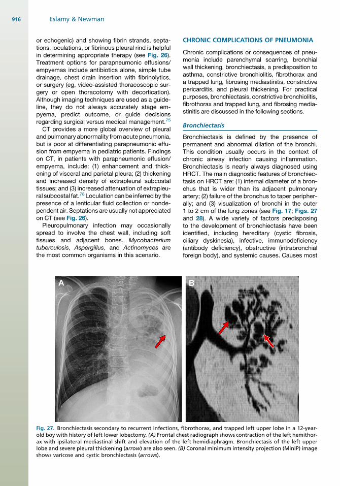
Fig. 27. Bronchiectasis secondary to recurrent infections, fold boy with history of left lower lobectomy. (A) Frontal chax with ipsilateral mediastinal shift and elevation of thelobe and severe pleural thickening (arrow) are also seen. (Bshows varicose and cystic bronchiectasis (arrows).
CHRONIC COMPLICATIONS OF PNEUMONIA
Chronic complications or consequences of pneu-monia include parenchymal scarring, bronchialwall thickening, bronchiectasis, a predisposition toasthma, constrictive bronchiolitis, fibrothorax anda trapped lung, fibrosing mediastinitis, constrictivepericarditis, and pleural thickening. For practicalpurposes, bronchiectasis, constrictive bronchiolitis,fibrothorax and trapped lung, and fibrosing media-stinitis are discussed in the following sections.
Bronchiectasis
Bronchiectasis is defined by the presence ofpermanent and abnormal dilation of the bronchi.This condition usually occurs in the context ofchronic airway infection causing inflammation.Bronchiectasis is nearly always diagnosed usingHRCT. The main diagnostic features of bronchiec-tasis on HRCT are: (1) internal diameter of a bron-chus that is wider than its adjacent pulmonaryartery; (2) failure of the bronchus to taper peripher-ally; and (3) visualization of bronchi in the outer1 to 2 cm of the lung zones (see Fig. 17; Figs. 27and 28). A wide variety of factors predisposingto the development of bronchiectasis have beenidentified, including hereditary (cystic fibrosis,ciliary dyskinesia), infective, immunodeficiency(antibody deficiency), obstructive (intrabronchialforeign body), and systemic causes. Causes most
ibrothorax, and trapped left upper lobe in a 12-year-est radiograph shows contraction of the left hemithor-left hemidiaphragm. Bronchiectasis of the left upper) Coronal minimum intensity projection (MinIP) image
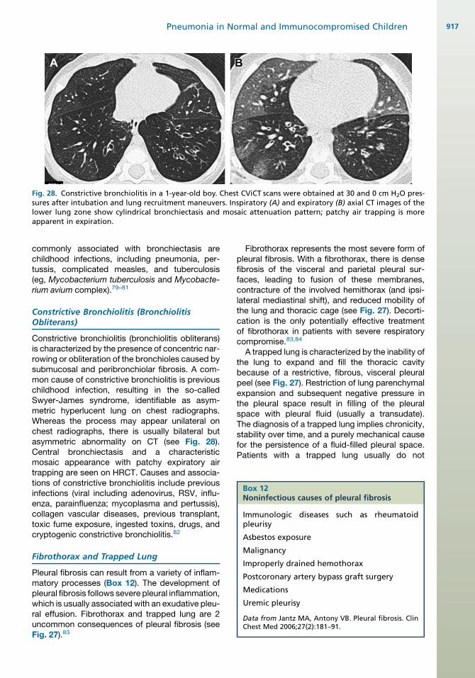
Fig. 28. Constrictive bronchiolitis in a 1-year-old boy. Chest CViCT scans were obtained at 30 and 0 cm H2O pres-sures after intubation and lung recruitment maneuvers. Inspiratory (A) and expiratory (B) axial CT images of thelower lung zone show cylindrical bronchiectasis and mosaic attenuation pattern; patchy air trapping is moreapparent in expiration.
Box 12Noninfectious causes of pleural fibrosis
Immunologic diseases such as rheumatoidpleurisy
Asbestos exposure
Malignancy
Improperly drained hemothorax
Postcoronary artery bypass graft surgery
Medications
Uremic pleurisy
Data from Jantz MA, Antony VB. Pleural fibrosis. ClinChest Med 2006;27(2):181–91.
Pneumonia in Normal and Immunocompromised Children 917
commonly associated with bronchiectasis arechildhood infections, including pneumonia, per-tussis, complicated measles, and tuberculosis(eg, Mycobacterium tuberculosis and Mycobacte-rium avium complex).79–81
Constrictive Bronchiolitis (BronchiolitisObliterans)
Constrictive bronchiolitis (bronchiolitis obliterans)is characterized by the presence of concentric nar-rowing or obliteration of the bronchioles caused bysubmucosal and peribronchiolar fibrosis. A com-mon cause of constrictive bronchiolitis is previouschildhood infection, resulting in the so-calledSwyer-James syndrome, identifiable as asym-metric hyperlucent lung on chest radiographs.Whereas the process may appear unilateral onchest radiographs, there is usually bilateral butasymmetric abnormality on CT (see Fig. 28).Central bronchiectasis and a characteristicmosaic appearance with patchy expiratory airtrapping are seen on HRCT. Causes and associa-tions of constrictive bronchiolitis include previousinfections (viral including adenovirus, RSV, influ-enza, parainfluenza; mycoplasma and pertussis),collagen vascular diseases, previous transplant,toxic fume exposure, ingested toxins, drugs, andcryptogenic constrictive bronchiolitis.82
Fibrothorax and Trapped Lung
Pleural fibrosis can result from a variety of inflam-matory processes (Box 12). The development ofpleural fibrosis follows severe pleural inflammation,which is usually associated with an exudative pleu-ral effusion. Fibrothorax and trapped lung are 2uncommon consequences of pleural fibrosis (seeFig. 27).83
Fibrothorax represents the most severe form ofpleural fibrosis. With a fibrothorax, there is densefibrosis of the visceral and parietal pleural sur-faces, leading to fusion of these membranes,contracture of the involved hemithorax (and ipsi-lateral mediastinal shift), and reduced mobility ofthe lung and thoracic cage (see Fig. 27). Decorti-cation is the only potentially effective treatmentof fibrothorax in patients with severe respiratorycompromise.83,84
A trapped lung is characterized by the inability ofthe lung to expand and fill the thoracic cavitybecause of a restrictive, fibrous, visceral pleuralpeel (see Fig. 27). Restriction of lung parenchymalexpansion and subsequent negative pressure inthe pleural space result in filling of the pleuralspace with pleural fluid (usually a transudate).The diagnosis of a trapped lung implies chronicity,stability over time, and a purely mechanical causefor the persistence of a fluid-filled pleural space.Patients with a trapped lung usually do not
Eslamy & Newman918
experience improvement in dyspnea after thora-centesis. In symptomatic patients, decorticationshould be considered. The underlying lung paren-chyma should be assessed before decortication. Ifthe trapped lung is severely diseased and fibrotic,decortication is unlikely to result in lung reexpan-sion and the procedure does not provide symp-tomatic benefit. In contrast, lung entrapment isthe result of an active inflammatory process ormalignancy in the pleural space, leading to arestricted pleural space. Pleural fluid from lungentrapment is an exudate, and symptoms inpatients with lung entrapment typically improveafter thoracentesis.83,85
Fibrosing Mediastinitis
Fibrosing mediastinitis is a rare condition charac-terized by proliferation of fibrous tissue within themediastinum. Symptoms are related to compres-sion of the central airways, superior vena cava,pulmonary veins, pulmonary arteries, and esoph-agus. The most common cause of this disorderis fungal infection, especially Histoplasma capsu-latum in the United States.86
SUMMARY
Pneumonia is an infection of the lung parenchymacaused by a wide variety of organisms in pediatricpatients. Imaging evaluation plays an importantrole in children with pneumonia by detecting thepresence of pneumonia and determining its loca-tion and extent, excluding other thoracic causesof respiratory symptoms, and showing complica-tions such as effusion/empyema and suppurativelung changes. Clear understanding of the under-lying potential cause, current role of imaging,proper imaging techniques, and characteristicimaging appearances of acute and chronic pneu-monias can guide optimal management of pedi-atric patients with pneumonia.
REFERENCES
1. Rudan I, Tomaskovic L, Boschi-Pinto C, et al. WHO
Child Health Epidemiology Reference Group. Global
estimate of the incidence of clinical pneumonia
among children under five years of age. Bull World
Health Organ 2004;82(12):895–903.
2. Murphy TF, Henderson FW, Clyde WA Jr, et al. Pneu-
monia: an eleven-year study in a pediatric practice.
Am J Epidemiol 1981;113(1):12–21.
3. Jokinen C, Heiskanen L, Juvonen H, et al. Incidence
of community-acquired pneumonia in the population
of four municipalities in eastern Finland. Am J Epide-
miol 1993;137(9):977–88.
4. Wardlaw T, Salama P, Johansson EW, et al. Pneu-
monia: the leading killer of children. Lancet 2006;
368(9541):1048–50.
5. Westra SJ, ChoyG. What imaging should we perform
for the diagnosis and management of pulmonary in-
fections? Pediatr Radiol 2009;39(Suppl 2):S178–83.
6. Virkki R, Juven T, Rikalainen H, et al. Differentiation
of bacterial and viral pneumonia in children. Thorax
2002;57(5):438–41.
7. Korppi M, Heiskanen-Kosma T, Jalonen E, et al. Aeti-
ology of community-acquired pneumonia in children
treated in hospital. Eur J Pediatr 1993;152(1):24–30.
8. Riccabona M. Ultrasound of the chest in children
(mediastinum excluded). Eur Radiol 2008;18(2):
390–9.
9. Kim OH, Kim WS, Kim MJ, et al. US in the diagnosis
of pediatric chest diseases. Radiographics 2000;
20(3):653–71.
10. NievelsteinRA,vanDamIM,vanderMolenAJ.Multide-
tectorCT inchildren:currentconceptsanddosereduc-
tion strategies. Pediatr Radiol 2010;40(8):1324–44.
11. Robinson TE. Computed tomography scanning
techniques for the evaluation of cystic fibrosis lung
disease. Proc Am Thorac Soc 2007;4(4):310–5.
12. Sargent MA, Jamieson DH, McEachern AM, et al.
Increased inspiratory pressure for reduction of atel-
ectasis in children anesthetized for CT scan. Pediatr
Radiol 2002;32(5):344–7.
13. Peltola V, Ruuskanen O, Svedstrom E. Magnetic
resonance imaging of lung infections in children. Pe-
diatr Radiol 2008;38(11):1225–31.
14. Hebestreit A, Schultz G, Trusen A, et al. Follow-up of
acute pulmonary complications in cystic fibrosis by
magnetic resonance imaging. Acta Paediatr 2004;
93:414–6.
15. Puderbach M, Eichinger M. The role of advanced
imaging techniques incystic fibrosis follow-up: is there
a place for MRI? Pediatr Radiol 2010;40(6):844–9.
16. Hansell DM, Lynch DA, McAdams HP, et al. Basic
patterns in lung disease. In: Hansell DM, Lynch DA,
McAdams HP, et al, editors. Imaging of diseases
of the chest. 5th edition. Philadelphia: Mosby; 2010.
p. 89, 139–43.
17. Selwyn BJ. The epidemiology of acute respiratory
tract infection in young children: comparison of find-
ings from several developing countries. Rev Infect
Dis 1990;12(Suppl 8):S870–88.
18. Vicencio AG. Susceptibility to bronchiolitis in infants.
Curr Opin Pediatr 2010;22(3):302–6.
19. Dawson KP, Long A, Kennedy J, et al. The chest
radiograph in acute bronchiolitis. J Paediatr Child
Health 1990;26(4):209–11.
20. Fisher RG, Boyce TG. Pneumonia syndromes. In:
Fisher RG, Boyce TG, editors. Moffet’s pediatric
infectious diseases: a problem-oriented approach.
4th edition. Philadelphia: Lippincott Williams &
Wilkins; 2005. p. 174–221.
Pneumonia in Normal and Immunocompromised Children 919
21. Kantor HG. The many radiologic facies of pneumo-
coccal pneumonia. AJR Am J Roentgenol 1981;
137(6):1213–20.
22. Kim YW, Donnelly LF. Round pneumonia: imaging
findings in a large series of children. Pediatr Radiol
2007;37(12):1235–40.
23. Brodzinski H, Ruddy RM. Review of new and newly
discovered respiratory tract viruses in children. Pe-
diatr Emerg Care 2009;25(5):352–60.
24. Powell DA, Hunt WG. Tuberculosis in children: an
update. Adv Pediatr 2006;53:279–322.
25. Inselman LS. Tuberculosis in children: an update.
Pediatr Pulmonol 1996;21(2):101–20.
26. Pitcher RD, Beningfield SJ, Zar HJ. Chest radio-
graphic features of lymphocytic interstitial pneumo-
nitis in HIV-infected children. Clin Radiol 2010;65(2):
150–4.
27. Wong KS, Lin TY, Huang YC, et al. Clinical and
radiographic spectrum of septic pulmonary embo-
lism. Arch Dis Child 2002;87(4):312–5.
28. de Benedictis FM, Carnielli VP, de Benedictis D.
Aspiration lung disease. Pediatr Clin North Am
2009;56(1):173–90.
29. Lodha R, Puranik M, Natchu UCM, et al. Recurrent
pneumonia in children: clinical profile and under-
lying causes. Acta Paediatr 2002;91(11):1170–3.
30. Wunderlich P, Rupprecht E, Trefftz F, et al. Chest
radiographs of near-drowned children. Pediatr Ra-
diol 1985;15(5):297–9.
31. Forler J, Carsin A, Arlaud K, et al. Respiratory
complications of accidental drownings in children.
Arch Pediatr 2010;17(1):14–8.
32. Eade NR, Taussig LM, Marks MI. Hydrocarbon
pneumonitis. Pediatrics 1974;54(3):351–7.
33. Lee KH, Kim WS, Cheon JE, et al. Squalene aspira-
tion pneumonia in children: radiographic and CT
findings as the first clue to diagnosis. Pediatr Radiol
2005;35(6):619–23.
34. Zanetti G, Marchiori E, Gasparetto TD, et al. Lipoid
pneumonia in children following aspiration of mineral
oil used in the treatment of constipation: high-
resolution CT findings in 17 patients. Pediatr Radiol
2007;37(11):1135–9.
35. Hadda V, Khilnani GC, Bhalla AS, et al. Lipoid pneu-
monia presenting as non resolving community
acquired pneumonia: a case report. Cases J 2009;
16(2):9332.
36. Passali D, Lauriello M, Bellussi L, et al. Foreign body
inhalation in children: an update. Acta Otorhinolar-
yngol Ital 2010;30(1):27–32.
37. Adaletli I, Kurugoglu S, Ulus S, et al. Utilization of low
dose multidetector CT and virtual bronchoscopy in
children with suspected foreign body aspiration. Pe-
diatr Radiol 2007;37:33–40.
38. Oermann CM, Panesar KS, Langston C, et al.
Pulmonary infiltrates with eosinophilia syndromes in
children. J Pediatr 2000;136(3):351–8.
39. Chitkara RK, Krishna G. Parasitic pulmonary eosino-
philia.SeminRespirCritCareMed2006;27(2):171–84.
40. Gaensler EA, Carrington CB. Peripheral opacities in
chronic eosinophilic pneumonia: the photographic
negative of pulmonary edema. AJR Am J Roentgenol
1977;128:1–13.
41. Allen JN, Davis WB. Eosinophilic lung diseases:
state of the art. Am J Respir Crit Care Med 1994;
150:1423–38.
42. Kawanami T, Bowen A. Juvenile laryngeal papillo-
matosis with pulmonary parenchymal spread. Case
report and review of the literature. Pediatr Radiol
1985;15(2):102–4.
43. Kaplan KA, Beierle EA, Faro A, et al. Recurrent
pneumonia in children: a case report and approach
to diagnosis. Clin Pediatr (Phila) 2006;45(1):15–22.
44. Couriel J. Assessment of the child with recurrent
chest infections. Br Med Bull 2002;61(1):115–32.
45. Manson D. Diagnostic imaging of neonatal pneu-
monia. In:DonoghueVK, editor. Radiological imaging
of the neonatal chest. 2nd edition. Berlin: Springer;
2007. p. 102.
46. Belady PH, Farkouh LJ, Gibbs RS. Intra-amniotic
infection and premature rupture of the membranes.
Clin Perinatol 1997;24(1):43–57.
47. NewmanB. Imagingofmedicaldiseaseof thenewborn
lung. Radiol Clin North Am 1999;37(6):1049–65.
48. Potter B, Philipps AF, Bierny JP, et al. Neonatal radi-
ology. Acquired diaphragmatic hernia with group B
streptococcal pneumonia. J Perinatol 1995;15(2):
160–2.
49. Leonidas JC, Hall RT, Beatty EC, et al. Radiographic
findings in early onset neonatal group b strepto-
coccal septicemia.Pediatrics 1977;59(Suppl[6Pt 2]):
1006–11.
50. Hammerschlag MR. Chlamydia trachomatis in chil-
dren. Pediatr Ann 1994;23(7):349–53.
51. Soares S, Rocha G, Pissarra S, et al. Pertussis with
severe pulmonary hypertension in a newborn with
good outcome–case report. Rev Port Pneumol 2008;
14(5):687–92.
52. Kundrat SL, Wolek TL, Rowe-Telow M. Malignant
pertussis in the pediatric intensive care unit. Dimens
Crit Care Nurs 2010;29(1):1–5.
53. McIntosh K, Harper M, Murray M. Pneumonia in
the immunocompromised host. In: Long SS,
Pickering LK, ProberCG, editors. Principles andprac-
tice of pediatric infectious diseases revised reprint.
3rd edition. Philadelphia: Saunders; 2009. p. 265–9.
54. Mori M, Galvin JR, Barloon TJ, et al. Fungal pulmo-
nary infections after bone marrow transplantation:
evaluation with radiography and CT. Radiology
1991;178(3):721–6.
55. Wilson S, Grundy R, Vyas H. Investigation and
management of a child who is immunocompromised
and neutropoenic with pulmonary infiltrates. Arch
Dis Child Educ Pract Ed 2009;94:129–37.
Eslamy & Newman920
56. Lee YR, Choi YW, Lee KJ, et al. CT halo sign: the
spectrum of pulmonary diseases. Br J Radiol 2005;
78(933):862–5.
57. McAdamsHP, Rosado-de-ChristensonML, Templeton
PA, et al. Thoracic mycoses from opportunistic fungi:
radiologic-pathologic correlation. Radiographics 1995;
15(2):271–86.
58. Marks MJ, Haney PJ, McDermott MP, et al. Thoracic
disease in children with AIDS. Radiographics 1996;
16(6):1349–62.
59. Winer-Muram HT, Rubin SA, Fletcher BD, et al.
Childhood leukemia: diagnostic accuracy of
bedside chest radiography for severe pulmonary
complications. Radiology 1994;193(1):127–33.
60. BrownMJ, Miller RR, Muller NL. Acute lung disease in
the immunocompromised host: CT and pathologic
examination findings. Radiology 1994;190(1):247–54.
61. Kang EY, Patz EF Jr, Muller NL. Cytomegalovirus
pneumonia in transplant patients: CT findings.
J Comput Assist Tomogr 1996;20(2):295–9.
62. George R, Andronikou S, Theron S, et al. Pulmonary
infections in HIV-positive children. Pediatr Radiol
2009;39(6):545–54.
63. Escuissato DL, Gasparetto EL, Marchiori E, et al.
Pulmonary infections after bone marrow transplanta-
tion: high-resolution CT findings in 111 patients. AJR
Am J Roentgenol 2005;185(3):608–15.
64. Gasparetto TD, Escuissato DL, Marchiori E. Pulmo-
nary infections following bone marrow transplanta-
tion: high-resolution CT findings in 35 paediatric
patients. Eur J Radiol 2008;66(1):117–21.
65. Donnelly LF, Klosterman LA. Pneumonia in children:
decreased parenchymal contrast enhancement–CT
sign of intense illness and impending cavitary
necrosis. Radiology 1997;205(3):817–20.
66. Donnelly LF, Klosterman LA. Cavitary necrosis
complicating pneumonia in children: sequential find-
ings on chest radiography. AJR Am J Roentgenol
1998;171(1):253–6.
67. Patradoon-Ho P, Fitzgerald DA. Lung abscess in
children. Paediatr Respir Rev 2007;8(1):77–84.
68. Leonardi S, del Giudice MM, Spicuzza L, et al. Lung
abscess in a child with Mycoplasma pneumoniae
infection. Eur J Pediatr 2010;169(11):1413–5.
69. Westcott JL, Volpe JP. Peripheral bronchopleural
fistula: CT evaluation in 20 patients with pneumonia,
empyema, or postoperative air leak. Radiology
1995;196:175–81.
70. Seo H, Kim TJ, Jin KN, et al. Multi-detector row
computed tomographic evaluation of bronchopleural
fistula: correlation with clinical, bronchoscopic, and
surgical findings. J Comput Assist Tomogr 2010;
34(1):13–8.
71. Refaely Y, Weissberg D. Gangrene of the lung: treat-
ment in two stages. Ann Thorac Surg 1997;64(4):
970–3.
72. Danner PK, McFarland DR, Felson B. Massive
pulmonary gangrene. Am J Roentgenol Radium
Ther Nucl Med 1968;103(3):548–54.
73. Penner C, Maycher B, Long R. Pulmonary gangrene.
A complication of bacterial pneumonia. Chest 1994;
105(2):567–73.
74. Kothari PR, Jiwane A, Kulkarni B. Pulmonary
gangrene complicating bacterial pneumonia. Indian
Pediatr 2003;40(8):784–5.
75. Calder A, Owens CM. Imaging of parapneumonic
pleural effusions and empyema in children. Pediatr
Radiol 2009;39(6):527–37.
76. Kurt BA, Winterhalter KM, Connors RH, et al. Therapy
of parapneumonic effusions in children: video-
assisted thoracoscopic surgery versus conven-
tional thoracostomy drainage. Pediatrics 2006;118:
e547–53.
77. Jaffe A, Balfour-Lynn IM. Management of empyema
in children. Pediatr Pulmonol 2005;40:148–56.
78. Donnelly LF, Klosterman LA. CTappearance of para-
pneumonic effusions in children: findings are not
specific for empyema. AJR Am J Roentgenol 1997;
169(1):179–82.
79. Pasteur MC, Helliwell SM, Houghton SJ, et al. An
investigation into causative factors in patients with
bronchiectasis. Am J Respir Crit Care Med 2000;
162(4 Pt 1):1277–84.
80. King PT. The pathophysiology of bronchiectasis. Int
J Chron Obstruct Pulmon Dis 2009;4:411–9.
81. PappaletteraM, Aliberti S, Castellotti P, et al. Bronchi-
ectasis: an update. Clin Respir J 2009;3(3):126–34.
82. Devakonda A, Raoof S, Sung A, et al. Bronchiolar
disorders: a clinical-radiological diagnostic algo-
rithm. Chest 2010;137(4):938–51.
83. Jantz MA, Antony VB. Pleural fibrosis. Clin Chest
Med 2006;27(2):181–91.
84. Morton JR, Boushy SF, Guinn GA. Physiological
evaluation of results of pulmonary decortication.
Ann Thorac Surg 1970;9:321–6.
85. Doelken P, Sahn SA. Trapped lung. Semin Respir
Crit Care Med 2001;22:631–5.
86. Miyata T, Takahama M, Yamamoto R, et al. Scle-
rosing mediastinitis mimicking anterior mediastinal
tumor. Ann Thorac Surg 2009;88(1):293–5.