Embed Size (px)
Citation preview
Published: September 12, 2011
r 2011 American Chemical Society 2666 dx.doi.org/10.1021/ci200168b | J. Chem. Inf. Model. 2011, 51, 2666–2679
ARTICLE
pubs.acs.org/jcim
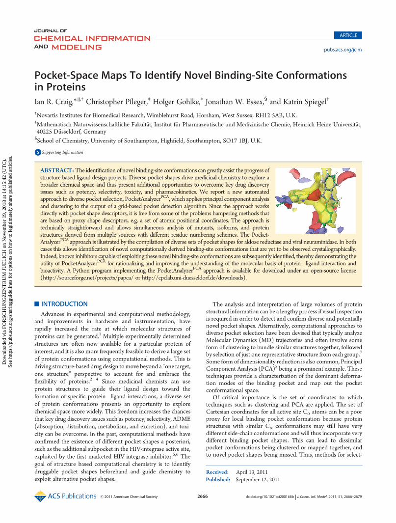
Pocket-Space Maps To Identify Novel Binding-Site Conformationsin ProteinsIan R. Craig,*,||,† Christopher Pfleger,‡ Holger Gohlke,‡ Jonathan W. Essex,§ and Katrin Spiegel†
†Novartis Institutes for Biomedical Research, Wimblehurst Road, Horsham, West Sussex, RH12 5AB, U.K.‡Mathematisch-Naturwissenschaftliche Fakult€at, Institut f€ur Pharmazeutische und Medizinische Chemie, Heinrich-Heine-Universit€at,40225 D€usseldorf, Germany§School of Chemistry, University of Southampton, Highfield, Southampton, SO17 1BJ, U.K.
bS Supporting Information
’ INTRODUCTION
Advances in experimental and computational methodology,and improvements in hardware and instrumentation, haverapidly increased the rate at which molecular structures ofproteins can be generated.1 Multiple experimentally determinedstructures are often now available for a particular protein ofinterest, and it is also more frequently feasible to derive a large setof protein conformations using computational methods. This isdriving structure-based drug design tomove beyond a “one target,one structure” perspective to account for and embrace theflexibility of proteins.2�4 Since medicinal chemists can useprotein structures to guide their ligand design toward theformation of specific protein�ligand interactions, a diverse setof protein conformations presents an opportunity to explorechemical space more widely. This freedom increases the chancesthat key drug discovery issues such as potency, selectivity, ADME(absorption, distribution, metabolism, and excretion), and toxi-city can be overcome. In the past, computational methods haveconfirmed the existence of different pocket shapes a posteriori,such as the additional subpocket in the HIV-integrase active site,exploited by the first marketed HIV-integrase inhibitor.5,6 Thegoal of structure based computational chemistry is to identifydruggable pocket shapes beforehand and guide chemistry toexploit alternative pocket shapes.
The analysis and interpretation of large volumes of proteinstructural information can be a lengthy process if visual inspectionis required in order to detect and confirm diverse and potentiallynovel pocket shapes. Alternatively, computational approaches todiverse pocket selection have been devised that typically analyzeMolecular Dynamics (MD) trajectories and often involve someform of clustering to bundle similar structures together, followedby selection of just one representative structure from each group.7
Some form of dimensionality reduction is also common, PrincipalComponent Analysis (PCA)8 being a prominent example. Thesetechniques provide a characterization of the dominant deforma-tion modes of the binding pocket and map out the pocketconformational space.
Of critical importance is the set of coordinates to whichtechniques such as clustering and PCA are applied. The set ofCartesian coordinates for all active site Cα atoms can be a poorproxy for local binding pocket conformation because proteinstructures with similar Cα conformations may still have verydifferent side-chain conformations and will thus incorporate verydifferent binding pocket shapes. This can lead to dissimilarpocket conformations being clustered or mapped together, andto novel pocket shapes being missed. Thus, methods for select-
Received: April 13, 2011
ABSTRACT:The identification of novel binding-site conformations can greatly assist the progress ofstructure-based ligand design projects. Diverse pocket shapes drive medicinal chemistry to explore abroader chemical space and thus present additional opportunities to overcome key drug discoveryissues such as potency, selectivity, toxicity, and pharmacokinetics. We report a new automatedapproach to diverse pocket selection, PocketAnalyzerPCA, which applies principal component analysisand clustering to the output of a grid-based pocket detection algorithm. Since the approach worksdirectly with pocket shape descriptors, it is free from some of the problems hampering methods thatare based on proxy shape descriptors, e.g. a set of atomic positional coordinates. The approach istechnically straightforward and allows simultaneous analysis of mutants, isoforms, and proteinstructures derived from multiple sources with different residue numbering schemes. The Pocket-AnalyzerPCA approach is illustrated by the compilation of diverse sets of pocket shapes for aldose reductase and viral neuraminidase. In bothcases this allows identification of novel computationally derived binding-site conformations that are yet to be observed crystallographically.Indeed, known inhibitors capable of exploiting these novel binding-site conformations are subsequently identified, thereby demonstrating theutility of PocketAnalyzerPCA for rationalizing and improving the understanding of the molecular basis of protein�ligand interaction andbioactivity. A Python program implementing the PocketAnalyzerPCA approach is available for download under an open-source license(http://sourceforge.net/projects/papca/ or http://cpclab.uni-duesseldorf.de/downloads).
Dow
nloa
ded
via
FOR
SCH
UN
GZ
EN
TR
UM
JU
EL
ICH
on
Nov
embe
r 19
, 201
8 at
14:
15:4
2 (U
TC
).
See
http
s://p
ubs.
acs.
org/
shar
ingg
uide
lines
for
opt
ions
on
how
to le
gitim
atel
y sh
are
publ
ishe
d ar
ticle
s.

2667 dx.doi.org/10.1021/ci200168b |J. Chem. Inf. Model. 2011, 51, 2666–2679
Journal of Chemical Information and Modeling ARTICLE
ing a diverse set of pocket conformations should instead usedescriptors that account for all the atoms lining the pocket. Anobvious choice is the set of Cartesian positions for all atomsbordering the binding site.7,9 However, collating this set ofdescriptors can be technically awkward when comparing proteinstructures with different residue numberings and/or atom order-ings. It also precludes the inclusion of protein structures withmutations of one or more binding pocket residues or structuresof more than one isoform. Furthermore, there can be consider-able ambiguity in defining the set of “binding site atoms”,particularly for flexible proteins.
In this paper, we address the problem of diverse pocket selec-tion from an alternative perspective by working directly withpocket shape descriptors rather than a set of proxy coordinates.In particular, we develop a procedure that reduces a largecollection of structures of the same protein to a subset that retainsa number of substantially distinct binding pocket conformations.The approach is based on applying PCA and clustering to theoutput of a grid-based pocket detection algorithm. The PCAyields (a) principal component (PC) eigenvectors, which revealthe dominant deformation modes of the pocket, and (b) PCprojections (“scores”), which provide characterization and visua-lization of the pocket conformational distribution (PCD). Cluster-ing of the PCD then results in an all-atom approach to diversepocket selection that is free from the problems hamperingmethodsbased on atomic coordinates. As our method builds upon thePocketAnalyzer pocket detection code developed in the Gohlkegroup, we call it PocketAnalyzerPCA. Although itself unpublished,the original PocketAnalyzer approach implements a variant ofthe LIGSITE algorithm.10,11 Minor differences between Pocket-Analyer and LIGSITE are described below. The approach istechnically straightforward and allows simultaneous analysis ofmutants, isoforms, and homologous proteins. Furthermore, althoughwe focus on the identification of novel pocket conformations fromMD simulations, our procedure is applicable to any source ofatomistic protein structural information, and all combinations thereof.
Here, we apply PocketAnalyzerPCA to two proteins that exhibitmoderate but significant active site flexibility, namely aldose reduc-tase and neuraminidase. Both proteins have been extensivelystudied, resulting in a well-characterized set of binding pocket
conformations with which to compare the output of our approachto diverse pocket selection. Since the PocketAnalyzerPCA techniqueis potentially applicable to problems beyond diverse pocketselection, the paper closes by highlighting some directions forfuture work.
’MATERIALS AND METHODS
PocketAnalyzerPCA. Outline.We first give a short overview ofthe PocketAnalyzerPCA approach (Figure 1), before dealing withthe methodological details in more depth. In the first step, a grid-based pocket detection algorithm is applied to an ensemble ofprotein structures. The identified pockets from each singleprotein structure are represented as a row vector of integers, eachencoding the inclusion (“1”) or exclusion (“0”) of a particular gridpoint. The row vector describes the pocket shape correspondingto a specific conformation of the protein. Since the same set ofgrid-points is used to analyze each protein structure, the rowvectors can be merged to produce a pocket shape matrix. Eachcolumn of this pocket shape matrix then represents the varyinginclusion/exclusion of a particular grid-point in the pockets of anensemble of protein structures.The pocket shape matrix is subjected to Principal Component
Analysis (PCA).12 This results in (a) a set of PC eigenvectors, (b)a set of eigenvalues that correspond to the variance along eachPC, and (c) the projections (or “scores”) of each protein structurealong each PC. The high-variance PCs describe the dominantchanges in pocket conformation within the given set of proteinstructures. The scores characterize the distribution of pocketshapes along the PCs, providing a map of the pocket conforma-tional space. Analysis and comparison of these pocket conforma-tional distributions (PCDs) may provide a useful perspective on avariety of questions related to molecular recognition and proteindynamics. Of particular relevance to medicinal chemistry effortsis the rephrasing of the diverse pocket selection problem in termsof finding a small number of protein structures whose pocketsnevertheless provide coverage of the significant regions of the PCD.In this work a clustering of the PCD is used to achieve this aim.Pocket Detection.The pocket detection algorithm implemented
within the PocketAnalyzer code is a variant of the LIGSITE
Figure 1. A graphical summary of the PocketAnalyzerPCA approach. The PocketAnalyzer pocket detection algorithm is applied to each protein structurein the set (A). For each structure, the resulting pocket shape (B) is encoded as a row vector of integers (C). Merging the row vectors from each proteinstructure produces the pocket shapematrix (D). Projecting the row vectors onto principal components derived from the pocket shapematrix generates amap of the pocket conformational space (E). The principal components themselves describe the dominant changes in pocket shape within the set ofprotein conformations (F).

2668 dx.doi.org/10.1021/ci200168b |J. Chem. Inf. Model. 2011, 51, 2666–2679
Journal of Chemical Information and Modeling ARTICLE
algorithm10 very similar to that described by Stahl and co-workers.11
As such, a grid map is defined across the protein where each gridpoint must meet a number of criteria in order to be included inthe pocket. First, the grid point should not be within the van derWaals radius of any protein atom. Second, it must be sufficientlyenclosed within the protein structure. This degree of buriednessis assessed by scanning away from the grid point along fourteenvectors (positive/negative in the x, y, and z axes and the four cubediagonals) and counting the number of directions in whichprotein atoms are encountered within a distance of 10 Å. A gridpoint is excluded if this count is less than the user-definedthreshold dob (degree of buriedness). Third, the grid point mustbe surrounded by a certain number of other well-buried neigh-boring grid points, determined by the parameter mnb (minimalnumber of neighbors). Fourth, after clustering of the grid pointsmeeting the preceding criteria, the point must belong to a clusterof size greater than the parametermcs (minimal cluster size). Thedefault values for the parameters are dob = 11, mnb = 15, andmcs = 50 at a grid-spacing of 0.8 Å.The parameters can be used to optimize the pocket detection
with respect to the protein system under investigation. Solvent-exposed pockets require a lower degree of buriedness threshold(dob) to adjust to the generally lower enclosure of grid-points inan open binding site. Smaller pockets might only be identified bysetting a lower minimal cluster size threshold (mcs). The numberof neighbors (mnb) affects the shape of the identified pockets.Increasing this value leads to pockets with a more globular shape,whereas lower values result in more disperse and filamentouspocket shapes, e.g. (sub)pockets that are connected by a tunnel.In this work the aldose reductase analysis used the default param-eter settings, whereas the values have been modified for theneuraminidase analysis. In particular, the dob threshold parameterwas reduced from the default value of 11 to 9 to account for thelarge and solvent-exposed binding pocket. As this then leads todisperse pocket shapes in the case of neuraminidase, the mnbvalue has been increased from the default of 15 to 18. Otherparameters were kept at their default values. As for the grid-spacing, changing this value within the boundaries of 0.5 to 1.5 Ådoes not grossly affect the identification of pockets. We thuschose a grid spacing of 0.8 Å, equivalent to approximately one-half of a C�C bond distance, in order to detect pockets reliablywith moderate computational effort.Note that two differences between the LIGSITE/Stahl algo-
rithm and the current one are that PocketAnalyzer: (a) does notignore hydrogen atoms and (b) does not add a tolerance of 0.8 Åto the van der Waals radii. Regarding the former, in this workthe Protein Preparation Wizard in Maestro13 was used to addhydrogens to the crystal structures. The same utility was alsoused to optimize the resulting hydrogen-bonding networks.Structural Alignment. A crucial aspect of the Pocket-
AnalyzerPCA approach is that all the protein structures are analyzedon the same set of grid points (currently initialized using the firststructure in the list). For this to be meaningful, the proteinstructures must be aligned before submission to the pocketdetection algorithm. Here, protein structures were aligned tominimize the rmsd between the Cartesian coordinates of the Cα
atoms of a specific set of active site residues (defined below). Asthe structural alignment depends on the set of atoms chosen,14 thequestion arises as to how sensitive the PocketAnalyzerPCA ap-proach is to a change in alignment. Notably, changing from an allCα alignment to an active site Cα alignment caused little change inthe resulting PCs and PCDs for large sets of protein structures (see
Supporting Information Figure S1): although the PC scores of afew individual structures sometimes changed more significantly,the spectrum of pocket shapes returned by the clustering processgenerally remained the same. This demonstrates that the Pock-etAnalyzerPCA approach is empirically rather insensitive to a switchbetween two sensible alignments.For ALR, Cα atoms of the following manually selected active
site residues were used for the alignment: 1ADS:W20, H46, V47,Y48, K77, W79, C80, H110, W111, T113, G114, F121, F122,L124, V130, N160, Q183, Y209,W219, V297, C298, L300, L301,S302, C303, H306, and Y309. For neuraminidase, the Cα atomsof all residues were used for the alignment.Principal Component Analysis. PCA is used to reduce the
dimensionality of the pocket shapematrix. Since allmatrix elementsare of the same type (i.e., binary integers representing grid-pointinclusion/exclusion) the covariance matrix is directly diagonalizedrather than first normalizing to the correlation matrix. Importantly,the PCA does not have to use all the grid-point inclusion/exclusionrow vectors. Some can be withheld and then projected onto theresulting principal components. This provides a mechanism toinvestigate how well one set of structures (e.g., derived crystal-lographically) overlap or cover the PCD derived from another set(e.g., derived from a MD simulation).To minimize the number of descriptors used by the PCA, only
grid points that are included in the pocket of interest for at leastone of the protein conformations are considered. For typicalbinding pockets only a few hundred grid-points meet thiscriterion, and the resulting PCA takes less than a minute for afew hundred protein structures. For both aldose reductase andneuraminidase the ‘pocket of interest’ in this work is the active(i.e., catalytic) site.Clustering. Clustering is used to derive a small subset of
protein structures whose pockets nevertheless provide coverageof the significant regions of the pocket conformational distribu-tion. In particular, the CLARA algorithm15 implemented in the Rpackage cluster16 is applied to group the protein structuresaccording to their scores along the PCs. Retaining only therepresentative protein structure from each cluster leaves a subsetof protein conformations corresponding to a diverse selection ofpocket shapes. In CLARA the cluster representative is the medoid,i.e. the member of the cluster with minimal average Euclideandistance to the other members of the cluster. The PocketAnaly-zerPCA program performs this clustering via an external call to R.The PocketAnalyzerPCA program allows clustering of pocket
shapes either on the original grid-point inclusion/exclusion de-scriptors or on the scores (i.e., projections) along a user-definednumber of the PCs. Data sets with a steep scree plot, which showsthe variance along the PC versus the PC index, could be mean-ingfully clustered using only a few PCs. In contrast, using all ofthe PCs is equivalent to clustering on the original grid-pointvariables. In between these extremes, based on their relatively flatscree plots, the examples presented here employ a CLARAclustering of the first 50 PC scores. Since this accounts for 83%of the variance in the aldose reductase data set, and 63% forneuraminidase, this corresponds to clustering on all pocket shapechanges except small and/or infrequently observed ones.Druggability Testing. SiteMap is used to assess the druggability
of the cluster representatives.17 The DScore druggability metriccomputed by SiteMap is a linear function of three pocket proper-ties: size, enclosure (akin to an average degree of buriedness), andhydrophilicity. These descriptors are calculated for each pocketusing a grid-based approach similar to the algorithm adopted by

2669 dx.doi.org/10.1021/ci200168b |J. Chem. Inf. Model. 2011, 51, 2666–2679
Journal of Chemical Information and Modeling ARTICLE
PocketAnalyzer. More details on SiteMap and DScore can befound in the literature.17 Many other methods for characteriz-ing the physicochemical properties and druggability of proteinpockets are available.18�21
Data Sets. Aldose Reductase.To provide a reference for activesite conformations generated by MD simulations, a set of eightcrystallographic ALR protein structures was assembled with thehelp of the CavBase module of Relibase+.22,23 The ALR struc-tures show two main active site conformations: an apo confor-mation in which the selectivity pocket illustrated in Figure 2 isclosed (PDB codes 1ADS, 1EL3) and a group of conforma-tions in which the selectivity pocket is open. Named after the ligandsbound to them,24 the latter are tolrestat (2FZD and 2FZB), zenarestat(1IEI and 1PWL), and IDD594 (1US0 and 2R24). Note that (a) allALR residue numberings in this work are those of the 1ADSstructure, (b) the ligand in the 1PWL structure is actuallyminalrestatnot zenarestat, and (c) the apo conformation was termed ‘holo’ insome previous work.24
It is natural to speculate whether any known ALR inhibitorswithout a crystallographically confirmed binding mode mightin fact occupy the novel pocket conformations identified byPocketAnalyzerPCA (see Results section). To address this issue,
inhibitors with structural similarity to a ligand with an availablecrystallographic binding mode were aligned to that templateusing the substructure-based ligand alignment method availablein ICM.25 Five crystallographic binding modes were initiallychosen as templates: tolrestat (from PDB code 2FZB), zenarestat(1IEI), IDD384 (1EL3), minalrestat (1PWL), and IDD594(1US0). The first three bind the catalytic site of aldose reductasevia an α-aminoacid substructure, minalrestat binds via a cyclicimide substructure, and IDD594 via an α-hydroxyacid substruc-ture. A set of 395 ALR inhibitors with IC50 < 1 μM was down-loaded from the bindingDB,26 of which 66 matched the substruc-ture of at least one of the templates (see Supporting Information,List S9). Since at least 36 of the 66 substructure-matchedbindingDB compounds were obvious derivatives of lidorestat,27
its crystallographic binding mode from PDB structure 1Z3N wasadded as a sixth alignment template.Neuraminidase. The set of crystallographic reference struc-
tures of neuraminidase N1 consists of an apo structure (2HTY),the oseltamivir bound structure in the closed 150-loop confor-mation (2HU4), the oseltamivir bound structure in the open150-loop conformation (2HU0), the zanamivir bound holo struc-ture (3B7E), a second apo structure (3BEQ), and the oseltamivirresistantH274Ymutant in complexwith zanamivir and oseltamivir(3CKZ, 3CL0). To this set of neuraminidase N1 protein con-formations, two structures of neuraminidase N8 are added inwhich the protein is bound to oseltamivir with the 150 loop in theopen and closed positions (2HT7, 2HT8). All neuraminidaseresidue numberings in this work correspond to those of the 2HTYstructure.As for ALR, structural alignments to crystallographic binding-
mode templates were used to predict whether known N1inhibitors might bind to any of the novel pocket shapes identifiedby the PocketAnalyzerPCA protocol. A set of 64 N1 inhibitorswith IC50 < 1 μM were downloaded from the bindingDB.26 Allbut one of these matched the substructure of oseltamivir. Inconsequence, the only crystallographic binding-mode used wasthat of oseltamivir from PDB entry 2HU4.MD Simulations. To generate computationally derived en-
sembles of ALR and neuraminidase structures, MD simulationswere performed using the Amber 9 package.28,29
Protocol for Aldose Reductase. Three trajectories were in-itiated from the 1US0 crystal structure of ALR in complex withthe carboxylic-acid type inhibitor IDD594. This initial structurewas chosen due to its high resolution (0.66 Å) and to be consistentwith previous simulations.24 All crystallographic waters, citrateions, and the ligand were deleted. The NADP+ cofactor wasretained. Side-chain tautomerization and protonation states wereassigned with the help of the Protein Preparation Wizard inMaestro.13 Hydrogens were deleted in Maestro before beingadded back in AMBER-compatible format using the tleapmodule. The Amber ff03 force-field30 was used for the protein,and the Ryde parametrization was used for the cofactor.31 Thetleapmodule was also used to solvate the protein in a rectangularbox of TIP3P water molecules, with a minimal distance betweenthe solute and the boundary of the box of 11 Å.32 Two potassiumions were then added to ensure overall charge neutrality. Thisresulted in a simulation cell with dimensions 81 � 70 � 81 Å3
and a total of 37299 atoms.One equilibration run was used to prepare all three trajec-
tories, beginning with a two-step minimization approach. First,the protein and cofactor were held fixed while the solventwas minimized using 500 steps of the steepest descent method
Figure 2. The large figure shows the active site of aldose reductase in anapo-like conformation (PDB 1EL3). The protein backbone is representedin gray ribbon with a more flexible section highlighted in yellow, as is thetorsionally labile Leu300 residue. The selectivity pocket and the catalyticsite are labeled (S) and (C) respectively. The ligand (IDD384) is shownright-of-center, and part of theNADP+ cofactor is visible in the lower right-hand corner. For reference, the gray box indicates the area displayed insubsequent figures. The smaller figures below illustrate pocket shapescharacterized by PocketAnalyzer for three representative ALR crystalstructures: (i) 1ADS (apo conformation), (ii) 1PWL (zenarestat), and(iii) 2FZD (tolrestat).

2670 dx.doi.org/10.1021/ci200168b |J. Chem. Inf. Model. 2011, 51, 2666–2679
Journal of Chemical Information and Modeling ARTICLE
followed by conjugate gradient minimization. In a second roundof minimization, using the same approach, the restraints on theprotein atoms were relaxed. The entire system was then heatedfrom 5 to 300 K over 200 ps at constant volume. In this and allfollowing simulations, a Langevin thermostat33 with a collisionfrequency of 5 ps�1 was employed to regulate the tempera-ture, and a time step of 1 fs was used. The SHAKE algorithm34
was used throughout to constrain the lengths of bonds involvinghydrogen, and the particle mesh Ewald method35 was employedto treat long-range electrostatic interactions. The nonbondedcutoff was set at 12 Å.The equilibrationwas completedwith a second stage of dynamics,
this time at constant pressure for 800 ps at a fixed temperature of300 K. The pressure was regulated to a reference of 1 bar usingAMBER’s weak-coupling barostat36 with a relaxation time of 4 ps.29
Three production simulations were then launched from the equili-brated structure. These trajectories differed only in the sequenceof random numbers used by the thermostat. Each of the simulationsran for 20 ns, and coordinates were saved at 500 ps intervals togive three sequences of 40 computationally derived protein con-formations.Protocol for Neuraminidase. To generate a computationally
derived ensemble of neuraminidase conformations, MD simula-tions were initiated from the 2.5 Å resolution apo structure (PDB2HTY) and the 2.4 Å resolution holo structure in complex withoseltamivir (PDB 2HU4). Topologies compatible with theAmber 9 program were prepared as outlined for aldose reductase.The calcium ion was retained as it is structurally important.37
Force field parameters for oseltamivir have been assigned usingthe generalized amber force field (GAFF).38 The atom types,charges, and prep-files for oseltamivir are included in the Support-ing Information (Section S2). To achieve charge neutrality,three potassium ions were added. The systems were solvatedwith TIP3P water molecules as described above for ALR. Thisresulted in a final box size of 80� 81� 81 Å3 and a total of 43201atoms for the apo simulation and a box of size 79 � 82 � 82 Å3
and a total of 43236 atoms for the two holo simulations.The neuraminidase structure features eight disulfide bridges,
which are expected to stabilize the protein framework. The mini-mization and equilibration protocol was shortened with respect toaldose reductase: in a first step, the entire system was minimizedwithout restraints by 5000 steps of steepest descent. In a second step,the temperature was gradually increased during 60 ps to 300K usingthe Andersen temperature coupling scheme, randomizing velocitiesevery 1000 steps, and a time step of 2 fs. Harmonic restraints of5 kcalmol�1 Å�2 were applied to the protein backbone. The densityof the system was allowed to adjust at the same time, again usingAMBER’s weak coupling barostat.36 Finally, the harmonic restraintswere released, and the system was allowed to relax during another100 ps before starting the production run. Throughout all MDsimulations, the SHAKE algorithm34 and the particle mesh Ewaldmethod35 were used. The nonbonded cutoff was set at 12 Å. Thetwo holo simulations differed only in the sequence of randomnumbers used by the thermostat. Each of the simulations ran for20 ns, and coordinates were saved at 10 ps intervals to give threesequences of 200 computationally derived protein conformations. Agreater number of conformations was extracted from the neurami-nidase simulations than from theALR trajectories to compensate forthe higher mobility of the protein in the former.
’RESULTS
PocketAnalyzerPCA Applied to Aldose Reductase. Pocket-AnalyzerPCA was first applied to aldose reductase (ALR). As atarget for the prevention of several diabetic complications, ALRhas been the subject of numerous drug design studies.39 Crystal-lographic and computational investigations have already identi-fied a set of distinct ALR active site conformations.24,40�42 Thismakes ALR a useful benchmark system for diverse pocketselection, with the crystal structures revealing an active site ofmoderate flexibility driven more by changes in side-chain con-formation rather than backbone rearrangement.Some important components of the ALR active site are
illustrated in Figure 2. Active site conformations identified byKlebe and co-workers24,40�42 differ principally in the region ofthe “selectivity” pocket. In contrast, the region around thecatalytic residues on the opposite side of the active site is markedlyrigid.24,43 The protein structure with PDB code 1ADS exemplifiesthe apo conformation in which the selectivity pocket is closed.The result is an active site of low volumemainly comprised of theresidues surrounding the catalytic site and the NADP+ cofactor.A side-chain rotation of residue L300, and an associated adjustmentof certain backbone torsional angles, opens the selectivity pocket inthe tolrestat-, zenarestat-, and IDD594-bound conformations.24
However, differences in the positioning of other side-chains,particularly those of residues W111, F115, F122, and C303,result in a distinct selectivity pocket conformation in each case.Diverse Pocket Selection. The PocketAnalyzerPCA diverse
pocket selection protocol was used to analyze and explore theactive site conformations visited in the three MD simulations ofaldose reductase (see Materials andMethods). The principal aimwas to compare theMD-derived binding-site conformations withthose observed in the set of available ALR crystal structures andto thus identify novel computationally generated pocket shapes.As a first step, the protein conformations contained in thecrystallographic and MD data sets were structurally aligned andprocessed with PocketAnalyzer. A Principal Component Anal-ysis was then applied to the grid-point inclusion/exclusion row
Figure 3. The highest-variance principal components of the aldosereductase data set: PC1 (left) and PC2 (right). Each is a linearcombination of the original grid-point descriptors. The size of the sphererepresenting each grid-point reflects the absolute magnitude of itscoefficient in the linear combination: the largest spheres indicate thelargest coefficients, i.e. the most important grid-points for that particularPC. Any two grid-points with the same color are positively correlated:according to that particular PC, they tend to be in the pocket shapesimultaneously. Any two grid points with different colors are negativelycorrelated: they do not tend to be in the pocket shape simultaneously.The dark gray spheres indicate grid-points which are included in morethan 80% of the pocket shapes. Also shown for reference are the side-chains of residues C298 and L300.

2671 dx.doi.org/10.1021/ci200168b |J. Chem. Inf. Model. 2011, 51, 2666–2679
Journal of Chemical Information and Modeling ARTICLE
vectors corresponding to the 120 computationally derived struc-tures (3 trajectories, 40 frames from each). In contrast, the grid-point row vectors corresponding to the 8 crystallographic pocketconformationswere not submitted to thePCAbutwere subsequentlyprojected onto the PCs resulting from the analysis of the MD-derived protein structures. However, including the crystallo-graphic pocket conformations in the PCA makes little differenceto the resulting PCs and the PCD (see Supporting InformationFigure S3).The two highest-variance PCs (out of 120 PCs in total) are
shown in Figure 3. The first, labeled PC1, describes an expansionof the pocket in the direction of the flexible L300 loop, simulta-neously opening both the selectivity pocket and another sub-pocket near residue C298. In contrast, the positive and negativelobes of PC2 show alternate, i.e. mutually exclusive, expansioninto different areas. According to PC2, pocket conformationswhich include the grid-points toward the selectivity pocket andL300 (blue) tend not to include certain grid-points around theC298 subpocket (red). Since these high-variance principalcomponents produced by PocketAnalyzerPCA characterize thedominant changes in pocket shape within the structural data set,they provide a novel and easily visualized perspective on proteinflexibility that is directly relevant to ligand binding and liganddesign.It is worth noting that, although PC1 and PC2 are the highest
variance principal components, in this case they are cumulativelyresponsible for only 21% of the data set’s total variance. Ingeneral it may be necessary to consider more than two PCs toobtain a full picture of the possible pocket deformations.PocketAnalyzerPCA also generates a score for each pocket
conformation along each PC by projecting the row vector of apocket conformation onto the PC. These scores collectivelycharacterize the pocket conformational distribution (PCD) andprovide a map of the regions of pocket space explored in thecurrent data set. Figure 4 exemplifies this with a plot of the scoresalong PC1 against those along PC2. Such plots enable compar-isons to be made between the PCDs of different parts of thestructural data set. For example, it is clear from Figure 4 that theMD simulations approach some of the crystallographic pocketconformations more closely than others.A second observation from Figure 4 is that the three MD
simulations explore different regions of pocket conformationalspace. In particular, the second trajectory visits an extensive regionof pocket space that is neither covered by the other two trajec-tories nor the crystallographic structures. This comparison oftheir respective PCDs indicates that the conformational samplingis incomplete in these 20 ns MD simulations. However, combin-ing multiple short trajectories increases the likelihood of achiev-ing a better coverage of the pocket shape space.24,44
To address the diverse pocket selection problem, the CLARAclustering algorithm was then applied to cluster the ALR pocketshapes according to their principal component scores. Here, tenclusters were identified based on the pockets’ scores along thefirst 50 PCs, which are cumulatively responsible for 83% of thetotal variance in the MD data set (see Supporting InformationFigure S4). Figure 5 shows the cluster representatives and indicatesthe number of members for each resulting cluster.The ten cluster representatives span a diversity of pocket shapes.
There are small, apo-like conformations such as clusters 2, 3, 5, 6,and 7. In contrast, the selectivity pocket is open in clusters 4, 8,and 9 and in fact adopts a different conformation in each case.24 Acollapsed pocket shape is apparent for cluster 10, whereasclusters 1 and 9 extend into the novel C298 subpocket that isinvolved in PC1 and PC2 (see Figure 3). This subpocket is ofsome interest because, unlike the selectivity pocket, it is notexploited by any of the ligands in the publicly available ALRcrystal structures.Notably, while at least two cluster representatives are drawn
from each of the three MD simulations (two from the first,three from the second, and two from the third), three cluster
Figure 4. The pocket conformational distribution for the aldosereductase data set along PC1 and PC2. Pocket shapes derived fromcrystal structures are represented by red squares. Pocket shapes derivedfrom the three trajectories MD1, MD2, and MD3 are colored blue,green, and yellow respectively.
Figure 5. The diverse pocket selection for the active site of aldose reductase. The ten cluster representatives are shown along with the size of each cluster(number of members). Each representative is viewed from the same angle as Figure 2 but zoomed in to better discriminate between pocket shapes. Forclarity, most of the protein is not shown � only the flexible L300 loop and the NADP+ cofactor are included for reference.

2672 dx.doi.org/10.1021/ci200168b |J. Chem. Inf. Model. 2011, 51, 2666–2679
Journal of Chemical Information and Modeling ARTICLE
representatives actually arise from crystallographically derivedprotein structures: number 4 is from 2FZD (tolrestat), number 6is from 1ADS (apo), and number 8 is from 1PWL (zenarestat).The PocketAnalyzerPCA diverse pocket selection has thereforeauto-matically distinguished three of the four pocket conformationscharacterized by Sotriffer and co-workers.24 The fourth crystal-lographic conformation (IDD594) is represented by structures1US0 and 2R24 that are both members of cluster 8 alongwith 1PWL.Within the context of the current clustering pattern, clusters
which do not contain a single crystallographically derived pocketshape (which are all except clusters 4, 6, and 8) represent novelcomputationally derived ALR binding-site conformations. Un-doubtedly, some of these “novel” conformations are in fact fairlysimilar to a pocket shape identified in one or other of the crystalstructures. Others seem genuinely distinct, such as the collapsedpocket of cluster 10 or the pocket shapes expanding into theC298 subpocket as in cluster representatives 1 and 9.Testing the Druggability of Novel Pocket Conformations.
Although novel pocket conformations may indeed providevaluable opportunities for diversifying ligand design, the questionarises as to whether any of these new computationally derivedpocket shapes are realistically druggable. To address this issue,the druggability of all ten cluster representatives was analyzedwith SiteMap17 (seeMaterials andMethods section). The resultsare displayed in Table 1.A higher DScore druggability metric is intended to indicate
a more druggable pocket conformation, with a threshold ofDScore = 0.98 taken to delineate “druggable” sites.17 Pocketswith DScore <0.83 are assumed to be “undruggable”, and any inbetween are predicted to be “difficult”. In Table 1 the MD-derived pocket conformations cover a wider range of druggabil-ities than the crystallographic pocket shapes. This is not entirelysurprising, given that this observation compares conformationsof protein�ligand cocrystal structures with those from ligand-free MD simulations. The more druggable computationallyderived pockets are cluster representatives 1, 7, and 9, each ofwhich has a DScore >1. Although cluster representative 7 israther similar to apo conformations like 1ADS, the other two aremore novel conformations that expand into the C298 subpocket.Cluster representative 9 is in fact much larger than is apparent
from Figure 5; its full extent is shown in Figure 6. It is formed by atwist of the protein backbone at residues C298 and A299, withassociated changes in side-chain conformation of C298 andY209. Smaller adjustments of side-chain torsional angles occurfor residues W112 and N160. The net effect is the opening of achannel past C298, underneath the L300 loop, connecting theALR binding site with a second cavity as shown in Figure 6. Thehigh DScore of this pocket derives in part from its size and in partfrom its low hydrophilic character.The predicted druggability of cluster representative 9 makes
this computationally derived pocket conformation a potentiallyattractive target for ALR ligand design. Therefore, the pocketconformation was analyzed to determine if already known ALRinhibitors bind to this region. By comparing 52 publicly availableligand-bound wild-type ALR crystal structures, no ligand wasfound that entered the region of the novel C298 subpocket. Thismay suggest that in reality the cluster representative 9 pocketshape is rarely formed, or equivalently that it has an unfavorablefree energy of formation. It seems plausible that the protein wouldhave to adopt a relatively high energy conformation in order toopen the C298 channel. Aspects such as protein strain andentropic changes are not explicitly incorporated into the currentDScore druggability metric.For the majority of known ALR inhibitors, however, there is
no publicly available crystallographic information on the bind-ing mode. Therefore, to address the question of whether theseligands might in fact occupy the C298 subpocket, those withstructural similarity to a ligand with an available crystallographicbinding mode were aligned to that template using the substruc-ture-based ligand alignment method available in ICM.25 Theresulting ligand alignments provide predicted binding modes fora set of 66 substructure-matched ALR inhibitors (for details seeMaterial and Methods).Remarkably, the alignments of two lidorestat derivatives predict
that they must bind to a pocket conformation similar to cluster
Table 1. Druggability Analysis of the Ten ALR ClusterRepresentatives
cluster
representative source DScore sizea enclosureb hydrophilicityc
1 MD 1 1.02 112 0.66 0.95
2 MD 1 0.94 88 0.65 1.02
3 MD 3 0.90 78 0.69 1.06
4 crystal 1.08 117 0.66 0.79
5 MD 3 0.97 124 0.66 1.12
6 crystal 0.98 95 0.68 1.06
7 MD 2 1.06 87 0.66 0.64
8 crystal 1.16 103 0.76 0.73
9 MD2 1.19 127 0.77 0.65
10 MD2 0.79 58 0.75 1.16aNumber of grid-points. b Fraction of radial rays which intersect theprotein within a certain distance;17 c In kcal mol�1 (but see SiteMapreference17 for calibration).
Figure 6. Full view of cluster representative 9 showing the open channeland the connection to a second cavity on the surface of the ALR proteinstructure. (A) The pocket shape identified by PocketAnalyzer. (B) TheSiteMap analysis of the same protein structure, showing the hydrophobic(yellow), hydrogen-bond donor (blue), and hydrogen-bond acceptor(red) fields. SiteMap site points are represented as green spheres.

2673 dx.doi.org/10.1021/ci200168b |J. Chem. Inf. Model. 2011, 51, 2666–2679
Journal of Chemical Information and Modeling ARTICLE
representative 9. The most striking example is shown in Figure 7,in which a phenyl substituent at the 2-position of the indole core oflidorestat is predicted to fill the hydrophobic part of the C298subpocket. This phenyl-derivative of lidorestat has an IC50 of100 nM in an in vitro ALR inhibition assay,27 providing experimen-tally based evidence that the novel computationally derived pocketconformation identified by PocketAnalyzerPCA is a plausible anddruggable target for structure-based drug design and a potentiallyvaluable opportunity to design novel ALR inhibitor chemotypes.Along these lines, since the phenyl-derivative of lidorestat alsoenters the more polar area toward the entrance to the channel, ahydrophilic meta or para substituent or the replacement of thephenyl ring with a nitrogen-containing heterocycle (e.g., pyridine,pyrrole, imidazole) might add further interactions with the protein.PocketAnalyzerPCA Applied to Neuraminidase. In a second
example the PocketAnalyzerPCA protocol was applied to viralneuraminidase subtype N1, which is a target for antiviral inhibi-tors useful in the treatment of influenza. Current drugs on themarket include zanamivir and oseltamivir, both of which bind tothe main catalytic (sialic acid-binding or SA) site of the enzyme.X-ray crystallography and extended MD simulations have revealed
significant conformational mobility in this region, which hasnot been observed for other neuraminidase subtypes.45 In particu-lar, the 150-loop can adopt an open or closed conformation; inits open conformation, it gives access to a second cavity, adjacentto the SA site. A third cavity in the neuraminidase binding sitehas been identified by means of MD simulations close to the430-loop.7 In the following we therefore refer to (i) the SA-cavitywhere oseltamivir binds, (ii) the 150-cavity, and (iii) the 430-cavity. Even though no current drugs are known to bind to theselatter two cavities, a technique called computational solventmapping has identified low energy binding-sites for small mo-lecular probes in both regions,46 and a virtual screening campaignhas identified several new chemotypes which could potentiallytarget these sites.7
Recently, McCammon and co-workers have characterized theconformational mobility of the neuraminidase active site usingextensive MD simulations.47,48 In their work, all-atom root-mean-square deviation-based (rmsd) clustering on a subset of 62residues lining the active site was used to identify and compareclusters of pocket conformations.7 Here we show that similarresults and insight about the binding site flexibility can be gainedby applying the PocketAnalyzerPCA diverse pocket selectionprotocol.Diverse Pocket Selection. The PocketAnalyzerPCA protocol
was used to analyze and cluster snapshots extracted from threeN1 MD simulations, including one apo and two holo simulations(see Materials and Methods). First, the aligned protein con-formations are processed with the PocketAnalyzer algorithm andthe grid-point inclusion/exclusion row vectors corresponding tothe 600 computationally derived structures (3 trajectories, 200frames from each) are fed into the PCA. The row vectorscorresponding to nine crystallographic pocket conformations(see Material and Methods section) are withheld from the PCAbut subsequently projected onto the resulting PCs. In Figure 8,the first two principal components, cumulatively responsible for25% of the total variance, show that the most variable regionsinvolve the 430-cavity and the 150-cavity, in agreement with thefindings of Amaro et al.47 The SA-cavity is more conserved, butin the apo simulation a side-chain movement of Arg152 causessome variation along a channel adjacent to the 150-loop. ThePCD for the neuraminidase data set along PC1 and PC2illustrates the different regions of pocket space explored by thethree MD simulations (Supporting Information Figure S6).To extract a representative set of pocket shapes we clustered
the pockets calculated from the three MD simulations using theirscores along the first 50 principal components, which are cumula-tively responsible for 63% of the total variance in this data set (seeSupporting Information Figure S5). This approach differs fromthe earlier rmsd-based clustering7 in that rather than clusteringeach simulation separately, all 600 snapshots were analyzedsimultaneously. We furthermore decided to define only 10clusters to facilitate further processing and visual inspection of theresulting representative structures. It would however be straight-forward to define a larger number of clusters or to extract additionalstructures from a given cluster of interest.An overlay of the MD snapshots corresponding to the cluster
representatives is shown in Figure 9 and compared to thecorresponding overlay of the crystallographic protein structures.The most striking difference between the experimental andcomputational ensembles is the much larger conformationalvariability in the 430-loop in the MD simulations. In previoussimulations,47 the most dramatic conformational changes have
Figure 7. Ligand alignment of the 2-phenyl derivative of lidorestat(green; bindingDB monomerid = 16471) with the crystallographicbinding mode of the parent compound from PDB structure 1Z3N(gray). The blue arrow indicates the occupation of the C298 subpocketby the 2-phenyl substituent. Lidorestat (A) and its 2-phenyl derivative(B) are sketched below the main figure. The SiteMap analysis of thecluster representative 9 is also shown in the same frame of referencefollowing alignment of the associated protein structure with 1Z3N usingthe Align Binding Sites tool in Maestro.13

2674 dx.doi.org/10.1021/ci200168b |J. Chem. Inf. Model. 2011, 51, 2666–2679
Journal of Chemical Information and Modeling ARTICLE
been observed in the apo simulations, with the 430-loop reachinga wide-open conformation that results in a 430-loop Cα rmsd of 4Å and a markedly increased solvent accessible surface area. In ourcase, it is the holo simulations that display the larger varia-tions in the 430-loop, with an average 430-loop Cα rmsd of 4Å (Supporting Information Figure S7, middle and lower panels).The opening of the 430-loop is accompanied by a positionalchange of Lys432 and Trp403, which swap positions (Figure 9).The 150-loop is more stable in the apo and first holo simulations,whereas it transitions into a wide-open state in the second holosimulation, leading to a final 150-loop Cα rmsd of 4 Å. This isagain in agreement with previous MD simulations and leads tothe opening of the 150-cavity.
More subtle differences in pocket-shapes are due to side-chainmovements and are apparent in the pocket shapes shown inFigure 10. Clusters representatives 2, 3, and 4 originate from theapo simulation and reflect the absence of the extensive protein�ligand hydrogen-bond and salt-bridge network that exists in theoseltamivir-bound holo simulations. In the latter, the positivelycharged exocyclic amino group on the ligand interacts with theconserved Glu119 and Asp151 of the 150-loop. In addition, theligand’s carboxylic acid functionality interacts with Arg292,Arg371, and Tyr347. In the absence of the ligand, Asp151 ismore mobile and moves outward, leading to a larger centralpocket shape. Cluster representatives 8, 9, and 10 expand signifi-cantly into the 150-cavity; they all originate from the second holo
Figure 9. Left: Superposition of a representative set of apo and holoX-ray structures of N1 andN8 neuraminidases (seeMaterial andMethods section).Right: Superposition ofMD-derived cluster representatives selected by PocketAnalyzerPCA. The 150-, 430-, and SA-loops are color-coded as in Figure 8.Selected active site residues are shown in ball and stick representation.
Figure 8. The highest variance principal components of the neuraminidase data set. The grid-points are sized and colored in the same way as in Figure 3.The 150-loop is shown in red, the 430-loop is shown in blue, and the SA-loop is shown in green. The latter comprises residues Pro245 to Ala250.
2675 dx.doi.org/10.1021/ci200168b |J. Chem. Inf. Model. 2011, 51, 2666–2679
Journal of Chemical Information and Modeling ARTICLE
simulation which has the largest 150-loop rmsd. Pockets that extendinto the 430-cavity were extracted from all three simulations,consider for example cluster representatives 1 (first holo simulation),4 (apo simulation), and 7, 8, and 9 (second holo simulation).To summarize the section up to this point, pocket shapes have
been identified which correspond to: (a) the wide-open con-formation of the 150-loop and (b) the wide-open conformationof the 430-loop. The PocketAnalyzerPCA results thus broadlymatch the all-atom rmsd-based analysis of Cheng et al.7
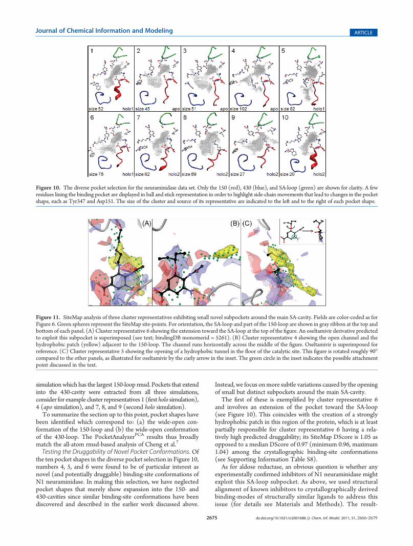
Testing the Druggability of Novel Pocket Conformations. Ofthe ten pocket shapes in the diverse pocket selection in Figure 10,numbers 4, 5, and 6 were found to be of particular interest asnovel (and potentially druggable) binding-site conformations ofN1 neuraminidase. In making this selection, we have neglectedpocket shapes that merely show expansion into the 150- and430-cavities since similar binding-site conformations have beendiscovered and described in the earlier work discussed above.
Instead, we focus onmore subtle variations caused by the openingof small but distinct subpockets around the main SA-cavity.The first of these is exemplified by cluster representative 6
and involves an extension of the pocket toward the SA-loop(see Figure 10). This coincides with the creation of a stronglyhydrophobic patch in this region of the protein, which is at leastpartially responsible for cluster representative 6 having a rela-tively high predicted druggability; its SiteMap DScore is 1.05 asopposed to a median DScore of 0.97 (minimum 0.96, maximum1.04) among the crystallographic binding-site conformations(see Supporting Information Table S8).As for aldose reductase, an obvious question is whether any
experimentally confirmed inhibitors of N1 neuraminidase mightexploit this SA-loop subpocket. As above, we used structuralalignment of known inhibitors to crystallographically derivedbinding-modes of structurally similar ligands to address thisissue (for details see Materials and Methods). The result-
Figure 10. The diverse pocket selection for the neuraminidase data set. Only the 150 (red), 430 (blue), and SA-loop (green) are shown for clarity. A fewresidues lining the binding pocket are displayed in ball and stick representation in order to highlight side-chain movements that lead to changes in the pocketshape, such as Tyr347 and Asp151. The size of the cluster and source of its representative are indicated to the left and to the right of each pocket shape.
Figure 11. SiteMap analysis of three cluster representatives exhibiting small novel subpockets around the main SA-cavity. Fields are color-coded as forFigure 6. Green spheres represent the SiteMap site-points. For orientation, the SA-loop and part of the 150-loop are shown in gray ribbon at the top andbottom of each panel. (A) Cluster representative 6 showing the extension toward the SA-loop at the top of the figure. An oseltamivir derivative predictedto exploit this subpocket is superimposed (see text; bindingDB monomerid = 5261). (B) Cluster representative 4 showing the open channel and thehydrophobic patch (yellow) adjacent to the 150-loop. The channel runs horizontally across the middle of the figure. Oseltamivir is superimposed forreference. (C) Cluster representative 5 showing the opening of a hydrophobic tunnel in the floor of the catalytic site. This figure is rotated roughly 90�compared to the other panels, as illustrated for oseltamivir by the curly arrow in the inset. The green circle in the inset indicates the possible attachmentpoint discussed in the text.

2676 dx.doi.org/10.1021/ci200168b |J. Chem. Inf. Model. 2011, 51, 2666–2679
Journal of Chemical Information and Modeling ARTICLE
ing alignments provide predicted binding modes for 64 sub-structure-matched derivatives of oseltamivir. Within this set ofsubmicromolar inhibitors, 95% involve a variationof thehydrophobicether substituent that is directed toward the SA-loop in thecrystallographic binding-mode of oseltamivir. The substructurealignments indeed predict that fire of the molecules occupythe novel SA-loop subpocket to some extent. One example is the4-propylpiperidine derivative that is superimposed on the fieldsfrom the SiteMap analysis of cluster representative 6 in panel A ofFigure 11. This molecule has an IC50 of 40 nM in an enzymaticN1 inhibition assay. Indeed, the report describing the synthesis ofthis compound also includes an illustration of a proprietary crystalstructure of the related piperidine derivative.49 This confirms thebinding mode of these compounds and provides experimentalsupport for plausibility of this novel binding-site conformationidentified by PocketAnalyzerPCA.A second variation of the SA-cavity is exhibited by cluster
representative 4 and is induced by a change in the conformation ofthe side-chain of Arg152. In particular, the guanidine group ofArg152moves toward, and forms a salt bridge with, the side-chainof Glu277. This rearrangement opens a narrow channel that isadjacent to the 150-loop (Figure 11, panel B). Interestingly, thisrotation of Arg152 side-chain would not be compatible with thebinding modes of known SA-cavity ligands (e.g., oseltamivir andzanamivir) and, indeed, is only observed in the apo MD simula-tion. Furthermore, although the novel channel includes a mod-erately sized hydrophobic patch, the rearrangement leads to anoverall decrease in depth and size of the catalytic binding side andhence a relatively low DScore of 0.92 (see Supporting Informa-tion Table S8). None of the substructure-matched oseltamivirderivatives are predicted to occupy this channel by the structuralalignment approach discussed above. However, some support forthe existence of this pocket conformation is provided by theearlier computational solvent mapping calculations.46 In thatwork it was observed that a low energy cluster of chemical probepositions is found in the region of this channel.Cluster representative 5 (Figure 11, panel C) exemplifies a
third conformation of the SA cavity that, to the best of ourknowledge, has not been previously observed in either experi-mental or computational studies. It involves the opening of a narrowbut hydrophobic tunnel in the floor of the catalytic site. Again, noneof the publicly available oseltamivir derivatives are predicted to enterthis tunnel by the structural alignment approach. However, thetunnel is also present to some extent in cluster representatives1, 2, 3, and 6, so it is a characteristic and recurrent feature of thesimulations. The addition of several rather hydrophobic site-points results in high predicted druggability with a DScore of1.08. Furthermore, the central cyclohexene ring of oseltamivirhas an obvious attachment point for hydrophobic substituentsthat might access this tunnel (Figure 11). It will therefore be ofinterest to see if future medicinal chemistry studies reportevidence of inhibitors binding to this novel pocket conformation.
’CONCLUDING REMARKS
We have introduced the PocketAnalyzerPCA methodology toaddress the problem of diverse pocket selection, i.e. how toreduce a large collection of structures of the same protein to asubset that retains a number of substantially distinct bindingpocket conformations. Diverse pocket shapes drive medicinalchemistry to explore a broader chemical space and so presentadditional opportunities to overcome key drug discovery issues
such as potency, selectivity, toxicity, and pharmacokinetics. Theidentification of diverse pocket shapes and novel binding-siteconformations can therefore greatly assist the progress of struc-ture-based ligand design projects.
The PocketAnalyzerPCA approach combines a grid-basedpocket detection algorithm with PCA and clustering. The result-ing principal component (PC) eigenvectors reveal the dominantbinding-site deformation modes within an ensemble of proteinstructures, and the corresponding PC scores provide character-ization and visualization of the pocket conformational distributions.From a methodological point of view, the PocketAnalyzerPCA
approach provides a novel and complementary perspective onprotein dynamics that may prove particularly relevant for ligandbinding and drug design. PocketAnalyzerPCA was primarilyenvisioned as a tool for analyzing trajectories of protein con-formations produced by MD simulations. However, the proce-dure is applicable to any source of atomistic protein structuralinformation and also to combinations of structures from severalsuch sources, as in the examples presented above. Therefore,PocketAnalyzerPCA may be useful for exploring the increasingvolume of experimentally derived structural information result-ing from high-throughput crystallography and advances inNMR-based techniques.
A technically related approach to a different problem com-bines pocket detection and clustering to track the opening andclosing of transient binding pockets in protein�protein interac-tion surfaces along MD trajectories.50 This ePOS methodanalyses each in a sequence of MD frames using the PASS pocketdetection algorithm51 and clusters the resulting pockets by the setof pocket lining atoms to define unique pockets and track theiropening and closing as time progresses. The recently announcedfpocket Web server uses a different pocket detection algorithmto perform a similar analysis.52 A precedent for combining grid-based pocket characterization with PCA is the GRID/CPCAapproach of Kastenholz et al.53,54 However, rather than focusingon the analysis of many structures of the same protein (as here)the GRID/CPCAmethod is directed at comparing structures ofdifferent targets to derive insights that assist in improvingcompound selectivity.
When applied to aldose reductase, a protein with moderatebinding-site flexibility and a well-characterized set of crystal-lographic binding-site conformations, PocketAnalyzerPCA distin-guishes three of the four crystallographically observed binding-site conformations previously reported by the Klebe group.24 Inaddition, the approach identifies a number of distinct pocketshapes that have not been observed experimentally and whichtherefore represent novel computationally derived binding-siteconformations. From a medicinal chemistry point of view, themost outstanding result is that one MD-derived pocket shape isparticularly striking in its difference to the crystallographic con-formations. A rotation of a short section of the protein backboneand accompanying adjustments in the positions of a few amino-acid side-chains open a channel connecting the active site withanother pocket on the protein surface. Although the channelitself and the second pocket are rather polar, the ‘entrance’ to thechannel forms a reasonably large hydrophobic subpocket, andSiteMap17 analysis predicted good druggability for this novelconformation of the ALR active site. Indeed, subsequent align-ment of known ALR inhibitors to the crystallographic bindingmodes of structurally similar ALR ligands identified a derivativeof lidorestat that is predicted to fill the novel hydrophobicsubpocket with a phenyl ring. This compound has an IC50 of

2677 dx.doi.org/10.1021/ci200168b |J. Chem. Inf. Model. 2011, 51, 2666–2679
Journal of Chemical Information and Modeling ARTICLE
100 nM in an in vitro ALR inhibition assay,27 providingexperimental evidence that the novel computational derivedpocket conformation identified by PocketAnalyzerPCA is a plau-sible and druggable target for structure-based drug designagainst ALR.
In a second example, the PocketAnalyzerPCA approach is usedto derive a diverse set of binding-site conformations from viralneuraminidase. Similarly to ALR, the binding-site flexibility ofneuraminidase is reasonably well-established, for example as aresult of MD simulations,48 and a number of distinct binding-siteconformations have been characterized crystallographically. ThePocketAnalyzerPCA diverse pocket approach was found to iden-tify a qualitatively similar range of binding-site conformations as apreviously reported atom-based rmsd clustering method,47,48
with the advantage of quickly highlighting conserved and variableregions in the pocket. The method also allows facile comparisonof structures from different sources and direct visualization ofdifferences in pocket shape rather than changes in proxy de-scriptors such as backbone and side-chain positions.
The N1 diverse pocket selection included three particularlyinteresting and novel subpockets adjacent to the main catalyticsite of N1 neuraminidase. Alignment of known submicromolarN1 inhibitors to the crystallographic binding-mode of oseltami-vir identified several molecules predicted to occupy the first ofthese subpockets, for example with a 4-propylpiperidine sub-stituent. Indeed, the report describing the synthesis of thiscompound includes an illustration of a proprietary crystalstructure of the unsubstituted piperidine derivative,49 confirm-ing the bindingmode and the plausibility of this computationallyderived binding-site conformation as a druggable target for N1inhibition.
In addition to direct application to structure-based liganddesign, the PocketAnalyzerPCA protocol produces an ensembleof protein structures incorporating diverse and potentiallynovel pocket shapes that could be useful as input to numerousstructure-based drug design methods.55 For example, thiswould provide an effective way to account for protein flex-ibility in docking and virtual screening,56�59 receptor-basedpharmacophore generation,3 and druggability analysis.60
Applied in this way, PocketAnalyzerPCA would be just onecomponent in a larger drug discovery workflow, providinga rational approach to selecting an ensemble of protein con-formations.61,62
Moving beyond the specific application of PocketAnalyzerPCA
to diverse pocket selection, in the future the approach may bemore broadly applied to address questions regarding the effect ofvarious perturbations on pocket conformational distributions.For example, when coupled with the appropriate MD simula-tions, PocketAnalyzerPCA may provide a useful perspective onthe change in binding-site conformation induced by factors suchas mutation, allosteric modulation, solvent pH, and post-transla-tional modification.
’ASSOCIATED CONTENT
bS Supporting Information. Additional PCD plots andPCA scree plots for the ALR and NA data sets, rmsd plots forall MD simulations, and details of the oseltamivir force fieldparametrization. This material is available free of charge via theInternet at http://pubs.acs.org.
’AUTHOR INFORMATION
Corresponding Author*E-mail: [email protected].
Present Addresses
)BASF SE, GVC/C - A030, 67056 Ludwigshafen, Germany.
’ACKNOWLEDGMENT
We are grateful to Teresa Jimenez Vaquero for providing aninitial version of the PocketAnalyzer code. Financial supportfrom the Education Office of Novartis Institutes for BiomedicalResearch is gratefully acknowledged (I.C.).
’ABBREVIATIONS:
ALR, aldose reductase; NA, neuraminidase; SA, sialic acid; PCA,principal component analysis; PC, principal component; PCD,pocket conformational distribution;MD, molecular dynamics;GAFF, generalized amber force field; rmsd, root mean squaredeviation
’REFERENCES
(1) Dutta, S.; Burkhardt, K.; Young, J.; Swaminathan, G.; Matsuura, T.;Henrick, K.; Nakamura, H.; Berman,H.DataDeposition andAnnotation atthe Worldwide Protein Data Bank. Mol. Biotechnol. 2009, 42, 1–13.
(2) Ahmed, A.; Kazemi, S.; Gohlke,H. Protein flexibility andmobility instructure-based drug design. Front. Drug Des. Discovery 2007, 3, 455–476.
(3) Carlson, H. A.; Masukawa, K. M.; Rubins, K.; Bushman, F. D.;Jorgensen, W. L.; Lins, R. D.; Briggs, J. M.; McCammon, J. A. Devel-oping a dynamic pharmacophore model for HIV-1 integrase. J. Med.Chem. 2000, 43, 2100–2114.
(4) Cozzini, P.; Kellogg,G. E.; Spyrakis, F.; Abraham,D. J.; Costantino,G.; Emerson, A.; Fanelli, F.; Gohlke, H.; Kuhn, L. A.; Morris, G. M.;Orozco,M.; Pertinhez, T. A.; Rizzi,M.; Sotriffer, C. A. Target flexibility: anemerging consideration in drug discovery and design. J. Med. Chem. 2008,51, 6237–6255.
(5) Schames, J. R.; Henchman, R. H.; Siegel, J. S.; Sotriffer, C. A.; Ni,H.; McCammon, J. A. Discovery of a novel binding trench in HIVintegrase. J. Med. Chem. 2004, 47, 1879–1881.
(6) Sotriffer, C. A.; Ni, H.; McCammon, J. A. Active site bindingmodes of HIV-1 integrase inhibitors. J. Med. Chem. 2000, 43, 4109–4117.
(7) Cheng, L. S.; Amaro, R. E.; Xu, D.; Li, W. W.; Arzberger, P. W.;McCammon, J. A. Ensemble-based virtual screening reveals potentialnovel antiviral compounds for avian influenza neuraminidase. J. Med.Chem. 2008, 51, 3878–3894.
(8) Zhou, Z.;Madrid,M.; Evanseck, J. D.;Madura, J. D. Effect of a boundnon-nucleosideRT inhibitor on the dynamics ofwild-type andmutantHIV-1reverse transcriptase. J. Am. Chem. Soc. 2005, 127, 17253–17260.
(9) Grant, B. J.; Rodrigues, A. P. C.; Elsawy, K. M.; McCammon,J. A.; Caves, L. S. D. Bio3d: an R package for the comparative analysis ofprotein structures. Bioinformatics 2006, 22, 2695–2696.
(10) Hendlich,M.; Rippmann, F.; Barnickel, G. LIGSITE: Automaticand efficient detection of potential small molecule-binding sites inproteins. J. Mol. Graphics Modell. 1997, 15, 359–363.
(11) Stahl, M.; Taroni, C.; Schneider, G. Mapping of protein surfacecavities and prediction of enzyme class by a self-organizing neuralnetwork. Protein Eng. 2000, 13, 83–88.
(12) Joliffe, I. T. Principal Component Analysis, 2nd ed.; Springer:New York, 2002.
(13) Maestro, version 9.1; Schr€odinger, LLC: New York, 2010.(14) Godzik, A. The structural alignment between two proteins: Is
there a unique answer? Protein Sci. 1996, 5, 1325–1338.(15) Kaufman, L.; Rousseeuw, P. J. Finding Groups in Data: An
Introduction to Cluster Analysis; Wiley: New York, 1990.

2678 dx.doi.org/10.1021/ci200168b |J. Chem. Inf. Model. 2011, 51, 2666–2679
Journal of Chemical Information and Modeling ARTICLE
(16) Available at http://cran.r-project.org/web/packages/cluster(accessed September 27, 2011).(17) Halgren, T. A. Identifying and Characterizing Binding Sites and
Assessing Druggability. J. Chem. Inf. Model. 2009, 49, 377–389.(18) Carl, N.; Konc, J.; Janezic, D. Protein surface conservation in
binding sites. J. Chem. Inf. Model. 2008, 48, 1279–1286.(19) Carl, N.; Konc, J.; Vehar, B.; Janezic, D. Protein-Protein
Binding Site Prediction by Local Structural Alignment. J. Chem. Inf.Model. 2010, 50, 1906–1913.(20) Cheng, A. C.; Coleman, R. G.; Smyth, K. T.; Cao, Q.; Soulard,
P.; Caffrey, D. R.; Salzberg, A. C.; Huang, E. S. Structure-based maximalaffinity model predicts small-molecule druggability. Nat. Biotechnol.2007, 25, 71–75.(21) Coleman, R. G.; Burr, M. A.; Souvaine, D. L.; Cheng, A. C. An
intuitive approach to measuring protein surface curvature. Proteins:Struct., Funct., Bioinf. 2005, 61, 1068–1074.(22) Hendlich, M.; Bergner, A.; Gunther, J.; Klebe, G. Relibase:
Design and development of a database for comprehensive analysis ofprotein-ligand interactions. J. Mol. Biol. 2003, 326, 607–620.(23) Schmitt, S.; Kuhn, D.; Klebe, G. A newmethod to detect related
function among proteins independent of sequence and fold homology.J. Mol. Biol. 2002, 323, 387–406.(24) Sotriffer, C. A.; Kramer, O.; Klebe, G. Probing flexibility and
“induced-fit” phenomena in aldose reductase by comparative crystalstructure analysis and molecular dynamics simulations. Proteins: Struct.,Funct., Bioinf. 2004, 56, 52–66.(25) ICM, version 3.6; MolSoft, LCC; La Jolla, CA, 2010.(26) Liu, T. Q.; Lin, Y. M.; Wen, X.; Jorissen, R. N.; Gilson, M. K.
BindingDB: a web-accessible database of experimentally determinedprotein-ligand binding affinities.Nucleic Acids Res. 2007, 35, D198–D201.(27) Van Zandt, M. C.; Jones, M. L.; Gunn, D. E.; Geraci, L. S.; Jones,
J. H.; Sawicki, D. R.; Sredy, J.; Jacot, J. L.; DiCioccio, A. T.; Petrova, T.;Mitschler, A.; Podjarny, A. D. Discovery of 3-[(4,5,7-trifluorobenzothiazol-2-yl)methyl]indole-N-acetic acid (Lidorestat) and congeners as highlypotent and selective inhibitors of aldose reductase for treatment of chronicdiabetic complications. J. Med. Chem. 2005, 48, 3141–3152.(28) Case, D. A.; Cheatham, T. E.; Darden, T.; Gohlke, H.; Luo, R.;
Merz, K. M.; Onufriev, A.; Simmerling, C.; Wang, B.; Woods, R. J. TheAmber biomolecular simulation programs. J. Comput. Chem. 2005,26, 1668–1688.(29) Case, D. A.; Darden, T. A.; Cheatham; Simmerling, C. L.;
Wang, J.; Duke, R. E.; Luo, R.; Merz, K. M.; Pearlman, D. A.; Crowley,M.; Walker, R. C.; Zhang, W.; Wang, B.; Hayik, S.; Roitberg, A.; Seabra,G.; Wong, K. F.; Paesani, F.; Wu, X.; Brozell, S.; Tsui, V.; Gohlke, H.;Yang, L.; Tan, C.; Mongan, J.; Hornak, V.; Cui, G.; Beroza, P.; Mathews,D. H.; Schafmeister, C.; Ross, W. S.; Kollman, P. A. Amber 9; Universityof California: San Francisco, 2006.(30) Duan, Y.; Wu, C.; Chowdhury, S.; Lee, M. C.; Xiong, G. M.;
Zhang, W.; Yang, R.; Cieplak, P.; Luo, R.; Lee, T.; Caldwell, J.; Wang,J. M.; Kollman, P. A point-charge force field for molecular mechanicssimulations of proteins based on condensed-phase quantum mechanicalcalculations. J. Comput. Chem. 2003, 24, 1999–2012.(31) Bryce, R. AMBER parameter database. http://www.pharmacy.
manchester.ac.uk/bryce/amber (accessed March 15, 2010).(32) Jorgensen, W. L.; Chandrasekhar, J.; Madura, J. D.; Impey,
R. W.; Klein, M. L. Comparison of Simple Potential Functions forSimulating Liquid Water. J. Chem. Phys. 1983, 79, 926–935.(33) Izaguirre, J. A.;Catarello,D.P.;Wozniak, J.M.; Skeel, R.D.Langevin
stabilization of molecular dynamics. J. Chem. Phys. 2001, 114, 2090–2098.(34) Ryckaert, J. P.; Ciccotti, G.; Berendsen, H. J. C. Numerical-
Integration of Cartesian Equations ofMotion of A SystemwithConstraints -Molecular-Dynamics of N-Alkanes. J. Comput. Phys. 1977, 23, 327–341.(35) Darden, T.; York, D.; Pedersen, L. Particle Mesh Ewald - An N.
Log(N) Method for Ewald Sums in Large Systems. J. Chem. Phys. 1993,98, 10089–10092.(36) Berendsen, H. J. C.; Postma, J. P. M.; Vangunsteren, W. F.;
Dinola, A.; Haak, J. R. Molecular-Dynamics with Coupling to AnExternal Bath. J. Chem. Phys. 1984, 81, 3684–3690.
(37) Lawrenz, M.; Wereszczynski, J.; Amaro, R.; Walker, R.; Roitberg,A.; McCammon, J. A. Impact of calcium on N1 influenza neuraminidasedynamics and binding free energy. Proteins: Struct., Funct., Bioinf. 2010,78, 2523–2532.
(38) Wang, J. M.; Wolf, R. M.; Caldwell, J. W.; Kollman, P. A.; Case,D. A. Development and testing of a general amber force field. J. Comput.Chem. 2004, 25, 1157–1174.
(39) Varkonyi, T.; Kempler, P. Diabetic neuropathy: new strategiesfor treatment. Diabetes Obes. Metab. 2008, 10, 99–108.
(40) Steuber, H.; Zentgraf, M.; Gerlach, C.; Sotriffer, C. A.; Heine,A.; Klebe, G. Expect the unexpected or caveat for drug designers:Multiple structure determinations using aldose reductase crystals treatedunder varying soaking and co-crystallisation conditions. J. Mol. Biol.2006, 363, 174–187.
(41) Steuber, H.; Zentgraf, M.; Podjarny, A.; Heine, A.; Klebe, G.High-resolution crystal structure of aldose reductase complexed with thenovel sulfonyl-pyridazinone inhibitor exhibiting an alternative active siteanchoring group. J. Mol. Biol. 2006, 356, 45–56.
(42) Steuber, H.; Zentgraf, M.; La Motta, C.; Sartini, S.; Heine, A.;Klebe, G. Evidence for a novel binding site conformer of aldosereductase in ligand-bound state. J. Mol. Biol. 2007, 369, 186–197.
(43) Luque, I.; Freire, E. Structural stability of binding sites: Con-sequences for binding affinity and allosteric effects. Proteins: Struct.,Funct., Genet. 2000, 63–71.
(44) Caves, L. S. D.; Evanseck, J. D.; Karplus, M. Locally accessibleconformations of proteins: Multiple molecular dynamics simulations ofcrambin. Protein Sci. 1998, 7, 649–666.
(45) Russell, R. J.; Haire, L. F.; Stevens, D. J.; Collins, P. J.; Lin, Y. P.;Blackburn, G. M.; Hay, A. J.; Gamblin, S. J.; Skehel, J. J. The structure ofH5N1 avian influenza neuraminidase suggests new opportunities fordrug design. Nature 2006, 443, 45–49.
(46) Landon, M. R.; Amaro, R. E.; Baron, R.; Ngan, C. H.; Ozonoff,D.; McCammon, J. A.; Vajda, S. Novel druggable hot spots in avianinfluenza neuraminidase H5N1 revealed by computational solventmapping of a reduced and representative receptor ensemble. Chem.Biol. Drug Des. 2008, 71, 106–116.
(47) Amaro, R. E.; Minh, D. D. L.; Cheng, L. S.; Lindstrom, W. M.;Olson, A. J.; Lin, J. H.; Li, W. W.; McCammon, J. A. Remarkable loopflexibility in avian influenza N1 and its implications for antiviral drugdesign. J. Am. Chem. Soc. 2007, 129, 7764.
(48) Amaro, R. E.; Xiaolin, C.; Ivaylo, I.; Dong, X.;McCammon, J. A.Characterizing Loop Dynamics and Ligand Recognition in Human andAvian Type Influenza Neuraminidases via Generalized Born MolecularDynamics and End-Point Free Energy Calculations. J. Am. Chem. Soc.2009, 131, 4702–4709.
(49) Lew, W.; Wu, H. W.; Chen, X. W.; Graves, B. J.; Escarpe, P. A.;MacArthur, H. L.; Mendel, D. B.; Kim, C. U. Carbocyclic influenzaneuraminidase inhibitors possessing a C-3-cyclic amine side chain: Synth-esis and inhibitory activity. Bioorg. Med. Chem. Lett. 2000, 10, 1257–1260.
(50) Eyrisch, S.; Helms, V. Transient pockets on protein surfacesinvolved in protein-protein interaction. J. Med. Chem. 2007, 50, 3457–3464.
(51) Brady, G. P.; Stouten, P. F. W. Fast prediction and visualizationof protein binding pockets with PASS. J. Comput.-Aided Mol. Des. 2000,14, 383–401.
(52) Schmidtke, P.; Le Guilloux, V.; Maupetit, J.; Tuffery, P. fpocket:online tools for protein ensemble pocket detection and tracking.NucleicAcids Res. 2010, 38, W582–W589.
(53) Afzelius, L.; Raubacher, F.; Karlen, A.; Jorgensen, F. S.; Andersson,T. B.; Masimirembwa, C.M.; Zamora, I. Structural analysis of CYP2C9 andCYP2C5 and an evaluation of commonly used molecular modelingtechniques. Drug Metab. Dispos. 2004, 32, 1218–1229.
(54) Kastenholz, M. A.; Pastor, M.; Cruciani, G.; Haaksma, E. E. J.;Fox, T. GRID/CPCA: A new computational tool to design selectiveligands. J. Med. Chem. 2000, 43, 3033–3044.
(55) Perot, S.; Sperandio, O.; Miteva, M. A.; Camproux, A. C.;Villoutreix, B. O. Druggable pockets and binding site centric chemicalspace: a paradigm shift in drug discovery. Drug Discovery Today 2010,15, 656–667.
2679 dx.doi.org/10.1021/ci200168b |J. Chem. Inf. Model. 2011, 51, 2666–2679
Journal of Chemical Information and Modeling ARTICLE
(56) Barril, X.; Morley, S. D. Unveiling the full potential of flexiblereceptor docking using multiple crystallographic structures. J. Med.Chem. 2005, 48, 4432–4443.(57) Craig, I. R.; Essex, J. W.; Spiegel, K. Ensemble Docking into
Multiple Crystallographically Derived Protein Structures: An EvaluationBased on the Statistical Analysis of Enrichments. J. Chem. Inf. Model.2010, 50, 511–524.(58) Huang, S. Y.; Zou, X. Q. Ensemble docking of multiple protein
structures: Considering protein structural variations in molecular dock-ing. Proteins: Struct., Funct., Bioinf. 2007, 66, 399–421.(59) Virtanen, S. I.; Pentikainen, O. T. Efficient Virtual Screening
UsingMultiple Protein Conformations Described as Negative Images ofthe Ligand-Binding Site. J. Chem. Inf. Model. 2010, 50, 1005–1011.(60) Egner, U.; Hillig, R. C. A structural biology view of target
drugability. Expert Opin. Drug Discovery 2008, 3, 391–401.(61) Bolstad, E. S. D.; Anderson, A. C. In pursuit of virtual lead
optimization: Pruning ensembles of receptor structures for increasedefficiency and accuracy during docking. Proteins: Struct., Funct., Bioinf.2009, 75, 62–74.(62) Rueda, M.; Bottegoni, G.; Abagyan, R. Recipes for the Selection
of Experimental Protein Conformations for Virtual Screening. J. Chem.Inf. Model. 2010, 50, 186–193.