Embed Size (px)
Citation preview

24 / Cucurbit Genetics Cooperative Report 33-34: 24-28 (2010-2011)
Podosphaera xanthii but not Golovinomyces cichoracearuminfects Cucurbits in a Greenhouse at Salinas, CaliforniaCosme Bojorques RamosResearch Center for Food and Development (CIAD, A. C.), Department of Phytopathology, km 5.5 Culiacan-EldoradoRoad, Culiacan, Sinaloa, Mexico, and University of Occident, Department of Biology, M. Gaxiola Street and InternationalRoad Los Mochis, Sinaloa, Mexico
Karunakaran Maruthachalam,Department of Plant Pathology, University of California, U.S. Agricultural Research Station, 1636 E. Alisal St., Salinas,CA 93905
James D. McCreightUnited States Department of Agriculture, Agricultural Research Service, U.S. Agricultural Research Station, 1636 E.Alisal St., Salinas, CA 93905
Raymundo S. Garcia EstradaResearch Center for Food and Development (CIAD. A. C.), Department of Phytopathology, km 5.5 Culiacan-EldoradoRoad, Culiacan, Sinaloa, Mexico
Traditionally, Erysiphe cichoracearum (nowGolovinomyces cichoracearum) and Podosphaera xanthii,formerly Sphaerotheca fuliginea, have been reported as thecausal agents of cucurbit powdery mildew (CPM) (2, 10).These two fungal species have often been misidentified(12), due to similarities in their anamorphic characteris-tics and the very scarce production of ascomata (13, 16),given that the ascocarpic structure was considered ba-sic for earlier systematics. After considering the fibrosininclusions in the conidia to differentiate S. fuliginea fromE. cichoracearum in Australia and North America (1, 18),S. fuliginea was determined to be the predominant or theonly CPM species in the United States (12), althoughboth species can be found infecting the same crops (2, 8,10).
Molecular analyses demonstrated that some char-acteristics of the ascomata were not associated with themonophyletic development of the Erysiphaceae, and thatPodosphaera and Sphaerotheca were not separate mono-phyletic groups (15). Based on the findings of Saenz andTaylor (15), Mori et al. (14) and Takamatsu et al. (17), onthe genetic relationships of the Erysiphaceae species,using the Internal Transcribed Spacers (ITS) of the ribo-somal DNA (rDNA), Braun and Takamatsu (7) proposedto merge Podosphaera and Sphaerotheca in the genusPodosphaera, and to divide the genus Podosphaera in twosections, based on morphological characteristics:Podosphaera sect. Podosphaera having dichotomously
branched ascomata appendages that in Podosphaera, sect.Sphaerotheca are miceliods.
The Section Sphaerotheca was then subdivided intwo subsections, in accordance to the size of the peridialcells of the ascomata: small (5 to 25 µm of diameter) inSphaerotheca and large (20 to 55 µm) in Magnicellulata (7).The species P. fusca and P. xanthii, of the SubsectionMagnicellulatae, were differentiated by Braun et al. (6),based on the size of chasmothecia and the thin walledportion of the asci (oculus), measuring 55 to 90 µm and8 to 15 µm, respectively, in P. fusca, and 75 to 100 µm and15 to 30 µm, respectively, in P. xanthii (3, 6).
Melon (Cucumis melo L.), cucumber (C. sativus L.),squash (Cucurbita pepo L.) and lettuce (Lactuca sativa L.)are normally grown in the same greenhouse at the USDA,ARS laboratory, Salinas, Calif. Chasmothecia were forthe first time noted in this greenhouse on squash andmelons infected by P. xanthii during the winter monthsof 2011 (Fig. 2). Wild lettuce (L. serriola L.) PI 491093plants grown on adjacent benches were infected by G.cichoracearum that was not observed to infect the neigh-boring cucurbits. We characterized morphological andmolecular parameters of these two species and cross-inoculated cucumber with G. cichoracearum.
Materials and MethodsPlants of several melon, squash and cucumber va-
rieties were planted in a greenhouse on 12 Jan. 2011, in
Cucurbit Genetics Cooperative Report 33-34: 24-28 (2010-2011) / 25
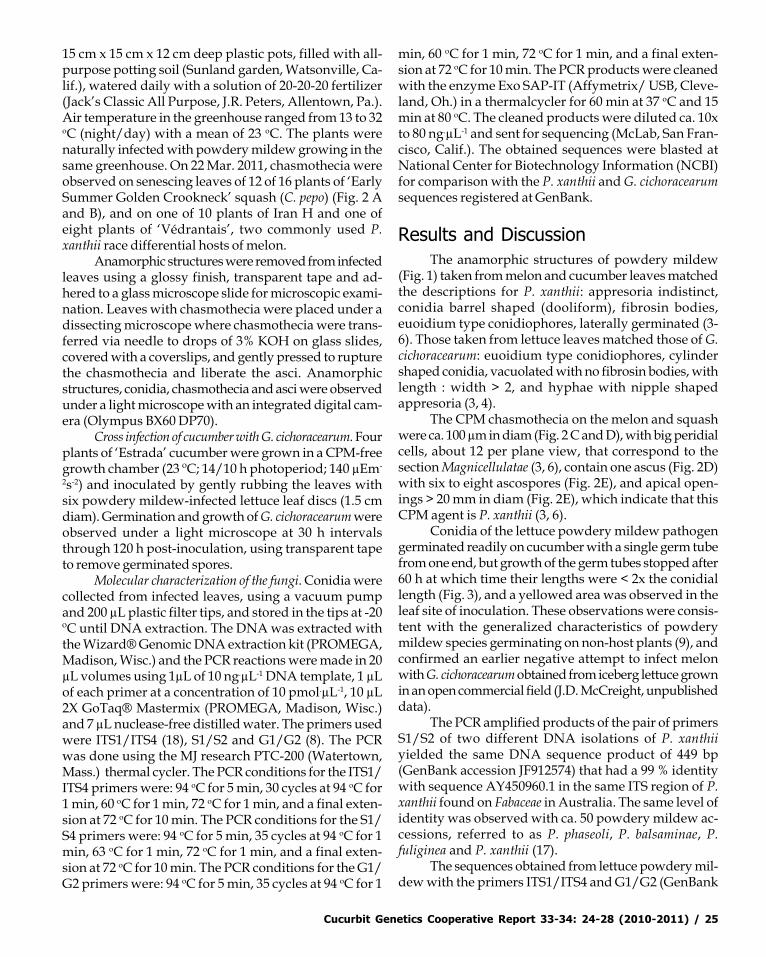
15 cm x 15 cm x 12 cm deep plastic pots, filled with all-purpose potting soil (Sunland garden, Watsonville, Ca-lif.), watered daily with a solution of 20-20-20 fertilizer(Jack’s Classic All Purpose, J.R. Peters, Allentown, Pa.).Air temperature in the greenhouse ranged from 13 to 32oC (night/day) with a mean of 23 oC. The plants werenaturally infected with powdery mildew growing in thesame greenhouse. On 22 Mar. 2011, chasmothecia wereobserved on senescing leaves of 12 of 16 plants of ‘EarlySummer Golden Crookneck’ squash (C. pepo) (Fig. 2 Aand B), and on one of 10 plants of Iran H and one ofeight plants of ‘Védrantais’, two commonly used P.xanthii race differential hosts of melon.
Anamorphic structures were removed from infectedleaves using a glossy finish, transparent tape and ad-hered to a glass microscope slide for microscopic exami-nation. Leaves with chasmothecia were placed under adissecting microscope where chasmothecia were trans-ferred via needle to drops of 3% KOH on glass slides,covered with a coverslips, and gently pressed to rupturethe chasmothecia and liberate the asci. Anamorphicstructures, conidia, chasmothecia and asci were observedunder a light microscope with an integrated digital cam-era (Olympus BX60 DP70).
Cross infection of cucumber with G. cichoracearum. Fourplants of ‘Estrada’ cucumber were grown in a CPM-freegrowth chamber (23 ºC; 14/10 h photoperiod; 140 µEm-
2s-2) and inoculated by gently rubbing the leaves withsix powdery mildew-infected lettuce leaf discs (1.5 cmdiam). Germination and growth of G. cichoracearum wereobserved under a light microscope at 30 h intervalsthrough 120 h post-inoculation, using transparent tapeto remove germinated spores.
Molecular characterization of the fungi. Conidia werecollected from infected leaves, using a vacuum pumpand 200 µL plastic filter tips, and stored in the tips at -20ºC until DNA extraction. The DNA was extracted withthe Wizard® Genomic DNA extraction kit (PROMEGA,Madison, Wisc.) and the PCR reactions were made in 20µL volumes using 1µL of 10 ng.µL-1 DNA template, 1 µLof each primer at a concentration of 10 pmol.µL-1, 10 µL2X GoTaq® Mastermix (PROMEGA, Madison, Wisc.)and 7 µL nuclease-free distilled water. The primers usedwere ITS1/ITS4 (18), S1/S2 and G1/G2 (8). The PCRwas done using the MJ research PTC-200 (Watertown,Mass.) thermal cycler. The PCR conditions for the ITS1/ITS4 primers were: 94 oC for 5 min, 30 cycles at 94 oC for1 min, 60 oC for 1 min, 72 oC for 1 min, and a final exten-sion at 72 oC for 10 min. The PCR conditions for the S1/S4 primers were: 94 oC for 5 min, 35 cycles at 94 oC for 1min, 63 oC for 1 min, 72 oC for 1 min, and a final exten-sion at 72 oC for 10 min. The PCR conditions for the G1/G2 primers were: 94 oC for 5 min, 35 cycles at 94 oC for 1
min, 60 oC for 1 min, 72 oC for 1 min, and a final exten-sion at 72 oC for 10 min. The PCR products were cleanedwith the enzyme Exo SAP-IT (Affymetrix/ USB, Cleve-land, Oh.) in a thermalcycler for 60 min at 37 oC and 15min at 80 oC. The cleaned products were diluted ca. 10xto 80 ng.µL-1 and sent for sequencing (McLab, San Fran-cisco, Calif.). The obtained sequences were blasted atNational Center for Biotechnology Information (NCBI)for comparison with the P. xanthii and G. cichoracearumsequences registered at GenBank.
Results and DiscussionThe anamorphic structures of powdery mildew
(Fig. 1) taken from melon and cucumber leaves matchedthe descriptions for P. xanthii: appresoria indistinct,conidia barrel shaped (dooliform), fibrosin bodies,euoidium type conidiophores, laterally germinated (3-6). Those taken from lettuce leaves matched those of G.cichoracearum: euoidium type conidiophores, cylindershaped conidia, vacuolated with no fibrosin bodies, withlength : width > 2, and hyphae with nipple shapedappresoria (3, 4).
The CPM chasmothecia on the melon and squashwere ca. 100 µm in diam (Fig. 2 C and D), with big peridialcells, about 12 per plane view, that correspond to thesection Magnicellulatae (3, 6), contain one ascus (Fig. 2D)with six to eight ascospores (Fig. 2E), and apical open-ings > 20 mm in diam (Fig. 2E), which indicate that thisCPM agent is P. xanthii (3, 6).
Conidia of the lettuce powdery mildew pathogengerminated readily on cucumber with a single germ tubefrom one end, but growth of the germ tubes stopped after60 h at which time their lengths were < 2x the conidiallength (Fig. 3), and a yellowed area was observed in theleaf site of inoculation. These observations were consis-tent with the generalized characteristics of powderymildew species germinating on non-host plants (9), andconfirmed an earlier negative attempt to infect melonwith G. cichoracearum obtained from iceberg lettuce grownin an open commercial field (J.D. McCreight, unpublisheddata).
The PCR amplified products of the pair of primersS1/S2 of two different DNA isolations of P. xanthiiyielded the same DNA sequence product of 449 bp(GenBank accession JF912574) that had a 99 % identitywith sequence AY450960.1 in the same ITS region of P.xanthii found on Fabaceae in Australia. The same level ofidentity was observed with ca. 50 powdery mildew ac-cessions, referred to as P. phaseoli, P. balsaminae, P.fuliginea and P. xanthii (17).
The sequences obtained from lettuce powdery mil-dew with the primers ITS1/ITS4 and G1/G2 (GenBank

26 / Cucurbit Genetics Cooperative Report 33-34: 24-28 (2010-2011)
accessions JF951305 and JF951306) had 98 and 100%identity with GenBank sequences AB077688.1 andAB07766.1, respectively, for G. cichoracearum. Similar lev-els of identity were found for G. orontii, which infectsmany families but not members of the Asteraceae ofwhich lettuce is a member (4, 5).
These results confirmed the identity of the CPMpathogen on cucurbits in a Salinas greenhouse as P.xanthii based on morphological and molecular charac-teristics. The powdery mildew pathogen on wild lettucein the same greenhouse was similarly confirmed as G.cichoracearum. Moreover, two G. cichoracearum isolates inSalinas (field and greenhouse) may be regarded as rep-resentatives of G. cichoracearum sensu stricto (4), which isrestricted to members of the Asteraceae (5, 11).
USDA is an equal opportunity provider and em-ployer.
Literature Cited1. Ballantyne, B. 1963. A preliminary note on the identity of cu-
curbit powdery mildews. Australian J. Sci. 25:360.2. Bardin, M., Carlier, J., and Nicot, P. C. 1999. Genetic differen-
tiation in the French population of Erysiphe cichoracearum, acausal agent of powdery mildew of cucurbits. Plant Pathol.48 (4):531-540.
3. Bolay, A. 2005. Les oïdiums de Suisse (Erysiphacées).Cryptogamica Helvética 20:1-176.
4. Braun, U. 1987. A monograph of the Erysiphales (Powderymildews). Hedwigia 89:1-700.
5. Braun, U. 1995. The powdery mildews (Erysiphales) of Eu-rope. Gustav Fischer Verlag, New York.
6. Braun, U., Shishkoff, N., and Takamatsu, S. 2001. Phylogenyof Podosphaera sect. Sphaerotheca subsect. Magnicellulatae(Sphaerotheca fuliginea auct. s. lat.) inferred from rDNA ITSsequences-a taxonomic interpretation. Schlechtendalia 7:45-52.
7. Braun, U., and Takamatsu, S. 2000. Phylogeny of Erysiphe,Microsphaera, Uncinula (Erysipheae) and Cistotheca,Podosphaera, Sphaerotheca (Cystotheceae) inferred from rDNAITS sequences–some taxonomic consequences.Schlechtendalia 4:1-33.
8. Chen, R.-S., Chu, C.-C., Cheng, C.-W., Chen, W.-Y., and Tsay,J.-G. 2008. Differentiation of two powdery mildews of sun-flower (Helianthus annuus) by PCR-mediated method basedon ITS sequences. European J. Plant Pathol. 121:1-8.
9. Glazebrook, J. 2005. Contrasting mechanisms of defense againstbiotrophic and necrotrophic pathogens. Annu. Rev.Phytopathol. 43:205-227.
10. Lebeda, A. 1983. The genera and species spectrum of cucum-ber powdery mildew in Czechoslovakia. Phytopath. Z.108:71-77.
11. Lebeda, A., and Mieslerov, B. 2011. Taxonomy, distributionand biology of lettuce powdery mildew (Golovinomycescichoracearum sensu stricto). Plant Pathol. 60:400-415.
12. McCreight, J. D. 2004. Notes on the change of the causalspecies of cucurbit powdery mildew in the U.S. CucurbitGenet. Coop. Rpt. 27:8-23.
13. McGrath, M. T., Staniszewska, H., Shishkoff, N., and Casella,G. 1996. Distribution of mating types of Sphaerotheca fuligineain the United States. Plant Dis. 80:1098-1102.
14. Mori, Y., Sato, Y., and Takamatsu, S. 2000. Molecular phy-logeny and radiation time of Erysiphales inferred from thenuclear ribosomal DNA sequences. Mycoscience 41:437-447.
15. Saenz, G. S., and Taylor, J. W. 1999. Phylogeny of theErysiphales (powdery mildews) inferred from internal tran-scribed spacer ribosomal DNA sequences. Can. J. Bot.77:150-168.
16. Stone, M. O. 1962. Alternate hosts of cucumber powderymildew. Ann. Appl. Biol. 50:203-210.
17. Takamatsu, S., Hirata, T., and Sato, Y. 2000. A parasitictransition from trees to herbs occurred at least twice in tribeCystotheceae (Erysifaseae): evidence from nuclear riboso-mal DNA. Mycological Research 104:1304-1311.
18. Thomas, C. E. 1978. A new biological race of powdery mil-dew of cantaloups. Plant Dis. Rptr. 62:223.

Cucurbit Genetics Cooperative Report 33-34: 24-28 (2010-2011) / 27
Figure 1. Podosphaera xanthii (Px) and Golovinomyces cichoracearum (Gc), anamorph(L) and conidia (R). Gc anamorph from dandelion (Taraxacum officinale) and conidiafrom lettuce (Lactuca sativa L.).
Figure 3. Germination of Golovinomyces cichoracearum conidia on ‘Estrada’ cucumber at30, 60 and 90 h post-inoculation.
Px Px
Gc
Gc
30 h 60 h 90 h

28 / Cucurbit Genetics Cooperative Report 33-34: 24-28 (2010-2011)
Figure 2. Podosphaera xanthii infection (A) and chasmothecia (B) on ‘EarlySummer Golden Crookneck’ squash (C. pepo) in a greenhouse, Salinas, Calif.Light microscope views of chasmothecia (C), ruptured chasmothecium withone ascus (D), and (E) ascus with eight ascospores and well developed apicalopening (AO).
A
B
C D
E
AO