Embed Size (px)
Citation preview

Point-of-Care Biosensor Systems
Arvind Sai Sarathi Vasan1, Ravi Doraiswami1, Dinesh Michael Mahadeo1, Yunhan Huang1 and Michael Pecht1,2
1Center for Advanced Life Cycle Engineering, University of Maryland, College Park, MD, USA
2Center for Prognostics and System Health Management, City University of Hong Kong, Kowloon, Hong Kong
ABSTRACT Point-of-care biosensor systems can potentially improve patient care through real-time and remote health monitoring. Over the past few decades, research has been conducted in the field of biosensors to detect patterns of biomarkers and provide information on their concentration in biological samples for robust diagnosis. In future point-of-care applications, requirements such as rapid label-free detection, miniaturized sensor size, and portability will limit the types of biosensors that can be used. This paper reviews label-free detection techniques using Biological MicroElectroMechanical Systems as a potential candidate for point-of-care biosensing applications. Furthermore, detailed surveys have been carried out on both the wireless networking schemes applicable for a point-of-care environment and prognostic techniques that will enable decision-support services. This paper concludes by providing a list of challenges that must be resolved before realizing biosensor systems for next-generation point-of-care applications. 1. INTRODUCTION
A study conducted by the Milken Institute reported that the total number of individuals in the U.S. affected by chronic diseases during 2003 was around 162 million. The treatment expenditures and loss in productivity projected for 2023 are as high as $790 billion and $3.363 trillion, respectively. It was also estimated that costs totaling $1.333 trillion could be avoided if improvements were made in the prevention and treatment of diseases [1]. Figures 1(a) and 1(b) present details on the number of people affected by the seven most common chronic diseases in the U.S. and the associated costs in 2003, respectively. Figure 1(c) presents the estimated costs incurred due to the chronic diseases.
Genomics and proteomics research has elucidated many new biomarkers that have the potential to improve healthcare [2–3]. The availability of multiple biomarkers is believed to be critical in the diagnosis of complex diseases [4]. Detection of biomarkers associated with different stages of disease pathogenesis could further facilitate early detection of diseases and their infection rate [5]. Widespread use of these biomarkers will depend upon the development of point-of-care (POC) biosensor devices that will allow real-time, rapid, label-free, and multiplexed detection with high selectivity and sensitivity.
In spite of the rapid explosion of genomics and proteomics research, very few of these biomarkers have transitioned into clinical settings. This is due to the lack of rapid diagnostic techniques that can successfully detect them. For example, tuberculosis (TB), a widespread public health problem [20], has proven to be curable [21]. However, WHO estimates for 2009 suggest a global detection rate of 63%, with only 50% of TB cases in Africa being detected [22]. Delayed diagnosis has serious consequences, because one untreated pulmonary case can increase the chances of an epidemic [23–24]. To obviate such problems, a diagnostic tool is needed that does not require laboratory facilities or demand specialist training. It has been estimated that a single diagnostic test offering 100% accuracy could save 625,000 lives per year if widely implemented, and a test with only 85% sensitivity and 97% specificity might save 392,000 lives, or 22.4% of the current annual worldwide deaths [25]. These numbers signify that there is a pressing need to develop new POC diagnostic tools to reduce the number of fatalities and the costs incurred due to growing health concerns.
As health care costs are increasing and with an ageing world population [26], there is an increasing need to remotely monitor the health condition of patients. This includes situations where patients are not confined to hospitals. To address this issue, a variety of POC system prototypes have been produced. Such systems provide real-time feedback information about the health condition of patients, either to the patients themselves or to a medical database accessible to health care providers. Thus, POC systems

constitute a new approach to address the issue of managing and monitoring the health of patients suffering from chronic diseases, elderly people, and postoperative rehabilitation patients by performing tasks that are traditionally performed using laboratory testing [27–28]. In this paper, information is presented on potentially new technologies in the form of POC biosensors and their integration with appropriate technology to provide clinically relevant information, thereby assisting physicians and clinicians in disease diagnosis. The major technology platform that will be the focus of this discussion is biosensors and their integration into POC systems for the analysis of clinically significant biomarkers.
Figure 1. (a) Number of people reported as having common chronic diseases in U.S. in 2003; (b) economic impact of chronic diseases in U.S. in 2003; and (c) projected annual avoidable costs for the year 2023.
POC systems for health monitoring may comprise different types of biosensors that are wearable or
implantable and capable of measuring physiological parameters such as heart rate, oxygen saturation, changes in plasma protein profile, and patterns of multiple biomarkers and their concentration. The sensed information is then communicated through a wired or wireless link to a central data acquisition node such as a Personal Digital Assistant (PDA), which can in turn transmit the sensed signals to a medical center [29]. These systems would not only reduce response time but would also make testing available in environments where laboratory testing is not feasible [6]. These systems not only have applications to medical diagnostics and biological warfare agent detection [7], but the sensor configuration can be modified to suit other applications, such as food quality assurance [8], environmental monitoring [9], and industrial process control [10–11]. Other possible applications are also emerging, such as the identification of animal and plant pathogens, field tissue and gene analysis, and diagnostic testing such as for malarial strains and water purity analysis [6].
Furthermore, early diagnosis of critical health changes could enable the prevention of fatal events [30]. Such early diagnosis requires continuous monitoring, yet current physiological sensing systems are unsuitable for unobtrusive, recurrent, long-term, low-cost health monitoring. The next generation of POC biosensor systems could enable better early detection of critical changes in a patient’s health condition.
(a) (b)
(c)

Hence, POC biosensor systems are expected to act not only as data collection systems, but they must also learn the health baselines of individual patients and discover problems autonomously by detecting alarming health trends using advanced information processing algorithms. Also, next-generation POC systems should satisfy certain medical criteria while operating under several ergonomic constraints and significant hardware resource limitations. Designing such systems is a very challenging task, since a lot of highly constraining and often conflicting requirements have to be considered. Specifically, the design needs to take into account size and weight factors; also, its presence should not hinder a patient’s actions. Furthermore, radiation concerns and other issues need to be accounted for. Also, the security and privacy of the collected clinical data must be guaranteed, while system power consumption must be minimal in order to increase the operational life of the system [29–32].
In this paper we identify the important elements of an integrated POC system that will satisfy the requirements of future health care services. We also discuss in detail the state of the art in each of these elements and the issues related to their implementation in a complete POC system. The aim of this review is not to criticize but rather to serve as a reference for researchers and developers to provide direction for future research requirements. Section 2 presents the important elements that have to be integrated in a POC system. In Section 3 biosensors as the main tool for diagnostics are discussed and their classification based on sensing method, transduction mechanism, and the type of receptor is presented. Section 4 reviews the requirements of wireless biosensors to enable remote health monitoring. This is followed by network-level architecture for a POC system to route information from the POC system to clinicians (Section 5). Section 6 introduces the concept of prognostics in health care using the information from POC systems. Research issues for implementing a POC wireless system are discussed in Section 7, and conclusions are presented in Section 8. 2. ELEMENTS OF FUTURE POC SYSTEMS
POC systems are viewed as integrated systems that can process clinical samples for a number of different types of biomarkers in a variety of settings, such as clinical laboratories, doctors’ offices, and, more. Basically, POC systems make state-of-the-art technology platforms accessible to a large population pool. From a diagnostic or prognostic perspective, POC systems must provide the clinician with the ability to have access to a wealth of molecular information for providing profiles of a variety of biomarkers using novel biosensing technology that in the past have been accessible only at major healthcare centers. The development of new biosensing technologies will provide opportunities for better screening of at-risk patients, tighter surveillance of disease recurrence, and better monitoring of treatment. In addition, POC technologies are by their very nature low-cost in their implementation, making large-scale screening for disease prevention more attractive to health care insurers [4].
The small size and simple construction of Biological MicroElectroMechanical Systems (BioMEMS) used for the detection of biological and chemical agents are ideally suited for POC lab-on-a-chip systems that are capable of detecting multiple biomarkers in the environment and on humans as well as performing sampling and analysis for critical data evaluation [33]. For this reason, BioMEMS has been receiving widespread attention in the biosensor research community in recent years. Applications dealing with diagnosis, sensing, and detection are the areas targeted most by BioMEMS engineers. The design, fabrication, and process steps for BioMEMS for diagnostics are different depending on the application and the materials used. BioMEMS have been used to handle biological samples for the detection of microorganisms, cells, proteins, viruses, and DNA [34]. Moreover, the areas of low-cost POC testing, battlefield diagnostics, and homeland security require inexpensive, low-power, reliable devices [35].
Current challenges in developing BioMEMS for diagnosis, sensing, and detection are 1) characterization and development of biosensors with low detection limits (as low as few femtamolar concentration or pg/mL), high sensitivity, and specificity [36]; 2) methods to identify and mitigate noise effects that are both intrinsic and extrinsic; 3) accurate identification of analytes; 4) use of materials that are biocompatible and have mechanical properties that enable improved sensitivity with low electrical losses; and 5) creating low-noise readout methods. Procedures for separation and manipulation of pathogens have also been one of the major problems in developing such sensors [33] [37]. Also, since

biosensors for POC applications are sometimes used as either implanted sensors or wearable sensors, it is important to ensure that the sensor materials are biocompatible so as to ensure that there are no harmful effects on patients; also, sensor materials should not degrade rapidly over time [38–39]. Biocompatibility in turn can cause sensor node failure [39] due to biofouling, hermiticity of encapsulation, electrode passivation, and limited lifetime of the immobilized enzymes [40–41].
Recently, there has been an increase in interest to integrate POC biosensors with microfluidics, which has permitted the miniaturization of conventional techniques to enable high-throughput and low-cost measurements through lab-on-a-chip systems [12]. Lab-on-a-chip systems take advantage of several intrinsic characteristics of microfluidics, including laminar flow, low consumption of costly reagents, minimal handling of hazardous materials, short reaction time required for analysis, multiple sample detection in parallel, portability, and versatility in design [13]. Systems for biomolecular assays [14–15] and bio-separations [16–17], including the separation of circulating tumor cells or plasma from whole blood, have been reported in the past [18–19].
In order to realize remote monitoring and provide real-time feedback information, biosensors with wireless link capability are desirable. Transmission of sensed data in the overall context of a POC biosensor system needs to be performed for communicating the collected clinical information to the healthcare provider or to a remote medical station. Typically, a biosensor network is deployed to aid data transfer from the POC to the medical database. A biosensor network is defined as a collection of sensor nodes that comprises a variety of biosensors, memory, data processing capabilities, and communication block [42]. A combination of wireless MEMS and biosensors meets the demands of future medical wireless biosensor systems. For reliable data transfer, a POC wireless biosensor network uses two-tiered network architecture [43-44]. The lower tier involves the distribution of biosensors in the POC, and the higher tier includes data routing to a central server. In a biosensor network, each biosensor senses its environment and sends the raw data collected to a base station [45]. Direct transmission by a biosensor to the base station is not a very efficient way of routing data. Hence, a network topology is employed to efficiently transfer the data. The most common network topologies used to route data are 1) the cluster-based approach and 2) the tree-based approach.
A cluster is basically a group of co-located sensor nodes that have formed a group or cluster to minimize their communication energy costs. A typical cluster is characterized by a leader mode called the cluster head. The cluster head is the sensor which is responsible for collecting data from its cluster members, aggregating it and forwarding it to the base-station. Therefore, in this topology the cluster heads are the nodes that perform the long distance communication to the base-station and hence consume more energy than the cluster member that performs only short-distance communication. In order to prevent the cluster head node from dying due to a lack of energy, the cluster node is periodically rotated. In the long run, therefore, the energy consumption due to communication is averaged out or spread over the entire network [45].
Another network topology that is commonly preferred is the tree-based approach. Here the nodes form a tree-based structure for routing information. The final data collection node is called the sink node. A set of nodes transmit data to the sink node and form the first level of the tree. These nodes in turn receive information from another set of nodes forming the second level of tree structure. In this level, the former set of nodes act as parent nodes to the latter. In the next level, the child nodes in the second level in turn act like parent nodes and receive information from another set of nodes. This hierarchy continues till all the nodes form a part of the network. Wireless Autonomous Spanning Tree Protocol (WASP) is a medium access protocol that is used in a tree-based approach. Here, each node will tell its children in which slot they can send their data by using a special message: a WASP scheme. This WASP scheme is unique for every node and constructed in the node. A node uses the schemes to control the traffic of its children and simultaneously to request more resources from its parents for these children. This minimizes the coordination overhead because each scheme is used by the parent and the children of the sending node. Everything the node has to know to generate this scheme can be obtained by listening to the WASP-schemes. Coming from its parent node (i.e., one level up in the tree) and from its children (i.e.,

one level down in the tree). Consequently, the division of the time slots is done in a distributed manner [46]. Figure 2 shows a typical cluster and tree topology.
Figure 2. (a) Cluster topology and (b) tree topology.
In addition to the above described requirements, an important and possibly required feature in a POC
system is the ability to provide embedded decision support, i.e., to extract higher level information from raw bio-signals obtained from the biosensors. In a scenario where numerous POC biosensor systems are deployed to continuously monitor several patients, a large quantity of multidimensional data will be accumulated for each patient. It will be tedious for the professionals to examine the entire data set in order to detect anomalies in health trends. Hence, the other requirement is to perform early identification (prognosis) and thus prevention (health management) of diseases and health episodes. This requires advanced inference logic and embedded signal processing capabilities to identify anomalies in the health condition of the patient. In related work [47–48], researchers have embedded machine learning algorithms in mobile phones to detect heart arrhythmias in the monitored ECG signals. Using a network of biosensors, additional physiological information can be obtained and can be fused using machine learning techniques to make an estimation of the patient’s health state [49]. 3. MEMS FOR BIOSENSING
Biosensors are devices used to selectively detect the presence of specific biomolecules or compounds in a given environment by carrying out physical or chemical transduction. Biosensors produce an electrical equivalent of the change reflected in the biologically sensitive element acting as the sensor head [50–51]. BioMEMS are defined as biosensors devices that are constructed using techniques based on micro/nano-scale fabrication. BioMEMS are used for identifying, immobilizing, growing, purifying, processing or manipulating, analyzing, and identifying biological and chemical analytes [37][52-53]. The increased surge in BioMEMS for biosensing applications is primarily due to the fact that microfabrication
(a)
(b)

technology has resulted in miniaturization of sensing devices. This has led to increased sensitivity resulting from sensors reduced in size to the scale of the analyte, leading to better performance, increased reliability of extracted data, and reduction in detection time. Also, effective reagent volumes are reduced, making the system cost-effective. Furthermore, miniaturization allows for portability, which is one of the main requirements for POC applications [50][54]. For example, Feltis et al. [6] constructed a fully self-contained, hand-held biosensor based on the Surface Plasmon Resonance (SPR) technique, with the size of the complete sensor unit being 15 x 8 cm. They were able to measure changes as small as 3.3×10⁻⁶ refractive index units and were able to detect the biological toxin ricin at 200 ng/mL in 10 min.
The range of potential applications of BioMEMS technology is broad. BioMEMS is the largest and most studied area is diagnostics for POC testing applications. Some of the recent research works in the field of POC diagnostics using BioMEMS include: Hwang et al. [55], who proposed and experimentally proved a novel detection method for detecting bacterial cells in blood; Abbas et al [56], who developed a millimeter wave BioMEMS for the detection of nitric oxide synthase (NOS) for ex-vivo applications; and Tremerler and Sarikaya [57], who combined genetic tools with synthetic nanoscale constructs to create a research methodology called Molecular Biomimetics. 3.1 Biosensors for POC technologies
A biosensor is traditionally defined as a bioanalytical device incorporating a biological material or a biomimic (e.g., antibodies or nucleic acids) that is intimately associated with or integrated within a physicochemical transducer or transducing microsystem. The transducer may be optical, electrochemical, thermometric, piezoelectric, or magnetic [4][58]. The purpose of the transducer is to aid in producing an electronic signal that is proportional to a single analyte or a related group of analytes. The essential elements of a biosensor (Figure 2) are (1) the bio-recognition element (bio-receptor), (2) the transducing element, (3) the excitation element, and (4) the readout modality. However, some biosensors might not contain any excitation element.
Figure 3. Schematic representation of a biosensor containing the biorecognition element, transducer, and physical
output that is related to the concentration of the analyte of interest.
One of the critical functionality requirements of biosensors is bio-recognition element selectivity for a
specific target analyte and the ability to maintain this selectivity in the presence of interfering species or compounds [59]. Selectivity depends on the ability of the bio-receptor to bind to the analyte. Highly selective biological recognition systems have been developed using bio-receptors that are developed from biological origins (such as antibodies and ligands) or that have been patterned after biological systems (synthetic-based recognition element such as aptamers, peptides, surface-imprinted polymers, or molecularly imprinted polymers) [57]. Another functionality requirement of a biosensor is sensitivity. Sensitivity depends on multiple factors, including the geometry of the sensing surface [60], sensor material properties [61], resolution of the measurement system, and the surface chemistry used in immobilizing the bio-recognition element on the sensing surface [62]. The two commonly used forms of surface chemistry are (1) silane chemistry and (2) thiol chemistry. Silane chemistry is preferred in situations where the sensing surface material is silica, silicon, or plastic. On the contrary, thiol chemistry is utilized when the sensing surface is potentially made of metals (mostly gold). In 2006, Bailey et al. [63] introduced the DNA-encoded antibody library (DEAL) technique. Both for pathological examination or

for fundamental biology studies, different classes of biomolecules are measured typically in heterogeneous samples, thus introducing unavoidable sources of noise that are hard to quantitate. The DEAL technique was proposed for spatially multiplexed detection of single-stranded DNAs (ssDNAs) and proteins on the same diagnostic platform. DEAL is based on the coupling of ssDNA oligomers onto antibodies that are then combined with the biological sample of interest. Spotted DNA arrays are then utilized to spatially stratify the biomolecules. Bailey et al demonstrated the DEAL technique for (1) the detection of multiple proteins, (2) the co-detection of ssDNAs, proteins, and cells, and (3) the sorting of cell lines and primary immune cells.
Widespread use of biosensors in POC applications will ultimately depend upon the development of biosensing techniques that allow rapid, label-free detection of multiple biomarkers with high selectivity and sensitivity. There are two major strategies for the detection of biomarkers: label-based and label-free detection. Existing bio-detection systems mainly rely on fluorescence transduction methods (also called labeled detection, as a fluorescent dye/tag is used to identify the biomarker concentration by either labeling the bounded biomarkers or the unbounded bio-recognition sites) to detect the binding of biomarkers to a biorecognition element due to its high sensitivity and selectivity and sufficient temporal and spatial resolution [65]. In other words, label-based techniques require the labeling of query molecules with labels such as fluorescent dyes, radioisotopes, or epitope tags [66]. This includes the enzyme-linked immunoabsorbent assay (ELISA), a common clinical approach for protein marker detection that has a detection limit as low as a few femtamolar concentrations, and the Frequency Resonant Energy Transfer (FRET) transduction method. However, these labeling strategies often alter the surface characteristics and natural activities of the query molecules. Moreover, labeling procedures are laborious and lengthy and they limit the number and types of query molecules that can be studied. This makes labeling impractical for use in POC applications [64][67]. In contrast to label-based techniques, label-free detection methods depend on the measurement of an inherent property of the query itself, such as mass or dielectric property, and it also avoids interference due to tagging molecules, thereby aiding in the rapid determination of reaction kinetics of biomolecular interactions in real time [68-70]. This is the reason that label-free detection holds potential for rapid diagnosis in a POC setting. 3.2 Biosensor classification
Apart from classification based on sensing strategy, i.e., label-based or label-free sensing (see Figure 4), biosensors are also classified based on the type of bio-receptor used (Table 1) and the transduction mechanism (Table 2).
The ability to recognize the “target” or biomarker in a mixed population is viewed as a critical step in any diagnostic assay. The biomarker can be present intracellularly or extracellularly. Whatever the sampling matrix might be, the biomarker must be recognized and collected from a heterogeneous population. In addition, the marker can be as complex as a whole cell or as simple as a single molecule (such as a prostate-specific antigen (PSA)) [4]. Hence, the specificity of the biosensor is determined by the receptor or the recognition element, and this directly affects the sensitivity of the biosensor. Biosensors can be classified into two types based on the bio-receptor used. The bio-receptors can be classified as (1) the bioaffinity type and (2) the biocatalytic type receptors [71]. In bioaffinity-based reception, the receptor does not affect or change the target material, while in a biocatalytic receptor; the receptor catalyzes a biochemical reaction. Most enzymes correspond to biocatalytic receptor types. In some cases where enzymes are not available to detect certain biomolecules, antibodies are used for highly selective reception.

Figure 4. Classification of biosensors based on sensing method.
Table 1. Biosensor classification based on transduction mechanism
Transduction Mechanism Method
Mechanical Stress sensing Mass sensing
Optical Fluorescence Chemiluminescence Bioluminescence Surface Plasmon Scattering Evanescent Waves Interferometry
Electrical Conductometric Capacitive
Piezoelectric Quartz Crystal Microbalance (QCM) Surface Acoustic Wave (SAW)
Electrochemical Potentiometric Amperometric Ion sensitive FET1 (ISFET) Chemical FET (ChemFET)
Thermal Calorimetric 1Field Effect Transistor
Table 2. Types of biosensors based on receptors
Receptor Type
Enzyme Bioaffinity/Biocatalysis
Antibody/Antigen Bioaffinity (immunosensor)
Nucleic Acids/DNA Biocatalysis
Biomimetic materials Bioaffinity
Cellular Structures/Cells Biocatalysis
Ionophore Bioaffinity
The most commonly used bio-receptors are 1) antibodies, 2) enzymes, 3) nucleic acids, and 4)
synthetic bio-recognition elements. Antibodies are biological molecules that exhibit very specific binding capabilities for specific structures. An antibody is a complex molecule made up of hundreds of individual amino acids arranged in a highly ordered sequence. Biosensors that depend on antigen-antibody binding are called immunosensors. One of the most widely used immuno assays is the enzyme-linked immunoabsorbent assay (ELISA) technique [73].
Enzymes are often chosen as bio-receptors based on their specific binding capabilities as well as their catalytic activities. In biocatalytic recognition mechanisms, the detection is amplified by a reaction catalyzed by macromolecules called biocatalysts. The catalytic activity provided by enzymes allows for much lower limits of detection than would be obtained with common binding techniques. Presnova et al

[72] demonstrated electrochemical biosensors based on horseradish peroxidase where the peroxidase is the enzyme that catalyzes the oxidation of a variety of organic molecules in the presence of hydrogen peroxide.
Another biorecognition mechanism that has been receiving a lot interest in the last decade involves the hybridization of deoxyribonucleic acid (DNA) or ribonucleic acid (RNA). Here, if the sequence of bases composing a certain part of the DNA is known, then the complementary sequence (which is called the probe) can be synthesized. By unwinding the double-stranded DNA into single strands and adding the probe, and then annealing the strands, the probe will hybridize to its complementary sequence on the target DNA. Ueberfeld et al. [74] designed a reversible fluorescent DNA probe that can be used to determine the concentration of single-stranded DNA in solution by using a ratiometric fluorescence measurement.
Recently, synthetic ligands have been studied for a variety of targets, such as aptamers and ligands. The advantage of using synthetic bio-receptors over other bio-recognition elements is that they are robust structures that can be placed in different environments without losing their specificity, can be made by using wet chemistry techniques, and can be easily structurally modified to support the addition of other sensing elements. Yao et al. [75] developed the “one-bead one-compound” (OBOC) combinatorial library method (where each resin bead displays a unique peptide) and whole cell binding assay to synthesize and identify D-amino acid-containing cancer cells.
As stated earlier, label-free detection techniques hold potential for inexpensive, noninvasive, and informative clinical diagnoses, particularly in point-of-care settings. Recently, there has been increasing interest among industry and the scientific community to use BioMEMS sensors for carrying out label-free detection. The miniaturized size of MEMS sensors aids in reducing measurement time, increasing sensitivity, and minimizing invasiveness. A couple of research efforts to realize label-free detection using RF MEMS have been reported recently. Kim et al. [76] demonstrated a detection method based on RF electric signals and MEMS to detect glucose oxidase (GOx). Dalmay et al. [77] developed a detection method using microwave frequencies to study cell electrical parameters. Arvind et al. [78] demonstrated two parallel modes of sensing using a single RF MEMS shunt capacitor to detect Staphylococcus Aureus using mouse monoclonal IgG3 proteins. Surface stress change and RF signal losses due to specific biomolecular binding are two parallel sensing modes realized.
3.3 Label-free detection techniques using BioMEMS sensors
Many label-free techniques (Figure 4) have been successfully demonstrated in the past using micron-size biosensors. However, these techniques also have issues regarding sensitivity and specificity. Further, expensive fabrication techniques, morphological anomalies, and insufficient knowledge of biosensors often restrict their use. Here we present the most commonly employed label-free detection techniques, namely 1) surface plasmon resonance (SPR), 2) microcantilevers, 3) quartz crystal microbalance (QCM), and 4) biological field effect transistors (BioFETs). All these sensors are constructed using techniques based on micro-/nano-scale fabrication and thus can be considered as BioMEMS sensors. Surface Plasmon Resonance (SPR)
SPR is a surface-sensitive spectroscopic method that measures change in the refractive index of bio-sensing material at the interface between metal surfaces, usually a thin gold film (50–100 nm) coated on a glass slide, and a dielectric medium [70]. Owing to high loss in the metal, the associated charge density wave, also called the surface plasma wave (SPW), propagates with high attenuation in the visible and near-infrared spectral regions. Changes in the refractive index of the sensing surface may be determined by optically interrogating the SPR. During interrogation, optical radiation is used to excite the SPW, which results in the resonant transfer of energy into the SPW. SPR manifests itself by resonant absorption of the energy of the optical wave [79]. In order to detect biomarkers, the gold surface in SPR is immobilized with the bio-recognition element. Unlabeled query molecules (target) are added, and any changes in the angle of reflection of light caused by binding of these molecules to the immobilized bio-receptors are measured to characterize biomolecular interactions in real time. The angle at which the

minimum intensity (corresponding to the maximum resonant energy transfer) of the reflected light is obtained is known as the “SPR angle” (see Figure 5), which is directly related to the amount of biomolecules bound to the gold surface. The advantages of SPR include real-time [85], multiplexed, qualitative, as well as quantitative detection. Also, SPR is sensitive to conformational changes on the gold surface. However, SPR is limited to the use of gold or silver surface alone.
Figure 5. Detection principle of an SPR device. Biomolecular interactions at the sensing surface layer are monitored
as a shift in the resonance wavelength.
SPR has been employed in a wide variety of bio-assays such as the analysis of association or
dissociation kinetics of biomolecules [80], drug discovery [81], rapid detection of cancer biomarkers [82], and antigen–antibody interactions in protein microarrays [83]. Yu et al. [84] exploited the highly sensitive nature of SPR to detect very low concentrations (0.1ng/mL) of domoic acid analyte having very low molecular weight (310g/mol). Recently, Feltis et al. [6] constructed a fully self-contained, handheld SPR device that is capable of duplicating many laboratory-based tests with a sensitivity range similar to many commercial ELISA-based immuno-assays. BioMEMS microcantilever
Electro-mechanical detection of biochemical entities and reactions has been demonstrated at the nano- and micro-scale using microcantilever structures [60]. Past research works have reported the observation that when specific biomolecular interactions occur on one surface of a microcantilever beam, the cantilever bends [86–89] (see Figure 6(b)). The recent discovery of the origin of nanomechanical motion generated by DNA hybridization and protein–ligand binding [88] provided some insight into the specificity of the technique. In addition, its use for DNA–DNA hybridization detection, including accurate positive/negative detection of one-base pair mismatches, has also been reported [88–89]. Besides being label-free, this technology readily lends itself to the formation of microarrays using well-known microfabrication techniques [90], thereby offering the promising prospect of high throughput protein analysis. However, it remained unclear whether the cantilever sensing technique had sufficient sensitivity and specificity to detect disease-related proteins at clinically relevant conditions and concentrations. Gu et
al. [91] in 2001 demonstrated the feasibility of using microcantilevers for detecting disease-related proteins by detecting PSA as an example of both protein–protein binding in general and tumor marker detection in particular.

In microcantilever biosensors there are two modes of sensing used for detection. They are (1) stress sensing and (2) mass sensing. Stress sensing is carried out by coating one side of the cantilever beam using a bio-receptor that adsorbs target biomolecules. The adsorption results in the expansion or compression of the bio-receptor layer, which then induces surface stress on the cantilever beam, and thus the cantilever bends due to stress. In the latter type of sensing, the cantilever is actuated to vibrate in its resonant frequency. The binding of the biomolecule with a bio-receptor changes the frequency of vibration. The shift in resonant frequency is analyzed to detect the concentration of the biomolecule [92].
Surface stress–based micro-cantilevers have been proposed and utilized because of their ease of operation, higher sensitivity, and the ease to study surface stress during adsorption through optical detection (as in atomic force microscopy (AFM)) and piezoresistive detection [92–94]. The microcantilever having precise deflection measurement techniques allows for the detection of surface stresses as small as 10-4 Nm-1 [95].
DNA hybridizations were the first biological sensing methods demonstrated using microcantilever- type sensors. McKendry et al. [96] used an eight-cantilever array to detect unlabeled DNA hybridizations at nanomolar concentrations within minutes. Zhang et al. [97] used microcantilevers in rapid and label-free detection of biomarker transcripts in human RNA in the picomolar range. Protein-based cantilever structures started evolving after the discovery of appropriate enzymes and antibodies. The late evolution of protein-based cantilevers is due to the fact that the sensitivity of microcantilevers for protein detection is much less compared to the detection of DNA. Biomolecule detection and analysis using proteins have also been reported by Wu et al. [91]. Arntz et al. [98] used a biosensor similar to the McKendry array [96] for real-time detection of cardiac biomarker proteins—myoglobin and kinase—whose level indicates heart disease. Further, Arntz et al. [98] were the first to report that the sensitivity of protein detection by a cantilever array technique is several orders of magnitude less than that reported in DNA hybridization.
The general concept of a BioMEMS cantilever device is that it detects changes in surface stress due to adsorption-induced force and resonant frequency variation due to mass loading [99]. Fritz et al. [86] reported that the bending motion is caused by changes in the surface stress of a cantilever beam, but the origin of the surface-stress change was first elucidated by Wu et al. [88], who also reported that deflection may be upward or downward depending on the type of molecules involved. Wu et al. [88] also suggested that cantilever deflection is created because of the interplay between changes in configurational entropy and intermolecular energy induced by specific biomolecular reactions between biomolecules such as antibodies and analytes. Figure 6(a) lists the operation modes, signal transductions, and read-out mechanisms with respect to microcantilever-based biosensors. The most commonly used molecular detection methods for measuring the bending of a microcantilever (Figure 6(b)) are the optical deflection method or piezoresistive techniques. In the optical deflection method a low-intensity laser is reflected off of the cantilever and is focused onto a position-sensitive detector (PSD) (Figure 6(c)). Low intensity laser beams are used so that the biomolecules coated on the surface of the cantilever are not affected [88][99].
The major advantage of the optical detection method is that it is capable of detecting deflections on the order of 10-10 m [100]. This detection scheme demands high precision and alignment, which makes the optical diagnosing system expensive. Also, the liqueous environment causes thermal management issues, leading to erroneous readings. Due to these disadvantages, a piezoresistive method of detection is preferred for portable sensors. In a piezoresistive microcantilever system (Figure 6(d)), a loop of piezoresistive material, such as polysilicon, is embedded onto the cantilever surface. When the cantilever bends, it causes the bending of piezo material, which results in a change in its resistance (i.e., deflection implies that the piezoresistive material undergoes a stress that causes a strain, which leads to a change in resistance). This change in resistance can be determined by using a Wheatstone circuit. Though less sensitive than optical detection methods, piezoresistive methods can detect deflections on the order of nanometers. The readout system can be integrated on the chip (which is not the case with optical detection) and is thus more appropriate for wireless sensing.

Quartz Crystal Microbalace Quartz crystal microbalance (QCM) based biosensors are a viable alternative to microcantilevers due
to their sensitive mass detection capabilities and their ability to monitor in real time. The general operating principle of a QCM biosensor is a decrease in the crystal’s resonant frequency following the binding of the target biomolecules. When a rigid mass binds to the oscillating crystal surface, the shift in the resonance frequency (∆f) is proportional to the mass bound. This was first proved by Sauerbrey [101] in 1959. Henceforth, QCM has been used to quantify the number of target cells in a sample. Targets detected by QCM include Chlamydia [102], Salmonella [103], Bacillus [104], Pseudomonas [105], and Escherichia coli [106]. Recently, Poitras et al. [107] developed a biosensor using QCM with dissipation monitoring (QCM-D) and validated it by detecting Escherichia coli (E. coli) O157:H7. The advantage of using QCM-D over traditional QCM is that it gives insight into the viscoelastic properties of the adsorbed mass. The biosensor response in QCM-D is the initial slope of the dissipation shift as a function of elapsed time (Dslope). The Dslope provides an important advantage of rapid online bacteria detection.
Figure 6. (a) Operation modes and transduction mechanism used in BioMEMS microcantilever. (b) Cantilever beam response: (i) initial state and (ii) sensing state. (c) Schematic of optical detection system for measuring microcantilever deflection. (d) Schematic of a piezoresistive BioMEMS microcantilever.
Electrochemical Biosensors
Recently, an impressive number of inventive designs for biosensors based on electrochemical sensing have appeared. These biosensors are also considered to be chemically modified electrodes (CMEs), since electronic conducting, semiconducting, and ionic conducting materials are coated with a biochemical films [108–109]. These types of sensors combine the bio-receptors with electrochemical transducers to produce a biosensor to provide a simple, accurate, and inexpensive platform for patient diagnosis.
(a) (b)
(c) (d)

There are different types of electrochemical biosensors that have been commonly reported in the past: 1) amperometric, 2) potentiometric, 3) field-effect transistor (FET), and 4) conductometric biosensors [110]. Amperometry is based on the measurement of the current resulting from the electrochemical oxidation or reduction of an electroactive species. It is usually performed by maintaining a constant potential at a working electrode (usually gold or carbon) or on an array of electrodes with respect to a reference electrode. The resulting current is directly correlated to the bulk concentration of the electroactive species [111–112]. Similarly, potentiometric measurements involve the determination of the potential differences between an indicator and reference electrode when there is no significant current flowing between them. The most common potentiometric devices are pH electrodes or gas selective (CO2 and NH3) electrodes [110]. The potential differences between these indicator and reference electrodes are proportional to the logarithm of the ion activity or gas fugacity (or concentration), as described by the Nernst-Donnan equation [113–114]. Also, many enzyme reactions, such as that of urease, and other biological membrane receptors may be monitored by ion conductometric or impedimetric devices using interdigitated microelectrodes [115]. Drummond et al. [116] carried out an in-depth survey of the approaches to electrochemical detection of selected DNA sequences, such as direct electrochemistry of DNA, electrochemistry at polymer-modified electrodes, electrochemistry of DNA-specific redox reporters, electrochemical amplification with nano-particles, and electrochemical devices based on DNA-modified charge transport chemistry.
Another important class of electrochemical devices is the biologically active field effect transistors (BioFETs). These are biosensor types that use a semiconductor transducer. The basic idea of BioFETs stems from ion-selective field-effect transistors (ISFETs). In ISFETS, the gate of a MOSFET (metal-oxide-semiconductor field-effect transistor) is replaced by an ion-selective layer, an aqueous solution, and a reference electrode [118–120]. Such devices measure the concentrations of certain ion species in the liquid and therefore act as pH sensors. More recently, biosensors such as EnFETs (enzyme FETs), ImmunoFETs, and DNAFETs were devised building upon the ISFET concept by replacing the ion-selective layer of ISFETs with layers of immobilized enzymes, antibodies, or DNA strands [121–123]. Gupta et al. [124] used a different kind of FET called the heterojunction (HFET) for in-vivo
electrochemical detection of physiological salt concentration. They reported the detection of streptavidin and clinically relevant levels of biotinylated monokine induced by interferon γ (MIG) at physiological salt concentrations with AlGaN heterojunction field effect transistors (HFETs). High charge sensitivity and electrical stability in buffers makes HFETs an attractive platform for in-vivo detection. 3.4 Lab-on-a-chip for point-of-care testing
Lab-on-a-chip is a system or device that can be used to perform a combination of analyses on a single miniaturized device for biological and clinical assays [125]. These systems are designed to either detect a single or a class of biomolecules, or they have system-level analytical capabilities for a broad range of biomolecules, known as a micro total analysis system (µTAS) [126]. These devices offer advantages such as integrating sample handling and preparation, mixing, separation, lysing of cells, and detection [37]. These devices are also capable of purifying, isolating, and characterizing samples, and such a platform integrates sample treatment modules together with separation and detection modules, all brought together using the functionality of MEMS techniques [127]. Currently, the focus on bioparticle manipulation methods to separate and identify biomolecules is based on electrostatic techniques such as electrophoresis, electroosmosis, electrofusion, electrowetting, and dielectrophoresis (DEP) [125]. Most of the bioparticle separation is done using DEP techniques. Pohl et al. [128] introduced the term DEP; they described the translational motion of neutral matter caused by the polarization effects in non-uniform electric fields.
Recently, there has been an increase in interest regarding the use of microfluidic paper-based electrochemical devices, also called µPEDs, for quantifying the concentration of various analytes in aqueous solutions, including biological fluids such as urine, serum, and blood [117]. The surge in interest is due to the advantages of µPEDs: (i) it is light-weight, portable, single-use, and disposable; (ii) it is flexible and foldable; (iii) it has excellent reproducibility with high sensitivity and accuracy; (iv) it does

not require professional medical personnel or complicated instruments; and (v) it allows for the integration of high-density detection systems into a small device [129-132].
Also, there have been research efforts in the past to realize rapid multiplexed detections using lab-on-a-chip devices. Fan et al. [133] reported an integrated microfluidic system to address the issue of multiplexed detection of protein in microliter quantities of blood using fluorescent labels. Fan et al. developed the integrated blood barcode chip (IBBC) to address the need for microchips that integrate on-chip plasma separations from microliter quantities of whole blood with rapid in-situ measurements of multiple plasma proteins. The immunoassay region of the chip is a microscopic barcode, integrated into a microfluidics channel and customized for the detection of many proteins and/or for the quantification of a single or few proteins over a broad concentration range. Zheng et al. [134] reported label-free, real-time multiplexed detection of protein cancer markers with high selectivity and femtamolar sensitivity using antibody-functionalized silicon-nanowire field-effect sensors.
4 WIRELESS BIOSENSORS Wireless networks allow secure and instant access to patient reports and medical and administration
records. This helps in reducing errors, making decisions more quickly, and increasing quality of care [135]. Wireless technology is credited with improving the flow of information unfettered by the constraints of location or time. Secure wireless healthcare enables healthcare organizations to confidently access medical applications and services, such as electronic health records, decision support, e-prescribing, and e-research [136]. Also, secure wireless healthcare offers the wireless foundation to deploy mobile care applications and location-aware services. This allows easy access to information for POC services regardless of the location. Access to patient information by healthcare professionals at the POC is critical for improving patient safety and medical care [137].
Biosensors with wireless link capabilities are desirable because wireless sensing systems can monitor health conditions in real time and also bring forward new health care services, such as real-time drug delivery. Although bio-assays for various pathogenic bacteria, proteins, and DNA based on BioMEMS systems have been reported, their potential holds only if they can be reused for further sensing by dissociating the detected pathogens from the corresponding antibodies. Traditionally, biosensors in the labs are reused by dissociating biomarkers such as detected proteins using strong chemical formulations. This method handicaps the use of both wearable and implantable sensors. Chen et al. [138] demonstrated a wireless BioMEMS sensor for the detection of C-Reactive Proteins (CRP). This BioMEMS sensor dealt with the problem of reusability through electrically controlled protein-antibody binding. The advantage of the sensor system was dissociation, which was realized by applying a low-frequency 0.2 Hz 1-V AC electric field to the sensor. Previously, Brod et al. [139] were the first to report and demonstrate electrical control on antigen-antibody binding for the modulation of molecular recognition next to an electrode in a pH biosensor. They demonstrated that the binding of an antibody to an antigen can be controlled electrically in a reversible manner. The antigen-antibody binding was monitored using electrochemical surface plasmon resonance (SPR). Application of a bias that is -0.5 V or more negative with respect to an Ag/AgCl reference electrode caused the antigens to detach from the antibodies. When the negative bias was removed, the antigens and antibodies had their binding ability restored. Similar efforts to control of molecular interactions using electrical means have been reported. This includes modulation of an enzyme immobilized on a membrane [140], redox reactions [141–143], dissociation of molecules by reducing covalent bonds [144], and electrophoretic and electrostatic control over DNA hybridization [145–146]. However, in some applications the antibody can potentially be desorbed from the sensing surface. In such applications, the DEAL approach circumvents the denaturation of antibodies. This is because DNA patterns are robust to dehydration and can survive elevated temperatures (80–100 °C) [133]. 4.1 Wireless biosensor architecture
A sensor provides the interface between the physical world and electronic systems, and a sensor node incorporates the sensor along with the signal conditioning unit, processing unit, and communication module. It is the function of the sensor nodes to acquire the sensed data and transmit them to the personal

server or data acquisition hardware. With advancements in MEMS technology, micro-size inexpensive sensor nodes are becoming more realizable [147]. A wide range of commercially available components-off-the-shelf (COTS) sensors have been deployed to monitor vital signals, the behavior of humans, and the surrounding environment. These include accelerometers and gyroscopes, electrocardiography (ECG), electromyography (EMG), electroencephalography (EEG) electrodes, pulse oximetry, respiration, carbon dioxide (CO2), blood pressure, blood sugar, humidity, and temperature sensors [148].
Figure 7. Functional block diagram of wireless sensor module.
Figure 7 shows the functional blocks of a wireless sensor module. The major four blocks of a sensor
module are the 1) power supply, 2) radio system, 3) processing unit, and 4) sensing module. The processing unit is composed of volatile and nonvolatile memory and a microcontroller. The sensor module in turn consists of a filter, analog to digital converter (ADC), and a sensor head. The type of sensor being used depends on the application [149]. The sensor senses the physiological data and is preconditioned (by amplification and filtering) to levels suitable for digitization. The digitized data is transmitted to the data acquisition device. A processor is programmed to control and coordinate the activities of the sensor node for internal data acquisition and establishing communication with the personal server. The processor also controls switching the individual components of the sensor node between sleep and wakeup modes to save battery power when not in use.
Power consumption is constrained in wireless sensor nodes for medical applications. Lowering the power consumption can prolong the lifetime of sensors. However, this adds an additional challenge to the digital signal processor (DSP) for supporting radio-frequency (RF) transmissions between the severely power-limited nodes. Self-powered wireless sensors are preferred in applications where sensors are implanted, because, once implemented, it is tough to access the sensor to recharge the battery.
Because of advances in MEMS along with improvements in nano-engineering techniques, the fabrication and manufacturing of micro-size sensors has become feasible [150]. This increased the potential for producing smaller implantable and wearable sensors and also changes the traditional way of measuring human physiological parameters. Recently, MEMS electrodes for ECG acquisition have been developed by the fabrication of micro-size needles or electrodes on silicon polymers [147]. Brown et al. [151] developed a fluorescent hydrogel-alginated microspherical optical glucose sensor, which serves as an example for miniaturized nano-engineered sensors. Najafi et al. [152] carried out a study on the use of battery-less MEMS implants for cardiovascular applications. This is a physiological sensor that includes an array of micro-needles for use in an implanted blood pressure sensor. A more detailed review of BioMEMS for implanted biosensors was provided by Grayson et al. [153].
4.2 POC wireless system model and architecture Prognostics in health care refers to methods of survival analysis and estimating the rate of health
deterioration. In order to carry out prognostics, physiological parameters and health deterioration parameters must be monitored. To understand and evaluate the prognostics of human health, biosensor

networks must be deployed both in the environment and on patients. Works related to wireless sensor networks for environment sensing, event detection, and specific target sensing have been studied in the past. Buratti et al. [154] reviewed wireless sensor technologies, their applications, standards, features in design, and evolution. Mahfuz and Ahmed [155] studied the suitability of wireless sensor networks (WSN) from the available communication system standards (ZigBee and Bluetooth) and CMOS-level architecture for environmental monitoring, protection, and control. Cao et al. [147] presented a comparison of IEEE 802 wireless standards for communication with an emphasis on the physical layer of the ISO/OSI layered architecture model.
In the context of POC systems, transmission of measured data needs to be performed 1) for communicating the data from the biosensors to the central node and 2) for sending the aggregated data from the central node to a remote medical database or the healthcare provider [29]. In the first scenario, the user’s mobility and comfortableness are hindered by the use of wires. Hence, in this case, the sensor nodes can form a body area network (BAN) for transmitting the data to a central node, which can be a personal digital assistant (PDA) or a custom-designed microcontroller-based device. Wireless BAN (WBAN) is a radio-frequency-based networking technology that interconnects small nodes around the human body, each of which has sensing capabilities [156–157]. A WBAN network is slightly different from a Wireless Personal Area Network (WPAN) in the sense that WPAN interconnect devices are centered on an individual person’s workspace and are intended to provide coverage distances of 10 m. Also, WBANs differ from general WSN, which imposes new technical challenges [157]. First, there are two types of nodes existing in WBANs: 1) sensors that are placed on the body or implanted inside the human body, and 2) router nodes whose main function is to relay data to the medical database [158-159]. This is different from WSNs wherein all nodes act both as sensor and relay nodes [160]. Second, the number of nodes deployed in a WBAN is comparatively less than in WSNs, as nodes are either placed on or inside the body. This places a limitation on the reliability of data due to the reduction in redundant information provided by multiple nodes sensing the same physiological parameter [156]. Third, WBANs exhibit relatively stable data rates compared to WSNs (which perform event-based monitoring) due to the need for periodic physiological monitoring, which demands continuous operation and thus requires power management. Finally, WBANs are used for critical parameter monitoring; latency must be largely avoided. In WSNs the nodes can be stationary or mobile, and node mobility does not occur in a group. WBAN nodes are associated with the wearer’s mobility, and thus mobility occurs in a group [160–161].
Figure 8. Two-level network architecture for point-of-care wireless systems.

A WBAN is a health monitoring network of sensor nodes that is implanted or worn by a person called the host [162]. The biosensor network consists of a group of biosensors, an external device called the control node that is placed on or closer to the human body, and an external base station to relay data to the global network (Figure 8) [167]. The sensor nodes in the WBAN are heterogeneous, possessing the ability to measure multiple stimuli from the host’s body [163–164] and each consist of a processor, memory, transceiver, sensors/actuators, and a power unit [167]. They are assumed to be built to survive extreme conditions, such as variation in temperature and humidity [165], and are powered using batteries or energy harvesting mechanisms that harvest energy from body movements, vibration, body heat production, and bio-fuels [166]. These biosensors perform the tasks of sensing physiological information, processing it, transmitting it to the control node, receiving external signals, and triggering actions such as drug delivery. The control node acts as both the data aggregation and routing point. The control node also sends the data collected periodically to an external base station, where it is stored for further processing. The control node and the base station have significantly higher transmission and processing capabilities than the biosensors [167].
The Vanderbilt Advanced NETwork Systems Lab in conjunction with the University of California, Berkeley and Cornell University developed an integrated wireless sensor network for remote health care called the CareNet (Figure 9). It was developed with to the goal of reliably and securely collecting physiological data and transmitting them to a medical database where healthcare providers can access patient health information [168–169]. Laerhoven and Gellersen [163] suggested that a large number of low quality sensors can perform the task of monitoring as effectively as a few high quality sensors. Such a network will have multiple sensors measuring the same stimuli, providing redundancy and increasing fault tolerance, while at the same time being less intrusive and more favorable for regular wearing due to their light-weight and small form factor [162]. In line with such a claim, networks up to 192 and 255 nodes have been proposed by Choi et al. [170].
Figure 9. CareNet network model.
Research projects such as CodeBlue, MobiHealth, and iSIM have also led to the concrete establishment of a WBAN system [171-173]. IEEE 1073 has been focusing on standardizing a layered architecture solution for wireless communication in wireless body area networks (WBANs) [158][174]. Figure 10 shows the IEEE 1073 layered architecture model for WBAN. The entire system architecture (presented by Jovanov et al. [175]) involves a two-tiered wireless heterogeneous networking infrastructure. Lo and Yang [176] developed a WBAN hardware platform based on this architecture using COTS (components off-the-shelf).
Sensor devices are lightweight, wearable and mobile, which also means they have low computation and communication power and a small amount of memory. A tele-healthcare system would employ a network comprising individual health monitoring systems that connects to the healthcare providers

(centered on a medical server) through the Internet [177–178]. The medical server resides at the top of this hierarchy and is optimized to provide service for a large number of users and encompasses a complex network that also connects medical personnel and healthcare professionals. This network is a multi-hop IEEE 802.11-based wireless network or mobile phone network (e.g., GPRS, 3G) [179]. The lower tier consists of a number of sensor nodes that are strategically placed on an individual’s body. The sensor nodes sample vital signals and transfer the relevant data to a personal server (PS; technically the control node), which is generally a PDA for mobile patients or a home-based PC for elderly patients for in-home monitoring [180–181] through a wireless link. The PS and the sensor nodes collectively form a network that is generally called a personal network [182] and has a computerless architecture to access sensor data. The personal server interfaces with the WBAN nodes through a network coordinator that implements ZigBee (IEEE 802.15.4) or Bluetooth connectivity [182]. Practically, this interconnectivity can be realized using radio waves, lasers, sound waves, light waves, or infrared radiation. Radio connectivity (IEEE 802.15.4) is preferred because for shorter range the connectivity can be established with lesser power and smaller antenna size than other schemes [183]. The personal server sets up and controls the WBAN and provides a graphical or audio interface to the user and transfers information about the individual’s health status to the health care provider. Table 3 compares commonly used short distance communication schemes.
Figure 10. ISO and IEEE 1073 layered architecture
Table 3. Comparison of short distance communication schemes
ZigBee
(IEEE 802.15.4)
Bluetooth
(Open Standard)
WLAN1
(IEEE 802.11)
Frequency Band 2.4 GHz 2.4 GHz 2.4 GHz
Bandwidth 5 MHz 1 MHz 20 MHz
Data rate 250 kbps2(2.4 GHz) 1 Mbps3 >11Mbps
Multiple Access Scheme CSMA/CA4 FHSS5 OFDMA6,CSMA/CA
Transmitter Power 0 dBm 4 dBm - 10 dBm 24 dBm
Range 0-10m 0-10m 0-100m 1Wireless Local Area Network 2kilo bits per second 3mega bits per second 4Carrier Sense Multiple Access/Collision Avoidance 5Frequency Hop Spread Spectrum 6Orthogonal Frequency Division Multiple Access
Yang et al. [183] fabricated and demonstrated a wearable RFID-enabled sensor node for continuous
biomedical monitoring and discussed the dielectric characterization of fabric substrates, inkjet-printing of conductive nano-particle silver ink, design of RFID antennas, and integration of sensor active and passive devices for a wireless biomedical application. A simple and cost-effective health care system was developed and named UbiMon [184]. NASA, in collaboration with Stanford University, developed a

wearable physiological monitoring system for astronauts for continual monitoring of physiological parameters during space missions [185]. The MIT Media Lab developed MIThril, a wearable computing platform compatible with both custom-made and COTS sensors that interfaces humans and computers [186–187]. Harvard University further extended and demonstrated the formation of ad-hoc networks in their project CodeBlue [188–189] to facilitate automatic vital signal collection and real-time analysis of human health when placed on patients in hospitals or disaster environments.
WBANs target diverse applications, including healthcare, real-time monitoring of athletes, workplace safety, consumer electronics (short range wireless technologies for human-computer interaction (HCI)) [190-191], secure authentication and safeguarding of uniformed personnel, and health monitoring of astronauts during space mission. Also, handicapped people can be assisted using intra-body communications (IBCs), as proposed by Riaz and Shimamoto in [190]. Categorization of WBANs is done based on factors such as the type of sensor for a particular application, radio system requirements (e.g., data rate), network topologies, and type of application [156].
WBANs extend conventional bedside monitoring to ambulatory monitoring, providing a POC to patients, the elderly [192], and others for both hospital and home-based scenarios [168]. The objective of POC wireless systems is to provide monitoring, diagnostic, autonomous diagnostic, alarm and emergency services along with management of electronic medical record (EMR) of patients [193]. The CodeBlue project at Harvard University was developed with an intention of providing a solution for pre-hospital and in-hospital emergency care, stroke patient rehabilitation, and disaster response. Some of the sensors developed for CodeBlue include a wireless two-lead EKG [194], accelerometer, gyroscope, and an EMG sensor for monitoring patients with strokes. It uses the ZigBee protocol to communicate among the nodes. It also uses a mesh topology and without any centralized server or database for storage and control of information. Due to multi-hop transmission the system experiences packet loss, and thus the bandwidth per receiver is limited to 40 kbps [163][184][195].
The Advanced Health and Disaster Aid Network (AID-N) developed at Johns Hopkins University has an architecture similar to that of CodeBlue [196]. It provides monitoring service for mass casualties during the time of disaster by using electronic tags similar to RFID tags. The system uses a global positioning system (GPS) for outdoor localization and a Mote Track system for indoor tracking. Further Wi-Fi and cellular networks are employed to facilitate the communication between personal servers and the central server.
Wearable health monitoring systems (WHMS) developed at the University of Alabama aim at providing telemedicine system for ambulatory health status monitoring. WHMS has a star-topology network. The personal server is implemented on a PDA or a PC, which co-ordinates data collection from sensor nodes using a TDMA scheme [195]. The MIThril project [186-187] includes an ECG, skin temperature, and galvanic skin response sensor that is wearable and used for sensing and context-aware interaction [197]. Further extensions were made to a multi-user wireless distributed wearable computing platform that utilizes Wi-Fi connectivity to relay data from a personal server (implemented on a PDA) to the central server [184].
Similar to WHMS, another research project involves the efforts of eight European countries and fifteen other partners to create an ambient intelligent microsystems platform for mobile services and applications (MIMOSA). It employs a mobile phone as a user-carried interface device. To enable local connectivity between the sensor nodes, WiBree (Bluetooth low-energy technology) and RFID tags were used. Also to provide an application programming interface for local connectivity and to facilitate sensor readings, NanoIP and simple sensor interface (SSI) protocols have been integrated into MIMOSA [198].
Human gesture recognition has been proven to be very useful for the handicapped. Schwiebert et al. [199] came up with a smart sensor with a novel approach to provide a complete system for restoring vision to visually-impaired persons from the signals generated by an external camera to an array of sensors that electrically stimulate the retina via a wireless interface. The Wireless Sensor Node for Motion Capture System with Accelerometers (WiMoCA) [200] is concerned with the design and implementation of a distributed gesture recognition system. The network system uses a star topology, and data

connectivity is realized using a time division multiple access (TDMA) approach. Sensing modules are made of tri-axial accelerometers operating at 868 MHz with a peak data rate of 100 kbps [201].
Lo and Yang [200][202] studied the need for gesture recognition and came up with a way to identify hand movement recognition. Other related work with respect to wireless medical system can be found in [203–210]. A more detailed discussion on WBAN application in cardiovascular disease, cancer detection, asthma, battlefield, and telemedicine systems can be found in [158].
In the context of WBAN, the IEEE 802.15.6 Task Group (TG) 6 is developing a communication standard for low-power wireless sensor nodes operating in and around a patient’s body. As pointed out by Pantelopoulos and Bourbakis [29], the standard under development should support scalable data rates ranging from 1 kbps to hundreds of Mbps over a distance of 2–5m with a network size of 100 nodes and guarantee very low latency and power consumption (around 0.1–1 mW) [157]. 4.3 WBAN infrastructure
Table 4 provides a reference to the most important frequency bands for POC wireless communication [157]. In order to design and develop a reliable system suitable for WBAN, knowledge of radio propagation channels is needed [211]. WMTS is the licensed WBAN for medical telemetry systems, but it has a restricted bandwidth (around 14 MHz). The other bands, such as ISM (2.4 GHz), include bands to protect adjacent channel interference. The MICS band operating in the range of 402–405 Hz is dedicated for implanted sensor communication. UWB operates at a higher frequency allocated between 3.1 to 10.6 GHz and will be looked upon more in future research. Currently, most of the WBAN services are confined to the 2.36–2.4 GHz band so as to leverage COTS integration and avoid interference from neighboring wireless channels.
Table 4. List of frequency bands for WBAN1
Description Frequency Band
Implant MICS2 402–405 MHz
On-body - 13.5 MHz
On-body - 5–50 MHz (HBC3)
On-body WMTS4 600 MHz
On-body ISM5 900 MHz
On-body ISM(ZigBee) 2.4 GHz
On-body UWB6 3.1–10.6 GHz 1Wireless Body Area Network 2Medical Implant Communications Service 3Human Body Communication 4Wireless Medical Telemetry Service 5Industrial, Scientific, and Medical 6Ultra-wideband
5 PROGNOSTICS IN POC APPLICATIONS
Reliability is a prime factor of concern for industrial and medical devices since the functional degradation of products during performance in the field can be catastrophic [212]. Such a situation occurred during World War II, when the reliability of military equipment became a serious matter of concern [213]. Since then there has been an increase in interest in industry to study the durability or lifetime of devices by monitoring the ongoing health of products and systems in order to predict failures and provide warnings in advance of catastrophic failure [214]. Vichare and Pecht [215] have defined prognostics as a method of prediction or estimation of the future state of health based on current and historical health conditions. Further, this lifetime prediction study was extended and applied to study the survival time of cancer patients. The term “remaining useful life (RUL)” used by industrial reliability engineers is called “survival analysis” by cancer researchers [213]. In recent times, survival analysis refers to a method for analyzing data in disciplines including medicine, epidemiology, environmental health, marketing, and astronomy. Analogous to system-level failure prediction, prognosis in health care

involves techniques that combine sensing, recording, and interpretation of environmental, operational, and performance parameters to carry out survival analysis for public health.
Health monitoring is one of the main application areas for pervasive computing [216]. The evolution of biosensors for POC diagnostics and prognostics has been motivated by the advancement in information and communication technologies, microfluidics, and microelectronics. Future POC diagnostic systems are expected to support artificial intelligent algorithms through the use of decision support process, in order to provide accurate and efficient diagnostic results in real time [213]. Decision support processing is a critical component of a POC system since they can assist novice practitioners and nurses in their diagnostic and clinical judgment [217]. POC wireless systems require the integration of current lab-on-a-chip technologies with decision support services operated by standalone devices.
In the context of POC systems, prognostics must be performed for two different purposes. First, prognostics is used to estimate the RUL of the POC system at each time instant so as to avoid system failure and reduce system downtime. This can be implemented through the use of traditional techniques of prognostics and health management (PHM) by interpreting the parameter indicative of (i) performance degradation, i.e., deviation of operating parameters from their expected values; (ii) physical degradation of critical subcomponents; or (iii) changes in operation environment [214]. The second type of health monitoring at POC refers to the diagnosis of diseases and estimating the survival time. Survival time no longer means the time to death [213]. It refers to the time period from a starting point to the occurrence of a certain event.
In POC systems, prognosis of human health conditions is a theoretical model, as in the traditional PHM approach. This theoretical model is built on the assumption that the health symptoms that are used to identify the presence of health risks are embedded in the physiological signals from the patient. Hence, the objective of these theoretical models is to represent the collected physiological information in an efficient manner, identify patterns from the extracted information that corresponds to pathological symptoms, and finally use this information to detect the health condition. Systems that provide this type of functionality are called clinical decision support systems (DSS). Some efforts in the past pertaining to DSS can be found in [218–221]. 5.1 Prognostics approaches for devices and systems
In this section, the different types of prognostic approaches as applied to devices and systems are discussed. Since POC systems have just started receiving attention in the clinical arena, no reports on previous work relating to prognostics and POC systems is available in the public domain. However, with the increasing demand for availability of medical systems, the need for prognostics will also soon be realized.
Prognostic approaches Early studies in prognostics focused on component-level reliability evaluation based on an
understanding of the component degradation and their loading conditions [222–223]. Based on these approaches, Pecht et al. [224] proposed methods to use life cycle models to improve design and manufacturing capability for electronic components. These studies led to the new theory of predicting failures based on physics-of-failure (PoF) models [225–226]. Methods to select parameters and modeling approaches for failure prediction have also been investigated [227–228]. In order to address situations where PoF methods are not suitable, data-driven approaches were developed that use statistical techniques to predict component failures based on their performance information [229]. These approaches rely on the assumption that the statistical characteristics of performance data are relatively unchanged unless there is a fault or malfunctioning of the system or component. Thus the main objective in data-driven approaches involves precisely detecting anomalies in performance data. Commonly used data-driven approaches include the sequential probability ratio test (SPRT) [230], support vector machines (SVMs) [231], Bayesian SVM [232], and Mahalanobis distance [233-238]. Cheng and Pecht [239] also investigated the use of a multivariate state estimation technique as an input for the prediction of RUL.

Fusion prognostics Data-driven approaches fail to distinguish between failure mechanisms when the physical
understanding of component failure is not incorporated. Recognition of this fact has led to the development of a fusion model that integrates a physical understanding of failure with data-driven techniques thereby exploiting the benefits of both PoF and data-driven methods [240–241]. Recent publications from the Center for Advanced Life Cycle Engineering (CALCE) at the University of Maryland have focused mainly on identifying a pre-cursor parameter related to the physics of component failure and employing machine learning techniques to forecast anomalies in the performance trend. Gu et
al. [242] developed a method to assess failures in electronic components under vibrational loading. Kwon et al. [243] presented a prognostic technique to detect interconnect failure precursors using RF impedance monitoring and the sequential probability ratio test (SPRT) under thermo-mechanical loading conditions. Patil et al. [244] focused on identifying precursor parameters for enabling prognostics of insulated gate bipolar transistors. System prognostics
In relation to system-level prognostics, Pecht [245] presented a road map for how prognostic approaches at the component level should be carried out to predict and avoid system failures. Sun et al.
[246] analyzed the benefits of applying prognostics to systems. However, due to the presence of multiple critical components in a system, there are multiple failure modes and mechanisms that need to be tracked to predict system failure. Wang et al. [247] developed a model to address this situation where each component and failure mode is modeled individually and then pooled together to form the system inspection model. 5.2 Prognosis of health condition
Prognostics in health care refers to the method incorporated in tracking the progression of health symptoms from which an estimate can be obtained about the patient’s health condition. According to Pantelopoulos and Bourbakis [49], “Prognostics uses a theoretical model to follow the progression of health symptoms so as to be able to derive, at any given time, an estimation of the user’s health condition in order to detect health risks by identifying dangerous health trends.”
In POC systems, these models utilize the information extracted using biosensors as input for estimating future health conditions. However, such signals are fuzzy in nature. Hence, Pantelopoulos and Bourbakis [49] proposed a prognostics methodology based on fuzzy language for estimating the health conditions of patients whereby the current state of the corresponding fuzzy finite-state machine (FSM) signifies the current estimated health state. Also a stochastic petri net model (SPN) has been employed for the interaction scheme between the user and the biosensor system. This approach classifies the bio-signals used in the identification of health symptoms into three categories to define the states of the FSM. The bio-signals that are used as input are classified as 1) value-specific signals, whose instantaneous values carry the diagnostic content; 2) morphology-specific signals, which contain diagnostic information in the structure of the signal; and 3) patient voice recording, which is used as feedback.
The most important component of the POC diagnostic/prognostic system concerns the modeling of knowledge from a patient’s clinical data and real-time monitored data. This component serves two purposes: (1) to extract new and useful knowledge and (2) to provide prediction methods for diagnosis as well as detection of susceptibility for a particular disease [248]. In order to extract new and potentially useful knowledge, cluster analysis [249] and association rule mining [250] are applied. Classification techniques [251] are applied to create predictive models for the diagnosis and detection of disease. 5.3 Survival analysis
Traditionally, prognostics in the medical arena have been performed using survival analysis techniques. Various types of statistical approaches have been used for analyzing survival data. As stated earlier, the primary variable in survival analysis is the survival time, which is modeled as a non-negative random variable [252]. Standard statistical methods are not suitable for analyzing survival time because

(1) the survival time may be longer than the study time (one type of censoring of survival data) and (2) the distribution of survival times is more skewed than symmetrical [213]. According to McGarty [253], in order to adopt a statistical technique for analyzing survival data, the evaluation of some natural process is embedded in the statistical model with the assumption that the model is a useful approximation of the real process [252]. The major applications of survival analysis in public health include estimation of the distribution of the survival time, testing hypotheses of equal survival distributions, and identification of prognostic factors [213].
Survival time can be defined by three survival functions [254]: (1) the survivorship function; (2) the probability density function, which is also known as unconditional failure rate; and (3) the hazard function which is also known as instantaneous failure rate, age-specific failure rate, or conditional mortality rate. The hazard function is a measure of the proneness to failure as a function of the age of the individual [215][255].
The objective in carrying out survival analysis is to estimate the survival function and cumulative hazard function, which helps in predicting the survival time. Various parametric and non-parametric approaches have been proposed in the past for estimation of the survival and hazard function [254].
Let T denote survival time, a non-negative random variable. As stated earlier, T is the time period from the starting point of the study to the occurrence of a certain event. The event may be death or hospitalization [213]. Mathematically, let 0T be a time-to-event variable [256], and let C be the fixed
right censoring time [252], which may occur when a patient leaves during the study or survives past the end of the study period. Then the survival variable T and the censoring indicator δ are defined as
follows:
},min{ 0 CTT = (1)
=0
1δ
if
if
CT
CT
≤
>
0
0 (2)
The survivorship or survival function is denoted by S(t), which is defined as
)(1)()( tFtTPtS −=>= (3)
where )(tF is the distribution function of T . The probability density function is defined as
t
ttTtPtf
t ∆
∆+<<=
→∆
)(lim)(
0 (4)
On the other hand the hazard function is defined as
t
tTttTtPth
t ∆
>∆+<<=
→∆
)|(lim)(
0 (5)
The three functions are mathematically equivalent, and even if one of them is known, the other two functions can be derived. According to the proportional hazard (PH) model the hazard function is proportional to the baseline hazard function, i.e.,
)exp()()( 0 Xthth β= (6)
where )(0 th denotes the baseline hazard function, β represents the distribution parameter, and X denotes
an observed predictor of T [253]. The baseline hazard function is estimated using parametric or non-parametric models [257]. Thus, once a baseline hazard function is estimated, the proportional hazard, survival function, and the probability density function of survival time can be predicted.
In certain cases the effects of left truncation and right censoring data are required. Joint survival analysis approach is required to tackle such problems. Shemayakin and Youn [258] presented a brief
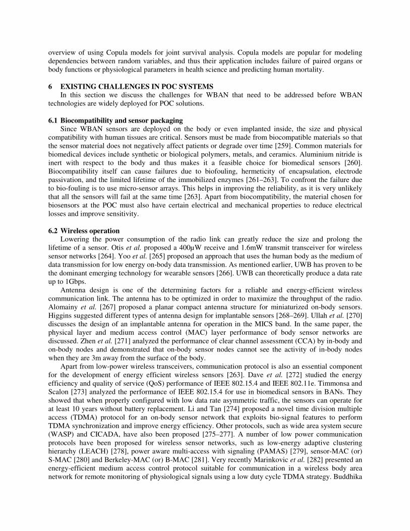
overview of using Copula models for joint survival analysis. Copula models are popular for modeling dependencies between random variables, and thus their application includes failure of paired organs or body functions or physiological parameters in health science and predicting human mortality.
6 EXISTING CHALLENGES IN POC SYSTEMS
In this section we discuss the challenges for WBAN that need to be addressed before WBAN technologies are widely deployed for POC solutions.
6.1 Biocompatibility and sensor packaging
Since WBAN sensors are deployed on the body or even implanted inside, the size and physical compatibility with human tissues are critical. Sensors must be made from biocompatible materials so that the sensor material does not negatively affect patients or degrade over time [259]. Common materials for biomedical devices include synthetic or biological polymers, metals, and ceramics. Aluminium nitride is inert with respect to the body and thus makes it a feasible choice for biomedical sensors [260]. Biocompatibility itself can cause failures due to biofouling, hermeticity of encapsulation, electrode passivation, and the limited lifetime of the immobilized enzymes [261–263]. To confront the failure due to bio-fouling is to use micro-sensor arrays. This helps in improving the reliability, as it is very unlikely that all the sensors will fail at the same time [263]. Apart from biocompatibility, the material chosen for biosensors at the POC must also have certain electrical and mechanical properties to reduce electrical losses and improve sensitivity. 6.2 Wireless operation
Lowering the power consumption of the radio link can greatly reduce the size and prolong the lifetime of a sensor. Otis et al. proposed a 400µW receive and 1.6mW transmit transceiver for wireless sensor networks [264]. Yoo et al. [265] proposed an approach that uses the human body as the medium of data transmission for low energy on-body data transmission. As mentioned earlier, UWB has proven to be the dominant emerging technology for wearable sensors [266]. UWB can theoretically produce a data rate up to 1Gbps.
Antenna design is one of the determining factors for a reliable and energy-efficient wireless communication link. The antenna has to be optimized in order to maximize the throughput of the radio. Alomainy et al. [267] proposed a planar compact antenna structure for miniaturized on-body sensors. Higgins suggested different types of antenna design for implantable sensors [268–269]. Ullah et al. [270] discusses the design of an implantable antenna for operation in the MICS band. In the same paper, the physical layer and medium access control (MAC) layer performance of body sensor networks are discussed. Zhen et al. [271] analyzed the performance of clear channel assessment (CCA) by in-body and on-body nodes and demonstrated that on-body sensor nodes cannot see the activity of in-body nodes when they are 3m away from the surface of the body.
Apart from low-power wireless transceivers, communication protocol is also an essential component for the development of energy efficient wireless sensors [263]. Dave et al. [272] studied the energy efficiency and quality of service (QoS) performance of IEEE 802.15.4 and IEEE 802.11e. Timmonsa and Scalon [273] analyzed the performance of IEEE 802.15.4 for use in biomedical sensors in BANs. They showed that when properly configured with low data rate asymmetric traffic, the sensors can operate for at least 10 years without battery replacement. Li and Tan [274] proposed a novel time division multiple access (TDMA) protocol for an on-body sensor network that exploits bio-signal features to perform TDMA synchronization and improve energy efficiency. Other protocols, such as wide area system secure (WASP) and CICADA, have also been proposed [275–277]. A number of low power communication protocols have been proposed for wireless sensor networks, such as low-energy adaptive clustering hierarchy (LEACH) [278], power aware multi-access with signaling (PAMAS) [279], sensor-MAC (or) S-MAC [280] and Berkeley-MAC (or) B-MAC [281]. Very recently Marinkovic et al. [282] presented an energy-efficient medium access control protocol suitable for communication in a wireless body area network for remote monitoring of physiological signals using a low duty cycle TDMA strategy. Buddhika

de Silva et al. [283] investigated the impact of inter-user interference and its behavior with respect to the number of networks and the rates at which these networks communicate. They observed that inter-user interference can cause up to 35 % loss in packet delivery ration.
Though energy-efficient MAC protocols have been devised, there is still a need for accurate models that take into account factors such as reliability, latency, mutual interference, energy consumption, and mobility effect. Sheryl Howard [284] studied the energy efficiency of error-correcting coding in low-power links that connect the implant and the external device. However, there is a need for more in-depth evaluation in all other scenarios.
6.3 Security Privacy requires effective and efficient authentication techniques in WBANs. It was suggested that
the ideal way to provide user authentication is to use complex but distinguishable human body characteristics. Sriram Cherukuri et al. [167] used the same method and proposed an approach wherein biometrics derived from the body are used for securing the keying material. A similar approach was presented by Venkatasubramanian et al. [285] with two schemes that rely on the novel technique of using physiological values from the wearer’s body for securing a cluster topology formation. Venkatasubramanian et al. [286] presented a novel key agreement scheme that allows two sensors in a WBAN to agree to a common key generated using electrocardiogram (EKG) signals for providing security on real EKG data. Still, a mechanism needs to be devised to differentiate between different levels of security to identify and distinguish between life-threatening events from other events with various security priorities. 6.4 Power restrictions
Sensors for WBAN application have power restrictions. This is mainly due to their small size and the absence of wires. Using an integrated power supply such as a battery will drain out of power. ence, there is a need to switch to passive power sources such as solar power, vibration, motion, air flow, temperature difference, and infrared radiation. Raghunathan et al. [287] discuss the key issues and tradeoffs that arise in the design of solar energy harvesting in wireless embedded systems and present a design, implementation, and performance evaluation of Heliomote sensors. Mitcheson et al. [288] developed a vibration-based generator designed for wearable/implantable devices that is capable of delivering up to 2µJ/cycle. Similarly, Reilly et al. [289] proposed a system to harvest vibration energy from a piezoelectric thin film. IMEC developed a 3 x 3 x 1.5 cm3 thermal micro-power generator that is capable of converting thermal energy to 4µW power at 5ºC temperature difference on the thermopile. Although techniques for generating power from passive sources have been proposed, the power generated is insufficient for continuous operation. Thus, to deal with this issue, there is a need for an external wireless power source to generate and wirelessly transfer power to the sensors. Recently, MIT researchers have reported transferring energy through a wireless medium to power electronic devices over a short range using evanescent waves [290]. Huang et al. [291] demonstrated a technique to harvest power from solar energy.
6.5 Standards and rules
Efforts have been made to develop standards for WBAN for POC [292]. However, pervasive health monitoring and treatments (POC) employing WBAN require standardized rules for operation in ambulatory environments and providing POC services without limitations on the wearer’s location or mobility while also taking into account individual privacy. Also, sensor fabrication, minimum biocompatible property requirements, power consumed, and minimum detectable levels of a particular biomarker must be standardized.
7 DISCUSSION
The growing number of fatalities resulting from chronic diseases has led to increased efforts to develop POC systems for remote health monitoring and patient care. To understand the improvements
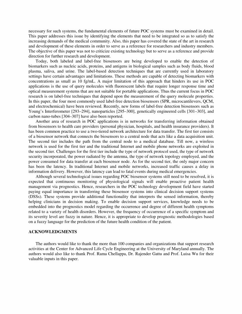
necessary for such systems, the fundamental elements of future POC systems must be examined in detail. This paper addresses this issue by identifying the elements that need to be integrated so as to satisfy the increasing demands of the medical community. Also, this paper has covered the state of the art in research and development of these elements in order to serve as a reference for researchers and industry members. The objective of this paper was not to criticize existing technology but to serve as a reference and provide direction for further research and development.
Today, both labeled and label-free biosensors are being developed to enable the detection of biomarkers such as nucleic acids, proteins, and antigens in biological samples such as body fluids, blood plasma, saliva, and urine. The label-based detection techniques that are currently used in laboratory settings have certain advantages and limitations. These methods are capable of detecting biomarkers with concentrations as small as 10 fg/mL. A major limitation of this approach that hinders its use in POC applications is the use of query molecules with fluorescent labels that require longer response time and optical measurement systems that are not suitable for portable applications. Thus the current focus in POC research is on label-free techniques that depend upon the measurement of the query molecule properties. In this paper, the four most commonly used label-free detection biosensors (SPR, microcantilevers, QCM, and electrochemical) have been reviewed. Recently, new forms of label-free detection biosensors such as Young’s Interferometer [293–294], nanoparticles [295–300], genetically engineered cells [301–303], and carbon nano-tubes [304–307] have also been reported.
Another area of research in POC applications is in networks for transferring information obtained from biosensors to health care providers (personal physician, hospitals, and health insurance providers). It has been common practice to use a two-tiered network architecture for data transfer. The first tier consists of a biosensor network that connects the biosensors to a central node that acts like a data acquisition unit. The second tier includes the path from the central node to a medical database. Till now, a wireless network is used for the first tier and the traditional Internet and mobile phone networks are exploited in the second tier. Challenges for the first tier include the type of network protocol used, the type of network security incorporated, the power radiated by the antenna, the type of network topology employed, and the power consumed for data transfer at each biosensor node. As for the second tier, the only major concern has been the latency. In traditional Internet and mobile networks, increased traffic causes a delay in information delivery. However, this latency can lead to fatal events during medical emergencies.
Although several technological issues regarding POC biosensor systems still need to be resolved, it is expected that continuous monitoring of physiological signals will enable proactive patient health management via prognostics. Hence, researchers in the POC technology development field have started paying equal importance in transferring these biosensor systems into clinical decision support systems (DSSs). These systems provide additional functionality that interprets the sensed information, thereby helping clinicians in decision making. To enable decision support services, knowledge needs to be embedded into the prognostics model regarding the occurrence and degree of different health symptoms related to a variety of health disorders. However, the frequency of occurrence of a specific symptom and its severity level are fuzzy in nature. Hence, it is appropriate to develop prognostic methodologies based on a fuzzy language for the prediction of the future health condition of the patient.
ACKNOWLEDGMENTS
The authors would like to thank the more than 100 companies and organizations that support research activities at the Center for Advanced Life Cycle Engineering at the University of Maryland annually. The authors would also like to thank Prof. Rama Chellappa, Dr. Rajender Gattu and Prof. Luisa Wu for their valuable inputs in this paper.

REFERENCES
1. Ross DeVol and Armen Bedroussian: An Unhealthy America: The Economic Burden of Chronic Diseases,
Milken Institute, New Jersey Briefing (2008). 2. Chris Sander: Genomic Medicine and the Future of Health Care. Science, 287 (5460), 1977-1978 (2000). 3. Huntington Willard, Misha Angrist and Geoffrey Ginsburg: Genomic Medicine: Genetic Variation and Its
Impact on the Future of Health Care. Philos. Trans. R. Soc. Lond. ,Ser. B, 360 (1460), 1543-1550 (2005). 4. Steven Soper, Kathlynn Brown, Andrew Ellington, Bruno Frazier, Guillermo Garcia-Manero, Vincent Gau,
Steven Gutman, Daniel Hayes, Brenda Korte, James Landers, Dale Larson, Frances Ligler, Arun Majumdar, Marco Mascini, David Nolte, Zeev Rosenzweig, Joseph Wang and David Wilson: Point-of-care biosensor systems for cancer diagnostics/prognostics. Biosensors Bioelectro., 2, 1932-1942 (2006).
5. Ruth Etzioni, Nicole Urban, Scott Ramsey, Martin McIntosh, Stephen Schwartz, Brian Reid, Jerald Radich, Garnet Anderson and Leland Hartwell: The case of early detection. Nat. Rev. Cancer, 3, 243-252 (2003).
6. Feltis BN, Sexton BA, Glenn FI, Best MJ, Wilkins M and Davis TJ: A hand-held surface plasmon resonance biosensor for the detection of ricin and other biological agents. Biosensors Bioelectron., 23, 1131-1136 (2008).
7. Justin Gooding: Biosensor Technology for Detecting Biological Warfare Agents: Recent Progress and Future Trends. Anal. Chim. Acta, 559, 137-151 (2006).
8. Leon Terry, Stephen White, Linda Tigwell: The Application of Biosensors to Fresh Produce and the Wider Food Industry. J. Agric. Food Chem., 53, 1309-1306 (2005).
9. Sara Rodriguez-Mozaz, Maria Lopez de Alda, Damia Barcelo: Biosensors as useful tools for environmental analysis and monitoring. Anal Bioanal Chem, 386, 1025-1041 (2006).
10. Antje Baeumner: Biosensors for environmental pollutants and food contaminants. 377, 434-445 (2003). 11. Donghui Yu, Bertrand Blankert, Jean-Claude Vire, and Jean-Michel Kauffmann: Biosensors in Drug
Discovery and Drug Analysis. Anal. Lette., 38, 1867-1701 (2005). 12. Seokheun Choi, Michael Goryll, Lai Yi Mandy Sin, Pak Kin Wong and Junseok Chae: Microfluidic-based
biosensors towards point-of-care detection of nucleic acids and proteins. Microfluid Nanofluid, 10, 231-247 (2011).
13. Seokheun Choi and Junseok Chae: A regenerative biosensing surface in microfluidics using electrochemical desorption of short-chain self-assembled monolayer. Microfluid Nanofluid, 7, 819-827 (2009).
14. Bo Huang, Hongkai Wu, Devaki Baya, Arthur Grossman, Sebastein Granier, Brian Kobilka and Richard Zare: Counting low-copy number proteins in a single cell. Science. 315, 81–84 (2007).
15. Elizabeth Ottesen, Jong Hong, Stephen Quake, Jared Leadbetter: Microfluidic digital PCR Enables Muiltgen analysis of individual environmental bacteria. Science 314, 1464–1467 (2006).
16. Lotien Huang, Edward Cox, Robert Austin and James Sturm: Continuous particle separation through deterministic lateral displacement. Science 304, 987–990 (2004).
17. Chia-Fu Chou, Robert Austin, Olgica Bakajin, Jonas Tegenfeldt, Judith Castelino, Shirley Chan, Edward Cox, Harold Craighead, Nicholas Darnton, Thomas Duke, Jongyoon Han, Steve Turner: Sorting biomolecules with microdevices. Electrophoresis 21, 81–90 (2000).
18. Mehmet Toner and Daniel Irimia: Blood-on-a-Chip. Annu. Rev. Biomed. Eng. 7, 77–103 (2005). 19. Sunitha Nagrath, Lecia Sequist, Shyamala Maheswaran, Daphne Bell, Daniel Irimia, Lindsey Ulkus, Matthew
Smith, Eunice Kwak, Subba Digumarthy, Alona Muzikansky, Paula Ryan, Ulysses Balis, Ronald Tompkins, Daniel Haber, Mehmet Toner. Isolation of rare circulating tumour cells in cancer patients by microchip technology. Nature 450, 1235–1239 (2007).
20. Ruth McNerney and Peter Daley: Towards a point-of-care test for active tuberculosis: obstacles and opportunities. Nat. Rev. Microbiol., 9, 204-213 (2011).
21. Fox W, Ellard GA and Mitchison DA: Studies on the treatment of tuberculosis undertaken by the British Medical Research Council tuberculosis units, 1946–1986, with relevant subsequent publications. Int. J.
Tuberc. Lung Dis. 3, S231–S279 (1999). 22. WHO. Global tuberculosis control: WHO Report 2010 (WHO, Geneva, 2010). 23. Lienhardt C, Rowley J, Manneh K, Lahai G, Needham D, Milligan P, McAdam KP: Factors affecting time
delay to treatment in a tuberculosis control programme in a sub-Saharan African country: the experience of The Gambia. Int. J. Tuberc. Lung Dis. 5, 233–239 (2001).
24. Golub JE, Bur S, Cronin WA, Gange S, Baruch N, Comstock GW, Chaisson RE: Delayed tuberculosis diagnosis and tuberculosis transmission. Int. J. Tuberc. Lung Dis. 10, 24–30 (2006).

25. Emmett Keeler, Mark Perkins, Peter Small, Christy Hanson, Steven Reed, Jane Cunningham, Julia Aledort, Lee Hillborne, Maria Rafael, Federico Girosi and Christopher Dye: Reducing the global burden of tuberculosis: the contribution of improved diagnostics. Nature 444 (Suppl. 1), 49–57 (2006).
26. Hao Y and Foster R: Wireless Body Sensor Networks for health Monitoring applications. Phys. Meas., 29, R27-R56 (2008).
27. Bonato P: Advances in wearable technology and applications in physical medicine and rehabilitation. J.
Neuro. Eng. Rehabil., 2, 2 (2005). 28. Bonato P: Wearable sensors/systems and their impact on biomedical engineering. IEEE Eng. Med. Biol.
Mag., 22 (3), 18-20 (2003). 29. Alexandros Pantelopoulos and Nikolaos Bourbakis: A Survey on Wearable Sensor-Based Systems for Health
monitoring and Prognosis. IEEE Trans. Syst. Man Cybern. Part C Appl. Rev., 40 (1), 1-12 (2010). 30. Bourbakis N and Gallagher J: DIAGNOSIS: A synergestic co-operative framework of new generation health
diagnosis systems for people with disabilities and the elderly. In Proc. Int. Conf. CS, 730-745 (2008). 31. Raskovic D, Martin T and Jovanov E: Medical monitoring applications for wearable computing. Comput. J.,
47, 495-504 (2004). 32. Anliker A, Beutel J, Dyer M, Enzler R, Luckowicz P, Thiele L, and Troster G: A systematic approach to the
design of distributed wearable systems. IEEE Trans. Comput., 53 (8), 1017-1033 (2004). 33. Arnab Choudhury: A Piezoresistive Microcantilever Array for Chemical Sensing applications. PhD
Dissertation, Dept. Mech. Eng., Georgia Institute of Technology, Atlanta, Georgia (2007). 34. Carlos Mastrangelo, Mark Burns and David Burke: Microfabricated devices for genetic diagnostics. Proc.
IEEE 86 (8), 1769-1787 (1998). 35. Che-Hsin Lin, Gwo-Bin Lee, Lung-Ming Fu and Bao Herng Hwey : Vertical focusing device utilizing
dielectrophoretic force and its application on microflow cytometer. J. Microelectromech. Syst 13 (6), 923-932, (2004).
36. James Rusling, Challa Kumar, Silvio Gutkind and Vyomesh Patel: Measurement of biomarker proteins for point-of-care early detection and monitoring of cancer. Analyst, 135, 2496-2511(2010).
37. Rashid Bashir: BioMEMS: state-of-art in detection , opportunities and prospects. Adv. Drug Delivery Rev., 56 (11), 1565-1586 (2004).
38. Yashwanth. Prakash, Suresh Lalwani, Sandeep Gupta, E. Elsharawy, and L. Schwiebert: Towards a propagation model for wireless biomedical applications. In: Proc. IEEE Intl. Conf. Commun. 3, 1993-1997 (2003).
39. Andrew Fort, Claude Desset, Philippe De Doncker, Piet Wambacq and Leo Van Biesen: An Ultra-Wideband Body Area Propagation Channel Model-From Statistic to Implementation. IEEE Trans. Microw. Theory Tech 54 (4), 1820-1826 (2006).
40. Frank Blum, Andrej Denisenko, Reinhart Job, Dietmar Borchert and Wolfgang R. Fahrner: Diamond Radiation Sensors for Medical Applications. In: Proc. ISIE Intl. Symp. Ind. Elec., 163-166 (1998).
41. Brian Otis, Yuen Hui Chee and Jan Rabaey: 400µW-RX, 1.6mW-TX Super-Regenerative Transceiver for Wireless Sensor Networks. In: Proc. IEEE ISSCC Intl. Solid-State Circuits Conf., 396-397 (2005).
42. Dummeruth M: Wireless wearable health monitoring system. M.S. Thesis, Case-Western Reserve University (2002).
43. Shanshan Jiang, Yanchuan Cao, Sameer Iyengar, Philip Kuryloski, Roozbeh Jafari, Yuan Xue, Ruzena Bajcsy, and Stephen Wicker: CareNet: An Integrated Wireless Sensor Networking Environment for Remote Healthcare. In: Proc. ICST 3
rd Intl. Conf. Body Area Networks (2008).
44. Shanshan Jiang, Yuan Xue, Annarita Giani and Ruzena Bajcsy: Providing QoS for Wireless Remote healthcare System. In: Proc. ICME IEEE Intl. Conf. Multimedia & Expo (2009).
45. Krishna Venkatasubramanian: A Secure Communication Protocol for Wireless Bio-medical Sensor Networks. MS Thesis, Dept. Computer Science, Arizona State University, AZ (2004).
46. Bart Braem, Benoit Latre, Ingrid Moerman, Chris Blondia, Piet Demeester: The Wireless Sutonomous Spanning tree Protocol for multihop Wireless Body Area Networks. Int. Conf. MOBIQW, 1-8 (2006).
47. Zhanpeng Jin, Joseph Oresko, Shimeng Huang and Allen Cheng: HeartToGo: A personalized Medicine Technology for Cardiovascular Disease Prevention and Detection. Proc. IEEE/NIH LiSSA, 80-83 (2009).
48. Jimena Rodriguez, Alfredo Goni and Arantza Illarramendi: Real-Time Classification of ECGs on a PDA. IEEE Trans. Inf Biomed., 9 (1), 23-34 (2005).
49. Alexandros Pantelopoulos and Nikolaos Bourbakis: Prognosis-A Wearable Health-Monitoring System for People at Risk: Methodology and modeling. IEEE Trans. Inf Biomed., 14 (3), 613-621 (2010).

50. Shantanu Bhattacharya, Jaesung Jang, Liju Yang, Demir Akin, and Rashid Bashir: BioMEMS and Nanotechnology-based approaches for rapid detection of biological entities. J. Rapid Methods Autom.
Microbiol., 15, 1-32 (2007). 51. Mohd. Ansari and Chongdu Cho: Design and Analysis of a high sensitive Microcantilever Biosensor for
Biomedical Applications. In: Intl. Conf. BioMed. Eng. & Informatics, IEEE Computer Society, 593-597 (2008).
52. Nabiollah Abolfathi: BioMEMS (Article) (2006). 53. Kua CH, Lam YC, Yang C, and Toumi KY: Review of bio-particle manipulation using dielectrophoresis.
Innovation in Manufacturing Systems and Technology (IMST) (2005). Available: http://hdl.handle.net/1721.1/7464.
54. Teena James, Manu Mannoor and Dentcho Ivanov: BioMEMS – Advancing the Frontiers of Medicine. Sensors 8, 6077-6107 (2008).
55. Kyu-Youn Hwang, Sung-Young Jeong, Young-Rok Kim, Kak Namkoong, Hee-Kyun Lim, Won-Seok Chung, Joon-Ho Kim and Nam Huh: Rapid Detection of bacterial cell from whole blood: Integration of DNA sample preparation into single micro-PCR chip. Sensors and Actuators B: Chemicals (2009).
56. Abdennour Abbas, Thomas Dargent, Dominique Croix, Michael Salzet and Bertrand Bocquet: Ex-vivo detection of neural events using THz BioMEMS. Med Sci Monit 15 (9), 121-125 (2009).
57. Candan Tamerler and Mehmet Sarikaya: Molecular biomimetics: nanotechnology and bionanotechnology using genetically engineered peptides. Phil. Trans. R. Soc. A, 1–22 (2009).
58. Thomas Ming-Hung Lee: Over-the-Counter Biosensors: Past, Present, and Future. Sensors, 8, 5535-5559 (2008).
59. Collings AF and Frank Caruso: Biosensors: Recent Advances. Rep. Prog. Phys 60, 1397-1445 (1997). 60. Mohd. Ansari and Chongdu Cho: Deflection, Frequency, and Stress Characteristics of Rectangular,
Triangular, and Step Profile Microcantilevers for Biosensors. Sensors, 9, 6046-6057 (2009). 61. Arvind Sai Sarathi Vasan, Ravi Doraiswami and Michael Pecht: Biocompatible Polymer Composite Material
for Highly Sensitive Point-of-Care BioMEMS Microcantilever sensors. Proc. SMTAI 2010, 279-288 (2010). 62. Nathalie Zammatteo, Laurent Jeanmart, Sandrine Hamels, Stephane Courtois, Pierre Louette, Laszlo Hevesi
and Jose Remacle: Comparison between Different Strategies of Covalent Attachment of DNA to Glass Surfaces to Build DNA Microarrays. Anal. Biochem., 280, 143-150 (2000).
63. Ryan Bailey, Gabriel Kwong, Caius Radu, Owen Witte and James Heath: DNA-encoded antibody library: A unified platform for multiplexed cell sorting and detection of genes and proteins. J. Am. Chem. Soc. 129, 1959–1967 (2007).
64. Jeroen Lijmer and Patrick Bossuyt: Various randomized designs can be used to evaluate medical tests. J.
Clin. Epidemiol 62, 364-373 (2009). 65. Kim Sapsford and Lisa Shriver-Lake: Bacterial Detection Using Evanescent Wave-Based Fluorescent
Biosensors. In: Principles of Bacterial Detection: Biosensors, Recognition Receptors and Microsystems. Eds: Springer, NY (2008).
66. Espina V, Woodhouse EC, Wulfkuhle J, Asmussen HD, Petricoin EF and Liotta LA: Protein microarray detection strategies: focus on direct detection technologies. J. Immunol. Methods, 290, 121-133 (2004).
67. Yu X, Xu D, Cheng Q: Label-free detection methods for protein microarray. Proteomics, 6, 5493-5503 (2006).
68. Chamritski I, Clarkson M, Franklin J, Li SW: Real-time detection of antigen-antibody reactions by imaging ellipsometry. Aust. J. Chem., 60, 667-671 (2007).
69. Ji J, O’Connell JG, Carter DJ, Larson DN: High-throughput nanohole array based system to monitor multiple binding events in real-time. Anal. Chem., 80, 2491-2498 (2008).
70. Sandipan Ray, Gunjan Mehta and Sanjeeva Srivastava: Label-free detection techniques for protein microarrays: Prospects, merits and challenges. Proteomics, 10, 731-748 (2010).
71. Tuan Vo-Dinh and Brian Cullum: Biosensors and biochips: advances in biological and medical diagnostics. J.
Anal. Chem 366, 540-551 (2000). 72. Presnova GV, Rybcova MY, Egorov AM: Electrochemical biosensors based on horseradish peroxidase. Russ.
J. Gen. Chem., 78 (12), 2482-2488 (2008). 73. Engvall E and Perlmann P: Enzyme linked immunosorbent assay (ELISA). Quantitative assay of
immunoglobulin G. Immunochemistry, 8, 871-874 (1971). 74. Jorn Ueberfeld and David Walt: Reversible Ratiometric Probe for Quantitative DNA Measurements. Anal.
Chem., 76, 947-952 (2004).
75. Nianhuan Yao, Wenwu Xiao, Xiaobing Wang, Jan Marik, See-H Park, Yoshikazu Takada and Kit Lam: Discovery of targeting ligands for breast cancer cells using the one-bead one-compund combinatorial method. J. Med. Chem. 52, 126-133 (2009).
76. Kim YI, Park TS, Kang JH, Lee MC, Kim JT, Park JH and Baik HK: Biosensors for label free detection based on RF and MEMS technology. Sens. Actuators B, 119, 592-599 (2006).
77. Claire Dalmay, Arnaud Pothier, Pierre Blondy, Mathilde Cheray, Fabrice Lalloue and Marie-Odile
Jauberteau: RF biosensor based Microwave filter for biological characterization. in Proc. 39th
European
Microwave Conference, 41-44 (2009).
78. Arvind Sai Sarathi Vasan, Ravi Doraiswami and Michael Pecht: Embedded 3D BioMEMS for Multiplexed Label Free Detection. To be published in Proc. IEEE ECTC (2011).
79. Jiri Homola, Sinclair Yee, Gunter Gauglitz: Surface plasmon resonance sensors: Review. Sens. Actuators, B, 54, 3–15 (1999).
80. Dina Wassaf, Guannan Kuang, Kris Kopacz, Qi-Long Wu, Que Nguyen, Mark Toews, Janja Cosic, Judith Jacques, Steve Wiltshire, Jeremy Lambert, Csaba Pazmany, Shannon Hogan, Robert Ladner, Andrew Nixon and Daniel Sexton: High throughput affinity ranking of antibodies using surface plasmon resonance microarrays. Anal. Biochem, 351, 241–253 (2006).
81. Shankaran DR, Gobi KV, Miura N: Recent advancements in surface plasmon resonance immunosensors for detection of small molecules of biomedical, food and environmental interest. Sens. Actuators, B, 121, 158–177 (2007).
82. Christine Campagnolo, Katherine Meyers, Thomas Ryan, Robert Atkinson, Yao Chen, Matt Scanlan, Gerd Ritter, Lloyd Old and Carl Batt: Real-time, label-free monitoring of tumor antigen and serum antibody interactions. J. Biochem. Biophys. Methods, 61, 283–298 (2004).
83. Ha Minh Hiep, Tatsuro Endo, Kagan Kerman, Miyuki Chikae, Do-Kyun Kim, Shohei Yamamura, Yuzuru Takamura and Eiichi Tamiya: A localized surface plasmon resonance based immunosensors for the detection of casein in milk. Sci. Technol. Adv. Mat., 8, 331–338 (2007).
84. Qiuming Yu, Shengfu Chen, Allen Taylor, Jiri Homola, Bertold Hock, Shaoyi Jiang: Detection of low-molecular-weight domoic acid using surface plasmon resonance sensor. Sens. Actuators, B, 107, 193–201 (2005).
85. Peter Pfeifer, Ulrich Aldinger, Gunter Schwotzer, Stephan Diekmann, Peter Steinrucke: Real time sensing of specific molecular binding using surface plasmon resonance spectroscopy. Sens. Actuators, B, 54, 166–175 (1999).
86. Fritz J, Baller MK, Lang HP, Rothuizen H, Vettiger P, Meyer E, Guntherodt HJ, Gerber Ch., Gimzewski JK: Translating Biomolecular Recognition into Nanomechanics. Science 288, 316–318 (2000).
87. Raiteri R, Nelles G, Butt HJ, Knol, W and Skladal P: Sensing of biological substances based on the bending of microfabricated cantilevers. Sens. Actuators, B, 61, 213–217 (1999).
88. Guanghua Wu, Haifeng Ji, Karolyn Hansen, Thomas Thundat, Ram Datar, Richard Cote, Michael Hagan, Arup Chakraborty, and Arunava Majumdar: Origin of nanomechanical cantilever motion generated from biomolecular interactions. Proc. Natl. Acad. Sci. USA, 98, 1560–1564 (2001).
89. Karolyn Hansen, Haifeng Ji, Guanghua Wu, Ram Datar, Richard Cote, Arunava Majumdar, Thomas Thundat: Cantilever-based optical deflection assay for discrimination of DNA single nucleotide mismatches. Anal.
Chem. 73, 1567–1571 (2001). 90. Madou M: Fundamentals of microfabrication. (CRC Press, New York; 1997). 91. Guanghua Wu, Ram Datar, Karolyn Hansen, Thomas Thundat, Richard Cote and Arun Majumdar: Bioassay
of prostate-specific antigen (PSA) using microcantilevers. Nat. Biotechnol., 19 (9), 856-860 (2001). 92. Hans-Jurgen Butt: A Sensitive Method to Measure Changes in the Surface Stress of Solids. J. Colloid
Interface Sci. 180, 251-260 (1996). 93. Thomas Brunt, Evans Chabala, Trevor Rayment, Sean O'Shea, Mark Welland: Measuring surface stress
induced by electrode processes using a micromechanical sensor. J. Chem. Soc. Faraday Trans 92 (20), 3807-3812 (1996).
94. Rudiger Berger, Emmanuel Delamarche, Hans Peter Lang, Christoph Gerber, James Gimzewski, Ernst Meyer, Hans-Joachim Guntherodt: Surface Stress in the Self-Assembly of Alkanethiols on Gold. Science, 276, 2021-2024 (1997).
95. Tian F, Pei JH, Hedden DL, Brown GM and Thundat T: Observation of the surface stress induced in microcantilevers by electrochemical redox processes. Ultramicroscopy, 100, 217-223 (2004).

96. Rachel McKendry, Jlayun Zhang, Youri Arntz, Torsten Strunz, Martin Hegner, Hans Peter Lang, Marko Baller, Ulrich Certa, Ernst Meyer, Hans-Joachim Guntherodt, and Christoph Gerber: Multiple label-free biodetection and quantitative DNA-binding assays on a nanomechanical cantilever array. Proc. Natl. Acad.
Sci. USA 99, 9783-9788 (2002). 97. Zhang J, Lang HP, Huber F, Bietsch A, Grange W, Certa U, McKendry R, Guntherodt H-J, Hegner M and
Gerber CH: Rapid and label-free nanomechanical detection of biomarker transcripts in human RNA. Nat.
Nanotechnol, 1, 214-220 (2006). 98. Arntz Y, Seelig JD, Lang HP, Zhang J, Hunziker P, Ramseyer JP, Meyer E, Hegner M, and Gerber C: Label-
free protein assay based on a nanomechanical cantilever array. Nanotechnology, 14, 86–90 (2003). 99. Sandeep Kumar Vashist: A Review of Microcantilevers for Sensing Applications. AZojono, Journal of
Nanotechnology Online, 3, 1-15 (2007). 100. Moulin AM, O’ Shea SJ and Welland ME: Microcantilever-based biosensors. Ultramicroscopy 82, 23-31
(2000). 101. Sauerbrey, Gunter: Verwendung von Schwingquarzen zur Wagung dünner Schichten und zur
Mikrowagung. Zeitschrift fur Physik 155 (2), 206–222 (1959). 102. Iddo Ben-Dov and Itamar Willner: Piezoelectric Immunosensors for Urine Specimens of Chlamydia
trachomatis Employing Quartz Crystal Microbalance Microgravimetric Analyses. Anal. Chem., 69 (17), 3506–3512 (1997).
103. In-Seon Park, Namsoo Kim: Thiolated Salmonella antibody immobilization onto the gold surface of piezoelectric quartz crystal. Biosens. Bioelectron., 13 (10), 1091–1097 (1998).
104. Vaughan RD, O’Sullivan CK, Guilbault GG: Development of quartz crystal microbalance (QCM) immunsensor for the detection of Listeria monocytogene. Enzyme Microb. Technol., 29, 635– 638 (2001).
105. Namsoo Kim, , In-Seon Park, Dong-Kyung Kim: Characteristics of a label-free piezoelectric immunosensors detecting Pseudomonas aeruginosa. Sens. Actuators, B, 100, 432–438 (2004).
106. Xiao-Li Su, Yanbin Li: A self-assembled monolayer-based piezoelectric immunosensors for rapid detection of Escherichia coli O157:H7. Biosens. Bioelectron., 19, 563–574 (2004).
107. Charles Poitras and Nathalie Tufenkji: A QCM-D-based biosensor for E. coli O157:H7 highlighting the relevance of the dissipation slope as a transduction signal. Biosens. Bioelectron., 24, 2137-2142 (2009).
108. Durst RA, Baumner AJ, Murray RW, Buck RP, Andrieux CP: Chemically Modified Electrodes (CME): Recommended terminology and definitions. Pure Appl. Chem., 69 (6), 1317-1323 (1997).
109. Wzodzimierz Kutner, Joseph Wang, Maurice L’Her, Richard Buck: Analytical aspects of chemically modified electrodes: classification, critical evaluation and recommendation. Pure Appl. Chem.,70 (6), 1301-1318 (1998).
110. Daniel Thevenot, Klara Toth, Richard Durst, George Wilson: Electrochemical biosensors: recommended definitions and classifications. Biosens. Bioelectron., 16, 121-131 (2001).
111. Shan D, Zhu M, Xue H, Cosnier S: Development of amperometric biosensor for glucose based on a novel attractive enzyme immobilization matrix: calcium carbonate nanoparticles. Biosens Bioelectron., 22 (8), 1612-1617 (2007).
112. Chen M, Zhang W, Jiang R, Diao G: Development of glucose amperometric biosensor based on a novel attractive enzyme immobilization matrix: amino derivative of thiacalix[4]arene. Anal Chim Acta., 687 (2), 177-183 (2011).
113. Yoshio Umezawa, Philippe Buhlmann, Kayoko Umezawa, Koji Tohda and Shigeru Amemiya: Potentiometric selectivity coefficients of ion-selective electrodes. Pure Appli, Chem., 72 (10), 1851-2082 (2000).
114. Wang Y, Cheng Q and Zeng X: Potentiometric biosensor for studying hydroquinone cytotoxicity in vitro. Biosens Bioelectron., 25 (6), 1356-62 (2010).
115. Chika Okafor, Daniel Grooms, Evangelyn Alocilja, and Steven Bolin: Fabrication of a Novel Conductometric Biosensor for Detecting Mycobacterium avium subsp. Paratuberculosis Antibodies. Sensors, 8, 6015-6025 (2008).
116. Gregory Drummond, Michael Hill and Jacqueline Barton: Electrochemical DNA sensors. Nat. Biotechnol., 21 (10), 1192-1199 (2003).
117. Zhihong Nie, Christian Nijhuis, Jinlong Gong, Xin Chen, Alexander Kumachev, Andres Martinez, Max Narovlyansky and George Whitesides: Electrochemical sensing in paper-based microfluidic devices. Lab
Chip, 10, 477-483 (2010). 118. Bergveld P: Development of an ion-sensitive solid-state device for neurophysiological measurements. IEEE
Trans. Biomed. Eng., BME-17, 70–71 (1970).

119. Bergveld P: Development, operation and application of the ion sensitive field effect transistor as a tool for electrophysiology. IEEE Trans. Biomed. Eng., BME-19, 342–351 (1972).
120. Bergveld P and Sibbald A: Analytical and Biomedical Applications of Ion-Selective Field-Effect Transistors. Elsevier, Amsterdam (1988).
121. Schoning MJ and Poghossian A: Recent advances in biologically sensitive field-effect transistors (BioFETs). Analyst, 127, 1137–1151 (2002).
122. Poghossian A, Cherstvy A, Ingebrandt S, Offenhauser A, and Schoning MJ: Possibilities and limitations of label-free detection of DNA hybridization with field-effect-based devices. Sens. Actuators, B, 111-112, 470–480 (2005).
123. Schoning MJ and Poghossian A: Bio FEDs (field-effect devices): State-of-the-art and new directions. Electroanalysis, 18 (19-20), 1893–1900 (2006).
124. Samit Gupta, Mark Elias, Xuejin Wen, John Shapiro, Leonard Brillson, Wu Lu, Stephen Craig Lee: Detection of clinically relevant levels of protein analyte under physiologic buffer using planar field effect transistors. Biosens.Bioelectron., 24, 505–511 (2008).
125. Shantanu Bhattacharya, Jaesung Jang, Liju Yang, Demir Akin, and Rashid Bashir: BioMEMS and Nanotechnology-based approaches for rapid detection of biological entities. J. Rapid Methods Autom.
Microbiol., 15, 1-32 (2007). 126. Bharat Bhusan: Nanotribology and Nanomechanics of MEMS/NEMS and BioMEMS/BioNEMS materials
and devices. Microelectron. Eng., 84, 387-412 (2007). 127. Teena James, Manu Mannoor and Dentcho Ivanov: BioMEMS – Advancing the Frontiers of Medicine.
Sensors 8, 6077-6107 (2008). 128. Herbert Pohl and Ira Hawk: Separation of Living and Dead cells by Dielectricphoresis. Science 152 (3722),
647-649 (1966). 129. Michael Brown: Lab on a piece of paper. Chemistry World, 7 (1), 33-33 (2010). 130. Chunyan Li, Lori Shutter, Pei-Ming Wu, Chong Ahn, Raj Narayan: Potential of a simple lab-on-a-tube for
point-of-care measurements of multiple analytes. Lab Chip, 10 (11), 1476-1479 (2010). 131. Rafaela Carvalhal, Emmanuel Carrilho, Laura Kubota: The potential and application of micro-fluidic paper
based separation devices. Bioanalysis, 2 (10), 1663-1665 (2010). 132. Zhihong Nie, Fredrique Deiss, Xinyu Liu, Ozge Akbulut, and George Whitesides: Integration of paper-based
microfluidic devices with commercial electrochemical readers. Lab Chip, 10 (22),3163-3169 (2010). 133. Rong Fan, Ophir Vermesh, Alok Srivastava, Brian Yen, Lidong Qin, Habib Ahmad, Gabriel Kwong, Chao-
Chao Liu, Juliane Gould, Leroy Hood & James R Heath: Integrated barcode chips for rapid, multiplexed analysis of proteins in microliter quantities of blood. Nat. Biotechnol., 26 (12), 1373 – 1378 (2008).
134. Gengfeng Zheng, Fernando Patolsky, Yi Cui, Wayne Wang and Charles Lieber: Multiplexed electrical detection of cancer markers with nanowire sensor arrays. Nat. Biotechnol., 23 (10), 1294 – 1301 (2005).
135. Cisco Securewireless: The Secure Wireless Network for Healthcare. Cisco Systems (2007). 136. Wireless in Healthcare. Cisco Systems (2006). 137. Delivering Mobile Point-of-Care with Pervasive Wireless Networks. Intel Corporation (2007). 138. Chun-Hao Chen, Rong-Zhang Hwang, Long-Sun Huang, Shi-Ming Lin, Hsiao-Chin Chen, Yu-Che Yang,
Yu-Tso Lin, Shih-An Yu, Yo-Sheng Lin, Yiao-Hong Wang, Nai-Kuan Chou, and Shey-Shi Lu: Wireless Bio-MEMS Sensors for C-Reactive Protein Detection Based on Nanomechanics. IEEE Trans. Biomed. Eng. 56 (2), 462-470 (2009).
139. Elad Brod, Shai Nimri, Boaz Turner, and Uri Sivan: Electrical Control over antibody-antigen binding. Sens.
Actuators, B, 128, 560-565 (2008). 140. Didier Vallin, Canh Tran-Minh: Electric field effects on immobilized urease activity. Biochemica et
Biophysica Acta – Enzymology, 571 (2), 321-332 (1979). 141. Tao NJ: Probing potential-tuned resonant tunneling through redox molecules with scanning tunneling
microscopy. Phys. Rev Lett, 76 (21), 4066-4069 (1996). 142. Andolfi L, Bruce D, Cannistraro S, Canters GW, Davis JJ, Hill HAO, Crozier J, Verbeet MPh, Wrathmell
CL, Astier Y: The electrochemical characteristics of blue copper protein monolayers on gold. J. Electroanal.
Chem, 565 (1), 21–28 (2004). 143. Jason Davis, Delphine Bruce, Gerard Canters, John Crozier, Hill Allen: Genetic modulation of metalloprotein
electron transfer at bare gold. Chem. Commun. (Camb) 5, 576–577 (2003). 144. Christian Hodneland, Milan Mrksich: Biomolecular surfaces that release ligands under electrochemical
control. J. Am. Chem. Soc,. 122, 4235–4236 (2000).
145. Carl Edman, Daniel Raymond, David Wu, Eugene Tu, Ronald Sosnowski, William Butler, Michael Nerenberg, Michael Heller: Electric field directed nucleic acid hybridization on microchips. Nucl Acids Res, 25 (24), 4907–4914 (1997).
146. Richard Heaton, Alexander Peterson and Rosina Georgiadis: Electrostatic surface Plasmon resonance: direct electric field-induced hybridization and denaturation in monolayer nucleic acid films and label free discrimination of base mismatches. PNAS, 98 (7), 3701–3704, (2001).
147. Huasong Cao, Victor Leung, Cupid Chow and Henry Chan: Enabling Technologies for Wireless Body Area Networks: A Survey and Outlook. IEEE Commun Mag 47 (12), 84-93 (2009).
148. Xu PJ, Zhang H and Tao XM: Textile-Structured Electrodes for Electrocardiogram. Textile Progress 40 (4), 183-213 (2008).
149. Marcos Vieira, Claudionor Coelho, Digenes Cecilio da Silva Jr., Jose da Mata.: Survey on wireless sensor network devices. In: Proc. IEEE ETFA 1, 537-544 (2003).
150. Benny Lo and Guang-Zhong Yang: Body Sensor Networks: Infrastructure for Life Science Research. IEEE/NLM Life Science Systems and Application Workshop, 1-2 (2006).
151. Quincy Brown, Rohit Srivastava and Michael McShane: Encapsulation of glucose oxidase and an oxygen-quenched fluorophore in polyelectrolyte-coated calcium alginated microspheres as optical glucose sensor systems. Biosensors and Bioelectronics 21 (1), 212-216 (2005).
152. Nader Najafi and Achiau Ludomirsky: Initial animal studies of a wireless, batteryless, MEMS implant for caridovascular applications. Biomedical Microdevices 6 (1), 61-65 (2004).
153. Amy Richards Grayson, Rebecca Shawgo, Audrey Johnson, Nolan Flynn, Yawen Li, Michael Cima, and Robert Langer: A BioMEMS Review: MEMS Technology for Physiologically Integrated Devices. Proc.
IEEE 92 (1), 6-21 (2004). 154. Chiara Buratti, Andrea Conti, Davide Dardari and Roberto Verdone: An Overview on Wireless Sensor
Networks Technologies and Evolution. Sensors, 9, 6869-6896 (2009). 155. Mohammad Upal Mahfuz and Kazi Ahmed: A review of micro-nano-scale wireless sensor networks for
environmental protection: Prospects and challenges. Science and Technology of Advanced Materials, 6, 302-306 (2005).
156. Jan Magne Tjensvold: IEEE Standard for Comparison of the IEEE 802.11, 802.15.1, 802.15.4 and 802.15.6 wireless standards. (2008).
157. Kamya Yazdandoost and Kamran Pour: Channel Model for Body Area Networks (BAN). IEEE P802.15-08-0780-09-0006, Technical Report (2009).
158. Sana Ullah, Pervez Khan, Niamat Ullah, Shahnaz Saleem, Henry Higgins and Kyung Sup Kwak: A Review of Wireless Body Area Networks for Medical Applications. IJCNS, 2 (8), 797-803 (2009).
159. Shoba G, Chittal RR, Kumar K: Medical Applications of Wireless Networks. In: Proc. ICSNC, 82 (2007). 160. Ning Xu: A Survey of Sensor Network Applications. IEEE Comm Mag, 102-114 (2002). 161. Mehmet Yuce, Steven Ng, Naung Myo, Jamil Khan and Wentai Liu: Wireless Body Sensor Network Using
Medical Implant Band. J Med Syst, 31, 467–474 (2007). 162. Krishna Venkatasubramanian and Sandeep Gupta: Physiological Value Based Efficient Usable Security
Solutions for Body Sensor Networks. ACM Transactions on Sensor Network, 6 (4), 31:1-31:36 (2010). 163. Kristof van Laerhoven, and Hans-Werner Gellersen: Spine versus Porcupine: A Study in distributed wearable
activity recognition. In: Proc ISWC, 142-149 (2004). 164. Kazushige Ouchi, Takuji Suzuki, and Miwako Doi: Lifeminder: A wearable healthcare support system using
users context. In: Proc ICDCSW, 791-792 (2002). 165. Rita Paradiso, Giannicola Loriga, and Nicola Taccini: A Wearable Healthcare system based on knitted
integrated sensors. IEEE Trans Inf Technol Biomed, 9 (3), 337-344 (2005). 166. Benny Lo, and Guang-Zhong Yang: Key Technical Challenges and Current Implementations of Body Sensor
Networks. In: Proc. IWWIBSN, (2005). 167. Sriram Cherukuri, Krishna Venkatasubramanian and Sandeep Gupta: BioSec: A Biometric Based Approach
for Securing Communication in Wireless Networks of Biosensors Implanted in the Human Body. In: Proc.
ICPPW’03 2003 Intl. Conf. Parallel Processing Workshops, (2003). 168. Shanshan Jiang, Yanchuan Cao, Sameer Iyengar, Philip Kuryloski, Roozbeh Jafari, Yuan Xue, Ruzena
Bajcsy and Stephen Wicker: CareNet: An Integrated Wireless Sensor Networking Environment for Remote Healthcare. In: Proc. ICST BAN, (2008).
169. Victor Shnayder, Bor-rong Chen, Konrad Lorincz, Thaddeus Fulford-Jones, and Matt Welsh: Sensor Networks for Medical Care. In: Proc SenSys (2005).

170. Sungdae Choi, Seong-Jun Song, Kyomin Sohn, Hyejung Kim, Jooyoung Kim, Jerald Yoo and Hoi-Jun Yoo: A Low Power Star-topology body area network controller for periodic data monitoring around and inside the human Body. In: Proc. ISWC IEEE, 139-140 (2006).
171. David Malan, Thaddeus Fulford-Jones, Matt Welsh, Steve Moulton: CodeBlue: An Ad Hoc Sensor Network Infrastructure for Emergency Medical Care. In: Proc MobiSys WAMES (2004).
172. Dimitri Konstantas, Aart Van Halteren, Richard Bults, Katarzyna Wac, Val Jones, Ing Widya, and Rainer Herzog: Mobihealth : Ambulant Patient Monitoring Over Public Wireless Networks. In: Proc MEDICON (2004).
173. Gerd Kortuem, Martin Bauer, and Zary Segall: Netman: The Design of a Collaborative Wearable Computer System. Mobile Networks and Applications 4 (1), 49-58 (1999).
174. Draft guide for health informatics-point-of-care medical Devices. Communication-technical report-Guidelines for the use of RF wireless technology, IEEE P1073.0.1.1/D01J (2006).
175. Emil Jovanov, Aleksandar Milenkovic, Chris Otto, and Piet de Groen: A Wireless Body Area Network of intelligent motion sensors for computer assisted physical rehabilitation. Journal of Neuro Engineering and
Rehabilitation 2 (6), 1-10 (2005). 176. Benny Lo and Guang-Zhong Yang: Architecture for Body Sensor Networks. IEE Proc. Perspective in
Pervasive Computing, 23-28 (2005). 177. Aleksandar Milenkovic, Chris Otto and Emil Jovanov: Wireless Sensor Network for Personal Health
Monitoring: Issues and an Implementation. J Comp Comm 29 (13-14), 521-533 (2006). 178. Mukesh R, Damodaran A, and Bharathi S: Energy Efficient Security Architecture for Wireless Biomedical
Sensor Networks. In: Proc IJCSIS 6 (1), 116-122 (2009). 179. Chris Otto, Aleksandar Milenkovic, Corey Sanders and Emil Jovanov: System Architecture of a wireless
Body Area Sensor Network for biquitous Health Monitoring. Journal of Mobile Multimedia 1 (4), 307-326 (2006).
180. Srdjan Krco: Implementation Solutions and Issues in Building a Personal Sensor Network for Health Care Monitoring. In: Proc IEEE EMBS, 350-353 (2003).
181. Lin Zong, Mike Sinclair and Ray Bittner: A Phone Centered Body Sensor Network Platform: Cost Energy Efficiency User Interface. In: Proc IEEE ICST BSN, 1-4 (2006).
182. Giorgetti G, Manes G, Lewiss JH, Mastroianni ST, and Gupta SKS: The Personal Sensor Network: A User-Centric Monitoring Solution. In: Proc. IEEE ICST BSN (2007).
183. Li Yang, Rushi Vyas, Amin Rida, Jonathan Pan and Manos Tentzeris: Wearable RFID-Enabled Sensor Nodes for Biomedical Applications. In: Proc ECTC, 2156-2159 (2008).
184. Muhammad Abdul Aleem: A Web Services-based System for Wireless Personal Area Network. In: Proc
ITSIM, 1467-1471 (2008). 185. Montgomery K, Mundt C, Thonier G, Tellier A, Udoh U, Barker V, Ricks R, Giovangrandi R, Davies P,
Cagle Y, Swain J, Hines J, Kovacs G: Lifeguard- A Personal Physiological Monitor For Extreme Environments. In: Proc IEMBS, 2192-2195 (2004).
186. Rich DeVaul, Michael Sung, Jonathan Gips, and Alex Pentland: MIThril 2003: Applications and Architecture. In: Proc ISWC (2003).
187. Alex Pentland: Healthwear: Medical Technology Becomes Wearable. IEEE Computer 37 (5), 34-41 (2004). 188. Konrad Lorincz, David Malan, Thaddeus Fulford-Jones, Alan Nawoj, Antony Clavel, Victor Shnayder,
Geoffrey Mainland, Matt Welsh and Steve Moulton: Sensor Networks for Emergency Response: Challenges and Opportunities. IEEE Pervasive Computing 3 (4), 805-813 (2004).
189. Victor Shnayder, Bor-Rong Chen, Konrad Lorincz, Thaddeus Fulford-Jones, and Matt Welsh: Sensor Networks for Medical Care. in Harvard university, Technical Report, TR-08-05 (2005).
190. Jordi Agud Ruiz, Shigeru Shimamoto: Novel Communication Services Based on Human Body and Environment Interaction: Applications inside Trains and Applications for Handicapped People. In: Proc.
IEEE WCNC, 2240-2245 (2006). 191. Lei Wang, Benny Lo and Guang-Zhong Yang: Multichannel Reflective PPG Earpiece Sensor with Passive
Motion Cancellation. IEEE Trans Biomed Circuits Syst 1 (4), 235-241 (2006). 192. Louis Atallah, Benny Lo, Guang-Zhong Yang and Frank Siegesmund: Wirelessly Accessible Sensor
Populations (WASP) for Elderly Care Monitoring. In: Proc PervasiveHealth, 2-7 (2008). 193. Chris Otto, Aleksandar Milenkovic, Corey Sanders, Emil Jovanov: System Architecture of a Wireless Body
Area Sensor Network for Ubiquitous Health Monitoring. J. Mob. Mult 1 (4), 307-326 (2007). 194. Thaddeus Fulford-Jones, Gu-Yeon Wei, Matt Welsh: A portable, low power, wireless two-lead EKG system.
In: Proc. IEEE EMBS, 2141-2144 (2004).

195. Aleksandar Milenkovic, Chris Otto, Emil Jovanov: Wireless Sensor Networks for Personal Health Monitoring: Issues and an Implementation. Comp. Commun 29 (13-14), 2521-2533 (2006).
196. Tia Gao, Tammara Massey, Leo Selavo, David Crawford, Bor-Rong Chen, Konrad Lorincz, Victor Shnayder, Logan Hauenstein, Foad Dabiri, James Jeng, Arjun Chenmugam, David White, Majid Sarrafzadeh, Matt Welsh: The Advanced Health and Disaster Aid Network: A Lightweight Wireless Medical System for Triage. IEEE Trans. Biomed. Circuits Syst 1 (3), 203-216 (2007).
197. Surapa Thiemjarus, Benny Lo, Guang-Zhong Yang: Context Aware Sensing - What's the Significance?. In: IEE Proc Perspective in Pervasive Computing, 163-170 (2005).
198. Iiro Jantunen, Hannu Laine, Pertti Huuskonen, Dirk Trossen, Vladimir Ermolov: Smart Sensor Architecture for Mobile-Terminal-Centric Ambient Intelligence. Sens. Act. 142 (1), 352-360 (2004).
199. Loren Schwiebert, Sandeep Gupta, Pepe Auner, Gary Abrams, Ray Iezzi, and Pat McAllister: A biomedical smart sensor for the visually impaired. In: Proc. IEEE Sensors 1, 693-698 (2002).
200. Rachel King, Benny Lo, Guang Zhong Yang: Hand Gesture Recognition With Body Sensor Networks. In: Proc. IEE BSN, 92-95 (2005).
201. Elisabetta Farella, Augusto Pieracci, Luca Benini, Laura Rocchi, Andrea Acquaviva: Interfacing uman and Computer with Wireless Body Area Sensor Networks: The WiMoCA Solution. Multimedia Tools App 38 (3), 337-363 (2008).
202. Surapa Thiemjarus, Benny Lo, Guang-Zhong Yang: A Spatio-Temporal Architecture for Context-Aware Sensing. In: IEEE Proc. IWWI BSN, 191-194 (2006).
203. Stankovic JA, Cao Q, Doan T, Fang L, He Z, Kiran R, Lin S, Son S, Stoleru R, Wood A: Wireless Sensor Networks for In-Home Healthcare: Potential and Challenges. In: Proc. HCMDSS (2005).
204. Emil Jovanov, Aleksandar Milenkovic, Chris Otto, and Piet de Groen, B. Johnson, S. Warren and G. Taibi: A WBAN System for Ambulatory Monitoring of Physical Activity and Health Status: Applications and Challenges. In: Proc. IEEE EMBS, 3810-3813 (2005).
205. Ken Fishkin, Min Wang: A Flexible, Low-Overhead Ubiquitous System foe Medication Monitoring. Intel Research Technical Report IRS-TR-03-011, (2003).
206. David Vawdrey, Eric Hall, Charles Knutson, James Archibald: A Self-Adapting Healthcare Information Infrastructure Using Mobile Computing Devices. In: Proc. Healthcom, 91-97 (2003).
207. Yuechun Chu and Aura Ganz: A mobile Teletrauma System using 3G Networks. IEEE Trans. Inf. Tech.
Biomed. 8 (4), 456-462 (2004). 208. Nicolas Chevrollier, Nada Golmie: On the use of Wireless Network Technologies in Healthcare
Environments. In: Proc. IEEE ASWN, 147-152 (2005). 209. Hsueh-Ting Chu, Chir-Chang Huang, Zhi-Hui Lian, Jeffrey Tsai: A Ubiquitous Warning System for asthma-
inducement. In: Proc IEEE SUTC, 186-191 (2006). 210. Robert Istepanian, Swamy Laxminarayan, Constantinos Pattichis: M-Health: Emerging Mobile Health
Systems. Eds: Springer (2005). 211. Cassim Munshi, Xiaoyuan Xu, Xiaodan Zou, Edward Soetiono, Chang Teo, Yong Lian: Wireless ECG
Plaster for Body Sensor Network. In: Proc. ISSS-MDBS, 310-313 (2008). 212. Kirby Keller, Kevin Swearingen, Jim Sheahan, Mike Bailey, Jon Dunsdon, Wojtek Przytula, Brett Jordan:
Aircraft Electrical Power Systems Prognostics and Health Management. In: Proc IEEE Aerospace Conf, 12 (2006).
213. Elisa Lee, Oscar Go: Survival Analysis in Public Health Research. Annu. Rev. Public. Health 18, 105-134 (1994).
214. Nikhil Vichare, Brian Tuchband, Michael Pecht: Prognostics and Health Monitoring of Electronics. Handbook on Performability Engineering., 1107-1122, EDS: Springer London (2008).
215. Nikhil Vichare, Micheal Pecht: Prognostics and Health Management of electronics. IEEE Trans. Comp. Pack.
Tech 29 (1), 222-229 (2006). 216. Rifat Shahriyar, Md. Faizul Bari, Gourab Kundu, Sheikh Iqbal Ahamed, Md. Mostofa Akbar: Intelligent
Mobile Health Monitoring System (IMHMS). Intl. J. Cont. Auto 2 (3) (2009). 217. Fanis Kalatzis, Nikolaos Giannakeas, Themis Exarchos, Leandro Lorenzelli, Andrea Adami, Massimiliano
Decarli, Sara Lupoli, Fabio Macciardi, Sofia Markoula, Ioannis Georgiou Dimitrios Fotiadis: Developing a genomic-based point-of-care diagnostic system for Rheumatoid arthritis and multiple sclerosis. In: Proc.
IEEE EMBS, 827-830 (2009). 218. Falas T, Papadopoulos G and Stafylopatis A: A review of decision support systems in telecare. J. Med. Syst.,
27, 347-356 (2003).

219. Carson ER, Cramp DG, Morgan A and Roudsari AV: Clinical decision support, systems methodology and telemedicine: Their role in the management of chronic disease. IEEE Trans. Inf. Techn. Biomed., 2 (2), 80-88 (1998).
220. Basilakis J, Lovell NH and Celler BG: A decision support architecture for telecare patient management of chronic and complex disease. In: Proc. 29
th Ann. Int. IEEE EMBS Conf., 4335-4338 (2007).
221. Berner E, Clinical Decision Support Systems: Theory and Practice, 2nd Eds. New York, Springer-Verlag (2006).
222. Pusarla C, Dasgupta A, Pecht M, and Christou A: A Physics-of-Failure Design Philosophy Applied to Flip-Chip Bonds. Microelectronics International, 36, 6-12 (1995).
223. Michael Pecht and Abjith Dasgupta: Physics-of-Failure: An Approach to Reliable Product Development. Journal of the Institute of Environmental Sciences, 38, 30-34 (1995).
224. Michael Pecht and Pradeep Lall: Life Cycle Approach to Design and Manufacturing Simulation of Multi-Chip Modules. International Journal of Modeling and Simulation, 16 (4), 173-183 (1996).
225. Michael Pecht and Jie Gu: Physics-of-failure-based Prognostics for Electronic Products. Trans. Inst. Meas.
Control, 31 (3/4), 309–322 (2009). 226. Jie Gu, Nikhil Vichare, Bilal Ayyub and Michael Pecht: Application of Grey Prediction Model for Failure
Prognostics of Electronics. International Journal of Performability Engineering, 6 (5), 435-442 (2010). 227. Kumar S, Dolev E and Pecht M: Parameter selection for health monitoring of electronic products.
Microelectronics Reliability, 50, 161-168 (2010). 228. Sachin Kumar and Michael Pecht: Modeling Approaches for Prognostics and Health Management of
Electronics. International Journal of Performability Engineering, 6 (5), 467-476 (2010). 229. Sachin Kumar and Michael Pecht: Data Analysis Approach for System Reliability, Diagnostics and
Prognostics. Pan Pacific Microelectronics Symposium, (2008). 230. Pecht M and Lopez L: Maxima-SPRT Methodology for Health Monitoring of Contact Resistance in IC
Sockets. Proc. 1st Intl. Conf. PHM, (2008). 231. Vasilis Sotiris and Michael Pecht: Support Vector Prognostics Analysis of Electronic Products and Systems.
AAAI Symp AI Prog, 120-127 (Nov, 2007). 232. Vasilis Sotiris and Michael Pecht: Anomaly Detection through a Bayesian Support Vector Machine. IEEE
Trans.Reliab, 59 (2), 277-286 (2010). 233. Gang Niu, Satnam Singh, Steven Holland, and Michael Pecht: Anomaly Detection of Notebook Computer
Based on Weibull Decision Metrics. Prognostics & System Health Management Conf., (2010). 234. Abraham Tomy Michael, Sachin Kumar, Sony Mathew and Michael Pecht: Anomaly Detection in Electronic
Products. Electronics System-Integration Tech Conf., (2008). 235. Sachin Kumar, Vasilis Sotiris and Michael Pecht: Health Assessment of Electronic Products using
Mahalanobis Distance and Projection Pursuit Analysis. International Journal of Computer, Information,
Systems Science, and Engineering, 2 (4), 242 - 250 (2008). 236. Sachin Kumar, Tommy Chow, and Michael Pecht: Approach to Fault Identification for Electronic Products
Using Mahalanobis Distance. IEEE Trans Instrum Meas., 59 (8), 2055-2064 (2010). 237. Nie L, Azarian M, Keimasi M, and Pecht M: Prognostics of Ceramic Capacitor Temperature-Humidity-Bias
Reliability Using Mahalanobis Distance Analysis. Circuit World, 33 (3), 21-28 (2007). 238. Nishad Patil, Diganta Das, and Michael Pecht: Mahalanobis Distance Approach for Insulated Gate Bipolar
Transistor (IGBT) Diagnostics. Proc. 17th Intl. Conf. Concurrent Engineering, 583-591 (2010). 239. Shunfeng Cheng and Michael Pecht: Multivariate State Estimation Technique for Remaining Useful Life
Prediction of Electronic Products. AAAI Symp. AI. Prog., 26-32 (2007). 240. Shunfeng Cheng and Michael Pecht: Fusion Prognostics Method for Remaining Useful Life Prediction of
Electronic Products. 5th IEEE Conf. Auto. Sci. Eng., 102-107 (2009). 241. Sun J, Zuo H, Yang H, and Pecht M: Study of Ensemble Learning-based Fusion Prognostics. Prog. Syst.
Health Management Conf. (2010). 242. Jie Gu, Donald Barker, and Michael Pecht: Prognostics Implementation of Electronics under Vibration
Loading. Microelectronics Reliability, 47 (12), 1849-1856 (2007). 243. Daeil Kwon, Michael Azarian and Michael Pecht: Prognostics of Interconnect Degradation using RF
Impedance Monitoring and Sequential Probability Ratio Test. International Journal of Performability
Engineering, 6 (5), 443-452 (2010). 244. Patil N, Celaya J, Das D, Goebel K, and Pecht M: Precursor Parameter Identification for Insulated Gate
Bipolar Transistor (IGBT) Prognostics. IEEE Trans. Reliab., 58 (2), 271-276 (2009).

245. Michael Pecht: A Prognostics and Health Management Roadmap for Information and Electronics-Rich Systems. IEICE Fundamentals Review, 3 (4), 25-32 (2010).
246. Sun, B., S. Zeng, R. Kang and M. Pecht: Benefits Analysis of Prognostics in Systems. Prog. Syst. Health
Management Conf. (2010). 247. Webin Wang, Dragan Banjevic and Michael Pecht: A multi-component and multi failure mode inspection
model based on the delay time concept. Reliability Engineering and System Safety, 95, 912–920 (2010). 248. Fanis Kalatzis, Nikolaos Giannakeas, Themis Exarchos, Leandro Lorenzelli, Andrea Adami, Massimiliano
Decarli, Sara Lupoli, Fabio Macciardi, Sofia Markoula, Ioannis Georgiou Dimitrios Fotiadis: Developing a genomic-based point-of-care diagnostic system for Rheumatoid arthritis and multiple sclerosis. In: Proc.
IEEE EMBS, 827-830 (2009). 249. Yorgos Goletsis, Themis Exarchos, Nikolaos Giannakeas, Dimitrios Fotiadis: Intelligent patient profiling for
diagnosis, staging and treatment selection in colon cancer. In: Proc IEEE BIBE, 1-6 (2008). 250. George Potamias, Lefteris Koumakis, Alexandros Kanterakis, Vassilis Moustakis, Dimitrsi Kafetzopoulos,
Manolis Tsiknakis, Artificial Intelligence and innovations 2007: from Theory to Applications. Eds: Springer, 37-145 (2007).
251. Jennifer Pittman, Eric Huang, Joseph Nevins, Mike West: Prediction tree models in clinico-genomics. Bulletin of the International Statistical Institute 54, 76-79 (2003).
252. Akram M, Aman Uullah M, Taj R: Survival Analysis of Cancer Patients using parametric and non-parametric approaches. Pakistan Vet. J 27 (4), 194-198 (2004).
253. Terrence McGarty: Stochastic System and State Estimation. Eds: John Wiley and sons Inc., New York, USA. (1974)
254. John Fox: Introduction to Survival Analysis. Lecture Notes. 255. Jie Gu, Micheal Pecht: Prognostics and Health Management Using Physics-of-Failure. In: Proc IEEE RAMS,
481-487 (2008). 256. Tihomir Asparouhov, Katherine Masyn, Bengt Muthen: Continuous time survival in latent variable models.
In: Proc. Joint Stat. Meeting, 180-187 (2006). 257. Tyler Smith, Besa Smith: Survival Analysis and The Application Of Cox's Proportional Hazards Modeling
Using SAS. In: Proc. SUGI, 244-252 (2001). 258. Arkady Shemyakin, Heekyung Youn: Copula models of joint last survivor analysis. Applied Stochastic
Models in Business and Industry 22 (2), 211–224 (2006). 259. Benny Lo, Guang-Zhong Yang: Body Sensor Networks - Research Challenges and Opportunities. In: Proc.
IET Seminar on Antennas and Propagation for Body-Centric Wireless Communications, 26-32 (2007). 260. Matti Hamalainen, Attaphongse Taparugssanagorn, Raffaello Tesi, Jari Iinatti: Wireless Medical
Communications Using UWB. In: Proc. IEEE ICUWB, 485-489 (2009). 261. Anna Radomska, Suket Singhal, Tony Cass: Protein Engineering for Biosensors, In: Body Sensor Networks,
Eds: Springer, London, 89-116 (2006). 262. Natalie Wisniewski, Francis Moussy, Reichert WM: Characterization of implantable biosensor membrane
biofouling. Fresenius. J. Anal. Chem 366, 611-621 (2000). 263. Bhavik Patel, Costas Anastassiou, Danny O’Hare: Biosensor Design and Interfacing, in Body Sensor
Networks, Springer London. 4, 1-88 (2006). 264. Otis B, Chee YH and Rabaey J: 400µW-RX, 1.6mW-TX Super-Regenerative Transceiver for Wireless
Sensor Networks. In: IEEE ISSCC, 396-398 (2005). 265. Hoi-Jin Yoo, Seong-Jun Song, Namjun Cho, Hye-Jeong Kim: Low Energy On-Body Communication for
BSN. In: Proc IFMBE 13, 15-20 (2007). 266. Dries Neirynck, Chris Williams, Andrew Nix and Mark Beach: Wideband Channel Characterisation for Body
and Personal Area Networks. In: Proc IEEE BSN (2004). 267. Akram Alomainy, Yang Hao, Frank Pasveer: Modelling and Characterisation of a Compact Sensor Antenna
for Healthcare Applications. In: Proc IFMBE 13, 3-8 (2007). 268. Henry Higgins: Wireless Communication in Body Sensor Networks, Eds: Springer, London, 117-144 (2006). 269. Henry Higgins: In-body Wireless Communication Made Real. In: Proc IFMBE 13, 49-52 (2007). 270. Sana Ullah, Henry Higgins, Riazul Islam, Pervez Khan, Kyung Kwak: On PHY and MAC Performance in
Body Sensor Networks. J. Wireless Comm. Net 2009, 7 pages (2009). 271. Bin Zhen, Huan-Bang Li, Ryuji Kohno: IEEE body area Networks and medical implant communications. In:
Proc. ICST BodyNets (2008). 272. Dave Cavalcanti, Ruediger Schmitt, Amjad Soomro: Performance Analysis of 802.15.4 and 802.11e for Body
Sensor Network Application. In: Proc. IFMBE BSN 13, 9-14 (2007).

273. Nicholas Timmons, William Scanlon: Analysis of the performance of IEEE 802.15.4 for medical sensor body area networking. In: Proc. IEEE SECON, 16-24 (2004).
274. Huaming Li and Jindong Tan: Heartbeat Driven Medium Access Control for Body Sensor Networks. In: Proc. ACM SIG-MOBILE HealthNets, 25-30 (2007).
275. Bart Braem, Benoit Latre, Ingrid Moerman, Chris Blondia, Piet Demeester: The wireless autonomous spanning tree protocol for multihop wireless body area networks. In: Proc. MobiQuitous, 1-8 (2006).
276. Benoit Latre, Bart Braem, Ingrid Moerman, Chris Blondia, Elisabeth Reusens, Wout Joseph, Piet Demeester: A low-delay protocol for multihop wirelessbody area networks. In: Proc. MobiQuitous, 1-8 (2007).
277. Huaming Li, Jindong Tan: Medium Access Control for Body Sensor Networks. In: Proc. ICCCN, 210-215 (2007).
278. Wendi Heinzelman, Anantha Chandrakasan, Hari Balakrishnan: An Application-Specific Protocol Architecture for Wireless Microsensor Networks. IEEE Trans. Wire. Comm. 1 (4), 660-670 (2004).
279. Suresh Singh, Cauligi Raghavendra: PAMAS-power aware multi-access protocol with signalling for ad hoc networks. ACM SIGCOMM Computer Communication Review 28 (3), 5-26 (1998).
280. Ye Wei, John Heidemann, Deborah Estrin: An Energy-Efficient MAC Protocol for Wireless Sensor Networks. In: Proc. IEEE INFOCOM 3, 1567-1576 (2002).
281. Joseph Polastre, Jason Hill, David Culler: Versatile Low Power Media Access for Wireless Sensor Networks. In: Proc. SenSys, 95-107 (2004).
282. Stevan Marinkovic, Emanuel Popovici, Christian Spagnol, Stephen Faul, William Marnane: Energy Efficient Low Duty Cycle MAC Protocol for Wireless Body Area Networks. In: IEEE Trans Inf Technol Biomed 13 (6), 1-11 (2009).
283. Buddhika de Silva, Anirudh Natarajan, Mehul Motani: Inter-User Interference in Body Sensor Networks: Preliminary Investigations and Infrastructure-Based Solution. In: Proc IEEE BSN, 35-40 (2009).
284. Sheryl Howard, Christian Schlegel, and Kris Iniewski: Error Control Coding in Low-Power Wireless Sensor Networks: When is ECC Energy Efficient? EURASIP Journal on Wireless Communications and Networking 2006, 1-14 (2006).
285. Krishna Kumar Venkatasubramanian, Sandeep Gupta: Security For Pervasive Health Monitoring Sensor Applications. In: Proc. ICISIP, 197-202 (2006).
286. Krishna Kumar Venkatasubramanian, Ayan Banerjee, Sandeep Gupta: EKG-based Key Agreement in Body Sensor Networks. In: Proc. IEEE INFOCOM, 1-6 (2008).
287. Vijay Raghunathan, Aman Kansal, Jason Hsu, Jonathan Friedman, and Mani Srivastava: Design considerations for solar energy harvesting wireless embedded systems. In: Proc IPSN (2005).
288. Paul Mitcheson, Tim Green, Eric Yeatman, Andrew S. Homes: Architectures for vibration driven micro power generators. J Microelectromech Syst 13 (3), 429-440 (2004).
289. Elizabeth Reilly, Eric Carleton, and Paul Wright: Thin Film Piezoelectric Energy Scavenging Systems for Long Term Medical Monitoring. In: Proc IEEE BSN 2006, 4 (2006).
290. Andre Kurs, Aristeidis Karalis, Robert Moffatt, Joannopoulos, Peter Fisher and Marin Soljacic: Wireless Power Transfer via Strongly Coupled Magnetic Resonances. Science 317, 83-86 (2007).
291. Yunhan Huang, Ravi Doraiswami and Michael Pecht: Energy Harvesting using RF MEMS. In: Proc. IEEE
ECTC, 1353-1358 (2010). 292. Steve Warren and Emil Jovanov: The Need for Rules of Engagement Applied to Wireless Body Area
Networks. Proc. IEEE CCNC, 979-983 (2006). 293. Aurel Ymeti, Jan Greve, Paul Lambeck, Thijs Wink, Stephan Hovell, Tom Beumer, Robert Wijn, Rene
Heideman, Vinod Subramaniam and Johannes Kanger: Fast, ultrasensitive virus detection using Young interferometer sensor. Nano Lette., 7 (2), 394-397 (2007).
294. Aurel Ymeti, Johannes Kanger, Jan Greve, Paul Lambeck, Robert Wijn, and Rene Heideman: Realization of multichannel integrated Young interferometer chemical sensor. Appl. Opt., 42 (28), 5649-5660 (2003).
295. Paul Alivisatos: The use of nanocrystals in biological detection. Nat. Biotechnol., 22, 47-52 (2004). 296. Xiaohu Gao, Yuanyuan Cui, Richard Levenson, Leland Chung and Shuming Nie: In vivo cancer targeting and
imaging with semiconductor quantum dots. Nat. Biotechnol., 22, 969-976 (2004). 297. Tero Soukka, Janika Paukkunen, Harri Harma, Stefan Lonnberg, Hanne Lindroos and Timo Lovgren:
Supersensitive time-resolved immunofluorometric assay of free prostate-specific antigen with nanoparticle label technology. Clin. Chem., 47, 1269-1278 (2001).
298. Deng Zhang, David Carr, Evangelyn Alocilja : Fluorescent bio-barcode DNA assay for the detection of Salmonella enterica serovar Enteritidis. Biosens. Bioelectron., 24, 1377-1381 (2009).

299. YunWei Cao, ROngchao Jing, Chad Mirkin: Nanoparticles with Raman spectroscopic fingerprints for DNA and RNA detection. Science, 297 (5586), 1536-1540 (2002).
300. Andrew Taton, Chad Mirkin and Robert Letsinger: Scanometric DNA array with nanoparticle probes. Science, 289 (5485), 1757-1760 (2000).
301. Todd Rider, Martha Patrovick, Frances Nargi, James Harper, Eric Schwoebel, Richard Mathews, David Blanchard, Laura Bortolin, Albert Young, Jianzhu Chen and Mark Hollis: A B cell-based sensor for rapid identification of pathogens. Science, 301 (5630), 213-215 (2003).
302. Pratik Banerjee, Dominik Lenz, Joseph Robinson, Jenna Rickus and Arun Bhunia: A novel and simple cell-based detection system with a collagen-encapsulated B-lymphocyte cell line as a biosensor for rapid detection of pathogens and toxins. Lab. Invest., 88 (2), 196-206 (2008).
303. Pratik Banerjee and Arun Bhunia: Cell-based biosensor for rapid screening of pathogens and toxins. Biosens.
Bioelectron., 26 (1), 99-106 (2010). 304. Gustavo Zelada-Guillen, Jordi Riu, Ali Duzgun and Xavier Rius: Immediate detection of living bacteria at
ultralow concentrations using a carbon nanotube based potentiometric aptasensor. Angewandte Chemie-
International Eds., 48 (40), 7334-7337 (2009). 305. Tasis D, Tagmatarchis N, Bianco A and Prato M: Chemistry of carbon nanotubes. Chem. Rev., 106 (3), 1105-
1136 (2006). 306. Sebastian Sorgenfrei, Chien-yang Chiu, Ruben Gonzalez, Young-Jun Yu, Philip Kim, Colin Nuckolls,
Kenneth Shepard: Label-free single-molecule detection of DNA-hybridization kinetics with a carbon nanotube field-effect transistor. Nat. Nanotechnol., 6 (2), 125-131 (2011).
307. Zhuo Chen, Scott Tabakman, Andrew Goodwin, Michae Kattah, Dan Daranciang, Xinran Wang, Guangyu Zhang, Guangyu Li, Guangyu Liu, Paul Utz, Shoushan Fan and Hongjie Dai: Protein microarrays with carbon nanotubes as multicolor Raman labels. Nat. Biotechnol., 26 (11), 1285-1292 (2008).