Embed Size (px)
Citation preview

REVIEW
Polarisation signals: a new currency for communicationN. Justin Marshall1,*, Samuel B. Powell1, Thomas W. Cronin2, Roy L. Caldwell3, Sonke Johnsen4, Viktor Gruev5,T.-H. Short Chiou6, Nicholas W. Roberts7 and Martin J. How7
ABSTRACTMost polarisation vision studies reveal elegant examples of howanimals, mainly the invertebrates, use polarised light cues fornavigation, course-control or habitat selection. Within the past twodecades it has been recognised that polarised light, reflected,blocked or transmitted by some animal and plant tissues, may alsoprovide signals that are received or sent between or within species.Much as animals use colour and colour signalling in behaviourand survival, other species additionally make use of polarisationsignalling, or indeedmay rely on polarisation-based signals instead. Itis possible that the degree (or percentage) of polarisation provides amore reliable currency of information than the angle or orientation ofthe polarised light electric vector (e-vector). Alternatively, signals withspecific e-vector angles may be important for some behaviours.Mixed messages, making use of polarisation and colour signals, alsoexist. While our knowledge of the physics of polarised reflections andsensory systems has increased, the observational and behaviouralbiology side of the story needsmore (andmore careful) attention. ThisReview aims to critically examine recent ideas and findings, andsuggests ways forward to reveal the use of light that we cannot see.
KEY WORDS: Polarised light, Signalling, Vision
IntroductionWe humans are largely blind to the polarisation of light, and mostvertebrates probably share this inability to see a form of light that isall around us (Foster et al., 2018; Temple et al., 2015). Conversely,many invertebrates, including the insects, spiders, crustaceans andcephalopods, and, in fact, a few vertebrates such as fish, are thoughtto use polarisation for local or global navigation or habitat selection(Dacke et al., 2001, 2002; Hawryshyn, 1992; Horváth and Varju,1997; Labhart, 1988; Labhart and Meyer, 2002; Schwind, 1983a;Wehner and Rossel, 1985). These behaviours rely on extended-source stimuli (see Glossary), such as the celestial hemisphere orreflective lake surfaces. However, some objects in nature, such asanimal skin or cuticle, as well as leaves and flowers, reflectpolarisation in smaller, discrete areas. These polarised reflectionsmay be used as signals (see Glossary) between or within species,in many respects similar to the way colours are used forcommunication. There is a clear distinction between an extended-source cue (see Glossary), such as a polarising light environment or
lake surface, and an animal- or plant-based signal (seeGlossary) – theformer is not subject to selective change through evolution, whereasthe latter may be subject to selective pressures. This Review examinesthis divide and points to a middle ground – the polarisation contrast ofsmall objects against large-field polarised backgrounds. In this case,while the polarisation visualised may be an extended source, it is theblocking or modification of this polarisation by a small object thatmay form the signal (Fig. 1). A critical question for this emergingfield is whether the polarisation reflections found on animals or plantshave evolved for a behavioural reason. Alternatively, as with bothultraviolet signals (Bennett and Cuthill, 1994; Cuthill et al., 2000;Hausmann et al., 2003) and fluorescent signals (Marshall andJohnsen, 2017), they may or may not be visually significant, andcould be a by-product of another feature or function of the tissue(Cronin et al., 2009, 2003; Horváth, 2014). These are complexquestions, but this is not to say that we shouldn’t attempt to addressthem. With this in mind, at the end of the Review we provide a userguide for the study of polarisation signalling.
Polarised lightPolarised light in nature (Fig. 1) has recently been summarised in theextensive book by Horváth and co-authors (Horváth, 2014) and incondensed form in Johnsen (2012) and Cronin et al. (2014). Fosterand co-workers also provide a comprehensive and constructiveguide to aid in the design and set-up of experiments where thepolarisation of light is used and controlled (Foster et al., 2018). Forthose wanting an introduction to the physics behind polarisation andpolarised light, books such as Goldstein, (2010) and Hecht (2017)are good places to begin.
Briefly, along with colour (wavelength or frequency) and intensity(‘brightness’), polarisation is a third physical property thatcharacterizes light. Light emitted from the sun is unpolarised. As itis scattered by particles in the atmosphere and in aquaticenvironments, or reflected and refracted by solid and liquid surfaces,it may become polarised to different degrees. Three quantities definepolarisation: angle, degree and ellipticity (see Glossary). The angle ofpolarisation describes the average direction in which the electric fieldof light oscillates, often referred to as the e-vector angle. The degree ofpolarisation (0–1), or percentage polarisation (0–100%), describes thedistribution of those angles, ranging from 0 (or 0%) for an unpolarisedlight beam to 1 (or 100%) for a beamwhere all thewaves oscillatewitha single angle of polarization. In nature, it is rare to find degrees ofpolarisation above 70%, whereas man-made polarisers may approach100%. Light can also carry angular momentum, a value we termellipticity. Ellipticities of −1 and 1 describe circularly polarised light,with intermediate values describing elliptically polarised light.Linearly polarised light has an ellipticity of 0.
Polarisation visionPolarisation vision (see Glossary) has been reviewed recently(Marshall and Cronin, 2011; Cronin et al., 2014; Labhart, 2016;Foster et al., 2018; and for specific animal groups, Horváth, 2014),
1Queensland Brain Institute, The University of Queensland, Brisbane, Queensland,4072, Australia. 2Department of Biological Sciences, University of MarylandBaltimore County, MD 21250, USA. 3University of California Berkeley, Departmentof Integrative Biology, Berkeley, CA 94720-3140, USA. 4Department of Biology,Duke University, Durham, NC 27708-0338, USA. 5Electrical and ComputerEngineering, University of Illinois, Urbana, IL 61801, USA. 6Department of LifeSciences, National Cheng-Kung University, Tainan City 701, Taiwan. 7School ofBiological Sciences, University of Bristol, Tyndall Avenue, Bristol BS8 1TQ, UK.
*Author for correspondence ([email protected])
N.J.M., 0000-0001-9006-6713
1
© 2019. Published by The Company of Biologists Ltd | Journal of Experimental Biology (2019) 222, jeb134213. doi:10.1242/jeb.134213
Journal
ofEx
perim
entalB
iology

sowe give only a brief summary here. Photoreceptors contain visualpigment molecules that consist of two parts, a transmembraneprotein, or opsin, and a chromophore bound within the opsin thatabsorbs the photons of light. Absorption is most likely when thee-vector or vibration direction of that photon is parallel to the longaxis of the chromophore, making the whole molecule dichroic(see Glossary; Kirschfeld, 1973; Cronin et al., 2014). The term‘e-vector’ in fact refers to single waves of light, but has beenextended, in polarisation biology, to refer to the overall or averageangle of oscillation within a beam of linearly polarised light.The rod and cone photoreceptors of vertebrates are constructed from
stacked plate-like lamellae, within which the visual pigments and theirchromophores are randomly arrayed relative to incoming photons.They are therefore, on average, insensitive to polarisation, or non-dichroic, without secondary modification (Roberts and Needham,2007; Roberts et al., 2004; Roberts, 2014). Such modification is foundin the anchovy, where the membrane plates of a specific population ofcones are arranged side-on and parallel to incoming light, and thecones themselves are oriented at right angles to each other (Flamariqueand Harosi, 2002; Kondrashev et al., 2012). This confines thechromophore relative to incoming e-vectors. Although similarorthogonal arrays of double cones exist in many other fish species,the mechanisms for polarisation sensitivity (see Glossary) in thesespecies are less clear (Hawryshyn, 1992, 2000; Roberts, 2014).
Invertebrate photoreceptors (rhabdomeres) are made from tubulesof membrane or microvilli that also restrict or partially orient thechromophore. The microvilli are arranged side-on to the light withina photoreceptor, generally in a single direction, like a pack ofdrinking straws (Fig. 2). The intrinsic molecular dichroism of visualpigment within the microvilli is enhanced at the cellular levelby such unidirectional membrane structures. Insects, crustaceansand cephalopods provide good examples of microvillar-typephotoreceptors exhibiting such overall linear polarisationsensitivity (Fig. 2). The eyes of these animal groups often havephotoreceptors arrayed with polarisation sensitivity at right angles toeach other. In some species, two sensitivity directions are commonand maintained throughout the whole eye and may be arrayedhorizontally (H) and vertically (V) relative to the outside world(Bernard and Wehner, 1977; Wehner, 2001; Talbot and Marshall,2010, 2011; How et al., 2014a). When considering polarisation-driven behaviours, it is important to work up from the molecularlevel to cellular (photoreceptor) dichroism and beyond to wholeeye, head and indeed animal orientation relative to the outside world(Alkaladi et al., 2013).
Visual systems may possess more than two angles of polarisationsensitivity, potentially to remove null-points (see Glossary; Bernardand Wehner, 1977; How and Marshall, 2014). Several insect species,including bees, crickets and ants, have the potential to compare
GlossaryCelestial fieldIn the context of this Review, this refers to the pattern of polarised light in the180 deg vault of the sky above an observer in terrestrial environments. Thispattern is the result of scatter in the atmosphere.Circular/elliptical polarisationAs well as degree and e-vector angle, an electromagnetic wavemay have angular momentum due to out of phase electric fieldcomponents of the wave. Ellipticities of −1 and 1 (phase differences of¼ of a wavelength) describe circularly polarised light, with intermediatevalues describing elliptically polarised light. Linearly polarised light has anellipticity of 0.Degree of polarisation/% polarisationThe proportion (0–1 or 0–100%) of waves in a light source of a particularpolarisation state. In this Review, only the degree of linear polarisation – theproportion of waves in a light source of a particular angular orientation – isconsidered.DichroismA material is described as dichroic if it absorbs one e-vector orientationpreferentially to others. Photoreceptor microvilli and some other animal andplant tissues are dichroic.E-vector angle/angle of polarisationThe angle, relative to an external reference such as horizontal or vertical,of the oscillation of waves in a polarised light beam. E-vector is theproperty of a single wave but is used synonymously with angle ofpolarisation in biology.Extended source/large fieldPolarisation cues for e.g. navigation generally come from visual systemsexamining large or extended areas of sky. Other large fields of polarisationmay come from reflective (e.g. lake) surfaces or from scattered lightunderwater.IridophoreA sub-class of chromatophores, colour- or light-reflecting cells that generateskin colour. Iridophore colours are produced using physical light interactionssuch as thin-film interference or scatter rather than absorptive pigmentsalone.MaxillipedModified limbs or appendages that function as or close to themouth-parts incrustaceans. In stomatopods, the second maxillipeds may have polarisedand coloured reflections on some segments and these are shown off duringencounters with other stomatopods and/or as general threat displays.
Null-pointA two-channel, orthogonal polarisation-sensitive visual system with maximallinear absorptions at, for example, 0 deg and 90 deg is described as havingnull-points relative to incoming linear polarisation stimuli at 45 deg and270 deg, as the comparative outputs of the visual system to these differentpolarisation angles (e-vectors) are indistinguishable.PhototaxisA behavioural response or bodily movement towards or away from light or aspecific quality of light.Polarisation cueA feature that is often (but not exclusively) an extended source ofpolarisation such as that in the celestial field or a lake surface used toguide behaviours such as navigation. Cues are not subject to evolution aspolarisation signals are. A small polarised cue for e.g. egg laying inbutterflies might be a specular leaf surface.Polarisation sensitivityVisual sensitivity to linearly or circularly polarised light. May be sensitivity toextended sources such as the sky or to small objects or specific signals.May or may not be distinct from polarisation vision.Polarisation signalOne feature that distinguishes signals from cues in general is that they aresubject to evolution. Polarisation signals known so far include smallsurfaces of integument or skin that may be used in mating display and arehence subject to sexual selection.Polarisation visionThe ability to distinguish polarisation degree or angle independent ofintensity. In its strictest sense, polarisation vision has only been shown incephalopods and stomatopods. In this Review, a more liberal definition isused, and includes some instances of sensitivity to linearly or circularlypolarised light that may confound both intensity and colour.Snell’s windowLooking up at the water surface from underwater, a viewer will see thewhole180 deg celestial hemisphere refracted into a 97 deg cone of light. Outsidethis window on the world above, total internal reflection makes the surfaceappear dark.SpecularitySpecular reflection is a property of shiny surfaces. A mirror-like reflectioncoming from the outer shiny surface of an object and at the reflection angleoften masking or obscuring the colour beneath. Such reflections (aside frommetallic surfaces) are usually plane-polarised at the angle of the surface.
2
REVIEW Journal of Experimental Biology (2019) 222, jeb134213. doi:10.1242/jeb.134213
Journal
ofEx
perim
entalB
iology

adjacent photoreceptors that, while maintaining a local orthogonalsensitivity pair, differ in primary e-vector angle sensitivity (Labhart,1999; Wehner, 2001). Mantis shrimps (stomatopods) possess fourlinear receptor types; horizontal and vertical,−45 deg and +45 deg, aswell as left and right circular types (↺ and↻), arranged in spatiallydiscrete but optically overlapping eye regions (Marshall et al., 2007,1991; Marshall, 1988). Stomatopods are exceptional and appear toencode the variables of light differently to any other animal.Labhart (2016) recently provided a careful summary examining
whether invertebrates observing extended field cues see e-vectorangle as a separate modality. He concluded that they probably donot extract angle information per se, and he extends the discussion toinclude potential functions of polarisation vision, including contrastenhancement and communication. While we agree with the first partof his conclusions, we suggest that polarisation does provide
information beyond that used in what may be termed ‘hard-wired’behaviours. This is notably the case where differences betweenpolarised objects may be learned through behavioural tests(Marshall et al., 2014).
One way to consider whether polarisation does provideinformation comes through a comparison with colour vision.Kelber and Osorio (2010) suggested four grades of colour visionbased on the behavioural uses and underlying mechanismsinvolved. These were: (1) phototaxis (see Glossary); (2) innatecolour preference; (3) colour learning and cognition; (4) colourcategorisation, or learning and memory beyond simple choice.While there remains much to be discovered about polarisationbehaviour and underlying visual mechanisms, the distinctionbetween Labhart’s view and ours is that we do think polarisationvision crosses the grade 2–3 boundary.
A
B
C
D
Bush
Grass
Reef
Mudflat
Fig. 1. Polarised light in the environment. (A) Polarisation camera images of a terrestrial scene, awaxy-leafed bushwith a small cut-out polaroid fish-shape on oneleaf (arrow). Left: intensity (black and white image). Centre: degree (%) polarisation, scale 0–100%, blue to white with deep-red at ∼45%, the limit of mostnatural polarised signals. Right: angle or e-vector direction, the circular key shows, for example, orange/red as horizontal and cyan as vertical (camera details inGruev et al., 2010 and Johnsen et al., 2016). Note the potential polarocryptic camouflage of ‘fish’ under these circumstances. (B) The same cut-out polaroid fish in ashaded-grass background with a low degree of polarisation, showing the potential for polarisation contrast and signalling. (C) Left: diagrammatic representationof the horizontal band of polarisation underwater at midday with the sun above, and the tilt of polarisation at low sun angles (modified fromHawryshyn, 1992). Centreand right: a typical reef scene at mid–late afternoon, approximately corresponding to the tilted angle in the left-most panel (scales as in A). Note the low degree ofpolarisation of reef substrate and high degree of backgroundwater. (D) Amud flat environment with a fiddler crab with a dry and contrasting claw. Scales and imagesare similar to those in A, but the left image is in normal colour. Note slightly different colour scale in middle panel so the background mudflat is ∼45%.
3
REVIEW Journal of Experimental Biology (2019) 222, jeb134213. doi:10.1242/jeb.134213
Journal
ofEx
perim
entalB
iology

Polarisation in aquatic environmentsMany of the putative polarisation signals currently known are inaquatic organisms. The aquatic realm provides four sources ofpolarisation: (1) refraction and reflection at the surface; (2) entry intothe water of the celestial polarisation pattern through Snell’s window
(see Glossary); (3) the intrinsic polarisation resulting from the scatterof light from particles in thewater column (similar but not identical tothe sky, Johnsen, 2012); and (4) the polarisation signals that animalsor other objects may generate themselves (Figs 3, 4, 5D and 6).Waterman and colleagues made excellent early studies of the
A
C
B
F
D
0.4
0.3
0.25
0.2
0.1
0400 500 600 700
Wavelength (nm)
Ret
arda
tion
(λ)
Experimental dataTheoretical calculation
Fig. 2. See next page for legend.
4
REVIEW Journal of Experimental Biology (2019) 222, jeb134213. doi:10.1242/jeb.134213
Journal
ofEx
perim
entalB
iology

first three, noting that the angle of the e-vector underwater due toscatter changed with the position of the sun during the day and thatwith increasing depth, intrinsic polarisation rapidly dominates thepolarisation light field (Ivanoff and Waterman, 1958; Waterman,1954). In later studies, Hawryshyn, (1992) and Cronin and Shashar(2001), Shashar et al. (2004) point out that, during the day, the patternof polarisation in background water is, on average, horizontal formuch of the time and only departs from this at the relatively low solarelevations around dawn and dusk (Fig. 1C, Fig. S1).As with the celestial field (see Glossary), this backdrop of
predictable polarisation is indicative of sun position and, althoughrarely considered, may be used in aquatic navigation (Powell et al.,2018). More relevant here, the mostly horizontal angle backdropmay also aid the visualisation of small objects against thispredictable polarised curtain (Figs 1 and 8; Fig. S1). The angle ofthe often orthogonal horizontal (H) and vertical (V) polarisationreceptors in the eye, relative to the outside world, would provide astrong object contrast against this backdrop and is examined furtherbelow (Fig. 2; and see Sharkey et al., 2015).Animal and plant tissues contain mostly water, and the refractive
index difference between them and an aquatic habitat is less than it isin air. The result is fewer specular reflections (see Glossary)underwater than would result from, for example, wet or waxy leaves,water or mudflat surfaces that polarise light for a terrestrial viewer(Fig. 1). While the background in water may be relatively highlypolarising (40–60% maximum in open ocean and more like 30%near reefs; Cronin and Shashar, 2001; Ivanoff andWaterman, 1958;Waterman and Westell, 1956), underwater structures and benthicareas, such as kelp and reef structures, are very low in degree ofpolarisation (Fig. 1C). One corollary of this is that intentionalsignals of polarisation may contrast well, boosting the efficiency ofpolarisation over colour for signalling in such a low-polarisationbut colour-confusing background (Figs 3B and 4D). Bothstomatopods and cuttlefish display against the benthos, and bothmay be superbly camouflaged in terms of colour, pattern andintensity; thus, polarisation may provide a means of communicationthat only they can see (Figs 3 and 4).
Polarising signals and polarisation informationThe information conveyed by polarisation signals in any species ispoorly understood. Detecting a polarised object against a
background or detecting an unpolarised object against a polarisedbackground is different to discriminating some form of polarisationinformation within the object. In common with colour, it isimportant to consider whether the potential signal conveysinformation about, for example, mate quality, territoriality or afood source. If so, is there evidence that evolution has optimisedconspicuousness and to which visual systems?
Polarised foodAt the moment, with the exception of humans (who use polarisationcameras to judge fruit quality on a factory conveyor-sorting system;Boyer et al., 2016), little evidence exists for polarisation visionbeing used to locate food or judge its quality. A few authorshave noted that flowers, and indeed other areas of plants,reflect polarisation patterns. In behavioural experiments usingbumblebees, Foster et al. (2014) constructed flower-like targetscontaining polarisation patterns to show that the bumblebees couldlearn to associate polarisation information with a food reward. Thisidea needs further confirmation – given all the other stimuli a flowerpresents at close range, such as colour, smell and taste, the additionof polarisation seems superfluous. Also, as these and other authorsnote, the receptors that mediate this behaviour have not beendetermined (Heinloth et al., 2018; Mathejczyk andWernet, 2017). Itshould also be noted that reward may drive learning that is outof context. For example, behavioural choice experiments instomatopods used polarised stimuli and a food reward (Marshallet al., 1999a,b; Chiou et al., 2008b). However, polarised food in thenatural environment of stomatopods has yet to be found.Stomatopods, bees and other animals may be able to associatepolarisation and food in the laboratory despite polarisation beingirrelevant in a normal foraging context.
It is important to distinguish whether the polarisation of the objectitself is the substrate for choice or whether contrast against apolarised background is enough. The detection of a small polarisedor non-polarised object against an unpolarised or polarisedbackground, respectively, may enable identification of somethingworth eating. Squid (Shashar and Cronin, 1996; Shashar et al.,2000, 1998), and both terrestrial and aquatic insects (Buschbecket al., 2007; Schwind, 1983b, 1984; Horváth, 2014) or their larvaemay use polarisation contrast in predatory events.
Mate choice, habitat choice, polarisation and colourIt has been proposed that the freshwater swordtail fish usespolarisation reflections for mate choice, with females preferring toassociate with males that reflect a higher degree of polarisation(Calabrese et al., 2014). These fish are a well-known model systemfor the study of sexual selection, and manipulations of the strikinglyasymmetrical tail ‘sword’ length and colour influence mate choice(Rosenthal and Lobel, 2006). Some of their colours are structural,produced using thin film interference, a reflective mechanism thatalso polarises light. These sorts of colours and their concomitantpolarisation are common in both freshwater and marine fish (e.g.pufferfish Canthigaster papua; Fig. 5D). Whether polarisation andcolour signals are combined in such mate-choice systems is not clear.The experiments manipulating the polarisation reflections ofswordtails relied on an increase in polarised light illumination in achoice chamber environment. It is difficult to determine whichelement of the environment – fish or background – elicited theresponse difference. Furthermore, while some attempt was made tomonitor colour and brightness changes during experimentalmanipulations, these variables were kept below an assumedthreshold of detection rather than varied over a large range. In order
Fig. 2. Polarisation photoreceptors in a variety of species. (A) Fiddler crab(Uca polita; photo courtesy of Jochen Zeil) and diagrammatic 3D structure of atypical crustacean rhabdom showing interdigitating orthogonal microvilli(Centre–after Stowe, 1983) seen in transmission electron micrograph (TEM) inlongitudinal section (right). Scale bar here and in TEMs below: 0.2 μm. Redarrows here and below denote bidirectional angle sensitivity. (B) Squid Loligopaelei along with a 3D diagrammatic representation of abutting orthogonalmicrovilli in its photoreceptors (middle; after Moody and Parriss, 1961) and theretina mounted flat with local photoreceptor angles superimposed to show V-and H-orientation relative to the eye view of the environment (right) (Talbot andMarshall, 2011). (C) Swallowtail butterfly Papilio xuthus (photo and TEMcourtesy of Kentaro Arikawa), 2D diagrammatic representation of ventral retinaproximal photoreceptor in transverse section (centre) and TEM of microvilli(right). (D) Details of circular polarisation sensitivity in stomatopod retinashowing the eye and mid-band region, a diagram of longitudinal section of theretina through mid-band from the area indicated by the line on photograph ofthe eye, the position of the specialised R8 photoreceptors in mid-band rows 5and 6, TEM of the unidirectional microvilli of this photoreceptor (top right) andarrows to a diagrammatic representation of its quarter-wave retardation opticalability, converting circularly polarised light to linearly polarised light that isabsorbed by photoreceptors below (Chiou et al., 2008b). The graph shows theremarkable spectral flatness resulting from this retardance (Roberts et al.,2009).
5
REVIEW Journal of Experimental Biology (2019) 222, jeb134213. doi:10.1242/jeb.134213
Journal
ofEx
perim
entalB
iology

a
B
DC
A
120
100
75
50
25
0100
80
60
40
20
0400 500 600 700 800
Wavelength (nm)
Par
tial p
olar
isat
ion
(%)
Ref
lect
ance
(%)
0 deg20 deg40 deg60 deg
10 deg30 deg50 deg
Fig. 3. Linear polarisation signals in cephalopods. (A) Sepia latimanus, the broad-club cuttlefish. (B) Polarisation video images (as detailed in Fig. 1) showinghighly polarised arm-stripe signals in the centre (% polarisation) panel. (C) Details of arm-stripe reflector in cephalopods, reflectance (top graph) and % polarisation(bottom graph) measured at several angles of tilt. The similar % or degree of polarisation at all angles of measurement clearly shows the independence of thissignal to angles of view also. The thick green curve shows the spectral sensitivity of cuttlefish, showing a good match to maximum polarisation efficiency at ∼500 nm.TEM of arm-stripe iridophores showing localised random reflection direction allowing the angle independence of% polarisation (Chiou et al., 2007). Scale bar: 7.5 μm.
6
REVIEW Journal of Experimental Biology (2019) 222, jeb134213. doi:10.1242/jeb.134213
Journal
ofEx
perim
entalB
iology
to determine that polarisation, or indeed colour, is detected as anindependent variable, this luminance difference control measure isessential (Bernard and Wehner, 1977; Foster et al., 2018), especiallygiven the uncertainties surrounding vertebrate polarisation sensitivity(Roberts and Needham, 2007; Roberts et al., 2004; Roberts, 2014).
Both electrophysiological (Hawryshyn et al., 2003) and behaviouralevidence (Mussi et al., 2005) suggest that the green chromisdamselfish Chromis viridis responds to polarised light. The fish areable to discriminate between different angles of polarisation associatedwith a food reward in a two-way choice test and do so independently of
BA
c
0
0.2
0.4
0.6
0.8
1.0
350 400 450 500 550 600 650 700
Wavelength (nm)
D
C
E F
% Po
lari
sati
on
Fig. 4. Linear polarisation signals in stomatopods. (A) Odontodactylus scyllarus (photo courtesy of Roy Caldwell) showing polarising antennal scales(B) photographed through H and V linear polarising filters denoted by white arrows. Maximal % polarisation is reached at a ∼45 deg tilt angle of the scale (far rightpair and appearing dark red to the vertical analyser) and is measured in related species in C (blue line). Green line shows that linear polarisation receptorspectral sensitivity is matched to polarisation reflection spectral efficiency in many stomatopods, as in cuttlefish (Fig. 3C), with peak sensitivity close to 500 nm(Chiou et al., 2012). (D) Haptosquilla trispinosa polarisation images (scales as in Fig. 1) showing highly H-polarised segments of maxillipeds corresponding toblue areas in E (photo courtesy of Roy Caldwell) and F (inset). (F) TEM of the elongated vesicular structure of the blue polarised maxillipeds of H. trispinosa: ananisotropic, dichroic, scattering nanostructure (Jordan et al., 2016). Scale bar: 0.5 μm.
7
REVIEW Journal of Experimental Biology (2019) 222, jeb134213. doi:10.1242/jeb.134213
Journal
ofEx
perim
entalB
iology

brightness (to 90%variation) and down to an angle difference between20 and 25 deg. The green structural colours of C. viridis also polariselight to a small degree (personal observation), so it is tempting tosuggest that they may respond to the polarisation of reflections fromconspecifics or indeed other species in their shallow waterenvironment (Figs 3, 4 and 5D). There is, however, conflictingevidence for polarisation sensitivity in this fish. C. viridis were testedusing a looming stimulus visible in polarisation contrast only (similarto that in Fig. 7) to look for escape responses. In this and three otherfish species – the goldfish (Carassius auratus), the zebrafish (Daniorerio) and the ambon damselfish (Pomacentrus ambionensis) – noresponse was found, implying a lack of polarisation sensitivity.Control looms of intensity contrasts engendered strong escapereactions in all species (Pignatelli et al., 2011 and see Foster et al.,
2018). However, a large object loom (Pignatelli et al., 2011) and asmall object choice (Mussi et al., 2005) represent tests of differentbehaviours, and more work is needed in order to determine thepolarisation vision capability of these and other fish species.
Stronger evidence exists for polarisation signals in mate choiceamong invertebrates. Males of the stomatopod crustaceanHaptosquilla trispinosa present polarising regions on forward-facingmaxillipeds (see Glossary) to prospective mates (Fig. 4). Chiou andcolleagues removed the polarisation from this signal and showed areduction in mate preference (Chiou et al., 2005). However, the areashown in display is also an intense blue, and stomatopods have superbcolour vision (Marshall and Arikawa, 2014; Marshall et al., 2007). Asthe manipulation also changed the colour, it was not possible todisentangle colour and polarisation components of the signal
B
0
2
4
6
8
10
AFl
ight
s pe
r tr
ial
Polarising Depolarising
C
D
Fig. 5. Functional and probable non-functional linear polarisation signals.(A) Heliconius cydno, a nymphalidbutterfly, in colour and % polarisationimage (right, scale similar to Fig. 1).(B) Preferential mate choice frequencyof males given females to interact withunder normal polarising conditions andwith a de-polarising filter placed over thewings (Sweeney et al., 2003). Barsindicate s.e.m. (C) The butterfly Curetisacuta with wings closed is thought toachieve polarisation and colourcamouflage among leaves in shade byreflecting both to match the background,probably in a similar manner to thatshown in Fig. 1A. (D) The toby pufferfish(Canthigaster papua), in common withmany marine and freshwater fish,displays iridescent coloured markingsthat are also polarising. This includesthe iridescent cornea and blue areasthat, while having colour and eye-shadefunction (Lythgoe and Shand, 1982),are most likely non-functionalpolarisation signals that are notvisible to this and other fish that lackpolarisation vision.
8
REVIEW Journal of Experimental Biology (2019) 222, jeb134213. doi:10.1242/jeb.134213
Journal
ofEx
perim
entalB
iology

completely. However, the comprehensive and comprehensivelyproven polarisation sensitivity in stomatopods (Marshall et al., 1991,2007; Kleinlogel and Marshall, 2006), and the very high degree ofpolarisation of this putative signal (Fig. 4; Chiou et al., 2005) suggestspolarisation as part of this communication code. Interestingly, someother Haptosquilla species may also combine blue and polarisedreflections, while others only use blue non-polarising colouration(How et al., 2014b). Several other stomatopod species also appear touse polarisation in mate choice, and the close-range examination anddisplay behaviours that they engage in (Figs 4 and 6 and Movie 1)suggest that judgments are being made based on the quality or contentof the signal area itself.Fiddler crabs may also use both colour and polarisation in
signalling. They are probably dichromatic (with UV and greensensitivities), enabling some colour vision, but are also sensitive toH- and V-oriented polarised light (Detto, 2007; Zeil and Hemmi,2006; Zeil and Hofmann, 2001). They are famous for their colouredclaw signals, used by males to attract females and ward off rivals,and the colour and motion information content in this behaviouralarena has been intensively studied (How et al., 2009; Zeil andHemmi, 2006; Fig. 1D and Fig. 2A). A wet carapace or shiny clawalso produces a polarising signal that conspecific females or rivalmales may attend to (Zeil and Hofmann, 2001). In this case, thepolarisation is due to surface specularity (Fig. 1A) rather than anintrinsic photonic mechanism within the carapace itself asexemplified in Figs 4 and 6. Even a dry non-polarising crab
against a strongly H-polarising wet mud-flat background provides apolarised object background contrast (Fig. 1D). Fiddler crabs areknown to be exquisitely sensitive to both angle and degreedifferences in polarised contrast in behavioural experiments (Howet al., 2014a,b, 2012, 2015). This is an example where the intrinsicpolarisation of the signalling object may be less important than theoverall scene contrast provided by a polarised or an unpolarisedobject in a large-field polarising background.
Moving further into terrestrial habitats, depolarising filters wereused to reduce wing polarisation in female Heliconius cydno, anymphalid butterfly, lowering mate choice frequency among males(Fig. 5A–C; Sweeney et al., 2003). Polarisation-based mate choice issuggested to be more frequent in forest-dwelling species of thenymphalid butterflies. Under a forest canopy, polarised light is rare(Fig. 1B); thus, the polarised flashes of a wing are a conspicuousbeacon for polarisation-sensitive eyes (Douglas et al., 2007). Effortswere made to control for intensity in the H. cydno experiments,and their wings are relatively dull compared with those ofsome butterflies. However, colour patterns are still present and, aswith stomatopods, the interaction between colour, intensity andpolarisation in butterfly mate choice needs further investigation,particularly given the complexity of their colour vision and complexphotoreceptor distribution (Arikawa et al., 1987; Arikawa andStavenga, 1997; Marshall and Arikawa, 2014; Stavenga et al., 2001).
Swallowtail butterflies (Papilio aegeus) also combine modalitiesand need the correct combination of colour and polarisation to choose
RL
A
B
CD
Fig. 6. Circular polarisation signals in stomatopods. Odontodactylus cultrifer has both linear and polarising reflections. (A) Linear reflections from abdominaland thoracic areas shown by photographing through linear H- and V-polarising filters (denoted by arrows). Note that the telson keel area does not alterreflectance (boxed area). (B) Keel from the boxed area in A shown in detail and photographed through left- and right-handed circular polarising filters. The colourchange indicates circular polarising activity. (C) Section of keel showing a red-orange layer, which is presumed to be an astaxanthin linear reflector (Chiou et al.,2012), and a clear layer, which is presumed to be a quarter-wave retarder with its fast axis at 45 deg to the linear reflector underneath, resulting in circularreflection. Note that circular polarisation is not from a chiral structure as known in beetles (Vukusic and Sambles, 2004). (D) A diagram showing the currentlyassumed structure of the keel-reflector, in cross section (centre), that allows left-handed reflection from one side and right-handed reflection from the other.
9
REVIEW Journal of Experimental Biology (2019) 222, jeb134213. doi:10.1242/jeb.134213
Journal
ofEx
perim
entalB
iology
02468
101214161820
1 2 3 4 5 6 7L L L L R R R
* * * * *
E-vector difference (deg)
Resp
onse
str
engt
h
Animal number
No.
of
choi
ces
ED
F G
A
C
1
0.8
0.6
0.4
0.2
00 1 2 3 8 16 26 47 88
B
Polarisationmonitor
Camera
Fiddler crab
Treadmill
0
5
10
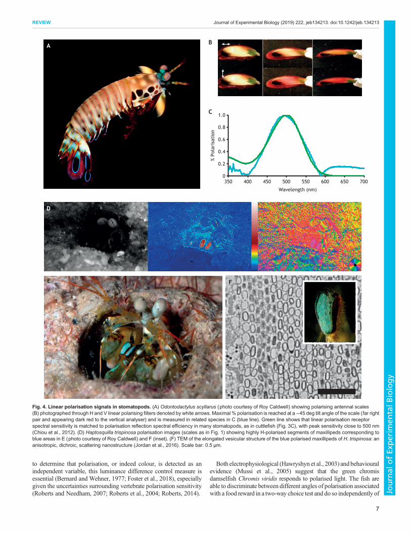
Fig. 7. Behavioural assessment of polarisation vision in the lab. (A) A fiddler crab on a floating ball treadmill and a polar-graph of the escape reaction(histogram showing the number of runs and their direction) to looming stimuli presented to the crab from a computer monitor that shows polarisation contrast only.Animals without polarisation vision see no image on the screen (Pignatelli et al., 2011; How et al., 2012). (B) The same experiment for cuttlefish; again, thelooming stimulus is only visible to polarisation-sensitive animals. (C) The cuttlefish reacts to the loom shown by skin-pattern change (split-screen image ofpattern before and after loom, top and bottom, respectively). (D) Graph showing high sensitivity of cuttlefish to polarisation angle difference of the stimulus down to1.5 deg (Temple et al., 2012). Bars show s.e.m. (E) Feeding containers with linear polarising filters (top) that are invisible until photographed through aV-polarising filter (bottom). The white lines were drawn after to show the angle orientation of the filters. (F) A stomatopod handling a feeding container in a choicetest where both linear polarisation e-vector angle (as in E) and circular polarisation handedness can be discriminated. (G) Results for animals trained to left- andright-handed reflecting feeding containers, as indicated by L or R. Asterisks indicate statistical significance based on a Fisher’s exact test (Marshall et al., 1999a;Chiou et al., 2008b).
10
REVIEW Journal of Experimental Biology (2019) 222, jeb134213. doi:10.1242/jeb.134213
Journal
ofEx
perim
entalB
iology

leaves for egg-laying. Behavioural tests indicate that they prefer to laytheir eggs under green horizontal leaves that also therefore reflectH-polarisation (Kelber et al., 2001), and H-polarisation is actuallyprioritised over colour. A horizontal leaf may provide both the bestshelter from weather and protection from the eyes of birds and otherpredators. This example amplifies an important difference betweencolour and polarisation vision. Judgements based on polarisationangle are usually (but not always, see Fig. 4B) necessarily bound tothe orientation of the object, such as the leaf surface. In other words,as a leaf changes angle, so does the reflected angle and, therefore,possibly the strength of the polarisation signal to a viewer (Fig. 1A).This is examined further in the next section.
Signal orientation, confounding parameters and polarisationcontrastColour, intensity and polarisation confounds may exist in a putativepolarisation signal and may or may not be disambiguated at theneural level (Glantz, 2001). As demonstrated by the swallowtail
butterfly story, e-vector direction is often related to object surfaceorientation or intrinsic polarisation mechanisms in animal cuticle(Fig. 4). Even when considering polarisation alone, qualityjudgements based on polarisation angle and/or degree may not beeasy to disambiguate, at least to the scientist observer (Bernard andWehner, 1977; How et al., 2014a). Considerable similaritiesbetween intensity contrast sensitivity functions and polarisationcontrast sensitivity functions have been found in dragonfly larvae(Sharkey et al., 2015) and in crayfish (Glantz, 1996). This meansthat great care is needed not to confuse what is being presented ortested when either observing nature or performing experiments(Marshall et al., 2014; Foster et al., 2018).
Examples exist where signals and/or their reception seemdeliberately simplified in nature. Rhabdom or photoreceptor twist,deliberate microvillar disorganisation or cell–cell cross-talk hasevolved in a number of species to reduce polarisation sensitivity infavour of another light modality such as colour (Marshall et al.,1991; Wehner and Bernard, 1993). As many waxy leaves or wet
A
B
% Polarisation of fish
% Po
lari
sati
on o
f ba
ckgr
ound
0
0.1
0.2
0.3
0.4
0 0.1 0.2 0.3 0.4
Fig. 8. Midwater camouflage and lack ofpolarocrypsis. (A) A diver among superblycamouflaged silvery big-eye trevally, Caranxsexfasciatus. The fish are also imaged using a polarisingcamera below (see Fig. 1 legend for explanation ofscales). In intensity and colour, the reflective camouflagemechanism functions well, but it breaks down in %polarisation (Johnsen et al., 2016). (B) Ratio of %polarisation of fish and background water in 8 species ofsilvery fish (Johnsen et al., 2016 and see Fig. S1)showing that silvery fish do not return polarisedreflections (Brady et al., 2013, 2015), as also predictedtheoretically (Jordan et al., 2012). Animals withpolarisation vision would therefore break this form ofcamouflage, suggesting that polarocrypsis, in thiscontext, does not work. Key: red, Caranx sexfasciatus;black, Sphyraena qenie; blue, Pseudocaranx dentex;bright green, Trachinotus blochii; dark green, Caranxmelampygus; yellow, Gnathanodon speciosus; grey,Pterocaesio marri; orange, Fistularia commersonii.
11
REVIEW Journal of Experimental Biology (2019) 222, jeb134213. doi:10.1242/jeb.134213
Journal
ofEx
perim
entalB
iology

surfaces polarise reflected light (Fig. 1) it has been suggested thatthis is seen as a sort of environmental noise, best removed if othercues or signals are sought. Ultimately, the apparent problem ofdisentangling colour and polarisation may be one of our ownmaking, as we perhaps try too hard to neatly define modalities andsignals. The ways in which polarisation is combined with colour orindeed intensity remains a challenge for the emerging ideas inpolarisation signalling.The cephalopods appear to have solved the colour–polarisation
confound by ignoring colour information altogether (Marshall andMessenger, 1996). They also seem to address the potentialpolarisation angle–degree confound with their signals. Cuttlefishpossess dynamic polarised stripes on their arms and, in commonwith other skin-based signals in cephalopods such as squid, thestripes can be masked or turned on and off, depending onbehavioural context (Chiou et al., 2007; Mathger and Hanlon,2006; Mathger et al., 2009a,b; Shashar et al., 2019). A de-couplingof e-vector angle from the angle of the object (the arm stripes) inspace is achieved through a randomly arranged array of plateletstructures or modified iridophores (see Glossary) in the arms(Fig. 3). Such independence from angle may serve to maintainpolarised signal consistency from a number of viewing angles,leaving degree-only information that is relevant to the behaviouraljudgement underway. The actual message conveyed remainsunknown in either cuttlefish or squid (Mathger et al., 2009a,b),but cuttlefish at least are known to expose the stripes during bothmating and agonistic displays (Shashar et al., 1996).Stomatopods also present signals that are independent of, or at least
different to, the object surface angle. The antennal scales in thepeacock mantis shrimp Odontodactylus scylarus are two largepaddle-shaped appendages that reflect a polarising signal best at a∼45 deg oblique angle to the paddle surface (Fig. 4; Chiou et al.,2012). This is achieved with an unusual astaxanthin-based molecularpolarisation mechanism that aligns polarisation to cell membranesand the molecules’ position within that membrane. The uropods (tail-fans) of this and other stomatopods from this genus also polarise lightat angles unrelated to the surface plane, apparently using the samemechanism, and both these and the antennal scales are used in displayto potential mates (Fig. 2 and Movie 1). H. trispinosa, also uses aninternal photonic mechanism to produce the blue polarised signaldiscussed above (Fig. 4). The result is a strong horizontal signalagainst a non-polarising background, (Fig. 4; Chiou et al., 2005,2008a; How et al., 2014b; Cronin, 2018).Talbot and Marshall (2010, 2011) showed that cuttlefish and
squid orient the microvilli in adjacent photoreceptors mainly H andV relative to the outside world (Fig. 2B). As noted, particularlyunderwater, this H–V arrangement may be important for lookinginto the mainly horizontal distant light field and for detectingobjects against this relatively predictable polarisation curtain(Fig. 8; Fig. S1). Where known, crustaceans also tend to show anH–V arrangement in their retina relative to the outside world. Boththe semi-terrestrial mud-flat and beach-dwelling fiddler crabs andghost crabs view a horizontally reflected polarised source (Zeil andHemmi, 2006; How et al., 2014a). Whether permanently submergedspecies also match an H–V array with the mostly horizontalscattered spacelight remains mostly unknown (Figs 1 and 2;Marshall et al., 1999a,b; Marshall and Cronin, 2014). Theexceptional stomatopods possess four angles of linear polarisationsensitivity and are able to rotate their eyes to align photoreceptorsand signals – this apparently enables them to optimise linearpolarisation contrast in their environment (Land et al., 1990;Marshall et al., 2007; Marshall and Cronin, 2014; Daly et al., 2016).
Polarisation camouflageAnimals use intensity and colour for camouflage in a variety of ways,such as simple matching, disruptive camouflage and countershading(Rowland, 2009; Stevens and Merilaita, 2009; Cott, 1940; Thayer,1909). As well as searching for polarisation signals, it is worth askingwhether there are examples of polarisation reflections and patternsthat camouflage or match a uniform or patterned background ofpolarisation. This has been termed polarocrypsis, and for a variety ofreasons explained below and previously (Cronin et al., 2016) we thinkit unlikely, at least in some of the contexts so far imagined.
An intriguing but untested possibility concerns shiny-leafed andtherefore polarising bushes (Fig. 1A). Such habitat certainly providesa disruptive polarised background into which a polarising animalmight disappear. As noted already, twisted rhabdoms and othermechanisms of disrupting polarisation sensitivity remove this sort ofglare and confounding information (Ribi, 1979;Wehner andBernard,1993; Wehner and Meyer, 1981; Marshall and Cronin, 2014). Manyinsects, however, do not eliminate polarisation sensitivity in theirphotoreceptors, and may indeed visualise colour patterns andpolarisation patterns confounded together in such a background.This might benefit a polarising animal in such a bush. It has beensuggested that the linearly polarised reflections from Japanese jewelbeetles (Chrysochroa fulgidissima) may help in either camouflage orintraspecific communication in forest-edge habitat against waxyfoliage (Fig. 1A; Stavenga et al., 2011).
The sunbeam butterfly,Curetis acuta, and the ‘glass scales’ of theswordtail butterfly, Graphum sarpedon, polarise light from theirwings like the heliconids already mentioned (Fig. 5A–C; Stavengaet al., 2012; Vukusic et al., 2000). In these instances, however, it isscatter or thin film interference from the under surface of the wingsthat both adds polarisation and reflects colours of the localenvironment. The wings display a polarising signal in flight,which is presumably not visible to predators (such as birds) but iscovertly visible to other butterflies. When at rest and with wingsfolded, a combination of their green or silvery-white colour,polarisation and local reflection from the wings may aid incamouflage in a forest edge leafy habitat. Both of these butterflyand beetle examples remain interesting ideas only, and are in need ofpositive behavioural and experimental verification.
The term ‘polarocrypsis’ in fact comes from the suggestion that asimpler background-matching camouflage exists in the marineenvironment. As detailed above, a background curtain of mostlyhorizontally polarised light exists underwater, except near dawn anddusk when it tilts substantially. In open water, there is nowhere tohide, and silvery fish possess a well-documented camouflage strategyin this habitat. Using guanine crystals as reflectors and arranging thecrystals over their curved surface to be vertical, silvery fish can act asflat mirrors and reflect the local surrounding light efficiently, thuslooking like water itself, both in terms of colour and intensity (Fig. 8;Denton and Land, 1971; Jordan et al., 2012). Brady and colleaguessuggest that this extends to polarisation in the lookdown Selenevomer, a relatively large (∼30 cm) semi-pelagic fish (Brady et al.,2015, 2013). That is, they claim S. vomer reflects polarised light aswell, and would thus be less visible against a polarising backgroundto a possible predator or any animal with polarising sensitivity.
A number of concerns about this study have previously been putforward (Cronin et al., 2016 and see counter-examples in Fig. 8 andFig. S1). Jordan et al. (2012) also demonstrated, both theoreticallyand using direct measurements, that the guanine crystals of silveryfish are arranged, almost ideally, to reflect intensity and notpolarisation. They conclude that for optimal camouflage, the bestcombination is spectrally broad-band, high-percentage and low-
12
REVIEW Journal of Experimental Biology (2019) 222, jeb134213. doi:10.1242/jeb.134213
Journal
ofEx
perim
entalB
iology

polarising reflectivity. Finally, Johnsen et al. (2016) quantified andphotographed many different silvery fish species at many depthsusing a polarisation camera specifically designed to work in water.One conclusion is that the degree of polarisation of all fish fell wellbelow that of the background, in addition to generally having smallvalues (Fig. 8; Fig. S1).S. vomer may be different to other silvery fish in its mechanism
(an untested possibility), but the 11 species of other silvery fish inthe study (Johnsen et al., 2016) were all conspicuous against thebackground to a predator with polarisation sensitivity (Fig. 8;Fig. S1). Polarisation vision in large fish or their predators has yet tobe shown, and while there are open-water squid species possiblylarge enough to tackle S. vomer, they typically prey upon smalleranimals. It might be worthwhile to conduct a closer examination ofsmall silvery fish in the size and distance range relevant tocrustacean and cephalopod predators.
Circular polarisation and the case for covert communicationDo polarising signals, only visible to animals with polarisationsensitivity, constitute a ‘secret’ communication mechanism? Someanimals can also see UV and other wavelength ranges that areinvisible to other species. The discussion of ‘secret’ communicationhas already occurred in these contexts, and we can learn from thatdiscussion (Bennett and Cuthill, 1994; Cuthill et al., 2000; Hausmannet al., 2003; Siebeck et al., 2006; Siebeck and Marshall, 2001). Doescovert communication occur in polarisation? For example, are theexposed polarisation dances of the Odontodactyloid stomatopods(Figs 4 and 6; Fig. S2 and Movie 1) made possible and not tooobvious by the ‘for-your-eyes-only’ nature of the event? The animalsin the video, although rather well camouflaged in intensity andpattern, are moving, making them conspicuous in other ways. It is notknown to what extent polarisation may be helping to keep this event‘private’. More likely, much like a displaying bird, they run the risk ofdetection by predators.Stomatopods and cephalopods eat each other and probably have
done for over 400 million years. Both use linear polarising signalsand both possess linear polarisation sensitivity. In the marine worldof these highly successful invertebrate groups is the arms race orselection for polarisation secrecy still underway? Stomatopodspossess circular polarisation vision and circular polarising signals,and these are not visible to cephalopods as far as we know (Figs 2D,6 and 7G; Chiou et al., 2005, 2007, 2008a,b). Does this indicate astep ahead in the communication arms race?Controlled circular polarisation behavioural experiments have
been conducted for stomatopods in three behavioural scenarios. Inone,O. scyllaruswas trained to discriminate left- from right-handedcircular polarisation in a food-rewarded binary choice test (Fig. 7G;Chiou et al., 2008b), while H. trispinosa discriminated linear fromcircular polarisation under similar circumstances but could notdiscriminate left from right circular polarisation (Templin, 2017).The reef-dwelling stomatopod Gonodactylus falcatus shows innateavoidance of burrow entrances containing circular polarisation,suggesting that this species may use circular polarisation ininterspecific communication (Gagnon et al., 2015). Further tobehavioural evidence, the optical mechanism (quarter-waveretarder) required for circular polarisation sensitivity has beenidentified in a specific set of stomatopod photoreceptors, directoptical measurements of the quarter-wave components have beenmade, electrophysiology has shown circular polarisation sensitivityin several species and a partially understood photonic mechanism toproduce the reflection has been documented (Chiou et al., 2008b;Roberts et al., 2009).
Despite these multiple lines of evidence, several holes also existin the stomatopod story, including the fact that many of the speciesthat appear to have circular polarisation sensitivity lack the relevantbody reflections. As noted already, the food-based experimentsindicate that they can discriminate different forms of linear andcircular polarisation, and can transfer this conditioning to a feedingscenario that they apparently never meet in the real world (Chiouet al., 2008a,b). That is, we are yet to discover potential stomatopodfood that selectively reflects circular or indeed linear polarisation.
Scarabs, some other beetles and a few other insects also reflectcircular polarisation, and their metallic beauty tempts us to believethis could be a signal perceived by their own species (Pye, 2010).Many are bright green and well camouflaged in a green bush, and itis easy to imagine they rely on their circular polarised reflections for‘secret’ communication. The photonic mechanism has been knownfor some time (Neville and Caveney, 1969; Vukusic and Sambles,2004; Jewell et al., 2007), and recent behavioural evidencewas citedto suggest that scarabs might also see circular polarisation (Bradyand Cummings, 2010). This idea, based on phototaxis rather thansignal recognition, has since been comprehensively countered, withseveral different species of scarab (Blahó et al., 2012; Horváth et al.,2014). There is also no evidence at the retinal, optical or neural levelfor circular polarisation sensitivity in beetles.
A guide to studying polarisation signallingBehaviourInvestigations of polarisation signalling are usually triggered by theobservation of a behaviour relative to a small object that is suspectedto reflect polarised light (Fig. 7). These behaviours are distinct fromnavigational or polarotactic behaviours that cue on large- or medium-sized fields of view such as sky or water. However, a signal may alsoinclude a small unpolarised object against a large polarised field.When investigating behavioural responses to polarised signals,appropriate control measures to avoid unintentional presentation ofpolarisation or intensity confounds are essential and difficult (Fosteret al., 2018). If, as is often the case, the polarised region of reflectancecoincides with a colour, some attempt to disambiguate or discountcolour and intensity is worthwhile. It is also valid to recognise andquantify mixed signals (Kelber et al., 2001).
In experimental manipulation, is the test relevant to the animal’snatural behavioural repertoire? Large, looming polarised objects andpolarised food are rare in nature but a response to such stimuli doesindicate polarisation sensitivity and may be used to quantify angle ordegree sensitivity. Polarisation sensitivity that can be transferred ontotasks that do not usually involve polarisation is indicative of higher-level polarisation vision, including signalling. Mate choice has adirect association with polarisation, so behavioural manipulationsaround this task may be most productive (Sweeney et al., 2003;Marshall and Cronin, 2011; Chiou et al., 2011).
Retinal substrates, electrophysiological recordings and polarisation-specific pathwaysIt is both instructive for experimental design and a key element ofour understanding to demonstrate a receptor mechanism, or at least asubsequent neuronal signal, that responds to polarised light and thatmight drive the relevant behaviour (Flamarique and Harosi, 2002;Kondrashev et al., 2012; Roberts, 2014). In invertebrates,microvillar direction and regularity provide an easy-to-quantifyproxy for e-vector angle sensitivity. The mechanism for detection ofpolarisation by vertebrates is less well defined, and animals such asfish and birds probably do not see object-based (or indeed any)polarisation (Marshall et al., 1991; Horváth, 2014; Mussi et al.,
13
REVIEW Journal of Experimental Biology (2019) 222, jeb134213. doi:10.1242/jeb.134213
Journal
ofEx
perim
entalB
iology

2005; Coemans et al., 1994). Neuronal responses to polarisationvariation, such as angle rotation or varying degree, are indicative ofpolarisation sensitivity but not necessarily polarisation vision.Electrophysiological data are hard to obtain, compared withindicative photoreceptor structures, but are instructive at everylevel, from receptors through to processing, to brain and motoroutput (Labhart and Meyer, 2002; Heinze and Homberg, 2007;Doujak, 1984; Shaw, 1966; Chiou et al., 2008b; Kleinlogel andMarshall, 2006; Moody and Parriss, 1961; Glantz, 1996, 2008).
Quantification of polarisation propertiesWhen investigating polarisation signalling, the polarised reflection,its area and location, should at the very least be photographed toqualitatively detail its altered appearance through changing linearand/or circular polarising filters (Douglas et al., 2007; and Gagnonand Marshall, 2016; Cronin and Marshall, 2011; Cronin et al.,2003). This can be taken further with relatively simple polarisationphotography to quantify angle, intensity and degree (Foster et al.,2018). Small area polarimetry using simple filters is easy withportable spectrophotometers (Cronin et al., 2009). Quantificationmay extend to photonic characterisation and molecularnanostructure, using electron microscopy, x-ray diffraction andellipsometry. As with colour, the efficacy of polarisation signals isdefined through the evolution of these structures by natural or sexualselection (Chiou et al., 2005, 2012; Jewell et al., 2007; How et al.,2014b).
In situ observation and natural historyOne of the least attended to but most necessary set of observations inany potential signalling scenario comes from observing the animalin situ and thinking through its biology and behavioural repertoire.The alternating behaviours of revealing and covering up polarisedarm stripes on cuttlefish or the polarised wings and carapace inbutterflies and stomatopods, respectively, are good examples of thissort of desirable data (Sweeney et al., 2003; Chiou et al., 2008a;Mathger et al., 2009a,b). Quantification of the polarised lightenvironment and, in particular, polarised background is particularlyworthy of attention, and it is relatively straightforward with the nowfrequently used portable spectrophotometers and appropriate filters.Such measurements allow estimates of polarisation contrast in thereal world (Fig. 1 and Shashar et al., 1995).
ConclusionsMultifaceted evidence for polarisation signalling exists inbutterflies, cephalopods and stomatopods, but not yet in fish,beetles and flowers. While we generally agree with Labhart’s viewthat polarisation vision is not designed to extract angle informationper se [aside from some exceptional behaviours in butterflies(Kelber et al., 2001) and possibly stomatopods (Cronin, 2018)], wepropose that polarisation signals contain specific information. Thesesignals, unlike large-field cues, may be subject to evolutionarypressures, and, in common with colour, may become in some wayoptimised or matched to specific receivers. Some evidence suggeststhat degree might be more reliable than angle in polarisation signals,and mixed modalities of colour and polarisation also exist.In signalling systems where both a colour and polarisation are
present and a colour vision system exists that can see the colour,studies must address whether there is an advantage to the additionalpolarisation information. It is convenient for us to dividepolarisation and colour or, indeed, to divide each into components(e.g. hue and saturation for colour, and degree, angle and ellipticityfor polarisation). However, both behaviourally and evolutionarily,
these distinctions are possibly misleading. While it is important tomeasure and parameterise as much as possible, especially whendealing with a form of light we don’t see, considering theperspective of the animal in its natural environment may lead tomore likely conclusions. What this requires is a return to the carefulobservations and time spent in the field by the likes of Lorenz andvon Frisch, who have strongly influenced our thoughts on behaviourand polarisation (e.g. von Frisch, 1949; Lorenz, 1962).
Competing interestsThe authors declare no competing or financial interests.
Supplementary informationSupplementary information available online athttp://jeb.biologists.org/lookup/doi/10.1242/jeb.134213.supplemental
ReferencesAlkaladi, A., How, M. J. and Zeil, J. (2013). Systematic variations in microvilli
banding patterns along fiddler crab rhabdoms. J. Comp. Physiol. A 199, 99-113.Arikawa, K. and Stavenga, D. G. (1997). Random array of colour filters in the eyes
of butterflies. J. Exp. Biol. 200, 2501-2506.Arikawa, K., Inokuma, K. and Eguchi, E. (1987). Pentochromatic visual system in
a butterfly. Naturwissenschaften 74, 297-298.Bennett, A. T. D. and Cuthill, I. C. (1994). Ultraviolet vision in birds: what is its
function? Vision Res. 34, 1471-1478.Bernard, G. D. andWehner, R. (1977). Functional similarities between polarization
vision and color vision. Vision Res. 17, 1019-1028.Blaho, M., Egri, Á., Hegedus, R., Josvai, J., Toth, M., Kertesz, K., Biro, L. P.,
Kriska, G. and Horvath, G. (2012). No evidence for behavioral responses tocircularly polarized light in four scarab beetle species with circularly polarizingexocuticle. Physiol. Behav. 105, 1067-1075.
Boyer, J., Keresztes, J. C., Saeys, W. Koshel, J. (2016). An automated imagingBRDF polarimeter for fruit quality inspection. In SPIE Optical Engineering+Applications, Vol. 9948, pp. 9. SPIE.
Brady, P. C. and Cummings, M. E. (2010). Differential response to circularlypolarized light by the jewel scarab beetle chrysina gloriosa. Am. Nat. 175,614-620.
Brady, P. C., Travis, K. A., Maginnis, T. and Cummings, M. E. (2013). Polaro–cryptic mirror of the lookdown as a biological model for open ocean camouflage.Proc. Natl Acad. Sci. USA 110, 9764-9769.
Brady, P. C., Gilerson, A. A., Kattawar, G. W., Sullivan, J. M., Twardowski, M. S.,Dierssen, H. M., Gao, M., Travis, K., Etheredge, R. I., Tonizzo, A. N. et al.(2015). Open-ocean fish reveal an omnidirectional solution to camouflage inpolarized environments. Science 350, 965-969.
Buschbeck, E., Sbita, S. and Morgan, R. (2007). Scanning behavior by larvae ofthe predacious diving beetle, Thermonectusmarmoratus (Coleoptera: Dytiscidae)enlarges visual field prior to prey capture. J. Comp. Physiol. A 193, 973-982.
Calabrese, G. M., Brady, P. C., Gruev, V. and Cummings, M. E. (2014).Polarization signaling in swordtails alters female mate preference. Proc. NatlAcad. Sci. USA 111, 13397-13402.
Chiou, T.-H., Cronin, T. W., Caldwell, R. L. and Marshall, N. J. (2005). Biologicalpolarized light reflectors in stomatopod crustaceans. In Optics & Photonics 2005,pp. 58881B-58881B-9. International Society for Optics and Photonics.
Chiou, T.-H., Mathger, L. M., Hanlon, R. T. and Cronin, T. W. (2007). Spectral andspatial properties of polarized light reflections from the arms of squid (Loligopealeii) and cuttlefish (Sepia officinalis L.). J. Exp. Biol. 210, 3624-3635.
Chiou, T.-H., Caldwell, R. L., Hanlon, R. T. and Cronin, T. W. (2008a). Finestructure and optical properties of biological polarizers in crustaceans andcephalopods. Proc. SPIE 6972, 1-10.
Chiou, T.-H., Kleinlogel, S., Cronin, T. W., Caldwell, R. L., Loeffler, B., Siddiqi,A., Goldizen, A. and Marshall, N. J. (2008b). Circular polarization vision in astomatopod crustacean. Curr. Biol. 18, 429-434.
Chiou, T.-H., Marshall, N. J., Caldwell, R. L. and Cronin, T. W. (2011). Changes inlight-reflecting properties of signalling appendages alter mate choice behaviour ina stomatopod crustacean Haptosquilla trispinosa. Mar. Freshwater Behav.Physiol. 44, 1-11.
Chiou, T.-H., Place, A. R., Caldwell, R. L., Marshall, N. J. and Cronin, T. W.(2012). A novel function for a carotenoid: astaxanthin used as a polarizer for visualsignalling in a mantis shrimp. J. Exp. Biol. 215, 584-589.
Coemans, M., Hzn, J. and Nuboer, J. (1994). The orientation of the e-vector oflinearly polarized light does not affect the behaviour of the pigeon Columba livia.J. Exp. Biol. 191, 107-123.
Cott, H. B. (1940). Adaptive Coloration in Animals. London: Methuen & Co. Ltd.Cronin, T. W. (2018). A different view: sensory Drive in the polarized-light realm.
Curr. Zool. 64, 513-523.
14
REVIEW Journal of Experimental Biology (2019) 222, jeb134213. doi:10.1242/jeb.134213
Journal
ofEx
perim
entalB
iology
Cronin, T. W. and Marshall, N. J. (2011). Patterns and properties of polarized lightin air and water. Phil. Trans. R. Soc. B 366, 619-626.
Cronin, T. W. and Shashar, N. (2001). The linearly polarized light field in clear,tropical marine waters: spatial and temporal variation of light intensity, degree ofpolarization and e-vector angle. J. Exp. Biol. 204, 2461-2467.
Cronin, T. W., Shashar, N., Caldwell, R. L., Marshall, N. J., Cheroske, A. G. andChiou, T.-H. (2003). Polarization vision and its role in biological signaling. Integr.Comp. Biol. 43, 549-558.
Cronin, T. W., Chiou, T.-H., Caldwell, R. L., Roberts, N. W. and Marshall, N. J.(2009). Polarization signals in mantis shrimps. In Polarization Science andRemote Sensing IV, Vol. 7461 (ed. J. A. Shaw and J. S. Tyo), pp. 74610C-10. SanDiego, CA, USA: SPIE.
Cronin, T. W., Johnsen, S., Marshall, N. J. and Warrant, E. J. (2014). VisualEcology. Woodstock: Princeton University Press.
Cronin, T. W., Gagnon, Y. L., Johnsen, S., Marshall, N. J. and Roberts, N. W.(2016). Comment on ‘Open-ocean fish reveal an omnidirectional solution tocamouflage in polarized environments’. Science 353, 552-552.
Cuthill, I. C., Partridge, J. C., Bennett, A. T. D., Church, S. C., Hart, N. S. andHunt, S. (2000). Ultraviolet vision in birds. Adv. Study Behav. 29, 159-214.
Dacke, M., Doan, T. A. and O’Carroll, D. C. (2001). Polarized light detection inspiders. J. Exp. Biol. 204, 2481-2490.
Dacke, M., Nordstrom, P., Scholtz, C. H. and Warrant, E. J. (2002). A specializeddorsal rim area for polarized light detection in the compound eye of the scarabbeetle Pachysoma striatum. J. Comp. Physiol. A 188, 211-216.
Daly, I. M., How, M. J., Partridge, J. C., Temple, S. E., Marshall, N. J., Cronin,T. W. and Roberts, N. W. (2016). Dynamic polarization vision in mantis shrimps.Nat. Commun. 7, 12140.
Denton, E. J. and Land, M. F. (1971). Mechanism of reflexion in silvery layers of fishand cephalopods. Proc. R. Soc. Lond. Series B 178, 43-61.
Detto, T. (2007). The fiddler crab Uca mjoebergi uses colour vision in mate choice.Proc. R. Soc. B 274, 2785-2790.
Douglas, J. M., Cronin, T. W., Chiou, T.-H. and Dominy, N. J. (2007). Lighthabitats and the role of polarized iridescence in the sensory ecology of neotropicalnymphalid butterflies (Lepidoptera: Nymphalidae). J. Exp. Biol. 210, 788-799.
Doujak, F. E. (1984). Electrophysiological measurement of photoreceptormembrane dichroism and polarization sensitivity in a Grapsid crab. J. Comp.Physiol. A 154, 597-605.
Flamarique, I. N. and Harosi, F. I. (2002). Visual pigments and dichroism ofanchovy cones: a model system for polarization detection. Vis. Neurosci. 19,467-473.
Foster, J. J., Sharkey, C. R., Gaworska, A. V. A., Roberts, N. W., Whitney, H. M.and Partridge, J. C. (2014). Bumblebees learn polarization patterns. Curr. Biol.24, 1415-1420.
Foster, J. J., Temple, S. E., How, M. J., Daly, I. M., Sharkey, C. R., Wilby, D. andRoberts, N.W. (2018). Polarisation vision: overcoming challenges of working witha property of light we barely see. Naturwissenschaften 105, 27.
Gagnon, Y. L. and Marshall, N. J. (2016). Intuitive representation ofphotopolarimetric data using the polarization ellipse. J. Exp. Biol. 219, 2430-2434.
Gagnon, Y. L., Templin, R. M., How, M. J. and Marshall, N. J. (2015). Circularlypolarized light as a communication signal in mantis shrimps. Curr. Biol. 25,3074-3078.
Glantz, R. M. (1996). Polarization sensitivity in crayfish lamina monopolar neurons.J. Comp. Physiol. 178, 413-425.
Glantz, R. M. (2001). Polarization analysis in the crayfish visual system. J. Exp. Biol.204, 2383-2390.
Glantz, R. M. (2008). Polarization vision in crayfish motion detectors. J. Comp.Physiol. A 194, 565-575.
Goldstein, D. H. (2010). Polarised Light. CRC Press.Gruev, V., Perkins, R. and York, T. (2010). CCD polarization imaging sensor withaluminum nanowire optical filters. Opt. Express 18, 19087-19094.
Hausmann, F., Arnold, K. E., Marshall, N. J. and Owens, I. P. (2003). Ultravioletsignals in birds are special. Proc. R. Soc. Lond. Series B 270, 61-67.
Hawryshyn, C. W. (1992). Polarization vision in fish. Am. Sci. 80, 164-175.Hawryshyn, C. W. (2000). Ultraviolet polarization vision in fishes: possiblemechanisms for coding e-vector. Phil. Trans. R. Soc. Lond. Series B 355,1187-1190.
Hawryshyn, C.W., Moyer, H. D., Allison,W. T., Haimberger, T. J. andMcFarland,W. N. (2003). Multidimensional polarization sensitivity in damselfishes. J. Comp.Physiol. 189, 213-220.
Hecht, E. (2017). Optics. Harlow: Pearson Education Limited.Heinloth, T., Uhlhorn, J. and Wernet, M. F. (2018). Insect responses to linearlypolarized reflections: orphan behaviors without neural circuits. Front. Cell.Neurosci. 12, 50.
Heinze, S. and Homberg, U. (2007). Map-like representation of celestial e-vectororientations in the brain of an insect. Science 315, 995-997.
Horvath, G. (2014). Polarised light and polarisation vision in animal sciences. InSpringer Series in Vision Research (ed. N. J. Marshall and S. P. Collin). New York:Springer.
Horvath, G. and Varju, D. (1997). Polarization pattern of freshwater habitatsrecorded by video polarimetry in red, green and blue spectral ranges and itsrelevance for water detection by aquatic insects. J. Exp. Biol. 200, 1155-1163.
Horvath, G., Blaho, M., Egri, Á., Hegedus, R. and Szel, G. (2014). Circularpolarization vision of scarab beetles. In. Polarised light and Polarisation Vision inAnimal Sciences. Springer Series in Vision Research (ed. N. J. Marshall and S. P.Collin), pp. 147-170. New York: Springer.
How, M. J. and Marshall, N. J. (2014). Polarization distance: a framework formodelling object detection by polarization vision systems. Proc. R. Soc. B 281,20131632.
How, M. J., Zeil, J. and Hemmi, J. M. (2009). Variability of a dynamic visual signal:the fiddler crab claw-waving display. J. Comp. Physiol. A 195, 55-67.
How, M. J., Pignatelli, V., Temple, S. E., Marshall, N. J. and Hemmi, J. M. (2012).High e-vector acuity in the polarisation vision system of the fiddler crab Ucavomeris. J. Exp. Biol. 215, 2128-2134.
How, M. J., Christy, J., Roberts, N. W. and Marshall, N. J. (2014a). Null point ofdiscrimination in crustacean polarisation vision. J. Exp. Biol. 217, 2462-2467.
How, M. J., Porter, M. L., Radford, A. N., Feller, K. D., Temple, S. E., Caldwell,R. L., Marshall, N. J., Cronin, T. W. and Roberts, N. W. (2014b). Out of the blue:the evolution of horizontally polarized signals in Haptosquilla (Crustacea,Stomatopoda, Protosquillidae). J. Exp. Biol. 217, 3425-3431.
Ivanoff, A. and Waterman, T. H. (1958). Factors, mainly depth and wavelength,affecting the degree of underwater light polarization. J. Mar. Res. 16, 283-307.
Jewell, S. A., Vukusic, P. and Roberts, N. W. (2007). Circularly polarized colourreflection from helicoidal structures in the beetle Plusiotis boucardi. New J. Phys.9, 99.
Johnsen, S. (2012). The Optics of Life: a Biologists Guide to Light in Nature.Princeton: Princeton University Press.
Johnsen, S., Gagnon, Y. L., Marshall, N. J., Cronin, T. W., Gruev, V. and Powell,S. B. (2016). Polarization vision seldom increases the sighting distance of silveryfish. Curr. Biol. 26, R752-R754.
Jordan, T. M., Partridge, J. C. and Roberts, N. W. (2012). Non-polarizingbroadband multilayer reflectors in fish. Nat. Photonics 6, 759-763.
Jordan, T. M., Wilby, D., Chiou, T.-H., Feller, K. D., Caldwell, R. L., Cronin, T. W.and Roberts, N. W. (2016). A shape-anisotropic reflective polarizer in astomatopod crustacean. Sci. Rep. 6, 21744.
Kelber, A. and Osorio, D. (2010). From spectral information to animal colour vision:experiments and concepts. Proc. R. Soc. B 277, 1617-1625.
Kelber, A., Thunell, C. and Arikawa, K. (2001). Polarisation-dependent colourvision in Papilio butterflies. J. Exp. Biol. 204, 2469-2480.
Kirschfeld, K. (1973). Vision of polarised light. In International BiophysicsCongress, Vol. 4, pp. 289-296. Moscow.
Kleinlogel, S. and Marshall, N. J. (2006). Electrophysiological evidence for linearpolarization sensitivity in the compound eyes of the stomatopod crustaceanGonodactylus chiragra. J. Exp. Biol. 209, 4262-4272.
Kondrashev, S. L., Gnyubkina, V. P. and Zueva, L. V. (2012). Structure andspectral sensitivity of photoreceptors of two anchovy species: Engraulis japonicusand Engraulis encrasicolus. Vision Res. 68, 19-27.
Labhart, T. (1988). Polarization-opponent interneurons in the insect visual-system.Nature 331, 435-437.
Labhart, T. (1999). How polarization-sensitive interneurones of crickets see thepolarization pattern of the sky: a field study with an optoelectronic model neurone.J. Exp. Biol. 202, 757-770.
Labhart, T. (2016). Can invertebrates see the e-vector of polarization as a separatemodality of light? J. Exp. Biol. 219, 3844-3856.
Labhart, T. and Meyer, E. P. (2002). Neural mechanisms in insect navigation:polarization compass and odometer. Curr. Opin. Neurobiol. 12, 707-714.
Land, M., Marshall, J., Brownless, D. and Cronin, T. (1990). The eye-movementsof the mantis shrimp Odontodactylus scyllarus (Crustacea: Stomatopoda).J. Comp. Physiol. A 167, 155-166.
Lorenz, K. (1962). The function of colour in coral reef fishes. Proc. R. Inst. GreatBritain 39, 282-296.
Lythgoe, J. N. and Shand, J. (1982). Changes in spectral reflections from theiridophores of the neon tetra. J. Physiol Lond. 325, 23.
Marshall, N. J. (1988). A unique color and polarization vision system in mantisshrimps. Nature 333, 557-560.
Marshall, N. J. and Arikawa, K. (2014). Unconventional colour vision. Curr. Biol.24, R1150-R1154.
Marshall, N. J. and Cronin, T. W. (2011). Polarisation vision. Curr. Biol. 21,R101-R105.
Marshall, N. J. and Cronin, T. W. (2014). Polarisation vision of crustaceans. InPolarised Light and Polarisation Vision in Animal Sciences (ed. G. Horvath), pp.171-216. New York: Springer.
Marshall, N. J. and Johnsen, S. (2017). Fluorescence as a means of colour signalenhancement. Philos. Trans. R. Soc. B 372, 20160335.
Marshall, N. J. andMessenger, J. B. (1996). Colour blind camouflage.Nature 382,408-409.
Marshall, N. J., Land, M. F., King, C. A. and Cronin, T. W. (1991). The compoundeyes of mantis shrimps (Crustacea, Hoplocarida, Stomatopoda).1. Compound
15
REVIEW Journal of Experimental Biology (2019) 222, jeb134213. doi:10.1242/jeb.134213
Journal
ofEx
perim
entalB
iology

eye structure-the detection of polarized-light. Philos. Trans. R. Soc. Lond. SeriesB 334, 33-56.
Marshall, J., Cronin, T. W., Shashar, N. and Land, M. (1999a). Behaviouralevidence for polarisation vision in stomatopods reveals a potential channel forcommunication. Curr. Biol. 9, 755-758.
Marshall, J., Kent, J. and Cronin, T. (1999b). Visual adaptations in crustaceans:spectral sensitivity in diverse habitats. In Adaptive Mechanisms in the Ecology ofVision (ed. S. Archer, M. Djamgoz, E. Lowe, J. Partridge and S. Vallerga), pp.285-328. London: Kluwer.
Marshall, J., Cronin, T. W. and Kleinlogel, S. (2007). Stomatopod eye structureand function: a review. Arthropod. Struct. Dev. 36, 420-448.
Marshall, N. J., Roberts, N. W., Cronin, T. W. (2014). Polarisation signals. InPolarised light and Polarisation Vision in Animal Sciences (ed. G. Horvath), pp.407-442. New York: Springer.
Mathejczyk, T. F. and Wernet, M. F. (2017). Sensing polarized light in insects.Oxford Res. Ency. Neurosci.
Mathger, L. M. and Hanlon, R. T. (2006). Anatomical basis for camouflagedpolarized light communication in squid. Biol. Lett. 2, 494-496.
Mathger, L. M., Denton, E. J., Marshall, N. J. and Hanlon, R. T. (2009a).Mechanisms and behavioural functions of structural coloration in cephalopods.J. R. Soc. Interface 6, S149-S163.
Mathger, L. M., Shashar, N. and Hanlon, R. T. (2009b). Do cephalopodscommunicate using polarized light reflections from their skin? J. Exp. Biol. 212,2133-2140.
Moody, M. F. and Parriss, J. R. (1961). The discrimination of polarized light byoctopus-a behavioural and morphological study. Zeitschrift Fur VergleichendePhysiologie 44, 268-291.
Mussi, M., Haimberger, T. J. and Hawryshyn, C. W. (2005). Behaviouraldiscrimination of polarized light in the damselfish Chromis viridis (familyPomacentridae). J. Exp. Biol. 208, 3037-3046.
Neville, A. C. and Caveney, S. (1969). Scarabaeid beetle exocuticle as an opticalanalogue of cholesteric liquid crystals. Biol. Rev. Camb. Philos. Soc. 44, 531-562.
Pignatelli, V., Temple, S. E., Chiou, T.-H., Roberts, N. W., Collin, S. P. andMarshall, N. J. (2011). Behavioural relevance of polarization sensitivity as a targetdetection mechanism in cephalopods and fishes. Phil. Trans. R. Soc. B 366,734-741.
Powell, S. B., Garnett, R., Marshall, J., Rizk, C. and Gruev, V. (2018). Bioinspiredpolarization vision enables underwater geolocalization. Sci. Adv. 4, 6841.
Pye, J. D. (2010). The distribution of circularly polarized light reflection in theScarabaeoidea (Coleoptera). Biol. J. Linn. Soc. 100, 585-596.
Ribi, W. A. (1979). Do the rhabdomeric structures in bees and flies really twist.J. Comp. Physiol. 134, 109-112.
Roberts, N. W. (2014). Polariszation vision in fishes. In Polarised light andPolarisation Vision in Animal Sciences (G. Horvath), pp. 171-216. New York:Springer.
Roberts, N. W. and Needham, M. G. (2007). A mechanism of polarized lightsensitivity in cone photoreceptors of the goldfish Carassius auratus. Biophys. J.93, 3241-3248.
Roberts, N. W., Gleeson, H. F., Temple, S. E., Haimberger, T. J. and Hawryshyn,C. W. (2004). Differences in the optical properties of vertebrate photoreceptorclasses leading to axial polarization sensitivity. J. Opt. Soc. Am. A 21, 335-345.
Roberts, N. W., Chiou, T.-H., Marshall, N. J. and Cronin, T. W. (2009). A biologicalquarter-wave retarder with excellent achromaticity in the visible wavelengthregion. Nat. Photonics 3, 641-644.
Rosenthal, G. G. and Lobel, P. (2006). Communication. In Behaviour andPhysiology of Fish, Vol. 24 (ed. K. Sloman, S. Balshine and R. Wilson), pp.39-78. Academic Press.
Rowland, H. M. (2009). From Abbott Thayer to the present day: what have welearned about the function of countershading? Phil. Trans. R. Soc. B Biol. Sci.364, 519-527.
Schwind, R. (1983a). A polarization-sensitive response of the flying water bugnotonecta-glauca to UV-light. J. Comp. Physiol. A 150, 87-91.
Schwind, R. (1983b). Zonation of the optical environment and zonation in therhabdom structure within the eye of the backswimmer, notonecta-glauca. CellTissue Res. 232, 53-63.
Schwind, R. (1984). Evidence for true polarization vision based on a 2-channelanalyzer system in the eye of the water bug, Notonecta glauca. J. Comp. Physiol.A 154, 53-57.
Sharkey, C. R., Partridge, J. C. and Roberts, N. W. (2015). Polarization sensitivityas a visual contrast enhancer in the Emperor dragonfly larva, Anax imperator.J. Exp. Biol. 218, 3399-3405.
Shashar, N. and Cronin, T. W. (1996). Polarization contrast vision in Octopus.J. Exp. Biol. 199, 999-1004.
Shashar, N., Addessi, L. and Cronin, T. W. (1995). Polarization vision as amechanism for detection of transparent objects. In Ultraviolet Radiation and Coral
Reefs (ed. D. Gulko and P. L. Jokiel), pp. 207-211. Honolulu, HI: University ofHawai’i.
Shashar, N., Rutledge, P. S. and Cronin, T. W. (1996). Polarization vision incuttlefish-A concealed communication channel? J. Exp. Biol. 199, 2077-2084.
Shashar, N., Hanlon, R. T. and Petz, A. D. (1998). Polarization vision helps detecttransparent prey. Nature 393, 222-223.
Shashar, N., Hagan, R., Boal, J. G. and Hanlon, R. T. (2000). Cuttlefish usepolarization sensitivity in predation on silvery fish. Vision Res. 40, 71-75.
Shashar, N., Sabbah, S. and Cronin, T. W. (2004). Transmission of linearlypolarized light in seawater: implications for polarization signaling. J. Exp. Biol.207, 3619-3628.
Shaw, S. R. (1966). Polarized light responses from crab retinula cells. Nature 211,92-93.
Siebeck, U. E. and Marshall, N. J. (2001). Ocular media transmission of coral reeffish-can coral reef fish see ultraviolet light? Vision Res. 41, 133-149.
Siebeck, U. E., Losey, G. S. andMarshall, J. (2006). UV communication in Fish. InCommunication in Fishes, Vol. 2 (ed. F. Laddich, S. P. Collin, P. Moller and B. G.Kapoor), pp. 423-455. Plymouth, UK: Science Publications Inc.
Stavenga, D. G., Kinoshita, M., Yang, E. C. and Arikawa, K. (2001). Retinalregionalization and heterogeneity of butterfly eyes. Naturwissenschaften 88,477-481.
Stavenga, D. G., Matsushita, A., Arikawa, K., Leertouwer, H. L. and Wilts, B. D.(2012). Glass scales on the wing of the swordtail butterflyGraphium sarpedon actas thin film polarizing reflectors. J. Exp. Biol. 215, 657-662.
Stavenga, D. G., Wilts, B. D., Leertouwer, H. L. and Hariyama, T. (2011).Polarized iridescence of the multilayered elytra of the Japanese jewel beetle,Chrysochroa fulgidissima. Phil. Trans. R. Soc. B 366, 709-723.
Stevens, M. and Merilaita, S. (2009). Animal camouflage: current issues and newperspectives. Phil. Trans. R. Soc. B 364, 423-427.
Stowe, S. (1983). A theoretical explanation of intensity-independant variation ofpolarisation sensitivity in crustacean retinular cells. J. Comp. Physiol. 153,435-441.
Sweeney, A., Jiggins, C. and Johnsen, S. (2003). Insect communication:polarized light as a butterfly mating signal. Nature 423, 31-32.
Talbot, C. M. andMarshall, J. (2010). Polarization sensitivity and retinal topographyof the striped pyjama squid (Sepioloidea lineolata–Quoy/Gaimard 1832). J. Exp.Biol. 213, 3371-3377.
Talbot, C. M. and Marshall, J. N. (2011). The retinal topography of three species ofcoleoid cephalopod: significance for perception of polarized light. Phil.Trans. R. Soc. B 366, 724-733.
Temple, S. E., Pignatelli, V., Cook, T., How, M. J., Chiou, T.-H., Roberts, N. W.andMarshall, N. J. (2012). High-resolution polarisation vision in a cuttlefish.Curr.Biol. 22, R121-R122.
Temple, S. E., McGregor, J. E., Miles, C., Graham, L., Miller, J., Buck, J., Scott-Samuel, N. E. and Roberts, N. W. (2015). Perceiving polarization with the nakedeye. Proc. R. Soc. B 282, 20150338.
Templin, R. (2017). Circular Polarisation Vision in Stomatopod Crustaceans. PhDThesis. University of Queensland.
Thayer, G. H. (1909). Concealing-Coloration in the Animal Kingdom: An Expositionof the Laws of Disguise Through Color and Pattern: Being a Summary of AbbottH. Thayer’s Discoveries. New York: Macmillan.
von Frisch, K. R. (1949). Die Polarisation des Himmelslichtes als orientierenderFaktor bei den Tanzen der Bienen. Experientia 5, 142-148.
Vukusic, P. and Sambles, J. R. (2004). Photonic structures in biology. Nature 429,680-680.
Vukusic, P., Sambles, J. R. and Lawrence, C. R. (2000). Structural colour - Colourmixing in wing scales of a butterfly. Nature 404, 457-457.
Waterman, T. H. (1954). Polarization patterns in submarine illumination. Science120, 927-932.
Waterman, T. H. and Westell, W. E. (1956). Quantitative effect of the suns positionon submarine light polarization. J. Mar. Res. 15, 149-169.
Wehner, R. (2001). Polarization vision–a uniform sensory capacity? J. Exp. Biol.204, 2589-2596.
Wehner, R. and Bernard, G. D. (1993). Photoreceptor twist: a solution to the false-color problem. Proc. Natl Acad. Sci. USA 90, 4132-4135.
Wehner, R. and Meyer, E. (1981). Rhabdomeric twist in bees - artefact or in vivostructure? J. Comp. Physiol. 142, 1-17.
Wehner, R. and Rossel, S. (1985). The bees celestial compass - a case study inbehavioural neurobiology. Fortschr. Zool. 31, 11-53.
Zeil, J. and Hemmi, J. M. (2006). The visual ecology of fiddler crabs. J. Comp.Physiol. A 192, 1-25.
Zeil, J. and Hofmann, M. (2001). Signals from ‘crabworld’: cuticular reflections in afiddler crab colony. J. Exp. Biol. 204, 2561-2569.
16
REVIEW Journal of Experimental Biology (2019) 222, jeb134213. doi:10.1242/jeb.134213
Journal
ofEx
perim
entalB
iology