Embed Size (px)
Citation preview

Journal of Cell Science, Supplem ent 17, 9-12 (1993)Printed in G reat Britain © The Company o f Biologists Limited 1993
9
Polarity signals in epithelial cells
Enrique Rodriguez-Boulan* and Chiara Z u r z o lo t
Department of Cell Biology, Cornell University Medical College, 1300 York Ave, New York, NY 10021, USA'Author for correspondencetPresent address: Dipartimento di Biologia e Patologia Cellulare e Molecolare, CEOS, CNR, Il Policlinico, Via Pansini 5, 80131 Napoli, Italy
SUMMARY
In simple epithelia, specialized vectorial functions such as transport and secretion are made possible by the segregation of proteins and lipids into opposite surface domains. This polarized distribution results from selective delivery to and retention at the appropriate domain. In the case of direct delivery, the sorting site for apical and basolateral proteins is the iram-Golgi network (TGN) where they are incorporated into distinct apical and basolateral vesicles that are targeted to the respective surfaces. The machinery that controls this simple process is in fact rather complicated. It involves many different steps from the recognition event (between
‘sorting signal(s)’ and ‘sorting receptor(s)’) to the formation of the vesicles, their budding, and the docking to the specialized plasma membrane domain. Here we summarize the latest developments in the sorting of apical and basolateral proteins, focusing in particular on the signals that are involved in this process and the current hypotheses about the mechanisms responsible for it, in both epithelia and in non-polarized cells.
Key words: epithelial polarity, sorting signals, irawi-Golgi network, GPI-anchored proteins
INTRODUCTION
Work in the past decade has greatly increased our knowledge of the mechanisms responsible for epithelial cell polarity. These mechanisms fall into three groups: intracellular sorting and polarized delivery of proteins and lipids to the cell surface (Rodriguez-Boulan and Powell, 1992; van Meer and Burger, 1992), retention by domain-specific cytoskele- tal interactions (Nelson, 1991) and diffusive restriction by the tight junctional fence (Cerejido et al., 1980; Gumbiner, 1990). One of the principal intracellular sorting sites for both proteins and lipids is the trans-Golgi network (TGN).
THE TGN: CROSSROADS OF ENDOCYTIC AND EXOCYTIC TRAFFIC
Intense research has demonstrated the crucial role of the TGN in the sorting of proteins to lysosomes, secretory granules and the cell surface. Lysosomal hydrolases are segregated into clathrin-coated vesicles via specific interaction of mannose 6-phosphate residues with a receptor that recycles between the TGN and a prelysosomal compartment; the cytoplasmic domain of this receptor carries a determinant that is recognized by Golgi-specific adaptor molecules (Pearse and Robinson, 1990; Robinson, 1992) that induce the formation of a clathrin-coated bud (Griffiths et al., 1988). Aggregation, presumably triggered by the ionic conditions in the TGN, is presumed to promote the incorporation of regulated secretory proteins into secretory granules.
In epithelial cells, proteins destined to the apical and to the basolateral surface follow a similar pathway through the ER and the Golgi apparatus until, in the TGN, they are incorporated into distinct apical and basolateral vesicles, which are targeted to the respective surface (Rodriguez-Boulan and Powell, 1992; Simons and Wandinger-Ness, 1990). The mechanisms that direct the budding of these vesicles and the docking into and fusion with specific surface domains are still unknown.
Clear progress has been made in the identification of features in the proteins and lipids that direct them apically or basolaterally. We will analyze these features separately for apical and basolateral sorting.
APICAL SORTING IN EPITHELIA
Unlike basolateral proteins, no discrete protein features have been identified in apical proteins that promote apical sorting. The hypothesis by van Meer and Simons (van Meer and Simons, 1988), that partition into hydrogen-bonded gly- colipid rafts serves to recruit apical proteins into apical targeted vesicles, is, prima facie, supported by the remarkable apical localization of proteins anchored to the cell surface by glycosylphosphatidyl inositol (GPI) (Ali and Evans, 1990; Lisanti et al., 1988, 1990). In GPI-anchored proteins, the C-terminal amino acid is linked by an amide bond to ethanolamine, which in turn is linked by a phosphodiester bond to a mannosyl-glycosaminyl core glycan anchored to the membrane by phosphatidylinositol (Cross, 1990; Fer-

10 E. Rodriguez-Boulan and C. Zurzolo
Table 1. Localization of GPI-anchored proteins in epithelial cells
GPI-anchored protein Localization Cell type
5' Nucleotidase apical intestine, kidney^1'Trehalase apical intestine, kidney^1'Alkaline phosphatase apical intestine, kidney, MDCK*'1 >Renal dipeptidase apical kidney^N-CAM (GPI-anchored) apical MDCK*'2'Thy-1 apical MDCK*®Carcinoembryonic apical intestine, Caco-2, SKC015'3)
antigen (CEA)Decay accelerating apical MDCK*, Caco-2, SKCO-15®
factor (DAF)Decay accelerating unpolarized FRT*(4>
factor (DAF)gDl-DAF apical MDCK*'5'gDl-DAF Basolateral P R T * (4 )
VSV G-PLAP apical MDCK*'6'PLAP apical MDCK*<7>
*Transfected GPI-anchored proteins.(1)Hooper et al. (1988); (2)Powell et al. (1991); (3)Lisanti et al. (1990);
WZurzolo et al. (1993); <5>Lisanti et al. (1989); (6>Brown et al. (1989); <7>Brown and Rose (1992).
guson and Williams, 1988; Low and Saltiel, 1988). The addition of GPI to the protein is a transamidation event in the lumen of the RER directed by a transient C-terminal hydrophobic signal (Berger et al., 1988; Caras et al., 1989; Gerber et al., 1992). GPI-anchored proteins are usually identified by their sensitivity to cleavage by Pi-specific phospholipase C; considerable variability of the GPI structure can be generated by substitutions of the glycan with ethanolamine phosphate or by sugars (aGal, aMan or bGalNAc) (Deeg et al., 1992; Ferguson and Williams, 1988). Table 1 shows a list of endogenous and exogenous GPI-anchored proteins that, with few exceptions (see below), have been found in the apical surface of diverse epithelia.
Evidence for the apical sorting role of GPI was provided by the transfection of cDNAs encoding transmembrane and GPI-anchored isoforms of a protein (Powell et al., 1991), and by the apical localization of GPI-anchored fusion proteins of basolateral viral glycoproteins VSV G and Herpes simplex gDl (Brown et al., 1989; Lisanti et al., 1989).
The ‘cluster hypothesis’ for apical targeting is supported by the observation that GPI-anchored proteins and GSLs show resistance to dissociation by certain mild non-ionic detergents (TX-100, TX-114) at low temperature (Hoessli and Rungger-Brandle, 1985; Hooper and Turner, 1988). This resistance is acquired at, or just before, arrival to the Golgi apparatus of the GPI-anchored protein (Brown, 1992). Since GSLs are produced in the Golgi apparatus, these experiments suggest that clusters of GPI-anchored proteins and GSLs are formed in the Golgi apparatus.
Hannan et al. (1993), using fluorescence energy tranfer and fluorescence recovery after photobleaching, observed that a fusion GPI-anchored protein, gDl-DAF, was clustered and immobile at the moment of arriving to the surface of wild-type (Con A-resistant MDCK) MDCK cells but was mobile in a mutant MDCK cell line that failed to sort gDl-DAF. After long periods of residence at the cell surface, the new surface molecule population acquired char
acteristics of the stable population in that their mobility increased to high levels (R=90% as detected by FRAP). These experiments suggest the existence of a transmembrane ‘sorting receptor’ that may be involved in linking the GSL-GPI-protein aggregates in the luminal leaflet of the TGN to the vesicle-forming machinery on the cytoplasmic side; the reason for the slow dissociation of this immobilizing mechanism at the cell surface is unknown.
Recent experiments with the Fischer Rat Thyroid (FRT) cell line have shown that, unlike in other epithelial cell lines, GPI-anchored proteins are mainly basolateral (Table 1); gDl-DAF is targeted basolaterally whereas exogenous DAF is unpolarized (Zurzolo et al., 1993a). Interestingly, GPI-anchored proteins fail to become detergent-unex- tractable during passage through the Golgi complex of these cells (Zurzolo et al., 1993b). Since GSLs are as poorly extractable as in MDCK cells, it is that GPI has structural differences in FRT cells that prevent it from associating with GSL patches. On the other hand we found that VIP21/caveolin (Rothberg et al., 1992; Kurzchalia et al., 1992), suggesting an important role of this factor in the clustering of GSLs and GPI-anchored proteins, is absent in FRT cells (Zurzolo et al., 1993b). Study of the mechanisms responsible for the alternative sorting of GPI-anchored proteins in FRT cells and ConA resistant MDCK cells may help elucidate the molecular basis of apical sorting in epithelial cells.
BASOLATERAL SORTING SIGNALS
Discrete cytoplasmic signals, partially resembling endocytic signals, have been discovered in several basolateral proteins (Table 2) (Brewer and Roth, 1991; Casanova et al., 1991; Dagermont et al., 1993; Hunziker et al., 1991; Le Bivic et al., 1991; Matter et al., 1992; Prill et al., 1993; Yokode et al., 1992). Whereas in some cases a tyrosine residue is a critical part of the signal, other basolateral signals do not exhibit such a requirement. Two mechanisms may be invoked for the role of such signals in basolateal sorting. They may facilitate incorporation of proteins carrying such
Table 2. Signals for basolateral sortingProtein Sequence
Basolateral signals overlapping with coated pit localization domain:p75 NGF receptor FKRTNSLYSSLPd)Influenza HA (Tyr mut.) CSNGSLQYRICI®Lgp 120 RKRSHAGYQTI<3>Fc Receptor EAENTITYSLLKH'3'Lysosomal acid phosphatase (LAP) RMQAQPPGYRHV<4>LDL Receptor FNDPVY«)
Basolateral signals distinct from coated pit localization domain:LDL Receptor HICRNQDGYSYPSPoly IgA Receptor RHRNVDRVSIGSY<6>Transferrin Receptor*
Y=Tyr required for coated pit localization.(1)Le Bivic et al. (1991); (2)Brewer and Roth (1991); (3)Hunziker et al.
(1991); WPrill et al. (1993); (»Matter et al. (1992), Yokode et al. (1992);®Casanova et al. (1991); ^Dargermont et al. (1993).
*Basolateral sequence still undefined but distinct from YTRF coated pitlocalization motif.
P olarity s igna ls in ep ithe lia l ce lls 11
Polarized cell Unpolarized cell
cellsurface
lysosome
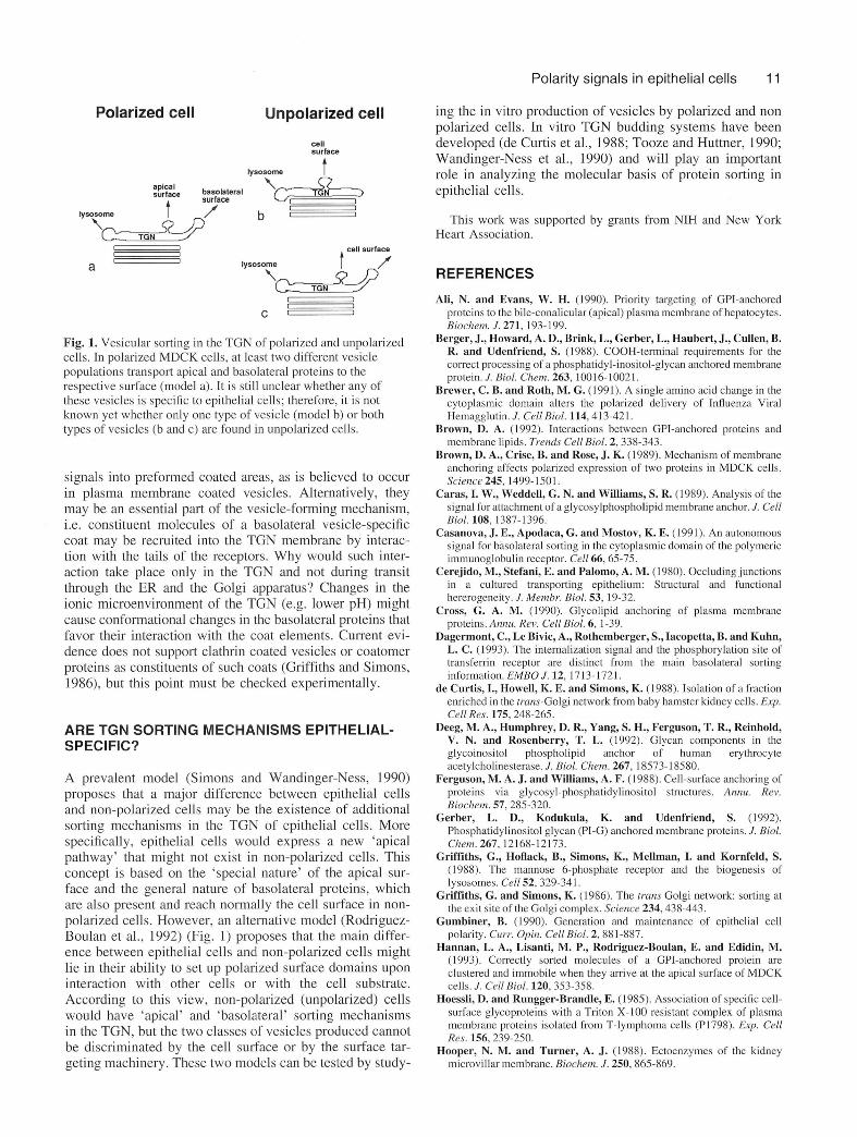
Fig. 1. Vesicular sorting in the TGN of polarized and unpolarized cells. In polarized MDCK cells, at least two different vesicle populations transport apical and basolateral proteins to the respective surface (model a). It is still unclear whether any of these vesicles is specific to epithelial cells; therefore, it is not known yet whether only one type of vesicle (model b) or both types of vesicles (b and c) are found in unpolarized cells.
signals into preformed coated areas, as is believed to occur in plasma membrane coated vesicles. Alternatively, they may be an essential part of the vesicle-forming mechanism, i.e. constituent molecules of a basolateral vesicle-specific coat may be recruited into the TGN membrane by interaction with the tails of the receptors. Why would such interaction take place only in the TGN and not during transit through the ER and the Golgi apparatus? Changes in the ionic microenvironment of the TGN (e.g. lower pH) might cause conformational changes in the basolateral proteins that favor their interaction with the coat elements. Current evidence does not support clathrin coated vesicles or coatomer proteins as constituents of such coats (Griffiths and Simons, 1986), but this point must be checked experimentally.
ARE TGN SORTING MECHANISMS EPITHELIAL- SPECIFIC?
A prevalent model (Simons and Wandinger-Ness, 1990) proposes that a major difference between epithelial cells and non-polarized cells may be the existence of additional sorting mechanisms in the TGN of epithelial cells. More specifically, epithelial cells would express a new ‘apical pathway’ that might not exist in non-polarized cells. This concept is based on the ‘special nature’ of the apical surface and the general nature of basolateral proteins, which are also present and reach normally the cell surface in nonpolarized cells. However, an alternative model (Rodriguez- Boulan et al., 1992) (Fig. 1) proposes that the main difference between epithelial cells and non-polarized cells might lie in their ability to set up polarized surface domains upon interaction with other cells or with the cell substrate. According to this view, non-polarized (unpolarized) cells would have ‘apical’ and ‘basolateral’ sorting mechanisms in the TGN, but the two classes of vesicles produced cannot be discriminated by the cell surface or by the surface targeting machinery. These two models can be tested by study
ing the in vitro production of vesicles by polarized and non polarized cells. In vitro TGN budding systems have been developed (de Curtis et al., 1988; Tooze and Huttner, 1990; Wandinger-Ness et al., 1990) and will play an important role in analyzing the molecular basis of protein sorting in epithelial cells.
This work was supported by grants from NIH and New York Heart Association.
REFERENCES
Ali, N. and Evans, W. H. (1990). Priority targeting of GPI-anchored proteins to the bile-conalicular (apical) plasma membrane of hepatocytes. Biochem. J. 271 ,193-199.
Berger, J., Howard, A. D., Brink, L., Gerber, L., Haubert, J., Cullen, B. R. and Udenfriend, S. (1988). COOH-terminal requirements for the correct processing of a phosphatidyl-inositol-glycan anchored membrane protein. J. Biol. Chem. 263, 10016-10021.
Brewer, C. B. and Roth, M. G. (1991). A single amino acid change in the cytoplasmic domain alters the polarized delivery of Influenza Viral Hemagglutin. J. CellBiol. 114.413-421.
Brown, D. A. (1992). Interactions between GPI-anchored proteins and membrane lipids. Trends CellBiol. 2, 338-343.
Brown, D. A., Crise, B. and Rose, J. K. (1989). Mechanism of membrane anchoring affects polarized expression of two proteins in MDCK cells. Science 245, 1499-1501.
Caras, I. W., Weddell, G. N. and Williams, S. R. (1989). Analysis of the signal for attachment of a glycosylphospholipid membrane anchor. J. Cell Biol. 108, 1387-1396. ~ '
Casanova, J. E., Apodaca, G. and Mostov, K. E. (1991). An autonomous signal for basolateral sorting in the cytoplasmic domain of the polymeric immunoglobulin receptor. Cell 66, 65-75.
Cerejido, M., Stefani, E. and Palomo, A. M. (1980). Occluding junctions in a cultured transporting epithelium: Structural and functional hererogeneity. J. Membr. Biol. 53, 19-32.
Cross, G. A. M. (1990). Glycolipid anchoring of plasma membrane proteins. Annu. Rev. CellBiol. 6, 1-39.
Dagermont, C., Le Bivic, A., Rothemberger, S., Iacopetta, B. and Kuhn, L. C. (1993). The internalization signal and the phosphorylation site of transferrin receptor are distinct from the main basolateral sorting information. EMBO J. 12, 1713-1721.
de Curtis, I., Howell, K. E. and Simons, K. ( 1988). Isolation of a fraction enriched in the trans-Golgi network from baby hamster kidney cells. Exp. Cell Res. 175,248-265.
Deeg, M. A., Humphrey, D. R., Yang, S. H., Ferguson, T. R., Reinhold, V. N. and Rosenberry, T. L. (1992). Glycan components in the glycoinositol phospholipid anchor of human erythrocyte acetylcholinesterase. J. Biol. Chem. 267, 18573-18580.
Ferguson, M. A. J. and Williams, A. F. (1988). Cell-surface anchoring of proteins via glycosyl-phosphatidylinositol structures. Annu. Rev. Biochem. 57, 285-320.
Gerber, L. D., Kodukula, K. and Udenfriend, S. (1992). Phosphatidylinositol glycan (PI-G) anchored membrane proteins. J. Biol. Chem. 267,12168-12173.
Griffiths, G., Hoflack, B., Simons, K., Mellman, I. and Kornfeld, S. (1988). The mannose 6-phosphate receptor and the biogenesis of lysosomes. Cell 52, 329-341.
Griffiths, G. and Simons, K. (1986). The trans Golgi network: sorting at the exit site of the Golgi complex. Science 234,438-443.
Gumbiner, B. (1990). Generation and maintenance of epithelial cell polarity. Curr. Opin. Cell Biol. 2, 881-887.
Hannan, L. A., Usanti. M. P., Rodriguez-Boulan, E. and Edidin, M. (1993). Correctly sorted molecules of a GPI-anchored protein are clustered and immobile when they arrive at the apical surface of MDCK cells. J. CellBiol. 120, 353-358.
Hoessli, D. and Rungger-Brandle, E. (1985). Association of specific cell- surface glycoproteins with a Triton X-100 resistant complex of plasma membrane proteins isolated from T-lymphoma cells (P1798). Exp. Cell Res. 156,239-250.
Hooper, N. M. and Turner, A. J. (1988). Ectoenzymes of the kidney microvillar membrane. Biochem. J. 250, 865-869.
12 E. Rodriguez-Boulan and C. Zurzolo
Hunziker, W., Harter, C., Matter, K. and Mellman, I. (1991). Basolateral sorting in MDCK cells requires a distinct cytoplasmic domain determinant. Cell 66 ,907-920.
Kurzchalia, T. V., Dupree, P., Parton, R. G., Kellner, R., Virta, H., Lehnert, M. and Simons, K. (1992). VIP21, a 21 kD membrane protein is an integral component of trans-Golgi-network-derived transport vesicles. / CellBiol. 118, 1003-1014.
Le Bivic, A., Sambuv, Y., Patzak, A., Patil, N., Chao, M. and Rodriguez- Boulan, E. (1991). An internal deletion in the cytoplasmic tail reverses the apical localization of human NGF receptor in transfected MDCK cells. J. CellBiol. 115, 607-618.
Lisanti, M., Caras, I. P., Davitz, M. A. and Rodriguez-Boulan, E. (1989). A glycophospholipid membrane anchor acts as an apical targeting signal in polarized epithelial cells. J. Cell Biol. 109,2145-2156.
Lisanti, M., Sargiacomo, M„ Graeve, L., Saltiel, A. and Rodriguez- Boulan, E. (1988). Polarized apical distribution of glycosyl phosphatidylinositol anchored proteins in a renal epithelial line. Proc. Nat. Acad. Sci. USA. 85, 9557-9561.
Lisanti, M. P., Le Bivic, A., Saltiel, A. and Rodriguez-Boulan, E. (1990). Preferred apical distribution of glycosyl-phosphatidylinositol (GPI) anchored proteins: a highly conserved feature of the polarized epithelial cell phenotype. J. Membr. Biol. 113, 155-167.
Low, M. G. and Saltiel, A. R. (1988). Structural and fuctional roles of glycosyl-phosphatidylinositol in membranes. Science 239, 268-275.
Matter, K., Hunziker, W. and Mellman, I. (1992). Basolateral sorting of LDL receptor in MDCK cells: The cytoplasmic domain contains two Tyrosine-dependent targeting determinants. Cell 71, 741-753.
Nelson, W. J. (1991). Cytoskeleton functions in membrane traffic in polarized epithelial cells. Semin. Cell Biol. 2,375-385.
Pearse, B. M. F. and Robinson, M. S. (1990). Clathrin, adaptors and sorting. Ann«. Rev. CellBiol. 6, 151-171.
Powell, S. K., Cunningham, B. A., Edelman, G. M. and Rodriguez- Boulan, E. (1991). Transmembrane and GPI anchored forms of NCAM are targeted to opposite domains of a polarized epithelial cell. Nature 353, 76-77
Prill, V., Lehmann, L., von Figura, K. and Peters, C. (1993). The cytoplasmic tail of lysosomal acid phosphatase contains overlapping but distinct signals for basolateral sorting and rapid internalization in polarized MDCK cells. EMBO J. 12, 2181-2193.
Robinson, M. S. (1992). Adaptins. Trends Cell Biol. 2, 293-297.Rodriguez-Boulan, E. and Powell, S. K. (1992). Polarity of epithelial and
neuronal cells. Annu. Rev. Cell Biol. 8, 395-427.Rothberg, K. G., Heuser, J. E., Donzell, W. C., Ying, Y.-S., Glenney, J.
R. and Anderson, R. G. W. (1992). Caveolin, a protein component of caveolae membrane coats. Cell 68,673-682.
Simons, K. and Wandinger-Ness, A. (1990). Polarized sorting in epithelia. Cell 62, 207-210.
Tooze, S. A. and Huttner, W. B. (1990). Cell-free protein sorting to the regulated and constitutive secretory pathways. Cell 60, 837847.
van Meer, G. and Burger, K. N. J. (1992). Sphingolipid trafficking-sorted out?. Trends Cell Biol. 2,332-337.
van Meer, G. and Simons, K. (1988). Lipid polarity and sorting in epithelial cells. J. Cell. Biochem. 36,51-58.
Wandinger-Ness, A., Bennett, M. K., Antony, C. and Simons, K. (1990). Distinct transport vesicles mediate the delivery of plasma membrane proteins to the apical and basolateral domains of MDCK cells. J. Cell Biol. I l l , 987-1000.
Yokode, M., Pathak, R. K., Hammer, R. E., Brown, M. S., Goldstein, J. L. and Anderson, R. G. W. (1992). Cytoplasmic sequence required for basolateral targeting of LDL receptor in livers of transgenic mice. J. Cell Biol. 117, 39-46.
Zurzolo, C., Lisanti, M. P., Caras, I. W., Nitsch, L. and Rodriguez- Boulan, E. (1993a). Glycosylphosphatidilinositol-anchored proteins are preferentially targeted to the basolateral surface in Fischer Rat Thyroid epithelial cells. J. Cell Biol. 121, 1031-1039.
Zurzolo, C., v a n ’t Hof, W., van Meer, G. and Rodriguez-Boulan, E. (1993b). VIP21/caveolin, glysphingolipid clusters, and the sorting of glycosylphosphatidylinositol-anchored proteins in epithelial cells. EMBO J. 13 (in press).