Embed Size (px)
Citation preview

Journal of Chromatography A, 1081 (2005) 238–244
Polyacrylamide gel electrophoresis separation and detection ofpolyamidoamine dendrimers possessing various cores
and terminal groups
Ajit Sharmaa,∗, Ankur Desaia, Riaz Alia, Donald Tomaliaa,b
a Department of Chemistry, Central Michigan University, Mt. Pleasant, MI 48859, USAb Dendritic NanoTechnologies, Inc., 2625 Denison Drive, Mt. Pleasant, MI 48858, USA
Received 21 March 2005; received in revised form 24 May 2005; accepted 31 May 2005Available online 13 June 2005
Abstract
Detection and separation of polyamidoamine dendrimers possessing various cores and surface groups was studied by polyacrylamide gelelectrophoresis. Although many dyes and staining techniques were able to detect dendrimers on polyacrylamide gels, Coomassie Blue wasf itions, whiled cur duringt using smallp ration bute ensive, andr©
K
1
prndutsgmgjd
vity,rgerculesie-mble
iletech-ery,ents
s isapil-ationf
milarapil-AMound
0d
ound to be the most sensitive and convenient. Amine and hydroxyl terminated dendrimers were best separated under acidic condendrimers with carboxyl surfaces required alkaline buffers. Some dendrimers were very susceptible to diffusion that could oc
heir separation, staining or destaining steps. In the absence of an appropriate fixation step, dendrimers could be resolved byore size gels and low voltage or current. Increasing core lengths did not significantly affect migration of a given dendrimer genexhibited improved separation and staining characteristics. Polyacrylamide gel electrophoresis was found to be a rapid, inexpeliable procedure to characterize many different water-soluble dendritic macromolecules.
2005 Elsevier B.V. All rights reserved.
eywords:Polyamidoamine dendrimers; Polyacrylamide gel electrophoresis; Staining
. Introduction
Polyamidoamine or PAMAM dendrimers are syntheticolymer-based nanoparticles (with diameters in the 1–15 nmange), which are widely recognized as important quantizedanoscale building blocks in biomedical research[1,2]. Den-rimers have unique structural or architectural components,nlike other traditional polymers. The various parts of a
ypical dendrimer molecule include a core, an interior con-isting of repetitive branch cell units and terminal functionalroups. In addition, cavities are also present in the macro-olecule (Fig. 1). The high density of terminal functionalroups, formed as a result of branching, can be used to con-
ugate other molecules to the dendrimer. For example, manyrug molecules may be covalently attached and carried into
∗ Corresponding author. Tel.: +1 989 774 3303; fax: +1 989 774 3883.E-mail address:[email protected] (A. Sharma).
the blood stream by each dendrimer molecule. The cawhich increases in size as the dendrimer grows to lagenerations, may be used to encapsulate small molelike drugs [3]. The core may be cleaved to provide pshaped dendrimers or dendrons, which can self-asseto form large nanoparticles[4]. Dendrimers are versatmolecules that have many potential applications in nanonology and life sciences, including gene and drug delivcontrast agents for imaging and in vitro diagnostic reag[5].
Characterization of dendrimers and their conjugatecritical for their successful synthesis and applications. Clary electrophoresis (CE) has been used for characterizof low generation PAMAM dendrimers[6–8]. Separation ohigher generations become problematic due to their sicharge densities and interactions with the walls of the clary [6]. The absence of any strong chromophore in PAMdendrimers requires the use of very low wavelengths (ar
021-9673/$ – see front matter © 2005 Elsevier B.V. All rights reserved.oi:10.1016/j.chroma.2005.05.074

A. Sharma et al. / J. Chromatogr. A 1081 (2005) 238–244 239
Fig. 1. Structure of an amine-terminated G3 PAMAM dendrimer with an EDA core.
214 nm) for their detection in CE. The buffer and sampletherefore has to be very clean to avoid false peaks due tolight scattering effects. In addition, CE requires expensiveinstrumentation and a trained operator. A much less expensivealternative is polyacrylamide gel electrophoresis (PAGE),which has been used to separate PAMAM dendrimers of var-ious generations[6,9]. PAGE has many potential advantagesfor dendrimer characterization. It allows separation underphysiological conditions, requires small sample size, yieldsa visual end signal, and is a mild, non-destructive techniquethat does not cause fragmentation of the dendrimer sample[10].
PAMAM dendrimers can be successfully separated byPAGE[6,9]. Most of this work has been directed to PAMAMdendrimers with amine surfaces and ethylenediamine (EDA)cores. However, there are many other kinds of PAMAM den-drimers that are commercially available today. These includedendrimers possessing various dimensions (generations) andcores like diaminobutane (DAB), diaminohexane (DAH),diaminododecane (DAD) and cystamine. Besides amines,other surface functional groups include carboxyls, hydroxylsand hexylamides. The physicochemical variations found inthese dendritic structures may require modified electrophore-sis conditions for their separation, as in the case for separationof acidic and basic proteins. The main objective of this reportis to study the effect of the various dendrimer cores and theirs con-d
2. Experimental
2.1. Apparatus
2.1.1. PAGEElectrophoresis was performed on polyacrylamide gels
in acidic or basic conditions. Mini gels (10 cm× 8 cm×0.75 mm) were used during the study. The electrophoresissystem was a Dual Mini-Vertical Slab Gel Electrophore-sis Unit from Sigma (St. Louis, MO, USA). This apparatusallows gel cooling by running water. Gels were photographedwith a Kodak DC 40 digital camera and images analyzed withKodak Digital Science 1D image analysis software (EastmanKodak, Rochester, NY).
2.2. Procedures
2.2.1. PAGEElectrophoresis under acidic or basic conditions was
performed as described before[7]. Amine-terminated den-drimers were fixed with glutaraldehyde. Unless specified, allelectrophoresis steps were performed at 4◦C.
2.2.2. Dendrimer purification from polyacrylamide gelsAfter electrophoresis, the desired dendrimer band was
excised from the gel and minced in an appropriate solvent( gelw 5 min
urfaces on electrophoretic migration and to find suitableitions for their separation and detection.
e.g., water if the dendrimer is very water soluble). Theas then subjected to three cycles of freeze and thaw (1

240 A. Sharma et al. / J. Chromatogr. A 1081 (2005) 238–244
each). It was incubated overnight at room temperature in ashaker. The sample was then centrifuged at 16,000×gon anEppendorf centrifuge 5415C for 10 min. The supernatant wasremoved and lyophilized.
2.2.3. Staining of amine surface PAMAM dendrimersCoomassie Blue R-250 (0.2%, w/v) was prepared in 50%
methanol/10% acetic acid solution. After electrophoresis, thegel was carefully removed from between the glass platesand placed in a small dish for staining. The gel was placedin the cold (4◦C) staining solution overnight (about 16 h).After staining, the gel was destained for 10–15 min in cold50% methanol/10% acetic acid and then placed in cold 10%methanol/10% acetic acid until the background was clear.
Dendrimer staining was also performed with the aminereagent, naphthoquinone. Ponceau S, Brilliant Blue G per-chloric acid (Sigma), 1% (w/v) Fast Green, Silver stain(Sigma procedure P3040), and Colloidal Coomassie Blue(Sigma). A carbon disulphide–silver nitrate (CS2/AgNO3)staining was also carried out. In this procedure, the post-electrophoresis gel was stained in a CS2 solution (made 1:1in absolute ethanol) for about 5 min and then excess of CS2was removed by rinsing the gel with deionized water for 15 s,five times. The gel was then placed in a 1% aqueous AgNO3solution for 24 h.
2nics
M er-a ple(h rateu thens
2
il-w sedf nt,M
3
3
lnesso n ofa var-i ered asicc te,a BlueR he geO ld not
find any previous reports on the staining characteristics ofPAMAM dendrimers on PAGE gels. Staining is an importantpart of electrophoresis. This step allows visualization of theseparated components on the gel.
Using EDA core PAMAM dendrimers with amine surface,a number of other stains used for proteins and or amines inelectrophoresis or histochemistry were evaluated in this partof the investigation. The objective was to study the stainingcharacteristics of dendrimers towards various dyes and todetermine if there was a more sensitive staining method foramine-terminated PAMAM dendrimers than Coomassie BlueR-250.
The dye is slightly soluble in water because ofits hydrophobic side-chains. The exact mechanism ofCoomassie Blue staining for proteins or dendrimers is uncer-tain, but electrostatic interactions between the dye’s sulfonicacid groups and amine groups in proteins and van der Waalsforces have been implicated[11,12].
Coomassie Blue R-250 was prepared in methanol/aceticacid. Staining of dendrimers was performed in the cold. Vari-ous incubation times were tested (30, 180 min and overnight)in order to optimize staining time. Results from this studyrevealed that the best results were obtained by overnight stain-ing of the gel (results not shown). After staining, the gel wasdestained for 10–15 min in cold 50% methanol/10% aceticacid and then placed in cold 10% methanol/10% acetic acidu n thec tiond foraG er
atedw iumsa withn er 10t wn).D lliantBe thanC thodfH tect1 anicf n ofa Them id)t edf in-i atedP m-p ionso eress 1
.2.4. Mass spectrometryMass spectra were recorded using a Bruker Dalto
ALDI-TOF mass spectrometer (Billerica, MA, USA) opting in a pulse ion extraction linear mode. The sam1�l) was mixed with 10�l of matrix (10 mg/ml of 3-ydroxyphenyl acetic acid). Solvent was allowed to evaponder ambient conditions. The dendrimer-matrix waspotted on the target plate.
.3. Chemicals
All routine chemicals were obtained from Aldrich (Maukee, WI, USA). All dendrimers were either purcha
rom Aldrich or Dendritic NanoTechnologies (Mt. PleasaI, USA; URL: http://dnanotech.com).
. Results and discussion
.1. Dendrimer staining
Previous work has clearly demonstrated the usefuf polyacrylamide gel electrophoresis for the separatiommonia or EDA-core PAMAM dendrimers possessing
ous surfaces[6,9]. Optimum electrophoresis conditions wescribed for dendrimer separation under acidic or bonditions with various buffers (Tris–glycine, Tris–boracetate or citrate). The common protein stain Coomassie-250 was used to detect the separated dendrimers on tther than the reported use of Coomassie Blue, we cou
l.
ntil the background was clear. Staining and destaining iold helped reduce diffusion, especially for lower generaendrimers[9]. The sensitivity of Coomassie Blue R-250mine surface EDA-core PAMAM dendrimers was 1.5�g for0, under optimized acidic conditions[9]. For proteins, th
eported sensitivity is 50 ng[13].Many primary amines develop a blue color when tre
ith ortho-quinones. The preferred reagent is the sodalt of 1,2-naphthoquinone-4-sulphonic acid[14]. Althoughmine-terminated PAMAM dendrimers could be stainedaphthoquinone, the sensitivity of this technique was ov
imes less than that of Coomassie Blue (results not shoendrimers could also be detected with Ponceau S, Brilue G perchloric acid, and Fast Green staining[15,16]. How-ver, all of these stains displayed much lower sensitivityoomassie Blue. Silver staining is a highly sensitive me
or visualization of proteins in electrophoresis gels[12].owever, unlike Coomassie Blue, it could not even de0�g EDA-core amine surface G0. A spot test used in org
unctional group analysis for amines involves the reactiomines with carbon disulphide to form dithiocarbamates.ixture is then reacted with 1% silver nitrate (in nitric ac
o give black silver sulphide[17]. This reaction was evaluator detection of PAMAM amines on PAGE gels. This stang methodology was quite sensitive for amine-terminAMAM dendrimers. The sensitivity of the stain was coared with Coomassie Blue R 250. Different concentratf G5–G9 and G0–G4 mixtures were run on gels that wtained with either Coomassie Blue R 250 or CS2/AgNO3tain. Both staining procedures could detect down to�g

A. Sharma et al. / J. Chromatogr. A 1081 (2005) 238–244 241
of G2–G9 (results not shown). Attempts to further improvethe sensitivity of the stain for dendrimers were unsuccess-ful. Although sensitivity of this method was comparable toCoomassie Blue R 250, it required more steps and involvedthe use of hazardous materials. Another protein stain, Bril-liant Blue G-colloidal has been utilized in the staining ofproteins in polyacrylamide and agarose gels. As a proteinstain, this dye is reported to be five times more sensitive thantraditional Coomassie Blue, takes only 1 h to stain and clearbackground is achieved in 7 h[18]. However, it could not staindendrimers. The lack of staining may be due to the inabilityof the large stain particles to penetrate the cavities present ina dendrimer.
3.2. Electrophoretic separation of dendrimers
Sharma et al. showed that PAMAM dendrimer mix-tures could be separated in non-gradient gels[9]. Employ-ing conditions that reduced diffusion, even low generationdendrimers (G0–G2) could be clearly resolved. These con-ditions included carrying out all steps (separation, stainingand destaining) at 4◦C, running the separation at low currentor voltage to reduce heat generation, and whenever possi-ble, fixation of the dendrimer samples after electrophoresis.PAMAM dendrimers with neutral or positively charged sur-f hosew here-f hiletl eens fer-e paredt fold( e)i easedc rchi-t driticm ithv
3er
c
Fig. 2. Electrophoresis of amine surface PAMAM dendrimers with EDAand cystamine cores. Acidic electrophoresis was performed on 15% gel for55 min at 200 V. Gel was stained with Coomassie Blue R-250. Lanes 1 and4, G0–G5 EDA core ladder; lane 2, G1 cystamine core; lane 3, G3 cystaminecore.
separation is therefore better at low pH[9]. Using conditionsdescribed before[9], amine-terminated PAMAM dendrimerswith various cores were separated on polyacrylamide gelswith T andC values of 15 and 2.6%, respectively.
Separation of a mixture of G0–G5 EDA core PAMAMdendrimers is shown inFig. 2 (lanes 1 and 4). Staining wasperformed with Coomassie Blue R 250. In accordance withprevious reports, acidic electrophoresis under conditions thatreduced diffusion and inclusion of a glutaraldehyde-fixationstep led to sharp, well-resolved bands. The largest differencein charge/mass ratio is between G0 and G1. The differencein ratio between two consecutive generations then progres-sively decreases with increasing generation number (Table 1).This effect is clearly observed on PAGE gels. The differ-ence in migration is greatest between G0 and G1 and thenit progressively decreases (Fig. 2, lanes 1 and 4). A simi-lar phenomenon is observed under alkaline separations[6,9].Separation of higher generation dendrimers involves morethan just differences in their charge/mass ratios. Molecularsieving and possible sample-gel interactions may also aid intheir separation. The acidic separation procedure also worksvery well in separation of dendrimers with other cores. Asshown inFig. 2 (lanes 2 and 3), cystamine dendrimers with
TT rimers (EDA core)
D G3
M 6909 162D 36N 32 8N 30 6T 6 94C 46C 89.7
s ands and ines)
aces have higher charge-mass ratios at low pH while tith negative charges have higher ratios at basic pH. T
ore, the former were separated under acidic conditions whe latter were separated under basic conditions[6,9]. In theast few years, many new PAMAM dendrimers have bynthesized. Currently, PAMAM dendrimers of many difnt cores and surfaces are commercially available. Com
o the commonly used EDA core, dendrimers with two-DAB core), three-fold (DAH core) and six-fold (DAD corncreased core lengths have been prepared. The incrore lengths may have some interesting effects on the aecture and physicochemical properties of these denacromolecules[19]. Electrophoresis of dendrimers w
arious cores and surfaces is discussed below.
.2.1. Separation of dendrimers with amine surfacesAmine-terminated PAMAM dendrimers have high
harge/mass ratios under acidic conditions (Table 1). Their
able 1heoretical molecular characteristics of amine surface PAMAM dend
endrimer G0 G1 G2
olecular weight 517 1430 3256iameter (A) 15 22 29umber of primary amines 4 8 16umber of tertiary amines 2 6 14otal number of amine groups 6 14 30harge/mass ratio at pH 8.8 (×10−4) (C/g)a 77 56 49harge/mass ratio at pH 2.9 (×10−4) (C/g)b 116 97.9 92.1a Charge/mass ratio was calculated assuming theoretical MW valueb Charge/mass ratio was calculated assuming theoretical MW value
G4 G5 G6 G7 G8 G9
14215 28826 58048 116493 233383 46745 54 67 81 97 114
64 128 256 512 1024 20462 126 254 510 1022 204
2 126 254 510 1022 2046 4045 44.4 44.1 43.9 43.87 43.83
88.6 88.1 87.8 87.3 87.66 87.63
full protonation of only terminal primary amines.full protonation of all amines (terminal primary and interior tertiary am.

242 A. Sharma et al. / J. Chromatogr. A 1081 (2005) 238–244
surface amine groups show sharp bands in which trailing gen-erations, dimers and other contaminants (which are producedduring synthesis) were clearly resolved.
The acidic electrophoresis procedure was also utilized forseparation of amine-terminated PAMAM dendrimers of vary-ing core lengths. Changing the core length has been reportedto dramatically alter the morphology and properties ofthese macromolecules[19]. Amine terminated PAMAM den-drimers with diaminododecane core, diaminohexane core,and diaminobutane core were run under acidic conditions.In general, dendrimers with higher chain length cores gavesharper bands than EDA core dendrimers, although theircharge/mass ratios were not significantly different. It is likelythat the increased core length made it more hydrophobic,which resulted in decreased dendrimer diffusion during elec-trophoresis, fixing, staining and destaining steps, especiallyfor the lower generations that are more susceptible to diffu-sion artifacts. It is also possible that the increased core lengthmay have led to enhanced interaction of Coomassie Blue dyewith the dendrimer.
3.2.2. Separation of dendrimers with carboxyl surfacesBrothers et al. used alkaline conditions to resolve 3.5 and
higher generation carboxyl terminated dendrimers[6]. Weused similar conditions on 15% gels, except that all steps werep ◦ forv er,G den-d e tot iesw iums likea xyl-t wella allerp ed tor t toa theb . Theu ltage( in a
better separation. G1.5 could be clearly seen and a mixtureof G1.5–4.5 was well resolved with sharp bands. However,G1.5 and G2.5 have very similar migrations, and cannot beresolved in mixtures. Trailing generations and higher molecu-lar weight species could be clearly seen and variations in corelengths did not appreciably affect migration times of den-drimers under these conditions (results not shown). Anothercarboxyl-containing class of dendrimers contains succinamicacid surface groups. These dendrimers are commerciallyavailable with various core lengths and types, and can alsobe well-resolved under these conditions. An example of thisis shown inFig. 3. Cystamine core PAMAM G4 dendrimerwith succinamic acid surface (MW 20 711, lane 1,Fig. 3A andB) was denatured with 10-fold excess DTT for 24 h at roomtemperature. The products of this cleavage (dendrons withhalf the molecular weight of the dendrimer) migrated similarto a G3 dendrimer (MW 10 203) (lane 2,Fig. 3A). Highergeneration contaminants in the G4 sample were broken downinto species that migrated like G4 dendrimers. Purification ofG4 gave a single band (lane 3,Fig. 3A and lane 2,Fig. 3B).Reduction of the purified G4 dendrimer under similar condi-tions, showed the original band and a new band that migratedlike a non-reduced G3 (lanes 3–5,Fig. 3B). Some G4 wasstill visible even after 24 h reduction under these conditions(Fig. 3C). As with proteins, PAGE is a convenient way tomonitor dendrimer cleavage reactions.
3para-
t inalg )a ablest assr en-d 3 h)i sedt tedd sep-a cidice detec-t ning
F dend on 20% gelf Lane 1 d G4. (B)L uction f
erformed at 4C. Although sharp bands were obtainedarious EDA core, carboxyl-terminated PAMAM dendrim1.5 could not be visualized (results not shown). Theserimers seem to be very sensitive to diffusion probably du
heir excellent water solubility. Some fixative methodologere tried (crosslinking by EDC/diamine, use of chromalts) but did not yield any improvements. Cationic dyeslcian blue and toluidine were used to stain the carbo
erminated dendrimers. However, they did not perform ass Coomassie Blue (results not shown). Therefore, smore size gels and reduced voltage (or current) was useduce diffusion. In addition, destaining time was kep
minimum (less than 30 min instead of 3–4 h). Onceands were clearly visible, the gels were photographedse of 20% polyacrylamide gel, separation at a lower voless than 100 V), and reduced destaining time, resulted
ig. 3. Reduction and separation of succinamic acid surface PAMAMor 30 min at 150 V. Gel was stained with Coomassie Blue R-250. (A)ane 1, unpurified G4, lane 2, purified G4; lanes 3–5, purified G4 red
.2.3. Separation of dendrimers with hydroxyl surfacesA major advantage of acidic electrophoresis is the se
ion of PAMAM dendrimers that possess uncharged termroups[9]. The interior tertiary amines (with pK around 4–5re protonated under acidic conditions. This charge en
heir electrophoretic migration. However, their charge/matios are almost half that of amine surface PAMAM drimers. Electrophoresis times would be very long (2–
f low voltage (or currents) or small pore gels were uo minimize diffusion (as in case of carboxyl-terminaendrimers). Thus, the diffusion problem is the worst forration of hydroxyl surface dendrimers. Nevertheless, alectrophoresis can provide adequate separation and
ion when all steps are carried out in the cold and stai
rimers with cystamine core. Alkaline electrophoresis was performed, unpurified G4; lane 2, G4 reduced with DTT for 24 h; lane 3, purifieor 5, 3, and 1 h, respectively. (C) Purified G4 reduction for 24 h.
A. Sharma et al. / J. Chromatogr. A 1081 (2005) 238–244 243
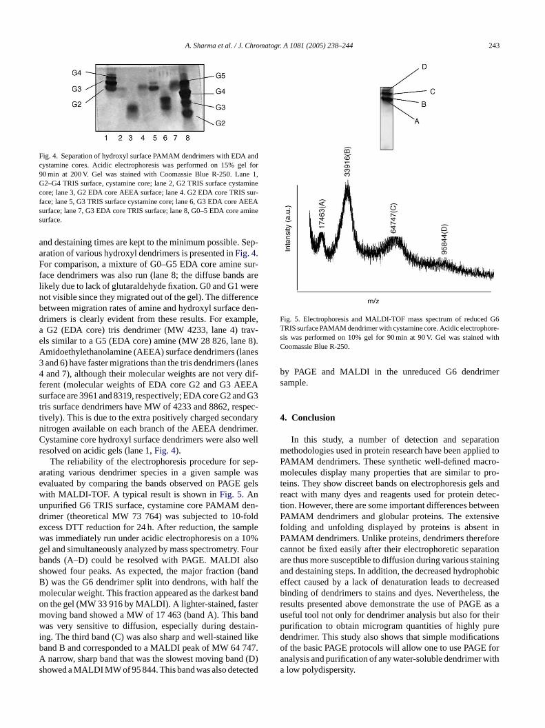
Fig. 4. Separation of hydroxyl surface PAMAM dendrimers with EDA andcystamine cores. Acidic electrophoresis was performed on 15% gel for90 min at 200 V. Gel was stained with Coomassie Blue R-250. Lane 1,G2–G4 TRIS surface, cystamine core; lane 2, G2 TRIS surface cystaminecore; lane 3, G2 EDA core AEEA surface; lane 4. G2 EDA core TRIS sur-face; lane 5, G3 TRIS surface cystamine core; lane 6, G3 EDA core AEEAsurface; lane 7, G3 EDA core TRIS surface; lane 8, G0–5 EDA core aminesurface.
and destaining times are kept to the minimum possible. Sep-aration of various hydroxyl dendrimers is presented inFig. 4.For comparison, a mixture of G0–G5 EDA core amine sur-face dendrimers was also run (lane 8; the diffuse bands arelikely due to lack of glutaraldehyde fixation. G0 and G1 werenot visible since they migrated out of the gel). The differencebetween migration rates of amine and hydroxyl surface den-drimers is clearly evident from these results. For example,a G2 (EDA core) tris dendrimer (MW 4233, lane 4) trav-els similar to a G5 (EDA core) amine (MW 28 826, lane 8).Amidoethylethanolamine (AEEA) surface dendrimers (lanes3 and 6) have faster migrations than the tris dendrimers (lanes4 and 7), although their molecular weights are not very dif-ferent (molecular weights of EDA core G2 and G3 AEEAsurface are 3961 and 8319, respectively; EDA core G2 and G3tris surface dendrimers have MW of 4233 and 8862, respec-tively). This is due to the extra positively charged secondarynitrogen available on each branch of the AEEA dendrimer.Cystamine core hydroxyl surface dendrimers were also wellresolved on acidic gels (lane 1,Fig. 4).
The reliability of the electrophoresis procedure for sep-arating various dendrimer species in a given sample wasevaluated by comparing the bands observed on PAGE gelswith MALDI-TOF. A typical result is shown inFig. 5. Anunpurified G6 TRIS surface, cystamine core PAMAM den-drimer (theoretical MW 73 764) was subjected to 10-folde plew 10%g Fourb lsos andB them bando term andw ain-i likeb 47.A (D)s ted
Fig. 5. Electrophoresis and MALDI-TOF mass spectrum of reduced G6TRIS surface PAMAM dendrimer with cystamine core. Acidic electrophore-sis was performed on 10% gel for 90 min at 90 V. Gel was stained withCoomassie Blue R-250.
by PAGE and MALDI in the unreduced G6 dendrimersample.
4. Conclusion
In this study, a number of detection and separationmethodologies used in protein research have been applied toPAMAM dendrimers. These synthetic well-defined macro-molecules display many properties that are similar to pro-teins. They show discreet bands on electrophoresis gels andreact with many dyes and reagents used for protein detec-tion. However, there are some important differences betweenPAMAM dendrimers and globular proteins. The extensivefolding and unfolding displayed by proteins is absent inPAMAM dendrimers. Unlike proteins, dendrimers thereforecannot be fixed easily after their electrophoretic separationare thus more susceptible to diffusion during various stainingand destaining steps. In addition, the decreased hydrophobiceffect caused by a lack of denaturation leads to decreasedbinding of dendrimers to stains and dyes. Nevertheless, theresults presented above demonstrate the use of PAGE as auseful tool not only for dendrimer analysis but also for theirpurification to obtain microgram quantities of highly puredendrimer. This study also shows that simple modificationso fora witha
xcess DTT reduction for 24 h. After reduction, the samas immediately run under acidic electrophoresis on ael and simultaneously analyzed by mass spectrometry.ands (A–D) could be resolved with PAGE. MALDI ahowed four peaks. As expected, the major fraction (b) was the G6 dendrimer split into dendrons, with halfolecular weight. This fraction appeared as the darkestn the gel (MW 33 916 by MALDI). A lighter-stained, fasoving band showed a MW of 17 463 (band A). This bas very sensitive to diffusion, especially during dest
ng. The third band (C) was also sharp and well-stainedand B and corresponded to a MALDI peak of MW 64 7narrow, sharp band that was the slowest moving band
howed a MALDI MW of 95 844. This band was also detec
f the basic PAGE protocols will allow one to use PAGEnalysis and purification of any water-soluble dendrimerlow polydispersity.

244 A. Sharma et al. / J. Chromatogr. A 1081 (2005) 238–244
Acknowledgements
The authors wish to thank to thank Dr. D. Hedstrand forthe mass spectrometry analysis. This research was financiallysupported by a grant from U.S. Army Research Laboratories(ARL Contract No. W911NF-04-2-0030).
References
[1] R. Esfand, D.A. Tomalia, Drug Discov. Today 6 (2001) 427.[2] U. Boas, P.M.H. Heegaard, Chem. Soc. Rev. 33 (2004) 43.[3] P. Kolhe, E. Misra, R.M. Kannan, S. Kannan, M. Lieh-Lai, Int. J.
Pharm. 259 (2003) 143.[4] D.A. Tomalia, B. Huang, D.R. Swanson, H.M. Brothers II, J.W.
Klimash, Tetrahedron 59 (2003) 3799.[5] J.M.J. Frechet, D.A. Tomalia (Eds.), Dendrimers and Other Dendritic
Polymers, Wiley, New York, 2001.[6] H.M. Brothers III, L.T. Piehler, D.A. Tomalia, J. Chromatogr. A 814
(1998) 233.
[7] A. Ebber, M. Vaher, J. Peterson, M. Loop, J. Chromatogr. A 949(2002) 351.
[8] J. Peterson, A. Ebber, V. Allikmaa, M. Lopp, Proc. Estonian Acad.Sci. Chem. 50 (2001) 156.
[9] A. Sharma, D.K. Mohanty, A. Desai, R. Ali, Electrophoresis 24(2003) 2733.
[10] J. Peterson, V. Allikmaa, J. Subbi, T. Pehk, M. Loop, Eur. Polym.J. 39 (2003) 33.
[11] C.M. Wilson, Methods Enzymol. 91 (1983) 236.[12] C.R. Merril, Methods Enzymol. 182 (1990) 477.[13] Life Sciences Catalog, National Diagnostics, 2002.[14] K. Hartke, U. Lohmann, Chem. Lett. (1983) 693.[15] A.H. Reisner, P. Nemes, C. Bucholtz, Anal. Biochem. 64 (1975)
509.[16] R.E. Allen, K.C. Masak, P.K. McAllister, Anal. Biochem. 104 (1980)
494.[17] M.T.M. Zaki, M.H. Fawzy, M.M. Assey, Microchim. Acta[Wien] 1
(1989) 221.[18] V. Neuhoff, N. Arold, D. Taube, W. Ehrhardt, Electrophoresis 9
(1988) 255.[19] D.M. Watkins, Y. Sayed-Sweet, J.W. Klimash, N.J. Turro, D.A.
Tomalia, Langmuir 13 (1997) 3136.