Embed Size (px)
Citation preview
HAL Id: hal-01579649https://hal-univ-rennes1.archives-ouvertes.fr/hal-01579649
Submitted on 31 Aug 2017
HAL is a multi-disciplinary open accessarchive for the deposit and dissemination of sci-entific research documents, whether they are pub-lished or not. The documents may come fromteaching and research institutions in France orabroad, or from public or private research centers.
L’archive ouverte pluridisciplinaire HAL, estdestinée au dépôt et à la diffusion de documentsscientifiques de niveau recherche, publiés ou non,émanant des établissements d’enseignement et derecherche français ou étrangers, des laboratoirespublics ou privés.
Poly(malic acid) bearing Doxorubicin and N-AcetylGalactosamine as a site-specific prodrug for targeting
hepatocellular carcinomaNivishna Venkatraj, M. J. Nanjan, Pascal Loyer, M. J. N. Chandrasekar,
Sandrine Cammas-Marion
To cite this version:Nivishna Venkatraj, M. J. Nanjan, Pascal Loyer, M. J. N. Chandrasekar, Sandrine Cammas-Marion.Poly(malic acid) bearing Doxorubicin and N-Acetyl Galactosamine as a site-specific prodrug for tar-geting hepatocellular carcinoma. Journal of Biomaterials Science, Polymer Edition, Taylor & Francis,2017, 28 (10-12), pp.1140-1157. �10.1080/09205063.2017.1311294�. �hal-01579649�
1
Poly(malic acid) bearing Doxorubicin and N-Acetyl Galactosamine as a
site-specific prodrugs for targeting HepatoCellular Carcinoma
Nivishna Venkatraj1,3, M.J. Nanjan1, Pascal Loyer2, M.J.N. Chandrasekar1,*
Sandrine Cammas Marion2,3,*.
1 JSS College of Pharmacy, Department of Pharmaceutical Chemistry, Rocklands,
Ootacamund, Tamil Nadu, India (Constituent of JSS University, Mysore, India).
2 INSERM, INRA, University of Rennes 1, University of Bretagne Loire, Nutrition
Metabolisms and Cancer (NuMeCan), Rennes, France.
3 Ecole Nationale Supérieure de Chimie de Rennes (ENSCR), Team Corint, ISCR, University
of Rennes 1, Rennes, France.
* Corresponding authors: [email protected]; [email protected]
2
Poly(malic acid) bearing Doxorubicin and N-Acetyl Galactosamine as a
site-specific prodrug for targeting HepatoCellular Carcinoma
Abstract: In the past, several systems of drug delivery carriers have been designed
with a high capacity to target specific cells and/or tissues and a reduced non-specific
toxicity. In this context, we synthesized and characterized novel poly(malic acid)
derivatives bearing Doxorubicin (Dox), Poly(ethylene glycol) (PEG) and/or N-Acetyl
Galactosamine (NAcGal) for drug delivery. These poly(malic acid) derivatives were
obtained by chemical modification of the carboxylic acid lateral groups of poly(malic
acid) (PMLA). The resulting nanoplatforms were evaluated for their in vitro
cytotoxicity using the human HepaRG hepatoma cell line. Results reveal that the
PMLA nanoplatform modified with PEG and Dox has an IC50 of 936 nM
corresponding to a Dox concentration of 47 nM, while the grafting of NAcGal onto
the nanoplatform reduced the IC50 to 527 nM corresponding to a Dox concentration of
26 nM. The presence of the targeting moiety, NAcGal, thus improves the cellular
toxicity of the Dox.
Keywords: Poly(malic acid) prodrug, Doxorubicin, N-Acetyl Galactosamine,
hepatocellular carcinoma.
3
Introduction
Hepatocellular carcinoma (HCC) is one of the most lethal cancers widely prevalent in the
world with only limited treatment options that depend on the stage of the disease, namely very
early/early, intermediate or advanced/late stage [1,2]. The available therapies range from
surgical resection, liver transplantation to chemotherapy with only limited success [1,3]. Lin
et al. have summarized the available treatments of HCC in a recent review and report that,
despite the existence of several curative treatments for very early/early stage, only
transarterial therapy or chemotherapy (mainly based on Sorafenib) can be proposed to patients
with intermediate or advanced/late stage of HCC with medium to short survival time [1].
Therefore, there is an urgent need for new treatments better suited not only to the patient but
also to the stage of the HCC, especially for intermediate and advanced/late stages. The aims
of designing new therapeutic forms for HCC treatments are to ameliorate the efficiency of the
anticancer drug by increasing its accumulation at the cancer site in the liver and decreasing its
uncontrolled biodistribution as well as the drug efflux by membrane channels while reducing
its systemic toxicity and improving the quality of the patient’s life [3]. These goals, however,
represent major challenges.
To answer these challenges, several drug delivery systems (DDS) have been developed in the
past and some of these have been approved by the Food and Drug Administration (FDA) for
clinical uses or are under clinical trials [4].
Among the several DDS developed, polymer-drug conjugates might be considered as
promising nanovectors. They are based on the model proposed by Ringsdorf in 1975 which
consists of a polymeric backbone bearing a solubilizer (mainly PEG), a drug (linked through a
degradable bond), a targeting molecule and/or a fluorescence probe allowing to follow the
biodistribution of the prodrug [5]. Tumor cell targeting is usually achieved by both passive
targeting, through the Enhanced Permeability Retention (EPR) effect [6,7] and active
4
targeting, using the ligand/receptor interaction [4]. Several targeting agents (TA) have been
described for their capacity to target human hepatic cancer cells and to promote cellular
uptake of the carriers thus improving the drug efficiency [8-12]. Among the HCC specific cell
membrane receptors, the Asialoglycoprotein (ASGP) receptor is largely present on the hepatic
cell surface and more specifically in human HCC [3]. Moreover, ASGP receptor is known to
be capable of specifically recognizing and binding to galactose and galactosamine residues
[3,13]. For example, the NAcGal has been shown to bind the ASGP receptor expressed on the
surface of human hepatic cancer cells and trigger efficient cellular uptake via the receptor-
mediated endocytosis [14]. Therefore, NAcGal can be used as a targeting moiety in the design
of site-specific drug carriers for HCC treatments.
Besides, a DDS needs to be protected against non-specific recognition by the
Reticuloendothelial System (RES) or macrophage system in order to increase its circulation
time in the body. A hydrophilic polymer, PEG, approved by the FDA in Humans has been
widely used for this purpose [15-17]. The stealth property, conferred to a DDS thanks to the
presence of PEG, depends on both the molar mass and the density of PEG [15-17]. In view of
the published results, a PEG having an amine end group (for the chemical coupling to the
polymer backbone) and a molar mass of 5,056 g/mol may be selected for designing a long
term circulating DDS.
Various biocompatible and/or (bio)degradable polymers have been evaluated as potential drug
carriers [18,19]. Among them, polyesters represent an attractive family for biomedical
applications as a result of their good biocompatibility and (bio)degradability. In this context,
PMLA attracts our attention because, in addition of being (bio)degradable, biocompatible,
non-toxic and non-immunogenic, it also has carboxylic acid lateral functional groups, which
can be used to chemically bind various molecules of interest such as drugs, targeting moieties,
etc. PMLA can be obtained either from the slime mold (Physarum polycephalum) [20] or by
5
the deprotection of the benzyl lateral groups of the poly(benzyl malate) prepared by anionic
ring opening polymerization of the corresponding monomer, the benzyl malolactonate
(MLABe) [21]. The chemical synthesis of PMLA is of interest because a large family of
polymers can be obtained by varying the nature of the lateral groups, the molar masses, and
the nature of the polymers (homopolymers, random or block copolymers) [22].
In the present study, we have chemically modified a PMLA obtained by chemical synthesis in
order to introduce several molecules of interest: (i) Doxorubicin, an anti-cancer drug, (ii)
NAcGal, a targeting moiety for liver cancer cells and (iii) PEG, a polymer allowing to confer
stealth properties to the DDS. The synthesized polymer-drug nanoconjugates were well
characterized and evaluated in vitro on a hepatoma cell line, the HepaRG. The cytotoxicity of
the Dox linked to the PMLA was thus evaluated and related to the presence or absence of the
NAcGal targeting moiety.
Experimental part
Materials
The racemic benzyl malolactonate (MLABe) was synthesized from DL-aspartic acid
according to the previously reported method [21]. In our method, we used dichloromethane
instead of benzene.
All the chemicals were used as received. Anhydrous THF was obtained from Aldrich.
Anhydrous ethanol was prepared just before use by distillation over sodium under N2
atmosphere. Nuclear magnetic resonance spectra (1HNMR) were recorded on a Brucker
ARX400 instrument (1H at 400MHz). Data are reported as follows: chemical shift
(multiplicity, number of hydrogen). The chemical shifts are reported as parts per million
(ppm) referenced to the appropriate residual solvent peak. Abbreviations are as follows: s
(singlet), d (doublet), t (triplet), q (quartet), dd (doublet of doublet), m (multiplet).
6
Weight average molar masses ( ) and molar mass distribution (Đ = ) of PMLABe
were determined by size exclusion chromatography (SEC) in THF at 40°C (flow
rate=1.0mL/min) on a GPC2502 Viscoteck apparatus equipped with a refractive index
detector, a LT5000L mixed medium org 300*7.8mm gel column, a TGuard Org Guard Col
10*4.6 mm and a GPC/SEC OmniSEC software. The polymer samples were dissolved in
THF at a concentration of 2 mg/mL. All elution curves were calibrated with poly(styrene)
standards.
and Đ = of the PMLA were determined by size exclusion chromatography
(SEC) in a 0.15 M phosphate buffer solution of pH7.4, 1 M NaCl at 35°C (flow
rate=0.5mL/min) on a GPC/SEC Malvern apparatus equipped with a refractive index detector
(VE3580) and a LALS-RALS 270 Dual Detector, a A4000-300*8 mm gel column, a 50*6
mm guard column and a GPC/SEC OmniSEC software. The polymer samples were dissolved
in 0.15 M phosphate buffer solution pH 7.4, 1 M NaCl at a concentration of 2 mg/mL. All
elution curves were calibrated with Pullulan standards.
The size (average diameter obtained by the CONTIN method), polydispersity and zeta
potential, (the measured electrophoretic mobility (μ) was converted to zeta potential (ζ) using
the Smoluchowski approximation) of the formulations were measured by Dynamic Light
Scattering using a DelsaTM Nano Beckman Coulter apparatus at 25°C.
Fourrier Transform Infra-Red (FTIR) spectra were recorded using a Fourier Nicolet 250
apparatus at 25°C.
UV spectra were recorded using a microplate reader (BioTek) at 485 nm for quantification of
Dox in the nanoconjugates and on a Secoman apparatus at 515 nm for MTT assays.
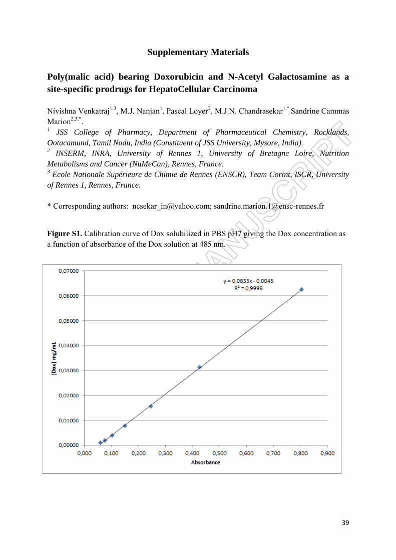
For the UV characterization of the nanoconjugates modified with Dox, we first realized a
calibration curve using Dox dissolved into PBS at pH7 with concentrations ranging from
7
0.0625 mg/mL to 0.00098 mg/mL (Figure S1). The equation giving the concentration of Dox
(mg/mL) as a function of the absorbance (Abs) at 485 nm is the following:
with R2 = 0.9998 (Figure S1).
The HepaRG hepatoma cells [23-25] were cultured as previously described in William’s E
medium (Lonza) supplemented with 2 mM of glutamine (Gibco), 5 mg/Lof insulin (Sigma),
10-5 M hydrocortisone hemisuccinate and 10% of fetal calf serum(Lonza).
Synthesis of poly(benzyl malate), PMLABe:
Poly(benzyl malate) was synthesized as described previously [21] starting from 3 g (1.46*10-2
mol) of racemic MLABe in presence of benzoate tetrabutylammonium (5.02*10-5 mol) as
initiator in bulk at 37°C. The polymerization was followed by FTIR and stopped when the
band, characteristic of MLABe, at 1,850 cm-1 had totally disappeared from the spectrum. The
resulting PMLABe was solubilized in a minimum volume of acetone and precipitated using a
large excess of ethanol. The precipitate was recovered and dried under vacuum leading to
2.53 g (yield= 84%) of PMLABe which was then analyzed by 1H NMR (structure) and SEC
in THF (molar masses and molar mass distribution).
1H NMR (CD3COCD3, ppm, 400MHz):2.84-3.10 (m, 2nH, CHCH2CO2), 5.09-5.19 (m,
2nH, CO2CH2C6H5), 5.47-5.63 (m, 1nH, CHCH2CO2), 7.25-7.46 (m, 5nH, CO2CH2C6H5).
SEC (THF, polystyrene standards, 1 mL/min, 40°C): 5,540 g/mol, Đ = 1.63
Synthesis of PMLA:
The PMLA was obtained by catalytic hydrogenolysis of the synthesized PMLABe. PMLABe
(2.53 g) was solubilized in 10 mL of acetone and 20 wt% of palladium on activated charcoal
(Pd/C, 0.51 g) were added to the solution. The round bottomed flask containing the PMLABe
and the Pd/C was placed under hydrogen atmosphere. The mixture was vigorously stirred for
12 h under hydrogen atmosphere. The solution was then filtered over celite and the acetone
8
was evaporated under vacuum. The obtained crude PMLA was solubilized in water, poured
into a dialysis bag (Molecular weight cut off, MCWO, of 3,500 g/mol) and dialyzed against
water overnight. The solution contained into the dialysis bag was lyophilized leading to a
white powder of PMLA (0.99 g, yield= 78%). It was characterized by 1H NMR and SEC in
0.15M phosphate buffer solution (PBS) pH7.4 containing 1M of NaCl.
1H NMR (CD3COCD3, ppm, 400MHz): 2.86-3.12 (m, 2nH, CHCH2CO2), 5.44-5.58(m,
1nH, CHCH2CO2), 6.78-7.85(m, 1nH, CO2H).
SEC (0.15 M PBS pH7.4, 1 M of NaCl, Pullulan standards): 7,910 g/mol, Đ = 1.2.
Degree of polymerization= 7,910/116= 68.
Activation of PMLA with N-hydroxysuccinimide (NHS):
PMLA (0.84 g, 7.24*10-3 mol, Mrepeat unit= 116 g/mol) was dissolved in 12 mL of anhydrous
acetone under N2 atmosphere and the flask was placed in an ice bath. In parallel, 2 eq.
(1.45*10-2 mol) of N-hydroxysuccinimide (NHS) were dissolved in 5 mL of anhydrous
dimethyl formamide (DMF) and transferred into the PMLA solution under N2 atmosphere.
Then, 2 eq. (1.45*10-2 mol) of dicyclohexylcarbodiimide (DCC) were dissolved into 5 mL of
anhydrous DMF and transferred under N2 atmosphere into the PMLA/NHS solution
maintained in the ice bath. The mixture was stirred at room temperature (RT) for 4 h. The
dicyclohexyl urea (DCU) formed was eliminated by filtration and the acetone was evaporated
under vacuum. The activated polymer, PMLA-NHS, was precipitated using a large excess of
ether. The precipitate was recovered, dried under vacuum and solubilized in acetone. This
solution was poured into a dialysis bag (MWCO=3,500 g/mol) and was dialysis against
acetone overnight. The solution contained into the dialysis bag was evaporated under vacuum
leading to 0.54 g of PMLA-NHS (yield= 64%). The purified polymer was characterized by 1H
NMR.
9
1H NMR (CD3COCD3, ppm, 400MHz): 2.67 (s, 4H, NHS, 55%), 2.86-3.12 (m, 2nH,
CHCH2CO2), 5.45-5.58 (m, 1nH, CHCH2CO2).
Grafting PEG onto activated PMLA-NHS:
The activated polymer (0.3 g, 1.4310-3 mol, Mrepeat unit= 213 g/mol) was dissolved in 4mL of
anhydrous DMF under N2 atmosphere. -methoxy,-amino PEG (PEG5056-NH2; 5,056
g/mol; number of repeating unit: 113; 5 mol%; 7.04*10-5 mol) and trimethylamine (TEA, 5
mol%, 7.04*10-5mol), dissolved in 2 mL of anhydrous DMF, were transferred to the PMLA-
NHS solution under N2 atmosphere. The mixture was stirred under N2 atmosphere for 1 h.
The solution was then separated into three batches of 2 mL each, containing 0.1 g of PMLA-
NHS-PEG5056. Two batches were used for further chemical modifications as described in the
following paragraphs.
One batch [containing 0.1 g (4.69*10-4 mol) of PMLA-NHS + 5mol% of PEG5056 (0.119 g)]
was stirred overnight at room temperature. Then 5 mL of sodium buffer solution pH5.5 were
added to the PMLA-NHS-PEG5056 to transform the residual NHS groups into carboxylic acid
functions. This solution was stirred at room temperature for 1 h and then centrifuged at 1,500
g for 10 min. The supernatant was lyophilized and the resulting polymer was dialyzed against
water (MWCO= 3,500 g/mol) for 12 h. The solution contained into the dialysis bag was then
lyophilized leading to 0.113 g of PMLA-PEG5056 (yield= 52 %). This polymer obtained was
characterized by 1H NMR.
1H NMR (CD3COCD3 + 2 drops of D2O, ppm, 400MHz): 2.86-3.12 (m, 2nH, CHCH2CO2),
3.60 (s, 4*113H, PEG5056, 5 mol%), 5.45-5.58 (m,1nH, CHCH2CO2).
Molar mass of PMLA-PEG5056= 23,053 g/mol [Molar mass= 68*0.95*116 +
68*0.05*(115+5,056)].
10
Grafting NAcGal onto PMLA-NHS-PEG5056:
Five mol% of NAcGal (M= 221.21 g/mol, 2.35*10-5 mol) dissolved in 1 mL of anhydrous
DMF, was added to one batch of PMLA-NHS (0.1 g, 4.69*10-4 mol) + 5 mol% of PEG5056.
The solution was stirred overnight at room temperature. Then, 5mL of sodium buffer solution
of pH5.5 were added to the PMLA-NHS-PEG5056-NAcGal to transform the NHS groups into
carboxylic acid functions. This solution was stirred at room temperature for 1 h and
centrifuged at 1,500 g for 10 min. The supernatant was lyophilized and the resulting polymer
was dialyzed against water (MWCO= 3,500 g/mol) for 12 h. The solution contained into the
dialysis bag was then lyophilized leading to 0.129 g of PMLA-PEG5056-NAcGal (yield= 58
%). The polymer obtained was characterized by 1H NMR.
1H NMR (CD3COCD3 + 2 drops of D2O, ppm, 400MHz): 2.86-3.12 (m, 2nH, CHCH2CO2),
3.60 (s, 4*113H, PEG5056, 5 mol%), 5.45-5.58 (m, 1nH, CHCH2CO2).
Molar mass of PMLA-PEG5056-NAcGal= 23,714 g/mol [Molar mass= 68*0.90*116 +
68*0.05*(115+5,056) + 68*0.05*(115+221.21)].
Grafting Dox onto PMLA-NHS-PEG5056:
Five mol% of Dox (M= 579.98 g/mol, 2.35*10-5 mol), dissolved into 2mL of anhydrous DMF
containing 3 µL of trimethylamine (NEt3), were added to one batch of PMLA-NHS (0.1 g,
4.69*10-4 mol) + 5 mol% of PEG5056. The solution was stirred for 1 h at room temperature
under N2 atmosphere in the dark. This solution was then separated into two batches of 1 mL,
each containing 0.115 g of PMLA-NHS-PEG5056-Dox. One batch was used to link the
NAcGal as described in the following paragraph.
The other batch was stirred overnight at room temperature in the dark. Then, 2.5 mL of
sodium buffer solution pH5.5 were added to the PMLA-NHS-PEG5056-Dox to transform the
NHS groups in carboxylic acid functions. This solution was stirred at room temperature for
1 h in the dark and centrifuged at 1,500 g for 10 min. The supernatant was lyophilized and the
11
resulting polymer was dialyzed against water (MWCO= 3,500 g/mol) for 12 h. The solution
contained into the dialysis bag was then lyophilized leading to 0.102 g of PMLA-PEG5056-
Dox (yield= 87 %). This polymer obtained was characterized by 1H NMR and UV at 485 nm
(quantification of Dox).
1H NMR (CD3COCD3 + 2 drops of D2O, ppm, 400MHz): 2.86-3.12 (m, 2nH, CHCH2CO2),
3.60 (s, 4*113H, PEG5056, 5 mol%), 5.45-5.58 (m, 1nH, CHCH2CO2).
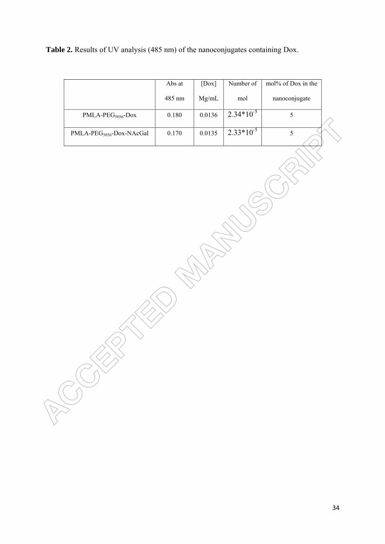
UV at 485 nm: Abs= 0.180, [Dox]= 0.0136mg/mL, Dox: 2.34*10-5 mol (5mol%).
Molar mass of PMLA-PEG5056-Dox= 24,790 g/mol [Molar mass= 68*0.90*116 +
68*0.05*(115+5,056) + 68*0.05*(115+579.98)].
Grafting NAcGal onto PMLA-NHS-PEG5056:
Five mol% of NAcGal (M= 221.21 g/mol, 2.35*10-5 mol) dissolved in 1 mL of anhydrous
DMF were added to one batch of PMLA-NHS (0.1 g, 4.69*10-4 mol) + 5 mol% of PEG5056 +
5 mol% of Dox. The solution was stirred overnight at room temperature in the dark. Then, 2.5
mL of sodium buffer solution of pH5.5 were added to the PMLA-NHS-PEG5056-Dox-NAcGal
to transform the NHS groups into carboxylic acid functions. This solution was stirred at room
temperature for 1 h in the dark and centrifuged at 1,500 g for 10 min. The supernatant was
lyophilized and the resulting polymer was dialyzed against water (MWCO= 3,500 g/mol) for
12 h. The solution contained into the dialysis bag was then lyophilized leading to 95 mg of
PMLA-PEG5056-Dox-NAcGal (yield= 81 %). The polymer obtained was characterized by 1H
NMR and UV at 485 nm (quantification of Dox).
1H NMR (CD3COCD3 + D2O, ppm, 400MHz): 2.86-3.12 (m, 2nH, CHCH2CO2), 3.60 (s,
4*113H, PEG5056, 5 mol%), 5.45-5.58 (m, 1nH, CHCH2CO2).
UV at 485 nm: Abs= 0.179, [Dox]= 0.0135mg/mL, Dox: 2.33*10-5 mol (5mol%).
Molar mass of PMLA-PEG5056-Dox-NAcGal= 25,103 g/mol [Molar mass= 68*0.85*116 +
68*0.05*(115+5,056) + 68*0.05*(115+579.98) + 68*0.05*(115+221.21)].
12
Preparation of the nanoconjugate solutions:
The nanoconjugates obtained were directly dissolved into a phosphate buffer solution (pH
7.5) at a concentration of 1 to 2 mg/mL. The resulting clear solutions were filtered through a
0.2 µm pore membrane and analyzed using a DelsaNano apparatus to determine their average
diameter ( , size dispersity (PDI) and zeta potential (). Results obtained are shown in Table
2 in Results & Discussion section.
The stability of the nanoconjugates was followed by DLS measurements after 24h, 48h and
72h of storage at 4°C. The results obtained are shown in Table 3 in Results & Discussion
section.
In vitro cytotoxic studies:
The prepared nanoconjugates were evaluated for their in vitro cytotoxicity on HepaRG
hepatoma cells using a 3-(4.5-dimethylthiazol-2-yl)-2,5-diphenyltetrazolium bromide (MMT)
assay. During sub-culturing, 2*106 cells were seeded in a 75 cm2 flask. The medium was
renewed every 48 h. The sub-culturing was realized by trypsinization every 2 weeks in order
to maintain the progenitor phenotype. For an optimal differentiation, the cells were
maintained at confluence after the 2 weeks and the medium was supplemented with 2% of
dimethylsulfoxide (DMSO) [25]. For the cytotoxicity assays, 96 well culture plates were
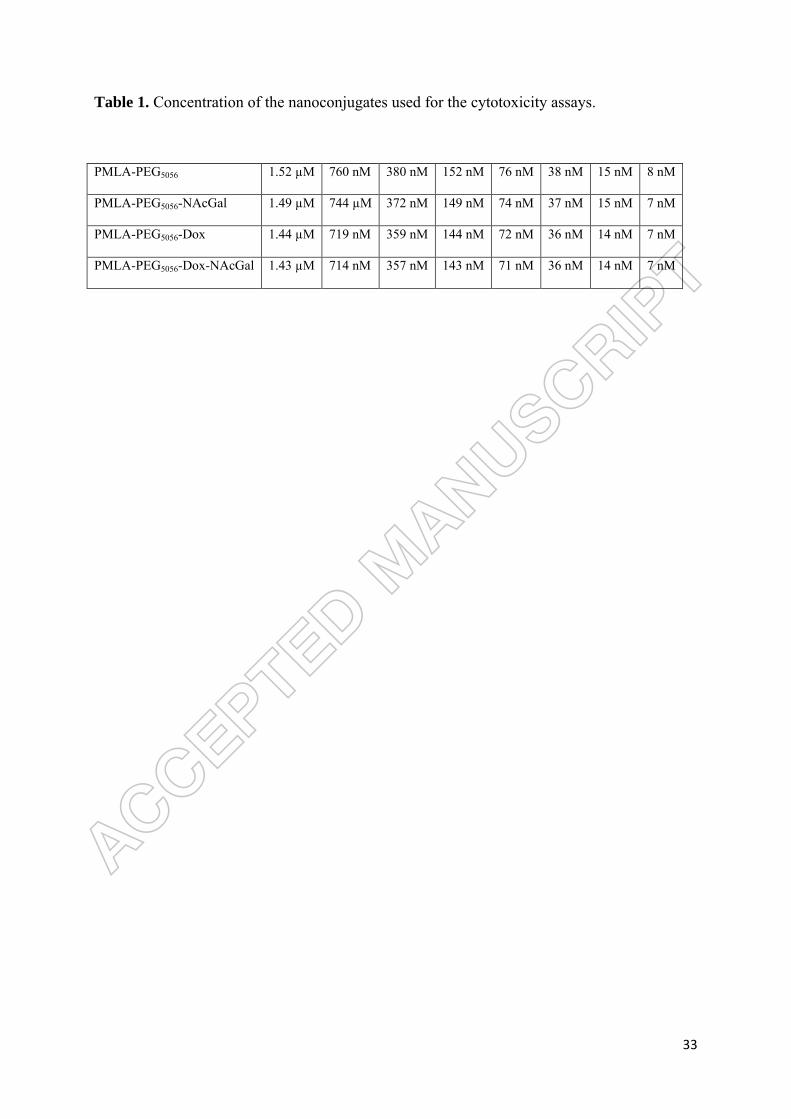
seeded 105 cells per well. The nanoconjugate preparations were then added to the wells at
concentrations ranging from 2 µM to 11 nM as shown in Table 1.The initial solutions of
nanoconjugates at a concentration of 2 mg/mL were prepared by diluting the nanoconjugate
with PBS.
Table 1
The cells were incubated for 24, 48 and 72h. After incubation, 100 µL of MMT solution were
added to each well. The culture plate was incubated for 2 h. The medium was then removed
by vacuum and a few µL of PBS were added to each well. This PBS was then removed by
13
vacuum and 100 µL of DMSO were added to each well to solubilize the formazane crystals.
The absorbance of each well was read by UV at 515 nm.
Results and Discussion
Synthesis and characterization of PMLA derivatives
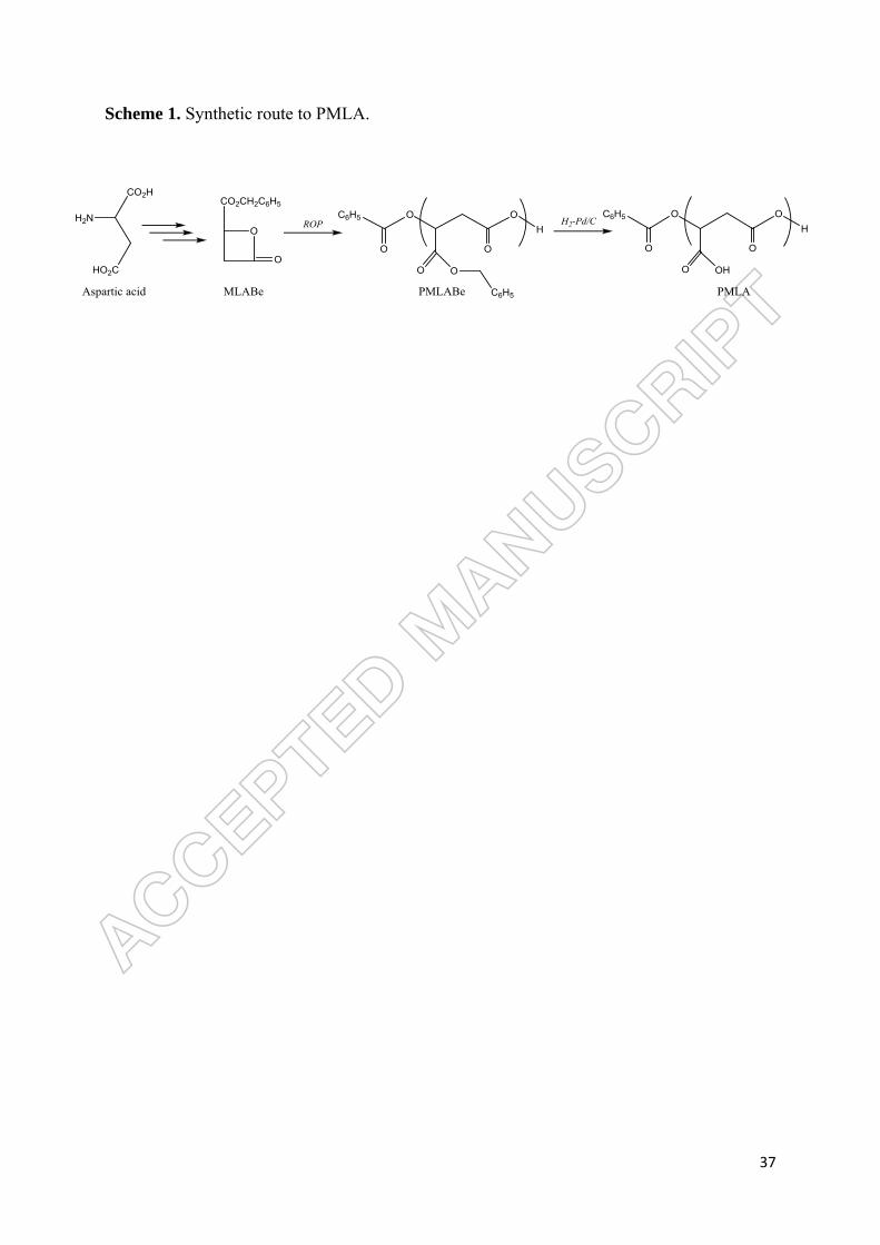
As shown in Scheme 1, the PMLABe was synthesized by anionic ring opening
polymerization of MLABe which was obtained in four steps starting from aspartic acid, as
described previously [21].
Scheme 1
The molar mass of PMLABe ( 5,540 g/mol) measured by SEC in THF was lower than
the theoretical value because the analytical technique involves a calibration curve (to link the
retention time or volume to the molar mass) based on poly(styrene) standards, that have a
chemical structure different from PMLA. Moreover, the solvent (THF) used to realize this
SEC analysis is not the best solvent for PMLABe. On the other hand, it was not possible to
determine the molar mass using the 1H NMR spectrum of the PMLABe because the end
chains, a benzoate group at one end and a carboxylic acid at the other hand, are not visible on
the spectrum. Consequently, we are only able to conclude that the dispersity of the
synthesized PMLABe (Đ = 1.63) is in good agreement with the data given in the literature for
this kind of polymerization [21].
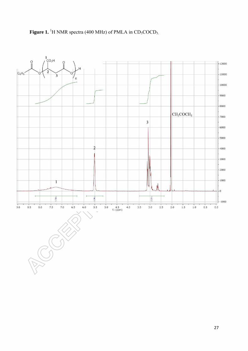
The catalytic hydrogenolysis of benzyl lateral groups of the synthesized PMLABe gives
access to the expected PMLA without any degradation of the ester groups of the polymer
backbone, as shown by the 1H NMR spectrum of the obtained PMLA (Figure 1).
Figure 1
Indeed, such catalytic deprotection of lateral benzyl ester groups is known to be specific and
does not affect the ester groups in the main chain [26]. The number of units of the PMLA is,
14
therefore, the same as the ones of the PMLABe and no degradation of the polymer occurs
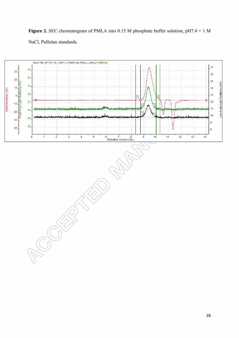
under such conditions. The molar mass of PMLA was measured by SEC in a 0.15 M
phosphate buffer solution (PBS) at pH7.4 in presence of 1 M NaCl with Pullulan as standards
(Figure 2).
Figure 2
As shown by Figure 2, only one peak was observed in the chromatogram at a molar mass of
7,950 g/mol and a quite narrow dispersity. The number of malic acid units was calculated
from the molar mass (7,950 g/mol) and the molar mass of the repeating unit (116 g/mol) as
follow: n= 7,950/116= 68.
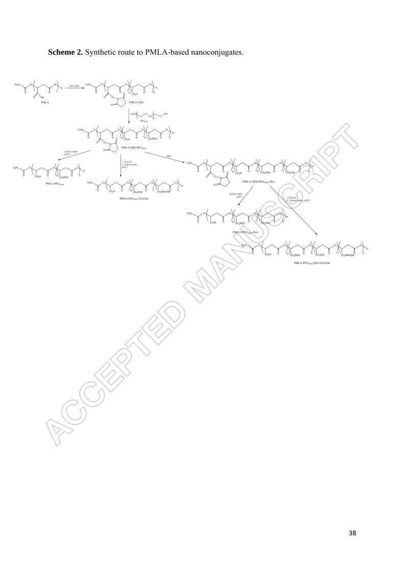
Starting with this PMLA, we have used the lateral carboxylic acid functions to introduce the
molecules of interest we have selected for this study, namely the PEG5056 to confer stealth
properties to the nanoconjugate [15-17], Dox, an anticancer drug, as a drug model [27] and
the NAcGal as the targeting agent for cancer cells [3,13,14] (Scheme 2).
Scheme 2
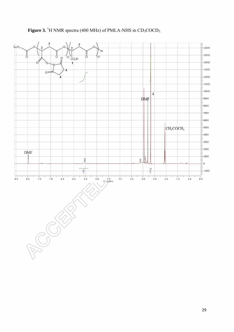
The carboxylic acid lateral functions of the PMLA were first activated by NHS through the
reaction between the carboxylic acids and NHS in the presence of DCC [28]. After
purification, the PMLA-NHS was analyzed by 1H NMR. The NMR spectrum showed that the
chemical modification is successful (Figure 3). However, only 55% of the lateral carboxylic
acid functions are activated under NHS form. Such a result is not surprising because the
chemical modification of lateral groups of polymers is hardly quantitative [29].
Figure 3
However, even if only 55% of the carboxylic acid functions are activated, it is enough to
continue the modification of the PMLA because we have chosen to introduce only 5 mol% of
each selected molecules. This amount was selected based on the results obtained by
Ljubimova et al. with natural PMLA [28+ references cited within]. These authors have
15
highlighted that the modification of the natural PMLA with 5 mol% of each selected molecule
(Dox, Temozoline Hydrazide, anti-sense oligonucleotides, PEG, Alexa Fluor 680, etc.) leads
to the expected results, both in vitro and in vivo. These nanoconjugates, called Polycephin®,
were designed to treat brain and/or breast cancers [30,31]. As shown in Scheme 1, we
introduced first a telechelic PEG with a molar mass of 5,056 g/mol with an amine group at
one end and a methoxy group at the other end. Starting from this PMLA-NHS-PEG5056
polymer, we introduced 5 mol% of NAcGal and/or 5 mol% of Dox (Scheme 2), to the
remaining activated carboxylic acid functions in order to obtain, four PMLA derivatives,
namely PMLA-PEG5056, PMLA-PEG5056-NAcGal, PMLA-PEG5056-Dox and PMLA-PEG5056-
Dox-NAcGal from the same PMLA batch. Each time, the remaining NHS groups were
eliminated under mild conditions by the addition of sodium buffer solution at pH5.5. Such
mild conditions avoid the possible degradation of the ester groups of the PMLA main chain
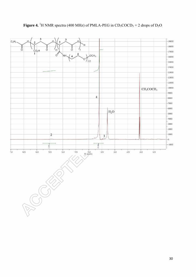
[28]. The synthesized macromolecular pro-drugs were characterized by 1H NMR. The 1H
NMR spectrum of the PMLA-PEG5056 (Figure 4) shows the peak characteristic of the two
methylene (CH2) protons at 3.6 ppm, and only the peak characteristic of the methine (CH)
protons of the PMLA backbone between 5.45-5.58 ppm, the peak characteristic of the
methylene protons of the PMLA backbone being hidden by the peak of water.
Figure 4
Nevertheless, we were able to calculate the percent of PEG linked to the PMLA backbone
from the relative integration of the PMLA’s methine protons and the PEG’s methylene
protons as follow:
%PEG= ([(Integration of CH2 of PEG)/4/113]/[Integration of CH of PMLA])*100
We found that 5 mol% of PEG were effectively linked to the PMLA backbone, as expected.
Unfortunately, the 1H NMR spectra of the three other nanoconjugates do not allowed us to
determine the percent of Dox and/or NAcGal linked to the PMLA backbone because the
16
peaks characteristics of these molecules were hidden either by the peaks of the solvent used
for the NMR (CD3CODC3 + 2 drops of D2O) or by the ones of the PEG. We realized the
proton NMR of our nanoconjugates in other deuterated solvents, D2O and DMSO-d6, but the
resulting spectra showed only the characteristic peak of the methylene protons of the PEG as a
result of the specific conformation adopted by the nanoconjugates in these solvents. Indeed,
water and DMSO promote the aggregation of the nanoconjugates and only the more
hydrophilic parts of the nanoconjugates are visible. However, because the binding of the PEG
chains to the PMLA backbone was shown to be as expected, we can reasonably assume that
the binding of Dox and/or NAcGal, which are small molecules, is also successful.
Consequently, in view of the purification steps done on our nanoconjugates (dialysis allowing
to eliminate all the unbounded small molecules) and results obtained for PMLA modification
with PEG, we considered that 5 mol% of Dox and/or NAcGal were effectively linked to the
PMLA backbone. To confirm that 5 mol% of Dox were effectively linked to the PMLA
backbone, we analyzed the nanoconjugates containing Dox by UV at 485 nm.
Table 2
Indeed, such analysis allows to link the absorbance of the solution containing Dox to its
concentration using a calibration curve (Figure S1). As shown by the results gathered in Table
2, the amount contained in the PMLA-PEG5056-Dox and PMLA-PEG5056-Dox-NAcGal
nanoconjugates were 2.34*10-5 mol and 2.33*10-5 mol, respectively, corresponding to around
5 mol% of Dox in each nanoconjugate.
Nevertheless, we are still working on the characterization of the PMLA-PEG5056-NAcGal,
PMLA-PEG5056-Dox and PMLA-PEG5056-Dox-NAcGal nanoconjugates (structure, percent of
molecules linked to the PMLA backbone). Therefore, we are currently looking for a solvent to
realize the proton NMR in which all the parts of the nanoconjugates can be visible and thus
analyzed. Moreover, we are also investigating the possibilities to use MALDI and ESI to
17
characterize our nanoconjugates with a major difficulty coming from the presence of the
carboxylic acid lateral function which can prevent the correct ionization of the
nanoconjugates together with the multiple ways for the nanoconjugates to be degraded under
the conditions used for MALDI and ESI measurements. Finally, HPLC is an analytical
method which we also intend to use to determine the amounts of Dox and NAcGal present in
our compounds. The results obtained thanks to these three analytical methods will have to be
compared in order to confirm: i). that the Dox and/or NAcGal were effectively linked to the
PMLA backbone and ii). the amount of Dox and/or NAcGal present in our nanoconjugates.
Preparation of the nanoconjugates
The four prodrugs synthesized were then solubilized in a phosphate buffer solution at pH7.5
containing 0.15N NaCl at a concentration of 1 to 2 mg/mL. The resulting clear solutions were
analyzed by dynamic light scattering (DLS) to obtain the average diameter and dispersity of
the nanoconjugates, in addition to zeta potential measurements to obtain the charge on the
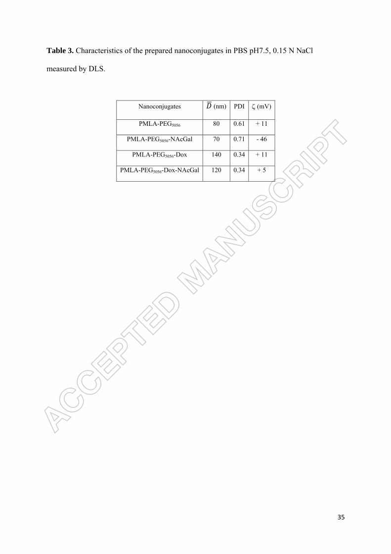
nanoconjugates. As shown in Table 3, we were able to obtain aggregates from all the
macromolecular prodrugs with apparent hydrodynamic diameter ranging from 70 to 140 nm.
Table 3
The dispersity of the nanoconjugates obtained, however, are quite high, ranging from 0.34 to
0.71, denoting that they form aggregates rather than well-defined nano-objects. Such large
dispersity values are in good agreement with the nature of the polymers used in this study.
Indeed, the synthesized macromolecular prodrugs are made up of macromolecular chains with
different side groups with various hydrophilicity/hydrophobicity properties and distributed
randomly throughout the chain. Such structure can only lead to the formation of loose
aggregates in aqueous media, unlike amphiphilic block copolymers known to self-aggregate
in aqueous media leading to the formation of nanoparticles [32].
18
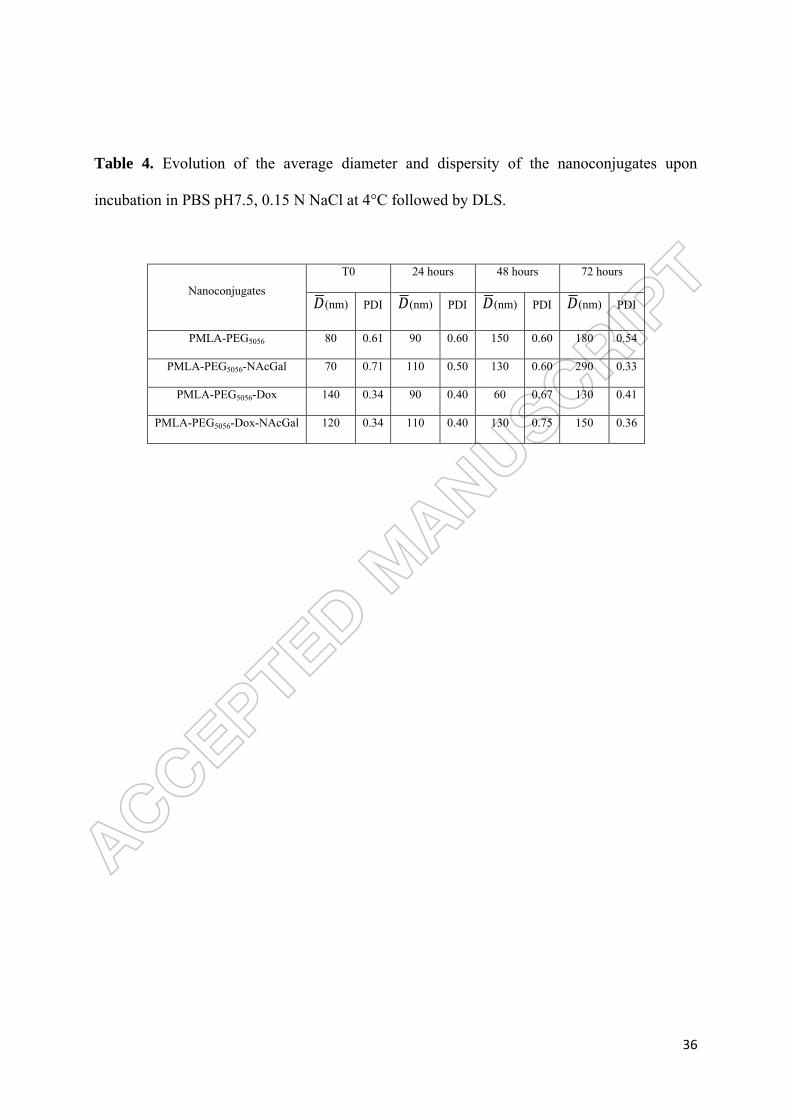
The behavior of the nanoconjugates was obtained under storage at 4°C in PBS pH7.5, 0.15 N
NaCl by measuring the average diameter and dispersity by DLS. Results obtained (Table 4)
reveal a significant increase in the average diameter for two nanoconjugates, namely the
PMLA-PEG5056 and the PMLA-PEG5056-NAcGal, from 70/80 nm to 180/290 nm after 72 h of
incubation at 4°C together with a stability of the dispersity at around 0.60. Such observation
might be due to the formation of very loose aggregates that undergo severe changes upon
incubation such as the fusion of aggregates. Further studies are, however, required using other
techniques such as the Asymmetrical Field-Flow Fractionation (AFFF), which allows not only
to characterize nanoparticles [33] but also the behavior of macromolecules in solution [34].
For the nanoconjugate PMLA-PEG5056-Dox, a significant decrease in the average diameter,
from 140 to 60 nm, was first observed after 48 h of incubation at 4°C together with an
increase in the dispersity (from 0.34 to 0.67), probably showing the formation of
nanoparticles-like structures as a result of the presence of hydrophobic Dox molecules.
Table 4
After 72 h of incubation at 4°C, however, the average diameter reached the initial value,
showing possible degradation of the aggregates as a result of too weak hydrophobic
interactions within the nanoconjugate in comparison to that observed for nanoparticles. In this
case also, the AFFF techniques might bring more information on the behavior of this
nanoconjugate in aqueous solutions. Finally, the nanoconjugate PMLA-PEG5056-Dox-NAcGal
seems to be quite stable in aqueous media over 72 h of incubation at 4°C, probably as a result
of the presence of both the Dox and NAcGal increasing the forces driving the formation of
aggregates.
We are currently studying the Dox release from the PMLA-PEG5056-Dox and PMLA-PEG5056-
Dox-NAcGal nanoconjugates under different conditions of pH and temperature.
19
In vitro cytotoxicity assays
As the nanoconjugates were synthesized to be used as site-specific drug carriers to treat
hepatocellular carcinoma (HCC), we first evaluated their in vitro cytotoxicity on the human
HepaRG hepatoma cell line [23-25]. The nanoconjugates were incubated at concentrations
ranging from 7 nM to 1.52 µM with HepaRG cells and the cell viability was measured using
the MMT assay after 1, 3 and 7 days of incubation (Figure 5 & Figure 6). The red line, plotted
on each graph, is the half maximal inhibitory concentration (IC50), a concentration at which
50% of the cells are dead, and allow to determine the cytotoxicity of the material studied. No
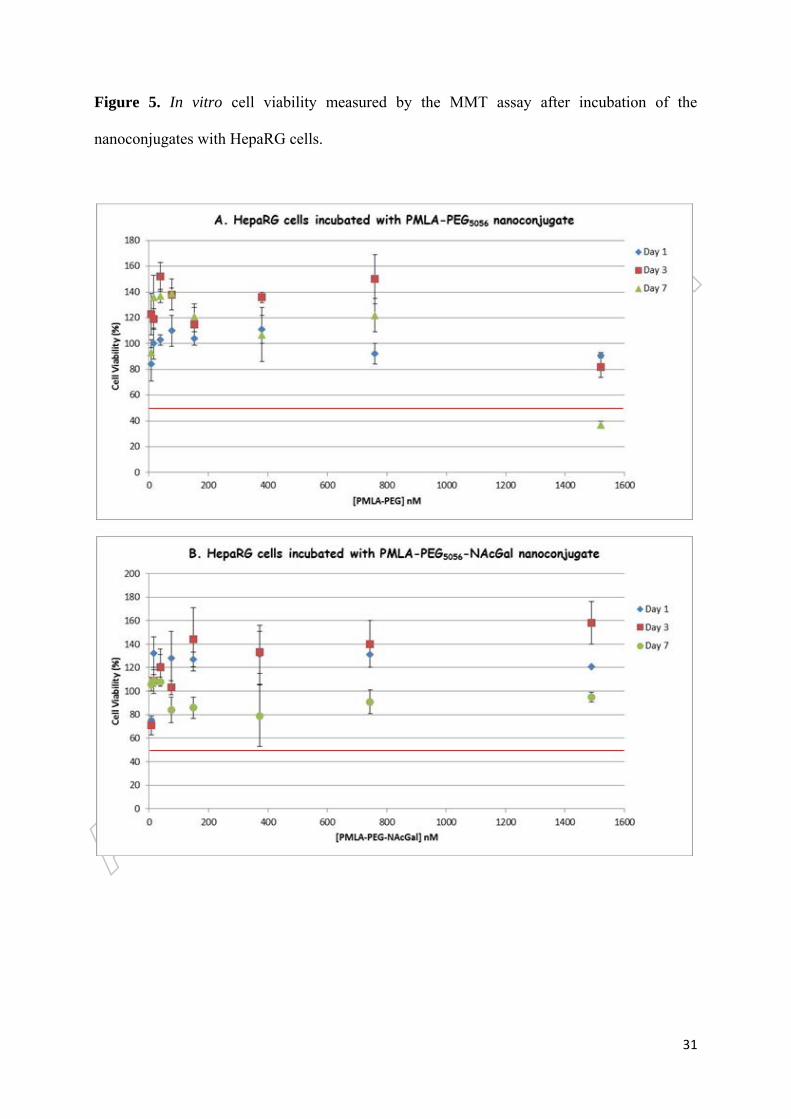
cytotoxicity effect was observed for the PMLA-PEG5056-NAcGal (Figure 5 B), at the different
time points of incubation even at a very high concentration as the cell viability stayed above
80%. We can conclude, therefore, that this compound does not have an acute toxicity on the
HepaRG cell line.
Figure 5
The nanoconjugate, PMLA-PEG5056 does not show in vitro cytotoxicity up to a concentration
of 1.2 µM (Figure 5 A). Surprisingly, the cell viability decreases to about 80% for a
concentration of 1.52 µM after 1 and 3 days of incubation and about 40% for a concentration
of 1.52 µM after 7 days of incubation. An IC50 of 1.25 µM was obtained for the PMLA-
PEG5056 nanoconjugate after 7 days of incubation. Since the PMLA is biocompatible even at
high concentrations [35], this cytotoxicity might come from the presence of PEG chains.
Indeed, in a recent study on the comparison of the in vitro cytotoxicity and hemocompatibility
of PEG and poly(2-ethyl-2-oxazoline), Fischer et al. have reported that PEGs with molar
masses ranging from 400 to 1,000 g/mol show an effect on cell viability after 12 and 24 h of
incubation. This effect was shown to be depending on PEG concentration and the molar mass
[23]. It is, therefore, not surprising that we also observe a cytotoxicity effect of the PMLA-
PEG5056 nanoconjugate at a higher concentration and longer incubation time.
20
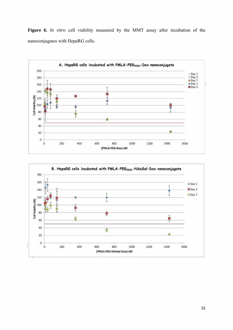
The PMLA-PEG5056-Dox nanoconjugate showed a cell viability decreasing in a dose
dependent manner (Figure 6 A) after 7 days of incubation, while the cell viability stays stable
regardless of the concentration after 1 or 3 days of incubation. The IC50 for this PMLA-
PEG5056-Dox nanoconjugate is 936 nM corresponding to a concentration in Dox of 47 nM.
Figure 6
Interestingly, the nanoconjugate of PMLA-PEG5056-Dox-NAcGal induces a significant
decrease in the cell viability after 3 and 7 days of incubation in a concentration dependent
manner (Figure 6 B). The cell viability decreases to around 60% after 3 days of incubation for
a concentration of 1.43 µM. After 7 days of incubation, an IC50 of 527 nM was obtained for
the nanoconjugate, PMLA-PEG5056-Dox-NAcGal corresponding to a concentration of 26 nM
in Dox.
The IC50 obtained for free Dox is around 94 nM after 3 days of incubation and below 20 nM
after 7 days of incubation while almost no cytotoxic effects were observed after 1 day of
incubation for the free Dox with HepaRG cells (data not shown).
The results reveal that, the presence of NAcGal on the nanoconjugate, PMLA-PEG5056-Dox-
NAcGal improves the effect of Dox since the IC50 of this nanoconjugate (IC50= 26 nM) is
lower than the one of the corresponding nanoconjugate without the NAcGal moieties (IC50=
47 nM). NAcGal groups seem therefore to improve the cellular uptake of the nanoconjugates
thus leading to a more efficient of cytotoxicity this Dox conjugate. Nevertheless, such
conclusion has to be proven by further experiments demonstrating the mechanism of the
nanoconjugate uptake by HepaRG cells and whether the NAcGal really plays a role in the
nanoconjugate internalization.
21
Conclusion
As hypothesized, the present work clearly demonstrates that PMLA is a suitable carrier for
targeting anticancer drugs to cancer cells. Moreover, the synthesized PMLA-PEG5056-Dox-
NAcGal pro-drug nanoconjugate has a higher cellular toxicity when compared to the PMLA-
PEG5056-Dox nanoconjugate without the targeting moiety. The targeting moiety NAcGal may
enhance the cellular uptake of the nanoconjugate PMLA-PEG5056-Dox-NAcGal, thus
improving the cytotoxic effect of the bound Dox in vitro against the HepaRG hepatoma cell
line. Further works, using confocal fluorescence microscopy and flow cytometry, are
presently in progress to prove the target specificity and the cellular uptake of the PMLA based
nanoconjugates substituted by the NAcGal targeting molecule.
Acknowledgement
This work was supported by Inserm and CNRS.
N.V. thanks the Women Scientists Scheme, DST, New Delhi for funding this work.

22
References
[1] Lin S, Hoffmann K, Schemmer P. Treatment of Hepatocellular Carcinoma: A
systematic Review. Liver Cancer. 2012; 1: 144-158.
[2] Jernal A, Bray F, Center MM et al. Global cancer statistics. CA Cancer J. Clin.
2011; 61(2): 69-90.
[3] Zhang X, Helena Ng HL, Lu A et al. Drug delivery system targeting advanced
hepatocellular carcinoma: Current and future. Nanomed.: Nanotechnol., Biol., Med. 2016; 12:
853-869.
[4] Jin SE, Jin HE, Hong SS. Targeted delivery system of nanobiomaterials in
anticancer therapy: from cells to clinics. BioMed Res. Int. Vol. 2014; Article ID 814208, 23
pages. http://dx.doi.org/10.1155/2014/8114208.
[5] Ringsdorf H. Structure and properties of pharmacologically active polymers. J.
Polym. Sci. 1975; 51: 135-153.
[6] Mastumura Y, Maeda H. A new concept for macromolecular therapeutics in cancer
chemotherapy: mechanism of tumoritropic accumulation of proteins and antitumor agent
smancs. Cancer Research. 1986; 46(12): 6387-6392.
[7] Fang J, Nakamura H, Maeda H. The EPR effect: unique features of tumor blood
vessels for drug delivery, factors involved, and limitations and augmentation effect. Adv.
Drug Del. Rev. 2011; 63(3): 136-151.
[8] Wang L, Tian B, Zhang J et al. Coordinated pH/redox dual-sensitive and
hepatoma-targeted multifunctional polymeric micelle system for stimuli-triggered
doxorubicin release: synthesis, characterization and in vitro evaluation. Int. J. Pharm. 2016;
501: 221-235.
23
[9] Craparo EF, Licciardi M, Connigliaro A et al. Hepatocyte-targeted fluorescent
nanoparticles based on polyaspartamide for potential theranostic applications. Polymer. 2015;
70: 257-270.
[10] Sarika PR, James NR, Nishna N et al. Galactosylated pullulan-curcumin
conjugate micelles for site-specific anticancer activity to hepatocarcinoma cells. Colloids and
Surfaces B: Biointerfaces; 2015; 133: 347-355.
[11] Qi X, Rui Y, Fan Y et al. Galactosylated chitosan-grafted multiwall carbon
nanotubes for pH-dependent sustained release and hepatic tumor-targeted delivery of
doxorubicin in vivo. Colloids and Surfaces B: Biointerfaces. 2015; 133: 314-322.
[12] Ishiwara T, Kaneko K, Ishihara T et al. Development of biodegradable
nanoparticles for liver-specific ribavirin delivery. J. Pharm. Sci. 2014; 103(2): 4005-4011.
[13] Weigel PH, Oka JA. Endocytosis and degradation mediated by the
asialoglycoprotein receptor in isolated rat hepatocytes. J. Biol. Chem. 1982; 257: 1201-1207.
[14] Medina SH, Tekumalla V, Chevliakov MV et al. N-acetylgalactosamine-
functionalized dendrimers as hepatic cancer cell-targeted carriers. Biomaterials. 2011; 32(17):
4118-4129.
[15] Dunn SE, Brindley A, Davis SS et al. Polystyrene-poly(ethylene glycol) (PS-
PEG2000) particles as model systems for site-specific drug delivery. 2. The effect of PEG
surface density on the in vitro cell interaction and in vivo biodistribution. Pharm. Res. 1994;
11: 1016-1022.
[16] Gref R, Minamitake Y, Peracchia MT et al. Biodegradable long-circulating
polymer nanospheres. Science. 1994; 263: 1600-1603.
[17] Klibanov AL, Murayama M, Torchilin VP et al. Amphipatic poly(ethylene
glycols) effectively prolong the circulation time of liposomes. FEBS Lett. 1990; 268: 235-
237.
24
[18] Park JH, Lee S, Kim JH et al. Polymeric nanomedicine for cancer therapy. Prog.
Polym. Sci. 2008; 33: 113-137.
[19] Neffe TA, Grijpma DW, Lendlein A. Advanced Functional Polymers for
Medicine. Macromol. Biosci. 2016; 16(12): 1743-1744.
[20] Holler E. Poly(malic acid) from Natural Sources. In Handbook of Engineering
Polymeric Materials: Marcel Dekker: New York, NY, USA. 1997; 93-103.
[21] Cammas S, Renard I, Langlois V et al. Poly(-malic acid): obtaining of high
molecular weights by improvement of the synthesis route. Polymer. 1996; 37(18): 4215-4220.
[22] Cammas-Marion S, Loyer P. Natural and synthetic poly(malic acid)-based
derivates: A family of versatile biopolymers for the design of drug nanocarriers. J. Drug Targ.
2014; 22(7): 556-575.
[23] Gripon P, Rumin S, Urban S et al. Infection of a human hepatoma cell line by
hepatitis B virus. Proc. Natl. Acad. Sci. U.S.A. 2002; 99: 15655-15660.
[24] Laurent V, Fraix A, Montier T et al. Highly efficient gene transfer into
hepatocyte-like cells: new means for drug metabolism and toxicity studies. Biotechnol. J.
2010; 5: 314-320.
[25] Laurent V, Glaise D, Nübel T et al. Highly efficient SiRNA and gene transfer into
hepatocyte-like HepaRG cells and primary human hepatocytes. Methods Mol. Biol. 2013;
987: 295-314.
[26] Vert M, Lenz RW. Preparation and properties of poly--malic acid: a functional
polyester of potential biomedical importance. ACS Polym. Preprint. 1979; 20(1): 608-611.
[27] Lown WJ et al. Anthracycline and anthraquinone anticancer agents: Current
status and recentdevelopments. Pharmacol.Ther.1993; 60: 185-214.
25
[28] Patil R, Portilla-Arias J, Ding H et al. Cellular delivery of doxorubicin via pH-
controlled hydrazine linkage using multifunctional nano vehicle based on poly(-L-malic
acid). Int. J. Mol. Sci. 2012; 13: 11681-11693.
[29] Soutif JC, Brosse JC. Chemical modification of polymers. III. Kinetic aspects.
Reactive. Polymers. 1990; 13: 1-26.
[30] Ding H, Inoue S, Ljubimov AV et al. Inhibition of brain tumor growth by
intravenous poly(-L-malic) acid nanobioconjugate with pH-dependent drug release. Proc.
Nat. Acad. Sci. USA (PNAS). 2010; 107: 18143-18148.
[31] Patil R, Ljubimov AV, Gangalum PR et al. MRI Virtual Biopsy and Treatment of
Brain Metastatic Tumors with Targeted Nanobioconjugates. Nanoclinic in the Brain. ACS
Nano. 2015; Apr 23, [Epub ahead of print] PMID: 25906400.
[32] Letchford K, Burt H. A review of the formation and classification of amphiphilic
block copolymer nanoparticulate structures: micelles, nanospheres, nanocapsules and
polymersomes. Eur. J. Pharm. Biopharm. 2007; 65: 259-269.
[33] Kang DY, Kim MJ, Kim ST et al. Size characterization of drug-loaded polymeric
core/shell nanoparticles using asymmetrical flow field-flow fractionation. Anal. Bioanal.
Chem. 2008; 390: 2183-2188. Doi: 10.1007/s00216-008-1984-1.
[34] Bourgoin A, Zablackis E, Poli JB. Characterization of -carrageenan solution
behavior by field-flow fractionation and multi angle light scattering. Food Hydrocolloids.
2008; 22: 1607-1611.
[35] Bauer M, Lautenschlaeger C, Kempe K et al. Poly(2-ethyl-2-oxazoline) as
Alternative for the Stealth Polymer Poly(ethylene glycol): Comparison of in vitro
Cytotoxicity and Hemocompatibility. Macromol. Biosci. 2012; 12: 986-998. Doi:
10.1002/mabi.201200017.

26
Figures
Figure 1. 1H NMR spectra (400 MHz) of PMLA in CD3COCD3.
Figure 2. 1H NMR spectra (400 MHz) of PMLA-NHS in CD3COCD3.
Figure 3. 1H NMR spectra (400 MHz) of PMLA-PEG5056 in CD3COCD3 + 2 drops of D2O.
Figure 4. SEC chromatogram of PMLA into 0.15 M phosphate buffer solution, pH7.4 + 1 M
NaCl, Pullulan standards.
Figure 5. In vitro cell viability measured by the MMT assay after incubation with HepaRG
cells of PMLA-PEG5056 (A) and PMLA-PEG5056-NAcGal (B).
Figure 6. In vitro cell viability measured by the MMT assay after incubation with HepaRG
cells of PMLA-PEG5056-Dox (A) and PMLA-PEG5056-Dox-NAcGal (B).
Tables
Table 1. Concentration of the nanoconjugates used for the cytotoxicity assays.
Table 2. Results of UV analysis (485 nm) of the nanoconjugates containing Dox.
Table 3. Characteristics of the prepared nanoconjugates in PBS pH7.5, 0.15 N NaCl measured
by DLS.
Table 4. Evolution of the average diameter and dispersity of the nanoconjugates upon
incubation in PBS pH7.5, 0.15 N NaCl at 4°C followed by DLS.
Schemes
Scheme 1. Synthetic route to PMLA.
Scheme 2. Synthetic route to PMLA-based nanoconjugates.
27
Figure 1. 1H NMR spectra (400 MHz) of PMLA in CD3COCD3.
28
Figure 2. SEC chromatogram of PMLA into 0.15 M phosphate buffer solution, pH7.4 + 1 M
NaCl, Pullulan standards.
29
Figure 3. 1H NMR spectra (400 MHz) of PMLA-NHS in CD3COCD3.
30
Figure 4. 1H NMR spectra (400 MHz) of PMLA-PEG in CD3COCD3 + 2 drops of D2O.
31
Figure 5. In vitro cell viability measured by the MMT assay after incubation of the
nanoconjugates with HepaRG cells.
32
Figure 6. In vitro cell viability measured by the MMT assay after incubation of the
nanoconjugates with HepaRG cells.
33
Table 1. Concentration of the nanoconjugates used for the cytotoxicity assays.
PMLA-PEG5056 1.52 µM 760 nM 380 nM 152 nM 76 nM 38 nM 15 nM 8 nM
PMLA-PEG5056-NAcGal 1.49 µM 744 µM 372 nM 149 nM 74 nM 37 nM 15 nM 7 nM
PMLA-PEG5056-Dox 1.44 µM 719 nM 359 nM 144 nM 72 nM 36 nM 14 nM 7 nM
PMLA-PEG5056-Dox-NAcGal 1.43 µM 714 nM 357 nM 143 nM 71 nM 36 nM 14 nM 7 nM
34
Table 2. Results of UV analysis (485 nm) of the nanoconjugates containing Dox.
Abs at
485 nm
[Dox]
Mg/mL
Number of
mol
mol% of Dox in the
nanoconjugate
PMLA-PEG5056-Dox 0.180 0.0136 2.34*10-5 5
PMLA-PEG5056-Dox-NAcGal 0.170 0.0135 2.33*10-5 5
35
Table 3. Characteristics of the prepared nanoconjugates in PBS pH7.5, 0.15 N NaCl
measured by DLS.
Nanoconjugates (nm) PDI (mV)
PMLA-PEG5056 80 0.61 + 11
PMLA-PEG5056-NAcGal 70 0.71 - 46
PMLA-PEG5056-Dox 140 0.34 + 11
PMLA-PEG5056-Dox-NAcGal 120 0.34 + 5
36
Table 4. Evolution of the average diameter and dispersity of the nanoconjugates upon
incubation in PBS pH7.5, 0.15 N NaCl at 4°C followed by DLS.
Nanoconjugates T0 24 hours 48 hours 72 hours
(nm) PDI (nm) PDI (nm) PDI (nm) PDI
PMLA-PEG5056 80 0.61 90 0.60 150 0.60 180 0.54
PMLA-PEG5056-NAcGal 70 0.71 110 0.50 130 0.60 290 0.33
PMLA-PEG5056-Dox 140 0.34 90 0.40 60 0.67 130 0.41
PMLA-PEG5056-Dox-NAcGal 120 0.34 110 0.40 130 0.75 150 0.36
37
Scheme 1. Synthetic route to PMLA.
38
Scheme 2. Synthetic route to PMLA-based nanoconjugates.
39
Supplementary Materials
Poly(malic acid) bearing Doxorubicin and N-Acetyl Galactosamine as a
site-specific prodrugs for HepatoCellular Carcinoma Nivishna Venkatraj1,3, M.J. Nanjan1, Pascal Loyer2, M.J.N. Chandrasekar1,* Sandrine Cammas Marion2,3,*. 1 JSS College of Pharmacy, Department of Pharmaceutical Chemistry, Rocklands,
Ootacamund, Tamil Nadu, India (Constituent of JSS University, Mysore, India). 2 INSERM, INRA, University of Rennes 1, University of Bretagne Loire, Nutrition
Metabolisms and Cancer (NuMeCan), Rennes, France. 3 Ecole Nationale Supérieure de Chimie de Rennes (ENSCR), Team Corint, ISCR, University
of Rennes 1, Rennes, France.
* Corresponding authors: [email protected]; [email protected]
Figure S1. Calibration curve of Dox solubilized in PBS pH7 giving the Dox concentration as a function of absorbance of the Dox solution at 485 nm.