Embed Size (px)
Citation preview
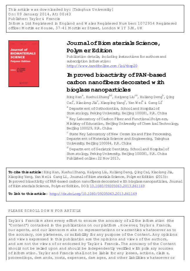
This article was downloaded by: [Tsinghua University]On: 09 January 2014, At: 00:43Publisher: Taylor & FrancisInforma Ltd Registered in England and Wales Registered Number: 1072954 Registeredoffice: Mortimer House, 37-41 Mortimer Street, London W1T 3JH, UK
Journal of Biomaterials Science,Polymer EditionPublication details, including instructions for authors andsubscription information:http://www.tandfonline.com/loi/tbsp20
Improved bioactivity of PAN-basedcarbon nanofibers decorated withbioglass nanoparticlesBing Hana, Xuehui Zhangbd, Haiyang Liucd, Xuliang Dengb, QingCaic, Xiaolong Jiac, Xiaoping Yangc, Yan Weib & Gang Lica Department of Orthodontics, School and Hospital ofStomatology, Peking University, Beijing 100081, P.R. Chinab Key Laboratory of Carbon Fiber and Functional Polymers,Ministry of Education, Beijing University of Chemical Technology,Beijing 100029, P.R. Chinac State Key Laboratory of New Ceramics and Fine Processing,Department of Materials Science and Engineering, TsinghuaUniversity, Beijing 100084, P.R. Chinad Department of Geriatric Dentistry, School and Hospital ofStomatology, Peking University, Beijing 100081, P.R. ChinaPublished online: 22 Nov 2013.
To cite this article: Bing Han, Xuehui Zhang, Haiyang Liu, Xuliang Deng, Qing Cai, Xiaolong Jia,Xiaoping Yang, Yan Wei & Gang Li , Journal of Biomaterials Science, Polymer Edition (2013):Improved bioactivity of PAN-based carbon nanofibers decorated with bioglass nanoparticles, Journalof Biomaterials Science, Polymer Edition, DOI: 10.1080/09205063.2013.861169
To link to this article: http://dx.doi.org/10.1080/09205063.2013.861169
PLEASE SCROLL DOWN FOR ARTICLE
Taylor & Francis makes every effort to ensure the accuracy of all the information (the“Content”) contained in the publications on our platform. However, Taylor & Francis,our agents, and our licensors make no representations or warranties whatsoever as tothe accuracy, completeness, or suitability for any purpose of the Content. Any opinionsand views expressed in this publication are the opinions and views of the authors,and are not the views of or endorsed by Taylor & Francis. The accuracy of the Contentshould not be relied upon and should be independently verified with primary sourcesof information. Taylor and Francis shall not be liable for any losses, actions, claims,proceedings, demands, costs, expenses, damages, and other liabilities whatsoever or

howsoever caused arising directly or indirectly in connection with, in relation to or arisingout of the use of the Content.
This article may be used for research, teaching, and private study purposes. Anysubstantial or systematic reproduction, redistribution, reselling, loan, sub-licensing,systematic supply, or distribution in any form to anyone is expressly forbidden. Terms &Conditions of access and use can be found at http://www.tandfonline.com/page/terms-and-conditions
Dow
nloa
ded
by [
Tsi
nghu
a U
nive
rsity
] at
00:
44 0
9 Ja
nuar
y 20
14

Improved bioactivity of PAN-based carbon nanofibers decoratedwith bioglass nanoparticles
Bing Hana#
, Xuehui Zhangb,d#
, Haiyang Liuc,d, Xuliang Dengb, Qing Caic, Xiaolong Jiac,Xiaoping Yangc, Yan Weib* and Gang Lic*
aDepartment of Orthodontics, School and Hospital of Stomatology, Peking University, Beijing100081, P.R. China; bKey Laboratory of Carbon Fiber and Functional Polymers, Ministry ofEducation, Beijing University of Chemical Technology, Beijing 100029, P.R. China; cState Key
Laboratory of New Ceramics and Fine Processing, Department of Materials Science andEngineering, Tsinghua University, Beijing 100084, P.R. China; dDepartment of Geriatric
Dentistry, School and Hospital of Stomatology, Peking University, Beijing 100081, P.R. China
(Received 24 August 2013; accepted 25 October 2013)
Composite nanofibers composed of polyacrylonitrile (PAN)-based carbon nanofibersand bioactive glass (BG) nanoparticles have been prepared by electrospinning andin situ sintering. Morphology observation showed that the BG nanoparticles of size20–50 nm were uniformly distributed on the surface of composite nanofibers with350 nm average diameter after carbonization. Biological mineralization indicated theformation of apatite-like layer on the surface of composite nanofibers, in which thecomposition of carbonate hydroxyapatite was proved by FTIR and XRD analysis.Cell growth dynamics according to cellular morphology, CCK-8 assay, and alkalinephosphatase activity assay exhibited better cell adhesion, proliferation, andosteogenic induction of bone marrow-derived mesenchymal stem cells cultured onthe composite nanofibers, which suggested the higher bioactivity of compositenanofibers compared to pure PAN-based carbon nanofibers.
Keywords: carbon nanofiber; bioglass; eletrospinning
Introduction
Polyacrylonitrile (PAN)-based carbon fibers (CFs) have long been considered for theapplication of tissue engineering due to exceptional mechanical properties, such asflexural and fatigue strength, high strength-to-weight ratio, etc.[1–3] Compared to thecommercial CFs, PAN-based carbon nanofibers (CNFs) were more favorable tofabricate biomedical materials for their intrinsic advantages such as larger surface,higher aspect ratio, and more prominent surface effect.[4–7] Furthermore, thesmaller-scale CNFs (diameter 100 nm or less) have been considered to promote theadhesion of osteoblasts and simultaneously impair the adhesion of fibroblast,[6] whichwas regarded as potential biomaterials in bone regeneration. However, as bio-inertmaterials, CNFs did not contain functional groups that could attract calcium cationsand phosphate anions to initiate the crystallization of carbonate hydroxyapatite (CHA).[8–10] Therefore, it was highly desirable to decorate CNFs with bioactive materials toexpand and optimize the application of CNFs in bio-technological fields.
*Corresponding authors. Email: [email protected] (Y. Wei); [email protected] (G. Li)#These authors contributed equally to this work.
© 2013 Taylor & Francis
Journal of Biomaterials Science, Polymer Edition, 2013http://dx.doi.org/10.1080/09205063.2013.861169
Dow
nloa
ded
by [
Tsi
nghu
a U
nive
rsity
] at
00:
44 0
9 Ja
nuar
y 20
14
Bioactive glass (BG) with excellent bioactivity and osteointegration ability has beenwidely used in orthopaedic applications. A certain composition of silicate glass hasbeen reported to chemically bond with living bone, and the mechanism of which wasattributed to the formation of an apatite layer on its surface.[11–13] BG also showedexcellent bioactive behavior in physiological fluids to release ions favoring apatiteformation.[14] Meanwhile, BG has been demonstrated to be capable of creating a bondwith soft tissue.[15] Taken together, BG may be a promising bioactive modifier toimprove the intrinsic physiological drawback of CNFs.
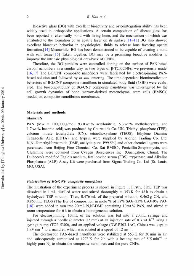
Therefore, the BG particles were controlled doping on the surface of PAN-basedcarbon nanofibers in a similar way as two types of β-TCP/CNFs, we previously made.[16,17] The BG/CNF composite nanofibers were fabricated by electrospinning PAN-based solution and followed by in situ sintering. The time-dependent biomineralizationbehaviors of BG/CNF composite nanofibers in simulated body fluid (5SBF) were evalu-ated. The biocompatibility of BG/CNF composite nanofibers was investigated by thecell growth dynamics of bone marrow-derived mesenchymal stem cells (BMSCs)seeded on composite nanofibrous membranes.
Materials and methods
Materials
PAN (Mw = 100,000 g/mol, 93.0 wt.% acrylonitrile, 5.3 wt.% methylacrylate, and1.7 wt.% itaconic acid) was produced by Courtaulds Co. UK. Triethyl phosphate (TEP),calcium nitrate tetrahydrate (CN), tetraethoxysilane (TEOS), Ethylene DiamineTetraacetic Acid (EDTA), and trypsin were supplied by Aldrich Trading Co. Ltd.N,N′-Dimethylformamide (DMF, analytic pure, P99.5%) and other chemical agents werepurchased from Beijing Fine Chemical Co. Rat BMSCs, Penicillin-Streptomycin, andGlutamine were obtained from Cyagen Biosciences Inc. (Guangzhou, China). TheDulbecco’s modified Eagle’s medium, fetal bovine serum (FBS), trypsinase, and AlkalinePhosphatase (ALP) Assay Kit were purchased from Sigma Trading Co. Ltd (St. Louis,MO, USA).
Fabrication of BG/CNF composite nanofibers
The illustration of the experiment process is shown in Figure 1. Firstly, 3 mL TEP wasdissolved in 1 mL distilled water and stirred thoroughly at 353 K for 48 h to obtain ahydrolyzed TEP solution. Then, 0.476 mL of the prepared solution, 0.462 g CN, and0.865 mL TEOS (The BG of composition in mole % of 58% SiO2–33% CaO–9% P2O5
[18]) were added in turn into 20 mL N,N′-DMF containing 10 wt.% PAN, and stirred atroom temperature for 6 h to obtain a homogeneous solution.
For electrospinning, 10 mL of the solution was fed into a 20 mL syringe andinjected through a needle (diameter 0.5 mm) at an injection rate of 0.3 mL h−1 using asyringe pump (TOP 5300), and an applied voltage (DW-P303-1AC, China) was kept at1 kV cm−1 to a mandrel, which was rotated at a speed of 12 ms−1.
The electrospun PAN-based nanofibers were stabilized at 553 K for 30 min in air,and subsequently carbonized at 1273 K for 2 h with a heating rate of 5 K min−1 inhighly pure N2 to obtain the composite nanofibers and the pure CNFs.
2 B. Han et al.
Dow
nloa
ded
by [
Tsi
nghu
a U
nive
rsity
] at
00:
44 0
9 Ja
nuar
y 20
14
Characterizations
The surface morphologies of nanofibers and cellular morphology were observed byfield emission scanning electron microscope (FE-SEM, Hitachi S-4700) at 20 kV usingPt coated samples. The elements analysis was performed under energy disperse X-rayspectroscopy (EDS, EDAXGENESIS2000). The chemical interaction was analyzedusing the diffuse reflectance Fourier transform infra-red spectroscopy (FTIR Magna750R, Nicolet, USA). X-ray diffraction patterns were collected using a X-raydiffractometer (XRD, Rigaku D/max 2500 VB2+/PC) operating at 40 kV and 200 mA.
Biological mineralization
In order to assess the bioactivity of the composite nanofibers, the in vitro mineralizationwas carried out in five times concentrated simulated body fluid (5SBF), which acceleratedthe classical biomimetic process from 7–14 days to 1 day, as shown in the schematicillustration of Figure 1. The 5SBF was prepared by following Barrere’s method with theion concentrations as below: NaCl (733.5 mM), MgCl2·6H2O (7.5 mM), CaCl2·2H2O(12.5 mM), Na2HPO4 (5.0 mM), and NaHCO3 (21.0 mM). The solution was kept at 37 °C, and the pH was regulated to be between 6.3 and 6.5.[19] The composite nanofiberswere soaked in SBF solution for 6, 18, and 30 h, respectively, and were removed andwashed with ethanol. The composite nanofibers without soaking in SBF solution werechoosed as the control.
Cell growth dynamics
Cell culture and seeding
The culture medium contained Rat Mesenchymal Stem Cell Basal Medium supplementedwith 10% Mesenchymal Stem Cell-Qualified FBS, 100 IU/mL Penicillin-Streptomycin,
Figure 1. Schematic illustration for the preparation of BG/CNF composite nanofibers and itsmineralization.
Journal of Biomaterials Science, Polymer Edition 3
Dow
nloa
ded
by [
Tsi
nghu
a U
nive
rsity
] at
00:
44 0
9 Ja
nuar
y 20
14
and 2 mM Glutamine. Once BMSCs reached 80–90% confluence, cells were detachedwith 0.25% trypsin/EDTA and subcultured at a density of 5 × 10⁵ cells in a T75 flask.BMSCs were passaged three times before being used. BG/CNF nanofibrous membraneswere placed into 6-well plates and sterilized with ultraviolet light for 1 h. Cells werecultured on these membranes in medium with osteogenic additives, including 50 mg/mLascorbic acid, 10 mM sodium b-glycerol phosphate, and 10−8 M dexamethasone. Controlcells were cultured on TCPs without any osteogenic additives. All experiments were donein triplicate.
Cellular morphology observation
The cellular morphology was observed after cells were cultured on nanofibrousmembranes for 1, 3, and 5 days. BMSCs were washed with PBS and fixed with 2.5%glutaraldehyde for 2 h, and then treated with 0.18M sucrose solution. The sampleswere rinsed three times with water, and then dehydrated through a series of gradedalcohol solutions before being air-dried overnight. The scaffolds were coated with goldusing a sputter coater before observation.
Cell proliferation analysis
Cell proliferation was evaluated using a Cell Counting Kit based on WST-8. After cellswere cultured on nanofibrous membranes for 4 h, 1, 3, and 5 days, the WST-8 reagentwas added, and incubated for another 4 h protected from light. Then, 200 μL of eachsample was added to a 96-well plate, and the absorbance was measured at 450 nmusing a microplate reader (Bio-Rad 680, Microplate Master, Hercules, CA, USA).
ALP activity assay
ALP activity of BMSCs cultured for 3, 7, and 14 days was measured using the ALPAssay Kit according to the manufacturer’s instructions. The kit uses p-nitrophenylphosphate (pNPP) as a phosphatase substrate which turns yellow when dephosphoryl-ated by ALP. From each well 30 μL culture supernatants were collected. Samples wereput in alkaline buffer, and added with 50 μL pNPP solution. After incubation for 60min, the reaction was stopped with 20 μL stop solution. Then the absorbance wasmeasured with 405 nm. This experiment was based on standard curve which from Kititself to quantify ALP activity.
The above experimental results and measurements were performed in triplicate andexpressed as the mean ± standard deviation. Statistical analysis was performed usingthe Student’s paired t-test, and p values less than 0.05 were considered significant.
Results and discussion
Mechanism schematic of BG/CNF composite nanofiber preparation
For PAN-based CNFs doped with different contents, different morphologies in the sin-tering process were observed, which was attributed to the dopant particle migration andaggregation,[16,17] as shown in the mechanism schematic of Figure 2. As seen fromFigure 2(a), in situ phase separation occurs between the matrix and dopant as the tem-perature increased. In the meantime, many small dopant particles were formed in matrixwhich had a large surface free energy. However, these particles were thermodynamically
4 B. Han et al.
Dow
nloa
ded
by [
Tsi
nghu
a U
nive
rsity
] at
00:
44 0
9 Ja
nuar
y 20
14

unstable, which led them migrate to the surface of the matrix and aggregate in the matrixfor reducing the energy. The model used for the sintering process was based on theGibbs-Thompson relation, and intrinsic to this formula were two constants: the surfacefree energy and the particle volume.[20] According to this formula, we defined the rateconstant of particle migration as k1, corresponding aggregation rate constant as k2. Asseen in Figure 2(b), the space between particles was large when doped with low content,and the particle migration is dominant (k1 > k2). As a contrary, when the matrix wasdoped with high content, the aggregation was dominant (k2 > k1), shown in Figure 2(c).
Morphology of BG/CNF composite nanofibers
Figure 3 shows the SEM images of the as-spun PAN-based nanofibers and nanofibersafter carbonization. As seen from Figure 3(a), the PAN-based nanofibers exhibited amillimeter-length-scale fibrous morphology with an average diameter ~450 nm, whichshowed a partial alignment along the rolling direction. After stabilization at 533 K inair and carbonization at 1273 K in nitrogen, small BG nanoparticles with 20–50 nm sizewere decorated and distributed uniformly on the surface of PAN-based CNFs, as shownin Figure 3(b). Furthermore, the average diameter of the nanofibers decreased to 350nm, due to the cyclization, decyanation, and denitrogenation during the carbonization,which resulted in the loss of the methylacrylate comonomer and other substitutionalgroups from the nanofibers.[21,22]
Biological mineralization
Morphology development of BG/CNF composite nanofiber mineralization
In order to investigate the degradation and bioactivity of BG/CNF composite nanofibersin vitro physiological environment, the morphology development and constituent
Figure 2. Mechanism schematic for particle migration and aggregation in sintering process,TEM images for different stage ((a) composite nanofiber after in situ phase separation, (b) com-posite nanofiber after migration, and (c) composite nanofiber after aggregation) are set on the topof the diagram.
Journal of Biomaterials Science, Polymer Edition 5
Dow
nloa
ded
by [
Tsi
nghu
a U
nive
rsity
] at
00:
44 0
9 Ja
nuar
y 20
14
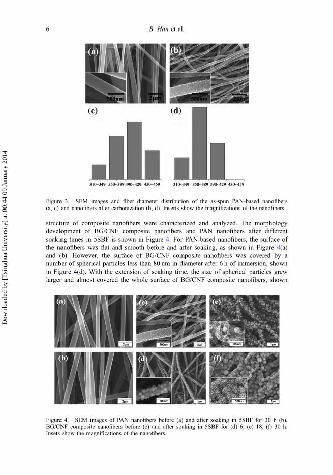
structure of composite nanofibers were characterized and analyzed. The morphologydevelopment of BG/CNF composite nanofibers and PAN nanofibers after differentsoaking times in 5SBF is shown in Figure 4. For PAN-based nanofibers, the surface ofthe nanofibers was flat and smooth before and after soaking, as shown in Figure 4(a)and (b). However, the surface of BG/CNF composite nanofibers was covered by anumber of spherical particles less than 80 nm in diameter after 6 h of immersion, shownin Figure 4(d). With the extension of soaking time, the size of spherical particles grewlarger and almost covered the whole surface of BG/CNF composite nanofibers, shown
Figure 3. SEM images and fiber diameter distribution of the as-spun PAN-based nanofibers(a, c) and nanofibers after carbonization (b, d). Inserts show the magnifications of the nanofibers.
Figure 4. SEM images of PAN nanofibers before (a) and after soaking in 5SBF for 30 h (b),BG/CNF composite nanofibers before (c) and after soaking in 5SBF for (d) 6, (e) 18, (f) 30 h.Insets show the magnifications of the nanofibers.
6 B. Han et al.
Dow
nloa
ded
by [
Tsi
nghu
a U
nive
rsity
] at
00:
44 0
9 Ja
nuar
y 20
14

in Figure 4(e). However, after soaking in 5SBF for 30 h, an obvious change in bothsize and morphology of spherical particles was observed in Figure 4(f) due to theincreased mineralization rate. In fact, the spherical particles with average diameter~700 nm were constituted of hundreds of acicular crystals, which suggested that theapatite-like layer was formed on the surface of the BG/CNF composite nanofibers.
Figure 5 shows energy dispersive spectroscopy (EDS) patterns of BG/CNF compos-ite nanofibers obtained at different time and element analysis results before and aftersoaking in 5SBF. The obtained element analysis of the BG/CNF composite nanofiberswithout soaking in the 5SBF agreed well with the nominal glass composition, that is58% SiO2-33% CaO-9% P2O5 (in mole %). With increasing the soaking time, the EDSpattern exhibited a gradual increase in the Ca and P signal and a corresponding attenua-tion of the Si signals, and the sharp increase of Ca and P signal after 30 h of soakingindicated the formation of an apatite-like material, which was consistent with the SEMresults. The Ca/P molar ratio exhibited gradual decreasing tendency, and the valueobtained by EDS was 1.68 for 30 h of incubation, which was very similar to the Ca/Pmolar ratio characteristic of stoichiometric HA (1.67).[23]
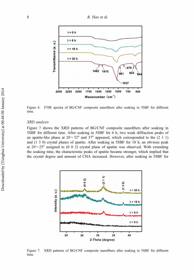
FTTR analysis
Figure 6 shows the FTIR spectra of BG/CNF composite nanofibers after soaking in5SBF for different time. The peaks at 1242 and 460 cm−1 in the spectra of BG/CNFcomposite nanofibers without soaking were corresponded to P=O double band andSi–O–Si band, respectively. After 6 h soaking in 5SBF solution, the obvious peak at1037 cm−1 was attributed to P–O stretching vibration, and the peaks at 879 and 564cm−1 were resulted from CO and P–O bending vibration. After 30 h soaking in 5SBFsolution, the broad and one-component bands at 1037 and 564 cm−1 corresponded tophosphate groups (PO3�
4 ), while the band located at 868 cm−1 corresponded to HPO2�4
groups, which were the disordered characteristic of featureless PO3�4 bands.[18] The
three bands at 1462, 1410 and 879 cm−1 were assigned as to CO2�3 groups, which
implied that the poorly crystallized or amorphous carbonated Ca–P phase wasprecipitated on the surface of nanofibers. With the prolong of soaking time in the 5SBFsolution, an increase in the intensity of the phosphate and carbonate peaks can beobserved, which was similar to those observed in synthetic CHA.[24] FTIR analysisindicated the formation of CHA on the surface of BG/CNF composite nanofibers afterbiological mineralization.
Figure 5. (a) EDS patterns obtained at different time and (b) SEM images and element analysisresults after soaking in 5SBF for (A) 0 h and (B) 30 h.
Journal of Biomaterials Science, Polymer Edition 7
Dow
nloa
ded
by [
Tsi
nghu
a U
nive
rsity
] at
00:
44 0
9 Ja
nuar
y 20
14
XRD analysis
Figure 7 shows the XRD patterns of BG/CNF composite nanofibers after soaking in5SBF for different time. After soaking in 5SBF for 6 h, two weak diffraction peaks ofan apatite-like phase at 2θ = 32o and 37o appeared, which corresponded to the (2 1 1)and (1 3 0) crystal planes of apatite. After soaking in 5SBF for 18 h, an obvious peakat 2θ = 25o assigned to (0 0 2) crystal plane of apatite was observed. With extendingthe soaking time, the characteristic peaks of apatite became stronger, which implied thatthe crystal degree and amount of CHA increased. However, after soaking in 5SBF for
Figure 6. FTIR spectra of BG/CNF composite nanofibers after soaking in 5SBF for differenttime.
Figure 7. XRD patterns of BG/CNF composite nanofibers after soaking in 5SBF for differenttime.
8 B. Han et al.
Dow
nloa
ded
by [
Tsi
nghu
a U
nive
rsity
] at
00:
44 0
9 Ja
nuar
y 20
14
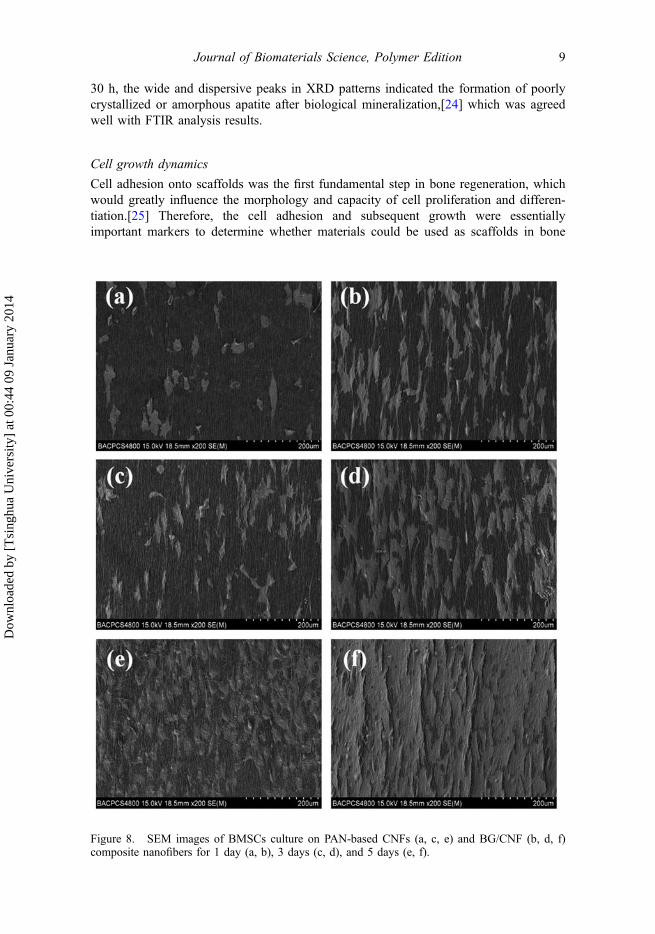
30 h, the wide and dispersive peaks in XRD patterns indicated the formation of poorlycrystallized or amorphous apatite after biological mineralization,[24] which was agreedwell with FTIR analysis results.
Cell growth dynamics
Cell adhesion onto scaffolds was the first fundamental step in bone regeneration, whichwould greatly influence the morphology and capacity of cell proliferation and differen-tiation.[25] Therefore, the cell adhesion and subsequent growth were essentiallyimportant markers to determine whether materials could be used as scaffolds in bone
Figure 8. SEM images of BMSCs culture on PAN-based CNFs (a, c, e) and BG/CNF (b, d, f)composite nanofibers for 1 day (a, b), 3 days (c, d), and 5 days (e, f).
Journal of Biomaterials Science, Polymer Edition 9
Dow
nloa
ded
by [
Tsi
nghu
a U
nive
rsity
] at
00:
44 0
9 Ja
nuar
y 20
14
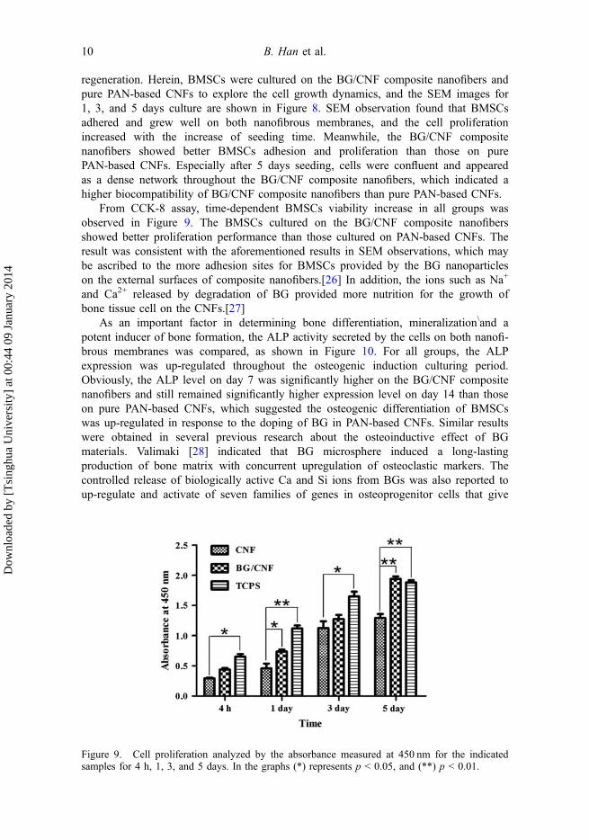
regeneration. Herein, BMSCs were cultured on the BG/CNF composite nanofibers andpure PAN-based CNFs to explore the cell growth dynamics, and the SEM images for1, 3, and 5 days culture are shown in Figure 8. SEM observation found that BMSCsadhered and grew well on both nanofibrous membranes, and the cell proliferationincreased with the increase of seeding time. Meanwhile, the BG/CNF compositenanofibers showed better BMSCs adhesion and proliferation than those on purePAN-based CNFs. Especially after 5 days seeding, cells were confluent and appearedas a dense network throughout the BG/CNF composite nanofibers, which indicated ahigher biocompatibility of BG/CNF composite nanofibers than pure PAN-based CNFs.
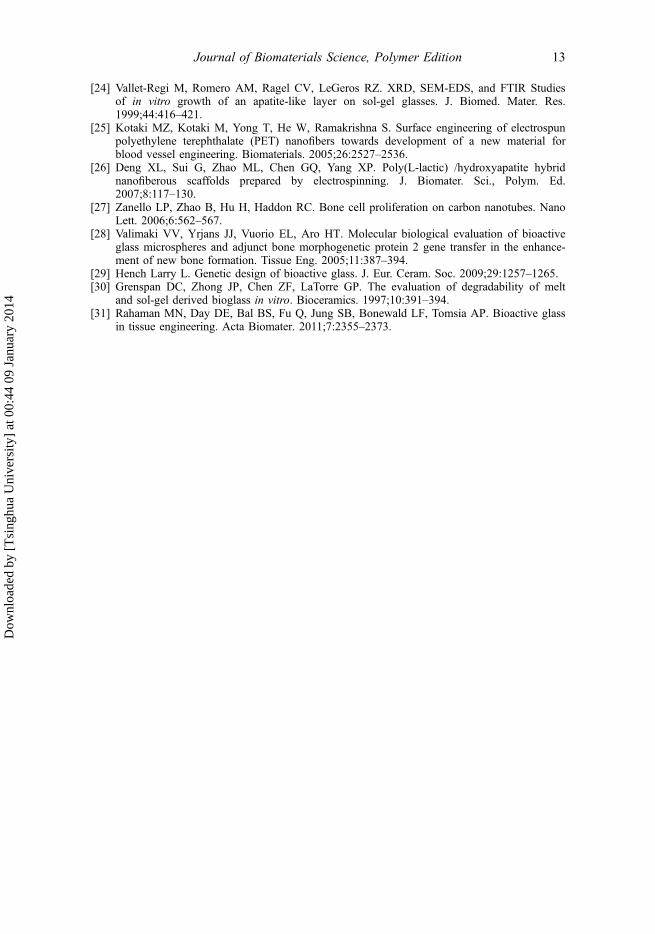
From CCK-8 assay, time-dependent BMSCs viability increase in all groups wasobserved in Figure 9. The BMSCs cultured on the BG/CNF composite nanofibersshowed better proliferation performance than those cultured on PAN-based CNFs. Theresult was consistent with the aforementioned results in SEM observations, which maybe ascribed to the more adhesion sites for BMSCs provided by the BG nanoparticleson the external surfaces of composite nanofibers.[26] In addition, the ions such as Na+
and Ca2+ released by degradation of BG provided more nutrition for the growth ofbone tissue cell on the CNFs.[27]
As an important factor in determining bone differentiation, mineralization\and apotent inducer of bone formation, the ALP activity secreted by the cells on both nanofi-brous membranes was compared, as shown in Figure 10. For all groups, the ALPexpression was up-regulated throughout the osteogenic induction culturing period.Obviously, the ALP level on day 7 was significantly higher on the BG/CNF compositenanofibers and still remained significantly higher expression level on day 14 than thoseon pure PAN-based CNFs, which suggested the osteogenic differentiation of BMSCswas up-regulated in response to the doping of BG in PAN-based CNFs. Similar resultswere obtained in several previous research about the osteoinductive effect of BGmaterials. Valimaki [28] indicated that BG microsphere induced a long-lastingproduction of bone matrix with concurrent upregulation of osteoclastic markers. Thecontrolled release of biologically active Ca and Si ions from BGs was also reported toup-regulate and activate of seven families of genes in osteoprogenitor cells that give
Figure 9. Cell proliferation analyzed by the absorbance measured at 450 nm for the indicatedsamples for 4 h, 1, 3, and 5 days. In the graphs (*) represents p < 0.05, and (**) p < 0.01.
10 B. Han et al.
Dow
nloa
ded
by [
Tsi
nghu
a U
nive
rsity
] at
00:
44 0
9 Ja
nuar
y 20
14

rise to rapid bone regeneration.[29] The mechanism of improved osteointegration ofBG has been attributed to the formation of an apatite layer on its surface. The drivingforce for apatite formation is the release of ions such as Na+ and Ca2+ during degrada-tion of BG when it is in contact with body fluid.[30,31] With the improved bioactivityby incorporating BG, the BG/CNF composite nanofibers were more favorable than purePAN-based CNFs to be applied as scaffolds for bone tissue engineering. These positiveresults constitute the necessary prerequisites for further investigations into the potentialof the BG materials to direct osteogenesis, leading to subsequent bone tissueregeneration.
Conclusions
BG/CNF composite nanofibers has been successfully fabricated by electrospinning fromPAN/CN/TEP/TEOS solution and followed by in situ sintering. Microstructure andphase composition analysis indicated that the resulting materials were fine bioglassparticles attached to the surface of the PAN-based CNFs. The biological mineralizationevaluation identified the formation of the apatite-like layer consisting of crystallites ofCHA. Cellular morphology, CCK-8 assay, and ALP activity assay suggested that BG/CNF composite nanofibers possessed favorable degradation and bioactivity, which maybe one of promising biomaterials for facilitation of osteogenesis.
Funding
The authors would acknowledge financial support from National Basic Research Program of China[number 2012CB933900], the Key Technologies R&D Program of China [2012BAI07B00]; theNational High Technology Research and Development Program of China [2011AA030100];the Beijing municipal Science & Technology Commission Projects [number Z121100005212007];the Key International S&T Cooperation Projects [number 2011DFA32190]; and the ScienceFoundation of Peking University School and Hospital of Stomatology [number YS020212].
Figure 10. Quantification of ALP activity in culture supernatants of BMSCs after 3, 7, and14 days cultured with osteogenic differentiation medium in the indicated groups. In the graphs(*) represents p < 0.05.
Journal of Biomaterials Science, Polymer Edition 11
Dow
nloa
ded
by [
Tsi
nghu
a U
nive
rsity
] at
00:
44 0
9 Ja
nuar
y 20
14

References[1] Kettunen J, Makela EA, Miettinen H, Nevalainen T, Heikkila M, Pohjonen T, Tormala P,
Rokkanen T. Mechanical properties and strength retention of carbon fiber-reinforced liquidcrystalline polymer (LCP/CF) composite: an experimental study on rabbits. Biomaterials.1998;19:1219–1228.
[2] Peters JL, Miner LH, Michael AC, Sesack SR. Ultrastructure at carbon fiber microelectrodeimplantation sites after acute voltammetric measurements in the striatum of anesthetized rats.J. Neurosci. Methods. 2004;137:9–23.
[3] Aoki K, Usui Y, Narita N, Ogiwara N, Iashigaki N, Nakamura K, Kato H, Sano K, OgiwaraN, Kametani K, Kim C, Taruta S, Kim YA, Endo M, Saito N. A thin carbon-fiber web as ascaffold for bone-tissue regeneration. Small. 2009;5:1540–1546.
[4] Webster TJ, Waid MC, McKenzie JL, Price RL, Ejiofor JU. Nano-biotechnology: carbonnanofibres as improved neural and orthopaedic implants. Nanotechnology. 2004;15:48–54.
[5] McKnight TE, Ericson MN, Jones SW, Melechko AV, Simpson ML. Vertically alignedcarbon nanofiber arrays: an electrical and genetic substrate for tissue scaffolding. Conf. Proc.IEEE Eng. Med. Biol. Soc. 2007;26:5381–5383.
[6] Elias KL, Price RL, Webster TJ. Enhanced functions of osteoblasts on nanometer diametercarbon fibers. Biomaterials. 2002;23:3279–3287.
[7] Kobayashi S, Kawai W. Development of carbon nanofiber reinforced hydroxyapatite withenhanced mechanical properties. Composites Part A. 2007;38:114–123.
[8] Zhang YZ, Venugopal J, Huang ZM, Lim CT, Ramakrishna S. Crosslinking of theelectrospun gelatin nanofibers. Polymer. 2006;47:2911–2917.
[9] Kashyap S, Griep K, Nychka JA. Crystallization kinetics, mineralization and crack propaga-tion in partially crystallized bioactive glass 45S5. Mater. Sci. Eng., C. 2011;31:762–769.
[10] Tomsia AP, Saiz E, Song J, CR. Biomimetic bonelike composites and novel bioactive glasscoatings. Adv. Eng. Mater. 2005;7:999–1004.
[11] Hench LL. The role of ceramics in an age of biology. Bioceramics: Mater. Appl. IV.2003;147:3–12.
[12] Hench LL. Genetic design of bioactive glass. J. Eur. Ceram. Soc. 2009;29:1257–1265.[13] Gunawidjaja PN, Lo AYH, Izquierdo-Barba I, Garcia A, Arcos D, Stevensson B, Grins J,
Vallet-Regi M, Eden M. Biomimetic apatite mineralization mechanisms of mesoporousbioactive glasses as probed by multinuclear P-31, Si-29, Na-23 and C-13 solid-state NMR.J. Phys. Chem. C. 2010;114:19345–19356.
[14] Labbaf S, Tsigkou O, Muller KH, Stevens MM, Porter AE, Jones JR. Spherical bioactiveglass particles and their interaction with human mesenchymal stem cells in vitro.Biomaterials. 2011;32:1010–1018.
[15] Hench LL, Xynos ID, Polak JM. Bioactive glasses for in situ tissue regeneration. J. Biomater.Sci., Polym. Ed. 2004;15:543–562.
[16] Liu HY, Cai Q, Lian PF, Fang Z, Duan S, Ryu S, Yang XP, Deng XL. The biologicalproperties of carbon nanofibers decorated with beta-tricalcium phosphate nanoparticles.Carbon. 2010;48:2266–2272.
[17] Liu HY, Cai Q, Lian PF, Fang Z, Duan S, Yang XP, Deng XL, Ryu S. Beta-tricalciumphosphate nanoparticles adhered carbon nanofibrous membrane for human osteoblasts cellculture. Mater. Lett. 2010;64:725–728.
[18] Xia W, Chang J. Preparation, in vitro bioactivity and drug release property of well-orderedmesoporous 58S bioactive glass. J. Non-Cryst. Solids. 2008;354:1338–1341.
[19] Barrere F, van Blitterswijk CA, de Groot K, Layrolle P. Nucleation of biomimetic Ca-Pcoatings on Ti6Al4V from a SBF × 5 solution: influence of magnesium. Biomaterials.2002;23:2211–2220.
[20] Bowker M. Surface science – the going rate for catalysts. Nat. Mater. 2002;1:205–206.[21] Dunham MG, Edie DD. Model of stabilization for PAN-based carbon-fiber precursor
bundles. Carbon. 1992;30:435–450.[22] Pacault A, Trinquecoste M. Carbonization kinetics of carbon-fiber precursors. Carbon.
1980;18:61–62.[23] Ostomel TA, Shi QH, Tsung CK, Liang HJ, Stucky GD. Spherical bioactive glass with
enhanced rates of hydroxyapatite deposition and hemostatic activity. Small.2006;2:1261–1265.
12 B. Han et al.
Dow
nloa
ded
by [
Tsi
nghu
a U
nive
rsity
] at
00:
44 0
9 Ja
nuar
y 20
14
[24] Vallet-Regi M, Romero AM, Ragel CV, LeGeros RZ. XRD, SEM-EDS, and FTIR Studiesof in vitro growth of an apatite-like layer on sol-gel glasses. J. Biomed. Mater. Res.1999;44:416–421.
[25] Kotaki MZ, Kotaki M, Yong T, He W, Ramakrishna S. Surface engineering of electrospunpolyethylene terephthalate (PET) nanofibers towards development of a new material forblood vessel engineering. Biomaterials. 2005;26:2527–2536.
[26] Deng XL, Sui G, Zhao ML, Chen GQ, Yang XP. Poly(L-lactic) /hydroxyapatite hybridnanofiberous scaffolds prepared by electrospinning. J. Biomater. Sci., Polym. Ed.2007;8:117–130.
[27] Zanello LP, Zhao B, Hu H, Haddon RC. Bone cell proliferation on carbon nanotubes. NanoLett. 2006;6:562–567.
[28] Valimaki VV, Yrjans JJ, Vuorio EL, Aro HT. Molecular biological evaluation of bioactiveglass microspheres and adjunct bone morphogenetic protein 2 gene transfer in the enhance-ment of new bone formation. Tissue Eng. 2005;11:387–394.
[29] Hench Larry L. Genetic design of bioactive glass. J. Eur. Ceram. Soc. 2009;29:1257–1265.[30] Grenspan DC, Zhong JP, Chen ZF, LaTorre GP. The evaluation of degradability of melt
and sol-gel derived bioglass in vitro. Bioceramics. 1997;10:391–394.[31] Rahaman MN, Day DE, Bal BS, Fu Q, Jung SB, Bonewald LF, Tomsia AP. Bioactive glass
in tissue engineering. Acta Biomater. 2011;7:2355–2373.
Journal of Biomaterials Science, Polymer Edition 13
Dow
nloa
ded
by [
Tsi
nghu
a U
nive
rsity
] at
00:
44 0
9 Ja
nuar
y 20
14