Embed Size (px)
DESCRIPTION
phoneee....
Citation preview
A Comparative Study of Clinicopathological Featuresbetween Chronic Cholecystitis Patients with and withoutHelicobacter pylori Infection in Gallbladder MucosaDi Zhou1., Wen-bin Guan2., Jian-dong Wang1*, Yong Zhang1, Wei Gong1, Zhi-wei Quan1*
1Department of General Surgery, Xinhua Hospital, Shanghai JiaoTong University, School of Medicine, Shanghai, China, 2Department of Pathology, Xinhua Hospital,
Shanghai JiaoTong University, School of Medicine, Shanghai, China
Abstract
Background: Helicobacter pylori has been isolated from 10%–20% of human chronic cholecystitis specimens but thecharacteristics of ‘‘Helicobacter pylori positive cholecystitis’’ remains unclear. This study aims to compare theclinicopathological features between chronic cholecystitis patients with and without Helicobacter pylori infection ingallbladder mucosa.
Methods: Three hundred and twenty-six chronic cholecystitis patients were divided into two groups according to whetherHelicobacter pylori could be detected by culture, staining or PCR for Helicobacter 16s rRNA gene in gallbladder mucosa.Positive samples were sequenced for Helicobacter pylori-specific identification. Clinical parameters as well as pathologicalcharacteristics including some premalignant lesions and the expression levels of iNOS and ROS in gallbladder werecompared between the two groups.
Results: Helicobacter pylori infection in gallbladder mucosa was detected in 20.55% of cholecystitis patients. These patientshad a higher prevalence of acid regurgitation symptoms (p= 0.001), more histories of chronic gastritis (p= 0.005), gastriculcer (p= 0.042), duodenal ulcer (p= 0.026) and higher presence of Helicobacter pylori in the stomach as compared topatients without Helicobacter pylori infection in the gallbladder mucosa. Helicobacter pylori 16s rRNA in gallbladder andgastric-duodenal mucosa from the same individual patient had identical sequences. Also, higher incidences ofadenomyomatosis (p= 0.012), metaplasia (p= 0.022) and higher enhanced expressions of iNOS and ROS were detected inHelicobacter pylori infected gallbladder mucosa (p,0.05).
Conclusions: Helicobacter pylori infection in gallbladder mucosa is strongly associated with Helicobacter pylori existed instomach. Helicobacter pylori is also correlated with gallbladder premalignant lesions including metaplasia andadenomyomatosis. The potential mechanism might be related with higher ROS/RNS production but needs furtherinvestigation.
Citation: Zhou D, Guan W-b, Wang J-d, Zhang Y, Gong W, et al. (2013) A Comparative Study of Clinicopathological Features between Chronic CholecystitisPatients with and without Helicobacter pylori Infection in Gallbladder Mucosa. PLoS ONE 8(7): e70265. doi:10.1371/journal.pone.0070265
Editor: Ivo G. Boneca, Institut Pasteur Paris, France
Received December 28, 2012; Accepted June 18, 2013; Published July 30, 2013
Copyright: � 2013 Zhou et al. This is an open-access article distributed under the terms of the Creative Commons Attribution License, which permitsunrestricted use, distribution, and reproduction in any medium, provided the original author and source are credited.
Funding: This work was supported by the National ‘‘Twelfth Five-Year’’ special science and technology major project (No.2012ZX10002016), (http://www.nmp.gov.cn/zxjs/crb/201012/t20101208_2127.htm); and the College Fund of Shanghai Jiaotong University School of Medicine (No.10XJ22003) (http://kjc.shsmu.edu.cn/Default.aspx). The funders had no role in study design, data collection and analysis, decision to publish, or preparation of the manuscript.
Competing Interests: The authors have declared that no competing interests exist.
* E-mail: [email protected] (J-dW); [email protected] (Z-wQ)
. These authors contributed equally to the work.
Introduction
Chronic cholecystitis is one of the most prevalent diseases
requiring surgical intervention. In China, more than 90% of the
cholecystitis cases are claimed to be caused by symptomatic
cholelithiasis, the incidence of which is approximately 10% of the
adult population. [1,2] Histologically, chronic cholecystitis pre-
sents a large range of related inflammatory epithelial changes
including mononuclear infiltrate, fibrosis, thickness of muscular
layer, dysplasia, hyperplasia and metaplasia-the last three have
been considered as premalignant lesions. [3–5].
The causes of chronic cholecystitis still remain unclear.
Recently, many findings obtained from microbiological studies
suggest that bacterial infection in biliary system might play a role.
Our previous meta-analysis demonstrated that Helicobacter pylori
(H.pylori) in human biliary system was correlated with chronic
cholecystitis, especially in the regions with higher prevalence of
this infectious agent such as South Asia, East Asia and Latin
America. [6] Evidences supporting the association between
H.pylori infection and chronic cholecystitis could be found by
using direct culture or staining of H.pylori in gallbladder tissues as
well as indirect techniques such as PCR, ELISA and serology for
detecting H.pylori-specific genes or antibodies.[7–9] The positive
rate of H.pylori in gallbladder is reported to be 10%–20% by
culture. [10].
PLOS ONE | www.plosone.org 1 July 2013 | Volume 8 | Issue 7 | e70265
H.pylori can induce oxidative stress through producing reactive
oxygen species (ROS) and reactive nitrogen species (RNS), which
are considered to be the important causes of chronic inflamma-
tion, ulcer and canceration of the stomach. [11] In H. pylori-
infected stomach, possible sources of ROS/RNS include neutro-
phils, vascular endothelial cells, gastric mucosal cells, and H. pylori
itself. One of the most important pathways of H.pylori-induced
RNS is mediated by overproduction of endogenous synthesis nitric
oxide (NO) through inducible NO synthase (iNOS) expression.
[12] In benign inflammatory and malignant gallbladder diseases,
ROS and iNOS also play an important role. [13] However, in
biliary system, the correlation between H.pylori and ROS/RNS
production still needs further investigation.
Two-thirds of the world population is infected with H.pylori. [14]
The findings of H.pylori in biliary tract implicated that the stomach
might not be the only arena of activity of this agent. However, few
studies by far have specifically assessed the characteristics of
‘‘Helicobacter pylori positive cholecystitis’’. Therefore, this study aims
to compare the clinicopathological features between chronic
cholecystitis patients with and without Helicobacter pylori infection
in gallbladder mucosa.
Materials and Methods
PatientsOf 378 patients who underwent cholecystectomy in Department
of General Surgery, Xinhua Hospital from December 2011 to July
2012, three hundred and twenty-six patients (97 males and 229
females, aged 21–87 years) who fulfilled the pathological criteria of
chronic cholecystitis were enrolled in this study. The exclusion
criteria were: (1) patients with history of hepato-biliary or
pancreatic surgery which changed the normal structure and
function of the biliary system, (2) patients who had previously
received standard triple therapy for H. pylori eradication, (3)
patients who had taken antibiotics or proton pump inhibitors 4–6
weeks prior to cholecystectomy. According to whether H. pylori was
detected positive in gallbladder mucosa, patients were divided into
two groups. The study protocol was approved by the Ethics
Committee of Shanghai JiaoTong University, School of Medicine
and signed informed consent was obtained from all the patients.
GastroscopyBefore or after cholecystectomy, all patients enrolled in this
study received gastroscopy with biopsy in order to clarify the
infection status of H. pylori in their stomach. Gastroscopy was
performed with video endoscopes that worked in high-resolution,
Table 1. Definitions of Pathological Changes of Chronic Cholecystitis.
Pathological Changes of Chronic Cholecystitis Definition
Inflammatory mononuclear infiltrate
Mild Diffuse, #10 inflammatory cells per HPF in any layer
Moderate Diffuse, between 11 to 30 cells per HPF
Severe Diffuse, more than 31 cells per HPF or follicular
Degree of fibrosis
Mild Uneven collagen deposition in #20% of material
Moderate Uneven collagen deposition in 21% to 70% of material
Severe Uneven collagen or lamellar fibroplasia in $71% of material
Thickness of the muscular layer
Mild Less than one third of the whole thickness
Moderate One third to two thirds of the wall
Severe More than two thirds of the wall thickness
Addipose tissue deposition
Mild Up to 10% of the material
Moderate 11% to 60% of the material
Severe More than 60% of the material
Degree of hyperplasia
Diffuse $70% of the whole sections
Focal ,70% of the whole sections
Degree of dysplasia
Low-grade Resemble tubular adenomas of the colon without intestinal metaplasia
High-grade Markedly pleomorphic nuclei and/or prominent nucleoli
Metaplasia
Pyloric type Structures similar to the pyloric glands in the lamina propria
Intestinal type Goblet cells and enterocitlike cells
Gastric surface type Epithelial cells of gallbladder mucosa replaced by tall columnar cells with abundant mucin and basallylocated nuclei
HPF: high power field.doi:10.1371/journal.pone.0070265.t001
Helicobacter pylori and Chronic Cholecystitis
PLOS ONE | www.plosone.org 2 July 2013 | Volume 8 | Issue 7 | e70265
white light mode and AFI mode (EVIS-FQ260Z; Olympus
Medical Systems Co. Ltd, Tokyo, Japan). Two biopsy specimens
were taken at each site from the greater curvature of the antrum,
and the greater and lesser curvature of the corpus. Each of the two
specimens from the above parts of the stomach were used
respectively for culture and Warthin-Starry Staining of H. pylori.
Cholecystectomy and Gallbladder BiopsyLaparoscopic cholecystectomy was performed by a single
surgeon using a standardized 4-port technique (Laparoscopic
Device, KARL STORZ GmbH, Tuttlingen, Germany). Two
biopsy specimens were taken aseptically at each site from the
fundus, body and neck of the gallbladder. Each of the two
specimens from the above parts of the gallbladder were used
respectively for culture and Warthin-Starry Staining of H. pylori.
The stomach and gallbladder specimens were aseptically
transferred to the microbiology laboratory immediately after
gastroscopy or cholecystectomy.
Verification of H. pylori Infection in Gallbladder andStomach
The presence of H. pylori in gastric or gallbladder mucosa was
determined by either positive culture, Warthin-Starry Staining or
positive nest PCR for specific 16s rRNA of this bacterium. At least
one positive test was regarded as confirmation of infection of this
agent in gallbladder or gastric mucosa.
Culture of H. pyloriThe gallbladder and gastric mucosa specimens were inoculated
onto sterile plates containing endo agar and Brucella agar
supplemented with 5% horse blood (Becton, Dickinson and
Company, Sparks, Maryland, USA) for nonspecific bacterial and
Campylobacter cultures, respectively. For isolation of H.pylori, we
prepared specific media containing BHI agar supplemented with
7% sheep blood, 0.4% IsoVitaleX (Becton, Dickinson and
Company, Sparks, Maryland, USA) and Skirrow-selective supple-
ments (Oxoid Limited, Thermo Fisher Scientific, Hampshire,
UK). H.pylori was incubated at a microaerophilic condition (5%
O2, 10% CO2, 85% N2) for 3–7 days at 37uC. Bacteria colonies
were examined with biochemical tests as well as microscopy for
conformation. H. pylori colony was identified as showing the typical
white, pin-point and transparent morphology. Oxidase and fast
urease activity were also performed at colonies grown on H. pylori
specific media. Bacterium with typical morphology, positive
oxidase and urease activity were verified as H. pylori.
Warthin-Starry Staining of H. pyloriWarthin-Starry Staining was performed using the specific kit
(Diagnostic BioSystems, Pleasanton, California, USA). Four-
micrometer thick paraffin sections of gallbladder and gastric
mucosa tissues from each patient were backed for 1 h at 60uC.
After dewaxing and re-hydration, sections were incubated at 56uCfor 1 h in 1% silver nitrate buffer in the dark box and then dipped
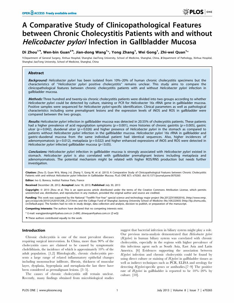
Figure 1. H.pylori infection in metaplastic gallbladder mucosa (oil immersion lens,61000, red arrow indicates H.pylori).doi:10.1371/journal.pone.0070265.g001
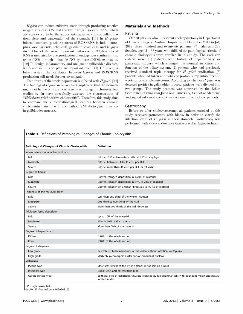
Figure 2. PCR products of Helicobacter specific 16s rRNA genefrom gallbladder and gastric mucosa samples. (lanes M: step-ladder marker; 1: positive control of gastric biopsy-derived H. pyloriDNA; 2: negative control of gastric biopsy; 3: negative 16s rRNA gene ingallbladder; 4 and 5: negative 16s rRNA gene in gallbladder and gastricmucosa acquired from one individual patient; 6 and 7: positive 16s rRNAgene in gallbladder and gastric mucosa acquired from anotherindividual patient).doi:10.1371/journal.pone.0070265.g002
Helicobacter pylori and Chronic Cholecystitis
PLOS ONE | www.plosone.org 3 July 2013 | Volume 8 | Issue 7 | e70265
in developer solution and stained for 5–8 min. Finally, sections
were dehydrated with 100% alcohol, cleared with xylene. H. pylori
was identified as stained into buffy or black color in a light yellow
background under microscope with oil immersion lens (61000).
The results were also determined independently by the above two
pathologists. Warthin-Starry staining and H. pylori culture were
blindly assayed for all the specimens from each patient.
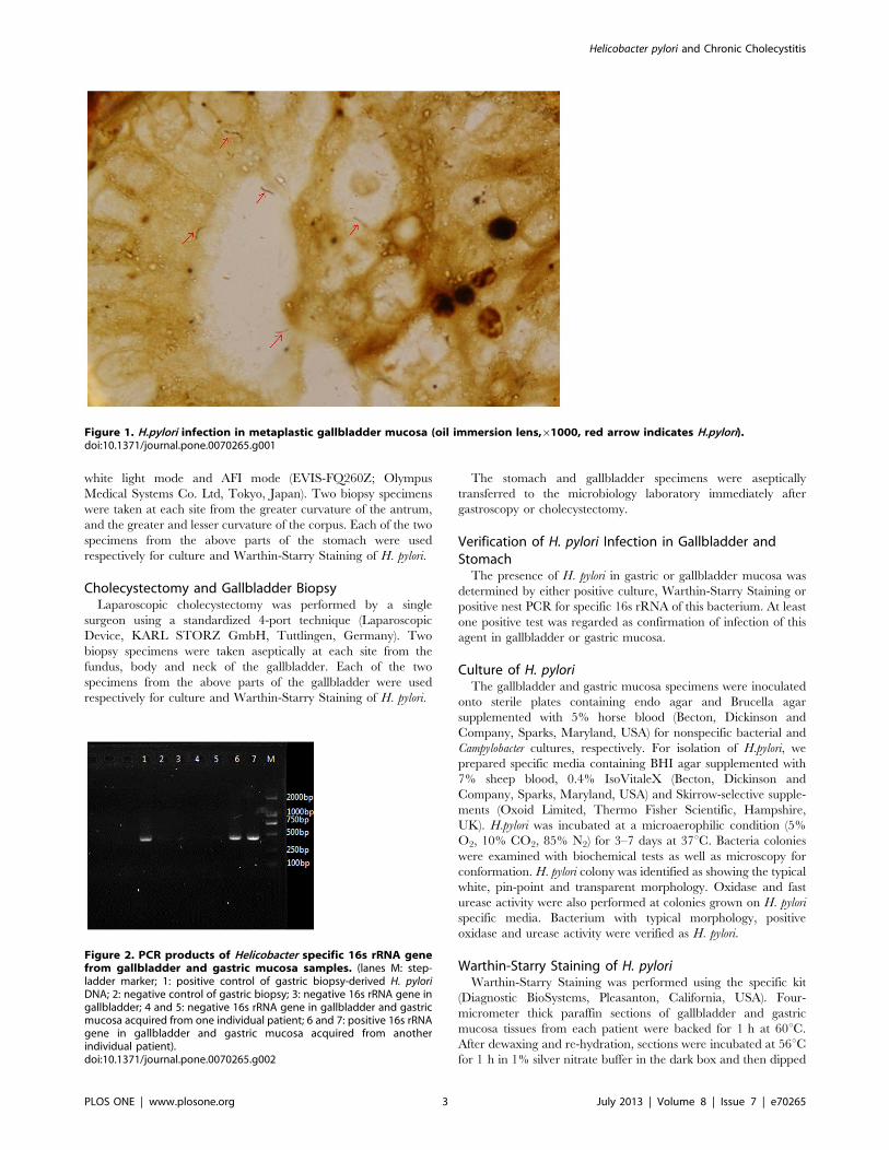
Figure 3. Comparison of complete sequence data of H. pylori 16s rRNA gene tested in gallbladder mucosa sample from publishedGenBank data: sequence ID ref|NR_044761.1|. (nucleotides 263–695 were listed).doi:10.1371/journal.pone.0070265.g003
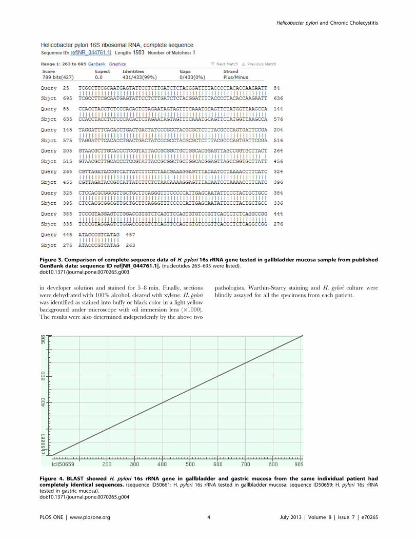
Figure 4. BLAST showed H. pylori 16s rRNA gene in gallbladder and gastric mucosa from the same individual patient hadcompletely identical sequences. (sequence ID50661: H. pylori 16s rRNA tested in gallbladder mucosa; sequence ID50659: H. pylori 16s rRNAtested in gastric mucosa).doi:10.1371/journal.pone.0070265.g004
Helicobacter pylori and Chronic Cholecystitis
PLOS ONE | www.plosone.org 4 July 2013 | Volume 8 | Issue 7 | e70265
PCR for Helicobacter 16s rRNA GeneThe DNA extracts were prepared from the paraffin specimens
as the kit (TIAN amp Micro DNA kit, TIANGEN Biotech,
Beijing, China) instructed. A semi-nested PCR assay specific for
Helicobacter 16s rRNA gene (16s rDNA) was amplified as previously
described [15], using primers 1F (59CTATGACGGG-
TATCCGGC39), 1R (59CTCACGACACGAGCTGAC39) and
2R (59TCGCCTTCGCAATGAGTATT39). Primers 1F and 1R
were used in the first step, whereas primers 1F and 2R were used
in the second step. The PCR reaction mixture contained 1 ml of
10 mM each primer (1F and 1R), 2 mL of 10 mM dNTP, 16PCR
reaction buffer, 2.5 mM MgCl2, 0.05% casein, 0.05% formamid,
1.25 U rTaq DNA polymerase (Takara, Dalian, China), and 2 mL
extracted DNA as templates. The amplification conditions for the
first step were 94uC for 2 min; 30 cycles of 94uC for 30 s, 55uC for
30 s, and 72uC for 30 s; and finally 72uC for 10 min. The reaction
mixture of the second step (25 mL) contained 0.5 ml of 10 mM each
primer each primer (1F and 2R), 0.2 mmol/L each dNTP,
16PCR reaction buffer, 2.5 mmol/L MgCl2, 1.0 U ExTaq DNA
polymerase (Takara, Dalian, China), and 2 mL 106diluted PCR
product from the first step. The 416-bp PCR products were
visualized by 1.2% agarose gel electrophoresis.
Sequence AnalysisThe PCR products were subsequently ligated into pMD-19T
simple vector (Takara, Dalian, China) for sequencing. The
product was sequenced after PCR amplification and double
enzyme digestion. The sequences were compared with the 16S
rRNA gene sequences of the strains that have been registered in
the GenBank database. Products of the sequence reaction were
aligned and the closest homologous sequence was identified by
Standard Nucleotide BLAST analysis tool on the internet
(BLASTn, http://blast.ncbi.nlm. nih.gov).
Table 2. Clinical characteristics of H.pylori-positive and negative chronic cholecystitis.
Characteristics No. of Patients n (%)
H.pylori (+) in gallbladdermucosa (n =67)
H.pylori (2) in gallbladdermucosa (n=259) p value
Age (yr) 45.54612.58 47.82611.56 NS
Gender
Male: Female (% Male) 19:48(28.36) 78:181(30.12) NS
BMI (kg/m2) 23.1062.24 22.7261.85 NS
Symptom
Mild Abdominal Pain 44(65.67) 149(57.53) NS
Biliary Colic 32(47.76) 102(39.38) NS
Loss of Appetite 12(17.91) 28(10.81) NS
Acid Regurgitation 22(32.84) 39(15.06) 0.001*
Heartburn 20(29.85) 50(19.31) NS
Preoperative Ultrasound Diagnosis
Gallstone Disease
No. of Gallstones
Single: Multiple (% Single) 21:34(38.18) 104:108(49.06) NS
Polypoid Lesion
No. of Polypoid Lesions
Single: Multiple (% Single) 8:4(66.67) 18:31(35.29) NS
Gallbladder Wall Thickening 12(17.91) 39(15.06) NS
Atrophic Gallbladder 15(22.39) 35(13.51) NS
History of Other Gastrointestinal Diseases
Chronic Gastritis 45(67.16) 124(47.88) 0.005*
Gastric Ulcer 11(16.42) 21(8.11) 0.042m
Duodenal Ulcer 8(11.94) 12(4.63) 0.026m
Reflux Esophagitis 12(17.91) 25(9.65) NS
Chronic Enteritis 3(4.48) 8(3.09) NS
H.pylori (+) in Gastric or Duodenal Mucosa
Warthin-Starry Stain 39(58.21) 106(40.93) 0.011m
H.pylori Culture16s rRNA gene PCR
33(49.25)42(62.69)
90(34.75)115(44.40)
0.029m
0.008*
*p,0.01.mp,0.05.NS: not significant.N/A: not applicable.doi:10.1371/journal.pone.0070265.t002
Helicobacter pylori and Chronic Cholecystitis
PLOS ONE | www.plosone.org 5 July 2013 | Volume 8 | Issue 7 | e70265
Histological Analysis of Chronic CholecystitisGallbladder specimens were fixed in 10% buffered formalin and
embedded in paraffin. Four micrometer-thick sections were cut
and stained with hematoxylin-eosin (HE). Sample slides were
examined independently by two attending pathologists who
specialized in biliary diseases and in the case of discrepancy, the
decision was made by discussion or in consultation with a third
experienced pathologist. Chronic inflammation was diagnosed in
the presence of a predominantly mononuclear inflammatory
infiltrate, fibrosis, or metaplastic changes. A scoring system
proposed by Barcia JJ [16] was used to semi-quantitatively assess
the histological changes of chronic cholecystitis (Table 1).
Immunohistochemical Staining of iNOS and ROSFor iNOS and ROS detection, immunohistochemistry was
performed on 4-mm thick, mounted on silane-coated slides of
gallbladder mucosa tissue sections. Sections were deparaffinized
and rehydrated, then washed in distilled water and 0.05 mol/L
Tris buffer. After blocking the nonspecific binding sites by using
Protein blocking agent (Coulter-Immunotech, Marseille, France),
sections were incubated with the primary polyclonal rabbit anti-
iNOS and ROS antibody (1:200, Transduction Laboratories,
kentucky, USA) at 4uC for 24 h. After sections were washed, a
biotinylated immunoglobulin (anti-rabbit serum for iNOS and
ROS) was applied for 30 min. Finally, all sections were incubated
with the avidine-biotin-complex (ABC) with alkaline phosphatase
(Vectastain, Vector Laboratories, Burlingame, California, USA).
The staining was visualized with 3,39-diaminobenzidine and
hydrogen peroxide.
Semiquantitative analysis of the iNOS and ROS immunostain-
ing was performed independently by the above two pathologists
and in the case of discrepancy, the decision was made by
discussion or in consultation with a third experienced pathologist.
In ten randomly selected areas of the whole section, the number
and percentage of positive cells were calculated for determining
staining intensity and proportion of iNOS or ROS staining. A case
without positive cells was considered negative. A case with less
than 10% positive cells was scored 1, 10–50% was scored 2, 50–
80% was scored 3 and more than 80% was scored 4. A staining
intensity was classified as weak (I), moderate (II) and strong (III). A
immunoreactive score was calculated as staining intensity6a-
mount of positive cells (from lowest score 0 to highest 12) and
specimen with a grade of more than 1 was defined as positive.
[17].
Statistical AnalysisThe software SAS 9.13 (SAS Institute, Gary, North Carolina,
USA) was used for conducting statistical analysis. Student’s t-test
was performed for comparing age and BMI. The immunoreactive
score of iNOS and ROS were calculated and statistically
compared between the two groups using Mann-Whitney U-test.
Chi-square test or Fischer’s exact test was used to examine the rest
clinicopathological parameters. For all statistical analyses, signif-
icance levels were set at p,0.05.
Results
Evidence of H. pylori in gallbadder mucosa was demonstrated by
Warthin-Starry staining in 64 (19.63%) patients and H. pylori
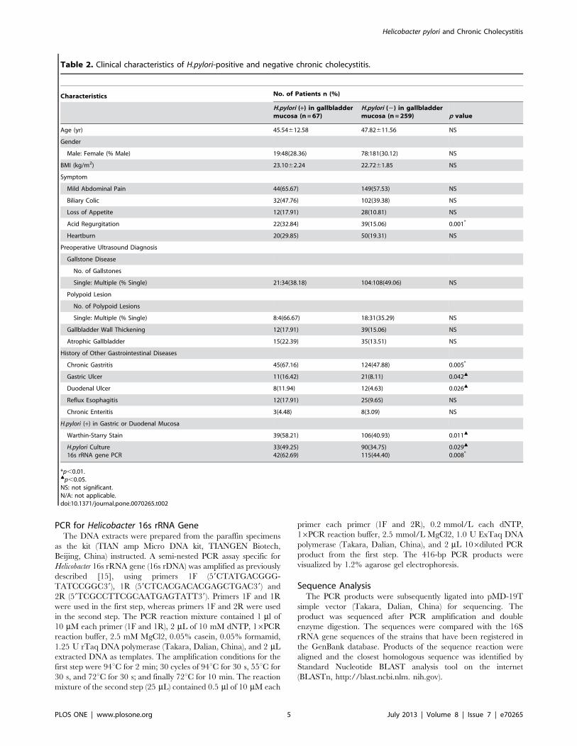
Figure 5. Metaplasia of Chronic Cholecystitis (hematoxylin-eosin stain,6100).doi:10.1371/journal.pone.0070265.g005
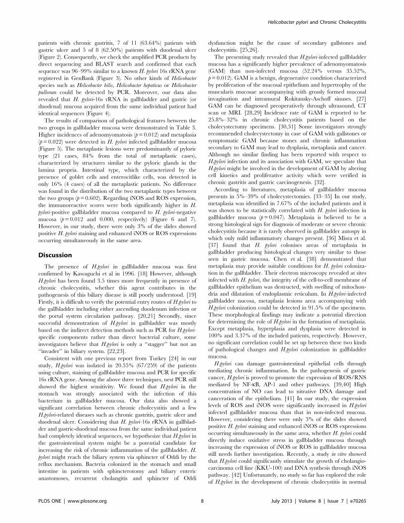
Figure 6. iNOS expression in gallbladder mucosa of chronic cholecystitis with H. pylori infection (A) and without H. pylori infection(B) (6100).doi:10.1371/journal.pone.0070265.g006
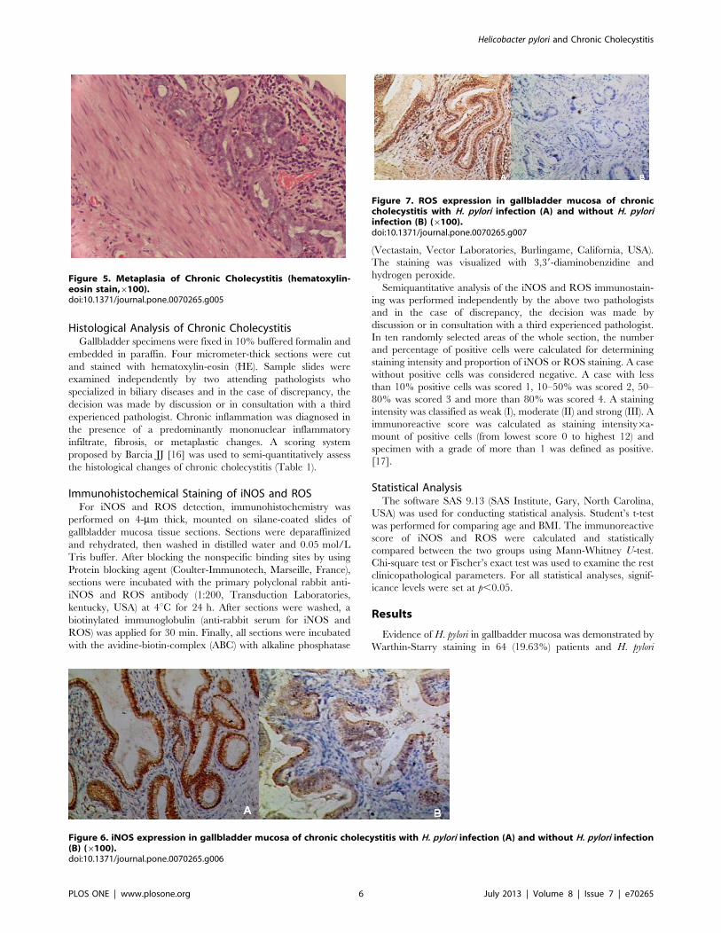
Figure 7. ROS expression in gallbladder mucosa of chroniccholecystitis with H. pylori infection (A) and without H. pyloriinfection (B) (6100).doi:10.1371/journal.pone.0070265.g007
Helicobacter pylori and Chronic Cholecystitis
PLOS ONE | www.plosone.org 6 July 2013 | Volume 8 | Issue 7 | e70265
colonies were identified upon culture in 55 (16.87%) patients.
Among them, 52 (77.61%) patients were both positive in staining
and culture. In PCR test for Helicobacter-16s rRNA gene, 67
(20.55%) patients were positive. From all the gallbladder
specimens, only the positive samples which detected by staining
or culture were positive in nest PCR test. All samples positive for
first-step amplicon were also positive for the nested PCR. Finally,
H. pylori infection in gallbladder mucosa was detected in 20.55%
(n = 67) of the cholecystitis patients (Figure 1 and 2). These
patients had a higher prevalence of acid regurgitation symptoms
(p= 0.001), more histories of chronic gastritis (p= 0.005), gastric
ulcer (p= 0.042), duodenal ulcer (p= 0.026) and a higher positive
rate of Helicobacter pylori (p,0.05) in the stomach as compared to
patients without Helicobacter pylori infection in the gallbladder
(Table 2). Of the 67 patients (20.55%) who were positive in H.
pylori 16s rRNA detection in gallbladder mucosa, amplications of
16s rRNA in their gastric or duodenal specimens were also
succeed in 42 patients (62.69%). These were 30 of 45 (66.67%)
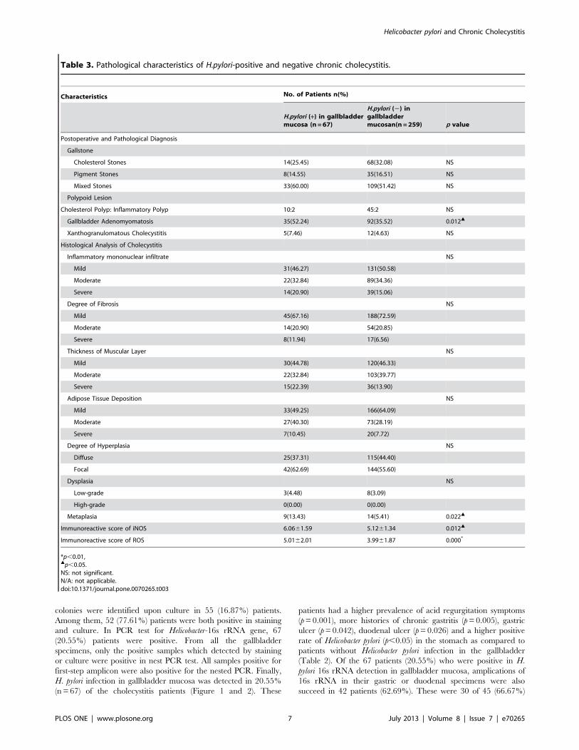
Table 3. Pathological characteristics of H.pylori-positive and negative chronic cholecystitis.
Characteristics No. of Patients n(%)
H.pylori (+) in gallbladdermucosa (n =67)
H.pylori (2) ingallbladdermucosan(n =259) p value
Postoperative and Pathological Diagnosis
Gallstone
Cholesterol Stones 14(25.45) 68(32.08) NS
Pigment Stones 8(14.55) 35(16.51) NS
Mixed Stones 33(60.00) 109(51.42) NS
Polypoid Lesion
Cholesterol Polyp: Inflammatory Polyp 10:2 45:2 NS
Gallbladder Adenomyomatosis 35(52.24) 92(35.52) 0.012m
Xanthogranulomatous Cholecystitis 5(7.46) 12(4.63) NS
Histological Analysis of Cholecystitis
Inflammatory mononuclear infiltrate NS
Mild 31(46.27) 131(50.58)
Moderate 22(32.84) 89(34.36)
Severe 14(20.90) 39(15.06)
Degree of Fibrosis NS
Mild 45(67.16) 188(72.59)
Moderate 14(20.90) 54(20.85)
Severe 8(11.94) 17(6.56)
Thickness of Muscular Layer NS
Mild 30(44.78) 120(46.33)
Moderate 22(32.84) 103(39.77)
Severe 15(22.39) 36(13.90)
Adipose Tissue Deposition NS
Mild 33(49.25) 166(64.09)
Moderate 27(40.30) 73(28.19)
Severe 7(10.45) 20(7.72)
Degree of Hyperplasia NS
Diffuse 25(37.31) 115(44.40)
Focal 42(62.69) 144(55.60)
Dysplasia NS
Low-grade 3(4.48) 8(3.09)
High-grade 0(0.00) 0(0.00)
Metaplasia 9(13.43) 14(5.41) 0.022m
Immunoreactive score of iNOS 6.0661.59 5.1261.34 0.012m
Immunoreactive score of ROS 5.0162.01 3.9961.87 0.000*
*p,0.01,mp,0.05.NS: not significant.N/A: not applicable.doi:10.1371/journal.pone.0070265.t003
Helicobacter pylori and Chronic Cholecystitis
PLOS ONE | www.plosone.org 7 July 2013 | Volume 8 | Issue 7 | e70265
patients with chronic gastritis, 7 of 11 (63.64%) patients with
gastric ulcer and 5 of 8 (62.50%) patients with duodenal ulcer
(Figure 2). Consequently, we check the amplified PCR products by
direct sequencing and BLAST search and confirmed that each
sequence was 96–99% similar to a known H. pylori 16s rRNA gene
registered in GenBank (Figure 3). No other kinds of Helicobacter
species such as Helicobacter bilis, Helicobacter hepaticus or Helicobacter
pullorum could be detected by PCR. Moreover, our data also
revealed that H. pylori-16s rRNA in gallbladder and gastric (or
duodenal) mucosa acquired from the same individual patient had
identical sequences (Figure 4).
The results of comparison of pathological features between the
two groups in gallbladder mucosa were demonstrated in Table 3.
Higher incidences of adenomyomatosis (p= 0.012) and metaplasia
(p= 0.022) were detected in H. pylori infected gallbladder mucosa
(Figure 5). The metaplastic lesions were predominantly of pyloric
type (21 cases, 84% from the total of metaplastic cases),
characterized by structures similar to the pyloric glands in the
lamina propria. Intestinal type, which characterized by the
presence of goblet cells and enterocitlike cells, was detected in
only 16% (4 cases) of all the metaplastic patients. No difference
was found in the distribution of the two metaplastic types between
the two groups (p= 0.602). Regarding iNOS and ROS expression,
the immunoreactive scores were both significantly higher in H.
pylori-positive gallbladder mucosa compared to H. pylori-negative
mucosa (p= 0.012 and 0.000, respectively) (Figure 6 and 7).
However, in our study, there were only 3% of the slides showed
positive H. pylori staining and enhanced iNOS or ROS expressions
occurring simultaneously in the same area.
Discussion
The presence of H.pylori in gallbladder mucosa was first
confirmed by Kawaguchi et al in 1996. [18] However, although
H.pylori has been found 3.5 times more frequently in presence of
chronic cholecystitis, whether this agent contributes in the
pathogenesis of this biliary disease is still poorly understood. [19]
Firstly, it is difficult to verify the potential entry routes of H.pylori to
the gallbladder including either ascending duodenum infection or
the portal system circulation pathway. [20,21] Secondly, since
successful demonstration of H.pylori in gallbladder was mostly
based on the indirect detection methods such as PCR for H.pylori-
specific components rather than direct bacterial culture, some
investigators believe that H.pylori is only a ‘‘stagger’’ but not an
‘‘invader’’ in biliary system. [22,23].
Consistent with one previous report from Turkey [24] in our
study, H.pylori was isolated in 20.55% (67/259) of the patients
using culture, staining of gallbladder mucosa and PCR for specific
16s rRNA gene. Among the above three techniques, nest PCR still
showed the highest sensitivity. We found that H.pylori in the
stomach was strongly associated with the infection of this
bacterium in gallbladder mucosa. Our data also showed a
significant correlation between chronic cholecystitis and a few
H.pylori-related diseases such as chronic gastritis, gastric ulcer and
duodenal ulcer. Considering that H. pylori-16s rRNA in gallblad-
der and gastric-duodenal mucosa from the same individual patient
had completely identical sequences, we hypothesize that H.pylori in
the gastrointestinal system might be a potential candidate for
increasing the risk of chronic inflammation of the gallbladder. H.
pylori might reach the biliary system via sphincter of Oddi by the
reflux mechanism. Bacteria colonized in the stomach and small
intestine in patients with sphincterotomy and biliary enteric
anastomoses, recurrent cholangitis and sphincter of Oddi
dysfunction might be the cause of secondary gallstones and
cholecystitis. [25,26].
The presenting study revealed that H.pylori-infected gallbladder
mucosa has a significantly higher prevalence of adenomyomatosis
(GAM) than non-infected mucosa (52.24% versus 35.52%,
p= 0.012). GAM is a benign, degenerative condition characterized
by proliferation of the mucosal epithelium and hypertrophy of the
muscularis mucosae accompanying with grossly formed mucosal
invagination and intramural Rokitansky-Aschoff sinuses. [27]
GAM can be diagnosed preoperatively through ultrasound, CT
scan or MRI. [28,29] Incidence rate of GAM is reported to be
25.8%–32% in chronic cholecystitis patients based on the
cholecystectomy specimens. [30,31] Some investigators strongly
recommended cholecystectomy in case of GAM with gallstones or
symptomatic GAM because stones and chronic inflammation
secondary to GAM may lead to dysplasia, metaplasia and cancer.
Although no similar finding has been reported with respect to
H.pylori infection and its association with GAM, we speculate that
H.pylori might be involved in the development of GAM by altering
cell kinetics and proliferative activity which were verified in
chronic gastritis and gastric carcinogenesis. [32].
According to literatures, metaplasia of gallbladder mucosa
presents in 5%–39% of cholecystectomies. [33–35] In our study,
metaplasia was identified in 7.67% of the included patients and it
was shown to be statistically correlated with H. pylori infection in
gallbladder mucosa (p= 0.047). Metaplasia is believed to be a
strong histological sign for diagnosis of moderate or severe chronic
cholecystitis because it is rarely observed in gallbladder autospy in
which only mild inflammatory changes present. [36] Misra et al.
[37] found that H. pylori colonises areas of metaplasia in
gallbladder producing histological changes very similar to those
seen in gastric mucosa. Chen et al. [38] demonstrated that
metaplasia may provide suitable conditions for H. pylori coloniza-
tion in the gallbladder. Their electron microscopy revealed at sites
infected with H. pylori, the integrity of the cell-to-cell membrane of
gallbladder epithelium was destructed, with swelling of mitochon-
dria and dilatation of endoplasmic reticulum. In H.pylori-infected
gallbladder nucosa, metaplasia lesions area accompanying with
H.pylori colonization could be detected in 91.5% of the specimens.
These morphological findings may indicate a potential direction
for determining the role of H.pylori in the formation of metaplasia.
Except metaplasia, hyperplasia and dysplasia were detected in
100% and 3.37% of the included patients, respectively. However,
no significant correlation could be set up between these two kinds
of pathological changes and H.pylori colonization in gallbladder
mucosa.
H.pylori can damage gastrointestinal epithelial cells through
mediating chronic inflammation. In the pathogenesis of gastric
cancer, H.pylori is proved to promote the expression of ROS/RNS
mediated by NF-kB, AP-1 and other pathways. [39,40] High
concentration of NO can lead to nitrative DNA damage and
canceration of the epithelium. [41] In our study, the expression
levels of ROS and iNOS were significantly increased in H.pylori
infected gallbladder mucosa than that in non-infected mucosa.
However, considering there were only 3% of the slides showed
positive H. pylori staining and enhanced iNOS or ROS expressions
occurring simultaneously in the same area, whether H. pylori could
directly induce oxidative stress in gallbladder mucosa through
increasing the expression of iNOS or ROS in gallbladder mucosa
still needs further investigation. Recently, a study in vitro showed
that H.pylori could significantly stimulate the growth of cholangio-
carcinoma cell line (KKU-100) and DNA synthesis through iNOS
pathway. [42] Unfortunately, no study so far has explored the role
of H.pylori in the development of chronic cholecystitis in normal
Helicobacter pylori and Chronic Cholecystitis
PLOS ONE | www.plosone.org 8 July 2013 | Volume 8 | Issue 7 | e70265
gallbladder epithelium with respect to cell proliferation, apoptosis,
and inflammation.
ConclusionsIn summary, our study indicated that Helicobacter pylori infection
in gallbladder mucosa is strongly associated with Helicobacter pylori
existed in the stomach. Helicobacter pylori is also correlated with
gallbladder premalignant lesions including metaplasia and adeno-
myomatosis. The potential mechanism might be related with
higher ROS/RNS production in Helicobacter pylori-positive gall-
bladder mucosa.
Acknowledgments
We thank Dr. Yong Yang and Dr. Ying-Bin Liu, Department of General
Surgery, Xinhua Hospital, Shanghai JiaoTong University, School of
Medicine, for their help of data collection.
Author Contributions
Conceived and designed the experiments: JDW DZ ZWQ. Performed the
experiments: DZ WBG WG YZ. Analyzed the data: JDW DZ ZWQ YZ.
Contributed reagents/materials/analysis tools: WG ZWQ. Wrote the
paper: DZ ZWQ JDW.
References
1. Hsing AW, Gao YT, Han TQ, Rashid A, Sakoda LC, et al. (2007) Gallstonesand the risk of biliary tract cancer: a population-based study in China.
Br J Cancer 97: 1577–1582.
2. Andreotti G, Liu E, Gao YT, Safaeian M, Rashid A, et al.(2011) Medical historyand the risk of biliary tract cancers in Shanghai, China: implications for a role of
inflammation. Cancer Causes Control 22: 1289–1296.
3. Roa I, de Aretxabala X, Araya JC, Roa J (2006) Preneoplastic lesions ingallbladder cancer. J Surg Oncol 93: 615–623.
4. Arora VK, Kumar S, Singh N, Bhatia A (2005) Intraoperative bile cytology of
the dysplasia-carcinoma in situ sequence of gallbladder carcinoma. Cancer 105:277–281.
5. Duarte I, Llanos O, Domke H, Harz C, Valdivieso V (1993) Metaplasia and
precursor lesions of gallbladder carcinoma. Frequency, distribution, andprobability of detection in routine histologic samples. Cancer 72: 1878–1884.
6. Zhou D, Zhang Y, Gong W, Mohamed SO, Ogbomo H, et al. (2011) Are
Helicobacter pylori and other Helicobacter species infection associated withhuman biliary lithiasis? A meta-analysis. PLoS One 6: e27390.
7. Lee JW, Lee DH, Lee JI, Jeong S, Kwon KS, et al. (2010) Identification of
Helicobacter pylori in Gallstone, Bile, and Other Hepatobiliary Tissues of Patientswith Cholecystitis. Gut Liver 4: 60–67.
8. Yakoob J, Khan MR, Abbas Z, Jafri W, Azmi R, et al. (2011) Helicobacter
pylori: association with gallbladder disorders in Pakistan. Br J Biomed Sci 68:59–64.
9. Pandey M (2007) Helicobacter species are associated with possible increase in risk ofbiliary lithiasis and benign biliary diseases. World J Surg Oncol 5: 94.
10. Chen DF, Hu L, Yi P, Liu WW, Fang DC, et al. (2007) H. pylori exist in the
gallbladder mucosa of patients with chronic cholecystitis. World J Gastroenterol13: 1608–1611.
11. Handa O, Naito Y, Yoshikawa T (2010) Helicobacter pylori: a ROS-inducing
bacterial species in the stomach. Inflamm Res 59: 997–1003.
12. Tonkic A, Tonkic M, Lehours P, Megraud F (2012) Epidemiology and Diagnosis
of Helicobacter pylori Infection. Helicobacter 17 Suppl 1: 1–8.
13. Zhang M, Pan JW, Ren TR, Zhu YF, Han YJ, et al. (2003) Correlatedexpression of inducible nitric oxide synthase and P53, Bax in benign and
malignant diseased gallbladder. Ann Anat 185: 549–554.
14. Correa P, Houghton J (2007) Carcinogenesis of Helicobacter pylori. Gastroenter-ology 133: 659–672.
15. Karagin PH, Stenram U, Wadstrom T, Ljungh A (2010) Helicobacter species
and common gut bacterial DNA in gallbladder with cholecystitis.World J Gastroenterol 16: 4817–4822.
16. Barcia JJ (2003) Histologic analysis of chronic inflammatory patterns in the
gallbladder: diagnostic criteria for reporting cholecystitis. Ann Diagn Pathol 7:147–153.
17. Kasper HU, Wolf H, Drebber U, Wolf HK, Kern MA (2004) Expression of
inducible nitric oxide synthase and cyclooxygenase-2 in pancreatic adenocarci-noma: correlation with microvessel density. World J Gastroenterol 10: 1918–
1922.
18. Kawaguchi M, Saito T, Ohno H, Midorikawa S, Sanji T, et al. (1996) Bacteriaclosely resembling Helicobacter pylori detected immunohistologically and geneti-
cally in resected gallbladder mucosa. J Gastroenterol 31: 294–298.
19. Bulajic M, Maisonneuve P, Schneider-Brachert W, Muller P, Reischl U, et al.(2002) Helicobacter pylori and the risk of benign and malignant biliary tract
disease. Cancer 95: 1946–1953.
20. Pellicano R, Menard A, Rizzetto M, Megraud F (2008) Helicobacter species andliver diseases: association or causation? Lancet Infect Dis 8: 254–260.
21. Tiwari SK, Khan AA, Ibrahim M, Habibullah CM (2006) Helicobacter pylori and
other Helicobacter species DNA in human bile samples from patients with varioushepato-biliary diseases. World J Gastroenterol 12: 2181–2186.
22. Shukla HS, Tewari M (2012) Discovery of Helicobacter pylori in gallbladder.Indian J Gastroenterol 31: 55–56.
23. Arnaout AH, Abbas SH, Shousha S (1990) Helicobacter pylori is not identified in
areas of gastric metaplasia of gall bladder. J Pathol 160: 333–334.24. Abayli B, Colakoglu S, Serin M, Erdogan S, Isiksal YF, et al. (2005) Helicobacter
pylori in the etiology of cholesterol gallstones. J Clin Gastroenterol 39: 134–137.25. Cetta F (1993) Do surgical and endoscopic sphincterotomy prevent or facilitate
recurrent common duct stone formation? Arch Surg 128: 329–336.
26. Lary MA, Meier DE (1983) Sphincter incompetence caused by common bileduct stones. Surgery 93: 538–540.
27. Jutras JA (1960) Hyperplastic cholecystoses; Hickey lecture, 1960.Am J Roentgenol Radium Ther Nucl Med 83: 795–827.
28. Stunell H, Buckley O, Geoghegan T, O’Brien J, Ward E, et al. (2008) Imaging ofadenomyomatosis of the gallbladder. J Med Imaging Radiat Oncol 52: 109–117.
29. Poonam Y, Ashu S, Rohini G (2008) Clinics in diagnostic imaging (121).
Gallbladder adenomyomatosis. Singapore Med J 49: 262–264.30. Tanno S, Obara T, Maguchi H, Fujii T, Mizukami Y, et al. (1998) Association
between anomalous pancreaticobiliary ductal union andadenomyomatosis of thegall-bladder. J Gastroenterol Hepatol 13: 175–180.
31. Ootani T (1992) Relationship between gallbladder carcinoma and the segmental
type of adenomyomatosis of the gallbladder. Cancer 69: 2647–2652.32. Bechi P, Balzi M, Becciolini A, Maugeri A, Raggi CC, et al. (1996) Helicobacter
pylori and cell proliferation of the gastric mucosa: possible implications forgastric carcinogenesis. Am J Gastroenterol 91: 271–276.
33. Meirelles-Costa AL, Bresciani CJ, Perez RO, Bresciani BH, Siqueira SA, et al.(2010) Are histological alterations observed in the gallbladder precancerous
lesions? Clinics 65: 143–150.
34. Stancu M, Caruntu ID, Giusca S, Dobrescu G (2007) Hyperplasia, metaplasia,dysplasia and neoplasia lesions in chronic cholecystitis - a morphologic study.
Rom J Morphol Embryol 48: 335–342.35. Khan MR, Raza SA, Ahmad Z, Naeem S, Pervez S, et al. (2011) Gallbladder
intestinal metaplasia in Pakistani patients with gallstones. Int J Surg 9: 482–485.
36. Fernandes JE, Franco MI, Suzuki RK, Bromberg SH (2008) Intestinalmetaplasia in gallbladders: prevalence study. Sao Paulo Med J 126: 220–222.
37. Misra V, Misra SP, Dwivedi M, Shouche Y, Dharne M, et al. (2007) Helicobacterpylori in areas of gastric metaplasia in the gallbladder and isolation of H. pylori
DNA from gallstones. Pathology 39: 419–424.
38. Chen DF, Hu L, Yi P, Liu WW, Fang DC, et al. (2007) H pylori are associatedwith chronic cholecystitis. World J Gastroenterol 13: 1119–1122.
39. Lee JS, Kim HS, Hahm KB, Sohn MW, Yoo M, et al. (2007) Inhibitory effectsof 7-carboxymethyloxy -39,49,5-trimethoxyflavone (DA-6034) on Helicobacter
pylori-induced NF-kappa B activation and iNOS expression in AGS cells.Ann N Y Acad Sci 1095: 527–535.
40. Cho SO, Lim JW, Kim KH, Kim H (2010) Involvement of Ras and AP-1 in
Helicobacter pylori-induced expression of COX-2 and iNOS in gastric epithelialAGS cells. Dig Dis Sci 55: 988–996.
41. Naito Y, Takagi T, Okada H, Nukigi Y, Uchiyama K, et al. (2008) Expression ofinducible nitric oxide synthase and nitric oxide-modified proteins in Helicobacter
pylori-associated atrophic gastric mucosa. J Gastroenterol Hepatol 23 Suppl 2:
S250–257.42. Boonyanugomol W, Chomvarin C, Baik SC, Song JY, Hahnvajanawong C, et
al. (2011) Role of cagA-positive Helicobacter pylori on cell proliferation,apoptosis, and inflammation in biliary cells. Dig Dis Sci 56: 1682–1692.
Helicobacter pylori and Chronic Cholecystitis
PLOS ONE | www.plosone.org 9 July 2013 | Volume 8 | Issue 7 | e70265