Embed Size (px)
Citation preview

Fax +41 61 306 12 34E-Mail [email protected]
ENETS Guidelines
Neuroendocrinology 2008;87:40–46 DOI: 10.1159/000109976
Poorly-Differentiated Endocrine Carcinomas of Midgut and Hindgut Origin
Hakan Ahlman a Ola Nilsson b Anne M. McNicol c Philippe Ruszniewski d
Bruno Niederle e Jens Ricke f Robert Jensen g Beata Kos-Kudła h
Kjell Öberg i Juan M. O’Connor j Marianne E. Pavel k Marie-Pierre Vullierme l
all other Frascati Consensus Conference participants 1
a Department of Surgery and b
Pathology, Sahlgrenska University Hospital, Göteborg , Sweden; c Department of
Oncology and Pathology, Royal Infirmary Hospital, Glasgow , UK; d Department of Gastroenterology, Beaujon Hospital,
Clichy , France; e Division of General Surgery, Department of Surgery, Medical University of Vienna ,
Austria; f Department of Radiology and Nuclear Medicine University of Magdeburg , Germany; g National Institutes
of Health, Cell Biology Section, Bethesda, Md. , USA; h Slaska Akademia Medyczna, Klinika Endokrynologii,
Zabrze , Poland; i Department of Internal Medicine, Endocrine Unit, University Hospital, Uppsala , Sweden; j Instituto Alexander Fleming, Buenos Aires , Argentina; k
Charité Universitätsmedizin, Department of Internal Medicine, Division of Hepatology and Gastroenterology, Campus Virchow Klinikum, Berlin , Germany; l Service de Gastroentérologie, Hôpital Beaujon, Clichy , France
last decades; from SEER cancer database colonic carci-noids are uncommon with a rather constant incidence [2] . These trends may relate to an increased awareness about rectal carcinoids and an increased number of diag-nostic examinations and endoscopic polypectomies. In western Sweden, the number of patients with clinically diagnosed rectal carcinoids were studied over three de-cades (1959–1988) showing a rising trend with 11, 21 and 37 cases per decade – only 1 patient had metastatic dis-ease [5] . In contrast, during a recent 5-year period (2000–2005) 7 patients with PDEC tumors of the colon and rec-tum were diagnosed in the same geographical region. These figures indicate that PDEC may have been misdi-agnosed as adenocarcinoma prior to the publication of the WHO 2000 criteria [6] . Two histological types of PDEC of the gastrointestinal tract can be identified: small or large cell carcinomas [6] . A nationwide SEER study on rare tumors of colon and rectum from the USA showed
Introduction
Epidemiology and Classification Retrospective clinical and epidemiological studies
show that the rectum is the third most common site for carcinoid tumors, overrepresented in black and Asian populations [1, 2] . Risk factors for malignancy are tumor size exceeding 2 cm and invasion beyond the muscularis propria; the 5-year survival relates to tumor stage [3, 4] . However, detailed information regarding poorly-differ-entiated endocrine carcinomas (PDEC) in the colon and rectum is greatly lacking. Most series report an increas-ing incidence of rectal carcinoids, which include both well-differentiated (WDEC) and PDEC tumors, over the
Published online: October 16, 2007
Hakan Ahlman Institute for Surgical Sciences Department of Surgery, Sahlgrenska sjukhuset SE–41345 Gothenburg (Sweden) Tel. +46 31 342 1778, Fax +46 31 82 21 04, E-Mail [email protected]
© 2007 S. Karger AG, Basel 0028–3835/08/0871–0040$24.50/0
Accessible online at: www.karger.com/nen
H. Ahlman wrote the paper, which was revised by O. Nilsson ac-cording to the comments and suggestions from A.M. McNicol, P. Ruszniewski, B. Niederle, and J. Ricke; they and the other authors listed in alphabetical order equally contributed to the preparation of the Guidelines. 1 See list at the end of the paper.

PDEC of Midgut and Hindgut Origin Neuroendocrinology 2008;87:40–46 41
an incidence rate of 2 ‘neuroendocrine carcinomas’ per million; this term corresponds to PDEC, but does not dis-tinguish between small or large cell variants [7] . Other limitations of SEER studies are absence of data on comor-bidity, heredity and chemotherapy, which may closely re-late to survival. Data on the true incidence of PDEC in relation to carcinomas with neuroendocrine (NE) differ-entiation are still lacking. PDEC of midgut origin are very rare [8] .
Minimal Consensus Statement on Epidemiology The incidence rate of colorectal PDEC (small and large cell
variants) is 2 per million and even lower for the midgut/appen-diceal sites. Like for WDEC, the trend may be rising. Data on the true incidence of PDEC vs. carcinomas with NE differentiation is lacking.
Prognosis and Genetics In reports and reviews of colorectal PDEC, several
statements and hypotheses have been made: (1) Patients usually present with metastatic disease and few are cured by surgery or chemotherapy [9] . (2) The tumors may be derived from endocrine-committed primitive cells and have low expression of large dense core granules contain-ing chromogranin A (CgA) or synaptophysin but main-tained expression of cytosolic markers, e.g. neuron-spe-cific enolase (NSE), and protein gene product 9.5. This may be a consequence of lost NE differentiation (‘the CgA switch-gene’) leading to decreased hormone synthe-sis and secretion [10, 11] . (3) Loss at heterozygosity of TP53, DCC and APC suppressor loci may indicate com-mon genetic events in the development of colorectal PDEC and adenocarcinoma [12] . (4) PDEC have high chromosomal instability [13] and are often synchronous with adenoma/adenocarcinoma [2] . (5) Investigations of PDEC arising in tubulovillous adenomas indicate clonal divergence of these two tumor types [14] . It is evident that genetic studies on larger series of well-characterized PDEC with access to clinical data are required to better understand the etiology of this tumor type.
Minimal Consensus Statement on Prognosis The prognosis of PDEC (small and large cell variants) is poor
due to frequent presentation with metastases and relative lack of effective therapy. The tumor type may be derived from endo-crine-committed progenitor cells with loss of NE differentia-tion, but due to common genetic events it cannot be excluded that they develop during stepwise progression of adenocarci-noma.
Small Cell PDEC In a review of gastrointestinal small cell carcinomas
(1970–2003), Brenner et al. [15] found 138 reports on 544 cases; the first description of small cell carcinomas in the esophagus was credited to McKeown [16] in 1951. This tumor type constitutes ! 1% of all gastrointestinal malig-nancies and is most common in the esophagus (53%), fol-lowed by the colorectum (13 + 7 = 20%) and stomach (11%). Malignant and site-specific symptoms dominate, but ectopic hormonal secretion may occur [17] . The tu-mors are usually metastatic at presentation. The TNM classification can be used for staging of tumors [18] , but in the USA the classification of pulmonary oat-cell carci-noma, according to the VA Lung Study Group [19] , is of-ten used also for gastrointestinal small cell carcinoma: limited disease (LD) means the primary tumor with, or without, regional lymph node metastases, and extensive disease (ED) includes distant metastases. A proposal for a TNM classification for endocrine tumors of colon and rectum will soon be published [Rindi et al., in press, 2007].
Histopathology and Genetics Histologically small cell carcinoma is indistinguish-
able from pulmonary oat-cell carcinoma with tumor cells less than the size of three lymphocytes and with scant cytoplasm. Nuclei can be round-, ovoid-, or spindle-shaped with granular chromatin and small nucleoli and typically showing nuclear molding. Small cell carcino-mas usually have 1 10 mitoses/10 HPF (average 40–50) and show lymphovascular invasiveness. Half of the tu-mors contain non-small cell carcinoma components, e.g. adenocarcinoma components can dominate and colorec-tal tumors may even contain foci of squamous differen-tiation [20] . The NE differentiation of tumor cells can show immunoreactivity for CgA and synaptophysin and is often also positive for the carcinoembryonic antigen (CEA). Keratin expression (AE1:AE3, Cam 5.2) is com-mon in these tumors, but not of the high molecular forms. In contrast to pulmonary small cell carcinoma (85–100%), thyroid transcription factor-1 (TTF-1) is only occasion-ally positive (17%) in gastrointestinal small cell carcino-ma [21] . Otherwise, gastrointestinal and lung small cell carcinoma both overexpress p53, have high proliferation, telomerase activation and Rb loss; p16 loss and K-ras am-plification are infrequent. The etiology is unknown, but besides origin in pluripotent stem cells a late stage phe-nomenon during stepwise progression of adenocarcino-ma cannot be excluded [22, 23] . In a recent study of 15 cases with colorectal small cell carcinoma, the phenotype

Ahlman et al.
Neuroendocrinology 2008;87:40–46 42
was not generally associated with loss of mismatch repair proteins and the tumors are thus more likely to develop through chromosomal rather than microsatellite insta-bility [24] .
Minimal Consensus Statement on Clinical Presentation, Histopathology and Genetics Small cell PDEC can be clinically divided into those with LD
and those with ED including metastases. The tumors have high proliferation and are lymphoinvasive. They can express CgA, synaptophysin and often CEA and low-molecular keratins. They are likely to develop through chromosomal instability.
Diagnostic Procedures and Therapy When a patient presents with a small cell carcinoma
tumor it is of prime importance to exclude a primary pul-monary small cell carcinoma and assess if the patient has LD or ED. Diagnostic studies are aimed at the affected site, e.g. endoscopy and spiral CT. There are no studies other than case reports on the sensitivity of other imag-ing modalities, i.e. MRI, FDG-PET, or octreotide scintig-raphy. However, available data suggest that PDEC mostly do not express somatostatin receptors in quantities that will allow octreotide scintigraphy. On the other hand, these tumors commonly demonstrate intense metabo-lism mirroring their proliferative activity, thus offering reasonable FDG-PET results in the majority of cases. In patients with LD, there is a potential role for radical sur-gery, while patients with ED are best treated with chemo-radiotherapy. However, judging from the limited patient series available it seems as if surgically-treated patients rapidly develop recurrence [cf. 15 ]. In fact, in one series surgical treatment was a significant predictor of death [25] . In the experience from the M.D. Anderson Memo-rial Hospital, a period of chemo-radiotherapy prior to surgery was proposed for patients with esophageal small cell carcinoma and LD [26] . Chemotherapy combining several agents seems to result in the best response rates; in a survey of the literature only 12 of 52 patients had colorectal small cell carcinoma. The most commonly used drugs were cisplatin (45 mg/m 2 per day ! 3 days i.v.) and etoposide (130 mg/m 2 per day on days 2 and 3 i.v.), sometimes combined with cyclophosphamide, doxo-rubicin and vincristine. The overall response rates with cisplatin-based or CAV/ACE chemotherapy (cyclophos-phamide/doxorubicin with vincristine or etoposide) in ED were 70–90% [cf. 15 ]. These data correlate well with the early study of Moertel et al. [27] , who reported 67% response rate in 18 patients with ‘anaplastic NE carcino-
ma’; of 12 responders, 3 showed complete regression. Drug toxicity was significant with nausea, sensory neu-ropathy, alopecia and bone marrow suppression. To date there have been no prospective randomized studies, nor more specific drug combinations recommended for colorectal PDEC.
Thus, chemotherapy based on cisplatin and etoposide is the mainstay of treatment for ED; the initial response rate is high, but of short duration (6–9 months) [cf. 9 ]. In pulmonary small cell carcinoma, combined chemo- and radiotherapy may offer a better treatment alternative than chemotherapy alone, but requires a good perfor-mance status of the patient. However, there are molecular differences between small cell carcinoma of different or-igin that might imply divergent management, e.g. pa-tients with gastrointestinal small cell carcinomas seem to have better survival after surgical resection. Since many gastrointestinal small cell carcinomas have a non-small cell carcinoma component, chemotherapy directed against both components, e.g. cisplatin, may offer a ther-apeutic advantage. The prognosis is still dismal and the response to second-line therapy poor [15] . Therefore, ag-gressive primary chemotherapy is recommended. If rad-ical resection can be performed, it seems reasonable to continue with adjuvant chemotherapy during a period in an attempt to obtain a more durable effect. In a recent phase II study, patients with PDEC and WDEC tumors were treated with XELOX (oxaliplatin and the 5-FU pre-cursor, capecitabine) irrespective of the site of the pri-mary tumor. Patients with PDEC seemed to have a low responsiveness to this treatment in contrast to patients with WDEC [28] .
Minimal Consensus Statement on Diagnostic Procedures and Therapy In patients with distant metastases, pulmonary small cell car-
cinoma should first be excluded (usually immunopositive for thyroid transcription factor 1). Diagnostic studies by endoscopy and CT are aimed at the affected site. Due to high proliferation, FDG-PET may be helpful. Patients with LD may be amenable for surgical treatment, while patients with ED are best treated with chemotherapy. The initial response rate for cisplatin and etopo-side is usually high but associated with significant toxicity. The prognosis is still dismal and response to second-line therapy poor.
Large Cell PDEC Variant The large cell PDEC variant also includes so-called
medullary tumors. In a series from the University of Michigan, the highest age incidence was around 60 years
PDEC of Midgut and Hindgut Origin Neuroendocrinology 2008;87:40–46 43
with large variations [29] . The average survival was simi-lar as for differentiated adenocarcinoma, but reduced ex-pression of the transcription factor CDX2 and high mic-rosatellite instability were much more common for large cell PDEC than for differentiated adenocarcinoma. There also seemed to be a preponderance of right-sided colonic tumors. It was suggested at the meeting that these patients should be treated according to the guidelines of small cell carcinoma based on the lack of larger experience.
Minimal Consensus Statement on Large Cell PDEC The large cell PDEC variant should be treated similar to small
cell PDEC in the absence of larger experience.
Therapeutical Results There are two large retrospective clinical series on
colorectal NE carcinomas, one from Chicago [39 patients with NE tumors out of 988 resected colorectal neoplasms (1983–1993)] [30] and one from MSKCC, New York [38 patients out of 6,495 resected (1975–1998)] [31] . The inci-dence figures differ markedly (3.9 vs. 0.6%), while the av-erage patient age was similar, 65 vs. 57 years. The Chi-cago series, which also included a few WDEC and mixed endocrine-exocrine (amphicrine) tumors, showed an equal distribution of small cell and intermediate cell tu-mors with preferential site in the colon. In the MSKCC series, there were 22 small cell and 16 large cell tumors with equal distribution between the colon and rectum.
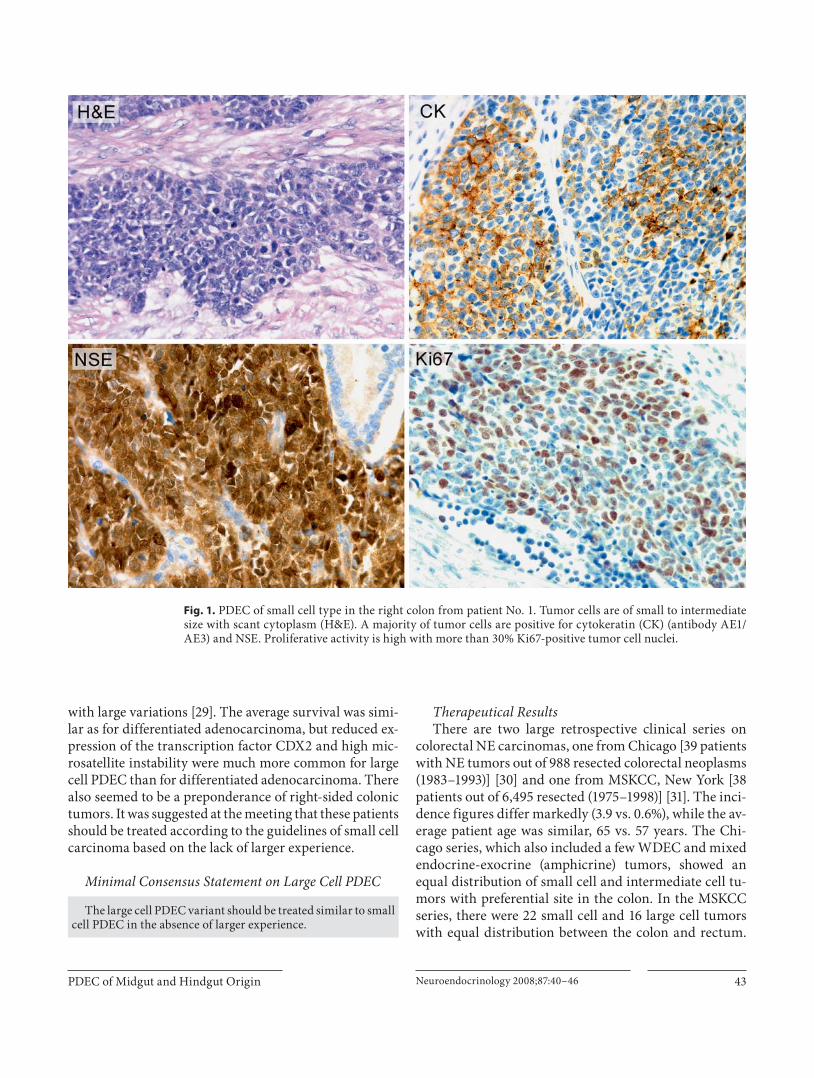
Fig. 1. PDEC of small cell type in the right colon from patient No. 1. Tumor cells are of small to intermediate size with scant cytoplasm (H&E). A majority of tumor cells are positive for cytokeratin (CK) (antibody AE1/AE3) and NSE. Proliferative activity is high with more than 30% Ki67-positive tumor cell nuclei.

Ahlman et al.
Neuroendocrinology 2008;87:40–46 44
The immunohistochemical studies were incomplete in the Chicago series and in the MSKCC only 15–19 tumors were investigated with expression of CgA and NSE in al-most all, while synaptophysin was expressed in about two-thirds. In both series, 80% of the patients presented with metastatic disease (Duke C and D or stage III–IV). The median survival was low: 7 and 10 months, respec-tively. The survival analysis in the first series was 58% at 6 months, 15% at 3 years and 6% at 5 years; in the second series the survival after 1 and 3 years was 46 and 13%, respectively [30, 31] .
Minimal Consensus Statement on Survival Despite active therapeutic efforts, the median survival of
high-stage PDEC tumors (ED) is very low (7–10 months). Sur-vival beyond 5 years is scarce.
Cases Illustrated in Figures 1 and 2 In figures 1 and 2 , patients with small or large cell
PDEC are presented with histopathological details. Pa-tient No. 1 (small cell carcinoma) was initially thought to have right-sided colonic adenocarcinoma as a cause of ileus and underwent removal of the primary tumor and resectable nodes. Due to asthma and poor general condi-tion, he rejected chemotherapy. After a progression-free
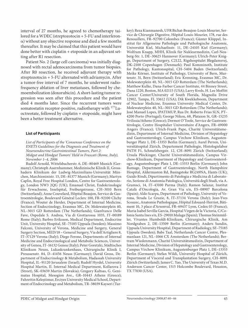
Fig. 2. PDEC of large cell type in the rectum from patient No. 2. Tumor cells are of large size with moderate amount of cytoplasm (H&E). A majority of tumor cells are positive for synaptophysin (SYN) and vesicular mono-amine transporter I (VMAT1). Proliferative activity is high with more than 30% Ki67-positive tumor cell nuclei.
PDEC of Midgut and Hindgut Origin Neuroendocrinology 2008;87:40–46 45
interval of 27 months, he agreed to chemotherapy tai-lored for a WDEC (streptozotocin + 5-FU and interferon- � ) without any objective response and died a few months thereafter. It may be claimed that this patient would have done better with cisplatin + etoposide in an adjuvant set-ting after R1 resection.
Patient No. 2 (large cell carcinoma) was initially diag-nosed with rectal adenocarcinoma from tumor biopsies. After R0 resection, he received adjuvant therapy with streptozotocin + 5-FU alternated with adriamycin. After a tumor-free interval of 7 months, he underwent radio-frequency ablation of liver metastases, followed by che-moembolization (doxorubicin). A short-lasting tumor re-gression was seen after this procedure and the patient died 4 months later. Since the recurrent tumors were somatostatin receptor-positive, radiotherapy with 177 Lu-octreotate, followed by cisplatin + etoposide, might have been a better treatment alternative.
List of Participants
List of Participants of the ‘Consensus Conference on the ENETS Guidelines for the Diagnosis and Treatment of Neuroendocrine Gastrointestinal Tumors, Part 2: Midgut and Hindgut Tumors’ Held in Frascati (Rome, Italy), November 1–4, 2006 Rudolf Arnold, Wittelsbacherstr. 6, DE–80469 Munich (Ger-
many); Christoph Auernhammer, Medizinische Klinik II, Gross-hadern Klinikum der Ludwig-Maximilians-Universität Mün-chen, Marchioninistr. 15, DE–81377 Munich (Germany); Martyn Caplin, Royal Free Hospital London, Centre for Gastroenterolo-gy, London NW3 2QG (UK); Emanuel Christ, Endokrinologiefür Erwachsene, Inselspital, Freiburgstrasse, CH–3010 Bern (Switzerland); Anne Couvelard, Hôpital Beaujon, Service de Gas-troentérologie, Boulevard Général Leclerc 100, FR–92100 Clichy (France); Wouter de Herder, Department of Internal Medicine, Section of Endocrinology, Erasmus MC, Dr. Molewaterplein 40, NL–3015 GD Rotterdam (The Netherlands); Gianfranco Delle Fave, Ospedale S. Andrea, Via di Grottarossa 1035, IT–00189 Rome (Italy); Barbro Eriksson, Medical Department, Endocrine Unit, University Hospital, SE–75185 Uppsala (Sweden); Massimo Falconi, University of Verona, Medicine and Surgery, General Surgery Section, MED/18 – General Surgery, Via dell’Artigliere 8, IT–37129 Verona (Italy); Diego Ferone, Departments of Internal Medicine and Endocrinological and Metabolic Sciences, Univer-sity of Genoa, IT–16132 Genoa (Italy); Peter Goretzki, Städtisches Klinikum Neuss, Lukaskrankenhaus, Chirurgische Klinik I, Preussenstr. 84, D–41456 Neuss (Germany); David Gross, De-partment of Endocrinology & Metabolism, Hadassah University Hospital, IL–91120 Jerusalem (Israel); Rudolf Hyrdel, University Hospital Martin, II. Internal Medical Department, Kollarova 2 (Street), SK–03659 Martin (Slovakia); Gregory Kaltsas, G. Geni-matas Hos pital, Mesogion Ave., GR–10443 Athens (Greece); Fahrettin Keleştimur, Erciyes University Medical School, Depart-ment of Endocrinology and Metabolism, TR–38039 Kayseri (Tur-
key); Reza Kianmanesh, UFR Bichat-Beaujon-Louis Mourier, Ser-vice de Chirurgie Digestive, Hôpital Louis-Mourier, 178, rue des Renouillers, FR–92700 Colombes (France); Günter Klöppel, In-stitut für Allgemeine Pathologie und Pathologische Anatomie, Universität Kiel, Michaelisstr. 11, DE–24105 Kiel (Germany); Wolfram Knapp, MHH, Klinik für Nuklearmedizin, Carl-Neu-berg-Str. 1, DE–30625 Hannover (Germany); Ulrich-Peter Knig-ge, Department of Surgery, C2122, Rigshospitalet Blegdamsvej, DK–2100 Copenhagen (Denmark); Paul Komminoth, Institute for Pathology, Kantonsspital, CH–5404 Baden (Switzerland); Meike Körner, Institute of Pathology, University of Bern, Mur-tenstr. 31, Bern (Switzerland); Eric Krenning, Erasmus MC, Dr. Molewaterplein 40, NL–3015 GD Rotterdam (The Netherlands); Matthew Kulke, Dana-Farber Cancer Institute, 44 Binney Street, Dana 1220, Boston, MA 02115 (USA); Larry Kvols, H. Lee Moffitt Cancer Center/University of South Florida, Magnolia Drive 12902, Tampa, FL 33612 (USA); Dik Kwekkeboom, Department of Nuclear Medicine, Erasmus University Medical Center, Dr. Molewaterplein 40, NL–3015 GD Rotterdam (The Netherlands); José Manuel Lopes, IPATIMUP, Rua Dr. Roberto Frias SLN, PT–4200 Porto (Portugal); George Nikou, 68, Plataion St, GR–15235 Vrilissia/Athens (Greece); Dermot O’Toole, Service de Gastroen-térologie, Centre Hospitalier Universitaire d’Angers, FR–49000 Angers (France); Ulrich-Frank Pape, Charité Universitätsme-dizin, Department of Internal Medicine, Division of Hepatology and Gastroenterology, Campus Virchow Klinikum, Augusten-burger Platz 1, DE–13353 Berlin (Germany); Aurel Perren, Uni-versitätsspital Zürich, Departement Pathologie, Histologielabor PATH F1, Schmelzbergstr. 12, CH–8091 Zürich (Switzerland); Ursula Plöckinger, Charité Universitätsmedizin, Campus Vir-chow-Klinikum, Department of Hepatology and Gastroenterol-ogy, Augustenburger Platz 1, DE–13353 Berlin (Germany); John Ramage, Department of Gastroenterology, North Hampshire Hospital, Aldermaston Rd, Basingstoke RG249NA, Hants (UK); Guido Rindi, Dipartimento di Patologia e Medicina di Laborato-rio, Sezione di Anatomia Patologica, Università degli Studi, via A. Gramsci, 14, IT–43100 Parma (Italy); Ramon Salazar, Institut Català d’Oncologia, Av. Gran Via s/n, ES–08907 Barcelona (Spain); Aldo Scarpa, Department of Pathology, University of Ve-rona, Strada Le Grazie, 8, IT–37134 Verona (Italy); Jean-Yves Scoazec, Anatomie Pathologique, Hôpital Edouard-Herriot, Bâti-ment 10, 3 place d’Arsonval, FR–69437 Lyon, Cedex 03 (France); Maria Isabel Sevilla Garcia, Hospital Virgen de la Victoria, C/Co-lonia Santa Ines s/n, ES–29010 Malaga (Spain); Thomas Steinmül-ler, Vivantes Humboldt-Klinikum, Chirurgische Klinik, Am Nordgraben 2, DE–13509 Berlin (Germany); Anders Sundin, Uppsala University Hospital, Department of Radiology, SE–75185 Uppsala (Sweden); Babs Taal, Netherlands Cancer Centre, Ples-manlaan 121, NL–1066 CX Amsterdam (The Netherlands); Ber-tram Wiedenmann, Charité Universitätsmedizin, Department of Internal Medicine, Division of Hepatology and Gastroenterology, Campus Virchow Klinikum, Augustenburger Platz 1, DE–13353 Berlin (Germany); Stefan Wildi, University Hospital of Zürich, Department of Visceral and Transplantation Surgery, CH–8091 Zürich (Switzerland); James C. Yao, The University of Texas M.D. Anderson Cancer Center, 1515 Holcombe Boulevard, Houston, TX 77030 (USA).

Ahlman et al.
Neuroendocrinology 2008;87:40–46 46
References
1 Modlin IM, Sandor A: An analysis of 8,305 cases of carcinoid tumors. Cancer 1997; 79: 813–829.
2 Modlin IM, Lye KD, Kidd M: A five-decade analysis of 13,715 carcinoid tumors. Cancer 2003; 97: 934–959.
3 Ballantyne GH, Savoca PE, Flannery JT, Ahlman BH, Modlin IM: Incidence and mortality of carcinoids of the colon. Data from the Connecticut Tumor Registry. Can-cer 1992; 69: 2400–2405.
4 Wang AY, Ahmed NA: Rectal carcinoids. Curr Opin Gastroenterol 2006; 22: 529–535.
5 Grönstad K, Grimelius L, Ekman R, Kewen-ter J, Ahlman H: Disseminated rectal car-cinoid tumor with production of immuno-reactive motilin. Endocr Pathol 1992; 3: 194–200.
6 Solcia E, Klöppel G, Sandler M: Histological Typing of Endocrine Tumors, ed 2. World Health Organization, International Histo-logical Classification of Tumors. Berlin, Springer, 2000.
7 Kang H, O’Connell JB, Leonardi MJ, Mag-gard MA, McGory ML, Ko CY: Rare tumors of the colon and rectum: a national review. Int J Colorectal Dis 2007; 22: 183–189.
8 Nilsson O, Van Cutsem E, Delle Fave G, Yao JC, Pavel ME, McNicol AM, Sevilla Garcia MI, Knapp WH, Kelestimur F, Sauvanet A, Pauwels S, Kwekkeboom DJ, Caplin M: Poor-ly differentiated carcinomas of the foregut (gastric, duodenal and pancreatic). Neuro-endocrinology 2006; 84: 212–215.
9 Arnold R, Rinke A, Schmidt C, Hofbauer L: Endocrine tumors of the gastrointestinal tract: chemotherapy. Best Pract Res Clin Gastroenterol 2005; 19: 649–656.
10 Kim T, Tao-Cheng J-H, Eiden LE, Loh PY: Chromogranin A, an ‘on-off ’ switch con-trolling dense-core secretory granule bio-genesis. Cell 2001; 106: 499–509.
11 Rindi G, Bordi C: Endocrine tumors of the gastrointestinal tract: etiology, molecular pathogenesis and genetics. Best Pract Res Clin Gastroenterol 2005; 19: 519–534.
12 Vortmeyer AO, Lubensky IA, Merino MJ, Wang CY, Pham T, Furth EE, Zhuang Z: Concordance of genetic alterations in poor - ly differentiated colorectal neuroendocrine carcinomas and associated adenocarcino-mas. J Natl Cancer Inst 1997; 19: 1448–1453.
13 Pizzi S, Azzoni C, Bassi D, Bottavelli L, Mil-ione M, Bordi C: Genetic alterations in poor-ly differentiated endocrine carcinomas of the gastrointestinal tract. Cancer 2003; 98: 1273–1282.
14 Ubiali A, Benetti A, Papotti M, Villaviacci V, Rindi G: Genetic alternations in poorly dif-ferentiated colon carcinomas developing in tubulovillous adenomas: a report of two cas-es. Virchows Arch 2001; 439: 776–781.
15 Brenner B, Tang LH, Klimstra DS, Kelsen DP: Small cell carcinomas of the gastrointes-tinal tract. J Clin Oncol 2004; 22: 2730–2739.
16 McKeown F: Oat-cell carcinoma of the esophagus. J Pathol Bacteriol 1952; 80: 889–891.
17 Hoff PM, Pazdur R: Small cell carcinoma of the gastrointestinal tract; in Raghavan D, Brecher M, Johnson DH (eds): Textbook of Uncommon Cancer. Chichester, Wiley, 1999, pp 463–467.
18 Vrouvas J, Ash DV: Extrapulmonary small cell cancer. Clin Oncol 1995; 7: 377–381.
19 Zelen M: Keynote address on biostatistics and data retrieval. Cancer Chemother Rep 1973; 4: 31–42.
20 Burke AB, Shekitha K, Sobin LH: Small cell carcinomas of the large intestine. Am J Clin Pathol 1991; 95: 315–321.
21 Ordonez NG: Value of thyroid transcription factor-1 immunostaining in distinguishing small cell lung carcinomas from other small cell carcinomas. Am J Surg Pathol 2000; 24: 1010–1023.
22 Ho KJ, Herrera GA, Jones JM, Alexander CB: Small cell carcinoma of the esophagus: evi-dence for a unified histogenesis. Hum Pathol 1984; 77: 460–468.
23 Latulippe E, Klimstra D: Retinoblastoma protein expression in colorectal high-grade neuroendocrine carcinoma. Med Pathol 2001; 14: 89A.
24 Stelon EB, Moskaluk CA, Mills SE: The mis-match repair protein status of colorectal small cell neuroendocrine carcinoma. Am J Surg Pathol 2006; 30: 1401–1404.
25 Casas F, Feuer F, Farrus B, Casals J, Biete A: Primary small cell carcinoma of the esopha-gus: a review of the literature with emphasis on therapy and prognosis. Cancer 1997; 80: 1366–1372.
26 Medgyesy CD, Wolff RA, Putnam JB Jr, Aja-ni JA: Small cell carcinoma of the esophagus: the University of Texas M.D. Anderson Can-cer Center experience and literature review. Cancer 2000; 88: 262–267.
27 Moertel C, Rubin J, O’Connell MJ, Rubin J: Phase II study of cisplatin therapy in patients with metastatic carcinoid tumor and the ma-lignant carcinoid syndrome. Cancer Treat Rep 1986; 70: 1459–1460
28 Bajetta E, Catena L, Procopio G, De Dosso S, Bichisao E, Ferrari L, Martinetti A, Platania M, Verzoni E, Formisano B, Bajetta R: Are capecitabine and oxaliplatin (XELOX) suit-able treatments for progressing low-grade and high-grade neuroendocrine tumors? Cancer Chemother Pharmacol 2007; 59: 637–642.
29 Hinoi T, Tani M, Lucas PC, Caca K, Daun RL, Macri E, Loda M, Appelman HD, Cho KR, Fearen ER: Loss of CDX2 expression and microsatellite instability are prominent fea-tures of large cell minimally differentiated carcinomas of the colon. Am J Pathol 2001; 159: 2239–2248.
30 Saclarides TJ, Szeluga D, Staven ED: Neuro-endocrine cancers of the colon and rectum. Results of a 10-year experience. Dis Colon Rectum 1994; 37: 635–642.
31 Bernick PE, Klimstra DS, Stua J, Minsley B, Saltz L, Ski W, Thaler H, Guillem J, Paty P, Cohen AM, Wang WD: Neuroendocrine carcinomas of the colon and rectum. Dis Co-lon Rectum 2004; 47: 163–169.